An Updated Review on EPR-Based Solid Tumor Targeting ...
17
Citation: Sharifi, M.; Cho, W.C.; Ansariesfahani, A.; Tarharoudi, R.; Malekisarvar, H.; Sari, S.; Bloukh, S.H.; Edis, Z.; Amin, M.; Gleghorn, J.P.; et al. An Updated Review on EPR-Based Solid Tumor Targeting Nanocarriers for Cancer Treatment. Cancers 2022, 14, 2868. https:// doi.org/10.3390/cancers14122868 Academic Editors: Patrycja Nowak- Sliwinska and Lingzhi Wang Received: 10 May 2022 Accepted: 6 June 2022 Published: 10 June 2022 Publisher’s Note: MDPI stays neutral with regard to jurisdictional claims in published maps and institutional affil- iations. Copyright: © 2022 by the authors. Licensee MDPI, Basel, Switzerland. This article is an open access article distributed under the terms and conditions of the Creative Commons Attribution (CC BY) license (https:// creativecommons.org/licenses/by/ 4.0/). cancers Review An Updated Review on EPR-Based Solid Tumor Targeting Nanocarriers for Cancer Treatment Majid Sharifi 1,2 , William C. Cho 3 , Asal Ansariesfahani 4 , Rahil Tarharoudi 4 , Hedyeh Malekisarvar 4 , Soyar Sari 4 , Samir Haj Bloukh 5,6 , Zehra Edis 6,7 , Mohamadreza Amin 8 , Jason P. Gleghorn 9, * , Timo L. M. ten Hagen 8, * and Mojtaba Falahati 8 1 Student Research Committee, School of Medicine, Shahroud University of Medical Sciences, Shahroud 3614773947, Iran; majidsharifi@tabrizu.ac.ir 2 Department of Tissue Engineering, School of Medicine, Shahroud University of Medical Sciences, Shahroud 3614773947, Iran 3 Department of Clinical Oncology, Queen Elizabeth Hospital, Hong Kong, China; [email protected] 4 Department of Cellular and Molecular Biology, Faculty of Advanced Science and Technology, Tehran Medical Sciences, Islamic Azad University, Tehran 1916893813, Iran; [email protected] (A.A.); [email protected] (R.T.); [email protected] (H.M.); [email protected] (S.S.) 5 Department of Clinical Sciences, College of Pharmacy and Health Sciences, Ajman University, Ajman P.O. Box 346, United Arab Emirates; [email protected] 6 Centre of Medical and Bio-allied Health Sciences Research, Ajman University, Ajman P.O. Box 346, United Arab Emirates; [email protected] 7 Department of Pharmaceutical Sciences, College of Pharmacy and Health Sciences, Ajman University, Ajman P.O. Box 346, United Arab Emirates 8 Laboratory Experimental Oncology and Nanomedicine Innovation Center Erasmus, Department of Pathology, Erasmus MC, 3015 GD Rotterdam, The Netherlands; [email protected] (M.A.); [email protected] (M.F.) 9 Department of Biomedical Engineering, University of Delaware, Newark, DE 19713, USA * Correspondence: [email protected] (J.P.G.); [email protected] (T.L.M.t.H.) Simple Summary: One of the important efforts in the treatment of cancers is to achieve targeted drug delivery by nanocarriers to be more effective and reduce adverse effects. However, due to the adverse responses of nanocarriers in clinical trials due to the very weak EPR effects, doubts have been raised in this regard. In this study, an attempt has been made to take a critical look at EPR approaches to enable the convergence of previous papers and the EPR critics to reach an appropriate therapeutic path. Although the effectiveness of EPR is highly variable due to the complex microenvironment of the tumor, there is high hope for cancer treatment by describing new strategies to overcome the challenges of EPR effect. Furthermore, in this paper an attempt was made to provide a reliable path for future to develop cancer therapeutics based on EPR effect. Abstract: The enhanced permeability and retention (EPR) effect in cancer treatment is one of the key mechanisms that enables drug accumulation at the tumor site. However, despite a plethora of virus/inorganic/organic-based nanocarriers designed to rely on the EPR effect to effectively target tumors, most have failed in the clinic. It seems that the non-compliance of research activities with clinical trials, goals unrelated to the EPR effect, and lack of awareness of the impact of solid tumor structure and interactions on the performance of drug nanocarriers have intensified this dissatisfac- tion. As such, the asymmetric growth and structural complexity of solid tumors, physicochemical properties of drug nanocarriers, EPR analytical combination tools, and EPR description goals should be considered to improve EPR-based cancer therapeutics. This review provides valuable insights into the limitations of the EPR effect in therapeutic efficacy and reports crucial perspectives on how the EPR effect can be modulated to improve the therapeutic effects of nanomedicine. Keywords: enhanced permeability and retention (EPR) effect; nanocarriers; solid tumor; drug delivery; vascular diffusion Cancers 2022, 14, 2868. https://doi.org/10.3390/cancers14122868 https://www.mdpi.com/journal/cancers
-
Upload
khangminh22 -
Category
Documents
-
view
3 -
download
0
Transcript of An Updated Review on EPR-Based Solid Tumor Targeting ...
Citation: Sharifi, M.; Cho, W.C.;
Ansariesfahani, A.; Tarharoudi, R.;
Malekisarvar, H.; Sari, S.; Bloukh,
S.H.; Edis, Z.; Amin, M.; Gleghorn,
J.P.; et al. An Updated Review on
EPR-Based Solid Tumor Targeting
Nanocarriers for Cancer Treatment.
Cancers 2022, 14, 2868. https://
doi.org/10.3390/cancers14122868
Academic Editors: Patrycja Nowak-
Sliwinska and Lingzhi Wang
Received: 10 May 2022
Accepted: 6 June 2022
Published: 10 June 2022
Publisher’s Note: MDPI stays neutral
with regard to jurisdictional claims in
published maps and institutional affil-
iations.
Copyright: © 2022 by the authors.
Licensee MDPI, Basel, Switzerland.
This article is an open access article
distributed under the terms and
conditions of the Creative Commons
Attribution (CC BY) license (https://
creativecommons.org/licenses/by/
4.0/).
cancers
Review
An Updated Review on EPR-Based Solid Tumor TargetingNanocarriers for Cancer TreatmentMajid Sharifi 1,2 , William C. Cho 3 , Asal Ansariesfahani 4, Rahil Tarharoudi 4, Hedyeh Malekisarvar 4,Soyar Sari 4, Samir Haj Bloukh 5,6 , Zehra Edis 6,7 , Mohamadreza Amin 8 , Jason P. Gleghorn 9,* ,Timo L. M. ten Hagen 8,* and Mojtaba Falahati 8
1 Student Research Committee, School of Medicine, Shahroud University of Medical Sciences,Shahroud 3614773947, Iran; [email protected]
2 Department of Tissue Engineering, School of Medicine, Shahroud University of Medical Sciences,Shahroud 3614773947, Iran
3 Department of Clinical Oncology, Queen Elizabeth Hospital, Hong Kong, China; [email protected] Department of Cellular and Molecular Biology, Faculty of Advanced Science and Technology,
Tehran Medical Sciences, Islamic Azad University, Tehran 1916893813, Iran; [email protected] (A.A.);[email protected] (R.T.); [email protected] (H.M.); [email protected] (S.S.)
5 Department of Clinical Sciences, College of Pharmacy and Health Sciences, Ajman University,Ajman P.O. Box 346, United Arab Emirates; [email protected]
6 Centre of Medical and Bio-allied Health Sciences Research, Ajman University,Ajman P.O. Box 346, United Arab Emirates; [email protected]
7 Department of Pharmaceutical Sciences, College of Pharmacy and Health Sciences, Ajman University,Ajman P.O. Box 346, United Arab Emirates
8 Laboratory Experimental Oncology and Nanomedicine Innovation Center Erasmus, Department of Pathology,Erasmus MC, 3015 GD Rotterdam, The Netherlands; [email protected] (M.A.);[email protected] (M.F.)
9 Department of Biomedical Engineering, University of Delaware, Newark, DE 19713, USA* Correspondence: [email protected] (J.P.G.); [email protected] (T.L.M.t.H.)
Simple Summary: One of the important efforts in the treatment of cancers is to achieve targeteddrug delivery by nanocarriers to be more effective and reduce adverse effects. However, due to theadverse responses of nanocarriers in clinical trials due to the very weak EPR effects, doubts have beenraised in this regard. In this study, an attempt has been made to take a critical look at EPR approachesto enable the convergence of previous papers and the EPR critics to reach an appropriate therapeuticpath. Although the effectiveness of EPR is highly variable due to the complex microenvironmentof the tumor, there is high hope for cancer treatment by describing new strategies to overcome thechallenges of EPR effect. Furthermore, in this paper an attempt was made to provide a reliable pathfor future to develop cancer therapeutics based on EPR effect.
Abstract: The enhanced permeability and retention (EPR) effect in cancer treatment is one of thekey mechanisms that enables drug accumulation at the tumor site. However, despite a plethora ofvirus/inorganic/organic-based nanocarriers designed to rely on the EPR effect to effectively targettumors, most have failed in the clinic. It seems that the non-compliance of research activities withclinical trials, goals unrelated to the EPR effect, and lack of awareness of the impact of solid tumorstructure and interactions on the performance of drug nanocarriers have intensified this dissatisfac-tion. As such, the asymmetric growth and structural complexity of solid tumors, physicochemicalproperties of drug nanocarriers, EPR analytical combination tools, and EPR description goals shouldbe considered to improve EPR-based cancer therapeutics. This review provides valuable insights intothe limitations of the EPR effect in therapeutic efficacy and reports crucial perspectives on how theEPR effect can be modulated to improve the therapeutic effects of nanomedicine.
Keywords: enhanced permeability and retention (EPR) effect; nanocarriers; solid tumor; drugdelivery; vascular diffusion
Cancers 2022, 14, 2868. https://doi.org/10.3390/cancers14122868 https://www.mdpi.com/journal/cancers
Cancers 2022, 14, 2868 2 of 17
1. Introduction
Despite various approaches to control solid tumor growth, such as surgery, chemother-apy, radiation therapy, and thermotherapy, solid tumors are still the leading cause of deathin cancer patients [1]. The need for better clinical outcomes in patients with cancer has ledresearchers to reconsider therapeutic strategies. To overcome these issues, the structuralcharacteristics of solid tumors, drug nanocarrier transport in tumor vessels and the intersti-tium, selective drug delivery systems, and analytical tools should be carefully considered todevelop cancer therapeutics [2,3]. Although it has long been thought that drug nanocarrierswill facilitate cancer therapeutics by directing drugs to solid tumors, nanomedicine devel-opment has not yet achieved promising clinical outcomes. Numerous reports indicate thatvirus-/inorganic nanoparticles-/organic nanoparticles-based nanocarriers could improvethe efficacy and safety of anticancer drugs through potential targeting, mitigated drugrelease in non-target tissues, and pH-sensitive drug release in the tumor microenvironment(TME) [4–7]. Tumor vascular permeability and the enhanced permeability and retention(EPR) mechanism in macromolecular drug delivery to solid tumors have been reported assuccessful strategies for cancer therapeutics [8–10]. However, the low EPR effect in humansolid tumors [8], poor tumor extravasation [11] and infiltration [11], and a diversity inthe tumor-immune microenvironment (TIME) [12] has resulted in the full realization ofEPR-mediated cancer therapeutics [13].
Today, simulation models have reported the dynamics of tumor vessel cooption andhave promoted the treatment of solid tumors [14,15]. However, different results wereobtained from different strategies aimed at investigating EPR-mediated drug deliveryto cancer cells. Different results can be related to the animal species used, the extent ofintratumoral ECM content, an unorganized tumor vasculature, the diverse propertiesof drug nanocarriers, variable fluid velocities, the types of malignant cells in the TIMEdespite having the exact origin, and the heterogeneous tissues of a solid tumor [16–21].Despite the certainty of the EPR-mediated tumor targeting and cancer nanomedicine treat-ment efficacy [22], the negative side of the EPR effect in cancer nano-therapy is derivedfrom the heterogeneous microvascular networks of solid tumors, which results in hetero-geneous distributions of nanomedicines. Therefore, a full exploration of the EPR effectin solid tumors and issues associated with dynamics, such as pathophysiological andpathoanatomic characteristics, drug nanocarrier formulations, physicochemical factors,and EPR analytical methodology, can provide useful information in the development ofEPR-based therapeutics.
In this regard, Nichols and Bae [9] discussed that the EPR approach in the therapeuticactivities of animal tumors is more prominent than in human tumors due to significantdifferences in tumor structure and function [9]. Furthermore, Maeda, et al. [23] reportedthat the use of EPR in the imaging process based on the presence of nano-probes andnanocarriers in the tumor for a short time (a few minutes) is more appropriate thanfor therapeutic activities. However, they showed that the EPR effect varies based on thephysicochemical properties of the nanocarriers [23]. Moreover, Danhier [17] discussed somefactors that result in EPR pitfalls in clinical trials, the future of EPR, and the required changesin drug nanocarriers. Recently, Wu [22] reported that despite the meager success of theEPR effect in clinical trials, it can be enhanced by regulating the physicochemical propertiesof nanocarriers, treatment duration, and the type of anti-cancer agents. At the same time,Izci, et al. [24] explained the advantages and challenges of EPR, the EPR enhancementmechanisms, and the study of independent EPR-based drug delivery strategies.
Although these review articles have comprehensively discussed the EPR-based ther-apeutics of solid tumors, people have not surveyed EPR-based tumor targeting with afocus on fluid flow and convection/diffusion mechanisms. Therefore, this review discusseschanges in structure as the tumor grows and how fluid flow, convection/diffusion, and thephysicochemical properties of nanocarriers affect the EPR-based transport of nanocarriersin solid tumors. Additionally, we discuss the need for improved novelty methodologies inanalyzing the EPR effect.
Cancers 2022, 14, 2868 3 of 17
2. Effective Vascular Structures for EPR in Solid Tumors
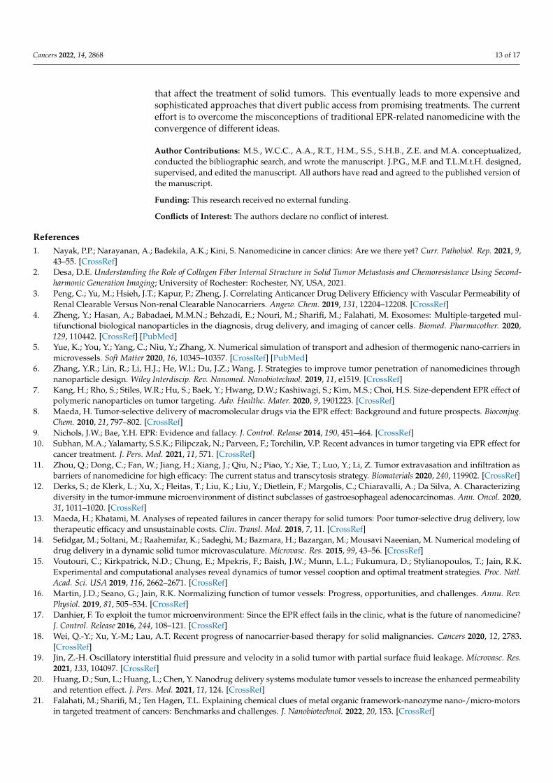
Solid tumors, unlike hematologic cancers, have a structure similar to normal tissue, in-cluding the parenchyma, which contains neoplastic cells and the surrounding stroma [25,26].The stroma of solid tumors is critical for cancer cell survival and metabolism through ex-tracellular matrix components and blood vessels [27,28]. Despite the similarity of themain components of the stromal extracellular matrix of solid tumors and normal tissue,solid tumors have different stromal patterns based on early- or late-forming blood vessels,which play an important role in therapeutic efficacy [29,30]. For example, the highestratio of stroma to tumor cells is found in gastric and pancreatic cancer tissues [31,32],while medullary breast carcinoma and lymphomas have the lowest stromal content [27].Hence, solid tumor therapy with one type of drug-based nanoplatform could not result incomparable clinical efficacy between these tumor types. Changes in the heterogeneity andaggressiveness of solid tumors are derived from the extent of interactions between tumorcell populations [33]. Targeting stromal cell-mediated pro-angiogenic signals in the TME isa key therapeutic strategy. The heterogeneity of angiogenesis and blood vessel maturationin human tumors can be regarded as a critical factor, resulting in conflicting outcomes intherapeutic studies. Based on the growth of solid tumors, it is possible to observe matureand immature vessels within the same tumor, resulting in different EPR effects. In fact,six groups of vessels in solid tumors can be observed with various sizes and shapes [34](Figure 1A), which include (1) mother vessels (very large, thin-walled, permeable, withweak pericytes), (2) glomeruli microvascular (poor organization of proliferated endothelialcells, pericytes, and basement membranes), (3) capillaries (including primary and microvas-cular glomeruloid vessels), (4) vascular malformations (often with asymmetric coverageof smooth muscle cells and or tissue), (5) feeding arteries (large vessels with completecapillary structure and often torturous), and (6) drainage veins (very large with completecapillary structure). Late detection of solid tumors in humans, unlike in model animals inwhich cancer is screened in the early stages, can also increase vascular growth, maturation,and diversity, which can serve as potential barriers to drug delivery in solid tumors [24]. Itis well known that biological sex and vascular development affect vascular architecture andpermeability [35]. Therefore, the impairment of newly formed blood vessels (types 1 to 4)during tumor angiogenesis could not lead to improved efficacy of conventional anticancertherapies. Moreover, during blood vessel maturation in solid tumors, the vessels show avariety of complexities that affect fluid flow resistance and may even generate reversedfluid flows [36]. Additionally, the asymmetric vascular growth, vasoconstriction, andsolid stress found in desmoplastic tumors create altered physical forces around the bloodvessels [16,37,38] (Figure 1B), which induce low red blood cell fluidity and increased flowresistance, which can induce hypoxia and acidosis. Furthermore, angiogenesis and vascularremodeling in normal tissues follow the law of diameter reduction in smaller branches,which is not seen in solid tumors [39]. Therefore, the heterogeneity of solid tumor mi-crovascular networks could result in alterations in vascular permeability and interstitialfluid flow, which act as key biological barriers to cancer drug delivery and efficacy insolid tumors. The overgrowth of solid tumors and a fibrotic response with increased ECMdeposition lead to the introduction of mechanical barriers, resulting in altered interstitialfluid flows [40,41]. Heterogeneity of vascular fluid flow due to physical barriers due toincreased cell proliferation in some solid tumor areas can reduce the efficiency of drugnanocarriers, especially in dimensions larger than 100 nm, intratumoral transport, and theEPR effect [20,41].
Regardless of the stages of vascular formation in solid tumors, which have been stud-ied by Fang et al. [42], exploring the molecular characteristics of tumor vessels can providea more accurate understanding of the active or inactive delivery of drug nanocarriers.Unlike normal vascular architecture with a homogeneous distribution of endothelial cellssurrounded by pericytes, the heterogeneous vasculature of a solid tumor results in differ-ent permeability models [43]. This morphological abnormality not only provides greatervascular permeability in solid tumors but also leads to increased interstitial pressure in

Cancers 2022, 14, 2868 4 of 17
the tumor tissue, which generates multiple distribution patterns of drug nanocarriers [44].It seems that determining the balance between vascular resistance and vascular leakageof solid tumors can be used as a basis for drug delivery mediated by the EPR effect toincrease therapeutic efficacy in interstitial fluid. Basically, momentary changes in fluid flowwith vascular resistance, and vascular leakage with vascular wall pore diameters rangingfrom 50–100 nm (tumor vessels with poor permeability) to 500–1000 nm (high permeabilityvessels) are explained. Overall, the EPR effect is expected to be more effective during theearly phases of cancer growth or peritumors than late phases due to improved leakagebehavior and the rate of fluid flow. This result could advance future therapeutics for solidtumors with a different approach.
Cancers 2022, 14, x 4 of 18
to physical barriers due to increased cell proliferation in some solid tumor areas can re-duce the efficiency of drug nanocarriers, especially in dimensions larger than 100 nm, in-tratumoral transport, and the EPR effect [20,41].
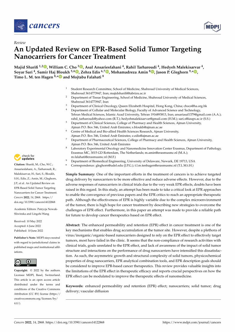
Figure 1. (A) Abnormally patterned vascular vessels in solid tumors. Six types of blood vessels with different characteristics can be identified. (B) A description of the overall structure of solid tumors and the solid stress phenomenon caused by tumor tissue growth that reduces fluid flow and even reverses fluid flow in the tumor.
Regardless of the stages of vascular formation in solid tumors, which have been stud-ied by Fang et al. [42], exploring the molecular characteristics of tumor vessels can provide a more accurate understanding of the active or inactive delivery of drug nanocarriers. Unlike normal vascular architecture with a homogeneous distribution of endothelial cells surrounded by pericytes, the heterogeneous vasculature of a solid tumor results in differ-ent permeability models [43]. This morphological abnormality not only provides greater vascular permeability in solid tumors but also leads to increased interstitial pressure in the tumor tissue, which generates multiple distribution patterns of drug nanocarriers [44]. It seems that determining the balance between vascular resistance and vascular leakage of solid tumors can be used as a basis for drug delivery mediated by the EPR effect to increase therapeutic efficacy in interstitial fluid. Basically, momentary changes in fluid flow with vascular resistance, and vascular leakage with vascular wall pore diameters ranging from 50–100 nm (tumor vessels with poor permeability) to 500–1000 nm (high permeability vessels) are explained. Overall, the EPR effect is expected to be more effec-tive during the early phases of cancer growth or peritumors than late phases due to im-proved leakage behavior and the rate of fluid flow. This result could advance future ther-apeutics for solid tumors with a different approach.
3. EPR-Mediated Drug Delivery to Solid Tumors In recent years, various perspectives on anticancer nanomedicine have been devel-
oped based on nanocarrier intratumoral transport pathways [22,45–47]. The structural
Figure 1. (A) Abnormally patterned vascular vessels in solid tumors. Six types of blood vessels withdifferent characteristics can be identified. (B) A description of the overall structure of solid tumorsand the solid stress phenomenon caused by tumor tissue growth that reduces fluid flow and evenreverses fluid flow in the tumor.
3. EPR-Mediated Drug Delivery to Solid Tumors
In recent years, various perspectives on anticancer nanomedicine have been developedbased on nanocarrier intratumoral transport pathways [22,45–47]. The structural complexi-ties of solid tumors that affect drug delivery pathways remain unclear [15]. As such, theblood flow complexities mentioned in Section 2, such as obstruction of fluid flow in thedeeper parts of the tumor, tumor heterogeneity due to different types of vessels that causeinterstitial fluid pressure, diversity of ECM of solid tumors, and lack of lymphatic drainagevessels, have caused a variety of therapeutic challenges based on the intratumoral transportand drug efficacy [20,25,48]. Regardless of the method and location of the injection, the drugnanocarriers introduced in vivo are distributed intratumorally using two basic concepts.Convection is the transfer of drugs by a moving fluid, such as blood or interstitial fluid, inwhich case the distance between the fluid and the cancer cell is critical [49], and diffusionis proportional to the concentration gradient, which is effective at short intervals of druginjection) [50]. Meanwhile, the intratumoral transport of drug nanocarriers in solid tumors
Cancers 2022, 14, 2868 5 of 17
is a function of both phenomena, based on fluid flow in the solid tumor, which stronglydepends on the tumor size [51]. In addition, diffusion is more prevalent in the case ofsmaller-sized drug nanocarriers in small vessels as well as in the intercellular spaces [52,53]similar to the pulmonary tissue vessels [54]. However, convection is more prevalent in thecase of larger drug nanocarriers present in a solid tumor core [55] and large vessels [53,56].In this regard, it has been determined that the specific entry of therapeutic payloads intotumors is inhibited when diffusion is involved in the intratumoral transport of nanocarriersdue to the increase of interstitial pressure in solid tumors [57–59]. Hence, diffusion cansometimes be considered an obstacle to yielding novel, effective therapies for solid tumors.Furthermore, if the interstitial fluid pressure stops as a function of tumor solid stress [60],or fluid flow in small vessels is reduced [61], EPR-mediated drug delivery could fail. Thus,the changes in fluid velocity in different parts of solid tumors based on their heterogeneousdevelopment and progression can be considered a very important indicator of the efficacyof EPR-mediated drug delivery in solid tumors. As the role of interstitial fluid pressure [59]and tumor solid stress [62] in modulating fluid velocity is very important, it is expectedthat the intratumoral transport of nanocarriers and the EPR effect in the peritumoral areaswill be greater than that of the core of solid tumors. In addition, the change in the directionof fluid flow in solid tumors [63,64] due to the irregular structure of vessels can result inthe mitigation of convection/diffusion-enhanced delivery of drugs for the treatment ofsolid tumors. By reversing the flow, the viscosity of the fluid in solid tumors is expectedto be increased several times that of normal tissue due to an increase in hematocrit [65].Therefore, this phenomenon can effectively manipulate convection and diffusion events ininterstitial transport in solid tumors mediated by EPR.
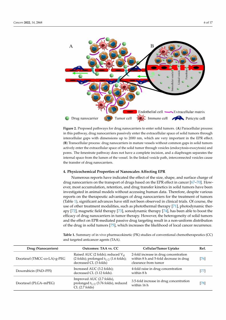
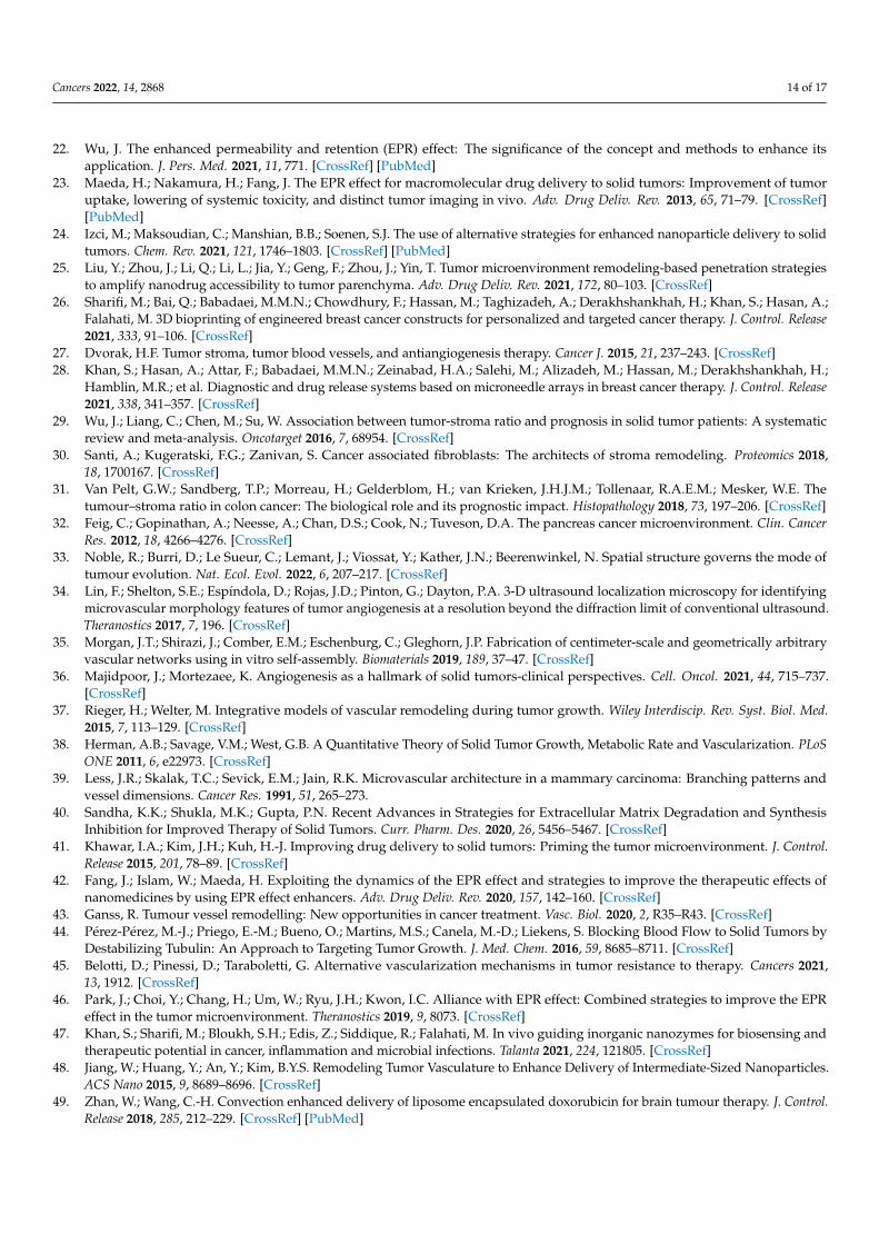
Tumor vessel development and maturation can also lead to the formation of anintegrated structure of endothelial cells and the presence of pericytes, which changesthe mechanism of intratumoral transport of drug nanocarriers due to an alteration inthe ratios of convection/diffusion status [58,64]. Although reducing the number andsize of vascular wall pores moderates the passive transport of drug nanocarriers into theinterstitial space, computational models predict that even without vessel pores/gaps dueto decreased interstitial fluid retention and reduced leakage into vessel lumen with higheruptake through intercellular and linked vesicles effectively increases the tumor delivery ofmacromolecular drugs based on the EPR effect (Figure 2) [64].
However, due to capillary growth and the reduction of leaky regions of the tumorvasculature, the passive transport of drug nanocarriers is significantly reduced. Increasingthe thickness and density between the vascular lumen and the interstitial space due tovascular maturation along with greater ECM and collagen accumulation can dramaticallyreduce the potential tumor-targeted drug delivery based on EPR-effect [66]. Therefore, anupdated view of the transport of drug nanocarriers based on the EPR effect due to theheterogeneous vasculature of a solid tumor is needed.
Cancers 2022, 14, 2868 6 of 17Cancers 2022, 14, x 6 of 18
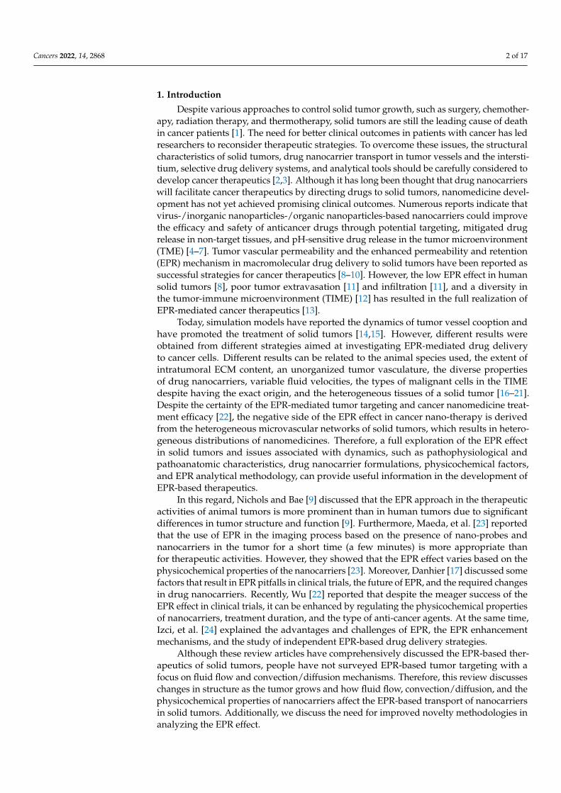
Figure 2. Proposed pathways for drug nanocarriers to enter solid tumors. (A) Paracellular process: in this pathway, drug nanocarriers passively enter the extracellular space of solid tumors through intercellular gaps with dimensions up to 2000 nm, which are very important in the EPR effect. (B) Transcellular process: drug nanocarriers in mature vessels without common gaps in solid tumors actively enter the extracellular space of the solid tumor through vesicles (endocytosis-exocytosis) and pores. The fenestrate pathway does not have a complete incision, and a diaphragm separates the internal space from the lumen of the vessel. In the linked vesicle path, interconnected vesicles cause the transfer of drug nanocarriers.
However, due to capillary growth and the reduction of leaky regions of the tumor vasculature, the passive transport of drug nanocarriers is significantly reduced. Increasing the thickness and density between the vascular lumen and the interstitial space due to vascular maturation along with greater ECM and collagen accumulation can dramatically reduce the potential tumor-targeted drug delivery based on EPR-effect [66]. Therefore, an updated view of the transport of drug nanocarriers based on the EPR effect due to the heterogeneous vasculature of a solid tumor is needed.
4. Physicochemical Properties of Nanoscales Affecting EPR Numerous reports have indicated the effect of the size, shape, and surface charge of
drug nanocarriers on the transport of drugs based on the EPR effect in cancer [67–70]. However, most accumulation, retention, and drug transfer kinetics in solid tumors have been investigated in animal models without accessing human data. Therefore, despite various reports on the therapeutic advantages of drug nanocarriers for the treatment of tumors (Table 1), significant advances have still not been observed in clinical trials. Of course, the use of other treatment modalities, such as photothermal therapy [71], photo-dynamic therapy [72], magnetic field therapy [73], sonodynamic therapy [74], has been able to boost the efficacy of drug nanocarriers in tumor therapy. However, the heteroge-neity of solid tumors and the effect on EPR-mediated passive drug targeting result in a non-uniform distribution of the drug in solid tumors [75], which increases the likelihood of local cancer recurrence.
Figure 2. Proposed pathways for drug nanocarriers to enter solid tumors. (A) Paracellular process:in this pathway, drug nanocarriers passively enter the extracellular space of solid tumors throughintercellular gaps with dimensions up to 2000 nm, which are very important in the EPR effect.(B) Transcellular process: drug nanocarriers in mature vessels without common gaps in solid tumorsactively enter the extracellular space of the solid tumor through vesicles (endocytosis-exocytosis) andpores. The fenestrate pathway does not have a complete incision, and a diaphragm separates theinternal space from the lumen of the vessel. In the linked vesicle path, interconnected vesicles causethe transfer of drug nanocarriers.
4. Physicochemical Properties of Nanoscales Affecting EPR
Numerous reports have indicated the effect of the size, shape, and surface charge ofdrug nanocarriers on the transport of drugs based on the EPR effect in cancer [67–70]. How-ever, most accumulation, retention, and drug transfer kinetics in solid tumors have beeninvestigated in animal models without accessing human data. Therefore, despite variousreports on the therapeutic advantages of drug nanocarriers for the treatment of tumors(Table 1), significant advances have still not been observed in clinical trials. Of course, theuse of other treatment modalities, such as photothermal therapy [71], photodynamic ther-apy [72], magnetic field therapy [73], sonodynamic therapy [74], has been able to boost theefficacy of drug nanocarriers in tumor therapy. However, the heterogeneity of solid tumorsand the effect on EPR-mediated passive drug targeting result in a non-uniform distributionof the drug in solid tumors [75], which increases the likelihood of local cancer recurrence.
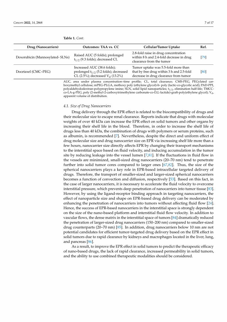
Table 1. Summary of in vivo pharmacokinetic (PK) studies of conventional chemotherapeutics (CC)and targeted anticancer agents (TAA).
Drug (Nanocarriers) Outcomes: TAA vs. CC Cellular/Tumor Uptake Ref.
Docetaxel (TMCC-co-LA)-g-PEGRaised AUC (2 folds); reduced Vd(2 folds); prolonged t1/2 (1.6 folds);decreased CL (3 folds)
2-fold increase in drug concentrationwithin 8 h and 5-fold decrease in drugclearance from tumor
[76]
Doxorubicin (PAD–PPI) Increased AUC (3.2 folds);decreased CL (3.12 folds).
4-fold raise in drug concentrationwithin 8 h [77]
Docetaxel (PLGA–mPEG)Improved AUC (2.7 folds),prolonged t1/2 (3.76 folds), reducedCL (2.7 folds)
3.5-fold increase in drug concentrationwithin 16 h [78]
Cancers 2022, 14, 2868 7 of 17
Table 1. Cont.
Drug (Nanocarriers) Outcomes: TAA vs. CC Cellular/Tumor Uptake Ref.
Doxorubicin (Mannosylated- SLNs) Raised AUC (5 folds); prolongedt1/2 (9.3 folds); decreased CL
2.8-fold raise in drug concentrationwithin 8 h and 2.4-fold decrease in drugclearance from the tumor
[79]
Docetaxel (CMC–PEG)Increased AUC (38.6 folds);prolonged t1/2 (5.2 folds); decreasedCL (2.5%); decreased Vd (13.2%)
Tumor uptake was 5.5-fold more thanthat by free drug within 3 h and 2.5-folddecrease in drug clearance from tumor
[80]
AUC, area under plasma concentration–time profile; CL, total clearance; CMS-PEG, PEGylated car-boxymethyl cellulose; mPEG-PLGA, methoxy poly (ethylene glycol)-b- poly (lactic-co-glycolic acid); PAD-PPI,polyaldehydodextran-polypropylene imine; SLN, solid lipid nanoparticles; t1/2, elimination half-life; TMCC-co-LA-g-PEG, poly (2-methyl-2-carboxytrimethylene carbonate-co-D,L-lactide)-graft-poly(ethylene glycol); Vd,apparent volume of distribution.
4.1. Size of Drug Nanocarriers
Drug delivery through the EPR effect is related to the biocompatibility of drugs andtheir molecular size to escape renal clearance. Reports indicate that drugs with molecularweights of over 40 kDa can increase the EPR effect on solid tumors and other organs byincreasing their shelf life in the blood. Therefore, in order to increase the shelf life ofdrugs less than 40 kDa, the combination of drugs with polymers or serum proteins, suchas albumin, is recommended [7]. Nevertheless, despite the direct and uniform effect ofdrug molecular size and drug nanocarrier size on EPR via increasing shelf life more than afew hours, nanocarrier size directly affects EPR by changing their transport mechanismsto the interstitial space based on fluid velocity, and inducing accumulation in the tumorsite by reducing leakage into the vessel lumen [7,81]. If the fluctuations in fluid flow inthe vessels are minimized, small-sized drug nanocarriers (20–70 nm) tend to penetratefurther into solid tumor cores compared to larger ones [67,82]. Thus, the size of thespherical nanocarriers plays a key role in EPR-based intracellular targeted delivery ofdrugs. Therefore, the transport of smaller-sized and larger-sized spherical nanocarriersbecomes a function of convection and diffusion, respectively [53]. Based on this fact, inthe case of larger nanocarriers, it is necessary to accelerate the fluid velocity to overcomeinterstitial pressure, which prevents deep penetration of nanocarriers into tumor tissue [83].However, by using the ligand-receptor binding approach in targeting nanocarriers, theeffect of nanoparticle size and shape on EPR-based drug delivery can be moderated byenhancing the penetration of nanocarriers into tumors without affecting fluid flow [24].Hence, the success of EPR-based nanocarriers in the interstitial space is strongly dependenton the size of the nano-based platform and interstitial fluid flow velocity. In addition tovascular flows, the dense matrix in the interstitial space of tumors [84] dramatically reducedthe penetration of larger-sized drug nanocarriers (150–200 nm) compared to smaller-sizeddrug counterparts (20–70 nm) [85]. In addition, drug nanocarriers below 10 nm are notpotential candidates for efficient tumor-targeted drug delivery based on the EPR effect insolid tumors due to rapid clearance by kidneys and macrophages located in the liver, lung,and pancreas [86].
As a result, to improve the EPR effect in solid tumors to predict the therapeutic efficacyof nano-based drugs, the lack of rapid clearance, increased permeability in solid tumors,and the ability to use combined therapeutic modalities should be considered.
Cancers 2022, 14, 2868 8 of 17
4.2. Shape and Surface Charge of the Drug Nanocarriers
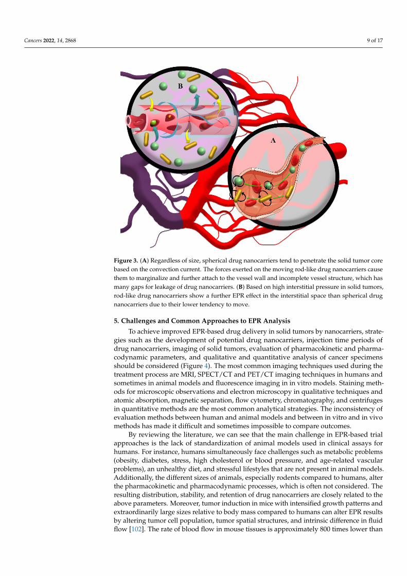
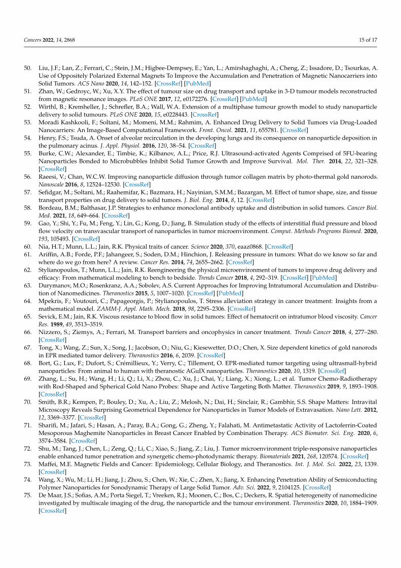
Other factors, including the shape and surface charge of drug nanocarriers based onchanging the nature of transport, improving shelf life, and intracellular permeability, canaffect the EPR-based therapeutics of solid tumors. It has been observed that non-sphericalnanocarriers can have higher diffusivity than spherical nanocarriers as a result of increasedinteractions with the vessel wall through improved radial thrust force deriving from rapidpressure changes [7,87,88]. Thus, the fluid provided a higher radial thrust force by applyingmultilateral force to different surfaces of non-spherical nanocarriers (Figure 3B). In contrast,spherical drug nanocarriers show convection-enhanced delivery of targeted drug penetra-tion into solid tumors (Figure 3A) [89]. Based on radial thrust, Chauhan, et al. [90] reportedthat rod nanocarriers were retained 4 times higher than spherical nanocarriers in solidtumors (Figure 3B). In addition, in a 3D spheroid model, it was recognized that cylindricalnanocarriers with 100 nm height and 325 nm diameter have maximum delivery to solidtumors and a more uniform penetration pattern than their nanorod counterparts, whilemuch smaller non-spherical nanocarriers penetrated deeper and more uniformly comparedto larger non-spherical nanocarriers [91]. Therefore, the deformation of nanocarriers basedon diffusion and convection results in heterogeneous distributions of nanomedicines insolid tumors. However, the combination of the size and shape factors of drug nanocarrierscan exhibit different responses based on the various behaviors of drug nanocarriers withrespect to interstitial fluid flow in tumors. For instance, it has been reported that spheri-cal drug nanocarriers with dimensions less than 70 nm have a higher radial thrust forcethan spherical nanocarriers with dimensions greater than 130 nm [92]. Furthermore, non-spherical nanocarriers have a longer half-life due to less uptake by macrophages, which canincrease the probability of deep penetration into the tumor [93]. The non-spherical shapeof nanocarriers also improves drug-mediated endocytosis due to increased interactionsbetween endothelial cells and drug nanocarriers [88]. Together, this will result in a moreeffective penetration of anticancer drugs through tumor tissue. However, due to the nega-tive charge of vessels [94], EPR-based drug delivery by nanocarriers can be manipulatedby using a positive charge on nanocarriers through modulation of radial thrust [95] basedon electrostatic interactions. For example, Campbell, et al. [96] and Krasnici, et al. [97]showed that liposomal nanocarriers with dimensions of ~150 nm and positively chargedsurface area could accumulate 1.5 and 7 times higher than that of anionic and highly anionicliposomal nanocarriers in the solid tumor, respectively. However, their results showed thatcationic liposomes did not potentially penetrate the intratumoral space compared to theother two samples. Therefore, the possibility of low penetration of drug nanocarriers in theinterstitial space due to the potential interaction of positively charged nanocarriers withthe vascular wall is also conceivable [98]. Nevertheless, the application of a positive chargefaces serious challenges due to its high toxicity to non-target tissues and rapid clearancethrough opsonization [99,100]. The use of chemical surface modification approaches fordrug nanocarriers, such as the use of compounds that are positively charged in the acidicenvironment of the tumor [101], can improve the surface charge drawbacks of nanocarriers.
Cancers 2022, 14, 2868 9 of 17Cancers 2022, 14, x 9 of 18
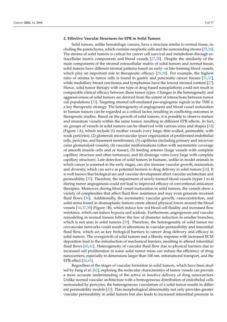
Figure 3. (A) Regardless of size, spherical drug nanocarriers tend to penetrate the solid tumor core based on the convection current. The forces exerted on the moving rod-like drug nanocarriers cause them to marginalize and further attach to the vessel wall and incomplete vessel structure, which has many gaps for leakage of drug nanocarriers. (B) Based on high interstitial pressure in solid tumors, rod-like drug nanocarriers show a further EPR effect in the interstitial space than spherical drug nanocarriers due to their lower tendency to move.
5. Challenges and Common Approaches to EPR Analysis To achieve improved EPR-based drug delivery in solid tumors by nanocarriers, strat-
egies such as the development of potential drug nanocarriers, injection time periods of drug nanocarriers, imaging of solid tumors, evaluation of pharmacokinetic and pharma-codynamic parameters, and qualitative and quantitative analysis of cancer specimens should be considered (Figure 4). The most common imaging techniques used during the treatment process are MRI, SPECT/CT and PET/CT imaging techniques in humans and sometimes in animal models and fluorescence imaging in in vitro models. Staining meth-ods for microscopic observations and electron microscopy in qualitative techniques and atomic absorption, magnetic separation, flow cytometry, chromatography, and centri-fuges in quantitative methods are the most common analytical strategies. The incon-sistency of evaluation methods between human and animal models and between in vitro and in vivo methods has made it difficult and sometimes impossible to compare out-comes.
Figure 3. (A) Regardless of size, spherical drug nanocarriers tend to penetrate the solid tumor corebased on the convection current. The forces exerted on the moving rod-like drug nanocarriers causethem to marginalize and further attach to the vessel wall and incomplete vessel structure, which hasmany gaps for leakage of drug nanocarriers. (B) Based on high interstitial pressure in solid tumors,rod-like drug nanocarriers show a further EPR effect in the interstitial space than spherical drugnanocarriers due to their lower tendency to move.
5. Challenges and Common Approaches to EPR Analysis
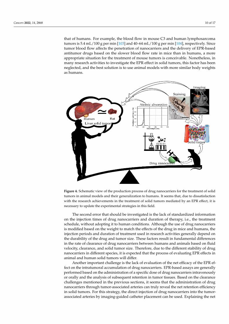
To achieve improved EPR-based drug delivery in solid tumors by nanocarriers, strate-gies such as the development of potential drug nanocarriers, injection time periods ofdrug nanocarriers, imaging of solid tumors, evaluation of pharmacokinetic and pharma-codynamic parameters, and qualitative and quantitative analysis of cancer specimensshould be considered (Figure 4). The most common imaging techniques used during thetreatment process are MRI, SPECT/CT and PET/CT imaging techniques in humans andsometimes in animal models and fluorescence imaging in in vitro models. Staining meth-ods for microscopic observations and electron microscopy in qualitative techniques andatomic absorption, magnetic separation, flow cytometry, chromatography, and centrifugesin quantitative methods are the most common analytical strategies. The inconsistency ofevaluation methods between human and animal models and between in vitro and in vivomethods has made it difficult and sometimes impossible to compare outcomes.
By reviewing the literature, we can see that the main challenge in EPR-based trialapproaches is the lack of standardization of animal models used in clinical assays forhumans. For instance, humans simultaneously face challenges such as metabolic problems(obesity, diabetes, stress, high cholesterol or blood pressure, and age-related vascularproblems), an unhealthy diet, and stressful lifestyles that are not present in animal models.Additionally, the different sizes of animals, especially rodents compared to humans, alterthe pharmacokinetic and pharmacodynamic processes, which is often not considered. Theresulting distribution, stability, and retention of drug nanocarriers are closely related to theabove parameters. Moreover, tumor induction in mice with intensified growth patterns andextraordinarily large sizes relative to body mass compared to humans can alter EPR resultsby altering tumor cell population, tumor spatial structures, and intrinsic difference in fluidflow [102]. The rate of blood flow in mouse tissues is approximately 800 times lower than
Cancers 2022, 14, 2868 10 of 17
that of humans. For example, the blood flow in mouse C3 and human lymphosarcomatumors is 5.4 mL/100 g per min [103] and 40–64 mL/100 g per min [104], respectively. Sincetumor blood flow affects the penetration of nanocarriers and the delivery of EPR-basedantitumor drugs based on the slower blood flow rate in mice than in humans, a moreappropriate situation for the treatment of mouse tumors is conceivable. Nonetheless, inmany research activities to investigate the EPR effect in solid tumors, this factor has beenneglected, and the best solution is to use animal models with more similar body weightsas humans.
Cancers 2022, 14, x 10 of 18
Figure 4. Schematic view of the production process of drug nanocarriers for the treatment of solid tumors in animal models and their generalization to humans. It seems that, due to dissatisfaction with the research achievements in the treatment of solid tumors mediated by an EPR effect, it is necessary to update the experimental strategies in this field.
By reviewing the literature, we can see that the main challenge in EPR-based trial approaches is the lack of standardization of animal models used in clinical assays for hu-mans. For instance, humans simultaneously face challenges such as metabolic problems (obesity, diabetes, stress, high cholesterol or blood pressure, and age-related vascular problems), an unhealthy diet, and stressful lifestyles that are not present in animal mod-els. Additionally, the different sizes of animals, especially rodents compared to humans, alter the pharmacokinetic and pharmacodynamic processes, which is often not consid-ered. The resulting distribution, stability, and retention of drug nanocarriers are closely related to the above parameters. Moreover, tumor induction in mice with intensified growth patterns and extraordinarily large sizes relative to body mass compared to hu-mans can alter EPR results by altering tumor cell population, tumor spatial structures, and intrinsic difference in fluid flow [102]. The rate of blood flow in mouse tissues is approxi-mately 800 times lower than that of humans. For example, the blood flow in mouse C3 and human lymphosarcoma tumors is 5.4 mL/100 g per min [103] and 40–64 mL/100 g per min [104], respectively. Since tumor blood flow affects the penetration of nanocarriers and the delivery of EPR-based antitumor drugs based on the slower blood flow rate in mice than in humans, a more appropriate situation for the treatment of mouse tumors is con-ceivable. Nonetheless, in many research activities to investigate the EPR effect in solid tumors, this factor has been neglected, and the best solution is to use animal models with more similar body weights as humans.
The second error that should be investigated is the lack of standardized information on the injection times of drug nanocarriers and duration of therapy, i.e., the treatment schedule, without adopting it to human conditions. Although the use of drug nanocarriers is modified based on the weight to match the effects of the drug in mice and humans, the injection periods and duration of treatment used in research activities generally depend on the durability of the drug and tumor size. These factors result in fundamental differ-ences in the rate of clearance of drug nanocarriers between humans and animals based on
Figure 4. Schematic view of the production process of drug nanocarriers for the treatment of solidtumors in animal models and their generalization to humans. It seems that, due to dissatisfactionwith the research achievements in the treatment of solid tumors mediated by an EPR effect, it isnecessary to update the experimental strategies in this field.
The second error that should be investigated is the lack of standardized informationon the injection times of drug nanocarriers and duration of therapy, i.e., the treatmentschedule, without adopting it to human conditions. Although the use of drug nanocarriersis modified based on the weight to match the effects of the drug in mice and humans, theinjection periods and duration of treatment used in research activities generally depend onthe durability of the drug and tumor size. These factors result in fundamental differencesin the rate of clearance of drug nanocarriers between humans and animals based on fluidvelocity, clearance, and solid tumor size. Therefore, due to the different stability of drugnanocarriers in different species, it is expected that the process of evaluating EPR effects inanimal and human solid tumors will differ.
Another important challenge is the lack of evaluation of the net efficacy of the EPR ef-fect on the intratumoral accumulation of drug nanocarriers. EPR-based assays are generallyperformed based on the administration of a specific dose of drug nanocarriers intravenouslyor orally and the analysis of subsequent retention in tumor tissues. Based on the clearancechallenges mentioned in the previous sections, it seems that the administration of drugnanocarriers through tumor-associated arteries can truly reveal the net retention efficiencyin solid tumors. For this strategy, the direct injection of drug nanocarriers into the tumor-associated arteries by imaging-guided catheter placement can be used. Explaining the net
Cancers 2022, 14, 2868 11 of 17
efficacy of drug nanocarriers in increasing EPR could influence the opinion of researchersto use a variety of drug nanocarriers to treat solid tumors by modifying the productionprocess. Nevertheless, few drug nanocarriers have been reported for arterial direct injectioninto solid tumors [105,106].
Other obvious errors in EPR detection processes include the method of collectingsamples based on the structural status of solid tumors and the accuracy of sample prepara-tion. As mentioned above, the penetration efficacy of drug nanocarriers in the solid tumorcore is lower than that of the peritumoral region. Thus, it is more desirable in therapeuticapproaches to make a detailed distinction between different areas of the tumor in theEPR examination to explain the effect of drug nanocarriers on therapeutic pathways in amore accurate and reproducible way. In addition, it seems that complete blood withdrawalshould be performed to prevent the report of drug nanocarriers in the blood, as nanocarriersare retained from solid tumor samples.
Although the mentioned challenges in controlling the concentration of drug nanocar-riers in solid tumors of animal models can be relaxed due to ethical sensitivities, possibletoxicities, high cost, and the complexity of experiments, the use of the above methods inclinical practice faces serious challenges.
6. Convergence of Theories to Reduce Conceptual Shortcomings in EPR
For almost 45 years, EPR theory has been pursued by researchers as an importantand reliable effect in cancer therapy. However, in recent years, a group of researchers havehypothesized the ineffectiveness of the EPR effect in cancer therapy [17]. They believe thatthe different outcomes in cancer treatment derived from animal and human models area challenge to EPR effect-based cancer therapeutics due to differences in body structureand solid tumor morphologies. At first glance, based on Sections 2 and 3, this hypothesisis logical. However, could the potential pitfall for the improvement of tumor uptake bynanocarriers be considered a criterion for accepting/rejecting the EPR effect hypothesis?Could the previous results of EPR effect-based tumor targeting and cancer nanomedicinetreatment efficacy be advanced with further strategies? The results of several studiesdemonstrate that the retention of drug nanocarriers by increasing the imbalance in theinflow (through vascular wall disorders) and fluid output (due to defects in the tumorlymphatic system) increases the killing of tumor cells in solid tumors [9,107]. Although thelack of reliable human data has challenged the EPR effect-based anticancer nanomedicinedrug targeting, assuming that clinical outcomes are always a function of solid tumorheterogeneity [108], physicochemical features of drug nanocarriers [109], and therapeuticdesigns with combined modalities (photo-therapy, thermal-therapy, magnetic-therapy,etc.) [24], it is not possible to precisely attribute the rates of treatment failure to one of theexisting therapeutic factors. Nevertheless, the convergence of the different theories canserve as a platform for optimizing cancer treatment strategies.
The first new cancer treatment strategy based on the EPR effect is to change theperspective of EPR effect-based therapeutic approaches. In the treatment process, thetransfer of drug nanocarriers from the cancer stroma to malignant cells is essential [110].As seen in several papers, the high accumulation of drug nanocarriers in normal tissuescan lead to undesirable toxicity through drug release and catalytic activity [111]. With thisnew perspective, it can be hoped that the actual accumulation of drug nanocarriers intosolid tumors will result in a promising therapeutic target for cancer treatment, especiallyby enhanced tumor penetration of the drug. Therefore, to enhance therapeutic activitiesin solid tumors, accepting the EPR effect, along with recruiting combined therapeuticmodalities to trigger the penetration of drug nanocarriers into malignant cells, can result inimproved nano-based precision carriers for targeting therapeutic and diagnostic agents inthe treatment of cancer. However, the accumulation of drug nanocarriers in solid tumors,especially in peritumoral areas, could be an EPR-independent strategy. Therefore, based onthe nature of the nanocarriers and drug availability, a new delivery nanoplatform could bedeveloped for tracking and treating poorly vascularized solid tumors.
Cancers 2022, 14, 2868 12 of 17
Another strategy to modulate EPR is the manipulation of drug nanocarriers throughsurface charge modifications, cluster size, flexible shape, and surface coatings for greater sta-bility, with or without external inducers, to augment the permeability of tumor-associatedvessels [6,112,113]. Modifications applied to drug nanocarriers to enhance the EPR effectusually do not result in potential active targeting. Since the effect of EPR indicates a highaccumulation of targeted nanocarriers in the stromal tissue of solid tumors, overtaking theinflow on outflow of drug nanocarriers can lead to inactive or semi-active accumulation ofnanocarriers in solid tumors using the EPR effect.
Competition is also one of the most important strategies in the development of thera-peutic nanomedicine for solid tumors. Indeed, the main competitors of EPR-based treat-ment of solid tumors are the liver, spleen, lung, and kidney. Therefore, reducing theclearance of drug nanocarriers by modulating the physicochemical properties of nanocar-riers matched to the TME of solid tumors can improve EPR-based tumor targeting andcancer nanomedicine treatment efficacy. As mentioned in Section 3, altering the physic-ochemical properties of drug nanocarriers can alter penetration into tumor tissue. Forexample, binding molecules or coatings of drug nanocarriers can modulate the renal filtra-tion rate [114], or natural and synthetic polymers can be used to minimize uptake by theliver [115]. On the other hand, the different morphological structures of solid tumors arekey factors in designing and developing potential targeting drug nanocarriers. This meansthat EPR effect-based drug delivery is much more effective in primary tumors with vascularstructures 1 to 4 than in mature tumors with vascular structures 5 to 6 [116]. Therefore,quantifying the morphological structures of solid tumors by various methods, such as imag-ing and histopathology, can highlight the ability of the EPR effect to improve the efficacyof anticancer nanomedicine. For instance, it has been determined that in micro-metastatictumor cell clusters the possibility of EPR effect-based drug delivery is much lower thanthat of other tumors [117].
Standardization of the methodology for analyzing the EPR effect in solid tumors canenable researchers to develop the function of EPR effect-based treatment of solid tumors.According to Sections 2 and 5, exploring the morphology of solid tumors and their de-velopment can provide useful information about the EPR effect-based tumor delivery ofnanodrugs, intracellular infiltration, cancer cell death, and changes in the physiologicalbehavior of the tumor. It should be noted that the main cause of EPR effect-based depen-dent or independent treatment of solid tumors is due to the intratumoral morphologicalheterogeneity of solid tumors and the different methodologies used in EPR effect analysis.Therefore, due to the presence of different methodologies, along with the variable structureof the solid tumor, reports on the EPR effect-based treatment of tumors are conflicting.
The timetable of treatment interventions is a critical challenge that can limit the EPReffect on the treatment of solid tumors. The review of Park, Choi, Chang, Um, Ryu, andKwon [46] confirms the alliance of the EPR effect and the reduction of hypoxia levels insolid tumors based on greater oxygen permeability using drug nanocarriers [118]. Reducingthe level of hypoxia by the EPR effect increases the possibility of the potential treatment ofsolid tumors [119,120]. However, different responses to solid tumors have been observedwith increasing oxygen levels [121]. Furthermore, due to the lack of sufficient informationon the relationship between physiological changes in solid tumors with decreasing hypoxiaand metastatic status, the role of the EPR effect in macromolecular therapeutics is stillunknown in detail [122,123].
7. Conclusions
Although therapeutic strategies used in solid tumors based on the EPR effect inanimal activities are more successful than in human activities, a successful EPR-basedtreatment process can be established by increasing the structural insight of solid tumorsin patients, changing the methods of manufacturing drug nanocarriers, and improvingEPR analysis approaches. Finally, we emphasize that addressing one-dimensional issuesrather than multidimensional concepts eliminates the possibility of integrating factors
Cancers 2022, 14, 2868 13 of 17
that affect the treatment of solid tumors. This eventually leads to more expensive andsophisticated approaches that divert public access from promising treatments. The currenteffort is to overcome the misconceptions of traditional EPR-related nanomedicine with theconvergence of different ideas.
Author Contributions: M.S., W.C.C., A.A., R.T., H.M., S.S., S.H.B., Z.E. and M.A. conceptualized,conducted the bibliographic search, and wrote the manuscript. J.P.G., M.F. and T.L.M.t.H. designed,supervised, and edited the manuscript. All authors have read and agreed to the published version ofthe manuscript.
Funding: This research received no external funding.
Conflicts of Interest: The authors declare no conflict of interest.
References1. Nayak, P.P.; Narayanan, A.; Badekila, A.K.; Kini, S. Nanomedicine in cancer clinics: Are we there yet? Curr. Pathobiol. Rep. 2021, 9,
43–55. [CrossRef]2. Desa, D.E. Understanding the Role of Collagen Fiber Internal Structure in Solid Tumor Metastasis and Chemoresistance Using Second-
harmonic Generation Imaging; University of Rochester: Rochester, NY, USA, 2021.3. Peng, C.; Yu, M.; Hsieh, J.T.; Kapur, P.; Zheng, J. Correlating Anticancer Drug Delivery Efficiency with Vascular Permeability of
Renal Clearable Versus Non-renal Clearable Nanocarriers. Angew. Chem. 2019, 131, 12204–12208. [CrossRef]4. Zheng, Y.; Hasan, A.; Babadaei, M.M.N.; Behzadi, E.; Nouri, M.; Sharifi, M.; Falahati, M. Exosomes: Multiple-targeted mul-
tifunctional biological nanoparticles in the diagnosis, drug delivery, and imaging of cancer cells. Biomed. Pharmacother. 2020,129, 110442. [CrossRef] [PubMed]
5. Yue, K.; You, Y.; Yang, C.; Niu, Y.; Zhang, X. Numerical simulation of transport and adhesion of thermogenic nano-carriers inmicrovessels. Soft Matter 2020, 16, 10345–10357. [CrossRef] [PubMed]
6. Zhang, Y.R.; Lin, R.; Li, H.J.; He, W.l.; Du, J.Z.; Wang, J. Strategies to improve tumor penetration of nanomedicines throughnanoparticle design. Wiley Interdiscip. Rev. Nanomed. Nanobiotechnol. 2019, 11, e1519. [CrossRef]
7. Kang, H.; Rho, S.; Stiles, W.R.; Hu, S.; Baek, Y.; Hwang, D.W.; Kashiwagi, S.; Kim, M.S.; Choi, H.S. Size-dependent EPR effect ofpolymeric nanoparticles on tumor targeting. Adv. Healthc. Mater. 2020, 9, 1901223. [CrossRef]
8. Maeda, H. Tumor-selective delivery of macromolecular drugs via the EPR effect: Background and future prospects. Bioconjug.Chem. 2010, 21, 797–802. [CrossRef]
9. Nichols, J.W.; Bae, Y.H. EPR: Evidence and fallacy. J. Control. Release 2014, 190, 451–464. [CrossRef]10. Subhan, M.A.; Yalamarty, S.S.K.; Filipczak, N.; Parveen, F.; Torchilin, V.P. Recent advances in tumor targeting via EPR effect for
cancer treatment. J. Pers. Med. 2021, 11, 571. [CrossRef]11. Zhou, Q.; Dong, C.; Fan, W.; Jiang, H.; Xiang, J.; Qiu, N.; Piao, Y.; Xie, T.; Luo, Y.; Li, Z. Tumor extravasation and infiltration as
barriers of nanomedicine for high efficacy: The current status and transcytosis strategy. Biomaterials 2020, 240, 119902. [CrossRef]12. Derks, S.; de Klerk, L.; Xu, X.; Fleitas, T.; Liu, K.; Liu, Y.; Dietlein, F.; Margolis, C.; Chiaravalli, A.; Da Silva, A. Characterizing
diversity in the tumor-immune microenvironment of distinct subclasses of gastroesophageal adenocarcinomas. Ann. Oncol. 2020,31, 1011–1020. [CrossRef]
13. Maeda, H.; Khatami, M. Analyses of repeated failures in cancer therapy for solid tumors: Poor tumor-selective drug delivery, lowtherapeutic efficacy and unsustainable costs. Clin. Transl. Med. 2018, 7, 11. [CrossRef]
14. Sefidgar, M.; Soltani, M.; Raahemifar, K.; Sadeghi, M.; Bazmara, H.; Bazargan, M.; Mousavi Naeenian, M. Numerical modeling ofdrug delivery in a dynamic solid tumor microvasculature. Microvasc. Res. 2015, 99, 43–56. [CrossRef]
15. Voutouri, C.; Kirkpatrick, N.D.; Chung, E.; Mpekris, F.; Baish, J.W.; Munn, L.L.; Fukumura, D.; Stylianopoulos, T.; Jain, R.K.Experimental and computational analyses reveal dynamics of tumor vessel cooption and optimal treatment strategies. Proc. Natl.Acad. Sci. USA 2019, 116, 2662–2671. [CrossRef]
16. Martin, J.D.; Seano, G.; Jain, R.K. Normalizing function of tumor vessels: Progress, opportunities, and challenges. Annu. Rev.Physiol. 2019, 81, 505–534. [CrossRef]
17. Danhier, F. To exploit the tumor microenvironment: Since the EPR effect fails in the clinic, what is the future of nanomedicine?J. Control. Release 2016, 244, 108–121. [CrossRef]
18. Wei, Q.-Y.; Xu, Y.-M.; Lau, A.T. Recent progress of nanocarrier-based therapy for solid malignancies. Cancers 2020, 12, 2783.[CrossRef]
19. Jin, Z.-H. Oscillatory interstitial fluid pressure and velocity in a solid tumor with partial surface fluid leakage. Microvasc. Res.2021, 133, 104097. [CrossRef]
20. Huang, D.; Sun, L.; Huang, L.; Chen, Y. Nanodrug delivery systems modulate tumor vessels to increase the enhanced permeabilityand retention effect. J. Pers. Med. 2021, 11, 124. [CrossRef]
21. Falahati, M.; Sharifi, M.; Ten Hagen, T.L. Explaining chemical clues of metal organic framework-nanozyme nano-/micro-motorsin targeted treatment of cancers: Benchmarks and challenges. J. Nanobiotechnol. 2022, 20, 153. [CrossRef]
Cancers 2022, 14, 2868 14 of 17
22. Wu, J. The enhanced permeability and retention (EPR) effect: The significance of the concept and methods to enhance itsapplication. J. Pers. Med. 2021, 11, 771. [CrossRef] [PubMed]
23. Maeda, H.; Nakamura, H.; Fang, J. The EPR effect for macromolecular drug delivery to solid tumors: Improvement of tumoruptake, lowering of systemic toxicity, and distinct tumor imaging in vivo. Adv. Drug Deliv. Rev. 2013, 65, 71–79. [CrossRef][PubMed]
24. Izci, M.; Maksoudian, C.; Manshian, B.B.; Soenen, S.J. The use of alternative strategies for enhanced nanoparticle delivery to solidtumors. Chem. Rev. 2021, 121, 1746–1803. [CrossRef] [PubMed]
25. Liu, Y.; Zhou, J.; Li, Q.; Li, L.; Jia, Y.; Geng, F.; Zhou, J.; Yin, T. Tumor microenvironment remodeling-based penetration strategiesto amplify nanodrug accessibility to tumor parenchyma. Adv. Drug Deliv. Rev. 2021, 172, 80–103. [CrossRef]
26. Sharifi, M.; Bai, Q.; Babadaei, M.M.N.; Chowdhury, F.; Hassan, M.; Taghizadeh, A.; Derakhshankhah, H.; Khan, S.; Hasan, A.;Falahati, M. 3D bioprinting of engineered breast cancer constructs for personalized and targeted cancer therapy. J. Control. Release2021, 333, 91–106. [CrossRef]
27. Dvorak, H.F. Tumor stroma, tumor blood vessels, and antiangiogenesis therapy. Cancer J. 2015, 21, 237–243. [CrossRef]28. Khan, S.; Hasan, A.; Attar, F.; Babadaei, M.M.N.; Zeinabad, H.A.; Salehi, M.; Alizadeh, M.; Hassan, M.; Derakhshankhah, H.;
Hamblin, M.R.; et al. Diagnostic and drug release systems based on microneedle arrays in breast cancer therapy. J. Control. Release2021, 338, 341–357. [CrossRef]
29. Wu, J.; Liang, C.; Chen, M.; Su, W. Association between tumor-stroma ratio and prognosis in solid tumor patients: A systematicreview and meta-analysis. Oncotarget 2016, 7, 68954. [CrossRef]
30. Santi, A.; Kugeratski, F.G.; Zanivan, S. Cancer associated fibroblasts: The architects of stroma remodeling. Proteomics 2018,18, 1700167. [CrossRef]
31. Van Pelt, G.W.; Sandberg, T.P.; Morreau, H.; Gelderblom, H.; van Krieken, J.H.J.M.; Tollenaar, R.A.E.M.; Mesker, W.E. Thetumour–stroma ratio in colon cancer: The biological role and its prognostic impact. Histopathology 2018, 73, 197–206. [CrossRef]
32. Feig, C.; Gopinathan, A.; Neesse, A.; Chan, D.S.; Cook, N.; Tuveson, D.A. The pancreas cancer microenvironment. Clin. CancerRes. 2012, 18, 4266–4276. [CrossRef]
33. Noble, R.; Burri, D.; Le Sueur, C.; Lemant, J.; Viossat, Y.; Kather, J.N.; Beerenwinkel, N. Spatial structure governs the mode oftumour evolution. Nat. Ecol. Evol. 2022, 6, 207–217. [CrossRef]
34. Lin, F.; Shelton, S.E.; Espíndola, D.; Rojas, J.D.; Pinton, G.; Dayton, P.A. 3-D ultrasound localization microscopy for identifyingmicrovascular morphology features of tumor angiogenesis at a resolution beyond the diffraction limit of conventional ultrasound.Theranostics 2017, 7, 196. [CrossRef]
35. Morgan, J.T.; Shirazi, J.; Comber, E.M.; Eschenburg, C.; Gleghorn, J.P. Fabrication of centimeter-scale and geometrically arbitraryvascular networks using in vitro self-assembly. Biomaterials 2019, 189, 37–47. [CrossRef]
36. Majidpoor, J.; Mortezaee, K. Angiogenesis as a hallmark of solid tumors-clinical perspectives. Cell. Oncol. 2021, 44, 715–737.[CrossRef]
37. Rieger, H.; Welter, M. Integrative models of vascular remodeling during tumor growth. Wiley Interdiscip. Rev. Syst. Biol. Med.2015, 7, 113–129. [CrossRef]
38. Herman, A.B.; Savage, V.M.; West, G.B. A Quantitative Theory of Solid Tumor Growth, Metabolic Rate and Vascularization. PLoSONE 2011, 6, e22973. [CrossRef]
39. Less, J.R.; Skalak, T.C.; Sevick, E.M.; Jain, R.K. Microvascular architecture in a mammary carcinoma: Branching patterns andvessel dimensions. Cancer Res. 1991, 51, 265–273.
40. Sandha, K.K.; Shukla, M.K.; Gupta, P.N. Recent Advances in Strategies for Extracellular Matrix Degradation and SynthesisInhibition for Improved Therapy of Solid Tumors. Curr. Pharm. Des. 2020, 26, 5456–5467. [CrossRef]
41. Khawar, I.A.; Kim, J.H.; Kuh, H.-J. Improving drug delivery to solid tumors: Priming the tumor microenvironment. J. Control.Release 2015, 201, 78–89. [CrossRef]
42. Fang, J.; Islam, W.; Maeda, H. Exploiting the dynamics of the EPR effect and strategies to improve the therapeutic effects ofnanomedicines by using EPR effect enhancers. Adv. Drug Deliv. Rev. 2020, 157, 142–160. [CrossRef]
43. Ganss, R. Tumour vessel remodelling: New opportunities in cancer treatment. Vasc. Biol. 2020, 2, R35–R43. [CrossRef]44. Pérez-Pérez, M.-J.; Priego, E.-M.; Bueno, O.; Martins, M.S.; Canela, M.-D.; Liekens, S. Blocking Blood Flow to Solid Tumors by
Destabilizing Tubulin: An Approach to Targeting Tumor Growth. J. Med. Chem. 2016, 59, 8685–8711. [CrossRef]45. Belotti, D.; Pinessi, D.; Taraboletti, G. Alternative vascularization mechanisms in tumor resistance to therapy. Cancers 2021,
13, 1912. [CrossRef]46. Park, J.; Choi, Y.; Chang, H.; Um, W.; Ryu, J.H.; Kwon, I.C. Alliance with EPR effect: Combined strategies to improve the EPR
effect in the tumor microenvironment. Theranostics 2019, 9, 8073. [CrossRef]47. Khan, S.; Sharifi, M.; Bloukh, S.H.; Edis, Z.; Siddique, R.; Falahati, M. In vivo guiding inorganic nanozymes for biosensing and
therapeutic potential in cancer, inflammation and microbial infections. Talanta 2021, 224, 121805. [CrossRef]48. Jiang, W.; Huang, Y.; An, Y.; Kim, B.Y.S. Remodeling Tumor Vasculature to Enhance Delivery of Intermediate-Sized Nanoparticles.
ACS Nano 2015, 9, 8689–8696. [CrossRef]49. Zhan, W.; Wang, C.-H. Convection enhanced delivery of liposome encapsulated doxorubicin for brain tumour therapy. J. Control.
Release 2018, 285, 212–229. [CrossRef] [PubMed]
Cancers 2022, 14, 2868 15 of 17
50. Liu, J.F.; Lan, Z.; Ferrari, C.; Stein, J.M.; Higbee-Dempsey, E.; Yan, L.; Amirshaghaghi, A.; Cheng, Z.; Issadore, D.; Tsourkas, A.Use of Oppositely Polarized External Magnets To Improve the Accumulation and Penetration of Magnetic Nanocarriers intoSolid Tumors. ACS Nano 2020, 14, 142–152. [CrossRef] [PubMed]
51. Zhan, W.; Gedroyc, W.; Xu, X.Y. The effect of tumour size on drug transport and uptake in 3-D tumour models reconstructedfrom magnetic resonance images. PLoS ONE 2017, 12, e0172276. [CrossRef] [PubMed]
52. Wirthl, B.; Kremheller, J.; Schrefler, B.A.; Wall, W.A. Extension of a multiphase tumour growth model to study nanoparticledelivery to solid tumours. PLoS ONE 2020, 15, e0228443. [CrossRef]
53. Moradi Kashkooli, F.; Soltani, M.; Momeni, M.M.; Rahmim, A. Enhanced Drug Delivery to Solid Tumors via Drug-LoadedNanocarriers: An Image-Based Computational Framework. Front. Oncol. 2021, 11, 655781. [CrossRef]
54. Henry, F.S.; Tsuda, A. Onset of alveolar recirculation in the developing lungs and its consequence on nanoparticle deposition inthe pulmonary acinus. J. Appl. Physiol. 2016, 120, 38–54. [CrossRef]
55. Burke, C.W.; Alexander, E.; Timbie, K.; Kilbanov, A.L.; Price, R.J. Ultrasound-activated Agents Comprised of 5FU-bearingNanoparticles Bonded to Microbubbles Inhibit Solid Tumor Growth and Improve Survival. Mol. Ther. 2014, 22, 321–328.[CrossRef]
56. Raeesi, V.; Chan, W.C.W. Improving nanoparticle diffusion through tumor collagen matrix by photo-thermal gold nanorods.Nanoscale 2016, 8, 12524–12530. [CrossRef]
57. Sefidgar, M.; Soltani, M.; Raahemifar, K.; Bazmara, H.; Nayinian, S.M.M.; Bazargan, M. Effect of tumor shape, size, and tissuetransport properties on drug delivery to solid tumors. J. Biol. Eng. 2014, 8, 12. [CrossRef]
58. Bordeau, B.M.; Balthasar, J.P. Strategies to enhance monoclonal antibody uptake and distribution in solid tumors. Cancer Biol.Med. 2021, 18, 649–664. [CrossRef]
59. Gao, Y.; Shi, Y.; Fu, M.; Feng, Y.; Lin, G.; Kong, D.; Jiang, B. Simulation study of the effects of interstitial fluid pressure and bloodflow velocity on transvascular transport of nanoparticles in tumor microenvironment. Comput. Methods Programs Biomed. 2020,193, 105493. [CrossRef]
60. Nia, H.T.; Munn, L.L.; Jain, R.K. Physical traits of cancer. Science 2020, 370, eaaz0868. [CrossRef]61. Ariffin, A.B.; Forde, P.F.; Jahangeer, S.; Soden, D.M.; Hinchion, J. Releasing pressure in tumors: What do we know so far and
where do we go from here? A review. Cancer Res. 2014, 74, 2655–2662. [CrossRef]62. Stylianopoulos, T.; Munn, L.L.; Jain, R.K. Reengineering the physical microenvironment of tumors to improve drug delivery and
efficacy: From mathematical modeling to bench to bedside. Trends Cancer 2018, 4, 292–319. [CrossRef] [PubMed]63. Durymanov, M.O.; Rosenkranz, A.A.; Sobolev, A.S. Current Approaches for Improving Intratumoral Accumulation and Distribu-
tion of Nanomedicines. Theranostics 2015, 5, 1007–1020. [CrossRef] [PubMed]64. Mpekris, F.; Voutouri, C.; Papageorgis, P.; Stylianopoulos, T. Stress alleviation strategy in cancer treatment: Insights from a
mathematical model. ZAMM-J. Appl. Math. Mech. 2018, 98, 2295–2306. [CrossRef]65. Sevick, E.M.; Jain, R.K. Viscous resistance to blood flow in solid tumors: Effect of hematocrit on intratumor blood viscosity. Cancer
Res. 1989, 49, 3513–3519.66. Nizzero, S.; Ziemys, A.; Ferrari, M. Transport barriers and oncophysics in cancer treatment. Trends Cancer 2018, 4, 277–280.
[CrossRef]67. Tong, X.; Wang, Z.; Sun, X.; Song, J.; Jacobson, O.; Niu, G.; Kiesewetter, D.O.; Chen, X. Size dependent kinetics of gold nanorods
in EPR mediated tumor delivery. Theranostics 2016, 6, 2039. [CrossRef]68. Bort, G.; Lux, F.; Dufort, S.; Crémillieux, Y.; Verry, C.; Tillement, O. EPR-mediated tumor targeting using ultrasmall-hybrid
nanoparticles: From animal to human with theranostic AGuIX nanoparticles. Theranostics 2020, 10, 1319. [CrossRef]69. Zhang, L.; Su, H.; Wang, H.; Li, Q.; Li, X.; Zhou, C.; Xu, J.; Chai, Y.; Liang, X.; Xiong, L.; et al. Tumor Chemo-Radiotherapy
with Rod-Shaped and Spherical Gold Nano Probes: Shape and Active Targeting Both Matter. Theranostics 2019, 9, 1893–1908.[CrossRef]
70. Smith, B.R.; Kempen, P.; Bouley, D.; Xu, A.; Liu, Z.; Melosh, N.; Dai, H.; Sinclair, R.; Gambhir, S.S. Shape Matters: IntravitalMicroscopy Reveals Surprising Geometrical Dependence for Nanoparticles in Tumor Models of Extravasation. Nano Lett. 2012,12, 3369–3377. [CrossRef]
71. Sharifi, M.; Jafari, S.; Hasan, A.; Paray, B.A.; Gong, G.; Zheng, Y.; Falahati, M. Antimetastatic Activity of Lactoferrin-CoatedMesoporous Maghemite Nanoparticles in Breast Cancer Enabled by Combination Therapy. ACS Biomater. Sci. Eng. 2020, 6,3574–3584. [CrossRef]
72. Shu, M.; Tang, J.; Chen, L.; Zeng, Q.; Li, C.; Xiao, S.; Jiang, Z.; Liu, J. Tumor microenvironment triple-responsive nanoparticlesenable enhanced tumor penetration and synergetic chemo-photodynamic therapy. Biomaterials 2021, 268, 120574. [CrossRef]
73. Maffei, M.E. Magnetic Fields and Cancer: Epidemiology, Cellular Biology, and Theranostics. Int. J. Mol. Sci. 2022, 23, 1339.[CrossRef]
74. Wang, X.; Wu, M.; Li, H.; Jiang, J.; Zhou, S.; Chen, W.; Xie, C.; Zhen, X.; Jiang, X. Enhancing Penetration Ability of SemiconductingPolymer Nanoparticles for Sonodynamic Therapy of Large Solid Tumor. Adv. Sci. 2022, 9, 2104125. [CrossRef]
75. De Maar, J.S.; Sofias, A.M.; Porta Siegel, T.; Vreeken, R.J.; Moonen, C.; Bos, C.; Deckers, R. Spatial heterogeneity of nanomedicineinvestigated by multiscale imaging of the drug, the nanoparticle and the tumour environment. Theranostics 2020, 10, 1884–1909.[CrossRef]

Cancers 2022, 14, 2868 16 of 17
76. Ho, K.S.; Aman, A.M.; Al-awar, R.S.; Shoichet, M.S. Amphiphilic micelles of poly (2-methyl-2-carboxytrimethylene carbonate-co-D, L-lactide)-graft-poly (ethylene glycol) for anti-cancer drug delivery to solid tumours. Biomaterials 2012, 33, 2223–2229.[CrossRef]
77. Agarwal, A.; Gupta, U.; Asthana, A.; Jain, N.K. Dextran conjugated dendritic nanoconstructs as potential vectors for anti-canceragent. Biomaterials 2009, 30, 3588–3596. [CrossRef]
78. Senthilkumar, M.; Mishra, P.; Jain, N.K. Long circulating PEGylated poly (D,L-lactide-co-glycolide) nanoparticulate delivery ofDocetaxel to solid tumors. J. Drug Target. 2008, 16, 424–435. [CrossRef]
79. Jain, A.; Agarwal, A.; Majumder, S.; Lariya, N.; Khaya, A.; Agrawal, H.; Majumdar, S.; Agrawal, G.P. Mannosylated solid lipidnanoparticles as vectors for site-specific delivery of an anti-cancer drug. J. Control. Release 2010, 148, 359–367. [CrossRef]
80. Ernsting, M.J.; Tang, W.-L.; MacCallum, N.W.; Li, S.-D. Preclinical pharmacokinetic, biodistribution, and anti-cancer efficacystudies of a docetaxel-carboxymethylcellulose nanoparticle in mouse models. Biomaterials 2012, 33, 1445–1454. [CrossRef]
81. Sheth, V.; Wang, L.; Bhattacharya, R.; Mukherjee, P.; Wilhelm, S. Strategies for delivering nanoparticles across tumor blood vessels.Adv. Funct. Mater. 2021, 31, 2007363. [CrossRef]
82. Chauhan, V.P.; Stylianopoulos, T.; Martin, J.D.; Popovic, Z.; Chen, O.; Kamoun, W.S.; Bawendi, M.G.; Fukumura, D.; Jain, R.K.Normalization of tumour blood vessels improves the delivery of nanomedicines in a size-dependent manner. Nat. Nanotechnol.2012, 7, 383–388. [CrossRef]
83. Matsumoto, Y.; Nichols, J.W.; Toh, K.; Nomoto, T.; Cabral, H.; Miura, Y.; Christie, R.J.; Yamada, N.; Ogura, T.; Kano, M.R. Vascularbursts enhance permeability of tumour blood vessels and improve nanoparticle delivery. Nat. Nanotechnol. 2016, 11, 533–538.[CrossRef]
84. Cox, T.R. The matrix in cancer. Nat. Rev. Cancer 2021, 21, 217–238. [CrossRef]85. Tang, L.; Gabrielson, N.P.; Uckun, F.M.; Fan, T.M.; Cheng, J. Size-Dependent Tumor Penetration and in Vivo Efficacy of
Monodisperse Drug–Silica Nanoconjugates. Mol. Pharm. 2013, 10, 883–892. [CrossRef]86. Ouyang, B.; Poon, W.; Zhang, Y.-N.; Lin, Z.P.; Kingston, B.R.; Tavares, A.J.; Zhang, Y.; Chen, J.; Valic, M.S.; Syed, A.M. The dose
threshold for nanoparticle tumour delivery. Nat. Mater. 2020, 19, 1362–1371. [CrossRef] [PubMed]87. Coclite, A.; Mollica, H.; Ranaldo, S.; Pascazio, G.; De Tullio, M.; Decuzzi, P. Predicting different adhesive regimens of circulating
particles at blood capillary walls. Microfluid. Nanofluid. 2017, 21, 168. [CrossRef] [PubMed]88. Jurney, P.; Agarwal, R.; Singh, V.; Choi, D.; Roy, K.; Sreenivasan, S.V.; Shi, L. Unique size and shape-dependent uptake behaviors
of non-spherical nanoparticles by endothelial cells due to a shearing flow. J. Control. Release 2017, 245, 170–176. [CrossRef][PubMed]
89. Toy, R.; Peiris, P.M.; Ghaghada, K.B.; Karathanasis, E. Shaping cancer nanomedicine: The effect of particle shape on the in vivojourney of nanoparticles. Nanomedicine 2014, 9, 121–134. [CrossRef] [PubMed]
90. Chauhan, V.P.; Popovic, Z.; Chen, O.; Cui, J.; Fukumura, D.; Bawendi, M.G.; Jain, R.K. Fluorescent Nanorods and Nanospheresfor Real-Time In Vivo Probing of Nanoparticle Shape-Dependent Tumor Penetration. Angew. Chem. Int. Ed. 2011, 50, 11417–11420.[CrossRef]
91. Agarwal, R.; Jurney, P.; Raythatha, M.; Singh, V.; Sreenivasan, S.V.; Shi, L.; Roy, K. Effect of Shape, Size, and Aspect Ratio onNanoparticle Penetration and Distribution inside Solid Tissues Using 3D Spheroid Models. Adv. Healthc. Mater. 2015, 4, 2269–2280.[CrossRef]
92. Toy, R.; Hayden, E.; Shoup, C.; Baskaran, H.; Karathanasis, E. The effects of particle size, density and shape on margination ofnanoparticles in microcirculation. Nanotechnology 2011, 22, 115101. [CrossRef]
93. Jindal, A.B. The effect of particle shape on cellular interaction and drug delivery applications of micro- and nanoparticles. Int. J.Pharm. 2017, 532, 450–465. [CrossRef]
94. Voutouri, C.; Polydorou, C.; Papageorgis, P.; Gkretsi, V.; Stylianopoulos, T. Hyaluronan-Derived Swelling of Solid Tumors, theContribution of Collagen and Cancer Cells, and Implications for Cancer Therapy. Neoplasia 2016, 18, 732–741. [CrossRef]
95. Stylianopoulos, T.; Soteriou, K.; Fukumura, D.; Jain, R.K. Cationic Nanoparticles Have Superior Transvascular Flux into SolidTumors: Insights from a Mathematical Model. Ann. Biomed. Eng. 2013, 41, 68–77. [CrossRef]
96. Campbell, R.B.; Fukumura, D.; Brown, E.B.; Mazzola, L.M.; Izumi, Y.; Jain, R.K.; Torchilin, V.P.; Munn, L.L. Cationic chargedetermines the distribution of liposomes between the vascular and extravascular compartments of tumors. Cancer Res. 2002, 62,6831–6836.
97. Krasnici, S.; Werner, A.; Eichhorn, M.E.; Schmitt-Sody, M.; Pahernik, S.A.; Sauer, B.; Schulze, B.; Teifel, M.; Michaelis, U.;Naujoks, K. Effect of the surface charge of liposomes on their uptake by angiogenic tumor vessels. Int. J. Cancer 2003, 105, 561–567.[CrossRef]
98. Duan, X.; Li, Y. Physicochemical characteristics of nanoparticles affect circulation, biodistribution, cellular internalization, andtrafficking. Small 2013, 9, 1521–1532. [CrossRef]
99. Rattan, R.; Bhattacharjee, S.; Zong, H.; Swain, C.; Siddiqui, M.A.; Visovatti, S.H.; Kanthi, Y.; Desai, S.; Pinsky, D.J.; Goonewardena,S.N. Nanoparticle-macrophage interactions: A balance between clearance and cell-specific targeting. Bioorgan. Med. Chem. 2017,25, 4487–4496. [CrossRef]
100. Gustafson, H.H.; Holt-Casper, D.; Grainger, D.W.; Ghandehari, H. Nanoparticle uptake: The phagocyte problem. Nano Today2015, 10, 487–510. [CrossRef]
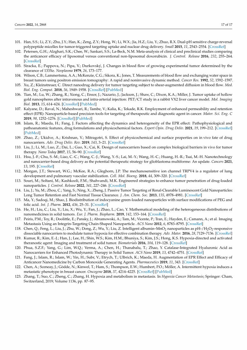
Cancers 2022, 14, 2868 17 of 17
101. Han, S.S.; Li, Z.Y.; Zhu, J.Y.; Han, K.; Zeng, Z.Y.; Hong, W.; Li, W.X.; Jia, H.Z.; Liu, Y.; Zhuo, R.X. Dual-pH sensitive charge-reversalpolypeptide micelles for tumor-triggered targeting uptake and nuclear drug delivery. Small 2015, 11, 2543–2554. [CrossRef]
102. Petersen, G.H.; Alzghari, S.K.; Chee, W.; Sankari, S.S.; La-Beck, N.M. Meta-analysis of clinical and preclinical studies comparingthe anticancer efficacy of liposomal versus conventional non-liposomal doxorubicin. J. Control. Release 2016, 232, 255–264.[CrossRef]
103. Siracka, E.; Pappova, N.; Pipa, V.; Durkovský, J. Changes in blood flow of growing experimental tumor determined by theclearance of 133Xe. Neoplasma 1979, 26, 173–177.
104. Wilson, C.B.; Lammertsma, A.A.; McKenzie, C.G.; Sikora, K.; Jones, T. Measurements of blood flow and exchanging water space inbreast tumors using positron emission tomography: A rapid and noninvasive dynamic method. Cancer Res. 1992, 52, 1592–1597.
105. Xu, Z.; Kleinstreuer, C. Direct nanodrug delivery for tumor targeting subject to shear-augmented diffusion in blood flow. Med.Biol. Eng. Comput. 2018, 56, 1949–1958. [CrossRef] [PubMed]
106. Tian, M.; Lu, W.; Zhang, R.; Xiong, C.; Ensor, J.; Nazario, J.; Jackson, J.; Shaw, C.; Dixon, K.A.; Miller, J. Tumor uptake of hollowgold nanospheres after intravenous and intra-arterial injection: PET/CT study in a rabbit VX2 liver cancer model. Mol. ImagingBiol. 2013, 15, 614–624. [CrossRef] [PubMed]
107. Kalyane, D.; Raval, N.; Maheshwari, R.; Tambe, V.; Kalia, K.; Tekade, R.K. Employment of enhanced permeability and retentioneffect (EPR): Nanoparticle-based precision tools for targeting of therapeutic and diagnostic agent in cancer. Mater. Sci. Eng. C2019, 98, 1252–1276. [CrossRef] [PubMed]
108. Islam, R.; Maeda, H.; Fang, J. Factors affecting the dynamics and heterogeneity of the EPR effect: Pathophysiological andpathoanatomic features, drug formulations and physicochemical factors. Expert Opin. Drug Deliv. 2021, 19, 199–212. [CrossRef][PubMed]
109. Zhao, Z.; Ukidve, A.; Krishnan, V.; Mitragotri, S. Effect of physicochemical and surface properties on in vivo fate of drugnanocarriers. Adv. Drug Deliv. Rev. 2019, 143, 3–21. [CrossRef]
110. Liu, J.; Li, M.; Luo, Z.; Dai, L.; Guo, X.; Cai, K. Design of nanocarriers based on complex biological barriers in vivo for tumortherapy. Nano Today 2017, 15, 56–90. [CrossRef]
111. Hsu, J.-F.; Chu, S.-M.; Liao, C.-C.; Wang, C.-J.; Wang, Y.-S.; Lai, M.-Y.; Wang, H.-C.; Huang, H.-R.; Tsai, M.-H. Nanotechnologyand nanocarrier-based drug delivery as the potential therapeutic strategy for glioblastoma multiforme: An update. Cancers 2021,13, 195. [CrossRef]
112. Morgan, J.T.; Stewart, W.G.; McKee, R.A.; Gleghorn, J.P. The mechanosensitive ion channel TRPV4 is a regulator of lungdevelopment and pulmonary vascular stabilization. Cell. Mol. Bioeng. 2018, 44, 309–320. [CrossRef]
113. Souri, M.; Soltani, M.; Kashkooli, F.M.; Shahvandi, M.K. Engineered strategies to enhance tumor penetration of drug-loadednanoparticles. J. Control. Release 2022, 341, 227–246. [CrossRef]
114. Liu, J.; Yu, M.; Zhou, C.; Yang, S.; Ning, X.; Zheng, J. Passive Tumor Targeting of Renal-Clearable Luminescent Gold Nanoparticles:Long Tumor Retention and Fast Normal Tissue Clearance. J. Am. Chem. Soc. 2013, 135, 4978–4981. [CrossRef]
115. Ma, Y.; Sadoqi, M.; Shao, J. Biodistribution of indocyanine green-loaded nanoparticles with surface modifications of PEG andfolic acid. Int. J. Pharm. 2012, 436, 25–31. [CrossRef]
116. He, H.; Liu, C.; Liu, Y.; Liu, X.; Wu, Y.; Fan, J.; Zhao, L.; Cao, Y. Mathematical modeling of the heterogeneous distributions ofnanomedicines in solid tumors. Eur. J. Pharm. Biopharm. 2019, 142, 153–164. [CrossRef]
117. Peiris, P.M.; Toy, R.; Doolittle, E.; Pansky, J.; Abramowski, A.; Tam, M.; Vicente, P.; Tran, E.; Hayden, E.; Camann, A.; et al. ImagingMetastasis Using an Integrin-Targeting Chain-Shaped Nanoparticle. ACS Nano 2012, 6, 8783–8795. [CrossRef]
118. Chen, Q.; Feng, L.; Liu, J.; Zhu, W.; Dong, Z.; Wu, Y.; Liu, Z. Intelligent albumin–MnO2 nanoparticles as pH-/H2O2-responsivedissociable nanocarriers to modulate tumor hypoxia for effective combination therapy. Adv. Mater. 2016, 28, 7129–7136. [CrossRef]
119. Kumar, R.; Kim, E.-J.; Han, J.; Lee, H.; Shin, W.S.; Kim, H.M.; Bhuniya, S.; Kim, J.S.; Hong, K.S. Hypoxia-directed and activatedtheranostic agent: Imaging and treatment of solid tumor. Biomaterials 2016, 104, 119–128. [CrossRef]
120. Phua, S.Z.F.; Yang, G.; Lim, W.Q.; Verma, A.; Chen, H.; Thanabalu, T.; Zhao, Y. Catalase-Integrated Hyaluronic Acid asNanocarriers for Enhanced Photodynamic Therapy in Solid Tumor. ACS Nano 2019, 13, 4742–4751. [CrossRef]
121. Fang, J.; Islam, R.; Islam, W.; Yin, H.; Subr, V.; Etrych, T.; Ulbrich, K.; Maeda, H. Augmentation of EPR Effect and Efficacy ofAnticancer Nanomedicine by Carbon Monoxide Generating Agents. Pharmaceutics 2019, 11, 343. [CrossRef]
122. Chen, A.; Sceneay, J.; Gödde, N.; Kinwel, T.; Ham, S.; Thompson, E.W.; Humbert, P.O.; Möller, A. Intermittent hypoxia induces ametastatic phenotype in breast cancer. Oncogene 2018, 37, 4214–4225. [CrossRef] [PubMed]
123. Zhang, T.; Suo, C.; Zheng, C.; Zhang, H. Hypoxia and metabolism in metastasis. In Hypoxia Cancer Metastasis; Springer: Cham,Switzerland, 2019; Volume 1136, pp. 87–95.


![Turning Back [updated 6.5.2015]](https://static.fdokumen.com/doc/165x107/6335f35102a8c1a4ec01fd86/turning-back-updated-652015.jpg)