Divergent MicroRNA targetomes of closely related circulating strains of a polyomavirus
Upload
independentCategory
view
3download
0
M O L E C U L A R O N C O L O G Y XXX ( 2 0 1 4 ) 1e1 0
ava i lab le a t www.sc ienced i rec t . com
ScienceDirect
www.elsevier .com/locate/molonc
Novel circulating microRNA signature as a potential
non-invasive multi-marker test in ER-positive early-stage
breast cancer: A case control study
Annette R. Kodahla,b,*, Maria B. Lyngc, Harald Binderd, Søren Colda,b,Karina Gravgaardc, Ann S. Knoope, Henrik J. Ditzela,b,c
aDepartment of Oncology, Odense University Hospital, Odense, DenmarkbUniversity of Southern Denmark, Odense, DenmarkcDepartment of Cancer and Inflammation, Institute of Molecular Medicine, University of Southern Denmark,
DenmarkdInstitute of Medical Biostatistics, Epidemiology and Informatics (IMBEI), Johannes Gutenberg University, Mainz,
GermanyeDepartment of Oncology, Rigshospitalet, Copenhagen University Hospital, Denmark
A R T I C L E I N F O
Article history:
Received 19 February 2014
Accepted 6 March 2014
Available online -
Keywords:
miRNA
Serum markers
Breast cancer
Abbreviations: miRNA, microRNA; mRNA,nent Analysis; ROC, Receiver Operating Cha* Corresponding author. Department of Onco
60138716; fax: þ45 66135477.E-mail addresses: [email protected]
i-mainz.de (H. Binder), [email protected]@health.sdu.dk (H.J. Ditzel).1574-7891/$ e see front matter ª 2014 Federhttp://dx.doi.org/10.1016/j.molonc.2014.03.00
Please cite this article in press as: Kodahmarker test in ER-positive early-stage brej.molonc.2014.03.002
A B S T R A C T
Introduction: There are currently no highly sensitive and specific minimally invasive bio-
markers for detection of early-stage breast cancer. MicroRNAs (miRNAs) are present in
the circulation and may be unique biomarkers for early diagnosis of human cancers. The
aim of this study was to investigate the differential expression of miRNAs in the serum of
breast cancer patients and healthy controls.
Methods: Global miRNA analysis was performed on serum from 48 patients with ER-positive
early-stage breast cancer obtained at diagnosis (24 lymph node-positive and 24 lymph
node-negative) and 24 age-matched healthy controls using LNA-based quantitative real-
time PCR (qRT-PCR). A signature of miRNAs was subsequently validated in an indepen-
dent set of 111 serum samples from 60 patients with early-stage breast cancer and 51
healthy controls and further tested for reproducibility in 3 independent data sets from the
GEO Database.
Results: A multivariable signature consisting of 9 miRNAs (miR-15a, miR-18a, miR-107, miR-
133a, miR-139-5p, miR-143, miR-145, miR-365, miR-425) was identified that provided
considerable discrimination between breast cancer patients and healthy controls. Further,
the ability of the 9 miRNA signature to stratify samples from breast cancer patients and
healthy controls was confirmed in the validation set ( p ¼ 0.012) with a corresponding
AUC ¼ 0.665 in the ROC-curve analysis. No association between miRNA expression and
tumor grade, tumor size, menopausal- or lymph node status was observed. The signature
was also successfully validated in a previously published independent data set of circu-
lating miRNAs in early-stage breast cancer ( p ¼ 0.024).
messenger RNA; ER, Estrogen Receptor; LNA, Locked Nucleic Acid; PCA, Principal Compo-racteristics; AUC, Area Under the Curve.logy, Odense University Hospital, Sdr. Boulevard 29, DK-5000 Odense C, Denmark. Tel.: þ45
k, [email protected] (A.R. Kodahl), [email protected] (M.B. Lyng), binderh@un-(S. Cold), [email protected] (A.S. Gravgaard), [email protected] (A.S. Knoop), hdit-
ation of European Biochemical Societies. Published by Elsevier B.V. All rights reserved.2
l, A.R., et al., Novel circulating microRNA signature as a potential non-invasive multi-ast cancer: A case control study, Molecular Oncology (2014), http://dx.doi.org/10.1016/
M O L E C U L A R O N C O L O G Y XXX ( 2 0 1 4 ) 1e1 02
Please cite this article in press as: Kodahlmarker test in ER-positive early-stage brej.molonc.2014.03.002
Conclusions: We present herein a 9 miRNA signature capable of discriminating between ER-
positive breast cancer and healthy controls. Using a specific algorithm based on the 9
miRNA signature, the risk for future individuals can be predicted. Since microRNAs are
highly stable in blood components, this signature might be useful in the development of a
blood-based multi-marker test to improve early detection of breast cancer. Such a test
could potentially be used as a screening tool to identify individuals who would benefit from
further diagnostic assessment.
ª 2014 Federation of European Biochemical Societies.
Published by Elsevier B.V. All rights reserved.
1. Introduction et al., 2011; Heneghan et al., 2010; Roth et al., 2010; van
The incidence of breast cancer, the most frequent cancer
among women and the second leading cause of cancer death
inwomen in Europe andNorth America, is steadily increasing.
Currently, mammography is the standard screening tool
worldwide, but this technique is not without limitations and
is associated with substantial over diagnosis as well as a
high false-positive rate (Taplin et al., 2008). About 3% of the
population screened annually with mammography will
show abnormalities, but about 65% of these will be false pos-
itives (Langagergaard et al., 2013). In addition, small cancers
are easily missed, especially in younger women due to denser
breast tissue (Carney et al., 2003). Therefore, there is a need for
highly sensitive and specific minimally invasive biomarker-
based assays for early detection of breast cancer, alone or in
combination with mammography.
MicroRNAs (miRNAs) are small, 18e25 nucleotide long,
non-coding RNA molecules that down-regulate the transla-
tion of messenger RNA (mRNA). MicroRNAs control a wide
array of physiological and pathological processes, including
development, differentiation, cellular proliferation, pro-
grammed cell death, oncogenesis and metastasis, by modu-
lating the expression of their target genes through cleaving
mRNA molecules or inhibiting their translation. A tumor sup-
pressor miRNA typically blocks the expression of a “true”
oncogenemRNA. Conversely,miRNA oncogenes block expres-
sion of “true” tumor suppressor genes, leading to increased
risk of tumor formation (Di Leva and Croce, 2010; Li et al.,
2009; Wijnhoven et al., 2007; Zhang et al., 2007). MicroRNAs
represent a promising new class of diagnostic biomarkers
that has been shown to be superior to corresponding mRNA
signatures based on over 20,000 genes when classifying
poorly-differentiated tumors (Lu et al., 2005). Circulating miR-
NAs have been proposed as promising novel biomarkers for
cancer and several other diseases (Weiland et al., 2012). One
of themost important advantages of using circulatingmiRNAs
as biomarkers, apart from being easily measured in blood
samples, is their remarkable stability in plasma and serum,
where they are most likely protected from RNAse degradation
due to their binding to Argonaut proteins (Arroyo et al., 2011;
Mitchell et al., 2008). However, it remains unclear whether a
particular combination of circulating miRNAs is superior to
single miRNAs as a diagnostic test in early breast cancer.
While previous studies on circulatingmiRNAs in breast cancer
have primarily focused on a few preselected miRNAs (Asaga
, A.R., et al., Novel circuast cancer: A case contr
Schooneveld et al., 2012; Wu et al., 2011), we focused on iden-
tifying amulti-miRNA signature optimal for distinguishing be-
tween early breast cancer and healthy controls.
Here, we report our study of prospectively collected serum
samples from 108 women with early-stage breast cancer and
75 concurrently collected healthy controls using a global
Locked Nucleic Acid (LNA�)-based miRNA qPCR platform
(Blondal et al., 2013) to assessdifferences inmiRNAexpression.
2. Materials and methods
2.1. Breast cancer patients and healthy controls
Serum samples (total 183, 108 cases and 75 healthy controls)
were selected from a larger cross sectional cohort collected at
the Department of Oncology, Odense University Hospital,
Denmark, in theperiod 1994e1997 (Espelundet al., 2008).All pa-
tientswere part of theDanish Breast Cancer Cooperative Group
(DBCG) treatment program (Blichert-Toft et al., 2008). Blood
samples were obtained from patients the morning prior to sur-
gery. Serum was prepared within 1 h of sample collection and
immediately stored at �80 �C. Histopathological diagnosis was
confirmed after surgical resection of the tumors. No patients
had received any treatment, such as neo-adjuvant chemo-
therapy or radiation therapy, prior to surgery. Sixty-three
percent were post-menopausal and 37% were pre-
menopausal.Thecontrol serumsampleswerecollectedconcur-
rently, using the same collection and sampling procedures,
from healthy women with no history of malignant diseases
and no inflammatory conditions. Furthermore, none of the
healthy controls were diagnosed with cancer or any inflamma-
tory condition within 5 years after collection of the blood sam-
ple. The age-matched healthy women were randomly selected
from the Danish Civil Registration System for a given case
with a birthday of �one day. The precise menopausal status
for the healthy controls were not available, however 65% were
above the age of 50 and thus were likely post-menopausal. All
cases and controls were ofWestern European descent.
2.2. Study design
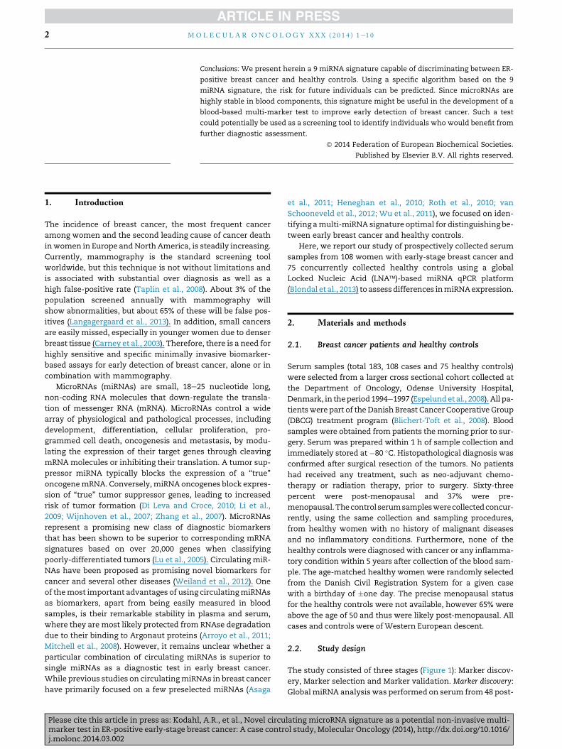
The study consisted of three stages (Figure 1): Marker discov-
ery, Marker selection and Marker validation. Marker discovery:
GlobalmiRNA analysis was performed on serum from 48 post-
lating microRNA signature as a potential non-invasive multi-ol study, Molecular Oncology (2014), http://dx.doi.org/10.1016/
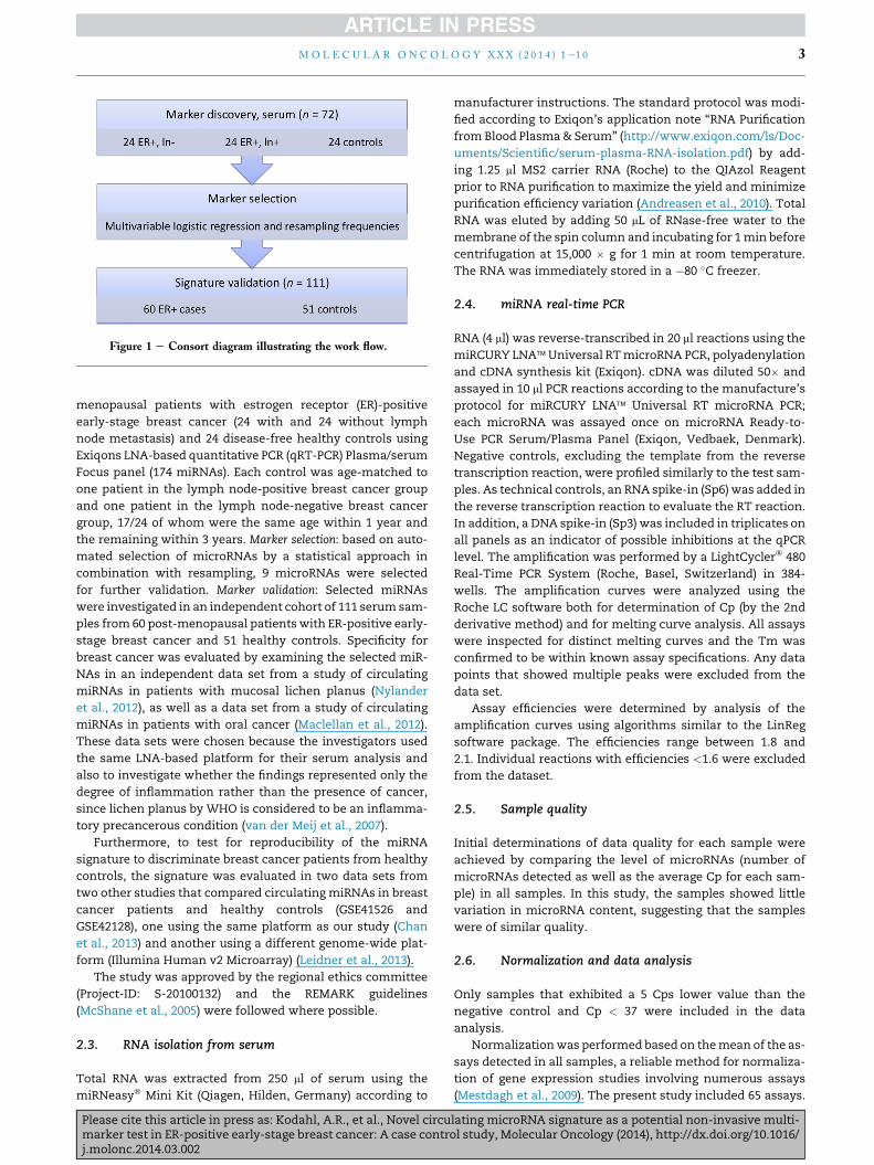
Figure 1 e Consort diagram illustrating the work flow.
M O L E C U L A R O N C O L O G Y XXX ( 2 0 1 4 ) 1e1 0 3
menopausal patients with estrogen receptor (ER)-positive
early-stage breast cancer (24 with and 24 without lymph
node metastasis) and 24 disease-free healthy controls using
Exiqons LNA-based quantitative PCR (qRT-PCR) Plasma/serum
Focus panel (174 miRNAs). Each control was age-matched to
one patient in the lymph node-positive breast cancer group
and one patient in the lymph node-negative breast cancer
group, 17/24 of whom were the same age within 1 year and
the remaining within 3 years. Marker selection: based on auto-
mated selection of microRNAs by a statistical approach in
combination with resampling, 9 microRNAs were selected
for further validation. Marker validation: Selected miRNAs
were investigated in an independent cohort of 111 serum sam-
ples from 60 post-menopausal patients with ER-positive early-
stage breast cancer and 51 healthy controls. Specificity for
breast cancer was evaluated by examining the selected miR-
NAs in an independent data set from a study of circulating
miRNAs in patients with mucosal lichen planus (Nylander
et al., 2012), as well as a data set from a study of circulating
miRNAs in patients with oral cancer (Maclellan et al., 2012).
These data sets were chosen because the investigators used
the same LNA-based platform for their serum analysis and
also to investigate whether the findings represented only the
degree of inflammation rather than the presence of cancer,
since lichen planus by WHO is considered to be an inflamma-
tory precancerous condition (van der Meij et al., 2007).
Furthermore, to test for reproducibility of the miRNA
signature to discriminate breast cancer patients from healthy
controls, the signature was evaluated in two data sets from
two other studies that compared circulating miRNAs in breast
cancer patients and healthy controls (GSE41526 and
GSE42128), one using the same platform as our study (Chan
et al., 2013) and another using a different genome-wide plat-
form (Illumina Human v2 Microarray) (Leidner et al., 2013).
The study was approved by the regional ethics committee
(Project-ID: S-20100132) and the REMARK guidelines
(McShane et al., 2005) were followed where possible.
2.3. RNA isolation from serum
Total RNA was extracted from 250 ml of serum using the
miRNeasy� Mini Kit (Qiagen, Hilden, Germany) according to
Please cite this article in press as: Kodahl, A.R., et al., Novel circumarker test in ER-positive early-stage breast cancer: A case controj.molonc.2014.03.002
manufacturer instructions. The standard protocol was modi-
fied according to Exiqon’s application note “RNA Purification
from Blood Plasma& Serum” (http://www.exiqon.com/ls/Doc-
uments/Scientific/serum-plasma-RNA-isolation.pdf) by add-
ing 1.25 ml MS2 carrier RNA (Roche) to the QIAzol Reagent
prior to RNA purification to maximize the yield and minimize
purification efficiency variation (Andreasen et al., 2010). Total
RNA was eluted by adding 50 mL of RNase-free water to the
membrane of the spin column and incubating for 1min before
centrifugation at 15,000 � g for 1 min at room temperature.
The RNA was immediately stored in a �80 �C freezer.
2.4. miRNA real-time PCR
RNA (4 ml) was reverse-transcribed in 20 ml reactions using the
miRCURY LNA�Universal RTmicroRNA PCR, polyadenylation
and cDNA synthesis kit (Exiqon). cDNA was diluted 50� and
assayed in 10 ml PCR reactions according to the manufacture’s
protocol for miRCURY LNA� Universal RT microRNA PCR;
each microRNA was assayed once on microRNA Ready-to-
Use PCR Serum/Plasma Panel (Exiqon, Vedbaek, Denmark).
Negative controls, excluding the template from the reverse
transcription reaction, were profiled similarly to the test sam-
ples. As technical controls, an RNA spike-in (Sp6) was added in
the reverse transcription reaction to evaluate the RT reaction.
In addition, a DNA spike-in (Sp3) was included in triplicates on
all panels as an indicator of possible inhibitions at the qPCR
level. The amplification was performed by a LightCycler� 480
Real-Time PCR System (Roche, Basel, Switzerland) in 384-
wells. The amplification curves were analyzed using the
Roche LC software both for determination of Cp (by the 2nd
derivative method) and for melting curve analysis. All assays
were inspected for distinct melting curves and the Tm was
confirmed to be within known assay specifications. Any data
points that showed multiple peaks were excluded from the
data set.
Assay efficiencies were determined by analysis of the
amplification curves using algorithms similar to the LinReg
software package. The efficiencies range between 1.8 and
2.1. Individual reactions with efficiencies <1.6 were excluded
from the dataset.
2.5. Sample quality
Initial determinations of data quality for each sample were
achieved by comparing the level of microRNAs (number of
microRNAs detected as well as the average Cp for each sam-
ple) in all samples. In this study, the samples showed little
variation in microRNA content, suggesting that the samples
were of similar quality.
2.6. Normalization and data analysis
Only samples that exhibited a 5 Cps lower value than the
negative control and Cp < 37 were included in the data
analysis.
Normalizationwas performed based on themean of the as-
says detected in all samples, a reliable method for normaliza-
tion of gene expression studies involving numerous assays
(Mestdagh et al., 2009). The present study included 65 assays.
lating microRNA signature as a potential non-invasive multi-l study, Molecular Oncology (2014), http://dx.doi.org/10.1016/

Table 1 e Characteristics of the individuals included in thediscovery set.
Parameters Lymphnode-positive
breastcancer (%)
Lymphnode-negative
breastcancer (%)
Controls(%)
Total 24 24 24
Median age, yr 58 58 57
Range 47e71 50e69 45e69
BMI 26 25 24
Range 19e35 20e37 19e39
Smoking status
Yes 6 (25) 4 (16) 6 (25)
No 13 (54) 10 (42) 18 (75)
Unknown 5 (21) 10 (42) 0
Post-menopausal 23 (96) 22 (92)
Pre-menopausal 1 (4) 2 (8)
ERþ 24 (100) 24(100)
Median
tumor size, mm
16.5 16.5
Range 5e36 2e35
Node-positive 24 (100) 0
1e4 19 (79)
5e9 2 (8)
10þ 3 (13)
Ductal carcinoma 24 (100) 21(88)
M O L E C U L A R O N C O L O G Y XXX ( 2 0 1 4 ) 1e1 04
The equation used to calculate the normalized Cp values was:
Normalized Cp ¼ mean Cp (n ¼ 65) e assay Cp.
For experiments involving only few assays, such as our
validation study, the most stably-expressed miRNAs (with
least variability) in the discovery setwere considered potential
normalization candidates. For this purpose,miR-10b andmiR-
30a were selected since high expression and low variability of
these twomiRNAs was seen in almost all individuals, while in
the remaining few, the expression was measurable and suffi-
cient for normalization.
2.7. Evaluation for potential hemolysis of the bloodsamples
To determine whether hemolysis of the blood samples had
occurred during sample preparation and thus released excess
blood component-related miRNAs, the ratio between the
expression level of two miRNAs was determined; miRNA-
451, expressed in red blood cells and miRNA-23a, relatively
stable in serum and plasma and not affected by hemolysis
(Blondal et al., 2013). Blondal et al. found that a delta Cp
(miR-23aemiR-451) of more than five was an indicator of
possible erythrocyte miRNA contamination and a delta Cp of
7e8 or more indicated a high risk of hemolysis. These param-
eters were evaluated for both the discovery and validation set.
2.8. Statistical analysis
To compare the 3 sample groups, we performed a one factor
ANOVA. MiRNAs with different expression levels between
BC patients and controls were identified by unpaired two-
tailed parametric t-test. P-values obtained for each miRNA
were adjusted for multiple testing by Bonferroni adjustment.
Component-wise likelihood-based boosting (Tutz and
Binder, 2007) was used as a statistical approach for automated
selection of a small set of important microRNAs for a logistic
regression model. The number of boosting steps, i.e. the tun-
ing parameter that determines the number of selected micro-
RNAs, was chosen by 10-fold cross-validation to optimize
prediction performance. Resampling was performed to iden-
tify stably-selected miRNAs informative concerning breast
cancer status (Sauerbrei et al., 2011). Specifically, the data
was randomly split into training and test data 100 times,
with training sets containing 63.2% of the observations
(Binder and Schumacher, 2008). A signature was developed
in each training data set and the resampling inclusion fre-
quency, meaning the relative frequency of signatures where
a certain miRNA is contained, was calculated. MicroRNAs
with a resampling inclusion frequency �0.3 were selected
for further validation. To evaluate whether BMI, smoking sta-
tus, tumor grade, tumor size, or lymph node status affected
the signature, we also provided these factors as candidates
for the boosting approach, which would assure their inclusion
in the model if they were important confounders. Receiver
Operating Characteristics (ROC) curve was calculated for the
validation samples to determine the discrimination perfor-
mance between new samples from women with or without
breast cancer. As the boosting approach estimates a logistic
regression model, odds ratios are obtained for interpretation,
where it should be noted that the odds ratios are biased
Please cite this article in press as: Kodahl, A.R., et al., Novel circumarker test in ER-positive early-stage breast cancer: A case contrj.molonc.2014.03.002
toward zero because of using this estimation approach for a
high-dimensional setting. Odds ratios larger than 1 indicate
increased levels of miRNA in cases, while values below 1 indi-
cate decreased levels of miRNA in cases.
All statistical analyses were performed using normalized
data. Any missing values were replaced by the median of a
given miRNA in the case or control group. Overall, there
were few missing values (Supplementary Table S1). This did
not change any p-values obtained when comparing the two
groups, but it does add to the statistical power. All p-values
were two-tailed and p-value<0.05 was considered statistically
significant. Power simulations were conducted to find the
minimal sample size necessary to find a true 1.5-fold change
with at least 80% statistical power. All statistical calculations
were performed using the statistical environments R and
STATA.
3. Results
3.1. Marker discovery and marker selection
GlobalmiRNA analysis was performed on serum from 48 post-
menopausal patients with ER-positive early-stage breast can-
cer (24 with and 24 without lymph node metastasis) and 24
age-matched and disease-free healthy controls using LNA-
based qRT-PCR (Table 1). Among the 175 miRNAs analyzed,
67 were differentially expressed in serum of breast cancer pa-
tients vs. healthy controls. NomiRNA signature was identified
that could significantly distinguish breast cancer patients
with vs. without lymph node metastasis. As a result, the two
groups were pooled in the further analysis.
lating microRNA signature as a potential non-invasive multi-ol study, Molecular Oncology (2014), http://dx.doi.org/10.1016/

Figure 2 e Principal Component Analysis (PCA) plot showing
unsupervised clustering analysis of the top 50 miRNAs with the
highest standard deviation (expressed in all samples) from the
discovery test set. The healthy controls (green dots) appear different
from the breast cancer patients with (red dots) and without lymph
node metastasis (blue dots).
Table 3 e Characteristics of the individuals included in thevalidation set.
Parameters Patients (%) Controls (%)
Total 60 51
Median age, yr 57 58
Range 32e69 33e70
Post-menopausal 38 (63) (65% above age 50)
Pre-menopausal 22 (37)
ERþ 60(100)
Median tumor size, mm 15
Range 2e65
Nodal status
0 42 (70)
1e4 15 (25)
5e9 1 (2)
10þ 2 (3)
M O L E C U L A R O N C O L O G Y XXX ( 2 0 1 4 ) 1e1 0 5
Principal Component Analysis (PCA), a method used to
reduce the dimension of large data sets, was performed on
the top 50 microRNAs that had the largest variation across
all samples. An overview of how the samples clustered based
on this variance was obtained (Figure 2).
Based on a multivariable logistic regression model esti-
mated by component-wise likelihood-based boosting and
Table 2 e Circulating microRNAs differentially expressed in the serum ofOdds ratios selected by the multivariate model. Discovery data.
MicroRNA Oddsratio
Resamplinginclusion
frequencies
MeanDCpcases
(N ¼ 48)
hsa-miR-145 0.52 0.87 �1.95
hsa-miR-107 1.23 0.42 0.44
hsa-miR-365 0.89 0.32 �4.17
hsa-miR-18a 1.12 0.31 �3.29
hsa-miR-425 1.1 0.30 �0.57
hsa-miR-139-5p 0.72 0.40 �3.19
hsa-miR-143 0.88 0.40 �2.61
hsa-miR-15a 1.33 0.33 3.13
hsa-miR-133a 0.75 0.37 �4.42
hsa-miR-142-3p 1.51 0.24 2.46
hsa-miR-378 0.95 0.21 �2.12
hsa-let-7b 0.79 0.21 0.36
hsa-miR-423-5p 1.05 0.20 �1.04
MicroRNAs selected for validation are in bold.
Please cite this article in press as: Kodahl, A.R., et al., Novel circumarker test in ER-positive early-stage breast cancer: A case controj.molonc.2014.03.002
resampling inclusion frequencies, a 9 miRNA signature,
including miR-15a, miR-18a, miR-107, miR-133a, miR-139-5p,
miR-143, miR-145, miR-365, and miR-425, was identified.
Four of these miRNAs were more highly expressed in the
breast cancer samples vs. healthy controls (miR-15a, miR-
18a, miR-107, and miR-425), while 5 were expressed at lower
levels (miR-133a, miR-139-5p, miR-143, miR-145, and miR-
365). Neither BMI, smoking status, tumor grade, tumor size,
nor lymph node status was selected by the boosting approach
for inclusion and thus do not appear to be important con-
founders. Results of the univariate analyses as well as resam-
pling inclusion frequencies for each miRNA in the profile are
summarized in Table 2. The parameters of the logistic regres-
sionmodel, as estimated by the boosting approach, provide an
algorithm for obtaining predictions for future individuals
based on the 9 miRNA signature. Basically, the algorithm pro-
vides a risk score for each sample analyzed. Subsequently,
this score can be converted into a predicted probability of hav-
ing breast cancer by the following equation 1/(1 þ expescore).
patients with early-stage breast cancer compared to healthy controls.
MeanDCp
controls(N ¼ 24)
Univariatep-values
Adjustedp-value (Bonferroni)
�1.32 2.08E�06 0.0004
�0.17 3.63E�05 0.0062
�3.29 3.59E�05 0.0061
�3.93 4.06E�05 0.0069
�0.82 0.0007 0.1191
�2.66 0.0037 0.6231
�1.88 0.0004 0.0727
2.87 0.0111 >1
�3.67 0.0028 0.4789
2.15 0.0086 >1
�1.56 0.0015 0.2579
0.58 0.0436 >1
�1.28 0.0111 >1
lating microRNA signature as a potential non-invasive multi-l study, Molecular Oncology (2014), http://dx.doi.org/10.1016/

Figure 3 e ROC curve analysis using the 9 miRNA profile (miR-15a,
miR-18a, miR-107, miR-133a, miR-139-5p, miR-143, miR-145,
miR-365, and miR-425) for discriminating breast cancer cases from
healthy controls in the validation set, which consisted of serum from
60 early-stage, ER-positive, breast cancer patients and 51 healthy age-
matched controls.
M O L E C U L A R O N C O L O G Y XXX ( 2 0 1 4 ) 1e1 06
As an example, the probability of having breast cancer with a
score of 1.1 is 1/(1 þ exp�1.1) ¼ 0.75.
Based on the ratio between miR-451 and miR-23a, which
have been shown to be associated with hemolysis under the
blood drawing process, we found a mean delta Cp (miR-
23aemiR-451) of 5.6 among the controls and 6.0 among cases,
indicating possible erythrocyte miRNA contamination, but no
overall high risk of hemolysis in the two groups.
3.2. Profile validation
To validate the ability of the 9 miRNA signature to stratify
breast cancer patients vs. healthy controls, we analyzed the
expression levels of these 9miRNAs in serum froman indepen-
dent group of 60 early-stage, ER-positive, breast cancer patients
and 51 healthy age-matched controls (Table 3). When evalu-
ating the samples for possible risk of hemolysis, we found a
mean delta Cp (miR-23aemiR-451) of 5.4 among the controls
and 5.8 among cases. Again, this indicates possible erythrocyte
miRNA contamination, but no overall high risk of hemolysis in
the two groups. Eight samples (7%) had a mean delta Cp
Table 4 e Signature miRNAs from current study compared in Leidner, C
Kodahl et al. Chan et al.
miRNA Log2 FC P value Log2 FC P va
miR-143 0.72 0.0004 �1.09 6.30E
miR-145 0.64 2.08E�06 1.24 0.293
miR-139-5p 0.53 0.004 �0.30 0.223
miR-365 0.91 3.59E�05 �1.01 0.014
miR-133a 0.75 0.003 �3.68 7.65E
miR-15a �0.26 0.011 �0.67 0.015
miR-18a �0.63 4.06E�05 �0.45 0.002
miR-107 �0.61 3.63E�05 �0.42 7.13E
miR-425 �0.25 0.0007 �0.57 0.002
Please cite this article in press as: Kodahl, A.R., et al., Novel circumarker test in ER-positive early-stage breast cancer: A case contrj.molonc.2014.03.002
between 7 and 8, but no samples had a mean delta Cp above
8. Most important, no significant difference in potential risk
of hemolysis was found between the two groups.
The 9 miRNA signature significantly discriminated be-
tween patients with breast cancer and healthy controls
( p ¼ 0.012, 95% CI 1.12e2.39). A ROC curve was calculated
with an Area Under the Curve (AUC) of 0.665 (95% CI
0.562e0.768; Figure 3) to discriminate early-stage breast can-
cer patients from healthy controls. Using a probability cut-
off of 0.48, the sensitivity was 83.3%, specificity 41.2%, positive
predictive value (PPV) 62.5% and a negative predictive value
(NPV) of 67.4%. To assess whether the 9 miRNA signature
was specific to post-menopausal breast cancer patients, we
compared the expression of the selected miRNAs in post-
(63%) and pre-menopausal (37%) breast cancer patients and
found no significant difference, suggesting that the signature
is not related to menopausal status.
Examining our 9miRNA signature in two separate data sets
using the same platform, one comparing serum miRNA
expression in patients with lichen planus to healthy controls
(Nylander et al., 2012), and the other comparing serummiRNA
expression in patients with oral cancer to healthy controls
(Maclellan et al., 2012), we found no difference in signature
expression of the 9 miRNAs between either patient set and
healthy controls, indicating that the signature is not just a
marker of inflammation or cancer in general (data not shown).
3.3. Test of reproducibility
We also evaluated our signature in data sets from two studies
of circulatingmiRNAs in early-stage breast cancer (Chan et al.,
2013; Leidner et al., 2013), applying our developed formula af-
ter normalizing tomiRNA-10b. Thus, scores for cases and con-
trols were obtained and compared by t-tests. A significant
difference between cases and controls was observed in the
data set of Chan et al. ( p ¼ 0.024), while no significant differ-
ence was observed for the data of Leidner et al. ( p ¼ 0.626)
(Table 4).
4. Discussion
In this study, we identified and validated a signature, consist-
ing of 9 circulating miRNAs (miR-15a, miR-18a, miR-107, miR-
han and Maclellan data sets.
Leidner et al. Maclellan et al.
lue Log2 FC P value Log2 FC P value
�04 0.90 0.054 N/A N/A
N/A N/A N/A N/A
N/A N/A N/A N/A
1.44 0.016 0.72 0.017
�09 N/A N/A 0.57 0.104
�0.25 0.041 N/A N/A
8 N/A N/A N/A N/A
�04 N/A N/A N/A N/A
1 N/A N/A 0.25 0.171
lating microRNA signature as a potential non-invasive multi-ol study, Molecular Oncology (2014), http://dx.doi.org/10.1016/

M O L E C U L A R O N C O L O G Y XXX ( 2 0 1 4 ) 1e1 0 7
133a, miR-139-5p, miR-143, miR-145, miR-365, and miR-425),
that discriminates between patients with newly diagnosed,
ER-positive, early-stage breast cancer and healthy controls.
A particular strength of this study is the long follow-up of
the DBCG-cohort and healthy controls that assured the latter
were not diagnosedwith any cancer or inflammatory diseases
within 5 years from blood collection. A second strength is the
identified miRNA signature consisting of several miRNAs, as
such signatures are likely less vulnerable than single miRNAs
to biological differences, which makes them more applicable
to daily clinical use.
In our miRNA selection for the signature, we accounted for
potential co-regulation of different miRNAs by using a multi-
variable approach that simultaneously considered all micro-
RNAs as candidates and automatically adjusted for other
microRNAs in the signature. Since miRNAs have numerous
target mRNAs, and a single mRNA is targeted by numerous
miRNAs, the effect of one dysregulated miRNA is most likely
dependent on the dysregulation of several others that might
perform similar biological roles. Our 9 miRNAs signature,
when put into a specific algorithm, generates a score that re-
flects the risk of having breast cancer. With only a moderate
performance (AUC of 0.665), the signature alone is not an
effective diagnostic test, but it may show promise as a valu-
able risk assessment tool in breast cancer screening in combi-
nation with screening mammography. In comparison, AUC
for screening mammography was found to be between 0.554
and 0.877, depending on age and breast tissue density
(Pisano et al., 2008). However, the performance of the test in
combination with screening mammography needs further
evaluation in larger, pre-diagnostic, studies.
One of the major concerns when analyzing circulating
miRNAs is the normalization procedure. In the discovery anal-
ysis, we normalized to the mean Cp-value of all samples,
which is recommended when dealing with multiple assays
(Mestdagh et al., 2009). However, this method was not appli-
cable in the validation study since it included only a few as-
says. Previous single assay studies lacked reliable
endogenous control microRNAs, and most used miR-16 for
endogenous normalization. However, recent studies have
found miR-16 to be unsuitable as reference (Cookson et al.,
2012; Kirschner et al., 2011). As an alternative, the most stably
and consistently expressed miRNAs may be used for normal-
ization, in our case miR-10b andmiR-30a. MiRNA-10b, howev-
er, has previously been described in relation to metastatic
breast cancer (Biagioni et al., 2012; Chen et al., 2013;
Eichelser et al., 2013; Iorio et al., 2005; Ma et al., 2007; Zhao
et al., 2012) with conflicting findings. Ma et al. found miR-
10b to be up-regulated in tumor tissues from patients with
metastatic breast cancer, but not in tissue from patients
with no metastatic disease (Ma et al., 2007). In contrast, Iori
et al. found miR-10b to be down-regulated in primary breast
cancer tissue compared to normal breast tissue (Iorio et al.,
2005), while Eichelser et al. recently reported no association
between serummiR-10b expression and the presence of overt
metastasis (Eichelser et al., 2013) despite having previously
found serumMiR-10b to be related to breast cancermetastasis
(Roth et al., 2010). Finally, Heneghan et al. did not find that
circulating miR-10b could discriminate between patients
with breast cancer (stages I-IV) and healthy controls
Please cite this article in press as: Kodahl, A.R., et al., Novel circumarker test in ER-positive early-stage breast cancer: A case controj.molonc.2014.03.002
(Heneghan et al., 2010). Thus, no clear evidence exists linking
miR-10b to early-stage breast cancer.
With regard to the other control miRNA, miR-30a, it has
been suggested that it might act as a tumor suppressor by tar-
geting Vim, a gene coding for vimentin (Cheng et al., 2012).
Examined on micro-dissected tumor cells, patients with
decreased levels of miRNA-30a at the site of primary tumors
had increased hazard ratios (HR) of recurrence. To our knowl-
edge, no studies have shown a linkage between the expression
of circulating miR-30a and early-stage breast cancer.
One of the major concerns when investigating circulating
miRNAs is the lack of reproducibility and the prevalence of
inconsistent findings between studies. This issue was previ-
ously addressed by Leidner et al., who examined the degree
of consensus between several genome-wide circulating
miRNA studies as well the reproducibility of these results in
a study involving serum samples from 20 women with newly
diagnosed breast cancer before surgery, 20 women after tu-
mor resection, 20 healthy controls and 10 women with can-
cers other than breast (Leidner et al., 2013). Comparing their
results to other published studies revealed a similar lack of
consistency for which there may be several reasons, such as
differences in study populations, sample material and prepa-
ration, platforms, miRNA extraction procedures, normaliza-
tion techniques as well as statistical approaches.
We investigated whether our miRNA signature discrimi-
nated between early-stage breast cancer patients and healthy
controls in a study using the same platform as we (Chan et al.,
2013), as well as in the data from the above-mentioned study
(Leidner et al., 2013), using a different platform. Comparison of
results across platforms has clear limitations, and thus we
were not surprised to find that the signature did not replicate
in the data set by Leidner et al. However, using the same
normalization approach on the data set by Chan et al., we suc-
cessfully validated our 9miRNA signature in this data set, sup-
porting the possible clinical utility of the signature although
this needs further investigation in a larger prospective study.
Several others have examined the expression of circulating
miRNAs (Supplementary Table S2) in relation to breast cancer
(Asaga et al., 2011; Chan et al., 2013; Cuk et al., 2013; Heneghan
et al., 2010; Leidner et al., 2013; Ng et al., 2013; Roth et al., 2010;
Schrauder et al., 2012; Wang et al., 2010; Wu et al., 2011, 2012;
Zhao et al., 2010), some using very limited numbers of probe-
sets (Asaga et al., 2011; Heneghan et al., 2010; Roth et al., 2010)
prone to becoming biased due to the way the selected probes
define the final observations. Others have used genome-wide
platforms to profile all circulating miRNAs (Cuk et al., 2013;
Leidner et al., 2013; Ng et al., 2013; Schrauder et al., 2012; Wu
et al., 2011, 2012; Zhao et al., 2010), a method that provides un-
biased discovery of putative circulating miRNA biomarkers. It
has been suggested that miRNA signatures consisting of a
combination ofmultiplemiRNAs are superior to singlemiRNA
models in discriminating blood samples from breast cancer
patients and healthy controls (Cuk et al., 2013; Ng et al.,
2013). Ng et al. found that a combination signature consisting
of miR-451 (up-regulated) and miR-145 (down-regulated)
discriminated between breast cancer cases and healthy con-
trols as well as other cancer types with an AUC of 0.96. How-
ever, this signature could not distinguish between benign
tumors and invasive breast cancer. In our study, we also found
lating microRNA signature as a potential non-invasive multi-l study, Molecular Oncology (2014), http://dx.doi.org/10.1016/
M O L E C U L A R O N C O L O G Y XXX ( 2 0 1 4 ) 1e1 08
miR-145 to be down-regulated in breast cancer vs. healthy
controls, while miR-451 expression did not differ between
the two groups. Cuk et al. showed that a signature consisting
of miR-148b, miR-409-3p and miR-801 was able to distinguish
between 127 female sporadic breast cancer patients and 80
healthy controls (AUC ¼ 0.69). MiR-801 was not included in
our study, and no difference in expression between miR-
148b and miR-409-3p was observed. These differing results
may be due to differences in the sample populations, since
Cuk et al. allowed inclusion of patients who were not primar-
ily operable.
All samples in the discovery set were analyzed for 174miR-
NAs (Exiqon Cancer Focus Panel). However, important miR-
NAs of more than 1800 human miRNAs currently listed in
the miRBase (http://www.mirbase.org/, accessed September
13, 2013) may have been missed.
Several of the miRNAs in our 9 miRNA signature have been
investigated by others. Interestingly, using prospectively
collected samples from the large Sister Cohort Study, serum
miR-18a was found to be expressed at higher levels in women
who subsequently developed breast cancer (within 18 months
from blood collection) compared to those who remained
breast cancer-free, although this could not be validated in
the relatively small validation set (Godfrey et al., 2013). Similar
to our findings, Chan et al. found down-regulation of miR-145
in serum of women with breast cancer compared to normal
controls, whereas in the same study, miR-133a and miR-143
were up-regulated, in contrast with our findings (Chan et al.,
2013). In agreement with our findings, Chen et al. also found
higher expression of miR-107 in breast cancer vs. normal tis-
sue (Chen et al., 2011). Finally, as in our study, low levels of
miR-139-5p and high levels of miR-425 were observed in
breast cancer vs. adjacent normal tissue, whereas, in contrast
to our findings, miRNA-365 was found to be more highly
expressed in breast cancer tissue (Romero-Cordoba et al.,
2012; Yan et al., 2008).
Recently, spike-in controls to evaluate the quality of the
RNA extraction have been introduced, but this technique
was not commonly used at the time of serum sample analysis
for this study and no control for the RNA extraction procedure
was performed.While thismay be a potential bias, all samples
in each study (discovery and validation set) were carefully
processed simultaneously and by the same bioanalyst to stan-
dardize the extraction procedure.
Technical replicates were not included in the present
study, however the qPCR platform used has previously
demonstrated a high reproducibilitywithmedian Pearson cor-
relation coefficients of 0.985, minimizing the risk of technical
variance (Jensen et al., 2011). Furthermore, technical valida-
tion is usually performed when the biological material is
sparse and the information gained by runningmore biological
samples instead of technical replicates is preferred.
To investigate whether the difference inmiRNA expression
between the breast cancer and the control group was simply
due to an inflammatory response, we investigated whether
our signature could distinguish mucosal lichen planus pa-
tients from healthy controls using a published circulating
miRNA data set (Nylander et al., 2012). This study was specif-
ically chosen because it was not a cancer being studied, but a
precancerous inflammatory disease, and because this study
Please cite this article in press as: Kodahl, A.R., et al., Novel circumarker test in ER-positive early-stage breast cancer: A case contrj.molonc.2014.03.002
employed the same platform as we. A limitation in this com-
parison is that the previous study included both men and
women, although 80% were women, and they were slightly
older (mean age women: 64 years, men: 58 years) than those
in our study (mean age: 55 years).
Sample handling, preparation and the use of different
collection tubes in serum vs. plasma could possibly affect
the miRNA profile. In our study, serum was used for the anal-
ysis of circulating miRNAs. Measurements of miRNAs in
serum and plasma have previously been found to be highly
correlated (Kroh et al., 2010; Mitchell et al., 2008). However,
serum samples have more detectable miRNAs than plasma
samples, and most miRNAs in serum show higher concentra-
tions than corresponding plasma samples (Wang et al., 2012).
Compared to whole blood, we would expect a difference since
miRNAs extracted from whole blood would include those
within blood cells. All patients in this study had diagnostic bi-
opsies taken prior to blood sampling, and whether this proce-
dure affected detection of circulating miRNAs remains to be
determined and thus should be considered a potential bias.
One method to increase the statistical power when
analyzing a limited amount of material is using a more ho-
mogenous group, which is why we focused on only women
with ER-positive breast cancer. The ATAC trial comprised of
9366 post-menopausal women with early-stage breast cancer
showed that 84% of the population was ER-positive (Baum
et al., 2002). Since then, the cut-off of 10% staining cells for
ER, required for classification as ER-positive, has been
changed to 1% (Viale et al., 2007), which means the number
of ER-positive breast cancers might be even higher than 84%.
Whether the signature presented in this study can distinguish
between ER-negative breast cancer and healthy controls has
not been investigated, but based on the above, the patient
characteristics in this study are close to the general Danish
mammography screening population. To assess whether our
signature is applicable to both pre- and post-menopausal
women requires larger studies. The present study did not pro-
vide enough power for such subgroup analysis.
5. Conclusion
We identified a novel circulating 9-miRNA signature that dis-
criminates between serum from early-stage, ER-positive,
breast cancer patients and healthy control subjects. The
signature was successfully validated in an independent
cohort of 111 serum samples as well as an independent data
set from the GEO database. This signature may be of use in
the development of a non-invasive, multi-marker blood test
to improve early detection of breast cancer. Specifically, the
signature provides a risk score reflecting the possibility of hav-
ing early-stage breast cancer. To further validate the potential
of this signature, we are planning a prospective study in the
pre-diagnostic setting at a mammography screening center.
Authors’ contributions
ARK, AK and HD participated in study design. ARK coordi-
nated the project, performed data interpretation and wrote
lating microRNA signature as a potential non-invasive multi-ol study, Molecular Oncology (2014), http://dx.doi.org/10.1016/

M O L E C U L A R O N C O L O G Y XXX ( 2 0 1 4 ) 1e1 0 9
the first draft of themanuscript. HB performed themajority of
the statistical analysis. HD, ML, HB and KG provided critical
advice on study design and data interpretation. HB, ML, KG,
and HD assisted with writing the manuscript. SC provided
all samples and patient data. All authors read and approved
the final manuscript.
Conflict of interest
The authors declare no conflict of interest.
Funding
The public and private foundations that supported the study
had no role in the design and conduct of the study; in the
collection, management, analysis, and interpretation of the
data; or in the preparation, review, or approval of the manu-
script. The authors had full access to all the data in the study
and had the final responsibility for the decision to submit the
manuscript for publication.
Acknowledgments
This study was supported by Odense University Hospital, the
Region of Southern Denmark, and University of Southern
Denmark. Further support was obtained from Svend H.A
Schrøder andwife Ketty L. Larsen Schrøder Foundation, Fabri-
kant EinarWillumsens Mindelegat, A.P. Møller and wife Chas-
tine Mc-Kinney Møllers Foundation, Beckett Foundation, King
Christian X Foundation, Mimi and Victor Larsen Foundation,
Danish Cancer Society, Danish Research Council, Danish Cen-
ter for Translational Breast Cancer Research (DCTB) and A
Race Against Breast Cancer. We thank M.K. Occhipinti for
editorial assistance.
Appendix A.Supplementary data
Supplementary data related to this article can be found at
http://dx.doi.org/10.1016/j.molonc.2014.03.002.
R E F E R E N C E S
Andreasen, D., Fog, J.U., Biggs, W., Salomon, J., Dahslveen, I.K.,Baker, A., Mouritzen, P., 2010. Improved microRNAquantification in total RNA from clinical samples. Methods 50,S6eS9.
Arroyo, J.D., Chevillet, J.R., Kroh, E.M., Ruf, I.K., Pritchard, C.C.,Gibson, D.F., Mitchell, P.S., et al., 2011. Argonaute2 complexescarry a population of circulating microRNAs independent ofvesicles in human plasma. Proc. Natl. Acad. Sci. U.S.A. 108,5003e5008.
Asaga, S., Kuo, C., Nguyen, T., Terpenning, M., Giuliano, A.E.,Hoon, D.S., 2011. Direct serum assay for microRNA-21
Please cite this article in press as: Kodahl, A.R., et al., Novel circumarker test in ER-positive early-stage breast cancer: A case controj.molonc.2014.03.002
concentrations in early and advanced breast cancer. Clin.Chem. 57, 84e91.
Baum,M., Budzar,A.U., Cuzick, J., Forbes, J.,Houghton, J.H., Klijn, J.G.,Sahmoud, T., 2002. Anastrozole alone or in combination withtamoxifen versus tamoxifen alone for adjuvant treatment ofpostmenopausal womenwith early breast cancer: first results ofthe ATAC randomised trial. Lancet 359, 2131e2139.
Biagioni, F., Bossel Ben-Moshe, N., Fontemaggi, G., Canu, V.,Mori, F., Antoniani, B., Di Benedetto, A., et al., 2012. miR-10b*, amaster inhibitor of the cell cycle, is down-regulated in humanbreast tumours. EMBO Mol. Med. 4, 1214e1229.
Binder, H., Schumacher, M., 2008. Adapting prediction errorestimates for biased complexity selection in high-dimensionalbootstrap samples. Stat. Appl. Genet. Mol. Biol. 7. Article12.
Blichert-Toft, M., Christiansen, P., Mouridsen, H.T., 2008. DanishBreast Cancer Cooperative GroupeDBCG: history,organization, and status of scientific achievements at 30-yearanniversary. Acta Oncol. 47, 497e505.
Blondal, T., Jensby Nielsen, S., Baker, A., Andreasen, D.,Mouritzen, P., Wrang Teilum, M., Dahlsveen, I.K., 2013.Assessing sample and miRNA profile quality in serum andplasma or other biofluids. Methods 59, S1eS6.
Carney, P.A., Miglioretti, D.L., Yankaskas, B.C., Kerlikowske, K.,Rosenberg, R., Rutter, C.M., Geller, B.M., et al., 2003. Individualand combined effects of age, breast density, and hormonereplacement therapy use on the accuracy of screeningmammography. Ann. Intern. Med. 138, 168e175.
Chan, M., Liaw, C.S., Ji, S.M., Tan, H.H., Wong, C.Y., Thike, A.A.,Tan, P.H., et al., 2013. Identification of circulating microRNAsignatures for breast cancer detection. Clin. Cancer Res. 19,4477e4487.
Chen, P.S., Su, J.L., Cha, S.T., Tarn, W.Y., Wang, M.Y., Hsu, H.C.,Lin, M.T., et al., 2011. miR-107 promotes tumor progression bytargeting the let-7 microRNA in mice and humans. J. Clin.Invest. 121, 3442e3455.
Chen, W., Cai, F., Zhang, B., Barekati, Z., Zhong, X.Y., 2013. Thelevel of circulating miRNA-10b and miRNA-373 in detectinglymph node metastasis of breast cancer: potential biomarkers.Tumour Biol. 34, 455e462.
Cheng, C.W., Wang, H.W., Chang, C.W., Chu, H.W., Chen, C.Y.,Yu, J.C., Chao, J.I., et al., 2012. MicroRNA-30a inhibits cellmigration and invasion by downregulating vimentinexpression and is a potential prognostic marker in breastcancer. Breast Cancer Res. Treat. 134, 1081e1093.
Cookson, V.J., Bentley, M.A., Hogan, B.V., Horgan, K.,Hayward, B.E., Hazelwood, L.D., Hughes, T.A., 2012.Circulating microRNA profiles reflect the presence of breasttumours but not the profiles of microRNAs within thetumours. Cell Oncol. (Dordr.) 35, 301e308.
Cuk, K., Zucknick, M., Heil, J., Madhavan, D., Schott, S.,Turchinovich, A., Arlt, D., et al., 2013. Circulating microRNAsin plasma as early detection markers for breast cancer. Int. J.Cancer 132, 1602e1612.
Di Leva, G., Croce, C.M., 2010. Roles of small RNAs in tumorformation. Trends Mol. Med. 16, 257e267.
Eichelser, C., Flesch-Janys, D., Chang-Claude, J., Pantel, K.,Schwarzenbach, H., 2013. Deregulated serum concentrationsof circulating cell-free microRNAs miR-17, miR-34a, miR-155,and miR-373 in human breast cancer development andprogression. Clin. Chem. 59, 1489e1496.
Espelund, U., Cold, S., Frystyk, J., Orskov, H., Flyvbjerg, A., 2008.Elevated free IGF2 levels in localized early-stage breast cancerin women. Eur. J. Endocrinol. 159, 595e601.
Godfrey, A.C., Xu, Z., Weinberg, C.R., Getts, R.C., Wade, P.A.,Deroo, L.A., Sandler, D.P., et al., 2013. Serum microRNAexpression as an early marker for breast cancer risk inprospectively collected samples from the Sister Study cohort.Breast Cancer Res. 15, R42.
lating microRNA signature as a potential non-invasive multi-l study, Molecular Oncology (2014), http://dx.doi.org/10.1016/

M O L E C U L A R O N C O L O G Y XXX ( 2 0 1 4 ) 1e1 010
Heneghan, H.M., Miller, N., Lowery, A.J., Sweeney, K.J., Newell, J.,Kerin, M.J., 2010. Circulating microRNAs as novel minimallyinvasive biomarkers for breast cancer. Ann. Surg. 251, 499e505.
Iorio, M.V., Ferracin, M., Liu, C.G., Veronese, A., Spizzo, R.,Sabbioni, S., Magri, E., et al., 2005. MicroRNA gene expressionderegulation inhumanbreast cancer.CancerRes. 65,7065e7070.
Jensen, S.G., Lamy, P., Rasmussen, M.H., Ostenfeld, M.S.,Dyrskjot, L., Orntoft, T.F., Andersen, C.L., 2011. Evaluation oftwo commercial global miRNA expression profiling platformsfor detection of less abundant miRNAs. BMC Genomics 12, 435.
Kirschner, M.B., Kao, S.C., Edelman, J.J., Armstrong, N.J.,Vallely, M.P., van Zandwijk, N., Reid, G., 2011. Haemolysisduring sample preparation alters microRNA content ofplasma. PLoS One 6, e24145.
Kroh, E.M., Parkin, R.K., Mitchell, P.S., Tewari, M., 2010. Analysisof circulating microRNA biomarkers in plasma and serumusing quantitative reverse transcription-PCR (qRT-PCR).Methods 50, 298e301.
Langagergaard, V., Garne, J.P., Vejborg, I., Schwartz, W., Bak, M.,Lernevall, A., Mogensen, N.B., et al., 2013. Existing datasources for clinical epidemiology: the Danish Quality Databaseof Mammography Screening. Clin. Epidemiol. 5, 81e88.
Leidner, R.S., Li, L., Thompson, C.L., 2013. Dampening enthusiasmfor circulating microRNA in breast cancer. PLoS One 8, e57841.
Li, M., Marin-Muller, C., Bharadwaj, U., Chow, K.H., Yao, Q.,Chen, C., 2009. MicroRNAs: control and loss of control inhuman physiology and disease. World J. Surg. 33, 667e684.
Lu, J., Getz, G., Miska, E.A., Alvarez-Saavedra, E., Lamb, J., Peck, D.,Sweet-Cordero, A., et al., 2005. MicroRNA expression profilesclassify human cancers. Nature 435, 834e838.
Ma, L., Teruya-Feldstein, J., Weinberg, R.A., 2007. Tumourinvasion and metastasis initiated by microRNA-10b in breastcancer. Nature 449, 682e688.
Maclellan, S.A., Lawson, J., Baik, J., Guillaud, M., Poh, C.F.,Garnis, C., 2012. Differential expression of miRNAs in theserum of patients with high-risk oral lesions. Cancer Med. 1,268e274.
McShane, L.M., Altman, D.G., Sauerbrei, W., Taube, S.E., Gion, M.,Clark, G.M., 2005. Reporting recommendations for tumormarker prognostic studies (REMARK). J. Natl. Cancer Inst. 97,1180e1184.
Mestdagh, P., Van Vlierberghe, P., De Weer, A., Muth, D.,Westermann, F., Speleman, F., Vandesompele, J., 2009. A noveland universal method for microRNA RT-qPCR datanormalization. Genome Biol. 10, R64.
Mitchell, P.S., Parkin, R.K., Kroh, E.M., Fritz, B.R., Wyman, S.K.,Pogosova-Agadjanyan, E.L., Peterson, A., et al., 2008.Circulating microRNAs as stable blood-based markers forcancer detection. Proc. Natl. Acad. Sci. U.S.A. 105,10513e10518.
Ng, E.K., Li, R., Shin, V.Y., Jin, H.C., Leung, C.P., Ma, E.S., Pang, R.,et al., 2013. Circulating microRNAs as specific biomarkers forbreast cancer detection. PLoS One 8, e53141.
Nylander, E., Ebrahimi, M., Wahlin, Y.B., Boldrup, L., Nylander, K.,2012. Changes in miRNA expression in sera and correlation toduration of disease in patients with multifocal mucosal lichenplanus. J. Oral Pathol. Med. 41, 86e89.
Pisano, E.D., Hendrick, R.E., Yaffe, M.J., Baum, J.K., Acharyya, S.,Cormack, J.B., Hanna, L.A., et al., 2008. Diagnostic accuracy ofdigital versus filmmammography: exploratory analysis ofselectedpopulationsubgroups inDMIST.Radiology246,376e383.
Romero-Cordoba, S., Rodriguez-Cuevas, S., Rebollar-Vega, R.,Quintanar-Jurado, V., Maffuz-Aziz, A., Jimenez-Sanchez, G.,Bautista-Pina, V., et al., 2012. Identification and pathwayanalysis of microRNAs with no previous involvement in breastcancer. PLoS One 7, e31904.
Roth, C., Rack, B., Muller, V., Janni, W., Pantel, K.,Schwarzenbach, H., 2010. Circulating microRNAs as blood-
Please cite this article in press as: Kodahl, A.R., et al., Novel circumarker test in ER-positive early-stage breast cancer: A case contrj.molonc.2014.03.002
based markers for patients with primary and metastaticbreast cancer. Breast Cancer Res. 12, R90.
Sauerbrei, W., Boulesteix, A.-L., Binder, H., 2011. Stabilityinvestigations of multivariable regression models derivedfrom low- and high-dimensional data. J. Biopharm. Stat. 21,1206e1231.
Schrauder, M.G., Strick, R., Schulz-Wendtland, R., Strissel, P.L.,Kahmann, L., Loehberg, C.R., Lux, M.P., et al., 2012. Circulatingmicro-RNAs as potential blood-based markers for early stagebreast cancer detection. PLoS One 7, e29770.
Taplin, S., Abraham, L., Barlow, W.E., Fenton, J.J., Berns, E.A.,Carney, P.A., Cutter, G.R., et al., 2008. Mammographyfacility characteristics associated with interpretive accuracyof screening mammography. J. Natl. Cancer Inst. 100,876e887.
Tutz, G., Binder, H., 2007. Boosting ridge regression. Comput. Stat.Data Anal. 51, 6044e6059.
van der Meij, E.H., Mast, H., van der Waal, I., 2007. The possiblepremalignant character of oral lichen planus and orallichenoid lesions: a prospective five-year follow-up study of192 patients. Oral Oncol. 43, 742e748.
van Schooneveld, E., Wouters, M.C., Van der Auwera, I.,Peeters, D.J., Wildiers, H., Van Dam, P.A., Vergote, I., et al.,2012. Expression profiling of cancerous and normal breasttissues identifies microRNAs that are differentially expressedin serum from patients with (metastatic) breast cancer andhealthy volunteers. Breast Cancer Res. 14, R34.
Viale, G., Regan, M.M., Maiorano, E., Mastropasqua, M.G.,Dell’Orto, P., Rasmussen, B.B., Raffoul, J., et al., 2007.Prognostic and predictive value of centrally reviewedexpression of estrogen and progesterone receptors in arandomized trial comparing letrozole and tamoxifen adjuvanttherapy for postmenopausal early breast cancer: BIG 1-98.J. Clin. Oncol. 25, 3846e3852.
Wang, F., Zheng, Z., Guo, J., Ding, X., 2010. Correlation andquantitation of microRNA aberrant expression in tissues andsera from patients with breast tumor. Gynecol. Oncol. 119,586e593.
Wang, K., Yuan, Y., Cho, J.H., McClarty, S., Baxter, D., Galas, D.J.,2012. Comparing the MicroRNA spectrum between serum andplasma. PLoS One 7, e41561.
Weiland, M., Gao, X.H., Zhou, L., Mi, Q.S., 2012. Small RNAs have alarge impact: circulating microRNAs as biomarkers for humandiseases. RNA Biol. 9, 850e859.
Wijnhoven, B.P., Michael, M.Z., Watson, D.I., 2007. MicroRNAsand cancer. Br. J. Surg. 94, 23e30.
Wu, Q., Lu, Z., Li, H., Lu, J., Guo, L., Ge, Q., 2011. Next-generationsequencing of microRNAs for breast cancer detection.J. Biomed. Biotechnol. 2011, 597145.
Wu, Q., Wang, C., Lu, Z., Guo, L., Ge, Q., 2012. Analysis of serumgenome-wide microRNAs for breast cancer detection. Clin.Chim. Acta 413, 1058e1065.
Yan, L.X., Huang, X.F., Shao, Q., Huang, M.Y., Deng, L., Wu, Q.L.,Zeng, Y.X., et al., 2008. MicroRNA miR-21 overexpression inhuman breast cancer is associated with advanced clinicalstage, lymph node metastasis and patient poor prognosis.RNA 14, 2348e2360.
Zhang, B., Pan, X., Cobb, G.P., Anderson, T.A., 2007. microRNAs asoncogenes and tumor suppressors. Development. Biol. 302,1e12.
Zhao, F.L., Hu, G.D., Wang, X.F., Zhang, X.H., Zhang, Y.K., Yu, Z.S.,2012. Serum overexpression of microRNA-10b in patients withbone metastatic primary breast cancer. J. Int. Med. Res. 40,859e866.
Zhao, H., Shen, J., Medico, L., Wang, D., Ambrosone, C.B., Liu, S.,2010. A pilot study of circulating miRNAs as potentialbiomarkers of early stage breast cancer. PLoS One 5, e13735.Web references. http://www.mirbase.org/.
lating microRNA signature as a potential non-invasive multi-ol study, Molecular Oncology (2014), http://dx.doi.org/10.1016/
Copyright © 2022 FDOKUMEN