NMDA-induced neuronal survival is mediated through nuclear factor I-A in mice
11
Research article 2446 The Journal of Clinical Investigation http://www.jci.org Volume 120 Number 7 July 2010 NMDA-induced neuronal survival is mediated through nuclear factor I-A in mice Sika Zheng, 1,2 Stephen M. Eacker, 1,3 Suk Jin Hong, 1,3 Richard M. Gronostajski, 4 Ted M. Dawson, 1,2,3 and Valina L. Dawson 1,2,3,5 1 Neuroregeneration and Stem Cell Programs, Institute for Cell Engineering, 2 Department of Neuroscience, and 3 Department of Neurology, Johns Hopkins University School of Medicine, Baltimore, Maryland, USA. 4 Department of Biochemistry and Program in Neurosciences, State University of New York at Buffalo, Developmental Genomics Group, Center of Excellence in Bioinformatics and Life Sciences, Buffalo, New York, USA. 5 Department of Physiology, Johns Hopkins University School of Medicine, Baltimore, Maryland, USA. Identification of the signaling pathways that mediate neuronal survival signaling could lead to new therapeutic targets for neurologic disorders and stroke. Sublethal doses of NMDA can induce robust endogenous protective mechanisms in neurons. Through differential analysis of primary library expression and microarray analyses, here we have shown that nuclear factor I, subtype A (NFI-A), a member of the NFI/CAAT-box transcription fac- tor family, is induced in mouse neurons by NMDA receptor activation in a NOS- and ERK-dependent manner. Knockdown of NFI-A induction using siRNA substantially reduced the neuroprotective effects of sublethal doses of NMDA. Further analysis indicated that NFI-A transcriptional activity was required for the neuropro- tective effects of NMDA receptor activation. Additional evidence of the neuroprotective effects of NFI-A was provided by the observations that Nfia –/– neurons were highly sensitive to NMDA-induced excitotoxicity and were more susceptible to developmental cell death than wild-type neurons and that Nfia +/– mice were more sen- sitive to NMDA-induced intrastriatal lesions than were wild-type animals. These results identify NFI-A as what we believe to be a novel neuroprotective transcription factor with implications in neuroprotection and neuronal plasticity following NMDA receptor activation. Introduction During mammalian development and in adulthood, a variety of molecular programs support the survival of neurons. Support from neurotrophic factors such as brain-derived neurotrophic factor (BDNF), nerve growth factor (NGF), and others activate or enhance intrinsic pro-survival pathways. Similarly, neuronal activ- ity plays a critical role in the maintenance of neuronal survival (1). Normal brain development requires neuronal activity, as blockade of physiological electrical activity induces dramatic cell death (2). Synaptic activity is also important for maintaining neuronal viabil- ity in the mature nervous system (3), and certain levels of neuronal activity through NMDA receptor activation in mature neurons can be protective against a variety of insults (4). How neuronal activ- ity engages and enhances the intrinsic pro-survival program is of growing interest for the development of new therapeutic strategies for the treatment of stroke and neurodegenerative diseases. Increasing evidence suggests that activity-dependent neuroprotec- tion persists long after the initial activity ceases. De novo protein synthesis is required for long-lasting neuroprotection, as inhibition of transcription or translation blocks the late phase of activity- dependent neuroprotection (5). Activity-dependent neuroprotection can be modeled in primary neuronal cultures through either mem- brane depolarization or nontoxic concentrations of glutamate act- ing primarily through the NMDA receptor, which leads to increases in intracellular calcium and activation of calcium-dependent cell survival pathways (1). This response has been termed “precondition- ing,” as a transient induction of activity protects the neuron from subsequent insults. This represents one important form of activity- dependent neuroprotection. Studies of activity-dependent neuro- protection have focused primarily on immediate-early genes that are induced by activation of cyclic AMP response element–binding protein (CREB) or myocyte enhancing factor 2 (MEF2). It is thought that these and related early changes set in motion long-term pro- cesses that lead to neuronal plasticity and enhanced survival. In contrast to an emerging understanding of the early changes in activity-dependent neuronal survival, the role of neuronal activity– regulated late response genes in neuroprotection is poorly under- stood. These genes are potentially coordinated by an array of tran- scription factors that are regulated by NMDA receptor activation. To gain a better understanding of the late responses that follow NMDA receptor stimulation, we previously carried out a differ- ential analysis of primary library expression (DAzLE) screening to identify plasticity-induced late-response genes (PLINGs) induced by a neuroprotective exposure to NMDA (50 μM of NMDA plus 10 μM glycine for 5 minutes) (6). PLINGs identified in this screen are likely to play important roles in long-term plasticity and neuronal survival. One PLING, the CCAAT-box transcription fac- tor nuclear factor I, subfamily A (Nfia), has an expression profile that suggests an important role in long-term responses to NMDA receptor activation (6). Here we explore the regulation and role of Nfia as an NMDA-induced late-response neuroprotective gene and show that NFI-A is a neuroprotective transcription factor that plays an important role in the late phase of neuroprotection fol- lowing a sublethal dose of NMDA. Results NFI-A is induced in neuroprotective models in vitro. To investigate Nfia’s expression pattern after a neuroprotective dose of NMDA (50 μM plus 10 μM glycine for 5 minutes), we used isoform-specific primer sets for quantitative real-time PCR to examine the mRNA levels of 3 NFI-A isoforms containing alternate first exons (Figure 1A). Conflict of interest: The authors have declared that no conflict of interest exists. Citation for this article: J Clin Invest. 2010;120(7):2446–2456. doi:10.1172/JCI33144.
-
Upload
independent -
Category
Documents
-
view
7 -
download
0
Transcript of NMDA-induced neuronal survival is mediated through nuclear factor I-A in mice

Research article
2446 TheJournalofClinicalInvestigation http://www.jci.org Volume 120 Number 7 July 2010
NMDA-induced neuronal survival is mediated through nuclear factor I-A in mice
Sika Zheng,1,2 Stephen M. Eacker,1,3 Suk Jin Hong,1,3 Richard M. Gronostajski,4 Ted M. Dawson,1,2,3 and Valina L. Dawson1,2,3,5
1Neuroregeneration and Stem Cell Programs, Institute for Cell Engineering, 2Department of Neuroscience, and 3Department of Neurology, Johns Hopkins University School of Medicine, Baltimore, Maryland, USA. 4Department of Biochemistry and Program in Neurosciences,
State University of New York at Buffalo, Developmental Genomics Group, Center of Excellence in Bioinformatics and Life Sciences, Buffalo, New York, USA. 5Department of Physiology, Johns Hopkins University School of Medicine, Baltimore, Maryland, USA.
Identificationofthesignalingpathwaysthatmediateneuronalsurvivalsignalingcouldleadtonewtherapeutictargetsforneurologicdisordersandstroke.SublethaldosesofNMDAcaninducerobustendogenousprotectivemechanismsinneurons.Throughdifferentialanalysisofprimarylibraryexpressionandmicroarrayanalyses,herewehaveshownthatnuclearfactorI,subtypeA(NFI-A),amemberoftheNFI/CAAT-boxtranscriptionfac-torfamily,isinducedinmouseneuronsbyNMDAreceptoractivationinaNOS-andERK-dependentmanner.KnockdownofNFI-AinductionusingsiRNAsubstantiallyreducedtheneuroprotectiveeffectsofsublethaldosesofNMDA.FurtheranalysisindicatedthatNFI-Atranscriptionalactivitywasrequiredfortheneuropro-tectiveeffectsofNMDAreceptoractivation.AdditionalevidenceoftheneuroprotectiveeffectsofNFI-AwasprovidedbytheobservationsthatNfia–/–neuronswerehighlysensitivetoNMDA-inducedexcitotoxicityandweremoresusceptibletodevelopmentalcelldeaththanwild-typeneuronsandthatNfia+/–miceweremoresen-sitivetoNMDA-inducedintrastriatallesionsthanwerewild-typeanimals.TheseresultsidentifyNFI-AaswhatwebelievetobeanovelneuroprotectivetranscriptionfactorwithimplicationsinneuroprotectionandneuronalplasticityfollowingNMDAreceptoractivation.
IntroductionDuring mammalian development and in adulthood, a variety of molecular programs support the survival of neurons. Support from neurotrophic factors such as brain-derived neurotrophic factor (BDNF), nerve growth factor (NGF), and others activate or enhance intrinsic pro-survival pathways. Similarly, neuronal activ-ity plays a critical role in the maintenance of neuronal survival (1). Normal brain development requires neuronal activity, as blockade of physiological electrical activity induces dramatic cell death (2). Synaptic activity is also important for maintaining neuronal viabil-ity in the mature nervous system (3), and certain levels of neuronal activity through NMDA receptor activation in mature neurons can be protective against a variety of insults (4). How neuronal activ-ity engages and enhances the intrinsic pro-survival program is of growing interest for the development of new therapeutic strategies for the treatment of stroke and neurodegenerative diseases.
Increasing evidence suggests that activity-dependent neuroprotec-tion persists long after the initial activity ceases. De novo protein synthesis is required for long-lasting neuroprotection, as inhibition of transcription or translation blocks the late phase of activity-dependent neuroprotection (5). Activity-dependent neuroprotection can be modeled in primary neuronal cultures through either mem-brane depolarization or nontoxic concentrations of glutamate act-ing primarily through the NMDA receptor, which leads to increases in intracellular calcium and activation of calcium-dependent cell survival pathways (1). This response has been termed “precondition-ing,” as a transient induction of activity protects the neuron from subsequent insults. This represents one important form of activity-dependent neuroprotection. Studies of activity-dependent neuro-
protection have focused primarily on immediate-early genes that are induced by activation of cyclic AMP response element–binding protein (CREB) or myocyte enhancing factor 2 (MEF2). It is thought that these and related early changes set in motion long-term pro-cesses that lead to neuronal plasticity and enhanced survival.
In contrast to an emerging understanding of the early changes in activity-dependent neuronal survival, the role of neuronal activity–regulated late response genes in neuroprotection is poorly under-stood. These genes are potentially coordinated by an array of tran-scription factors that are regulated by NMDA receptor activation. To gain a better understanding of the late responses that follow NMDA receptor stimulation, we previously carried out a differ-ential analysis of primary library expression (DAzLE) screening to identify plasticity-induced late-response genes (PLINGs) induced by a neuroprotective exposure to NMDA (50 μM of NMDA plus 10 μM glycine for 5 minutes) (6). PLINGs identified in this screen are likely to play important roles in long-term plasticity and neuronal survival. One PLING, the CCAAT-box transcription fac-tor nuclear factor I, subfamily A (Nfia), has an expression profile that suggests an important role in long-term responses to NMDA receptor activation (6). Here we explore the regulation and role of Nfia as an NMDA-induced late-response neuroprotective gene and show that NFI-A is a neuroprotective transcription factor that plays an important role in the late phase of neuroprotection fol-lowing a sublethal dose of NMDA.
ResultsNFI-A is induced in neuroprotective models in vitro. To investigate Nfia’s expression pattern after a neuroprotective dose of NMDA (50 μM plus 10 μM glycine for 5 minutes), we used isoform-specific primer sets for quantitative real-time PCR to examine the mRNA levels of 3 NFI-A isoforms containing alternate first exons (Figure 1A).
Conflictofinterest: The authors have declared that no conflict of interest exists.
Citationforthisarticle:J Clin Invest. 2010;120(7):2446–2456. doi:10.1172/JCI33144.

research article
TheJournalofClinicalInvestigation http://www.jci.org Volume 120 Number 7 July 2010 2447
Nfia1.1, NfIa1.1B, and Nfia1.1C show similar biphasic temporal induction profiles. The first phase of expression increases and falls rapidly, with peak expression at 3 hours after treatment. The second phase of expression increases slowly, peaks at 16 hours, and then falls but remains elevated at 24 hours compared with baseline.
A universal Nfia primer set recognizing all Nfia isoforms was used to measure the total Nfia mRNA levels. The expression profile of total Nfia mRNA after a protective dose of NMDA exhibits a simi-lar biphasic pattern, where the total Nfia mRNA expression peaks at 3 and 16 hours and then remains relatively high compared with
Figure 1NFI-A is induced by neuroprotec-tive models in vitro. (A) Induction of Nfia mRNA splice variants (left) and total Nfia mRNA (right) upon 50 μM NMDA (5 minutes) treatment in pri-mary cortical cultures. Message lev-els were measured by quantitative real-time PCR using isoform-specific primer sets. Total Nfia mRNA was measured in at least 3 independent experiments, with mRNA levels nor-malized relative to Gapdh internal control. (B) Immunoblot analysis of the induction of NFI-A following a 5-minute 50 μM NMDA treatment of primary cortical cultures with or with-out the MEK inhibitor U0126 (50 μM), the NOS inhibitor nitro-l-arginine (L-NNA, 100 μM), or the NMDA recep-tor antagonist APV (250 μM) applied 30 minutes before 50 μM NMDA treatment. (C) Quantification of NFI-A levels was normalized to β-tubulin expression. Experiments were rep-licated at least 3 times; *P < 0.05, **P < 0.01, ***P < 0.001, 1-way ANOVA followed by Tukey-Kramer post-hoc test. (D) Immunocytochemical stain-ing of cortical cultures at 0, 3, and 24 hours after 5-minute 50 μM NMDA treatment shows induction of NFI-A only in neurons (MAP2+), which is blocked by APV (250 μM). Note that the increased intensity of NFI-A staining in MAP2+ cells and stain-ing in non-neuronal cells (arrows) does not change following NMDA treatment. These data are repre-sentative of 3 separate experiments. Scale bar: 50 μm.

research article
2448 TheJournalofClinicalInvestigation http://www.jci.org Volume 120 Number 7 July 2010
baseline at 24 hours (Figure 1A). To study NFI-A expression, we generated and characterized an NFI-A–specific rabbit polyclonal antibody recognizing the C-terminal region of NFI-A. This anti-body recognizes a single band on immunoblot at the appropriate molecular weight in wild-type but not Nfia–/– brain homogenate and demonstrates that NFI-A is expressed throughout the cen-tral nervous system (Supplemental Figure 1, A and B; supple-mental material available online with this article; doi:10.1172/JCI33144DS1). Treatment with 50 μM NMDA upregulates NFI-A expression as early as 30 minutes after treatment (Figure 1, B and C). NFI-A expression modestly decreases at 6 hours but remains higher than basal levels and is elevated at 24 hours after NMDA treatment (Figure 1B). Although in vivo ischemic preconditioning did not induce NFI-A expression (data not shown), an induction profile similar to that resulting from in vitro stimulation with 50 μM NMDA was observed following maximal electroconvulsive shock in vivo (Supplemental Figure 2).
NO and MEK signaling contribute, in part, to the development of NMDA-induced neuroprotection following exposure to a neu-roprotection-inducing concentration of NMDA (7). To test wheth-er NMDA regulates induction of NFI-A by NMDA or MEK signal-ing cascades, we exposed primary cortical cultures to the nNOS inhibitor N-nitro-arginine or the MEK-specific inhibitor U0126
prior to and during the NMDA treatment. Under the conditions used in this study, N-nitro-arginine inhibited the generation of nitrite/nitrate, indicating inhibition of NOS (Supplemental Fig-ure 3A), and U0126 prevented the phosphorylation of ERK, indi-cating effective inhibition of MEK (Supplemental Figure 3B). Both the nNOS inhibitor and the MEK inhibitor blocked the late-phase (≥6 hours) induction of NFI-A, whereas nNOS inhibition blocked induction of NFI-A after 1 hour, and both treatments failed to block the early-phase (30 minutes) induction (Figure 1, B and C). The NMDA antagonist l-2-amino-5-phosphonovaleric acid (APV) completely blocked all phases of the induction of NFI-A, confirming the dependence on activation of the NMDA receptor (Figure 1, B and C). Immunohistochemical analysis of cortical cultures revealed that induction of NFI-A expression at 3 and 24 hours occurred in MAP2+ neuronal cells and not in MAP2– non-neuronal cells (Figure 1D). This induction was completely blocked by the NMDA antagonist APV (Figure 1D).
Preconditioning can be induced by a brief period of sublethal ischemia, which renders the tissue resistant to subsequent deleteri-ous effects of prolonged episodes of ischemia (8). Thus, neuronal protection rendered by preconditioning can be considered a patho-physiologically relevant form of plasticity. Oxygen glucose depri-vation (OGD) in primary neuronal cultures can model ischemia, and brief episodes of OGD can activate preconditioning through NMDA receptor–dependent mechanisms (7, 9). The induction of NFI-A after OGD preconditioning was similar to the induction following NMDA treatment, showing both an early and late induc-tion phase (Supplemental Figure 4, A and B). NFI-A was clearly upregulated at 6 hours and 2 days after treatment. OGD precon-ditioning–induced NFI-A expression was blocked by the nNOS inhibitor N-nitro-arginine and the MEK inhibitor U0126 (Supple-mental Figure 4, A and B).
To test whether NFI-A can be induced by other types of non-NMDA glutamate receptor activation, we treated neuronal cultures with 25 μM α-amino-3-hydroxyl-5-methyl-4-isoxazole-propionate (AMPA) for 5 minutes and monitored the expression of NFI-A (Supplemental Figure 4, C and D). Interestingly, NFI-A expression
Figure 2Blocking NFI-A induction by 50 μM NMDA neuroprotective treatment significantly inhibits its protective effects. (A) Knockdown of ectopic NFI-A expression by NFI-A siRNAs (1, 2, and 3) in HeLa cells. SCR, scrambled control siRNA. siRNA and NFI-A expression plasmid were cotransfected into HeLa cells. Twenty-four hours after transfection, NFI-A expression levels were examined from total cell lysates. These experiments were replicated 3 times. (B) Immunoblot analysis of blockade of NFI-A induction by 50 μM NMDA treatment using NFI-A siRNA 3, but not by SCR siRNA or DsRed siRNA molecules. Corti-cal cultures were transfected with siRNA 1 day and 3 days prior to 50 μM NMDA treatment and were harvested 24 hours after NMDA treatment. Experiments were replicated 3 times. (C) Quantification of NFI-A levels. *P < 0.01, 1-way ANOVA followed by Tukey-Kramer post-hoc test. (D) Neuronal viability after 500 μM NMDA excitotoxicity in cortical cultures. siRNAs were transfected twice at 3 days and 1 day prior to 50 μM NMDA treatment. The cultures were challenged with 500 μM NMDA toxicity for 5 minutes at 24 hours after the 5-minute 50 μM NMDA treatment. Experiments were replicated at least 3 times, with at least 6,000 neurons counted per experiment. NFI-A siRNA treatment (right gray bar) significantly blocks the protective effects of 50 μM NMDA against 500 μM NMDA treatment. *P < 0.01, 1-way ANOVA followed by Tukey-Kramer post-hoc test.

research article
TheJournalofClinicalInvestigation http://www.jci.org Volume 120 Number 7 July 2010 2449
was also induced by exposure to AMPA. However, AMPA-induced NFI-A expression was completely blocked by the NMDA receptor antagonist APV (Supplemental Figure 4, C and D), suggesting that secondary activation of the NMDA receptor is responsible for this induction of NFI-A expression.
Blocking NFI-A induction by siRNA significantly inhibits NMDA-induced neuroprotection. To determine NFI-A’s contribution to NMDA-mediated neuroprotection, we conducted NFI-A knock-down experiments. Three distinct siRNA molecules were designed to target unique regions of Nfia. A scrambled sequence was gen-erated as a negative control siRNA. When cotransfected with an NFI-A expression construct in HeLa cells, these siRNA molecules substantially knocked down exogenous NFI-A expression at 24 hours (Figure 2A) and 48 hours after transfection (data not shown). Scrambled siRNA molecules (SCR) did not affect NFI-A expression. siRNA 3 achieved the most significant knockdown effect and was used in subsequent experiments. Although exogenous NFI-A expression could be easily knocked down in HeLa cells, endogenous NFI-A expression was not significantly knocked down in quiescent primary cortical cultures (Figure 2, B and C). However, NFI-A expression induced by 50 μM NMDA could be partially and significantly blocked by siRNA silencing (Figure 2, B and C). To control for potential off-target effects, an siRNA targeted against DsRed was used in addition to the SCR. This DsRed siRNA has been shown to engage the RNA-induced silencing complex and knocks down DsRed protein efficiently (Supplemental Figure 5) (10). Similar to SCR siRNA, DsRed siRNA did not affect NMDA-induced NFI-A expression (Figure 2, B and C). Cultures transfected with siRNAs were then challenged with toxic 500 μM NMDA, and neuronal survival was assessed 24 hours later. Approximately 18% of naive neurons survived exci-totoxic treatment, whereas approximately 82% of neurons pre-treated with NMDA (50 μM, 5 minutes) 24 hours earlier survived
NMDA excitotoxicity (Figure 2D). Neither SCR siRNA nor DsRed siRNA application changed the viability of 50 μM NMDA-pre-treated neurons from excitotoxicity, as 80% were still resistant to 500 μM NMDA excitotoxicity. In contrast, knockdown of NFI-A expression during the 50-μM NMDA pretreatment rendered these cultures susceptible to excitotoxicity, as only 55% of these neu-rons survived the excitotoxic NMDA (500 μM) treatment (Figure 2D). The reversal of the protective effect of low-dose NMDA by knockdown of NFI-A was not due an inherent toxicity of NFI-A siRNA transfection in neurons, since the siRNA only–transfected neurons demonstrated normal viability. Moreover, SCR siRNA, DsRed siRNA, and NFI-A siRNA had no effect on NMDA exci-totoxicity, as siRNA-transfected neurons were still susceptible to NMDA excitotoxicity without 50 μM NMDA pretreatment. Thus, NFI-A siRNA treatment blocks the induction of NFI-A after low-dose NMDA treatment, leading to decreased NMDA-induced neuroprotection. These data taken together indicate that NFI-A may play an important role in NMDA-induced neuroprotection.
NFI-A protects neurons from a variety of toxic stimuli in vitro. Gain-of-function experiments were designed to explore the role of NFI-A in protection against toxicity in neuronal culture. Two different HA-tagged adenoviral NFI-A expression constructs to isoforms of NFI-A (Ad.HA-NFI-A1.1 and Ad.HA-NFI-A1.1B) were generated to express NFI-A, in addition to a GFP-express-ing adenovirus (Ad.GFP) that served as an experimental control. Adenovirus-mediated gene delivery could transduce approxi-mately 80% of neurons in dissociated cortical culture (Supple-mental Figure 6A). NFI-A expression via adenoviral transduction led to a greater than 5-fold increase in NFI-A levels, as assessed at its peak level of expression 40 hours after infection (Supple-mental Figure 5B and Supplemental Figure 6C). At this time, cultures were challenged with 500 μM NMDA for 5 minutes, and neuronal survival was assessed 24 hours later (Figure 3). Naive
Figure 3NFI-A protects against a variety of toxic insults. (A) Representative photomicrographs of cortical cultures infected with GFP adenovirus, NFI-A1.1 adenovirus, or NFI-A1.1B adenovirus and pretreated for 5 minutes with 50 μM NMDA or mock treated (CSS/CSS), then chal-lenged with 500 μM NMDA excitotoxicity. (B) Quantifi-cation of neuronal viability. Experiments were replicated at least 4 times. *P < 0.01, 1-way ANOVA followed by Tukey-Kramer post-hoc test. Scale bar: 50 μm. (C) Neurons transformed with Ad.HA-NFI-A1.1 or Ad.GFP were treated with 100 μM kainate or 100 μM AMPA or (D) 300 μM H2O2. Experiments were replicated at least 3 times. *P < 0.01, Student’s t test, treated versus GFP-overexpressing neurons. (E) SCG neurons prein-fected with GFP or NFI-A1.1 adenovirus were deprived of NGF. Experiments were replicated at least 4 times. *P < 0.01, 1-way ANOVA followed by Tukey-Kramer post-hoc test, compared with GFP control or mock con-trol SCG neurons deprived of NGF.

research article
2450 TheJournalofClinicalInvestigation http://www.jci.org Volume 120 Number 7 July 2010
cultures and Ad.GFP-transduced cultures were equally sensi-tive to NMDA excitotoxicity (Figure 3, A and B), with only 20% of neurons remaining alive after the toxic treatment. However, neurons infected with either NFIA1.1 or NFIA1.1B adenovirus showed a dramatic, 4-fold increase in survival, with more than 80% neurons viable after a toxic dose of NMDA.
Adenovirus infected both neurons and glial cells in primary cul-ture (Supplemental Figure 6A), obscuring the cell type in which NFI-A overexpression elicits its protective effect. To address this issue, we generated lentiviral constructs that express HA-tagged NFI-A. Both GFP- (GFP.Lv) and NFI-A–expressing (HA-NFI-A.Lv) lentiviruses preferentially infected neurons over glia (Supplemen-tal Figure 7A). HA-NFI-A.Lv expressed NFI-A at a level comparable to Ad.NFI-A1.1 (Supplemental Figure 7B). Neuronal expression of NFI-A using HA-NFI-A.Lv was sufficient to elicit a neuroprotective response, suggesting that NFI-A can function cell-autonomously to promote survival (Supplemental Figure 7C).
To understand whether the protective effect of NFI-A extends beyond NMDA receptor–mediated excitotoxicity, we treated cortical neurons with 100 μM kainate or 100 μM AMPA to trig-
ger non-NMDA excitotoxicity 40 hours after infection with NFI-A1.1 or GFP adenovirus (Figure 3C). Only 10% of Ad.GFP-infected neurons survived kainate treatment, but survival of Ad.HA-NFI-A1.1–infected neurons after kainate treatment was increased more than 3-fold (Figure 3C). Similarly, there was a 4-fold increase in survival of neurons transduced with Ad.HA-NFI-A1.1 following AMPA excitotoxicity (Figure 3C). These data show that NFI-A overexpression can protect against different forms of glutamate receptor–mediated excitotoxicity, but to varying degrees according to the subtype of glutamate receptor activated. To examine whether NFI-A can also protect cortical neurons from other types of cell death, we exposed Ad.HA-NFI-A1.1–infected neurons to hydrogen peroxide (Figure 3D). Only 13% of Ad.GFP-infected neurons survived a 300-μM hydrogen peroxide treatment, whereas Ad.HA-NFI-A1.1–infected neuronal survival was increased 3-fold to 40% (Figure 3D). Additionally, a role for NFI-A–mediated protection against NGF withdrawal in sympathetic neurons was explored (Figure 3E). Typically, less than 5% of superior cervical ganglion (SCG) neurons survived 24 hours after NGF withdrawal, whereas Ad.HA-NFI-A1.1–infected
Figure 4NFI-A protects neurons through its transcriptional activity. (A) Schematic representation of NFI luciferase reporter (NFI LUC), which was gener-ated from the basic luciferase reporter (Tal LUC) by inserting triplicate NFI response elements (TTGGCACGGAGCCAA) upstream of the basal transcriptional Tal promoter. NFI-A DBM is a DNA-binding mutant with 3 cysteine residues (Cys2, Cys4, Cys5) in the DNA-binding domain mutated to serine residues. NFI-A DBD is a deletion mutant in which the whole DNA-binding domain was truncated. (B) Luciferase activity in neurons transfected with combinations of NFI-A, NFI-A mutants, or luciferase reporters. Experiments were replicated at least 3 times. *P < 0.01, 1-way ANOVA followed by Tukey-Kramer post-hoc test. (C) Luciferase activity following 50 μM NMDA treatment in primary cortical cultures trans-fected with NFI LUC or Tal LUC. Experiments were replicated at least 3 times. (D) Neuronal viability after 500 μM NMDA excitotoxicity treatment in primary cortical cultures transfected with NFI-A, NFI-A mutants, or control vector (pCHA) and cotransfected with a GFP plasmid. Experiments were replicated at least 4 times. *P < 0.01, 1-way ANOVA followed by Tukey-Kramer post-hoc test, compared with control vector (pCHA), NFI-A DBM, or NFI-A DBD. (E) Immunoblot analysis of NFI-A, NFI-A DBM, and NFI-A DBD showing similar levels of expression in SHSY5Y cells 24 hours after plasmid transfection. Experiments were replicated 3 times. (F) Quantification of immunoblot analysis in E by laser densitometry.
research article
TheJournalofClinicalInvestigation http://www.jci.org Volume 120 Number 7 July 2010 2451
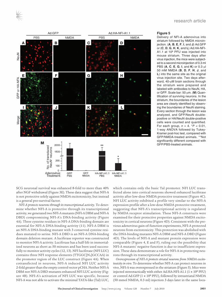
SCG neuronal survival was enhanced 8-fold to more than 40% after NGF withdrawal (Figure 3E). These data suggest that NFI-A is not protective solely against NMDA excitotoxicity, but instead is a general pro-survival factor.
NFI-A protects neurons through its transcriptional activity. To deter-mine whether NFI-A is protective through its transcriptional activity, we generated two NFI-A mutants (NFI-A DBM and NFI-A DBD) compromising NFI-A’s DNA-binding activity (Figure 4A). Three cysteine residues in NFI-A DNA-binding domain are essential for NFI-A DNA-binding activity (11). NFI-A DBM is an NFI-A DNA-binding mutant with 3 conserved cysteine resi-dues mutated to serine. NFI-A DBD is an NFI-A DNA-binding domain deletion mutant. A luciferase reporter was constructed to monitor NFI-A activity. Luciferase has a half-life in immortal-ized neurons as short as 50 minutes and has been used success-fully to monitor activity cycles (12, 13). NFI luciferase (NFI LUC) contains three NFI response elements (TTGGC[N5]GCCAA) in the promoter region of the LUC construct (Figure 4A). When cotransfected in neurons, NFI-A induced NFI LUC activity 2-fold greater than the empty control vector pCHA. Neither NFI-A DBM nor NFI-A DBD mutants enhanced NFI LUC activity (Fig-ure 4B). NFI-A’s activation of NFI LUC was specific, because NFI-A was not able to activate the minimal TATA-like (Tal) LUC,
which contains only the basic Tal promoter. NFI LUC trans-fected alone into cortical neurons showed enhanced luciferase activity after low-dose NMDA protective treatment (Figure 4C). NFI LUC activity exhibited a profile very similar to the NFI-A expression profile after a low-dose NMDA protective treatment, suggesting that NFI-A’s transcriptional activity is regulated by NMDA receptor stimulation. These NFI-A constructs were examined for their protective properties against NMDA excito-toxicity in cortical neurons (Figure 4D). Consistent with the pre-vious adenovirus gain-of-function experiments, NFI-A protected neurons from excitotoxicity. This protection was abolished with the DNA-binding mutants NFI-A DBM and NFI-A DBD (Figure 4D). The levels of NFI-A and mutant protein expression were comparable (Figure 4, E and F), ruling out the possibility that NFI-A mutants’ negative function is due to insufficient expres-sion. These data demonstrate a role for NFI-A in protecting neu-rons through its transcriptional activity.
Overexpression of NFI-A protects striatal neurons from NMDA excito-toxicity in vivo. To determine whether NFI-A can protect neurons in vivo, NFI-A was overexpressed in the striatum (Figure 5). Mice were injected stereotactically with either Ad.HA-NFI-A1.1 (1 × 10⁸ PFU) or control Ad.GFP (1 × 10⁸ PFU), followed by intrastriatal NMDA (50 mmol NMDA, 0.3 ml) injection 3 days later in the same loca-
Figure 5Delivery of NFI-A adenovirus into striatum followed by NMDA microin-jection. (A, B, E, F, I, and J) Ad.GFP or (C, D, G, H, K, and L) Ad.HA-NFI-A1.1 at 108 PFU was injected into mouse striatum. Three days after virus injection, the mice were subject-ed to a second microinjection of 0.3 ml PBS (A, C, E, G, I, and K) or 0.3 μl 50 mM NMDA (B, D, F, H, J, and L) into the same site as the original virus injection site. Two days after-ward, 40-μM brain sections through the striatum were prepared and labeled with antibodies to NeuN, HA, or GFP. Scale bar: 50 μm. (M) Quan-tification of surviving neurons. In the striatum, the boundaries of the lesion area are clearly identified by observ-ing the boundaries of NeuN staining. Every section through the lesion was analyzed, and GFP/NeuN double-positive or HA/NeuN double-positive cells were counted and quantified. For each group, n = 4. *P < 0.01, 1-way ANOVA followed by Tukey-Kramer post-hoc test, compared with GFP/NMDA-treated animals. **Not significantly different compared with GFP/PBS-treated animals.

research article
2452 TheJournalofClinicalInvestigation http://www.jci.org Volume 120 Number 7 July 2010
tion (rostral, 0.5 mm; lateral, 2.0 mm; ventral, 3.5 mm from bregma). Brain sections were stained with anti-NeuN antibody to identify neurons and anti-HA or anti-GFP antibodies to identify NFI-A and GFP adenovirus–infected cells, respectively. Cells positive for anti-HA and anti-NeuN staining and cells positive for anti-GFP and anti-NeuN staining were counted as live infected neurons. Although adenovirus infection efficiency in vivo was relatively low compared with that in vitro (Figure 5, A, C, E, and G, and Supplemental Figure 6, B and D), NFI-A adenovirus and GFP adenovirus achieved similar neuronal infection rates (Figure 5M). NMDA injection causes a mas-sive lesion in the striatum, whereas PBS injection did not kill striatal neurons (data not shown). In the Ad.GFP-infected striatum, there were few GFP-positive neurons that survived NMDA microinjection (Figure 5, B, F, J, and M). However, in the NFI-A adenovirus–injected striatum, there were significantly more surviving virus-infected neu-rons (Figure 5, D, H, L, and M). Thus, NFI-A overexpression in the striatum protects neurons against NMDA toxicity in vivo.
NFI-A–deficient neurons are sensitive to NMDA treatment. To ascer-tain whether a reduction or deficiency in NFI-A levels may lead to reduced neuronal survival or increased sensitivity to toxic insults, we cultured Nfia–/– neurons from embryonic brains (genotype in Figure 6A and protein levels in Figure 6B). We find that Nfia–/– neu-rons are difficult to maintain and are extremely fragile. The Nfia–/– neurons were very sensitive to a normally protective dose of 50 μM NMDA, with only 20% neuronal survival (Figure 6C). Surprisingly, these Nfia–/– neurons were not able to tolerate exposure to con-trolled saline solution (CSS) unless the NMDA receptor blocker MK801 or APV was added in the CSS solution (Figure 6D). More-over, the toxicity was partially blocked by tetrodotoxin (TTX) and substantially blocked when calcium in the CSS was replaced with cobalt. Thus, the sensitivity of Nfia–/– neurons to CSS is a calcium-dependent process. It is likely due to activation of the NMDA recep-tor following endogenous glutamate release secondary to enhanced synaptic activity following washing and CSS treatment of the cul-
tures. To determine whether the increased susceptibility of Nfia–/– neurons to CSS is due to the absence of NFI-A, we transduced Nfia–/– neurons with the NFI-A adenovirus (Ad.HA-NFI-A1.1) (Fig-ure 6E). Overexpression of NFI-A prevented the cell death of Nfia–/– neurons exposed to CSS (Figure 6E). Taken together, these results indicate that NFI-A is essential for survival of cortical neurons.
Nfia–/– mouse SCG neurons are depleted. The observations that NFI-A is important for neuronal protection against toxic insults in vitro and in vivo, as well as the extreme sensitivity of Nfia–/– cortical neu-rons in culture, raised the possibility that NFI-A is a general surviv-al factor for neurons in other physiological processes. To explore whether NFI-A is a survival factor for developing neurons in the peripheral nervous system, we examined the number of SCG neu-rons, by immunohistochemistry and Nissl staining at P0.5, just prior to the perinatal death of Nfia–/– pups. We found that Nfia–/– SCGs had 30% fewer neurons than the Nfia+/+ SCGs (Supplemental Figure 8, A–C), implying a role for NFI-A in neurogenesis, neural crest migration, and/or survival of SCG neurons. NFI-A was clearly expressed in SCG neurons at P0.5, as indicated by colocalization with MAP2 (Supplemental Figure 8D).
Loss of NFI-A function enhances NMDA excitotoxicity in vivo. To deter-mine whether NFI-A is required for neuronal survival in vivo, we examined the susceptibility of Nfia+/– mice to excitotoxic insults in vivo. Nfia–/– mice are perinatal lethal, but Nfia+/– mice are viable (14). NMDA was injected into the striatum of Nfia+/+ or Nfia+/– mice to test whether NFI-A promotes neuronal survival in response to a toxic treatment in vivo (Figure 7). Using measurements of direct lesion volume, Nfia+/– mice had 30% larger lesion volumes than the Nfia+/+ mice (Figure 7C), indicating that the reduction in NFI-A lev-els sensitizes the animal to NMDA excitotoxicity. This difference in susceptibility to NMDA toxicity remained significant when lesion volumes were determined using indirect estimates that accounted for swelling of tissue in response to insult (Supplemental Figure 9). Nfia+/– mice expressed less NFI-A in the striatum compared with
Figure 6NFI-A–deficient neurons are sensitive to NMDA receptor activation. (A) Genotyping of embryos whose brains were used for cortical cul-tures. (B) NFI-A expression in day 10 in vitro neuronal cultures with different genotypes. (C) Cortical cultures dissociated from Nfia+/+, Nfia+/–, and Nfia–/– embryos were treated with either CSS or 50 μM NMDA. Experiments were replicated at least 3 times. *P < 0.01, 1-way ANOVA followed by Tukey-Kramer post-hoc test. (D) Nfia–/– neurons were treated with CSS with or without MK801 (10 μM), APV (250 μM), 6-cyano-7-nitroquinoxaline-2,3,-dione (CNQX) (400 μM), TTX (2 μM), or CSS with calcium replaced by cobalt (–Ca2+). *P < 0.01 compared with CSS mock treatment, 1-way ANOVA followed by Tukey-Kramer post-hoc test. Experiments were replicated 3 times. (E) The increased sensitivity of Nfia–/– neurons is rescued by Ad.HA-NFI-A1.1–mediated NFI-A overexpression in primary cortical cultures. Experiments were replicated 3 times. *P < 0.01, Student’s t test, compared with Nfia–/– neurons transduced with GFP adenovirus.

research article
TheJournalofClinicalInvestigation http://www.jci.org Volume 120 Number 7 July 2010 2453
their wild-type littermates (Figure 7D). These results taken togeth-er indicate that NFI-A is an important survival factor for neurons against toxic insults and that a deficiency of NFI-A increases the susceptibility of neurons to injury.
DiscussionThe major finding of this study is the identification of NFI-A as a neuronal survival transcription factor. NFI-A levels are upregulated in long-lasting neuroprotection protocols, includ-ing administration of a nonlethal dose of NMDA and OGD preconditioning. Blocking NFI-A induction reduced the neu-roprotection that resulted from these preconditioning stimuli. NFI-A overexpression protected neurons from excitotoxic insults both in vitro and in vivo through its transcriptional activity, and reducing NFI-A levels sensitized neurons to excitotoxicity and led to an increased developmental cell loss of SCG neurons. These data taken together suggest that NFI-A is a critical player in the neuronal pro-survival program.
NFI-A belongs to the NFI transcription factor family, a distinct class of site-specific DNA-binding proteins. In vertebrates, the NFI family consists of 4 members: NFI-A, NFI-B, NFI-C, and NFI-X (15). Newborn Nfia–/– mice show severe hydrocephalus and agen-esis of corpus callosum, and approximately 95% exhibit perinatal lethality (14). A few survivors show male sterility and low female fertility, as well as axial tremor indicative of neurological defects. There is abnormal development of forebrain midline glial struc-tures and commissural projections in Nfia–/– mice, suggesting that NFI-A may regulate callosal development by regulating the devel-opment of midline glia (16). NFI-A also controls the onset of glio-genesis in the developing spinal cord (17). While the Nfia–/– mice revealed a developmental role for NFI-A, a function for NFI-A in adult brain was not known. Following a short, low-dose bath appli-cation of NMDA or nonlethal OGD, NFI-A was induced. Simply overexpressing NFI-A was sufficient to activate long-lasting neu-
roprotection. Consistent with this notion is the observation that NFI-A was transcriptionally active after low-dose exposure to NMDA, with an activation profile similar to its protein expres-sion. Moreover, NFI-A mutants lacking DNA-binding activity no longer protected neurons from NMDA excitotoxicity. Blockade of NFI-A induction by siRNA significantly decreased the neuro-protective effects of low-dose NMDA treatment, and this result is consistent with the notion that NFI-A upregulation is necessary to induce neuroprotection. Overexpression of NFI-A in neurons was sufficient to render neurons resistant to excitotoxicity and other insults. The greatest protection elicited by overexpression of NFI-A was against NMDA excitotoxicity, followed by AMPA and kainate excitotoxicity. This probably reflects the divergent cell death path-ways activated by these different glutamate receptor agonists. The protection against NMDA excitotoxicity was also observed in vivo, although to a lesser degree than observed in vitro. This is most likely due to the lower transduction rate of neurons by adenovirus in vivo (Supplemental Figure 6D). In addition, the more complex and dynamic interaction of neurons in vivo likely increases the dependency of neuronal survival on the local environment. Addi-tionally, neuronal-selective expression of NFI-A using lentivirus in vitro was sufficient to confer a protective phenotype. These obser-vations suggest that NFI-A acts in a cell-autonomous manner.
To assess whether NFI-A plays an important role in neuronal survival in the absence of a neuroprotective stimulus, we exam-ined the sensitivity of Nfia+/– mice. Nfia+/– mice were more sensi-tive than Nfia+/+ mice to NMDA excitotoxic treatment in vivo. Moreover, Nfia–/– cortical neurons were markedly sensitive not only to concentrations of NMDA that were nontoxic to wild-type cells, but they were sensitive to the small amount of glutamate that was released due to synaptic activity following a complete exchange of CSS. Cell death in the Nfia–/– neuronal cultures due to exchange of CSS was completely attenuated by the NMDA receptor antagonists MK801 and APV, the substitution of Co2+ for Ca2+, as well as restoration of the NFI-A expression. Thus, in the absence of NFI-A, NMDA receptor activation is no longer protective and instead leads to cell death.
To further test whether NFI-A is necessary for neuronal survival, we examined the integrity of SCG neurons in Nfia–/– mice. SCG neurons start to develop at E13.5 and become mature around birth (18, 19). We found significantly fewer sympathetic neurons in Nfia–/– animals, suggesting a critical role for NFI-A in developmen-tal survival. Although it is conceivable that NFI-A is required for the production and secretion of target-derived neurotrophins or neural crest migration or neurogenesis, increased susceptibility to cell death in the absence of NFI-A seems a more likely explanation. Consistent with this notion is the observation that overexpression of NFI-A in vitro protected SCG neurons against NGF withdrawal.
NMDA receptor–mediated neuroprotection is a transcription-dependent process (5) that potently protects against a variety of toxic insults (9, 15, 20–23). NMDA receptor activation plays important roles in ischemic tolerance both in vitro (9, 15) and in vivo (8), in excitotoxic tolerance (24), and in activity-depen-dent neuroprotection (4). Long-lasting neuroprotection induced by NMDA receptor activation seems to be dependent on acti-vation of synaptic NMDA receptors either through enhanced synaptic activity or low-dose and/or brief bath applications of NMDA (24, 25), whereas excitotoxic doses of NMDA lead to a shut-off of these cell survival pathways and/or activation of cell death pathways (26), ultimately leading to neuronal demise.
Figure 7NFI-A–deficient neurons are sensitive to NMDA treatment in vivo. (A and B) Representative photomicrograph of intrastriatal lesion of (A) Nfia+/+ mice and (B) Nfia+/– mice 2 days after microinjection of 0.3 ml NMDA (50 mM). Scale bar: 50 mm. (C) Quantification of lesion volumes. n = 4; *P < 0.01, Student’s t test. (D) Immunoblot of NFI-A expression in the striatum of Nfia+/+ and Nfia+/– mice.

research article
2454 TheJournalofClinicalInvestigation http://www.jci.org Volume 120 Number 7 July 2010
NMDA-induced cell survival is mediated, in part, through CREB and the CREB/cAMP response element (CRE) transcriptional pathway (4, 24). However, CREB-independent cell survival path-ways play prominent roles depending on the neuroprotective stim-ulus. For instance, an NMDA receptor– and NO/MEK-dependent cell survival pathway predominates in OGD tolerance in vitro (7), and NO-dependent processes play important roles in ischemic tolerance in vivo (27). Through DAzLE, we have identified NFI-A and other NMDA-induced late-response genes that are regulated by the NO/MEK-dependent signaling pathway (6). Accordingly, we found that NMDA-induced upregulation of NFI-A was blocked by inhibitors of NOS and MEK. The downstream transcriptional effectors of the NO/MEK cell survival pathway are not known and require further study.
NFI-A is induced by both the short (5-minute) nonlethal bath application of low-dose (50 μM) NMDA and nonlethal OGD, imparting long-lasting neuroprotection similar to that induced by disinhibition of GABAergic neurons by bicuculline adminis-tration (4). All three paradigms are NMDA receptor and calcium dependent (4, 7, 9, 28). Long-lasting neuroprotection due to bicuculline administration, which is thought to mimic synaptic activity, is due, in part, to CREB-dependent processes (4), where-as a short bath application of low-dose NMDA and nonlethal OGD leads to CREB-dependent and -independent cell survival pathways (7, 24, 29, 30). It is likely that these different neuro-protective induction paradigms may activate divergent cell sur-vival pathways through preferential activation of synaptic and extrasynaptic receptors, respectively (25, 28, 31). Here we show that nonlethal exposure to both OGD or NMDA induces NFI-A induction, in part, by NOS- and MEK-dependent pathways. In contrast to the relatively transient NMDA receptor–mediated activation of CREB through Ser133 phosphorylation (24, 32), NFI-A levels remained elevated for at least 24 hours after the neuroprotective stimulus. Thus, Nfia is an NMDA-induced late-response gene that is regulated, in part, through NO- and MEK-dependent pathways. Prolonged enhanced expression of NFI-A positions it as a potential important protein in long-lasting neu-roprotection-induced NMDA receptor activation.
In summary, we found what we believe to be a novel neuronal survival transcription factor, NFI-A. We show here that NFI-A is a transcription factor induced by multiple neuroprotective para-digms, its transcriptional activity is important for its neuroprotec-tive function, and neuronal survival is compromised in the absence of NFI-A. The discovery of NFI-A and its role in neuronal survival may shed light on the molecular mechanisms of long-lasting neu-roprotection. Future studies may uncover new therapeutic options for triggering neuronal survival to protect against neuronal loss due to disease or stroke.
MethodsAnimals. All experimental protocols using animals were approved by the Institutional Animal Care and Use Committee of Johns Hopkins Univer-sity. Nfia+/– mice on a C57BL/6J background were generated at the Lerner Research Institute and bred at the Johns Hopkins University School of Medi-cine. Heterozygous Nfia+/– mice were bred to acquire litters of Nfia+/+, Nfia+/–, and Nfia–/– mice for culture and Nfia+/+ or Nfia+/– littermate mice for intra-striatal injections. Genotyping was performed as previously described (14).
Cytotoxicity. Primary cortical cultures were exposed to experimental condi-tions as previously described (7). Cells were washed with control salt solu-tion (CSS, containing [in mM]: 120 NaCl, 5.4 KCl, 1.8 CaCl2, 25 Tris-Cl,
15 glucose, pH 7.4) and exposed to 50 μM NMDA plus 10 μM glycine, 500 μM NMDA plus 10 μM glycine, 25 μM AMPA, or 100 μM AMPA in CSS for 5 minutes and replaced with MEM/21 mM glucose. To study sen-sitivity of Nfia–/– neurons to calcium, CaCl2 in the CSS was replaced with 1.8 mM CoCl2. For exposure to kainate feeding, media was fully exchanged with feeding media containing 100 μM kainate for an overnight exposure. H2O2 (300 μM) was applied to cultures in the feeding media for 30 minutes. The exposure was terminated by replacement with normal feeding media. Combined OGD was performed by complete exchange of media with deoxy-genated, glucose-free Earle’s balanced salt solution (EBSS), bubbled with 10% H2/85% N2/5% CO2 (33). Cultures were kept in an anaerobic chamber for 15 minutes at 37°C. OGD was terminated by replacement of the EBSS solution with oxygenated feeding media. After the various treatment para-digms, the cultures were returned to the incubator and maintained over-night. NGF withdrawal experiments were performed as previously described (34). Twenty-four hours after treatments, cell viability was determined as described (35). The cultures were stained with 1 μg/ml Hoechst 33342, which stains all cell nuclei, and 7 μM propidium iodide, which stains dead cell nuclei. Cells were imaged by CCD camera under a fluorescence micro-scope and were counted by an observer blinded to the experimental proto-col with unbiased computer-assisted cell counting (Axiovision 4.3 software, Carl Zeiss Inc.). Glial nuclei fluoresce at an intensity different from that of neuronal nuclei and were gated out. Percent cell survival was determined as the ratio of total minus dead cells to total cells. At least 3 separate individual experiments using at least 3 separate wells were performed.
Real-time PCR. Total RNA from cultured neurons was prepared using TRIzol reagent (Invitrogen). First-strand cDNA was synthesized with random hexamer primers by using Superscript III First-Strand Synthe-sis Kit (Invitrogen). Real-time PCR was performed with cDNA, SYBR Green PCR Master Mix (Applied Biosystems), and specific forward and reverse primers using the ABI PRISM 7900HT Sequence Detec-tion System (Applied Biosystems). Relative amounts of mRNAs were calculated by using the comparative CT method. GAPDH was used as the invariant control. Primer sequences were as follows: NFI-A total (5′-TGGCATACTTTGTACATGCAGC-3′, 5′-ACCTGATGTGACAAAGCT-GTCC-3 ′) , NFI-A1.1 (5 ′ -AATGTGAACGCAAGAAGCAGGC-3 ′ , 5′-GCAGAAGTGCTTCAATGAAAGG-3′), NFI-A1.1B (5′-CTCCCGT-GAGTTAGCATCAGATG-3′, 5′-TGCAGGTTGAACCATGTGTAG-3′), NFI-A1.1C (5′-GCGATGAAGCAATTCGTCTGAG-3′, 5′-CAGGTTGAAC-CATGTGTAGGCG-3′), and GAPDH (5′-CCTGGCCAAGGTCATCCAT-GAC-3′, 5′-CATCACGCCACAGCTTTCCAGA-3′).
siRNA. dsRNA was prepared using the Silencer siRNA Construction Kit (Ambion) according to the instruction manual. The strategy for choosing siRNA target was based on the observation that siRNA with 3′ overhanging UU dinucleotides are the most effective (36). The C-ter-minal transactivation domain of NFI-A was used to screen for siRNA molecule candidates. Sequences used to generate siRNAs were as fol-lows: siRNA1 sense 5′-AGUUCUUCAUACUACAGCAUU-3′, antisense 5′-UGCUGUAGUAUGAAGAACUUU-3′; siRNA2 sense 5′-AGAGUUUGUC-CAACUUGUCUU-3′, antisense 5′-GACAAGUUGGACAAACUCUUU-3′; siRNA3 sense 5′-ACCGAUUCGUCAGUGUUGGUU-3′, antisense 5′-CCAA-CACUGACGAAUCGGUUU-3′; SCR siRNA sense 5′-CAAGUGCUUGC-GUUAGCUGUU-3′, antisense 5′-CAGCUAACGCAAGCACUUGUU-3′; DsRed siRNA sense 5′-AGUUCCAGUACGGCUCCAAUU-3′, antisense 5′-UUGGAGCCGUACUGGAACUUG-3′. These sequences were subjected to BLAST analysis to guarantee uniqueness across mouse and rat genomes. siRNA was transfected into cells using GeneSilencer siRNA Transfection Reagent (Gene Therapy Systems) according to the manufacturer’s instruc-tions. siRNA was transfected 72 and 24 hours before NMDA treatment to maximize knockdown efficiency.

research article
TheJournalofClinicalInvestigation http://www.jci.org Volume 120 Number 7 July 2010 2455
Transduction of cultured neurons with recombinant viruses. HA-tagged NFI-A cDNA (HA-NFI-A1.1 or HA-NFI-A 1.1B) was subcloned into the adenovirus packaging construct using EcoRI and XbaI restriction sites. The NFI-A adenovirus packaging construct was sent to Viraquest to produce crude viral extracts. Amplification and purification of high-titer viral stocks were prepared as previously described (37). Neuronal cultures were incubated with Ad.HA-NFI-A1.1, Ad.HA-NFI-A1.1B, or Ad.GFP (MOI, 1,000) in the feeding media with minimum toxicity over-night. The virus was washed out on the second day, and the neurons were allowed another 24 hours to express the transduced gene before experiments were performed. HA-tagged NFI-A1.1 lentivirus was pro-duced by replacing the EGFP ORF from the cFUGw lentiviral vector. Virus particles were produced using the Virapower Lentiviral packag-ing system (Invitrogen). Primary cortical neurons were transduced at an MOI of 10 with either EGFP- or NFI-A–expressing lentivirus on day 8 in vitro. Cells were allowed 72 hours to attain high levels of expression prior to excitotoxicity experiments.
Intrastriatal microinjection and lesion analysis. Male mice (20–26 g) were anesthetized with 45 mg/kg pentobarbital via intraperitoneal injection. The mouse head was fixed in a stereotactic frame (Kopf), and a burr hole was drilled above the striatum (rostral, 0.5 mm; lateral, 2.0 mm; ventral 3.5 mm from bregma). A 0.3-μl NMDA (50 mM) or vehicle (0.1 M PBS, pH 7.4) solution was injected over 2 minutes, and the needle was held in place for an additional 8 minutes after injection. For adenovirus injection, 1 μl of virus expressing NFI-A or GFP was injected over 10 minutes using a Micro4 microinjection pump (World Precision Instruments) in wild-type C57BL/6J mice. The needle was left in place for an additional 10 minutes. The identical stereotactic coordinates were used afterward for toxin or vehicle delivery 3 days later. Forty-eight hours after toxin or vehicle deliv-ery, the mice were deeply anesthetized with pentobarbital and perfusion fixed with 4% paraformaldehyde in PBS. The entire brain was removed after perfusion, post-fixed in the same fixative solution, and cryoprotect-ed with 30% sucrose in PBS for freezing and serial sectioning.
Brain tissue sections (40 μm) were permeabilized with 0.1% Triton X-100 in PBS for 1 hour and blocked with 10% normal goat serum and 0.1% Triton X-100 in PBS for 1 hour. Primary antibodies were diluted in block-ing solution and incubated overnight at 4°C. Signals were detected with fluoro-conjugated secondary antibody under a Carl Zeiss Laser Scanning System LSM 510 confocal microscope. Antibodies were used as follows: 1:200 polyclonal anti–NFI-A, 1:200 anti-NeuN (Chemicon), 1:200 Texas red–conjugated anti-mouse antibody (Molecular Probes), 1:200 Oregon green–conjugated anti-rabbit antibody (Molecular Probes, Invitrogen). The boundaries of the lesioned area can be clearly identified by the boundar-ies of NeuN staining. Every section through the lesion was analyzed for NeuN-positive cells costained with HA or GFP. These double-labeled cells were scored as viable.
For experiments in Nfia+/– mice, frozen 40-μm sections were Nissl stained. Each section was imaged using a digital camera, and lesion areas and total intrastriatal lesion volume were determined for each animal. Lesions were
defined by the loss of Nissl staining in the injected striatum and were quantified using SigmaScan software (Systat Software Inc.). Indirect lesion estimates were performed by measuring the volume of the lesion as a func-tion of the total volume of the striatum.
SCG assessment. SCG neurons were counted as previously described (38). Briefly, 10-μm sections were taken every 50 μm through the SCG and were Nissl stained. The size of SCG areas of each section was mea-sured using SigmaScan software and summed (S). For each animal, the 3 sections that contained the 3 largest SCG neurons (with their area size of A1, A2, and A3) were subjected to cell counts (cell number C1, C2, and C3). Cells were counted as neurons if they contained a clear nucleus. The cell density of SCG neurons was calculated as: D = (A1/C1 + A2/C2 + A3/C3)/3. Since SCG neurons are relatively homogenously distributed, the total SCG neuron number was calculated as: T = S × D × 5. The sec-tion that contained the largest SCG region of one animal was used as a representative picture of that animal’s SCG.
Statistics. Statistical analyses were carried out with GraphPad InStat. Differences among multiple means were evaluated by 1-way ANOVA fol-lowed by the Tukey-Kramer post-hoc test and 2-way ANOVA followed by Bonferroni post-test when applicable. Differences between paired means were assessed with the unpaired, 2-tailed Student’s t test. The null hypothesis was rejected at the 0.05 level. All error bars represent the standard deviation.
AcknowledgmentsWe thank Christopher Deppmann for help with SCG neuronal culture and Xi Chen for help with preparation of SCG sections and counting of SCG neurons. We thank Emily Pai for assistance with neurotoxicity assays. This work was supported by NIH DA00226 (to V.L. Dawson, T.M. Dawson), NIH NS40809 (to V.L. Dawson), the McKnight Endowment for the Neurosciences (to V.L. Daw-son), NIH HD34901 (to R.M. Gronostajski), an American Heart Association Predoctoral Fellowship (to S. Zheng), and a Frontiers in Advanced Research in the Medical Sciences fellowship (to S.M. Eacker). Ted M. Dawson is Leonard and Madlyn Abramson Profes-sor in Neurodegenerative Diseases.
Received for publication June 29, 2007, and accepted in revised form April 14, 2010.
Address correspondence to: Valina L. Dawson, Institute for Cell Engineering, Department of Neurology, Johns Hopkins University School of Medicine, 733 North Broadway, Suite 731, Baltimore, Maryland 21205, USA. Phone: 410.614.3359; Fax: 410.614.9568; E-mail: [email protected]. Or to: Ted M. Dawson, Institute for Cell Engineering, Department of Neurology, Johns Hopkins University School of Medicine, 733 North Broadway, Suite 719, Baltimore, Maryland 21205, USA. Phone: 410.614.3359; Fax: 410.614.9568; E-mail: [email protected].
1. West AE, Griffith EC, Greenberg ME. Regulation of transcription factors by neuronal activity. Nat Rev Neurosci. 2002;3(12):921–931.
2. Ikonomidou C, et al. Blockade of NMDA receptors and apoptotic neurodegeneration in the develop-ing brain. Science. 1999;283(5398):70–74.
3. Mennerick S, Zorumski CF. Neural activity and survival in the developing nervous system. Mol Neurobiol. 2000;22(1–3):41–54.
4. Papadia S, Stevenson P, Hardingham NR, Bading H, Hardingham GE. Nuclear Ca2+ and the cAMP response element-binding protein family mediate a late phase of activity-dependent neuroprotection.
J Neurosci. 2005;25(17):4279–4287. 5. Marini AM, Paul SM. N-methyl-D-aspartate recep-
tor-mediated neuroprotection in cerebellar granule cells requires new RNA and protein synthesis. Proc Natl Acad Sci U S A. 1992;89(14):6555–6559.
6. Hong SJ, Li H, Becker KG, Dawson VL, Dawson TM. Identification and analysis of plasticity-induced late-response genes. Proc Natl Acad Sci U S A. 2004;101(7):2145–2150.
7. Gonzalez-Zulueta M, et al. Requirement for nitric oxide activation of p21(ras)/extracellular regulated kinase in neuronal ischemic preconditioning. Proc Natl Acad Sci U S A. 2000;97(1):436–441.
8. Dirnagl U, Simon RP, Hallenbeck JM. Ischemic tolerance and endogenous neuroprotection. Trends Neurosci. 2003;26(5):248–254.
9. Grabb MC, Choi DW. Ischemic tolerance in murine cortical cell culture: critical role for NMDA recep-tors. J Neurosci. 1999;19(5):1657–1662.
10. Krichevsky AM, Kosik KS. RNAi functions in cul-tured mammalian neurons. Proc Natl Acad Sci U S A. 2002;99(18):11926–11929.
11. Novak A, Goyal N, Gronostajski RM. Four con-served cysteine residues are required for the DNA binding activity of nuclear factor I. J Biol Chem. 1992;267(18):12986–12990.

research article
2456 TheJournalofClinicalInvestigation http://www.jci.org Volume 120 Number 7 July 2010
12. Brandes C, et al. Novel features of drosophila peri-od Transcription revealed by real-time luciferase reporting. Neuron. 1996;16(4):687–692.
13. Nunez L, Faught WJ, Frawley LS. Episodic gonado-tropin-releasing hormone gene expression revealed by dynamic monitoring of luciferase reporter activ-ity in single, living neurons. Proc Natl Acad Sci U S A. 1998;95(16):9648–9653.
14. das Neves L, et al. Disruption of the murine nuclear factor I-A gene (Nfia) results in perina-tal lethality, hydrocephalus, and agenesis of the corpus callosum. Proc Natl Acad Sci U S A. 1999; 96(21):11946–11951.
15. Gronostajski RM. Roles of the NFI/CTF gene family in transcription and development. Gene. 2000;249(1–2):31–45.
16. Shu T, Butz KG, Plachez C, Gronostajski RM, Richards LJ. Abnormal development of forebrain midline glia and commissural projections in Nfia knock-out mice. J Neurosci. 2003;23(1):203–212.
17. Deneen B, Ho R, Lukaszewicz A, Hochstim CJ, Gronostajski RM, Anderson DJ. The transcrip-tion factor NFIA controls the onset of gliogen-esis in the developing spinal cord. Neuron. 2006; 52(6):953–968.
18. Levi-Montalcini R, Booker B. Destruction of the sympathetic ganglia in mammals by an antiserum to a nerve-growth protein. Proc Natl Acad Sci U S A. 1960;46(3):384–391.
19. Fagan AM, Zhang H, Landis S, Smeyne RJ, Silos-Santiago I, Barbacid M. TrkA, but not TrkC, recep-tors are essential for survival of sympathetic neu-rons in vivo. J Neurosci. 1996;16(19):6208–6218.
20. Chuang DM, Gao XM, Paul SM. N-methyl-D-aspartate exposure blocks glutamate toxicity in cultured cerebellar granule cells. Mol Pharmacol. 1992;42(2):210–216.
21. Balazs R, Jorgensen OS, Hack N. N-methyl-D-aspar-tate promotes the survival of cerebellar granule cells in culture. NeuroScience. 1988;27(2):437–451.
22. Rocha M, Martins RA, Linden R. Activation of NMDA receptors protects against glutamate neuro-toxicity in the retina: evidence for the involvement of neurotrophins. Brain Res. 1999;827(1–2):79–92.
23. Raval AP, Dave KR, Mochly-Rosen D, Sick TJ, Perez-Pinzon MA. Epsilon PKC is required for the induc-tion of tolerance by ischemic and NMDA-mediated preconditioning in the organotypic hippocampal slice. J Neurosci. 2003;23(2):384–391.
24. Lee HT, Chang YC, Wang LY, Wang ST, Huang CC, Ho CJ. cAMP response element-binding protein activation in ligation preconditioning in neonatal brain. Ann Neurol. 2004;56(5):611–623.
25. Soriano FX, Papadia S, Hofmann F, Hardingham NR, Bading H, Hardingham GE. Precondition-ing doses of NMDA promote neuroprotection by enhancing neuronal excitability. J Neurosci. 2006;26(17):4509–4518.
26. Hardingham GE, Fukunaga Y, Bading H. Extrasyn-aptic NMDARs oppose synaptic NMDARs by trig-gering CREB shut-off and cell death pathways. Nat Neurosci. 2002;5(5):405–414.
27. Huang PL. Nitric oxide and cerebral ischemic pre-conditioning. Cell Calcium. 2004;36(3–4):323–329.
28. Hardingham GE, Chawla S, Cruzalegui FH, Bading H. Control of recruitment and transcription-acti-vating function of CBP determines gene regulation by NMDA receptors and L-type calcium channels. Neuron. 1999;22(4):789–798.
29. Hara T, Hamada J, Yano S, Morioka M, Kai Y, Ushio Y. CREB is required for acquisition of ischemic tol-erance in gerbil hippocampal CA1 region. J Neuro-chem. 2003;86(4):805–814.
30. Mabuchi T, et al. Phosphorylation of cAMP response
element-binding protein in hippocampal neurons as a protective response after exposure to gluta-mate in vitro and ischemia in vivo. J Neurosci. 2001; 21(23):9204–9213.
31. Zhang SJ, et al. Decoding NMDA Receptor Sig-naling: Identification of Genomic Programs Specifying Neuronal Survival and Death. Neuron. 2007;53(4):549–562.
32. Sala C, Rudolph-Correia S, Sheng M. Developmen-tally regulated NMDA receptor-dependent dephos-phorylation of cAMP response element-binding protein (CREB) in hippocampal neurons. J Neurosci. 2000;20(10):3529–3536.
33. Monyer H, Giffard RG, Hartley DM, Dugan LL, Gold-berg MP, Choi DW. Oxygen or glucose deprivation-induced neuronal injury in cortical cell cultures is reduced by tetanus toxin. Neuron. 1992;8(5):967–973.
34. Riccio A, Ahn S, Davenport CM, Blendy JA, Ginty DD. Mediation by a CREB family transcription factor of NGF-dependent survival of sympathetic neurons. Science. 1999;286(5448):2358–2361.
35. Gonzalez-Zulueta M, et al. Manganese superoxide dismutase protects nNOS neurons from NMDA and nitric oxide-mediated neurotoxicity. J Neurosci. 1998;18(6):2040–2055.
36. Elbashir SM, Harborth J, Lendeckel W, Yalcin A, Weber K, Tuschl T. Duplexes of 21-nucleotide RNAs mediate RNA interference in cultured mam-malian cells. Nature. 2001;411(6836):494–498.
37. He TC, Zhou S, da Costa LT, Yu J, Kinzler KW, Vogelstein B. A simplified system for generating recombinant adenoviruses. Proc Natl Acad Sci U S A. 1998;95(5):2509–2514.
38. Lonze BE, Riccio A, Cohen S, Ginty DD. Apopto-sis, axonal growth defects, and degeneration of peripheral neurons in mice lacking CREB. Neuron. 2002;34(3):371–385.