Nitroreductase-mediated cell/tissue ablation in zebrafish: a spatially and temporally controlled...
15
Nitroreductase-mediated cell/tissue ablation in zebrafish: a spatially and temporally controlled ablation method with applications in developmental and regeneration studies Silvia Curado, Didier Y R Stainier, and Ryan M Anderson Department of Biochemistry and Biophysics, Programs in Developmental Biology, Genetics and Human Genetics, Liver Center, Diabetes Center and the Cardiovascular Research Institute, University of California, San Francisco, 1550 Fourth Street, San Francisco, California 94158-2324, USA. Abstract Ablation studies are used to elucidate cell lineage relationships, developmental roles for specific cells during embryogenesis and mechanisms of tissue regeneration. Previous chemical and genetic approaches to directed cell ablation have been hampered by poor specificity, limited efficacy, irreversibility, hypersensitivity to promoter leakiness, restriction to proliferating cells, slow inducibility or complex genetics. Here, we provide a step-by-step protocol for a hybrid chemical- genetic cell ablation method in zebrafish that, by combining spatial and temporal control, is cell-type specific, inducible, reversible, rapid and scaleable. Bacterial Nitroreductase (NTR) is used to catalyze the reduction of the innocuous prodrug metrodinazole (Mtz), thereby producing a cytotoxic product that induces cell death. Based on this principle, NTR is expressed in transgenic zebrafish using a tissue-specific promoter. Subsequent exposure to Mtz by adding it to the media induces cell death exclusively within NTR + cells. This approach can be applied to regeneration studies, as removing Mtz by washing permits tissue recovery. Using this protocol, cell ablation can be achieved in 12–72 h, depending on the transgenic line used, and recovery initiates within the following 24 h. INTRODUCTION Analysis of cell-ablated organisms can uncover the roles of specific tissues, or tissue interactions, during development and homeostasis. In addition, analysis of recovery after ablation may reveal novel cellular and molecular mechanisms underlying the regeneration process, thus bringing new insights to the field of regenerative medicine. Danio rerio (zebrafish) is a vertebrate model organism increasingly being used to study a variety of developmental mechanisms and disease pathologies. It combines genetic tractability (both forward and reverse genetics), optical transparency, accessibility during embryogenesis and a well-documented capacity to regenerate several tissues after injury, including fin, retinal axons, spinal cord, heart, hair cells, melanocytes and liver 1–7 . Additionally, drugs are easily administered to zebrafish via water, and the ability to generate large numbers of embryos allows large-scale analyses. The ability to precisely ablate cells and tissues in this model organism opens new avenues into developmental, disease and regeneration studies. © 2008 Nature Publishing Group Correspondence should be addressed to D.Y.R.S. (E-mail: [email protected]).. Reprints and permissions information is available online at http://npg.nature.com/reprintsandpermissions NIH Public Access Author Manuscript Nat Protoc. Author manuscript; available in PMC 2009 July 6. Published in final edited form as: Nat Protoc. 2008 ; 3(6): 948–954. doi:10.1038/nprot.2008.58. NIH-PA Author Manuscript NIH-PA Author Manuscript NIH-PA Author Manuscript
Transcript of Nitroreductase-mediated cell/tissue ablation in zebrafish: a spatially and temporally controlled...

Nitroreductase-mediated cell/tissue ablation in zebrafish: aspatially and temporally controlled ablation method withapplications in developmental and regeneration studies
Silvia Curado, Didier Y R Stainier, and Ryan M AndersonDepartment of Biochemistry and Biophysics, Programs in Developmental Biology, Genetics andHuman Genetics, Liver Center, Diabetes Center and the Cardiovascular Research Institute,University of California, San Francisco, 1550 Fourth Street, San Francisco, California 94158-2324,USA.
AbstractAblation studies are used to elucidate cell lineage relationships, developmental roles for specific cellsduring embryogenesis and mechanisms of tissue regeneration. Previous chemical and geneticapproaches to directed cell ablation have been hampered by poor specificity, limited efficacy,irreversibility, hypersensitivity to promoter leakiness, restriction to proliferating cells, slowinducibility or complex genetics. Here, we provide a step-by-step protocol for a hybrid chemical-genetic cell ablation method in zebrafish that, by combining spatial and temporal control, is cell-typespecific, inducible, reversible, rapid and scaleable. Bacterial Nitroreductase (NTR) is used to catalyzethe reduction of the innocuous prodrug metrodinazole (Mtz), thereby producing a cytotoxic productthat induces cell death. Based on this principle, NTR is expressed in transgenic zebrafish using atissue-specific promoter. Subsequent exposure to Mtz by adding it to the media induces cell deathexclusively within NTR+ cells. This approach can be applied to regeneration studies, as removingMtz by washing permits tissue recovery. Using this protocol, cell ablation can be achieved in 12–72h, depending on the transgenic line used, and recovery initiates within the following 24 h.
INTRODUCTIONAnalysis of cell-ablated organisms can uncover the roles of specific tissues, or tissueinteractions, during development and homeostasis. In addition, analysis of recovery afterablation may reveal novel cellular and molecular mechanisms underlying the regenerationprocess, thus bringing new insights to the field of regenerative medicine.
Danio rerio (zebrafish) is a vertebrate model organism increasingly being used to study avariety of developmental mechanisms and disease pathologies. It combines genetic tractability(both forward and reverse genetics), optical transparency, accessibility during embryogenesisand a well-documented capacity to regenerate several tissues after injury, including fin, retinalaxons, spinal cord, heart, hair cells, melanocytes and liver1–7. Additionally, drugs are easilyadministered to zebrafish via water, and the ability to generate large numbers of embryos allowslarge-scale analyses. The ability to precisely ablate cells and tissues in this model organismopens new avenues into developmental, disease and regeneration studies.
© 2008 Nature Publishing GroupCorrespondence should be addressed to D.Y.R.S. (E-mail: [email protected])..Reprints and permissions information is available online at http://npg.nature.com/reprintsandpermissions
NIH Public AccessAuthor ManuscriptNat Protoc. Author manuscript; available in PMC 2009 July 6.
Published in final edited form as:Nat Protoc. 2008 ; 3(6): 948–954. doi:10.1038/nprot.2008.58.
NIH
-PA Author Manuscript
NIH
-PA Author Manuscript
NIH
-PA Author Manuscript

Existing cell and tissue ablation methodsPrevious genetic methods designed to specifically ablate cells in different model organismsexhibit various limitations, as discussed below. Unless specified, these methods have not beentried in zebrafish.
• Diphtheria toxin A. Expression of diphtheria toxin A-chain (DTA) in a tissue-specific manner can lead to the effective ablation of the target cell population, but itcan also result in the undesired ablation of other cells, or even death of the organism,due to its potency8,9. This problem has prevented the establishment of stable zebrafishlines carrying the DTA transgene, so this method has only been used in transienttransgenesis9. This technique could be used in an inducible manner if a suitableinducible promoter that is completely silent in the off-state could be identified.
• Kid/Kis. This strategy consists of expressing the bacterial toxin Kid in the targetedcell population while, simultaneously, expressing its antidote (Kis) in the rest of theorganism to protect it from the cell death induced by Kid10. This method has beenused in zebrafish, but only in embryos that transiently express Kid/Kis.
• HSV tymidine kinase/ganciclovir. This approach uses promoter-based expressionof HSV tymidine kinase together with a chemical (ganciclovir) to inhibit cellproliferation and induce cell death. This method is routinely used ex vivo and has beensuccessfully used in rodents11,12 but only kills proliferating cells13. Thus, quiescentor slow-cycling cell populations are more refractory to this method.
• Tamoxifen-inducible c-Myc. Overexpression of tamoxifen-inducible c-MycER
results in elevated apoptosis rates, but significant ablation in mouse takes a long time(6–10 d; see ref. 14), probably because this transgene also causes a persistentlyelevated proliferation rate in the targeted cells.
• Toxic viral protein M2(H37A). Cell death is induced by expressing the M2(H37A)toxic ion channel of the influenza-A virus in the target cell population15,16. Thistoxic effect can be inhibited by the addition of the antiviral drug rimantadine inmammalian cell cultures and Xenopus embryos but not transgenic mice. Furthermore,this suggests that the establishment and maintenance of stable transgenic lines in manycases would require long-term exposure to this inhibitor.
The NTR-mediated technique described here can be used to genetically ablate cells in zebrafishin a specific and inducible manner17,18. It is germline transmissible and kills cells withoutregard to their cell-cycle status19.
Overview of NTR-mediated ablationThis technique is based on the ability of the Escherichia coli enzyme NTR to convert the non-toxic prodrug Mtz into a cytotoxic metabolite. Initially, NTR is reduced by NADH or NADPH.Then, Mtz binds to NTR and is electrochemically reduced and converted into a potent DNAinterstrand cross-linking agent, which subsequently causes the death of the NTR-expressingcell20–22. The ectopic expression of a fluorescent protein—NTR fusion protein (FP-NTR) viaa tissue-specific promoter (active in the cell population to be ablated) combined with exposureof the organism to Mtz can induce rapid destruction of the targeted cell population (see Fig.1). Because the toxic form of Mtz remains confined to the NTR-expressing cell, no neighboringcells are affected, leading to the exclusive ablation of the NTR+ cells. This characteristic makesMtz a better NTR substrate than others previously described, such as CB1954 (5-(aziridin-1-yl)-2,4-dinitrobenzamide), which exhibits a ‘bystander effect’, damaging neighboring cellstogether with the targeted NTR-expressing cells23. Moreover, the fusion of a fluorescent tagwith NTR allows the fate of the NTR+ cells to be monitored throughout the course of ablation(see Fig. 2).
Curado et al. Page 2
Nat Protoc. Author manuscript; available in PMC 2009 July 6.
NIH
-PA Author Manuscript
NIH
-PA Author Manuscript
NIH
-PA Author Manuscript

Applications of Mtz/NTR-mediated ablationBecause the technique described here offers rigorous spatial specificity as well as temporalcontrol, it has multiple applications. The examination of the effects of the absence of a tissueor cell population can, for instance, provide new insights into the understanding of the role ofa tissue, or cell population, for morphogenesis, patterning or cell survival, during developmentor homeostasis. Furthermore, the NTR/Mtz system can be used as a versatile tool forregeneration studies: analysis of tissue recovery after ablation will enable the elucidation ofcellular and molecular mechanisms underlying tissue regeneration. This approach can be usedin a wild-type context as well as in mutant or genetically modified backgrounds. This versatilityis particularly advantageous when investigating how certain signaling pathways or molecularplayers are involved during the process of regeneration (by coexpression of dominant negativefactors, mutant alleles or Morpholino antisense injection). Moreover, this system can becombined with the use of other chemicals that modulate specific signaling pathways toinvestigate their role in tissue regeneration.
This technique has been successfully used to ablate cardiomyocytes, hepatocytes andpancreatic β-cells in zebrafish embryos and larvae by using promoters specific for each of thesecell populations17,18. These studies also showed that the NTR/Mtz cell ablation technique isreversible, as tissue recovery could be observed after ablation. After damage of the larval heartwas induced, which resulted in failure of the heart to contract and pump blood to the rest ofthe organism, cardiomyocytes regenerated, recovering morphology and function of the heartand reestablishing blood circulation. Similarly, following ablation of β-cells, the larvalpancreas exhibited substantial recovery, generating new Insulin-producing cells in the islet.
There is no fundamental difference in applying this method to embryonic, larval, juvenile oradult zebrafish except the scale of the experiment (see Box 1). Thus, as with any other drugtreatment, it is expected that this ablation technique will work in adults, and, in fact, we haveobserved effective β-cell ablation at all developmental stages tested (R.M.A. and D.Y.R.S.,unpublished data).
Limitations of NTR-mediated ablationSome limitations may, however, be associated with this technique, which depend on the tissuebeing investigated.
Availability of suitable promoterAlthough the method offers great flexibility regarding the target tissue to be ablated, as it canbe adapted by selecting an appropriate promoter to drive NTR expression, the ability to ablatecertain cell populations, or tissues, will depend on the availability of such a promoter.
Some tissues resist complete ablationA second limitation can be the inability to ablate the target cell population completely,depending on which tissue is being ablated. It is possible that some tissues, or cells within atissue, will be more accessible to the prodrug than others. It is also possible that some tissuesrespond rapidly to the loss of cells and are replaced as quickly as they are destroyed. Thisscenario may explain why a few residual cells may still be detected after ablation. However,this mosaic effect may be advantageous when partial ablation is preferred. In addition,hepatocytes might metabolize Mtz and thus limit the efficacy of this method for theirablation17.
Curado et al. Page 3
Nat Protoc. Author manuscript; available in PMC 2009 July 6.
NIH
-PA Author Manuscript
NIH
-PA Author Manuscript
NIH
-PA Author Manuscript

Some tissues require longer treatment for ablationAnother possible limitation is the time required to induce ablation, as it includes diffusion andaccumulation of Mtz into the FP-NTR-expressing cells and induction of cell death. We haveobserved that some FP-NTR zebrafish transgenic lines require only a few hours of Mtztreatment, whereas others require longer than 24 h for an observable effect to be induced. Theablation time is variable and likely depends not only on the promoter used to drive FP-NTRexpression, but also on each particular transgenic line. Different lines using the same promotermay show variable levels of FP-NTR expression, possibly due to positional effects of thetransgene integration site. Optimization of the technique may thus require the generation ofmultiple transgenic lines and empirical determination of the necessary period of Mtz exposure.
Experimental designTransgene constructs—The use of this method requires generation of a transgenic line(stable or transient) carrying a cassette in which FP fused to NTR is driven by a tissue-specificpromoter (tsp) (Fig. 1a). The tsp defines the tissue to be ablated, NTR itself is the conditionaltoxigene and the FP enables monitoring of NTR-expressing cells during the procedure.Additionally, the FP facilitates the identification of transgenic carriers.
To generate the Tg(tsp:FP-NTR) zebrafish transgenic line, the promoter of interest can becloned into the multiple cloning site of the tol2_CFP_NTR plasmid17 and co-injected withTol2 transposase mRNA into 1-cell stage embryos24. Alternatively, the promoter and FP-NTRcassette can be flanked by I-SceI restriction sites and co-injected with I-SceImeganuclease25.
Within each stable transgenic line, extent of ablation may be affected by the following: (i)expression level of the transgene; (ii) mosaicism of promoter activity in the transgenic line;(iii) accessibility of the targeted cells to Mtz; (iv) turnover rate of the cells that are being ablated;and (v) ability of the cells to metabolize Mtz. Thus, each transgenic line will require thoroughcharacterization and precise determination of the optimal conditions for cell ablation.
For some studies, ablation of only a subset of a promoter’s expression domain will be desired;in this case, it is preferable to use transient transgenic lines. Bear in mind that with thisimplementation, expression will vary greatly among the embryos.
Optimization of Mtz concentration and exposure—Critical variables in this ablationprotocol are the concentration of Mtz used and the time of exposure to this prodrug. Eachtransgenic line will require optimization of the exact conditions for cell ablation/regeneration.Thus, when characterizing a new transgenic line, a range of different Mtz concentrations (suchas 1, 2.5, 5, 7.5 and 10 mM) should be used, and embryos/larvae should be checked regularlyfor signs of cell ablation. We have observed ablation at concentrations ranging from 1 to 10mM. See Table 1 for examples of time required for damage/regeneration for three establishedtransgenic lines using promoters specific for cardiomyocytes (cardiac myosin light chain 2—cmlc226), pancreatic β-cells (insulin—ins)17 and hepatocytes (fatty acid binding protein 10,liver basic—fabp1027).
BOX 1JUVENILES OR ADULTS
The following adaptation of our method should be applicable to fish older than 5 d post-fertilization (dpf):
1. Maintain juvenile or adult fish in the aquaculture system until needed.
Curado et al. Page 4
Nat Protoc. Author manuscript; available in PMC 2009 July 6.
NIH
-PA Author Manuscript
NIH
-PA Author Manuscript
NIH
-PA Author Manuscript

2. Prepare enough Mtz solution (as described in REAGENT SETUP but by usingaquarium water in place of egg water, 5 mM maximum concentration) for eachpair of adult fish to be bathed in 500 ml of solution. Store protected from light atroom temperature (23–25 °C) until use.
3. At the desired stage for ablation, remove FP-NTR+ transgenic fish from theaquaculture system and transfer to 1-liter chambers each containing two fish and500 ml of solution. The same experiment and control groups (A—D) apply as inStep 4 of the main PROCEDURE. Fish are netted directly into the Mtz or controlsolution.
4. Keep adult fish in 1-liter tanks in a heated space (28 °C) in an opaque box to preventphotoinactivation of Mtz.
5. During the course of treatment, feed fish once per day with a normal diet.Approximately 10 min after feeding, replace the water with fresh Mtz or controlsolution.
6. Monitor the effects of treatment; these may be more difficult to observe in adultfish, and some fish may have to be sacrificed throughout a course of treatment toestablish the baseline of ablation efficacy.
7. (Optional) Following treatment, wash adults in several changes of fresh systemwater over the course of 1 h, and then return them to the system for recovery.
Controls—Three negative control conditions are appropriate for these ablation experiments,all of which should demonstrate no effect on developing larvae (Fig. 1c). First, 0.2% dimethylsulfoxide (DMSO) (vol/vol) treatment of wild-type larvae provides a baseline for comparisonwith the ablated embryos and the other controls. Second, treatment of wild-type larvae withMtz solution reveals any off-target effects of the drug alone. Finally, treatment of NTR+ larvaewith DMSO alone reveals any drug-independent effects of the transgene.
It is critical to thoroughly analyze the FP-NTR expression pattern in each transgenic line fora correct interpretation of the results. And, of course, this system can be used to ablate differentcell populations simultaneously, in the same animal, by combining transgenes drivingexpression of FP-NTR in different tissues.
Although the protocol described here applies to zebrafish (embryos, larvae and adults), theNTR/Mtz system may also be applied to other aquatic animals, to mammals and also possiblyto insects such as fruitflies. The NTR/CB1954 combination has been previously used in mouse,to ablate neurons28, astrocytes29 and adipocytes30, and in several ‘gene suicide’ cancertherapy studies31. The use of Mtz instead of CB1954 as the NTR substrate should, however,avoid any undesired ‘bystander effect’. Zebrafish can be easily exposed to chemicals added tothe water, whereas for other organisms, such as mouse or fruitfly, this may require peritonealor subcutaneous administration or dissolving chemicals in the food. For this reason, zebrafishand other aquatic animals are ideal for this system, as the prodrug can be quickly and easilyadded or removed from the surrounding media.
The protocol below assumes that suitable transgenic lines have been previously generated(according to the guidelines above). Progeny are collected from these FP-NTR carriers andcontrols and are sorted and treated with freshly prepared Mtz or control solution. The progressof ablation is monitored until the desired effect is reached. For regeneration purposes, the drugis washed out and the recovery monitored.
Curado et al. Page 5
Nat Protoc. Author manuscript; available in PMC 2009 July 6.
NIH
-PA Author Manuscript
NIH
-PA Author Manuscript
NIH
-PA Author Manuscript
MATERIALSREAGENTS
• Wild-type and Tg(tsp:FP-NTR) zebrafish lines, generated according to guidelinesprovided in the INTRODUCTION. Note that a plasmid containing CFP-NTR and adetailed map are available from the authors upon request
• Egg water(0.075 g CaSO4, 0.3 g ‘Instant Ocean’ Sea Salt (Instant Ocean,http://www.instantocean.com), 1 liter of water)
• Mtz (Sigma, cat. no. M1547)• DMSO (J.T. Baker, cat. no. 9224-01)• Mtz solution: 1–10 mM Mtz in 0.2% DMSO (vol/vol) in egg water (see REAGENT
SETUP)• Control solution: 0.2% DMSO (vol/vol) in egg water
EQUIPMENT• Air incubator set at 28 °C• Epifluorescence-equipped dissecting stereo microscope• 6-well culture plates or plastic Petri dishes• Glass pipettes and pipetter• Disposable 50-ml polypropylene conical tubes• Divided zebrafish-breeding tanks
REAGENT SETUPMtz solution—Approximately 15–30 min before the chosen time for starting ablation (t1),prepare the appropriate Mtz solution by first adding DMSO to the egg water, then the requiredamount of Mtz powder. Shake vigorously until Mtz is dissolved. ! CAUTION Mtz may betoxic at high concentrations or after prolonged exposure. Use of gloves when preparing theMtz solution is advised. ▲ CRITICAL If unpigmented embryos are needed, add 0.2 mMphenylthiourea to the Mtz and control solutions. ▲ CRITICAL The required concentrationof Mtz should be determined for each transgenic line and for the rate/extent of damage desired.However, the concentration of Mtz should not exceed 10 mM because nonspecificteratogenesis will occur. DMSO is not essential, but it helps solubilize Mtz and may facilitatepermeation of the drug into the animals. See Troubleshooting section.
PROCEDUREPreparation of embryos/larvae for treatment
1| Collect eggs from timed pair matings (using divided tanks) of Tg(tsp:FP-NTR)(heterozygous) and wild-type fish to ensure synchronized development of progeny.
2| Transfer fertilized eggs to 100-mm Petri dishes with egg water at a density no greater than60 embryos per 25 ml of egg water.
3| Incubate eggs at 28 °C in egg water until they reach the desired stage for start of ablation(t1). The incubation period varies depending on the promoter used and the purpose of theexperiment (examples are provided in Table 1).
Curado et al. Page 6
Nat Protoc. Author manuscript; available in PMC 2009 July 6.
NIH
-PA Author Manuscript
NIH
-PA Author Manuscript
NIH
-PA Author Manuscript

▲ CRITICAL STEP If necessary, impede the development of pigmentation by adding 0.2mM phenylthiourea to the media after gastrulation is complete.
4| Distribute samples into experimental and control treatment groups. Before the desired timeto start ablation (t1), sort FP-NTR+ and FP-NTR- embryos/larvae (as determined by thepresence or lack of the appropriate fluorescent signal, respectively) into four chambers (e.g.,Petri-dishes or four wells of a multiwell plate) as follows: (A) FP-NTR-/DMSO (control); (B)FP-NTR+/DMSO (control); (C) FP-NTR-/Mtz (control); (D) FP-NTR+/Mtz (experiment) (Fig.1c). Use a minimum of 20 embryos/larvae per group to account for any in vivo assay variability—a maximum density of 6–7 embryos per ml minimizes crowding effects.
▲ CRITICAL STEP Unhatched embryos should be manually or enzymaticallydechorionated32 before Mtz treatment to ensure effective drug penetration.
Mtz treatment5| Approximately 15–30 min before the chosen time for ablation (t1), prepare the appropriateMtz solution as described in REAGENT SETUP.
6| Replace the water in each chamber with the freshly prepared Mtz solution in chambers Cand D, and DMSO control solution in chambers A and B.
! CAUTION Mtz may be toxic at high concentrations or after prolonged exposure. Use ofgloves when preparing the Mtz solution is advised.
7| Incubate with prodrug to induce cell ablation. Place the Petri dishes/multiwell plate in theincubator at 28 °C, in the dark, covered with aluminum foil or inside an opaque box, to preventphotoinactivation of Mtz.
Analysis of ablation8| At t2, end of ablation, check under the microscope for any indication of cell death, such asdecrease in the levels of FP, or obvious phenotype that may result from death of target tissue.If necessary, add 0.02 mg ml-1 tricaine to immobilize embryos/larvae for observation (afterobservation, replace medium with new Mtz and control solutions). Two examples ofphenotypes that we have observed include the following: 18–24 h after 48 h post-fertilization(hpf) Tg(cmlc2:CFP-NTR)s890 embryos have been exposed to 10 mM Mtz, cardiomyocytesdie, the heart stops contracting, and pooling of the blood can be observed using a bright fieldmicroscope; and 12–24 h after 84 hpf Tg(ins:CFP-NTR)s892 larvae have been exposed to 5–10 mM Mtz, strong reduction or complete ablation of fluorescence in the Insulin-producingβ-cells can be observed (Fig. 2). Note that no significant cell death should be detected in anyof the control samples: (A) FP-NTR-/DMSO; (B) FP-NTR+/DMSO; (C) FP-NTR-/Mtz. Insome cell populations/tissues, treatment may not result in an obvious defect or decrease in FP-NTR fluorescence. To assess subtle effects, embryos/larvae are fixed in 3% formaldehyde (wt/vol) and evaluated for apoptosis using the TUNEL assay or activated Caspase-3immunostaining17 (Fig. 3) or other analyses.
? TROUBLESHOOTING
Post-ablation regeneration analysis (optional)9| After damage has been satisfactorily completed (t2), wash samples with 3–5 changes of freshegg water using a pipette; this procedure will remove all traces of the Mtz solution. Place theembryos/larvae back into the 28 °C incubator.
Curado et al. Page 7
Nat Protoc. Author manuscript; available in PMC 2009 July 6.
NIH
-PA Author Manuscript
NIH
-PA Author Manuscript
NIH
-PA Author Manuscript

▲ CRITICAL STEP Ablation of certain tissues may cause secondary effects, which maylead to death of the organism or its distress to a point beyond recovery independent from anyintrinsic ability of the ablated tissue to recover. For example, extensive ablation ofcardiomyocytes will eliminate blood circulation; thus, it may be crucial to replace the Mtzsolution with Mtz-free solution within a certain time window after damage has been observed.
10| At t3, following washout of Mtz, check regularly for recovery of ablated cells/tissue undermicroscope (for recovery of function or morphology of affected tissue or increase in levels ofFP expression) or process sample for immunostaining, microarray, biochemical or otheranalyses.
▲ CRITICAL STEP The required period for regeneration, if it occurs, will vary significantlyamong NTR-expressing lines using different promoters, and even possibly among lines withthe same transgene inserted in different loci.
• TIMINGThe time required to perform this assay will vary significantly depending on the FP-NTRexpressing transgenic line used. Here, we provide an estimate of the time required for samplemanipulation during this assay.
Steps 1–4, preparation of embryos/larvae for treatment: 1–5 d
Steps 5–8, Mtz treatment and analysis of ablation: 12–72 h
Steps 9 and 10, post-ablation regeneration analysis (optional): 24–72 h
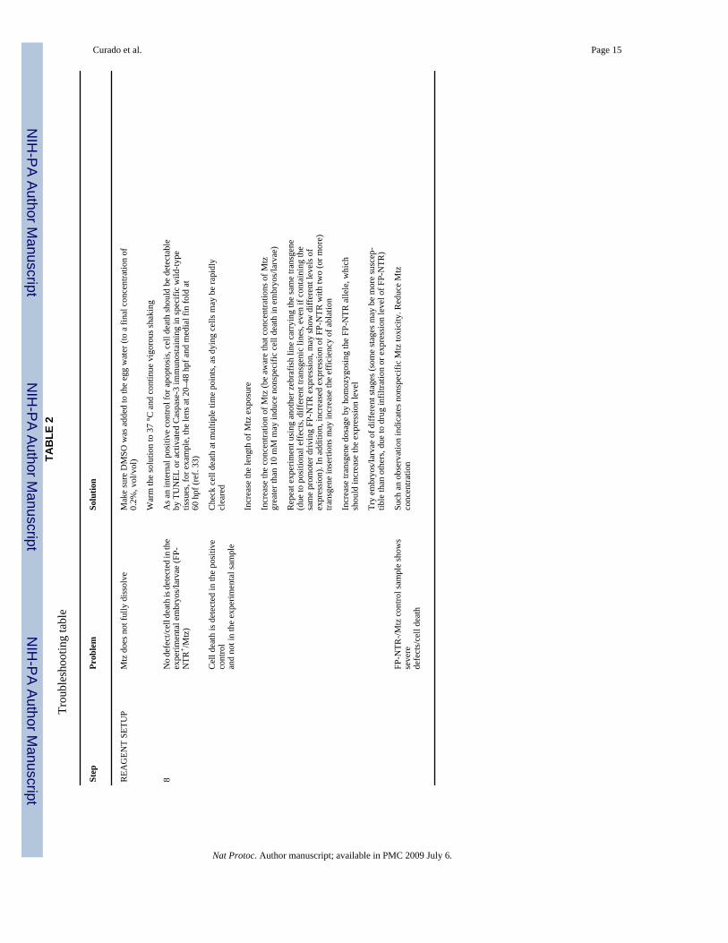
? TROUBLESHOOTINGTroubleshooting advice can be found in Table 2.
ANTICIPATED RESULTSCell death should be induced in all transgenic animals carrying the tsp:FP-NTR transgene afterexposure to a sufficient dose of Mtz, whereas no effect should be detected in any of the controlembryos/larvae used: FP-NTR- or FP-NTR+ exposed to DMSO alone, or FP-NTR- exposed toMtz. In our hands, all transgenic CFP-NTR embryos showed significant cell damage in thetargeted tissue after exposure to 10 mM Mtz, although the extent of ablation varied dependingon the tissue examined, the promoter used to drive FP-NTR and the transgenic line itself. Someexamples of our results are shown in Figures 2 and 3.
The period of time required to induce the death of cells expressing FP-NTR may vary from afew hours to several days (see Table 1) and will depend greatly on the stage, length of Mtztreatment, Mtz concentration, targeted tissue and FP-NTR transgenic line used. Using thissystem to ablate cells, it is possible to reverse damage as some injured tissues may regenerateafter removing Mtz by washing. However, the rate of recovery will depend both on the biologyof the targeted tissue and the extent of damage. It is possible that the percentage of animalsexhibiting recovery will never reach 100%; this situation may be advantageous for studyingenhancement versus impairment of regeneration in modified backgrounds—opening newpossibilities to discover factors that may inhibit or enhance cell and tissue regeneration.
ACKNOWLEDGMENTSWe thank Justin Bosch for comments on the manuscript and help in testing the protocol for adult ablations, StephenJ. Johnson (Washington University) for his suggestion to use the NTR/Mtz system in zebrafish, Jeff Mumm and Eric
Curado et al. Page 8
Nat Protoc. Author manuscript; available in PMC 2009 July 6.
NIH
-PA Author Manuscript
NIH
-PA Author Manuscript
NIH
-PA Author Manuscript

Schroeter (Washington University) for providing the CFP-NTR construct and Ana Ayala and Koroboshka Brand forexpert help maintaining the fish. R.M.A. was supported by a postdoctoral fellowship from the JDRF. This work wassupported in part by grants from the NIH (NHLBI and NIDDK) and the Packard Foundation to D.Y.R.S.
References1. Johnson SL, Weston JA. Temperature-sensitive mutations that cause stage-specific defects in Zebrafish
fin regeneration. Genetics 1995;141:1583–1595. [PubMed: 8601496]2. Bernhardt RR, Tongiorgi E, Anzini P, Schachner M. Increased expression of specific recognition
molecules by retinal ganglion cells and by optic pathway glia accompanies the successful regenerationof retinal axons in adult zebrafish. J. Comp. Neurol 1996;376:253–264. [PubMed: 8951641]
3. Becker T, Wullimann MF, Becker CG, Bernhardt RR, Schachner M. Axonal regrowth after spinal cordtransection in adult zebrafish. J. Comp. Neurol 1997;377:577–595. [PubMed: 9007194]
4. Poss KD, Wilson LG, Keating MT. Heart regeneration in zebrafish. Science 2002;298:2188–2190.[PubMed: 12481136]
5. Harris JA, et al. Neomycin-induced hair cell death and rapid regeneration in the lateral line of zebrafish(Danio rerio). J. Assoc. Res. Otolaryngol 2003;4:219–234. [PubMed: 12943374]
6. Yang CT, Sengelmann RD, Johnson SL. Larval melanocyte regeneration following laser ablation inzebrafish. J. Invest. Dermatol 2004;123:924–929. [PubMed: 15482481]
7. Sadler KC, Krahn KN, Gaur NA, Ukomadu C. Liver growth in the embryo and during liver regenerationin zebrafish requires the cell cycle regulator, uhrf1. Proc. Natl. Acad. Sci. USA 2007;104:1570–1575.[PubMed: 17242348]
8. Kurita R, et al. Suppression of lens growth by alphaA-crystallin promoter-driven expression ofdiphtheria toxin results in disruption of retinal cell organization in zebrafish. Dev. Biol 2003;255:113–127. [PubMed: 12618137]
9. Wan H, et al. Analyses of pancreas development by generation of gfp transgenic zebrafish using anexocrine pancreas-specific elastaseA gene promoter. Exp. Cell Res 2006;312:1526–1539. [PubMed:16490192]
10. Slanchev K, Stebler J, de la Cueva-Mendez G, Raz E. Development without germ cells: the role ofthe germ line in zebrafish sex differentiation. Proc. Natl. Acad. Sci. USA 2005;102:4074–4079.[PubMed: 15728735]
11. Jung J, et al. Ablation of tumor-derived stem cells transplanted to the central nervous system bygenetic modification of embryonic stem cells with a suicide gene. Hum. Gene Ther 2007;18:1182–1192. [PubMed: 18021021]
12. Chu QD, et al. Rat adenocarcinoma cell line infected with an adenovirus carrying a novel herpes-simplex virus-thymidine kinase suicide gene construct dies by apoptosis upon treatment withganciclovir. J. Surg. Res 2007;143:189–194. [PubMed: 17950092]
13. Springer CJ, Niculescu-Duvaz I. Approaches to gene-directed enzyme prodrug therapy (GDEPT).Adv. Exp. Med. Biol 2000;465:403–409. [PubMed: 10810644]
14. Pelengaris S, Khan M, Evan GI. Suppression of Myc-induced apoptosis in beta cells exposes multipleoncogenic properties of Myc and triggers carcinogenic progression. Cell 2002;109:321–334.[PubMed: 12015982]
15. Smith CA, et al. Conditional ablation of T-cell development by a novel viral ion channel transgene.Immunology 2002;105:306–313. [PubMed: 11918692]
16. Smith SJ, Kotecha S, Towers N, Mohun TJ. Targeted cell-ablation in Xenopus embryos using theconditional, toxic viral protein M2(H37A). Dev. Dyn 2007;236:2159–2171. [PubMed: 17615576]
17. Curado S, et al. Conditional targeted cell ablation in zebrafish: a new tool for regeneration studies.Dev. Dyn 2007;236:1025–1035. [PubMed: 17326133]
18. Pisharath H, Rhee JM, Swanson MA, Leach SD, Parsons MJ. Targeted ablation of beta cells in theembryonic zebrafish pancreas using E. coli nitroreductase. Mech. Dev 2007;124:218–229. [PubMed:17223324]
19. Bridgewater JA, et al. Expression of the bacterial nitroreductase enzyme in mammalian cells rendersthem selectively sensitive to killing by the prodrug CB1954. Eur. J. Cancer 1995;31A:2362–2370.[PubMed: 8652270]
Curado et al. Page 9
Nat Protoc. Author manuscript; available in PMC 2009 July 6.
NIH
-PA Author Manuscript
NIH
-PA Author Manuscript
NIH
-PA Author Manuscript

20. Lindmark DG, Muller M. Antitrichomonad action, mutagenicity, and reduction of metronidazole andother nitroimidazoles. Antimicrob. Agents Chemother 1976;10:476–482. [PubMed: 791102]
21. Anlezark GM, et al. The bioactivation of 5-(aziridin-1-yl)-2,4-dinitrobenzamide (CB1954)—I.Purification and properties of a nitroreductase enzyme from Escherichia coli—a potential enzymefor antibody-directed enzyme prodrug therapy (ADEPT). Biochem. Pharmacol 1992;44:2289–2295.[PubMed: 1472094]
22. Edwards DI. Nitroimidazole drugs—action and resistance mechanisms. II. Mechanisms of resistance.J. Antimicrob. Chemother 1993;31:201–210. [PubMed: 8463167]
23. Bridgewater JA, Knox RJ, Pitts JD, Collins MK, Springer CJ. The bystander effect of thenitroreductase/CB1954 enzyme/prodrug system is due to a cell-permeable metabolite. Hum. GeneTher 1997;8:709–717. [PubMed: 9113510]
24. Kawakami K, et al. A transposon-mediated gene trap approach identifies developmentally regulatedgenes in zebrafish. Dev. Cell 2004;7:133–144. [PubMed: 15239961]
25. Grabher C, Joly JS, Wittbrodt J. Highly efficient zebrafish transgenesis mediated by the meganucleaseI-SceI. Methods Cell Biol 2004;77:381–401. [PubMed: 15602923]
26. Huang CJ, Tu CT, Hsiao CD, Hsieh FJ, Tsai HJ. Germ-line transmission of a myocardium-specificGFP transgene reveals critical regulatory elements in the cardiac myosin light chain 2 promoter ofzebrafish. Dev. Dyn 2003;228:30–40. [PubMed: 12950077]
27. Her GM, Chiang CC, Chen WY, Wu JL. In vivo studies of liver-type fatty acid binding protein (L-FABP) gene expression in liver of transgenic zebrafish (Danio rerio). FEBS Lett 2003;538:125–133.[PubMed: 12633865]
28. Isles AR, et al. Conditional ablation of neurones in transgenic mice. J. Neurobiol 2001;47:183–193.[PubMed: 11333400]
29. Cui W, Allen ND, Skynner M, Gusterson B, Clark AJ. Inducible ablation of astrocytes shows thatthese cells are required for neuronal survival in the adult brain. Glia 2001;34:272–282. [PubMed:11360300]
30. Felmer R, Cui W, Clark AJ. Inducible ablation of adipocytes in adult transgenic mice expressing theE. coli nitroreductase gene. J. Endocrinol 2002;175:487–498. [PubMed: 12429046]
31. Searle PF, et al. Nitroreductase: a prodrug-activating enzyme for cancer gene therapy. Clin. Exp.Pharmacol. Physiol 2004;31:811–816. [PubMed: 15566399]
32. Westerfield, M. The Zebrafish Book. A Guide for the Laboratory Use of Zebrafish (Danio rerio). Vol.4th edn. University of Oregon Press; Eugene, Oregon: 2000.
33. Cole LK, Ross LS. Apoptosis in the developing zebrafish embryo. Dev. Biol 2001;240:123–142.[PubMed: 11784051]
Curado et al. Page 10
Nat Protoc. Author manuscript; available in PMC 2009 July 6.
NIH
-PA Author Manuscript
NIH
-PA Author Manuscript
NIH
-PA Author Manuscript

Figure 1.Experimental design for Mtz/NTR tissue-specific ablation. (a) Components of the toxigenecassette: a defined tissue-specific promoter (tsp) is cloned upstream of a cassette comprised ofa fluorescent protein (FP) fused to Nitroreductase (NTR) in a transgenesis vector. (b) FP-NTRfusion protein is expressed in a defined population of cells (blue) at the start of ablation (t1).Metronidazole (Mtz) added to the water is converted to a cytotoxin by FP-NTR, which causesapoptosis of the FP-NTR+ cells (brown), but not neighboring cells (tan; t2). (c) Samples aredivided into four groups for each ablation experiment: DMSO treatment of (A) NTR- and (B)NTR+ embryos/larvae and Mtz treatment of (C) NTR- embryos/larvae to control fornonspecific effects of Mtz or the transgene, and Mtz treatment of (D) NTR+ embryos for tissueablation. At t1 (start of ablation), the FP-NTR is visible in the tissue to be ablated (blue); at t2(end of ablation), FP-NTR+ cells will be depleted or absent (brown); and at t3 (during recoveryafter Mtz washout), new FP-NTR+ cells may be produced.
Curado et al. Page 11
Nat Protoc. Author manuscript; available in PMC 2009 July 6.
NIH
-PA Author Manuscript
NIH
-PA Author Manuscript
NIH
-PA Author Manuscript
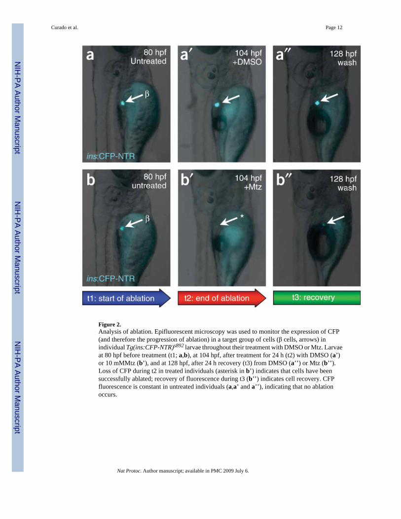
Figure 2.Analysis of ablation. Epifluorescent microscopy was used to monitor the expression of CFP(and therefore the progression of ablation) in a target group of cells (β cells, arrows) inindividual Tg(ins:CFP-NTR)s892 larvae throughout their treatment with DMSO or Mtz. Larvaeat 80 hpf before treatment (t1; a,b), at 104 hpf, after treatment for 24 h (t2) with DMSO (a’)or 10 mMMtz (b’), and at 128 hpf, after 24 h recovery (t3) from DMSO (a’’) or Mtz (b’’).Loss of CFP during t2 in treated individuals (asterisk in b’) indicates that cells have beensuccessfully ablated; recovery of fluorescence during t3 (b’’) indicates cell recovery. CFPfluorescence is constant in untreated individuals (a,a’ and a’’), indicating that no ablationoccurs.
Curado et al. Page 12
Nat Protoc. Author manuscript; available in PMC 2009 July 6.
NIH
-PA Author Manuscript
NIH
-PA Author Manuscript
NIH
-PA Author Manuscript
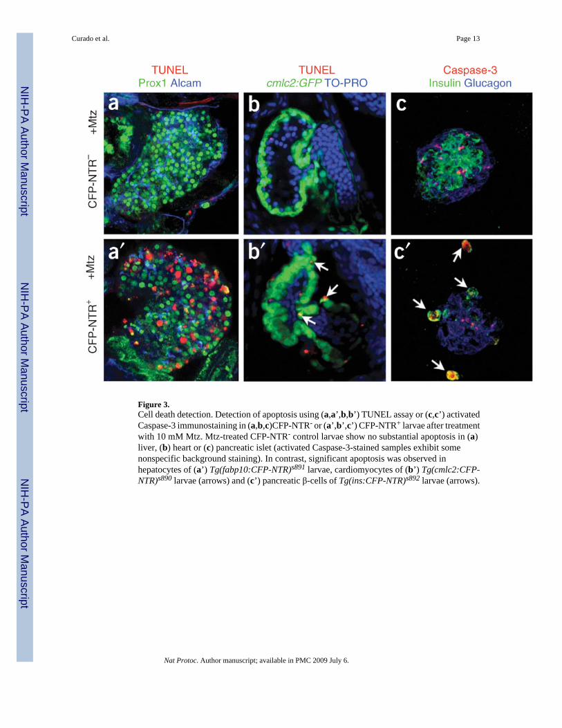
Figure 3.Cell death detection. Detection of apoptosis using (a,a’,b,b’) TUNEL assay or (c,c’) activatedCaspase-3 immunostaining in (a,b,c)CFP-NTR- or (a’,b’,c’) CFP-NTR+ larvae after treatmentwith 10 mM Mtz. Mtz-treated CFP-NTR- control larvae show no substantial apoptosis in (a)liver, (b) heart or (c) pancreatic islet (activated Caspase-3-stained samples exhibit somenonspecific background staining). In contrast, significant apoptosis was observed inhepatocytes of (a’) Tg(fabp10:CFP-NTR)s891 larvae, cardiomyocytes of (b’) Tg(cmlc2:CFP-NTR)s890 larvae (arrows) and (c’) pancreatic β-cells of Tg(ins:CFP-NTR)s892 larvae (arrows).
Curado et al. Page 13
Nat Protoc. Author manuscript; available in PMC 2009 July 6.
NIH
-PA Author Manuscript
NIH
-PA Author Manuscript
NIH
-PA Author Manuscript

NIH
-PA Author Manuscript
NIH
-PA Author Manuscript
NIH
-PA Author Manuscript
Curado et al. Page 14
TABLE 1Time points for Mtz treatment (examples)
Tg(tsp:CFP-NTR) t1 t2 t3
Tg(cmlc2:CFP-NTR)s890 48 hpf 72 hpf 96 hpf
Tg(fabp10:CFP-NTR)s891 96 hpf 168 hpf
Tg(ins:CFP-NTR)s892 84 hpf 110 hpf 145 hpf
Tg(ins:CFP-NTR)s892 90 dpf 93 dpf
t1, addition of Mtz and control (DMSO) solutions; t2, phenotype/tissue damage detected (wash out Mtz for regeneration studies); t3, recovery of damagedtissue observed; hpf, hours post-fertilization; dpf, days post-fertilization.
Nat Protoc. Author manuscript; available in PMC 2009 July 6.
NIH
-PA Author Manuscript
NIH
-PA Author Manuscript
NIH
-PA Author Manuscript
Curado et al. Page 15TA
BLE
2Tr
oubl
esho
otin
g ta
ble
Step
Prob
lem
Solu
tion
REA
GEN
T SE
TUP
Mtz
doe
s not
fully
dis
solv
eM
ake
sure
DM
SO w
as a
dded
to th
e eg
g w
ater
(to
a fin
al c
once
ntra
tion
of0.
2%, v
ol/v
ol)
War
m th
e so
lutio
n to
37
°C a
nd c
ontin
ue v
igor
ous s
haki
ng
8N
o de
fect
/cel
l dea
th is
det
ecte
d in
the
expe
rimen
tal e
mbr
yos/
larv
ae (F
P-N
TR+ /M
tz)
As a
n in
tern
al p
ositi
ve c
ontro
l for
apo
ptos
is, c
ell d
eath
shou
ld b
e de
tect
able
by T
UN
EL o
r act
ivat
ed C
aspa
se-3
imm
unos
tain
ing
in sp
ecifi
c w
ild-ty
petis
sues
, for
exa
mpl
e, th
e le
ns a
t 20–
48 h
pf a
nd m
edia
l fin
fold
at
60 h
pf (r
ef. 3
3)
Cel
l dea
th is
det
ecte
d in
the
posi
tive
cont
rol
and
not i
n th
e ex
perim
enta
l sam
ple
Che
ck c
ell d
eath
at m
ultip
le ti
me
poin
ts, a
s dyi
ng c
ells
may
be
rapi
dly
clea
red
Incr
ease
the
leng
th o
f Mtz
exp
osur
e
Incr
ease
the
conc
entra
tion
of M
tz (b
e aw
are
that
con
cent
ratio
ns o
f Mtz
grea
ter t
han
10 m
M m
ay in
duce
non
spec
ific
cell
deat
h in
em
bryo
s/la
rvae
)
Rep
eat e
xper
imen
t usi
ng a
noth
er z
ebra
fish
line
carr
ying
the
sam
e tra
nsge
ne(d
ue to
pos
ition
al e
ffec
ts, d
iffer
ent t
rans
geni
c lin
es, e
ven
if co
ntai
ning
the
sam
e pr
omot
er d
rivin
g FP
-NTR
exp
ress
ion,
may
show
diff
eren
t lev
els o
fex
pres
sion
). In
add
ition
, inc
reas
ed e
xpre
ssio
n of
FP-
NTR
with
two
(or m
ore)
trans
gene
inse
rtion
s may
incr
ease
the
effic
ienc
y of
abl
atio
n
Incr
ease
tran
sgen
e do
sage
by
hom
ozyg
osin
g th
e FP
-NTR
alle
le, w
hich
shou
ld in
crea
se th
e ex
pres
sion
leve
l
Try
embr
yos/
larv
ae o
f diff
eren
t sta
ges (
som
e st
ages
may
be
mor
e su
scep
-tib
le th
an o
ther
s, du
e to
dru
g in
filtra
tion
or e
xpre
ssio
n le
vel o
f FP-
NTR
)
FP-N
TR-/M
tz c
ontro
l sam
ple
show
sse
vere
defe
cts/
cell
deat
h
Such
an
obse
rvat
ion
indi
cate
s non
spec
ific
Mtz
toxi
city
. Red
uce
Mtz
conc
entra
tion
Nat Protoc. Author manuscript; available in PMC 2009 July 6.