Drug analysis by capillary electrophoresis and laser-induced fluorescence
Upload
independentCategory
view
3download
0
New Method Based on Capillary Electrophoresiswith Laser-Induced Fluorescence Detection(CE-LIF) to Monitor Interaction betweenNanoparticles and the Amyloid-� Peptide
Davide Brambilla,† Romain Verpillot,† Myriam Taverna,*,† Line De Kimpe,‡ Benjamin LeDroumaguet,† Julien Nicolas,† Mara Canovi,§ Marco Gobbi,§ Francesco Mantegazza,|
Mario Salmona,§ Valerie Nicolas,⊥ Wiep Scheper,‡ Patrick Couvreur,† and Karine Andrieux†
Laboratoire de Physico-Chimie, Pharmacotechnie et Biopharmacie, UMR CNRS 8612, Univ Paris-Sud 11, Faculte dePharmacie, 5 rue Jean-Baptiste Clement, F-92296 Chatenay-Malabry, France, Neurogenetics Laboratory, AcademicMedical Center, Amsterdam, The Netherlands, Istituto di Ricerche Farmacologiche “Mario Negri”, Milano, Italy,Department of Experimental Medicine, University of Milano-Bicocca, Monza, Italy, and Institut d’InnovationTherapeutique (IFR141 ITFM), Univ Paris-Sud, Faculte de Pharmacie, 5 rue Jean-Baptiste Clement,F-92296 Chatenay-Malabry, France
A novel application of capillary electrophoresis withlaser-induced fluorescence detection (CE-LIF) was pro-posed to efficiently detect and monitor the interactionbetween polymeric nanoparticles and the �-Amyloidpeptide (A�1-42), a biomarker for Alzheimer’s Disease(AD), at concentrations close to physiological condi-tions. The CE-LIF method allowed the interactionbetween PEGylated poly(alkyl cyanoacrylate) nanopar-ticles (NPs) and the soluble A�1-42 peptide monomersto be highlighted. These results were confirmed bysurface plasmon resonance (SPR) and confocal laserscanning microscopy (CLSM). Whereas SPR showedan interaction between the NPs and the A�1-42 peptide,CLSM allowed the formation of large aggregates/assemblies at high NP and peptide concentrations tobe visualized. All these results suggested that thesenanoparticles could bind the A�1-42 peptide and influ-ence its aggregation kinetics. Interestingly, the non-PEGylated poly(alkyl cyanoacrylate) NPs did not alterthe aggregation kinetics of the A�1-42 peptide, thusemphasizing the high level of discrimination of the CE-LIF method with respect to NPs.
The Alzheimer’s disease (AD) is a neurodegenerative disordercharacterized by a progressive loss of cognitive functions andcharacteristic pathological changes in the brain. It is the mostcommon elderly dementia, affecting 35 million people worldwide.1
It is histopathologically characterized by two main hallmarks: (i)the extracellular deposition of amyloid plaques mainly composedof �-amyloid peptides (A�) and (ii) intracellular neurofibrillar
tangles composed of hyperphosphorylated Tau protein.2,3 AD isa very complex disease which is likely the result of a multifactorialprocess strongly influenced by genetic and environmental com-ponents, but the mechanisms involved are not yet clearlyunderstood and still under debate.4-6
The treatment of AD represents a crucial challenge due toincomplete understanding of the etiology and the lack of diagnosticmethods able to discriminate between AD and other neurologicaldiseases. All currently approved therapies, such as the N-methyl-D-aspartate (NMDA) receptor antagonist7-9 and the acetylcho-linesterase (AchE) inhibitors,10-12 are only directed toward thealleviation of AD symptoms and exhibit many side effects.7
Among etiopathological hypotheses that have been proposedso far, the neuronal loss due to the toxicity of A� peptideaggregates, usually referred to as the “amyloid hypothesis”, isone of the most widely accepted.13,14 A� peptide is produced byneurons through sequential proteolytic cleavage of the amyloidprecursor protein (APP) by �- and γ-secretases, forming peptideswith a variable number of amino-acids, usually from 39 to 42.15,16
Among the different species, the A� peptide 1-42 (A�1-42) is
* To whom correspondence should be addressed. Phone: +33 (0)1 46 83 5462. Fax: +33 (0)1 46 83 59 44. E-mail: [email protected].
† UMR CNRS 8612, Univ. Paris Sud 11.‡ Academic Medical Center.§ Istituto di Ricerche Farmacologiche “Mario Negri”.| University of Milano-Bicocca.⊥ Institut d’Innovation Therapeutique.
(1) Querfurth, H. W.; LaFerla, F. M. N. Engl. J. Med. 2010, 362, 329.
(2) Aguzzi, A.; O’Connor, T. Nat. Rev. Drug Discovery 2010, 9, 237.(3) Panza, F.; Solfrizzi, V.; Frisardi, V.; Imbimbo, B. P.; Capurso, C.; D’Introno,
A.; Colacicco, A. M.; Seripa, D.; Vendemiale, G.; Capurso, A.; Pilotto, A.Aging: Clin. Exp. Res. 2009, 21, 386.
(4) Aliev, G.; Smith, M. A.; de la Torre, J. C.; Perry, G. Mitochondrion 2004,4, 649.
(5) de la Torre, J. C. Lancet Neurol. 2004, 3, 184.(6) Korolainen, M. A.; Nyman, T. A.; Aittokallio, T.; Pirttila, T. J. Neurochem.
2010, 112, 1386.(7) Kemp, J. A.; McKernan, R. M. Nat. Neurosci 2002, 5 (Suppl), 1039.(8) Parsons, C. G.; Danysz, W.; Quack, G. Neuropharmacology 1999, 38, 735.(9) Reisberg, B.; Doody, R.; Stoffler, A.; Schmitt, F.; Ferris, S.; Mobius, H. J.
N. Engl. J. Med. 2003, 348, 1333.(10) Sugimoto, H.; Iimura, Y.; Yamanishi, Y.; Yamatsu, K. J. Med. Chem. 1995,
38, 4821.(11) Munoz-Torrero, D. Curr. Med. Chem. 2008, 15, 2433.(12) Birks, J.; Grimley Evans, J.; Iakovidou, V.; Tsolaki, M.; Holt, F. E. Cochrane
Database Syst. Rev. 2009, CD001191.(13) Townsend, M.; Mehta, T.; Selkoe, D. J. J. Biol. Chem. 2007, 282, 33305.(14) Gralle, M.; Botelho, M. G.; Wouters, F. S. J. Biol. Chem. 2009, 284, 15016.(15) Pierrot, N.; Octave, J.-N. Curr. Alzheimer Res. 2008, 5, 92.
Anal. Chem. 2010, 82, 10083–10089
10.1021/ac102045x 2010 American Chemical Society 10083Analytical Chemistry, Vol. 82, No. 24, December 15, 2010Published on Web 11/18/2010

believed to be the most representative and the most toxicspecies in AD physiopathology due to its high tendency tospontaneously self-aggregate.17,18
The aggregation kinetics of A�1-42 peptide involves severalsteps and leads to the formation of species exhibiting variablesizes: typically small soluble oligomers, higher molar massoligomers, larger protofibrils and eventually insoluble fibrils.This folding and assembly are governed by remarkablycomplex processes leading to multiple coexisting physicalforms.
Even though it is well-known that the peptide adopts in vivo ahelical conformation when it is part of the trans-membrane domainof APP,19 spectroscopic studies have reported a random coilstructure in aqueous media.20 It may aggregate into multiple smalloligomers (up to six peptides) which coalesce into intermediateassemblies.21,22 The existence of a transitory conversion from therandom coil to a helical structure upon oligomerization has beenshown by nuclear magnetic resonance spectroscopy.19 A furtherconformational change has been proposed, leading to soluble�-sheet aggregates and then to nonsoluble fibrils.20
Several therapeutic approaches targeting A� peptides are underinvestigation. Among them, one can find the design of �- andγ-secretase inhibitors23 and monoamine oxidase inhibitors,24-27
together with A�1-42 passive and active immunization28,29 anddual inhibitor30-32 strategies. More recently, the development ofsmall molecules based on methylene blue and curcumin deriva-tives, which can interfere with the aggregation kinetics, may alsorepresent a promising therapeutic approach.33,34
In this context, we are currently developing novel nanopar-ticulate systems to target and/or to influence the aggregationkinetics of the A� peptides. Among suitable nanocarriers, biode-
gradable PEGylated poly(alkyl cyanoacrylate) nanoparticles (NPs)were selected as a first candidate to investigate this newtherapeutic approach due to their biodegradable properties andtheir well-established use for drug delivery purposes35 as well astheir in vivo ability to overpass the blood brain barrier (BBB).36,37
To screen the ability of our NPs to efficiently bind A�1-42, wehave for the first time applied capillary electrophoresis (CE)coupled to laser-induced fluorescence (LIF) detection to analyzefluorescently labeled A�. CE with UV detection has previouslybeen used for the in vitro identification of small molecules aspotential inhibitors of A� aggregation.38,39 This innovative work,was however limited due to the high concentrations of peptiderequired for the screening, which were about 5 orders ofmagnitude higher than those found in vivo. More recently, Katoet al.40 reported a CE-LIF method allowing the antiaggregationfeatures of molecules toward fibrils to be monitored. However,those species are no longer considered to be the main toxicspecies for AD.41-43 Consequently, a novel analytical method isrequired and must be adapted to A� monomers and resultingsoluble oligomers.
Based on the work of De Lorenzi’s research group related tothe screening of antifibrillogenic activity of small molecules,39 wereport herein an analytical method able to monitor in real timethe binding of soluble A�1-42 monomers to nanoparticles.Importantly, this is the first time that CE is developed toevaluate nanocarriers as potential therapeutic agents for ADtreatment using in vitro conditions compatible with clinicalapplication/trials. This analytical method was further associatedto confocal laser scanning microscopy (CLSM) experimentswhich demonstrated a binding effect of the nanoparticles tothe amyloid peptide, by influencing their aggregation kinetics.The physiological peptide concentration detectable with thismethod allowed nanoparticulate systems with potential bindingfeature to the amyloid peptide to be readily screened andquantitatively evaluated.
EXPERIMENTAL SECTIONMaterial and Chemicals. Poly[hexadecyl cyanoacrylate-co-
rhodamine B cyanoacrylate-co-methoxypoly(ethylene glycol) cy-anoacrylate] P(MePEGCA-co-RCA-co-HDCA) copolymers,44 poly-[hexadecyl cyanoacrylate-co-methoxypoly(ethylene glycol) cyano-
(16) Chow, V. W.; Mattson, M. P.; Wong, P. C.; Gleichmann, M. NeuroMol. Med.2009, 12, 1.
(17) Garcia-Matas, S.; de Vera, N.; Aznar, A. O.; Marimon, J. M.; Adell, A.; Planas,A. M.; Cristofol, R.; Sanfeliu, C. J. Alzheimer’s Dis. 2010, 20, 229.
(18) Allaman, I.; Gavillet, M.; Belanger, M.; Laroche, T.; Viertl, D.; Lashuel, H. A.;Magistretti, P. J. J. Neurosci. 2010, 30, 3326.
(19) Crescenzi, O.; Tomaselli, S.; Guerrini, R.; Salvadori, S.; D’Ursi, A. M.;Temussi, P. A.; Picone, D. Eur. J. Biochem. 2002, 269, 5642.
(20) Tomaselli, S.; Esposito, V.; Vangone, P.; van Nuland, N. A.; Bonvin, A. M.;Guerrini, R.; Tancredi, T.; Temussi, P. A.; Picone, D. ChemBioChem 2006,7, 257.
(21) Kayed, R.; Head, E.; Thompson, J. L.; McIntire, T. M.; Milton, S. C.; Cotman,C. W.; Glabe, C. G. Science 2003, 300, 486.
(22) Klein, W. L.; Krafft, G. A.; Finch, C. E. Trends Neurosci. 2001, 24, 219.(23) Shao, D.; Zou, C.; Luo, C.; Tang, X.; Li, Y. Bioorg. Med. Chem. Lett. 2004,
14, 4639.(24) Sano, M.; Ernesto, C.; Thomas, R. G.; Klauber, M. R.; Schafer, K.;
Grundman, M.; Woodbury, P.; Growdon, J.; Cotman, C. W.; Pfeiffer, E.;Schneider, L. S.; Thal, L. J. N. Engl. J. Med. 1997, 336, 1216.
(25) Riederer, P.; Danielczyk, W.; Grunblatt, E. Neurotoxicology 2004, 25, 271.(26) Youdim, M. B. H.; Buccafusco, J. J. Trends Pharmacol. Sci. 2005, 26, 27.(27) Huang, W.; Chen, Y.; Shohami, E.; Weinstock, M. Eur. J. Pharmacol. 1999,
366, 127.(28) Zou, J.; Yao, Z.; Zhang, G.; Wang, H.; Xu, J.; Yew, D. T.; Forster, E. L.
J. Neurol. Sci. 2008, 272, 87.(29) Dodart, J. C.; Bales, K. R.; Paul, S. M. Trends Mol. Med. 2003, 9, 85.(30) Bolognesi, M. L.; Andrisano, V.; Bartolini, M.; Banzi, R.; Melchiorre, C.
J. Med. Chem. 2005, 48, 24.(31) Camps, P.; Formosa, X.; Munoz-Torrero, D.; Petrignet, J.; Badia, A.; Clos,
M. V. J. Med. Chem. 2005, 48, 1701.(32) Toda, N.; Tago, K.; Marumoto, S.; Takami, K.; Ori, M.; Yamada, N.; Koyama,
K.; Naruto, S.; Abe, K.; Yamazaki, R.; Hara, T.; Aoyagi, A.; Abe, Y.; Kaneko,T.; Kogen, H. Bioorg. Med. Chem. 2003, 11, 1935.
(33) Yadav, A.; Sonker, M. Eur. J. Med. Chem. 2009, 44, 3866.(34) Hawkes, C. A.; Deng, L. H.; Shaw, J. E.; Nitz, M.; McLaurin, J. Eur.
J. Neurosci. 2010, 31, 203.
(35) Nicolas, J.; Couvreur, P. Wiley Interdiscip. Rev.: Nanomed. Nanobiotechnol.2009, 1, 111.
(36) Andrieux, K.; Couvreur, P. Wiley Interdiscip. Rev.: Nanomed. Nanobiotechnol.2009, 1, 463.
(37) Brigger, I.; Morizet, J.; Aubert, G.; Chacun, H.; Terrier-Lacombe, M. J.;Couvreur, P.; Vassal, G. J. Pharmacol. Exp. Ther. 2002, 303, 928.
(38) Sabella, S.; Quaglia, M.; Lanni, C.; Racchi, M.; Govoni, S.; Caccialanza, G.;Calligaro, A.; Bellotti, V.; De Lorenzi, E. Electrophoresis 2004, 25, 3186.
(39) Colombo, R.; Carotti, A.; Catto, M.; Racchi, M.; Lanni, C.; Verga, L.;Caccialanza, G.; De Lorenzi, E. Electrophoresis 2009, 30, 1418.
(40) Kato, M.; Kinoshita, H.; Enokita, M.; Hori, Y.; Hashimoto, T.; Iwatsubo, T.;Toyo’oka, T. Anal. Chem. 2007, 79, 4887.
(41) Dahlgren, K. N.; Manelli, A. M.; Stine, W. B., Jr.; Baker, L. K.; Krafft, G. A.;LaDu, M. J. J. Biol. Chem. 2002, 277, 32046.
(42) Cizas, P.; Budvytyte, R.; Morkuniene, R.; Moldovan, R.; Broccio, M.; Losche,M.; Niaura, G.; Valincius, G.; Borutaite, V. Arch. Biochem. Biophys. 2010,496, 84.
(43) Roychaudhuri, R.; Yang, M.; Hoshi, M. M.; Teplow, D. B. J. Biol. Chem.2009, 284, 4749.
(44) Brambilla, D.; Nicolas, J.; Droumaguet, B. L.; Andrieux, K.; Marsaud, V.;Couraud, P.-O.; Couvreur, P. Chem. Commun. (Cambridge, U. K.) 2010,46, 2602.
10084 Analytical Chemistry, Vol. 82, No. 24, December 15, 2010
acrylate] P(MePEGCA-co-HDCA) copolymers35 and poly(hexa-decyl cyanoacrylate) PHDCA homopolymers37 were obtainedfollowing previously reported procedures. NaH2PO4 (>99%) waspurchased from Merck & Co (Fontenay Sous Bois, France),Na2HPO4 (>98%) was obtained from Prolabo (Strasbourg,France), thioflavin (99%), ammonium hydroxide (NH4OH)28.1% (m/V), Pluronic F-68 (99%), 1,1,1,3,3,3-hexafluoro-2-propanol (HFIP) (99.8%), dimethyl sulfoxide (99.5%), sodiumdodecyl sulfate (SDS, 99%), acetic acid (99%), sodium acetate(99%), bovine serum albumin (BSA, 99%) and ethanolamine(99%) were purchased from Sigma-Aldrich (St. Quentin Falla-vier, France). Sodium hydroxide (NaOH, 1 M) was obtainedfrom VWR (Fontenay-sous Bois, France). Acetone was pur-chased at the highest grade from Carlo Erba (Val de Reuil,France). Lyophilized HiLyte Fluor 488 labeled A�1-42 and A�1-42
peptides were provided by ANASPEC (Le Perray en Yvelines,France). Anti-A� antibody 6E10 was from Covance (Princeton,New Jersey).
APPARATUSCapillary Electrophoresis. CE was performed on PA 800
instrument (Beckman Coulter, Roissy, France) using uncoatedsilica capillaries (Phymep, Paris) with an internal diameter of 50µm and 50 cm total length (40 cm effective length was employedfor the separation). All buffers were prepared with deionized waterand were filtered through a 0.22 µm membrane (VWR) beforeuse. Before analysis, the capillaries were preconditioned by thefollowing rinsing sequence: 0.1 M NaOH for 5 min, 1 M NaOHfor 5 min and then deionized water for 5 min. The in-between-runs rinsing cycles were carried out by pumping sequentiallythrough the capillary: water for 5 min, 50 mM SDS for 2 min (toinhibit the aggregation and subsequent peptide adsorption on thecapillary wall),45 and 0.1 M NaOH for 5 min. The samples wereintroduced into the capillary by hydrodynamic injection under 3.4kPa. The capillary was thermostated at 25 °C and the sampleswere maintained at 37 °C by the storage sample module of thePA 800 apparatus. The separations were carried out at 16 kV withpositive polarity at the inlet using 80 mM phosphate buffer pH7.4. The electrolyte was renewed after each run. The peptideswere detected by a laser-induced fluorescence (LIF) detectionsystem equipped with 3.5 mW argon-ion laser with a wavelengthexcitation of 488 nm, the emission being collected through a 520nm band-pass filter or by diode-array detector (DAD) at 190 nm.Peak areas were estimated using the 32 Karat software (BeckmanCoulter).
Confocal Laser Scanning Microscope. Observations weremade by sequential acquisition with a Zeiss LSM-510 confocalscanning laser microscope equipped with a 30 mW argon laserand 1 mW helium neon laser, using a Plan-Apochromat 63X
objective lens (NA 1.40, oil immersion). Red fluorescence wasobserved with a long-pass 560 nm emission filter and under a 543nm laser illumination. Green fluorescence was observed with aband-pass 505 and 550 nm emission filter and under a 488 nmlaser illumination. The pinhole diameter was set at 61 µm givingan optical section thickness of 0.6 µm. Stacks of images werecollected every 0.3 µm along the z axis. Twelve bit numericalimages were acquired with LSM 510 software version 3.2.
SPR Apparatus. ProteOn XPR36 (Biorad) apparatus, whichhas six parallel flow channels that can be used to uniformlyimmobilize strips of six ligands on the sensor surface, wasemployed. The fluidic system of Proteon XPR36 can automaticallyrotate 90° so that up to six different analytes (e.g., differentnanoparticle preparations, or different concentrations of the samenanoparticle) can be injected simultaneously over all the differentimmobilized molecules. All the injections were carried out for 3min at a flow rate of 30 µL ·min-1 at 30 °C in PBST (phosphatebuffer saline +0.005% Tween20, pH 7.4).
METHODSNanoparticle Preparation. Fluorescent and nonfluorescent
nanoparticles were prepared using P(MePEGCA-co-RCA-co-HDCA), P(MePEGCA-co-HDCA) copolymers, and PHDCA ho-mopolymer according to protocols recently published by ourgroup.44 The (co)polymer (10 mg) was dissolved in acetone (2mL) and this solution was added dropwise to an aqueous solution0.5% (w/v) of Pluronic F-68 (4 mL) under vigorous mechanicalstirring. A milky suspension was observed almost instantaneously.Acetone was then evaporated under reduced pressure andnanoparticles were purified by ultracentrifugation (150 000g, 1 h,4 °C, Beckman Coulter, Inc.). The supernatant was discarded andthe pellet was resuspended in the appropriate volume of deionizedwater to yield a 2.5 mg ·mL-1 nanoparticle suspension.
Nanoparticle Characterization. The nanoparticle diameter(Dz) was measured by dynamic light scattering (DLS) with aNano ZS from Malvern (173° scattering angle) at 25 °C. DLSmeasurements were used to monitor the nanoparticles stabilityas a function of time upon their incubation at 37 °C in the bufferemployed for capillary electrophoresis experiments. The nano-particle surface charge was investigated by �-potential measure-ment at 25 °C after dilution with 1 mM NaCl solution applyingthe Smoluchowski equation and using the same apparatus.
Peptide Sample Preparation and Storage. LyophilizedA�1-42 and HiLyte Fluor labeled A�1-42 peptide were dissolvedin 0.16% (m/V) ammonium hydroxide aqueous solution toreach a concentration of 2 mg ·mL-1. The fluorescent andnonlabeled peptide solutions were then divided into aliquotsindividually stored at -20 °C which were freshly defrosted prioranalysis.
The A�1-42 peptide used for SPR experiments was preparedfrom a depsi-A�1-42 peptide synthesized as previously de-scribed.46 This depsi-peptide is much more soluble than thenative peptide and has also a much lower propensity toaggregate, thus preventing the spontaneous formation of“seeds” in solution.46 The native A�1-42 peptide was thenobtained from the depsi-peptide by a “switching” procedureinvolving a change in pH.46,47 The A�1-42 peptide solutionobtained immediately after switching was shown to be free ofseed. The A�1-42 peptide obtained by this procedure is
(45) Han, Y.; He, C.; Cao, M.; Huang, X.; Wang, Y.; Li, Z. Langmuir 2009, 26,1583.
(46) Taniguchi, A.; Sohma, Y.; Hirayama, Y.; Mukai, H.; Kimura, T.; Hayashi,Y.; Matsuzaki, K.; Kiso, Y. ChemBioChem 2009, 10, 710.
(47) Balducci, C.; Beeg, M.; Stravalaci, M.; Bastone, A.; Sclip, A.; Biasini, E.;Tapella, L.; Colombo, L.; Manzoni, C.; Borsello, T.; Chiesa, R.; Gobbi, M.;Salmona, M.; Forloni, G. Proc. Natl. Acad. Sci. U.S.A. 2010, 107, 2295.
(48) Atha, D. H.; Ingham, K. C. J. Biol. Chem. 1981, 256, 12108.(49) Shulgin, I. L.; Ruckenstein, E. Biophys. Chem. 2006, 120, 188.(50) Auer, S.; Trovato, A.; Vendruscolo, M. PLoS Comput. Biol. 2009, 5,
e1000458.(51) Chiti, F.; Dobson, C. M. Annu. Rev. Biochem. 2006, 75, 333.
10085Analytical Chemistry, Vol. 82, No. 24, December 15, 2010
therefore in its original state and, for the sake of simplicity; itwill be referred here to as the “monomer”. Further character-izations, carried out by Gobbi’s group, indicated that the A�“monomer” used for the present study gave no thioflavin T(ThT) signal and was unstructured as observed by circulardichroism (manuscript submitted, 2010).
The peptide concentration required for the different methodsdescribed below were not strictly the same as long as differentdetection thresholds had to be taken into account.
Capillary Electrophoresis Experiments. To study the in-teraction between the monomeric form of the A�1-42 peptide andthe nanoparticles, aliquots of HiLyte Fluor labeled A�1-42
peptide stock solutions were diluted in 20 mM phosphatebuffer (NaH2PO4) at pH 7.4 containing a 20 µM P(MePEGCA-co-RCA-co-HDCA) nanoparticle suspension to obtain final pep-tide concentrations of 5, 1, 0.5, and 0.05 µM. The samples werethen incubated at 37 °C and analyzed by capillary electrophore-sis every 2 h. The same protocol was followed with P(Me-PEGCA-co-RCA-co-HDCA) nanoparticle suspension or A�1-42
peptide solution.Similarly, a mixture of nonfluorescent P(MePEGCA-co-HDCA)
or PHDCA nanoparticle suspensions (20 µM final concentration)and A�1-42 at 10 µM were analyzed by CE using the DADdetector at 190 nm as control.
These experiments allowed the evolution of % monomer peakas a function of incubation time to be determined. % monomerpeak is calculated as the ratio between the absolute peak area ofthe monomer observed at t ) 0 and the one observed at eachincubation time.
Surface Plasmon Resonance Experiments. A�1-42 mono-mers were immobilized in parallel-flow channels of a GLCsensor chip (Biorad) using amine-coupling chemistry. Briefly,after surface activation the peptide solutions (10 µM in acetatebuffer pH 4.0) were injected for 5 min at a flow rate of 30µL ·min-1 and the remaining activated groups were blockedwith ethanolamine at pH 8.0. The final immobilization levelswere about 2500 resonance units (1 RU ) 1 pg protein ·mm-2).Bovine serum albumin (BSA) was immobilized, in a parallelflow channel as a reference protein. Another reference surfacewas prepared in parallel using the same immobilizationprocedure but without addition of the peptide (naked surface).Before performing experiments with nanoparticles, we checkedthat A� species immobilized can bind with high affinity to theanti-A� antibody 6E10 (see Supporting Information (SI) FigureS6c). The suspension of P(MePEGCA-co-HDCA) NPs was dilutedat different concentrations (0.3-20 µM) and flowed into themachine simultaneously.
Confocal Laser Scanning Microscopy Analysis. The inter-action between the polymeric nanoparticles and the A�1-42 peptidewas investigated by confocal laser scanning microscopy (CLSM)using HiLyte Fluor labeled A�1-42 peptide and rhodamineB-labeled P(MePEGCA-co-RCA-co-HDCA) NPs.
The A�1-42 peptide aliquots were defrosted, immediatelydiluted with 20 mM phosphate buffer and incubated with 20µM nanoparticle suspension to reach a final peptide concentra-tion of 10, 1, or 0.05 µM. A 10 µL deposit of this final incubationsample on glass coverslips was immediately analyzed by CLSM.The remaining suspensions were incubated at 37 °C and, after
12 h, another 10 µL was withdrawn from the suspension andanalyzed in the same manner by CLSM.
Thioflavin T Aggregation Assay. A�1-42 was dissolved inhexafluoroisopropanol (HFIP) at a final concentration of 1mg ·mL-1, sampled and allowed to evaporate. For coaggrega-tion experiments, the peptide film was first dissolved in DMSOand sonicated in a bath sonicator for 10 min. Subsequently,A�1-42 was diluted in phosphate-buffer saline (PBS, 20 mMsodium phosphate buffer, pH 7.4, containing 137 mM NaCl)to a final concentration of 50 µM. This mixture was aggregatedin the presence or absence of P(MePEGCA-co-RCA-co-HDCA)NPs for 24 h at 37 °C. Aggregated A�1-42 was diluted to a finalconcentration of 5 µM into 50 mM glycine buffer at pH 7.4containing 10 µM ThT. Fluorescence was measured in 96 wellnonbinding plates (Greiner Bio One, Frickenhausen, Germany)using a Fluostar Omega microplate reader at an excitationwavelength of 450 nm and emission at 485 nm.
RESULTS AND DISCUSSION
The purified fluorescent and nonfluorescent nanoparticlesprepared by nanoprecipitation were characterized by DLS and�-potential measurements. PEGylated nanoparticles presented anaverage diameter in the 90-100 nm range with narrow particlesize distribution, whereas non-PEGylated counterparts werecentered around 160 nm (see SI Figure S1). Those nanoparticlesexhibited a negative �-potential (-30 ± 5 mV), which is in asuitable window for biomedical applications. Notably all kinds ofnanoparticles showed good colloidal stability in the capillaryelectrophoresis buffer (i.e., phosphate buffer pH 7.4, 20 mM) asassessed by DLS measurement during 72 h (SI Figure S1).
The CE-LIF method described in this study was developed toinvestigate the interaction between rhodamine B-labeled nano-particles and HiLyte Fluor labeled A�1-42. The LIF detectionallowed a lower concentration of peptide to be used than withUV detection, in order to better fit with physiological processesand conditions. Indeed, A� peptide is found in biological fluids,such as the cerebro-spinal fluid (CSF), at nanomolar concentra-tions and therefore a sensitive and discriminative analyticalmethod is required.
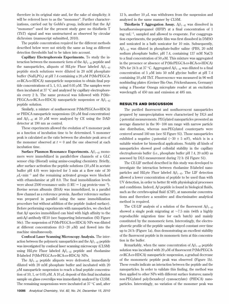
The CE-LIF analysis of a solution of the fluorescent A�1-42
showed a single peak migrating at ∼7.5 min (with a highlyreproducible migration time for each batch) and mainlyconstituted by the monomeric form. Interestingly, the electro-phoretic profile of the peptide sample stayed constant over timeup to 24 h (Figure 1a), thus demonstrating an excellent stabilityof the fluorescent peptide in its monomeric form at this concentra-tion in the buffer.
Remarkably, when the same concentration of A�1-42 peptidesolution was incubated with 20 µM of fluorescent P(MePEGCA-co-RCA-co-HDCA) nanoparticle suspension, a gradual decreaseof the monomeric peptide peak was observed (Figure 1b).These results indicate an interaction between the peptide and thenanoparticles. In order to validate this finding, the method wasthen applied to other NPs with different surface features; namelynon-PEGylated poly(hexadecyl cyanoacrylate) (PHDCA) nano-particles. Interestingly, no variation of the monomer peak was
10086 Analytical Chemistry, Vol. 82, No. 24, December 15, 2010
observed over time upon incubation with PHDCA NPs under thesame experimental conditions as the PEGylated counterparts(Figure 1c). It is well-known that PEG chains can interplay withprotein environment altering their solubility in aqueous media,48
although the mechanism has not yet been fully elucidated.49
In our case, it is believed that PEG chains locally modified thepeptide solubility and thus triggered its uptake by the nano-particles. Therefore, these results confirm that the analyticalmethod presented in this study is able to efficiently discriminatenanoparticles with respect to their respective aptitude to bindA�1-42.
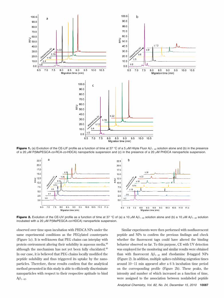
Similar experiments were then performed with nonfluorescentpeptide and NPs to confirm the previous findings and checkwhether the fluorescent tags could have altered the bindingbehavior observed so far. To this purpose, CE with UV detectionwas employed for the monitoring and similar results were obtainedthan with fluorescent A�1-42 and rhodamine B-tagged NPs(Figure 2). In addition, multiple spikes exhibiting migration timesaround 10-11 min appeared after a 6 h incubation time periodon the corresponding profile (Figure 2b). These peaks, theintensity and number of which increased as a function of time,were assigned to the association between nonlabeled peptide
Figure 1. (a) Evolution of the CE-LIF profile as a function of time at 37 °C of a 5 µM Hilyte Fluor A�1-42 solution alone and (b) in the presenceof a 20 µM P(MePEGCA-co-RCA-co-HDCA) nanoparticle suspension and (c) in the presence of a 20 µM PHDCA nanoparticle suspension.
Figure 2. Evolution of the CE-UV profile as a function of time at 37 °C of (a) a 10 µM A�1-42 solution alone and (b) a 10 µM A�1-42 solutionincubated with a 20 µM P(MePEGCA-co-HDCA) nanoparticle suspension.
10087Analytical Chemistry, Vol. 82, No. 24, December 15, 2010
species and the nanoparticles. However, these peaks were notobserved with the CE-LIF method likely due to optical featuresof the LIF detection.
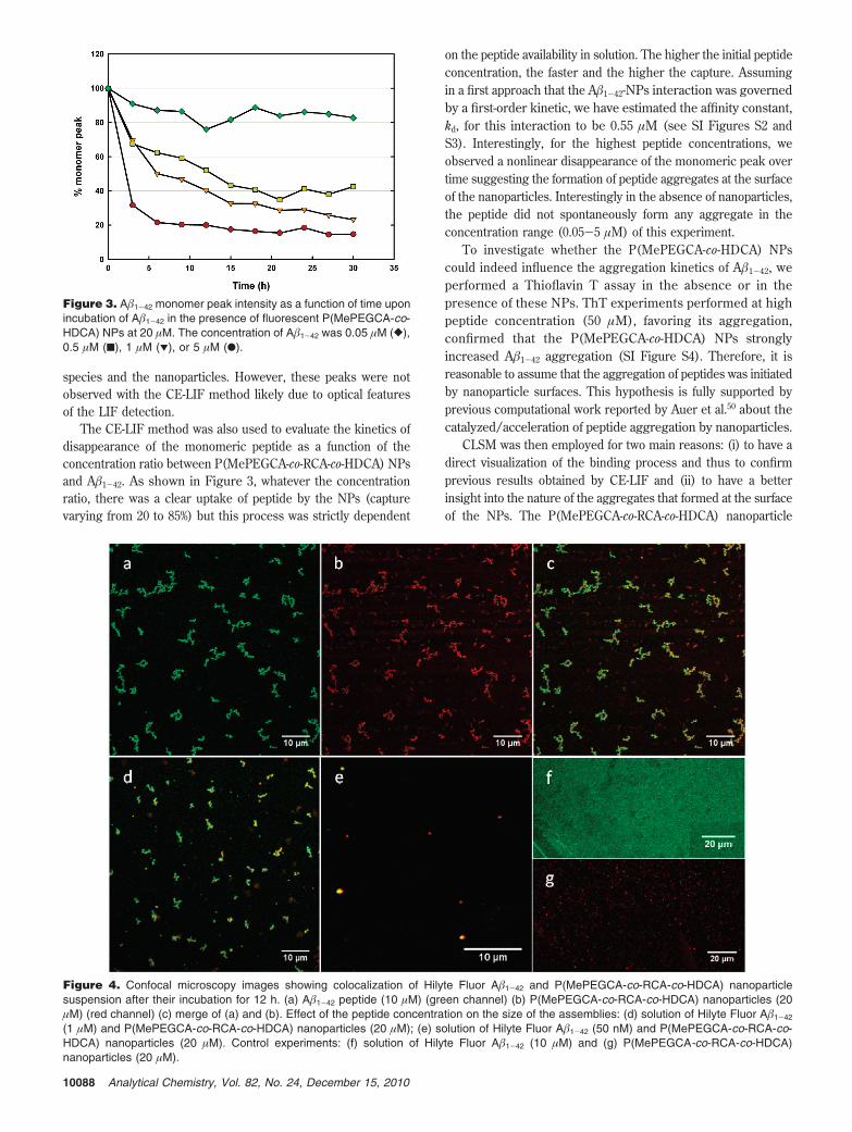
The CE-LIF method was also used to evaluate the kinetics ofdisappearance of the monomeric peptide as a function of theconcentration ratio between P(MePEGCA-co-RCA-co-HDCA) NPsand A�1-42. As shown in Figure 3, whatever the concentrationratio, there was a clear uptake of peptide by the NPs (capturevarying from 20 to 85%) but this process was strictly dependent
on the peptide availability in solution. The higher the initial peptideconcentration, the faster and the higher the capture. Assumingin a first approach that the A�1-42-NPs interaction was governedby a first-order kinetic, we have estimated the affinity constant,kd, for this interaction to be 0.55 µM (see SI Figures S2 andS3). Interestingly, for the highest peptide concentrations, weobserved a nonlinear disappearance of the monomeric peak overtime suggesting the formation of peptide aggregates at the surfaceof the nanoparticles. Interestingly in the absence of nanoparticles,the peptide did not spontaneously form any aggregate in theconcentration range (0.05-5 µM) of this experiment.
To investigate whether the P(MePEGCA-co-HDCA) NPscould indeed influence the aggregation kinetics of A�1-42, weperformed a Thioflavin T assay in the absence or in thepresence of these NPs. ThT experiments performed at highpeptide concentration (50 µM), favoring its aggregation,confirmed that the P(MePEGCA-co-HDCA) NPs stronglyincreased A�1-42 aggregation (SI Figure S4). Therefore, it isreasonable to assume that the aggregation of peptides was initiatedby nanoparticle surfaces. This hypothesis is fully supported byprevious computational work reported by Auer et al.50 about thecatalyzed/acceleration of peptide aggregation by nanoparticles.
CLSM was then employed for two main reasons: (i) to have adirect visualization of the binding process and thus to confirmprevious results obtained by CE-LIF and (ii) to have a betterinsight into the nature of the aggregates that formed at the surfaceof the NPs. The P(MePEGCA-co-RCA-co-HDCA) nanoparticle
Figure 4. Confocal microscopy images showing colocalization of Hilyte Fluor A�1-42 and P(MePEGCA-co-RCA-co-HDCA) nanoparticlesuspension after their incubation for 12 h. (a) A�1-42 peptide (10 µM) (green channel) (b) P(MePEGCA-co-RCA-co-HDCA) nanoparticles (20µM) (red channel) (c) merge of (a) and (b). Effect of the peptide concentration on the size of the assemblies: (d) solution of Hilyte Fluor A�1-42
(1 µM) and P(MePEGCA-co-RCA-co-HDCA) nanoparticles (20 µM); (e) solution of Hilyte Fluor A�1-42 (50 nM) and P(MePEGCA-co-RCA-co-HDCA) nanoparticles (20 µM). Control experiments: (f) solution of Hilyte Fluor A�1-42 (10 µM) and (g) P(MePEGCA-co-RCA-co-HDCA)nanoparticles (20 µM).
Figure 3. A�1-42 monomer peak intensity as a function of time uponincubation of A�1-42 in the presence of fluorescent P(MePEGCA-co-HDCA) NPs at 20 µM. The concentration of A�1-42 was 0.05 µM ([),0.5 µM (9), 1 µM (1), or 5 µM (b).
10088 Analytical Chemistry, Vol. 82, No. 24, December 15, 2010
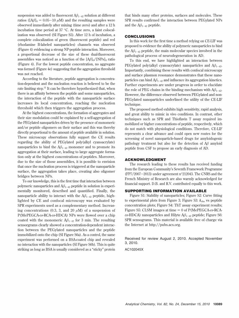
suspension was added to fluorescent A�1-42 solution at differentratios ([A�]0 ) 0.05-10 µM) and the resulting samples wereobserved immediately after mixing (time zero) and after a 12 hincubation time period at 37 °C. At time zero, a faint colocal-ization was observed (SI Figure S5). After 12 h of incubation, acomplete colocalization of green (fluorescent peptide) and red(rhodamine B-labeled nanoparticles) channels was observed(Figure 4) evidencing a strong NP-peptide interaction. Moreover,a proportional decrease of the size of these dual-fluorescentassemblies was noticed as a function of the [A�]0/[NPs]0 ratio(Figure 4). For the lowest peptide concentration, no aggregatewas formed (Figure 4e) suggesting that the aggregation thresholdwas not reached.
According to the literature, peptide aggregation is concentra-tion-dependent and the nucleation reaction is believed to be therate limiting step.51 It can be therefore hypothesized that, whenthere is an affinity between the peptide and some nanoparticles,the interaction of the peptide with the nanoparticle surfaceincreases its local concentration, reaching the nucleationthreshold which then triggers the aggregation process.
At the highest concentrations, the formation of aggregates andtheir size modulation could be explained by a self-aggregation ofthe PEGylated nanoparticles driven by the presence of monomericand/or peptide oligomers on their surface and this was therebydirectly proportional to the amount of peptide available in solution.These microscopy observations fully support the CE resultsregarding the ability of PEGylated poly(alkyl cyanoacrylate)nanoparticles to bind the A�1-42 monomer and to promote itsaggregation at their surface, leading to large aggregate forma-tion only at the highest concentrations of peptides. Moreover,due to the size of those assemblies, it is possible to envisionthat once the nucleation process is triggered at the nanoparticlesurface, the aggregation takes place, creating also oligomerbridges between NPs.
To our knowledge, this is the first time that interaction betweenpolymeric nanoparticles and A�1-42 peptide in solution is experi-mentally monitored, described and quantified. Finally, thenanoparticle ability to interact with the A�1-42 peptide, high-lighted by CE and confocal microscopy was evaluated bySPR experiments used as a complementary method. Increas-ing concentrations (0.3, 3, and 20 µM) of a suspension ofP(MePEGCA-co-RCA-co-HDCA) NPs were flowed over a chipcoated with the monomeric A�1-42 for 3 min. The resultingsensorgrams clearly showed a concentration-dependent interac-tion between the PEGylated nanoparticles and the peptideimmobilized onto the chip (SI Figure S6a). As a control, the sameexperiment was performed on a BSA-coated chip and revealedno interaction with the nanoparticles (SI Figure S6b). This is quitestriking as long as BSA is usually considered as a “sticky” protein
that binds many other proteins, surfaces and molecules. TheseSPR results confirmed the interaction between PEGylated NPsand the A�1-42 peptide.
CONCLUSIONSIn this work for the first time a method relying on CE-LIF was
proposed to evidence the ability of polymeric nanoparticles to bindthe A�1-42 peptide, the main molecular species involved in thepathological process of neurodegeneration in AD.
To this end, we have highlighted an interaction betweenPEGylated poly(alkyl cyanoacrylate) nanoparticles and A�1-42.Importantly, combining these results with confocal microscopyand surface plasmon resonance demonstrates that these nano-particles can bind A�1-42 and influence its aggregation kinetics.Further experiments are under progress in order to elucidatethe role of PEG chains in the binding mechanism with A�1-42.However, the difference observed between PEGylated and non-PEGylated nanoparticles underlined the utility of the CE-LIFtechnique.
The proposed method exhibits high sensitivity, rapid analysis,and great ability to mimic in vivo conditions. In contrast, othertechniques such as SPR and Thioflavin T assay required im-mobilized or higher concentrations of peptide, respectively, whichdo not match with physiological conditions. Therefore, CE-LIFrepresents a clear advance and could open new routes for thescreening of novel nanoparticle-based drugs for amyloidogenicpathology treatment but also for the detection of A� amyloidpeptide from CSF to propose an early diagnosis of AD.
ACKNOWLEDGMENTThe research leading to these results has received funding
from the European Community’s Seventh Framework Programme(FP7/2007-2013) under agreement n°212043. The CNRS and theFrench Ministry of Research are also warmly acknowledged forfinancial support. D.B. and R.V. contributed equally to this work.
SUPPORTING INFORMATION AVAILABLEFigure S1: Stability of nanoparticles; Figure S2: Curve fitting
to experimental plots from Figure 3; Figure S3: kobs vs peptideconcentration plots; Figure S4: ThT assay experiment results;Figure S5: CLSM images at time ) 0 of P(MePEGCA-co-RCA-co-HDCA) nanoparticles and Hilyte A�1-42 peptide; Figure S6:SPR sensograms. This material is available free of charge viathe Internet at http://pubs.acs.org.
Received for review August 2, 2010. Accepted November3, 2010.
AC102045X
10089Analytical Chemistry, Vol. 82, No. 24, December 15, 2010
Copyright © 2022 FDOKUMEN