New Insights into Ultrastructure, Lipid Composition and Organization of Vernix Caseosa
11
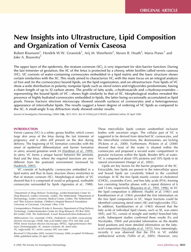
New Insights into Ultrastructure, Lipid Composition and Organization of Vernix Caseosa Robert Rissmann 1 , Hendrik W.W. Groenink 1 , Arij M. Weerheim 2 , Steven B. Hoath 3 , Maria Ponec 1 and Joke A. Bouwstra 1 The upper layer of the epidermis, the stratum corneum (SC), is very important for skin barrier function. During the last trimester of gestation, the SC of the fetus is protected by a cheesy, white biofilm called vernix caseosa (VC). VC consists of water-containing corneocytes embedded in a lipid matrix and the basic structure shows certain similarities with the SC. This study aimed to characterize VC, with the main focus on an integral analysis of free and (to the corneocytes) bound lipids, on the lipid organization, and on ultrastructure. Free lipids of VC show a wide distribution in polarity; nonpolar lipids such as sterol esters and triglycerides predominate, having a chain length of up to 32 carbon atoms. The profile of fatty acids, o-hydroxyacids and o-hydroxyceramides – representing the bound lipids of VC – shows high similarity to that of SC. Morphological studies revealed the presence of highly hydrated corneocytes embedded in lipids, the latter being occasionally accumulated as lipid pools. Freeze fracture electron microscopy showed smooth surfaces of corneocytes and a heterogeneous appearance of intercellular lipids. The results suggest a lower degree of ordering of VC lipids as compared to the SC. A small-angle X-ray diffraction study showed similar results. Journal of Investigative Dermatology (2006) 126, 1823–1833. doi:10.1038/sj.jid.5700305; published online 20 April 2006 INTRODUCTION Vernix caseosa (VC) is a white, greasy biofilm, which covers large skin areas of the fetus during the last trimester of pregnancy and is often still present on the skin during delivery. The beginning of VC formation coincides with the onset of epidermal differentiation and barrier formation in utero, around gestation week 24 (Hardman et al., 1999). In utero, VC forms a unique barrier between the amniotic fluid and the fetus, where the required functions are very different from the postnatal environment (reviewed by Haubrich, 2003). VC consists of hydrophilic corneocytes embedded in a lipid matrix and thus its basic structure shows similarities to that of stratum corneum (SC). Morphological studies of VC showed that it is comprised of ovoid or polygonal, malleable corneocytes surrounded by lipids (Agorastos et al., 1988). These intercellular lipids contain unidentified inclusion bodies with uncertain origin. The cellular part of VC is suggested to be derived from detached SC corneocytes, and the intercellular connections, the desmosomes, are lacking (Pickens et al., 2000). Furthermore, Pickens et al. (2000) showed that most of the water is situated within the corneocytes and proposed a second water compartment in granular inclusions within the lipids. Besides 80% of water, VC is composed of about 10% proteins and 10% lipids in its natural environment (Hoeger et al., 2002). Lipids are key factors for the barrier properties of the SC. Extractable, free lipids are present in the intercellular space and bound lipids are covalently linked to the cornified envelope. In SC the free lipids mainly consist of cholesterol (CHOL), ceramides (Cers), and free fatty acids (FFA), forming two coexisting lamellar phases with a repeat distance of 6 and 13 nm, respectively (Bouwstra et al., 1991, 1996). In VC the lipid composition is different. Haahti et al. (1961) and Kaerkkaeinen et al. (1965) provided the first information on the free lipid composition in VC. Major fractions could be identified containing sterol esters (SE) and triglycerides (TG). In addition, Kaerkkaeinen et al. (1965) showed that the fatty acids of individual lipid classes, such as SE, wax esters (WE), and TG, consist of straight and methyl branched fatty acids. Subsequent studies confirmed these results (Fu and Nicolaides, 1969) and provided information on the double bond positions and more detailed information on the fatty- acid composition (Nicolaides et al., 1972). Very interestingly, recently it was observed that the FFA in VC exhibit antibacterial activity (Tollin et al., 2005). However, very & 2006 The Society for Investigative Dermatology www.jidonline.org 1823 ORIGINAL ARTICLE Received 12 December 2005; revised 23 January 2006; accepted 10 February 2006; published online 20 April 2006 1 Department of Drug Delivery Technology, Leiden/Amsterdam Center for Drug Research, Leiden University, Leiden, The Netherlands; 2 Department of Dermatology, Leiden University Medical Center, Leiden, The Netherlands and 3 Skin Sciences Institute, Children’s Hospital Research Foundation, University of Cincinnati, Cincinnati, Ohio, USA Correspondence: Professor Dr Joke A. Bouwstra, Department of Drug Delivery Technology, Leiden/Amsterdam Center for Drug Research, PO Box 9502, RA Leiden 2300, The Netherlands. E-mail: [email protected] Abbreviations: Cer, ceramide; CHOL, cholesterol; cryo-SEM, cryoscanning electron microscopy; FAME, fatty-acid methyl ester; FFA, free fatty acid; FFEM, freeze fracture electron microscopy; HPTLC, high-performance thin layer chromatography; SC, stratum corneum; SE, sterol ester; TG, triglyceride; VC, vernix caseosa; WE, wax ester
-
Upload
cincinnatichildrens -
Category
Documents
-
view
0 -
download
0
Transcript of New Insights into Ultrastructure, Lipid Composition and Organization of Vernix Caseosa
New Insights into Ultrastructure, Lipid Compositionand Organization of Vernix CaseosaRobert Rissmann1, Hendrik W.W. Groenink1, Arij M. Weerheim2, Steven B. Hoath3, Maria Ponec1 andJoke A. Bouwstra1
The upper layer of the epidermis, the stratum corneum (SC), is very important for skin barrier function. Duringthe last trimester of gestation, the SC of the fetus is protected by a cheesy, white biofilm called vernix caseosa(VC). VC consists of water-containing corneocytes embedded in a lipid matrix and the basic structure showscertain similarities with the SC. This study aimed to characterize VC, with the main focus on an integral analysisof free and (to the corneocytes) bound lipids, on the lipid organization, and on ultrastructure. Free lipids of VCshow a wide distribution in polarity; nonpolar lipids such as sterol esters and triglycerides predominate, havinga chain length of up to 32 carbon atoms. The profile of fatty acids, o-hydroxyacids and o-hydroxyceramides –representing the bound lipids of VC – shows high similarity to that of SC. Morphological studies revealed thepresence of highly hydrated corneocytes embedded in lipids, the latter being occasionally accumulated as lipidpools. Freeze fracture electron microscopy showed smooth surfaces of corneocytes and a heterogeneousappearance of intercellular lipids. The results suggest a lower degree of ordering of VC lipids as compared tothe SC. A small-angle X-ray diffraction study showed similar results.
Journal of Investigative Dermatology (2006) 126, 1823–1833. doi:10.1038/sj.jid.5700305; published online 20 April 2006
INTRODUCTIONVernix caseosa (VC) is a white, greasy biofilm, which coverslarge skin areas of the fetus during the last trimester ofpregnancy and is often still present on the skin duringdelivery. The beginning of VC formation coincides with theonset of epidermal differentiation and barrier formationin utero, around gestation week 24 (Hardman et al., 1999).In utero, VC forms a unique barrier between the amnioticfluid and the fetus, where the required functions are verydifferent from the postnatal environment (reviewed byHaubrich, 2003).
VC consists of hydrophilic corneocytes embedded in alipid matrix and thus its basic structure shows similarities tothat of stratum corneum (SC). Morphological studies of VCshowed that it is comprised of ovoid or polygonal, malleablecorneocytes surrounded by lipids (Agorastos et al., 1988).
These intercellular lipids contain unidentified inclusionbodies with uncertain origin. The cellular part of VC issuggested to be derived from detached SC corneocytes, andthe intercellular connections, the desmosomes, are lacking(Pickens et al., 2000). Furthermore, Pickens et al. (2000)showed that most of the water is situated within thecorneocytes and proposed a second water compartment ingranular inclusions within the lipids. Besides 80% of water,VC is composed of about 10% proteins and 10% lipids in itsnatural environment (Hoeger et al., 2002).
Lipids are key factors for the barrier properties of the SC.Extractable, free lipids are present in the intercellular spaceand bound lipids are covalently linked to the cornifiedenvelope. In SC the free lipids mainly consist of cholesterol(CHOL), ceramides (Cers), and free fatty acids (FFA), formingtwo coexisting lamellar phases with a repeat distance of 6and 13 nm, respectively (Bouwstra et al., 1991, 1996). In VCthe lipid composition is different. Haahti et al. (1961) andKaerkkaeinen et al. (1965) provided the first information onthe free lipid composition in VC. Major fractions could beidentified containing sterol esters (SE) and triglycerides (TG).In addition, Kaerkkaeinen et al. (1965) showed that thefatty acids of individual lipid classes, such as SE, wax esters(WE), and TG, consist of straight and methyl branched fattyacids. Subsequent studies confirmed these results (Fu andNicolaides, 1969) and provided information on the doublebond positions and more detailed information on the fatty-acid composition (Nicolaides et al., 1972). Very interestingly,recently it was observed that the FFA in VC exhibitantibacterial activity (Tollin et al., 2005). However, very
& 2006 The Society for Investigative Dermatology www.jidonline.org 1823
ORIGINAL ARTICLE
Received 12 December 2005; revised 23 January 2006; accepted 10 February2006; published online 20 April 2006
1Department of Drug Delivery Technology, Leiden/Amsterdam Center forDrug Research, Leiden University, Leiden, The Netherlands; 2Department ofDermatology, Leiden University Medical Center, Leiden, The Netherlandsand 3Skin Sciences Institute, Children’s Hospital Research Foundation,University of Cincinnati, Cincinnati, Ohio, USA
Correspondence: Professor Dr Joke A. Bouwstra, Department of Drug DeliveryTechnology, Leiden/Amsterdam Center for Drug Research, PO Box 9502,RA Leiden 2300, The Netherlands. E-mail: [email protected]
Abbreviations: Cer, ceramide; CHOL, cholesterol; cryo-SEM, cryoscanningelectron microscopy; FAME, fatty-acid methyl ester; FFA, free fatty acid;FFEM, freeze fracture electron microscopy; HPTLC, high-performance thinlayer chromatography; SC, stratum corneum; SE, sterol ester;TG, triglyceride; VC, vernix caseosa; WE, wax ester
long chain fatty acids were often neglected as most of thesestudies were carried out focusing on acyl chain lengthdistribution up to a length of 20 carbon atoms. In addition tothe common SC Cers, Oku et al. (2000) identified thepresence of two additional Cers. Furthermore, Hoeger et al.(2002) found that all barrier lipids of SC are present in VC. Allstudies performed until now were fragmented, focusing eitheron the nonpolar free lipids SE, WE, and TG or on the barrierlipids of VC, in particular on the Cers. Currently, no data onthe overall free lipid composition of VC are available inliterature. Moreover, no information on the composition ofbound lipids in VC is available.
The objective of our study was to characterize VC in moredetail, with a main focus on the overall analysis of all lipidclasses present in VC and the profile of the fatty acids up to achain length of 32 carbon atoms using high-performance thinlayer and gas chromatography. Besides the free lipidcomposition, a detailed analysis of the bound lipids was alsocarried out. Ultrastructural studies using cryoscanningelectron microscopy (cryo-SEM) aimed to gather moreknowledge on the water distribution within the VC. Lipidorganization was investigated by freeze fracture electronmicroscopy (FFEM) and small-angle X-ray diffraction.
RESULTSNonpolar lipids dominate in VC
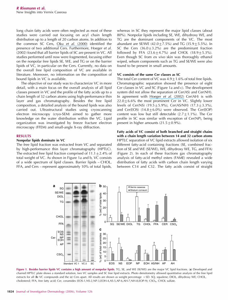
The free lipid fraction was extracted from VC and separatedby high-performance thin layer chromatography (HPTLC).The extracted free lipid fraction comprised of 11.172.4% oftotal weight of VC. As shown in Figure 1a and b, VC consistsof a wide spectrum of lipid classes. Barrier lipids – CHOL,FFA, and Cers – represent approximately 10% of total lipids,
whereas in SC they represent the major lipid classes (about80%). Nonpolar lipids including SE, WE, dihydroxy WE, andTG are the dominant components of the VC. The mostabundant are SE/WE (42.077.5%) and TG (35.975.5%). InSC the Cers (36.073.2%) are the predominant fractionfollowed by FFA (23.674.7%) and CHOL (18.975.3%).Even though SC from ex vivo skin was thoroughly ethanolwiped, sebum components such as TG and SE/WE were alsofound to be present in small amounts.
VC consists of the same Cer classes as SC
The total Cer content of VC was 4.971.6% of total free lipids.Chromatographic separation showed the presence of eightCer classes in VC and SC (Figure 1a and c). The developmentsystem did not allow the separation of Cer(AS) and Cer(NH).In agreement with Hoeger et al. (2002) Cer(AH) is with22.076.6% the most prominent Cer in VC. Slightly lowerlevels of Cer(NS) (19.575.9%), Cer(AS/NH) (17.373.3%),and Cer(EOS) (14.876.0%) were observed. The Cer(EOP)content was low but still detectable (2.771.1%). The Cerprofile in SC was similar with exception of Cer(NP), beingpresent in higher amounts (21.570.9%).
Fatty acids of VC consist of both branched and straight chainswith a chain length variation between 14 and 32 carbon atoms
HPTLC separation of VC lipid extracts allowed isolation of sixdifferent fatty-acid containing fractions (SE, combined frac-tion of SE and WE (SE/WE), WE, dihydroxy WE, TG, and FFA)(Figure 2). In each of these fractions gas chromatographyanalysis of fatty-acid methyl esters (FAME) revealed a widedistribution of fatty acids with carbon chain length varyingbetween C14 and C32. The fatty acids consist of straight
SQ
SQ
SE/WE
SE/WE
DIOL
DIOL
TG
TG
CHOL
CHOLFFA FFA
Cer
CER CSO4
AP
CSO4
EOH
EOH AS/NH AP AHEOP
EOS
EOS
NP
NP
NS
NSStandard VC 1 VC 2 SC
50
40
30
30
25
20
15
10
5
0
20
10
0
% (
wt)
of t
otal
lipi
ds%
(w
t) o
f tot
al c
eram
ides
Vernix caseosaStratum corneum
a b
c
Figure 1. Besides barrier lipids VC contains a high amount of nonpolar lipids. TG, SE, and WE (SE/WE) are the major VC lipid fractions. (a) Developed and
charred HPTLC plate shows a standard solution, two VC samples and SC free lipid extracts. Photo densitometry allowed quantitative analysis of the free lipid
extracts for all (b) VC compounds and the (c) Cers apart. All results are shown as weight percentage þ SD. SQ, squalene; DIOL, dihydroxy WE; CHOL,
cholesterol; FFA, free fatty acid; Cer, ceramides (EOS-1,NS-2,NP-3,EOH-4,AS-5,AP-6,AH-7,NH-8,EOP-9); CSO4, CHOL sulfate.
1824 Journal of Investigative Dermatology (2006), Volume 126
R Rissmann et al.New Insights into Vernix Caseosa
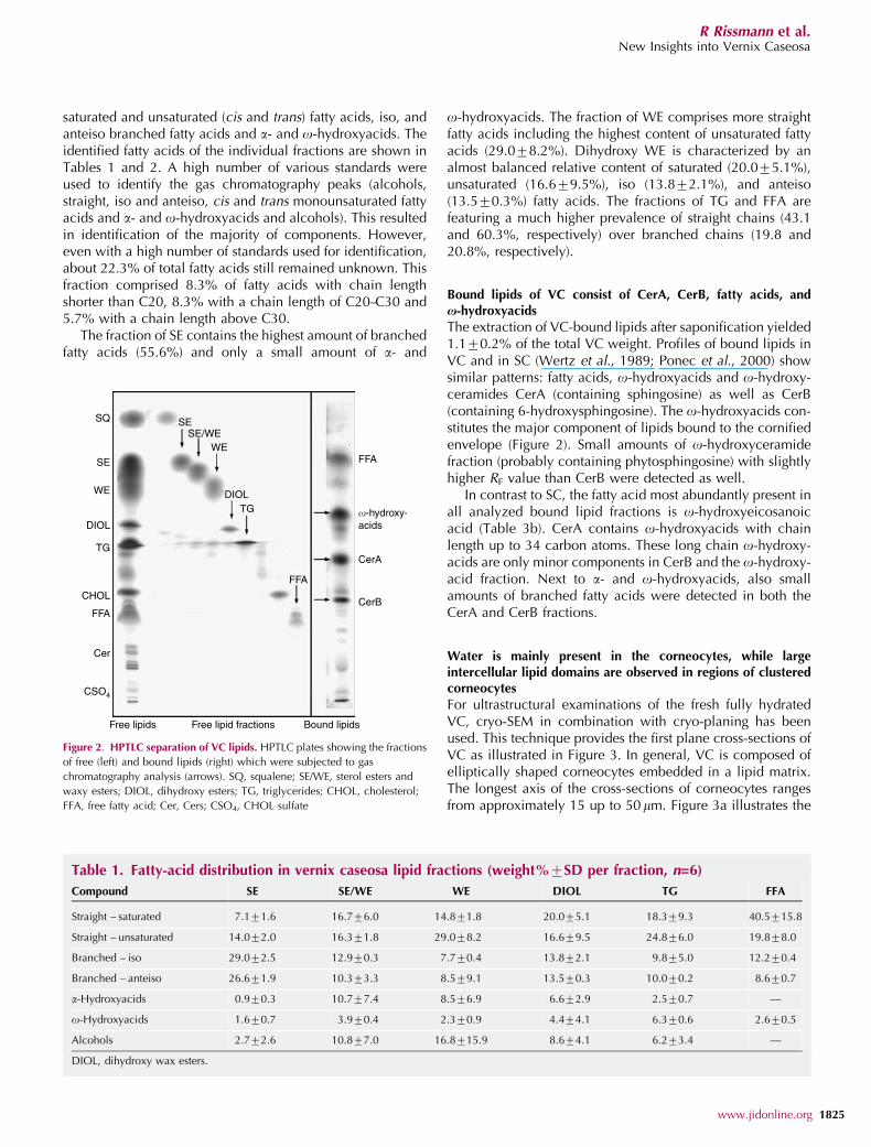
saturated and unsaturated (cis and trans) fatty acids, iso, andanteiso branched fatty acids and a- and o-hydroxyacids. Theidentified fatty acids of the individual fractions are shown inTables 1 and 2. A high number of various standards wereused to identify the gas chromatography peaks (alcohols,straight, iso and anteiso, cis and trans monounsaturated fattyacids and a- and o-hydroxyacids and alcohols). This resultedin identification of the majority of components. However,even with a high number of standards used for identification,about 22.3% of total fatty acids still remained unknown. Thisfraction comprised 8.3% of fatty acids with chain lengthshorter than C20, 8.3% with a chain length of C20–C30 and5.7% with a chain length above C30.
The fraction of SE contains the highest amount of branchedfatty acids (55.6%) and only a small amount of a- and
o-hydroxyacids. The fraction of WE comprises more straightfatty acids including the highest content of unsaturated fattyacids (29.078.2%). Dihydroxy WE is characterized by analmost balanced relative content of saturated (20.075.1%),unsaturated (16.679.5%), iso (13.872.1%), and anteiso(13.570.3%) fatty acids. The fractions of TG and FFA arefeaturing a much higher prevalence of straight chains (43.1and 60.3%, respectively) over branched chains (19.8 and20.8%, respectively).
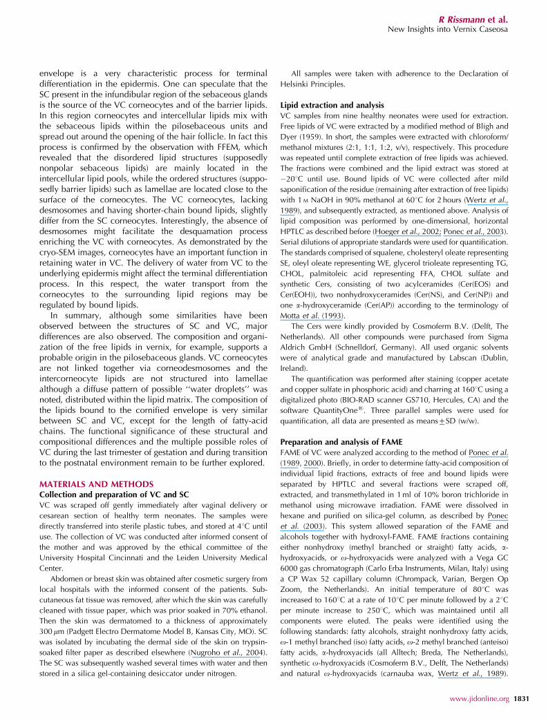
Bound lipids of VC consist of CerA, CerB, fatty acids, andx-hydroxyacids
The extraction of VC-bound lipids after saponification yielded1.170.2% of the total VC weight. Profiles of bound lipids inVC and in SC (Wertz et al., 1989; Ponec et al., 2000) showsimilar patterns: fatty acids, o-hydroxyacids and o-hydroxy-ceramides CerA (containing sphingosine) as well as CerB(containing 6-hydroxysphingosine). The o-hydroxyacids con-stitutes the major component of lipids bound to the cornifiedenvelope (Figure 2). Small amounts of o-hydroxyceramidefraction (probably containing phytosphingosine) with slightlyhigher RF value than CerB were detected as well.
In contrast to SC, the fatty acid most abundantly present inall analyzed bound lipid fractions is o-hydroxyeicosanoicacid (Table 3b). CerA contains o-hydroxyacids with chainlength up to 34 carbon atoms. These long chain o-hydroxy-acids are only minor components in CerB and the o-hydroxy-acid fraction. Next to a- and o-hydroxyacids, also smallamounts of branched fatty acids were detected in both theCerA and CerB fractions.
Water is mainly present in the corneocytes, while largeintercellular lipid domains are observed in regions of clusteredcorneocytes
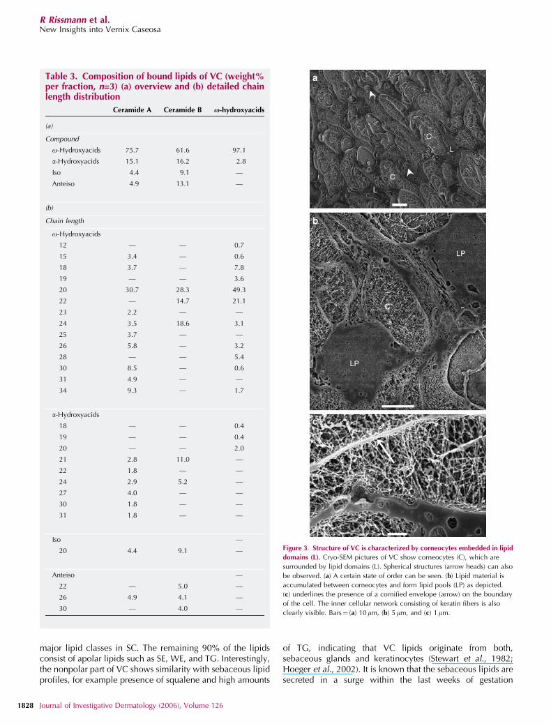
For ultrastructural examinations of the fresh fully hydratedVC, cryo-SEM in combination with cryo-planing has beenused. This technique provides the first plane cross-sections ofVC as illustrated in Figure 3. In general, VC is composed ofelliptically shaped corneocytes embedded in a lipid matrix.The longest axis of the cross-sections of corneocytes rangesfrom approximately 15 up to 50 mm. Figure 3a illustrates the
SQ
SE
SESE/WE
DIOL
DIOL
TG
TG
CHOL
FFA
FFA
FFA
Cer
CerB
CerA
CSO4
Free lipids Free lipid fractions Bound lipids
WE
WE
�-hydroxy-acids
Figure 2. HPTLC separation of VC lipids. HPTLC plates showing the fractions
of free (left) and bound lipids (right) which were subjected to gas
chromatography analysis (arrows). SQ, squalene; SE/WE, sterol esters and
waxy esters; DIOL, dihydroxy esters; TG, triglycerides; CHOL, cholesterol;
FFA, free fatty acid; Cer, Cers; CSO4, CHOL sulfate
Table 1. Fatty-acid distribution in vernix caseosa lipid fractions (weight%7SD per fraction, n=6)
Compound SE SE/WE WE DIOL TG FFA
Straight – saturated 7.171.6 16.776.0 14.871.8 20.075.1 18.379.3 40.5715.8
Straight – unsaturated 14.072.0 16.371.8 29.078.2 16.679.5 24.876.0 19.878.0
Branched – iso 29.072.5 12.970.3 7.770.4 13.872.1 9.875.0 12.270.4
Branched – anteiso 26.671.9 10.373.3 8.579.1 13.570.3 10.070.2 8.670.7
a-Hydroxyacids 0.970.3 10.777.4 8.576.9 6.672.9 2.570.7 —
o-Hydroxyacids 1.670.7 3.970.4 2.370.9 4.474.1 6.370.6 2.670.5
Alcohols 2.772.6 10.877.0 16.8715.9 8.674.1 6.273.4 —
DIOL, dihydroxy wax esters.
www.jidonline.org 1825
R Rissmann et al.New Insights into Vernix Caseosa
Table 2. Chain length distribution in various free lipid fractions (weight%7SD per fraction, n=6) (a) straight chainfatty acids and (b) branched chain fatty acids
Chain length SE SE/WE WE DIOL TG FFA
(a)
Saturated
14 — 0.470.5 0.170.2 0.971.2 0.970.8 —
15 0.270.2 0.870.6 0.170.1 1.271.7 0.370.3 6.272.7
16 0.870.8 1.271.7 3.674.0 6.579.1 10.877.4 24.6712.7
18 1.170.1 0.470.6 0.670.8 1.870.5 2.070.6 4.270.9
19 0.470.0 — 4.476.2 3.774.1 0.570.1 0.970.2
20 0.470.6 0.370.3 0.770.2 1.070.6 0.570.0 0.770.1
21 — 0.170.1 0.170.1 0.470.2 0.470.0 0.370.0
22 — — 3.471.9 — — —
23 — 0.270.3 — 2.070.9 0.270.2 1.370.1
24 0.871.1 0.370.5 0.170.2 1.070.1 0.470.0 —
25 2.872.8 4.672.1 1.171.5 1.370.7 0.470.1 1.770.0
26 0.170.1 0.370.5 0.270.3 0.170.1 0.170.0 —
27 0.670.7 0.971.2 0.470.3 — 1.370.1 0.670.1
32 — 7.3710.3 — — — —
Unsaturated
16:1t 0.270.3 0.370.4 0.670.8 2.673.6 10.974.8 8.571.6
16:1c 0.370.0 0.570.5 16.570.6 3.671.1 3.173.2 0.670.8
18:1t 0.470.4 6.272.5 0.370.4 3.773.7 4.474.5 5.773.2
18:1c 0.870.0 1.870.5 3.270.7 4.373.9 4.272.8 2.771.1
18:2t 0.570.6 0.170.2 0.270.3 0.870.0 —
18:2c — 0.170.1 6.377.0 1.070.9 0.370.3 1.170.8
20:1c 1.970.3 2.170.6 0.970.8 0.770.5 0.270.0 0.370.1
22:1t 2.673.7 2.670.5 0.270.3 — 0.370.1 0.370.2
22:1c 5.171.6 0.670.8 0.270.3 0.770.5 0.470.0 —
24:1t — 0.570.7 0.470.6 — —
24:1c 2.173.0 1.470.1 — — 0.370.4 0.670.2
(b)
Iso
14 0.470.6 0.570.8 0.370.4 1.070.6 0.170.1 —
16 3.070.0 3.973.3 3.271.7 2.874.0 3.773.4 3.271.9
18 1.071.5 0.670.1 1.570.3 1.571.0 1.870.2 2.071.7
20 13.070.8 0.871.0 — 4.070.4 0.370.0 0.370.2
22 — — — — 0.370.0 —
23 2.974.0 — — — — —
24 0.870.7 0.370.2 2.671.6 1.370.8 0.570.3 5.670.4
25 2.773.8 1.370.2 — 1.270.2 0.570.6 —
26 2.473.1 1.572.0 — — — —
27 0.570.8 1.170.9 — — 0.270.2 1.070.3
28 1.972.6 1.271.7 0.170.1 — — —
29 0.370.5 1.472.1 — 0.170.2 — 0.270.1
30 — 0.170.2 0.170.0 1.770.8 2.570.2 —
Anteiso
15 0.270.3 1.972.3 0.770.5 1.471.5 0.170.1 1.470.1
16 — 0.370.4 1.271.6 — 1.770.2 0.770.4
Table 2 Continued on the following page
1826 Journal of Investigative Dermatology (2006), Volume 126
R Rissmann et al.New Insights into Vernix Caseosa
preference of a certain degree of orientation of thecorneocytes but the high state of ordering as observed inSC is absent. As depicted in Figure 3b and c, the interior ofthe corneocytes is characterized by a network of white fibers,which represents the keratin filament of the cells. In betweenthe keratin, dark regions are observed. These dark regionsrepresent the water domains. In most cases the corneocytesare located closely to each other. However, Figure 3a alsoshows areas with a very smooth appearance located betweenthe cells. These domains correspond to the intercellular lipidregions (L) and are frequently surrounded by severalcorneocytes. In Figure 3b a higher magnification of the lipiddomains is shown and the accumulation of lipids to a lipidpool can be observed (LP). Very interestingly, in the lipiddomains small round structures are observed (Figure 3b). Asthese round structures are very dark in appearance, they mostprobably represent small water pools within the lipiddomains. The cross-section of these structures ranges from50 nm up to several mm.
A higher magnification is shown in Figure 3c. In thisimage, the cornified envelope is recognized by a bright,white line surrounding the cell (arrow). Besides the corneo-cytes also a domain of lipids is observed with the roundstructures. The dark appearances of these circular structurescontain small white dots and fibers. Although the origin is notfully understood, they might represent salt or proteins.
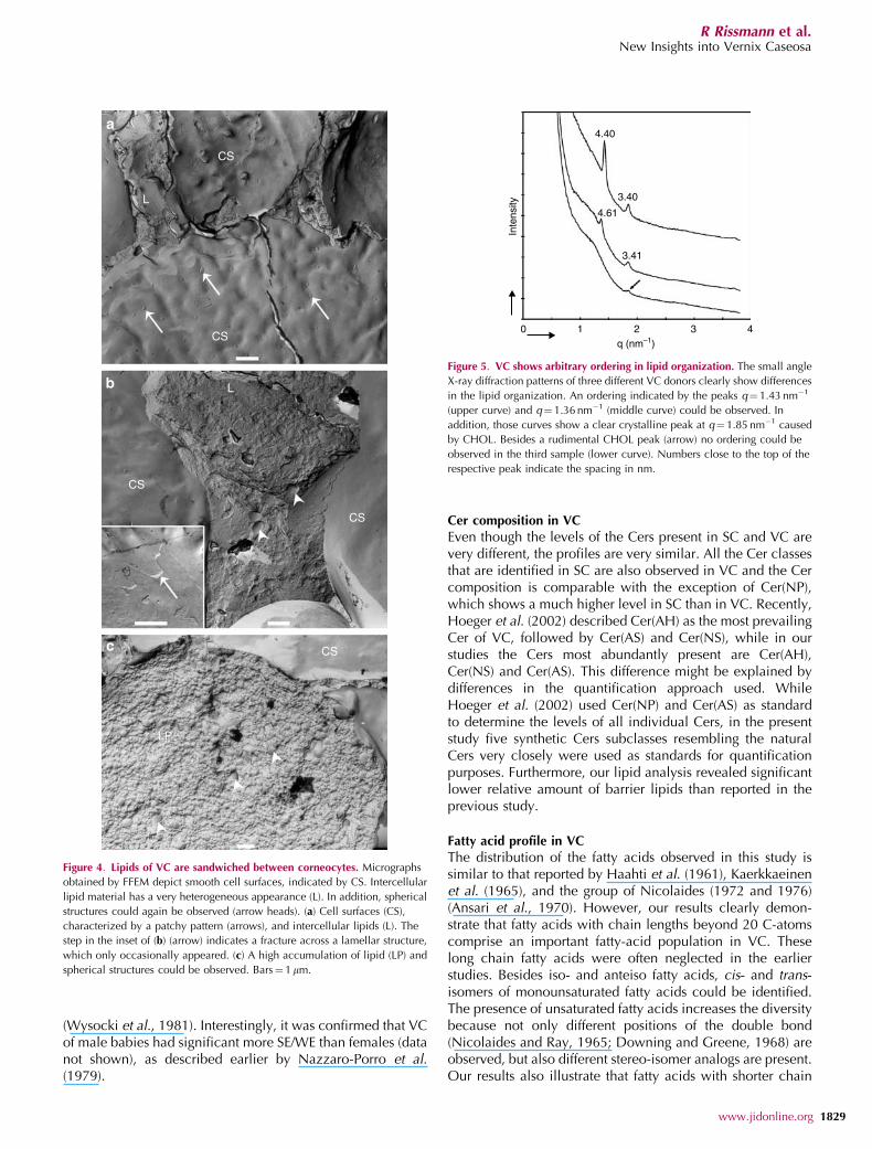
Lipids in between VC corneocytes show a heterogeneousappearance and form only occasionally lipid lamellae
A more detailed analysis of the lipid domains can beconducted with FFEM. The images depict mainly surfacesof corneocytes that are characterized by very smooth fractureplanes (Figure 4). This is a consequence of the fracturetechnique, which takes always the route of least resistance. InFigure 4a a cluster of five corneocytes, separated by sharpboundaries, is shown. In addition, several intercellularlipid domains are visualized. The smooth surfaces of thecorneocytes most probably represent the bound lipids.These smooth surfaces very often show a patchy pattern(see arrows Figure 4a). The lipid regions are characterized bya rough appearance, indicating the absence of a long rangeordering of the lipids (Figure 4b). Only occasionally sharp
steps could be observed suggesting the presence of lipidlamellae (inset in Figure 4b). In the large lipid domains(Figure 4c), very frequently spherical structures (arrowheads)are observed. No ordered distribution of the sphericalstructures in space was noticed, suggesting the absence oflong-range ordering.
No long-range ordering of the lipids in VC
The diffraction profiles of the VC obtained from differentdonors differ slightly (Figure 5). One out of the threediffraction profiles does not reveal any sharp peaks, whiletwo diffraction profiles exhibit one sharp peak at approxi-mately q¼ 1.43 nm�1 and q¼1.36 nm�1, respectively. How-ever, as no higher-order reflections were observed indicatingthat no well-defined long-range ordering, such as the occur-rence of lamellae stacks, is present in these samples. In thethree diffraction curves a diffraction peak at q¼ 1.85 nm�1
could be identified, which is attributed to crystallineCHOL.
DISCUSSIONIt has been suggested that VC protects the fetus of unwantedinfluences from its environment and might be an importantplayer in promoting the maturation of the SC in utero. As thelipid composition and ultrastructure of VC might be crucialfor its biological function, information on these properties isof great interest.
Although several groups examined the lipid compositionof VC, no information is available on the integral lipidcomposition of VC. Furthermore, in analyzing the fatty-acidchain length, often fatty-acid chains up to a chain length ofonly 20 C-atoms have been reported to be present insubstantial amounts, while it is expected that longer fatty-acid chains are also present in VC. In addition, noinformation is available on the composition of the boundlipids and only limited information is available on theultrastructure of VC. All these aspects have been addressedin this study and will be discussed below.
Free lipid composition in VC
The present study reveals that only 10% of the VC lipidsconsists of the barrier lipids CHOL, FFA, and Cers, being the
Table 2. continued
Chain length SE SE/WE WE DIOL TG FFA
17 0.871.1 1.470.7 1.371.5 0.771.0 0.270.2 1.270.2
18 1.071.5 — 3.774.9 1.071.0 0.570.0 0.770.1
21 7.570.5 0.970.8 — 0.670.3 0.270.1 0.470.1
22 12.470.2 1.371.8 0.170.1 4.670.3 0.570.2 —
23 — 1.370.4 — 0.370.4 — 0.870.2
24 3.474.8 1.171.1 0.570.7 0.370.2 — —
26 0.670.1 0.470.6 0.971.2 4.772.0 4.870.3 3.370.2
27 0.870.7 1.672.4 0.170.2 — 1.870.1 —
DIOL, dihydroxy wax esters; c, cis; t, trans.
www.jidonline.org 1827
R Rissmann et al.New Insights into Vernix Caseosa
major lipid classes in SC. The remaining 90% of the lipidsconsist of apolar lipids such as SE, WE, and TG. Interestingly,the nonpolar part of VC shows similarity with sebaceous lipidprofiles, for example presence of squalene and high amounts
of TG, indicating that VC lipids originate from both,sebaceous glands and keratinocytes (Stewart et al., 1982;Hoeger et al., 2002). It is known that the sebaceous lipids aresecreted in a surge within the last weeks of gestation
Table 3. Composition of bound lipids of VC (weight%per fraction, n=3) (a) overview and (b) detailed chainlength distribution
Ceramide A Ceramide B x-hydroxyacids
(a)
Compound
o-Hydroxyacids 75.7 61.6 97.1
a-Hydroxyacids 15.1 16.2 2.8
Iso 4.4 9.1 —
Anteiso 4.9 13.1 —
(b)
Chain length
o-Hydroxyacids
12 — — 0.7
15 3.4 — 0.6
18 3.7 — 7.8
19 — — 3.6
20 30.7 28.3 49.3
22 — 14.7 21.1
23 2.2 — —
24 3.5 18.6 3.1
25 3.7 — —
26 5.8 — 3.2
28 — — 5.4
30 8.5 — 0.6
31 4.9 — —
34 9.3 — 1.7
a-Hydroxyacids
18 — — 0.4
19 — — 0.4
20 — — 2.0
21 2.8 11.0 —
22 1.8 — —
24 2.9 5.2 —
27 4.0 — —
30 1.8 — —
31 1.8 — —
Iso —
20 4.4 9.1 —
Anteiso —
22 — 5.0 —
26 4.9 4.1 —
30 — 4.0 —
C
C
C
C
L
L
LP
LP
a
b
c
Figure 3. Structure of VC is characterized by corneocytes embedded in lipid
domains (L). Cryo-SEM pictures of VC show corneocytes (C), which are
surrounded by lipid domains (L). Spherical structures (arrow heads) can also
be observed. (a) A certain state of order can be seen. (b) Lipid material is
accumulated between corneocytes and form lipid pools (LP) as depicted.
(c) underlines the presence of a cornified envelope (arrow) on the boundary
of the cell. The inner cellular network consisting of keratin fibers is also
clearly visible. Bars¼ (a) 10 mm, (b) 5mm, and (c) 1 mm.
1828 Journal of Investigative Dermatology (2006), Volume 126
R Rissmann et al.New Insights into Vernix Caseosa
(Wysocki et al., 1981). Interestingly, it was confirmed that VCof male babies had significant more SE/WE than females (datanot shown), as described earlier by Nazzaro-Porro et al.(1979).
Cer composition in VC
Even though the levels of the Cers present in SC and VC arevery different, the profiles are very similar. All the Cer classesthat are identified in SC are also observed in VC and the Cercomposition is comparable with the exception of Cer(NP),which shows a much higher level in SC than in VC. Recently,Hoeger et al. (2002) described Cer(AH) as the most prevailingCer of VC, followed by Cer(AS) and Cer(NS), while in ourstudies the Cers most abundantly present are Cer(AH),Cer(NS) and Cer(AS). This difference might be explained bydifferences in the quantification approach used. WhileHoeger et al. (2002) used Cer(NP) and Cer(AS) as standardto determine the levels of all individual Cers, in the presentstudy five synthetic Cers subclasses resembling the naturalCers very closely were used as standards for quantificationpurposes. Furthermore, our lipid analysis revealed significantlower relative amount of barrier lipids than reported in theprevious study.
Fatty acid profile in VC
The distribution of the fatty acids observed in this study issimilar to that reported by Haahti et al. (1961), Kaerkkaeinenet al. (1965), and the group of Nicolaides (1972 and 1976)(Ansari et al., 1970). However, our results clearly demon-strate that fatty acids with chain lengths beyond 20 C-atomscomprise an important fatty-acid population in VC. Theselong chain fatty acids were often neglected in the earlierstudies. Besides iso- and anteiso fatty acids, cis- and trans-isomers of monounsaturated fatty acids could be identified.The presence of unsaturated fatty acids increases the diversitybecause not only different positions of the double bond(Nicolaides and Ray, 1965; Downing and Greene, 1968) areobserved, but also different stereo-isomer analogs are present.Our results also illustrate that fatty acids with shorter chain
a
b
c
LP
CS
CS
CS
CS
CS
L
L
Figure 4. Lipids of VC are sandwiched between corneocytes. Micrographs
obtained by FFEM depict smooth cell surfaces, indicated by CS. Intercellular
lipid material has a very heterogeneous appearance (L). In addition, spherical
structures could again be observed (arrow heads). (a) Cell surfaces (CS),
characterized by a patchy pattern (arrows), and intercellular lipids (L). The
step in the inset of (b) (arrow) indicates a fracture across a lamellar structure,
which only occasionally appeared. (c) A high accumulation of lipid (LP) and
spherical structures could be observed. Bars¼ 1mm.
4.40
3.40
4.61
3.41
Inte
nsity
0 1 2 3 4q (nm–1)
Figure 5. VC shows arbitrary ordering in lipid organization. The small angle
X-ray diffraction patterns of three different VC donors clearly show differences
in the lipid organization. An ordering indicated by the peaks q¼ 1.43 nm�1
(upper curve) and q¼ 1.36 nm�1 (middle curve) could be observed. In
addition, those curves show a clear crystalline peak at q¼1.85 nm�1 caused
by CHOL. Besides a rudimental CHOL peak (arrow) no ordering could be
observed in the third sample (lower curve). Numbers close to the top of the
respective peak indicate the spacing in nm.
www.jidonline.org 1829
R Rissmann et al.New Insights into Vernix Caseosa
length (C16–C18) are prevailing in TG and WE fractions,while very long chain fatty acids (4C20) are the major fattyacids of the SE fraction. Moreover, we observed, that thebranched/straight ratio of fatty acids is decreasing with lipidpolarity, being the highest in the least polar SE and decreasingin more polar components, such as TG and FFA. Only thefraction of dihydroxy WE does not fully follow this trend.When comparing our results with those of Tollin et al. (2005),in our VC samples main FFA fractions are C16:0, C16:1t,C15:0, and C18:1t, while the most abundant ones in their VCsamples are C16:0, C24:0, C18:2, and C18:1. Furthermore,we did not observe polyunsaturated fractions of FFA, whilethey did not notice branched fatty acids.
Bound lipids
As VC consists of the hydrated hydrophilic corneocytessurrounded by lipophilic intercellular lipids, the interface ofboth domains has an important mediating function. In SC thisinterface is formed by lipids covalently bound to the cornifiedenvelope (corneocyte-bound lipid envelope, Swartzendruberet al., 1987). Our results demonstrate for the first time that inVC – like in SC – bound lipids are present that are covalentlylinked to the cornified envelope. The bound lipids mainlyconsist of four lipid classes, namely o-hydroxyacids, fattyacids, and the o-hydoxyceramides, CerA and CerB, similarlyas observed in SC (Swartzendruber et al., 1987). However,the chain lengths of the bound lipids in VC are different fromthose in SC. Striking is the high level of o-hydroxyeicosanoicacid (Table 3(b)) in these lipids, while the o-hydroxyacid with30 C-atoms, abundantly present in SC (Wertz et al., 1989), isalmost absent in VC. Noticeable is also the presence ofbranched subtypes of fatty acids in CerA and CerB. Oku et al.(2000) described also the presence of branched fatty acids inVC’s free Cers.
Ultrastructure of VC
Cryo-SEM in combination with slicing and planing providesvery flat surfaces across hydrophilic as well as lipophilicdomains and thus provides structural information about bothdomains. FFEM reveals fracture planes of the VC samples, inwhich the fracture planes run through the regions of leastresistance, which are mainly the lipid domains.
Additionally to the study of Pickens et al. (2000), the cryo-SEM images also depict the internal structure of thecorneocytes and reveal that the keratin filaments constitutethe scaffold and create a body to store water within thecorneocytes. Furthermore, the corneocytes show a certaindegree of orientation in the VC samples. In the extracellularregions lipids are present similarly as in SC. The lipiddomains are characterized by smooth regions in the cryo-SEM images with many small round structures, see Figure 3band c. These structures were also described by Pickens et al.(2000). The origin and function of the round structures remainunclear. A former study showed the presence of antimicrobialpeptides in such inclusion bodies (Akinbi et al., 2004) buttheir larger size might be explained by coalescence inducedby their technique. However, we propose that the innercontent most probably consist of water, which is identified by
cryo-SEM as small dark holes. This also explains the roundshape structure as a high surface tension, normally observedbetween the lipophilic domains and the hydrophilic water,will minimize the interfacial surface creating the round-shaped structures. At higher magnification (Figure 3c) it isclearly observed that besides water, other material is presentin these little water pools. This might be proteins, peptides orsalt crystals which biological functions still remain unclear.
Cryo-SEM and FFEM reveal the presence of large lipidpools (Figures 3b and 4b) in regions of clustered corneocytes.Such large regions of lipids have not been observed in SC andmight partially be due to the absence of corneodesmosomesin VC allowing separation of the corneocytes. In the lipidregions many small spherical particles are observed withFFEM (Figure 4b and c). Most probably these represent theround-shaped structures identified with cryo-SEM, but nowfractured along the outer surface of the particles, whichseems to be of lipid nature. In addition, the lipid domainsshow a very heterogeneous and rough appearance (Figure 4b)indicating that lipids are not highly organized. Only on thecell surfaces (inset Figure 4b), lipid lamellae could beobserved occasionally. This is different from the lipids inSC, which are organized in very well-defined stacks of lipidlamellae (Madison et al., 1987). The results of the small-angleX-Ray diffraction studies corroborate these results. At thediffraction profiles, besides the peak attributed to crystallineCHOL, only two out of the three diffraction patterns revealone additional diffraction peak indicating the absence oflong-range ordering.
The difference in organization in VC and SC can beexplained by the difference in lipid composition at least intwo ways. (I) Barrier lipids, especially Cers with long fatty-acid chains play an important role in the characteristicorganization in SC (Bouwstra et al., 1998). Most probably, thelower level of this lipid class reduces its contribution to thefinal lipid organization. (II) In SC the majority of the lipidshave straight unsaturated fatty-acid chains, while in VC themajority of the fatty-acid chains are either unsaturated orbranched. This most probably will have important conse-quences for the lipid organization. However, in order to drawany final conclusions about the specific role of the reducedCer level and the increased levels of branched andunsaturated fatty acids more studies are required to addressthis issue. Those could also include studies of gestational-period-dependent changes in VC lipid composition asdevelopmental differences in VC may play an important rolefor the properties.
What have we learned from these studies about the formation,origin, and function of VC?
As far as the lipid composition is concerned, only a lowcontent of barrier lipids is present in VC. This might suggestthat the primary function of VC is not only to limit thediffusion of substances across VC, but also to protect theinitially formed SC of the fetus from direct contact with theamniotic fluid. This might also facilitate the formation of amature SC in utero. The synthesis of barrier lipids andchemical linkage of the bound lipids to the cornified
1830 Journal of Investigative Dermatology (2006), Volume 126
R Rissmann et al.New Insights into Vernix Caseosa
envelope is a very characteristic process for terminaldifferentiation in the epidermis. One can speculate that theSC present in the infundibular region of the sebaceous glandsis the source of the VC corneocytes and of the barrier lipids.In this region corneocytes and intercellular lipids mix withthe sebaceous lipids within the pilosebaceous units andspread out around the opening of the hair follicle. In fact thisprocess is confirmed by the observation with FFEM, whichrevealed that the disordered lipid structures (supposedlynonpolar sebaceous lipids) are mainly located in theintercellular lipid pools, while the ordered structures (suppo-sedly barrier lipids) such as lamellae are located close to thesurface of the corneocytes. The VC corneocytes, lackingdesmosomes and having shorter-chain bound lipids, slightlydiffer from the SC corneocytes. Interestingly, the absence ofdesmosomes might facilitate the desquamation processenriching the VC with corneocytes. As demonstrated by thecryo-SEM images, corneocytes have an important function inretaining water in VC. The delivery of water from VC to theunderlying epidermis might affect the terminal differentiationprocess. In this respect, the water transport from thecorneocytes to the surrounding lipid regions may beregulated by bound lipids.
In summary, although some similarities have beenobserved between the structures of SC and VC, majordifferences are also observed. The composition and organi-zation of the free lipids in vernix, for example, supports aprobable origin in the pilosebaceous glands. VC corneocytesare not linked together via corneodesmosomes and theintercorneocyte lipids are not structured into lamellaealthough a diffuse pattern of possible ‘‘water droplets’’ wasnoted, distributed within the lipid matrix. The composition ofthe lipids bound to the cornified envelope is very similarbetween SC and VC, except for the length of fatty-acidchains. The functional significance of these structural andcompositional differences and the multiple possible roles ofVC during the last trimester of gestation and during transitionto the postnatal environment remain to be further explored.
MATERIALS AND METHODSCollection and preparation of VC and SC
VC was scraped off gently immediately after vaginal delivery or
cesarean section of healthy term neonates. The samples were
directly transferred into sterile plastic tubes, and stored at 41C until
use. The collection of VC was conducted after informed consent of
the mother and was approved by the ethical committee of the
University Hospital Cincinnati and the Leiden University Medical
Center.
Abdomen or breast skin was obtained after cosmetic surgery from
local hospitals with the informed consent of the patients. Sub-
cutaneous fat tissue was removed, after which the skin was carefully
cleaned with tissue paper, which was prior soaked in 70% ethanol.
Then the skin was dermatomed to a thickness of approximately
300mm (Padgett Electro Dermatome Model B, Kansas City, MO). SC
was isolated by incubating the dermal side of the skin on trypsin-
soaked filter paper as described elsewhere (Nugroho et al., 2004).
The SC was subsequently washed several times with water and then
stored in a silica gel-containing desiccator under nitrogen.
All samples were taken with adherence to the Declaration of
Helsinki Principles.
Lipid extraction and analysis
VC samples from nine healthy neonates were used for extraction.
Free lipids of VC were extracted by a modified method of Bligh and
Dyer (1959). In short, the samples were extracted with chloroform/
methanol mixtures (2:1, 1:1, 1:2, v/v), respectively. This procedure
was repeated until complete extraction of free lipids was achieved.
The fractions were combined and the lipid extract was stored at
�201C until use. Bound lipids of VC were collected after mild
saponification of the residue (remaining after extraction of free lipids)
with 1 M NaOH in 90% methanol at 601C for 2 hours (Wertz et al.,
1989), and subsequently extracted, as mentioned above. Analysis of
lipid composition was performed by one-dimensional, horizontal
HPTLC as described before (Hoeger et al., 2002; Ponec et al., 2003).
Serial dilutions of appropriate standards were used for quantification.
The standards comprised of squalene, cholesteryl oleate representing
SE, oleyl oleate representing WE, glycerol trioleate representing TG,
CHOL, palmitoleic acid representing FFA, CHOL sulfate and
synthetic Cers, consisting of two acylceramides (Cer(EOS) and
Cer(EOH)), two nonhydroxyceramides (Cer(NS), and Cer(NP)) and
one a-hydroxyceramide (Cer(AP)) according to the terminology of
Motta et al. (1993).
The Cers were kindly provided by Cosmoferm B.V. (Delft, The
Netherlands). All other compounds were purchased from Sigma
Aldrich GmbH (Schnelldorf, Germany). All used organic solvents
were of analytical grade and manufactured by Labscan (Dublin,
Ireland).
The quantification was performed after staining (copper acetate
and copper sulfate in phosphoric acid) and charring at 1601C using a
digitalized photo (BIO-RAD scanner GS710, Hercules, CA) and the
software QuantityOnes. Three parallel samples were used for
quantification, all data are presented as means7SD (w/w).
Preparation and analysis of FAME
FAME of VC were analyzed according to the method of Ponec et al.
(1989, 2000). Briefly, in order to determine fatty-acid composition of
individual lipid fractions, extracts of free and bound lipids were
separated by HPTLC and several fractions were scraped off,
extracted, and transmethylated in 1 ml of 10% boron trichloride in
methanol using microwave irradiation. FAME were dissolved in
hexane and purified on silica-gel column, as described by Ponec
et al. (2003). This system allowed separation of the FAME and
alcohols together with hydroxyl-FAME. FAME fractions containing
either nonhydroxy (methyl branched or straight) fatty acids, a-
hydroxyacids, or o-hydroxyacids were analyzed with a Vega GC
6000 gas chromatograph (Carlo Erba Instruments, Milan, Italy) using
a CP Wax 52 capillary column (Chrompack, Varian, Bergen Op
Zoom, the Netherlands). An initial temperature of 801C was
increased to 1601C at a rate of 101C per minute followed by a 21C
per minute increase to 2501C, which was maintained until all
components were eluted. The peaks were identified using the
following standards: fatty alcohols, straight nonhydroxy fatty acids,
o-1 methyl branched (iso) fatty acids, o-2 methyl branched (anteiso)
fatty acids, a-hydroxyacids (all Alltech; Breda, The Netherlands),
synthetic o-hydroxyacids (Cosmoferm B.V., Delft, The Netherlands)
and natural o-hydroxyacids (carnauba wax, Wertz et al., 1989).
www.jidonline.org 1831
R Rissmann et al.New Insights into Vernix Caseosa
Integration of peak areas and calculation of weight percentages was
performed by a Baseline 810 integrator. Heptadecanoic acid was
used as the internal standard. To correct for background, lipid-free
samples underwent the same separation and transmethylation
procedure (HPTLC development, off-scraping, extraction from silica
gel, and final column separation) and appropriate peak areas were
subtracted in analyzed lipid samples. To refine the identification of
individual components, samples and standards were also injected
simultaneously to confirm the data obtained with the external
standards. Alcohols and o-hydroxyacids of the hydroxy FAME were
converted by acetylation with acetanhydride to their analogical ester.
Owing to acetylation, a shift in the retention time occurred. This
allowed confirmation of results obtained with external standards.
Cryo-SEM
A small amount of VC (B2 mg) was placed in a small cylindrical
sample holder and further processed, as described in detail elsewhere
(Bouwstra et al., 2003). Briefly, the samples were rapidly frozen at
�1801C in liquid propane using the plunging method (KF80, Reichert-
Jung, Vienna, Austria). The cryo-fixed samples were sliced at a
specimen temperature of �901C and the samples were freeze dried for
1 minute at �901C at 0.1 Pa and sputter coated with 5 nm platinum.
The samples were mounted into a field emission electron microscope
(6300 FESEM, Yeol, Tokyo, Japan) and examined at �1901C.
FFEMVC samples were fixed between two copper plates and immediately
cryo-fixed in liquid propane. The frozen specimens were stored in
liquid nitrogen until further use. The fracturing of the samples was
performed by a slightly modified method as described elsewhere
(Honeywell-Nguyen et al., 2003). Briefly, the samples were mounted
onto a special designed sample holder and then placed on a
precooled table (�1501C) in a Balzers BAF 400-D freeze fracture
device (Balzers, Lichtenstein). The samples were fractured and
replicated by evaporation of platinum and carbon. The replicas were
removed from the freeze fracture device and subsequently cleaned
by submersion into 0.5 M quaternary ammonium hydroxide in
toluene (SolueneTM-350, Packard, Groningen, The Netherlands)
followed by washing with toluene. The replicas were visualized in
a Philips EM410 (Philips, Eindhoven, The Netherlands) transmission
electron microscopy system.
Small-angle X-ray diffraction
The small-angle X-Ray diffraction measurements were conducted at
station BM26B at the European Synchrotron Radiation Facility in
Grenoble, France (Bras, 1998). The measurements were carried out
as described in detail by de Jager et al. (2004). The diffraction data
were collected by a two-dimensional gas-filled area detector. The
diffraction pattern was obtained at room temperature for a collection
period of 5 minutes. Duplicate measurements of VC samples were
performed to confirm reproducibility.
CONFLICT OF INTERESTThe authors state no conflict of interest.
ACKNOWLEDGMENTSThis work was financed by a grant from the Dutch Technology FoundationSTW (LGN6117). The Netherlands Organization for Scientific Research
(NWO) is acknowledged for providing beamtime at the ESRF in Grenoble. Weare also grateful to Igor Dolbnya and Wim Bras for the technical assistance atthe ESRF. The company Cosmoferm provided the synthetic ceramides whichwas highly appreciated.
REFERENCES
Agorastos T, Hollweg G, Grussendorf EI, Papaloucas A (1988) Features ofvernix caseosa cells. Am J Perinatol 5:253–9
Akinbi HT, Narendran V, Pass AK, Markart P, Hoath SB (2004) Host defenseproteins in vernix caseosa and amniotic fluid. Am J Obstet Gynecol191:2090–6
Ansari MN, Fu HC, Nicolaides N (1970) Fatty acids of the alkane diol diestersof vernix caseosa. Lipids 5:279–82
Bligh EG, Dyer WJ (1959) A rapid method of total lipid extraction andpurification. Can J Biochem Physiol 37:911–7
Bouwstra JA, de Graaff A, Gooris GS, Nijsse J, Wiechers JW, van Aelst AC(2003) Water distribution and related morphology in human stratumcorneum at different hydration levels. J Invest Dermatol 120:750–8
Bouwstra JA, Gooris GS, Cheng K, Weerheim A, Bras W, Ponec M (1996)Phase behavior of isolated skin lipids. J Lipid Res 37:999–1011
Bouwstra JA, Gooris GS, Dubbelaar FE, Weerheim AM, Ijzerman AP, PonecM (1998) Role of ceramide 1 in the molecular organization of the stratumcorneum lipids. J Lipid Res 39:186–96
Bouwstra JA, Gooris GS, van der Spek JA, Bras W (1991) Structuralinvestigations of human stratum corneum by small-angle X-ray scatter-ing. J Invest Dermatol 97:1005–12
Bras W (1998) A SAXS/WAXS beamline at the ESRF and future experiments.J Macromol Sci Phys B 37:557–66
de Jager M, Gooris G, Ponec M, Bouwstra J (2004) Acylceramide head grouparchitecture affects lipid organization in synthetic ceramide mixtures.J Invest Dermatol 123:911–6
Downing DT, Greene RS (1968) Double bond positions in the unsaturatedfatty acids of vernix caseosa. J Invest Dermatol 50:380–6
Fu HC, Nicolaides N (1969) The structure of alkane diols of diesters in vernixcaseosa lipids. Lipids 4:170–5
Haahti E, Nikkari T, Salmi AM, Laaksonen AL (1961) Fatty acids of vernixcaseosa. Scand J Clin Lab Invest 13:70–3
Hardman MJ, Moore L, Ferguson MW, Byrne C (1999) Barrier formation in thehuman fetus is patterned. J Invest Dermatol 113:1106–13
Haubrich KA (2003) Role of Vernix caseosa in the neonate: potentialapplication in the adult population. AACN Clin Issues 14:457–64
Hoeger PH, Schreiner V, Klaassen IA, Enzmann CC, Friedrichs K, Bleck O(2002) Epidermal barrier lipids in human vernix caseosa: correspondingceramide pattern in vernix and fetal skin. Br J Dermatol 146:194–201
Honeywell-Nguyen PL, Wouter Groenink HW, de Graaff AM, Bouwstra JA(2003) The in vivo transport of elastic vesicles into human skin: effects ofocclusion, volume and duration of application. J Control Rel 90:243–55
Kaerkkaeinen J, Nikkari T, Ruponen S, Haahti E (1965) Lipids of vernixcaseosa. J Invest Dermatol 44:333–8
Madison KC, Swartzendruber DC, Wertz PW, Downing DT (1987) Presenceof intact intercellular lipid lamellae in the upper layers of the stratumcorneum. J Invest Dermatol 88:714–8
Motta S, Monti M, Sesana S, Caputo R, Carelli S, Ghidoni R (1993) Ceramidecomposition of psoriatic scale. Biochim Biophysic Acta 1182:147–51
Nazzaro-Porro M, Passi S, Boniforti L, Belsito F (1979) Effects of aging on fattyacids in skin surface lipids. J Invest Dermatol 73:112–7
Nicolaides N, Apon JM (1976) Further studies of the saturated methylbranched fatty acids of vernix caseosa lipid. Lipids 11:781–90
Nicolaides N, Fu HC, Ansari MN, Rice GR (1972) The fatty acids of wax estersand sterol esters from vernix caseosa and from human skin surface lipid.Lipids 7:506–17
Nicolaides N, Ray T (1965) Skin lipids. 3. Fatty chains in skin lipids. the use ofvernix caseosa to differentiate between endogenous and exogenouscomponents in human skin surface lipid. J Am Oil Chem Soc 42:702–7
1832 Journal of Investigative Dermatology (2006), Volume 126
R Rissmann et al.New Insights into Vernix Caseosa
Nugroho AK, Li G, Grossklaus A, Danhof M, Bouwstra JA (2004) Transdermaliontophoresis of rotigotine: influence of concentration, temperature andcurrent density in human skin in vitro. J Control Rel 96:159–67
Oku H, Mimura K, Tokitsu Y, Onaga K, Iwasaki H, Chinen I (2000) Biaseddistribution of the branched-chain fatty acids in ceramides of vernixcaseosa. Lipids 35:373–81
Pickens WL, Warner RR, Boissy YL, Boissy RE, Hoath SB (2000)Characterization of vernix caseosa: water content, morphology, andelemental analysis. J Invest Dermatol 115:875–81
Ponec M, Boelsma E, Weerheim A (2000) Covalently bound lipids inreconstructed human epithelia. Acta Derm Venereol 80:89–93
Ponec M, Weerheim A, Kempenaar J, Elias PM, Williams ML (1989)Differentiation of cultured human keratinocytes: effect of cultureconditions on lipid composition of normal vs. malignant cells. In VitroCell Dev Biol 25:689–96
Ponec M, Weerheim A, Lankhorst P, Wertz P (2003) New acylceramide innative and reconstructed epidermis. J Invest Dermatol 120:581–8
Stewart ME, Quinn MA, Downing DT (1982) Variability in the fatty acidcomposition of wax esters from vernix caseosa and its possible relation tosebaceous gland activity. J Invest Dermatol 78:291–5
Swartzendruber DC, Wertz PW, Madison KC, Downing DT (1987) Evidencethat the corneocyte has a chemically bound lipid envelope. J InvestDermatol 88:709–13
Tollin M, Bergsson G, Kai-Larsen Y, Lengqvist J, Sjovall J, Griffiths W et al.(2005) Vernix caseosa as a multi-component defence system based onpolypeptides, lipids and their interactions. Cell Mol Life Sci 62:2390–9
Wertz PW, Madison KC, Downing DT (1989) Covalently bound lipids ofhuman stratum corneum. J Invest Dermatol 92:109–11
Wysocki SJ, Grauaug A, O’Neill G, Hahnel R (1981) Lipids in forehead vernixfrom newborn infants. Biol Neonate 39:300–4
www.jidonline.org 1833
R Rissmann et al.New Insights into Vernix Caseosa