Neural stem cell properties of Müller glia in the mammalian retina: Regulation by Notch and Wnt...
20
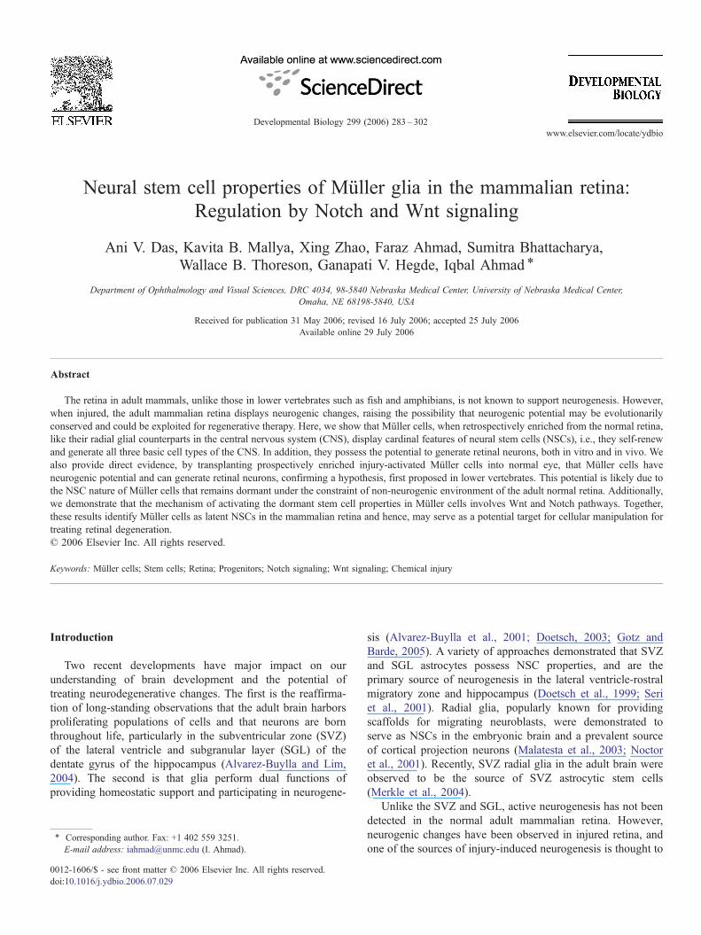
Neural stem cell properties of Müller glia in the mammalian retina: Regulation by Notch and Wnt signaling Ani V. Das, Kavita B. Mallya, Xing Zhao, Faraz Ahmad, Sumitra Bhattacharya, Wallace B. Thoreson, Ganapati V. Hegde, Iqbal Ahmad ⁎ Department of Ophthalmology and Visual Sciences, DRC 4034, 98-5840 Nebraska Medical Center, University of Nebraska Medical Center, Omaha, NE 68198-5840, USA Received for publication 31 May 2006; revised 16 July 2006; accepted 25 July 2006 Available online 29 July 2006 Abstract The retina in adult mammals, unlike those in lower vertebrates such as fish and amphibians, is not known to support neurogenesis. However, when injured, the adult mammalian retina displays neurogenic changes, raising the possibility that neurogenic potential may be evolutionarily conserved and could be exploited for regenerative therapy. Here, we show that Müller cells, when retrospectively enriched from the normal retina, like their radial glial counterparts in the central nervous system (CNS), display cardinal features of neural stem cells (NSCs), i.e., they self-renew and generate all three basic cell types of the CNS. In addition, they possess the potential to generate retinal neurons, both in vitro and in vivo. We also provide direct evidence, by transplanting prospectively enriched injury-activated Müller cells into normal eye, that Müller cells have neurogenic potential and can generate retinal neurons, confirming a hypothesis, first proposed in lower vertebrates. This potential is likely due to the NSC nature of Müller cells that remains dormant under the constraint of non-neurogenic environment of the adult normal retina. Additionally, we demonstrate that the mechanism of activating the dormant stem cell properties in Müller cells involves Wnt and Notch pathways. Together, these results identify Müller cells as latent NSCs in the mammalian retina and hence, may serve as a potential target for cellular manipulation for treating retinal degeneration. © 2006 Elsevier Inc. All rights reserved. Keywords: Müller cells; Stem cells; Retina; Progenitors; Notch signaling; Wnt signaling; Chemical injury Introduction Two recent developments have major impact on our understanding of brain development and the potential of treating neurodegenerative changes. The first is the reaffirma- tion of long-standing observations that the adult brain harbors proliferating populations of cells and that neurons are born throughout life, particularly in the subventricular zone (SVZ) of the lateral ventricle and subgranular layer (SGL) of the dentate gyrus of the hippocampus (Alvarez-Buylla and Lim, 2004). The second is that glia perform dual functions of providing homeostatic support and participating in neurogene- sis (Alvarez-Buylla et al., 2001; Doetsch, 2003; Gotz and Barde, 2005). A variety of approaches demonstrated that SVZ and SGL astrocytes possess NSC properties, and are the primary source of neurogenesis in the lateral ventricle-rostral migratory zone and hippocampus (Doetsch et al., 1999; Seri et al., 2001). Radial glia, popularly known for providing scaffolds for migrating neuroblasts, were demonstrated to serve as NSCs in the embryonic brain and a prevalent source of cortical projection neurons (Malatesta et al., 2003; Noctor et al., 2001). Recently, SVZ radial glia in the adult brain were observed to be the source of SVZ astrocytic stem cells (Merkle et al., 2004). Unlike the SVZ and SGL, active neurogenesis has not been detected in the normal adult mammalian retina. However, neurogenic changes have been observed in injured retina, and one of the sources of injury-induced neurogenesis is thought to Developmental Biology 299 (2006) 283 – 302 www.elsevier.com/locate/ydbio ⁎ Corresponding author. Fax: +1 402 559 3251. E-mail address: [email protected] (I. Ahmad). 0012-1606/$ - see front matter © 2006 Elsevier Inc. All rights reserved. doi:10.1016/j.ydbio.2006.07.029
-
Upload
independent -
Category
Documents
-
view
2 -
download
0
Transcript of Neural stem cell properties of Müller glia in the mammalian retina: Regulation by Notch and Wnt...
99 (2006) 283–302www.elsevier.com/locate/ydbio
Developmental Biology 2
Neural stem cell properties of Müller glia in the mammalian retina:Regulation by Notch and Wnt signaling
Ani V. Das, Kavita B. Mallya, Xing Zhao, Faraz Ahmad, Sumitra Bhattacharya,Wallace B. Thoreson, Ganapati V. Hegde, Iqbal Ahmad ⁎
Department of Ophthalmology and Visual Sciences, DRC 4034, 98-5840 Nebraska Medical Center, University of Nebraska Medical Center,Omaha, NE 68198-5840, USA
Received for publication 31 May 2006; revised 16 July 2006; accepted 25 July 2006Available online 29 July 2006
Abstract
The retina in adult mammals, unlike those in lower vertebrates such as fish and amphibians, is not known to support neurogenesis. However,when injured, the adult mammalian retina displays neurogenic changes, raising the possibility that neurogenic potential may be evolutionarilyconserved and could be exploited for regenerative therapy. Here, we show that Müller cells, when retrospectively enriched from the normal retina,like their radial glial counterparts in the central nervous system (CNS), display cardinal features of neural stem cells (NSCs), i.e., they self-renewand generate all three basic cell types of the CNS. In addition, they possess the potential to generate retinal neurons, both in vitro and in vivo. Wealso provide direct evidence, by transplanting prospectively enriched injury-activated Müller cells into normal eye, that Müller cells haveneurogenic potential and can generate retinal neurons, confirming a hypothesis, first proposed in lower vertebrates. This potential is likely due tothe NSC nature of Müller cells that remains dormant under the constraint of non-neurogenic environment of the adult normal retina. Additionally,we demonstrate that the mechanism of activating the dormant stem cell properties in Müller cells involves Wnt and Notch pathways. Together,these results identify Müller cells as latent NSCs in the mammalian retina and hence, may serve as a potential target for cellular manipulation fortreating retinal degeneration.© 2006 Elsevier Inc. All rights reserved.
Keywords: Müller cells; Stem cells; Retina; Progenitors; Notch signaling; Wnt signaling; Chemical injury
Introduction
Two recent developments have major impact on ourunderstanding of brain development and the potential oftreating neurodegenerative changes. The first is the reaffirma-tion of long-standing observations that the adult brain harborsproliferating populations of cells and that neurons are bornthroughout life, particularly in the subventricular zone (SVZ)of the lateral ventricle and subgranular layer (SGL) of thedentate gyrus of the hippocampus (Alvarez-Buylla and Lim,2004). The second is that glia perform dual functions ofproviding homeostatic support and participating in neurogene-
⁎ Corresponding author. Fax: +1 402 559 3251.E-mail address: [email protected] (I. Ahmad).
0012-1606/$ - see front matter © 2006 Elsevier Inc. All rights reserved.doi:10.1016/j.ydbio.2006.07.029
sis (Alvarez-Buylla et al., 2001; Doetsch, 2003; Gotz andBarde, 2005). A variety of approaches demonstrated that SVZand SGL astrocytes possess NSC properties, and are theprimary source of neurogenesis in the lateral ventricle-rostralmigratory zone and hippocampus (Doetsch et al., 1999; Seriet al., 2001). Radial glia, popularly known for providingscaffolds for migrating neuroblasts, were demonstrated toserve as NSCs in the embryonic brain and a prevalent sourceof cortical projection neurons (Malatesta et al., 2003; Noctoret al., 2001). Recently, SVZ radial glia in the adult brain wereobserved to be the source of SVZ astrocytic stem cells(Merkle et al., 2004).
Unlike the SVZ and SGL, active neurogenesis has not beendetected in the normal adult mammalian retina. However,neurogenic changes have been observed in injured retina, andone of the sources of injury-induced neurogenesis is thought to
Table 1List of antibodies used
Name Species Dilution Company
Vimentin Mouse 1:10 HybridomaGS Rabbit 1:5000 SigmaNestin Mouse 1:4 HybridomaSox2 Rabbit 1:500 ChemiconNotch1 Rat 1:1 Giftβ-Tubulin Rabbit 1:1000 BabcoGFAP Rabbit 1:100 SigmaO4 Mouse 1:30 ChemiconMap2 Mouse 1:200 ChemiconCalbindin Rabbit 1:10,000 GiftPax6 Rabbit 1:1000 BabcoChx10 Rabbit 1:250 GiftRx Mouse 1:250 GiftCD31 Mouse 1:50 ChemiconMac-1 Rabbit 1:100 Accurate Chemical and Corp.Brn3b Rabbit 1:300 BabcoOpsin Mouse 1:1000 GiftPKC Rabbit 1:1000 SigmaSyntaxin Mouse 1:100 GiftBrdU Rat 1:100 Accurate Chemical and Corp.
284 A.V. Das et al. / Developmental Biology 299 (2006) 283–302
be the major glial cell type of the retina, the Müller cells(Braisted et al., 1994; Ooto et al., 2004; Reh and Fischer, 2001).Are Müller cells latent NSCs in retina, that can be activated byextrinsic cues, including those that are injury-induced, toproliferate and generate new neurons? To answer this question,we purified Müller cells from the rodent retina and demon-strated that they display cardinal features of NSCs in vitro, i.e.,they self-renew, and generate both neurons and glia, in additionto generating retinal neurons in conducive culture conditions.Müller cells, identified and enriched prospectively as sidepopulation (SP) cells, when transplanted into retina generatedlamina-specific retinal neurons, thus providing a direct proof oftheir neurogenic potential. In addition, our results suggested thatNotch and Wnt pathways acted in concert to regulate the stemcell properties of Müller cells. This raises the possibility oftreating degenerating retinas from within, by targeted manip-ulation of these cells.
Materials and methods
Müller cell culture
Enrichment of Müller cells was done according to a previously describedmethod (Hicks and Courtois, 1990). Briefly, eyes from postnatal (PN) days 10–21 rats were enucleated and incubated overnight in DMEM. Eyecups weretransferred to dissociation solution (DMEM containing 0.1% Trypsin and 70 U/ml collagenase) and incubated at 37°C in CO2 incubator for 1 h. Eyecups werewashed with DMEM and the retina was dissected out with care to avoidcontamination from RPE and ciliary epithelium. The retina was mechanicallydissociated into small aggregates and cultured in DMEM containing 10% FBSfor 8–10 days. The floating retinal aggregates and debris were removed leavingpurified flat cell population of Müller cells attached to the bottom of the dish.Cells were trypsinized and cultured in DMEM containing 10% FBS for another5 days to get a further purified population. Cells were dissociated using Trypsin–EDTA and cultured in DMEM/F12 (containing 1× N2 supplement (GIBCO),2 mM L-glutamine, 100 U/ml penicillin, 100 μg/ml streptomycin) supplementedwith FGF2 (10 ng/ml; Collaborative Research) and EGF (20 ng/ml;Collaborative Research) at a density of ∼5×10⁴ cells/cm2 for 5–7 days togenerate neurospheres. To remove microglia, cell dissociates were subjected toimmunopanning using Mac-1 antibody (Barres et al., 1988; Meucci et al., 1998).To examine their neurosphere-forming ability, microglia was purified from retinaaccording to a previously described method (Roque and Caldwell, 1993) andcultured in the same medium mentioned above for 4–6 days. To ascertain theclonal generation of neurospheres by co-culturing green (GFP-expressing) andnon-green cells (Zhao et al., 2002), Müller cells were enriched from the retina ofthe green [constitutively expressing the green fluorescent protein (GFP)] andwild-type mice as described above. LDA analysis was carried out as previouslydescribed (Ahmad et al., 2004; Das et al., 2004). Cells were diluted to give aninitial concentration of 5000 cells/ml from which serial dilutions in 200 μlaliquots were plated in individual wells of a 96-well plate. Culture was carriedout for 7 days, after which the fraction of wells not containing neurospheres foreach cell-plating density was calculated. The negative logarithm of fraction ofnegative wells was plotted against the number of cells plated/well to provide astraight line in a semi logarithm plot. The zero term of Poisson equation (F0=e
−x,where F0 is the fraction of well without neurospheres and x is the mean numberof cell/well) predicts that when 37% of test wells are negative, there is an averageof one stem cell/well. To examine the differentiation potential of Müller cell-derived stem cells/progenitors, neurospheres were exposed to 10 μM BrdU(Sigma) for the final 48 h to tag the dividing cells and plated on poly-D-lysine(500 μg/ml) and laminin (5 μg/ml) coated 12-mm glass coverslips. In order topromote differentiation, mitogens were substituted with brain-derived neuro-trophic factor (BDNF; 1 ng/ml), retinoic acid (RA; 1 μM) and 1% FBS andculture was continued for 5–7 days. Cells were fixed using cold 4%paraformaldehyde for immunocytochemical analysis.
FACS analysis
In order to find out the purity of enriched Müller cells, we carried out FACSanalysis using antibodies specific to Müller glia such as glutamine synthetase(GS) and vimentin. Briefly, cells in the monolayer culture were dissociated intosingle cells and fixed using ice-cold ethanol. After washing with 1× PBScontaining 1% BSA, cells (2×10⁶) were incubated with appropriate dilution ofGS/vimentin (Table 1) in 1× PBS containing 1% BSA for 1 h at 4°C. Prior toantibody incubation, cells were blocked for non-specific binding in 1× PBScontaining 0.1% Triton-X100 for 30 min at 4°C. After antibody incubation, cellswere washed and incubated in PBS-BSA solution containing appropriatesecondary antibodies linked to FITC for 1 h at 4°C. Cells were washed withPBS-BSA and resuspended in PBS-BSA for FACS analysis.
Immunocytochemical analysis
Immunocytochemical analysis was carried out for the detection of BrdUand/or cell-specific markers as previously described (Zhao et al., 2002). Briefly,paraformaldehyde-fixed cells were incubated in PBS containing 5% NGS and 0,0.2 or 0.4% Triton-X100 followed by an overnight incubation in antibodies at4°C. The list of antibodies used is given in Table 1. Cells were examined forepifluorescence following incubation in IgG conjugated to Cy3/FITC. Imageswere captured using cooled CCD-camera (Princeton Instruments) and Openlabsoftware (Improvision).
RT-PCR analysis
Isolation of total RNA and cDNA synthesis were carried out as previouslydescribed (James et al., 2003). Briefly, 4 μg of RNAwas transcribed into cDNAin total volume of 50 μl. Specific transcripts were amplified with gene-specificforward and reverse primers using a step cycle program on a Robocycler(Stratagene). Amplifications were carried out for 25 cycles so that they remainedwithin the range of linearity to yield a semi-quantitative estimation of changes inthe relative levels of transcripts. Products were visualized by ethidium bromidestaining after electrophoresis on 2% agarose gel. Gene-specific primers used forRT-PCR analyses are given in Table 2.
Co-culture experiments
BrdU-labeled neurospheres derived from enriched Müller cells werecollected, washed extensively to remove excess BrdU and co-cultured on
Table 2List of primers used for the RT-PCR analysis
Name Sequence Annealing temp (°C) Size (bp) Acc. No
Vimentin Forward: 5′AAGGCACTAATGAGTCCCTGGAG3′ 56 251 NM031140Reverse: 5′GTTTGGAAGAGGCAGAGAAATCC3′
Glutamine synthetase (GS) Forward: 5′TCACAGGGACAAATGCCGAG3′ 58 362 M96152Reverse: 5′GTTGATGTTGGAGGTTTCGTGG3′
CRALBP Forward: 5′CTGAGTTTGGAGGAATCTTGC3′ 54 150 XM217702Reverse: 5′TGGATTTGGGGGAGAGTTC3′
Opsin Forward: 5′CATGCAGTGTTCATGTGGGA 3′ 64 422 U22180Reverse: 5′AGCAGAGGCTGGTGAGCATG 3′
mGluR6 Forward: 5′CACAGCGTGATTGACTACGAG3′ 56 317 D13963Reverse: 5′CTCAGGCTCAGTGACACAGTTAG3′
HPC1 Forward: 5′AAGAGCATCGAGCAGCAGAGCATC3′ 60 342 NM016801Reverse: 5′CATGGCCATGTCCATGAACAT3′
Brn3b Forward: 5′GGCTGGAGGAAGCAGAGAAATC 3′ 60 141 AF390076Reverse: 5′TTGGCTGGATGGCGAAGTAG 3′
β-Tubulin Forward: 5′TGCGTGTGTACAGGTGAATGC3′ 54 250 NM139254Reverse: 5′AGGCTGCATAGTCATTTCCAAG3′
GFAP Forward: 5′ATCTGGAGAGGAAGGTTGAGTCG3′ 58 310 NM017009Reverse: 5′TGGCGGCGATAGTCATTAGA3′
PLP Forward: 5′CGGGTGTGTCATTGTTTGGG3′ 58 310 NM030990Reverse: 5′ACAGGTGGAAGGTCATTTGGAAC3′
Notch1 Forward: 5′TCTGGACAAGATTGATGGCTACG3′ 56 329 NM008714Reverse: 5′CGTTGACACAAGGGTTGGACTC3′
Nestin Forward: 5′TGGAGCAGGAGAAGCAAGGTCTAC3′ 56 295 NM012987Reverse: 5′TCAAGGGTATTAGGCAAGGGGG3′
Pax6 Forward: 5′CCATCTTTGCTTGGGAAATCC3′ 56 310 NM_013001Reverse: 5′TCATCCGAGTCTTCTCCATTGG3′
Musashi1 Forward: 5′TGAAAGAGTGTCTGGTGATGCG3′ 52 309 NM148890Reverse: 5′GCCTGTTGGTGGTTTTGTCG3′
Sox2 Forward: 5′AGGGCTGGGAGAAAGAAGAG3′ 56 179 NM_011443Reverse: 5′GGAGAATAGTTGGGGGGAAG3′
ABCG2 Forward: 5′GATGGCTACACTCACTCACAAAG3′ 54 325 NM_011920Reverse: 5′ACTGAATCTAACCCAAGGAAGG3′
Clusterin Forward: 5′CCTCCAGTCCAAGATGCTCAAC 58 292 NM_053021Reverse: 5′TTTCCTGCGGTATTCCTGTAGC
Carbonic anhydrase Forward: 5′TTGCCAATGGAGACCGACAG 58 233 NM_019291Reverse: 5′TGAGCCCCAGTGAAAGTGAAAC
CD31 Forward: 5′AAGAGCAACTTCCAGACCGTCC 58 222 NM_031591Reverse: 5′AAGCACCATTTCATCTCCAGACTG
Tyrosinase Forward: 5′TCAGTCTATGTCATCCCCACAGG 56 252 NM_011661Reverse: 5′GTTCTCATCCCCAGTTAGTTCTCG
Ath5 Forward: 5′TGGGG(I)CA(GA)GA(CT)AA(GA)AA(GA)3′ 52 231 AF071223Reverse: 5′CAT(I)GG(GA)AA(I)GG(CT)TC(I)GG(CT)TG3′
Ath3 Forward: 5′CGACTGGCAAGGAACTACATCTG3′ 50 629 NM007501Reverse: 5′ACTAATGCTCAGGGGTGGTGTG3′
Otx2 Forward: 5′GAGAGGAGGTGGCACTGAAAATC3′ 56 391 U96488Reverse: 5′CCCCCAAAGTAGGAAGTTGAGC3′
NeuroD Forward: 5′AAGAGGAGGAGGAAGAGGAGGAG3′ 58 337 AF107728Reverse: 5′TTGGTAGTGGGCTGGGACAAAC3′
CyclinA Forward: 5′TACACACACGAGGTAGTGACGCTG3′ 56 307 X60767Reverse: 5′CCAAGCCGTTTTCATCCAGG3′
CyclinD1 Forward: 5′ACACCAATCTCCTCAACCGACC3′ 56 383 NM171992Reverse: 5′GTTCACCAGAAGCAGTTCCATTTG3′
CyclinD3 Forward: 5′CGAAACCACACCCCTGACTATTG3′ 60 201 NM012766Reverse: 5′TGCTTTTTGACCAGTGCCTGC3′
Delta1 Forward: 5′CGACCTCGCAACAGAAAAC3′ 56 397 NM032063Reverse: 5′CGACCTCGCAACAGAAAAC3′
Hes1 Forward: 5′GCTTTCCTCATCCCCAATG3′ 56 224 NM024360Reverse: 5′CGTATTTAGTGTCCGTCAGAAGAG3′
Crx Forward: 5′CCTCACTATTCGGTCAATGCC3′ 58 346 NM_021855Reverse: 5′ATGTGCCTGCCTTCCTCTTC3′
Lef1 Forward: 5′CACACAACTGGCATCCCTCATC3′ 56 205 NM130429Reverse: 5′TACACTCGGCTACGACATTCGC3′
Fzd1 Forward: 5′TGGTTTCGTGTCGCTCTTCC 3′ 60 223 XM216082
(continued on next page)
285A.V. Das et al. / Developmental Biology 299 (2006) 283–302
Table 2 (continued)
Name Sequence Annealing temp (°C) Size (bp) Acc. No
Reverse: 5′AGGGCAAGGGATGGCATAAC3′Fzd2 Forward: 5′CGCTTCTACTTTCTTCACGGTCAC3′ 58 211 NM172035
Reverse: 5′GCCTTCTTTCTTAGTGCCCTGC3′Fzd4 Forward: 5′TGCTTCATCTCTACCACCTTCACTG3′ 60 282 NM022623
Reverse: 5′CAGAATAACCCACCAAATGGAGC3′Ki67 Forward: 5′GAGCAGTTACAGGGAACCGAAG3′ 58 262 X82786
Reverse: 5′CCTACTTTGGGTGAAGAGGCTG3′p27Kip1 Forward: 5′GACTTTCAGAATCATAAGCCCCTG3′ 58 261 NM031762
Reverse: 5′TGGACACTGCTCCGCTAACC3′β-Actin Forward: 5′GTGGGGCGCCCCAGGCACCA 3′ 50 548 XM_037235
Reverse: 5′ CTCCTTAATGTCACGCACGATTTC 3′
286 A.V. Das et al. / Developmental Biology 299 (2006) 283–302
poly-D-lysine, laminin coated 12-mm glass coverslips with E3 chick/PN1 ratretinal cells in 1% FBS for 5–7 days, across 0.4 μm membrane (Millipore). Atthe end of culture, cells were fixed with 4% paraformaldehyde for 15 min at 4°Cfollowed by immunocytochemical analysis for various retinal neuronal markers.Media were changed every other day and neurospheres were allowed todifferentiate for 5–7 days. To obtain E3 chick retinal cells, fertilized hens eggs(SPAFAS, Wilmington, MA) were incubated in a humidified chamber at 37°Cfor 3 days. Embryos were staged according to Hamburger and Hamilton (1951).Retina were dissected out from eyes and dissociated into single cells aspreviously described (James et al., 2003).
Hoechst dye efflux assay
Hoechst dye efflux assay was carried as previously reported (Ahmad et al.,2004; Bhattacharya et al., 2003). Briefly, dissociated retinal cells wereresuspended in Iscove's modified Dulbecco's medium (IMDM) at a concentra-tion of 106 cells/ml and incubated at 4°C overnight followed by staining withHoechst 33342 (4 μg/ml) at 37°C for 60 min and sorted on a FACStar Plus (BDBiosciences, Lincoln Park, NJ) cell sorter. Hoechst dye was excited at 350 nm,and fluorescence was measured at two wavelengths using a 485 BP22 (485-nmbandpass filter) and a 675 EFLP (675-nm long-pass edge filter) optical filter(Omega Optical, Brattleboro, VT). The SP region was defined on the flowcytometer on the basis of its fluorescence emission in both blue and redwavelengths. Dead cells and debris were excluded by establishing a live gate onthe flow cytometer using forward versus side scatter. The sorted SP cells wereprocessed for immunocytochemical and RT-PCR analyses. The specificity of theassay was ascertained by incubating cells with 150 μM of verapamil during thestaining stage.
In vivo activation and enrichment of Müller cells
PN21 rats were used for this experiment. The rats were anesthetized withketamine and xylazine and right eyes were injected with 0.5 μl–10 μl of amixed solution of neurotoxins [kainate (40 mM) and NMDA (4 mM)], growthfactors (FGF2, 20 ng/eye; insulin, 1 μg/eye) and BrdU (1 μg/eye) by intravitrealinjection using a glass micropipette attached to a 10-μl Hamilton syringe aspreviously described (Zhao et al., 2005; Chacko et al., 2003). The left eyes wereinjected with the vehicle and were used as controls. Rats were givenintraperitoneal injections of BrdU (0.12 mg/g body weight) everyday untilthe end of the experiment on 4th day. Eyes were enucleated. Some eyes wereprocessed for immunohistochemical analysis and retina from the rest wassubjected to Hoechst dye efflux assay as described above. To examine the roleof Notch signaling on the in vivo activated Müller cells, DAPT (25 μM), a γ-secretase inhibitor (James et al., 2004), was injected along with the growthfactors. To examine the involvement of Wnt signaling in the in vivo activationof Müller cells, Wnt2b and FzdCRD conditioned medium was injectedintravitreously. Wnt2b and FzdCRD conditioned medium was obtained bytransiently transfecting pEF-Wnt2b vector (Kubo et al., 2003) and Mfz8CRD-IgG (Hsieh et al., 1999) in 293 cells by using Effectene transfection kit(Qiagen). One day after transfection, cells were transferred to serum-freeDMEM/F-12, and the medium was collected after an additional 24 h. Controlconditioned medium was obtained from untransfected 293T cells. To
compensate for the artifactual differences in levels of cell-specific transcriptsin the injured retina due to cell loss, examination of levels of transcripts by PCRwas carried out after normalization of levels of beta actin transcripts in inuredand uninjured retina.
Brain and retinal transplantation
Transplantation of GFP+ cells from Müller cell-derived neurospheres wascarried out as previously described (Vicario-Abejon et al., 1995; Zhao et al.,2002). Briefly, to implant cells in the hippocampus of the PN2 rat pups (n=4)animals were anesthetized by hypothermia. Each animal was placed on aminiaturized hypothermic stereotaxic instrument (Stoelting Co.). A hole wasdrilled in the skull using a dental burr at 1.7 mm lateral to midline and 1.2 mmposterior to bregma. A pulled glass pipette with an external diameter of∼70 μmwas placed ∼1.4 mm below dura to deliver 0.5 μl of cell suspension over aperiod of 2 min. The surgical area was cleaned and the incision was closed withSuperglue. The animal was revived on a 37°C heating pad and returned to itsmother. Intravitreal transplantation of GFP+ cells from Müller cell-derivedneurospheres cells or SP cells was performed in PN1 rats (n=3) as previouslydescribed (Chacko et al., 2003). Briefly, animals were anesthetized with 20–25 μl of a 1:1 dilution of ketamine (60 mg/ml) and xylazine (8 ng/ml), injectedintraperitoneally. A 30-gauge needle was inserted into the eye near the equator.The needle was retracted, and a glass micropipette connected to a 10 μlHamilton syringe was inserted through the wound to deliver 50,000–100,000cells in a 1–2 μl volume. Animals were sacrificed 1 week after transplantation.Eyes were enucleated and processed for immunohistochemical analysis. The SPcells were labeled with CFDA-SE as per manufacturer's instructions (MolecularProbes) for tracking purpose. In order to determine the lamina-specificincorporation of grafted cells, retinal sections (75 sections) from differenttransplantation experiments were examined and total cells integrated in aparticular section were counted. Next, the number of cells incorporated in eachlamina was counted to determine the lamina-specific proportion of cells withrespect to total cell integrated.
Brain overlay culture
To analyze whether Müller cells differentiate into physiologically activeneuronal cells in vivo, we carried out brain slice overlay culture according toa previously described method (Benninger et al., 2003; Polleux et al., 1998).Briefly, PN1 pups were sacrificed by decapitation, brains removed in ice-coldHBSS, and placed on a clean Whatman filter paper. A solution of low-melting agarose was poured on top of the brain and was allowed to solidifyby placing the filter on a cooled surface. 300-μm-thick coronal sections werecut using a mechanical tissue chopper (Stoelting Co.). Slices were collectedusing a flat brush and cultured in a 6-well plate on millicell filter (Millipore)covered with a film of slice culture medium (DMEM containing 25% HBSSand 10% FBS). Müller cell-derived neurospheres from GFP-transgenic micewere dissociated and resuspended in slice culture medium. Approximately1×105 cells were added onto and around the slices and cultured for 3–4 days. At the end of culture, the slices were analyzed for the presence ofGFP+ cells and used for electrophysiological and immunohistochemicalanalyses.
287A.V. Das et al. / Developmental Biology 299 (2006) 283–302
Electrophysiological analysis
For electrophysiological studies, brain slices with GFP+ cells from Müllercell-derived neurospheres were placed in a chamber, and perfused on the stage ofan upright, fixed-stage microscope (for electrophysiology, model BHWI;Olympus, Lake Success, NY; for imaging experiments, model E600FN; Nikon,Melville, NY) with an oxygenated solution containing NaCl, 140 mM; KCl,5 mM; CaCl2, 2 mM; MgCl2, 1 mM; HEPES, 10 mM; glucose, 10 mM (pH 7.4).Experiments were performed at room temperature. For whole-cell recording,patch pipettes were pulled on a vertical puller (model PB-7; Narishige, Tokyo,Japan) from borosilicate glass pipettes (1.2-mm outer diameter, 0.95-mm innerdiameter; with internal filament (World Precision Instruments) and had tips of 1-to 2-μm outer diameter with tip resistances of 6 to 12 MΩ. Pipettes were filledwith a bathing solution containing KCH3SO4, 98 mM; KCl, 44 mM; NaCl,3 mM; HEPES, 5 mM; EGTA, 3 mM; MgCl2, 3 mM; CaCl2, 1 mM; glucose,2 mM; Mg-adenosine triphosphate (ATP), 1 mM; guanosine triphosphate(GTP), 1 mM; and reduced glutathione, 1 mM (pH 7.2) (Das et al., 2005b; Jameset al., 2003). GFP-positive cells were identified under epifluorescence andpatched for whole-cell recording.
Adenovirus infection
To eliminate the immigrant astrocytes from the Müller cell culture, weinfected the purified Müller cells with Adgfap2Tk vector for 24 h and exposedthem to Ganciclovir (GCV; 150 μg/ml) (Das et al., 2006; Vandier et al., 2000) foranother 24 h. GCV specifically eliminate Tk-expressing cells. Cells werecollected, washed and then cultured in the presence of FGF2 to generateneurospheres.
Results
Enriched Müller cells display properties of NSCs
To examine whether or not Müller cells possess NSCproperties we carried out neurosphere assay (Doetsch et al.,1999; Laywell et al., 2000; Merkle et al., 2004) on Müller cells,enriched from rodent retina, ranging from PN10 to PN21. Weadopted a monolayer culture protocol that has been demon-strated to enrich Müller cells to the range of greater than 95%,free from neuronal, fibroblastic or astrocytic contaminations(Hicks and Courtois, 1990). In our hands, approximately 95%of cells in the monolayer culture were Müller cells as revealedby the proportion of cells expressing vimentin and glutaminesynthetase (GS) immunoreactivities (Figs. 1A–N). To furtherascertain the enrichment and purity of monolayer Müller cellculture, we examined the expression of cell-specific transcripts.RT-PCR analysis revealed that cells in culture expressed abattery of transcripts characteristic of Müller cells such as GS,CRALBP, Vimentin, Clusterin and Carbonic anhydrase (Black-shaw et al., 2004) (Fig. 1O). In contrast, transcriptscorresponding to rod photoreceptors (opsin), bipolar cells(mGluR6), amacrine cells (Syntaxin 1), retinal ganglion cells orRCGs (Brn3b), endothelial cells (CD31) and retinal pigmentedepithelium (RPE)/pigmented ciliary epithelium (tyrosinase)were not detected suggesting that the monolayer culture wasenriched for Müller cells and not contaminated with theabovementioned cells (Fig. 1P). However, low levels oftranscripts characteristic of microglia (Iba1) were detected,suggesting the presence of microglia in the enriched Müller cellculture. Subsequent immunocytochemical analysis revealed thepresence of microglia in the monolayer culture in the range of
2–4%, which could be removed by immunopanning (seebelow).
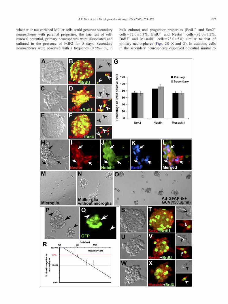
Having determined the purity of the culture, we firstexamined the proliferating and self-renewing abilities ofenriched Müller cells. When these cells were cultured in thepresence of FGF2 for 4–5 days, a small subset (0.5%–1%) ofcells proliferated to generate spheres of an average size of200 μM. The size of neurospheres was dependent on culturetime [Fig. 2A (5 days in culture) versus Fig. 2P (2 days inculture)]. The spheres displayed classic features of neuro-spheres; the majority (72.4±2.6%) of cells in spheresincorporated BrdU, attesting to their proliferative nature andexpressed general (Notch1; 83.0±3.0) and neural (Sox2:74.0±3.4; Nestin: 86.0±1.8, Musashi: 73.0±4.3) stem cellmarkers (Figs. 2A–F and G). The majority (88.0±1.5%) ofproliferating cells co-expressed Nestin and Notch, suggestingthat these cells did not represent distinct sub-populations of cellsbut rather a single population of neural progenitors, expressingmultiple neural progenitor markers (Figs. 2H–L). Although theabsence of cells expressing CD31, retinal neuron-specificmarkers and tyrosinase ensured that endothelial cells, retinalneurons, RPE/pigmented ciliary epithelium, respectively, werenot the source of neurospheres, a possibility remained that thesource could be the contaminating microglia. To examine thispossibility, we purified microglia from retina (Roque andCaldwell, 1993) and cultured them identically like enrichedMüller cells in the presence of FGF2. At the end of 7 days inculture, neurospheres were not detected (Fig. 2M). In addition,we observed that enriched Müller cells, following the removalof microglia by immunopanning, still formed neurospheres,when exposed to FGF2 for 2 days (Fig. 2N). Together, theseobservations suggested that enriched Müller cells and notcontaminating microglia were the source of neurospheres. Itcould be argued that immigrant astrocytes, as a possible minorcontaminant in the monolayer culture, could be the source ofneurospheres. To address this issue, we adapted an approachof selectively ablating GFAP-expressing astrocytes in themonolayer culture, followed by examining the generation ofneurospheres (Das et al., 2006; Morshead et al., 2003). Cellsin the monolayer culture, before their exposure to FGF2, wereinfected with adenoviral vector Adgfap2Tk in which the HSV-Tk gene is driven by GFAP promoter. Following infectionand exposure to ganciclovir (GCV) for 24 h, cells werecultured in the presence of FGF2 for 1–2 days. GCV isconverted into a toxic triphosphate form in the presence ofHSV-Tk, killing dividing cells by incorporating in replicatingDNA (Vandier et al., 2000). We observed the generation ofneurospheres in culture of Adgfap2Tk infected cells and thosethat were infected with the control virus, providing evidenceagainst immigrant astrocytes as the source of neurospheres(Fig. 2O).
To address the issue that neurospheres generated in bulkculture may not have clonal origin but arise due to cellaggregation, we co-cultured green [obtained from greenfluorescent protein (GFP)-expressing mice] and non-green(obtained from wild-type mice) enriched Müller cells in thepresence of FGF2. The culture was carried out for 2 days since
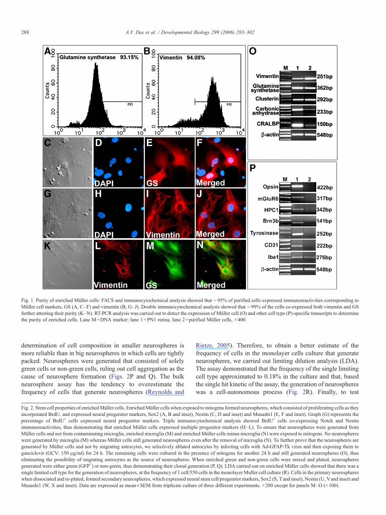
Fig. 1. Purity of enriched Müller cells: FACS and immunocytochemical analysis showed that ∼95% of purified cells expressed immunoreactivities corresponding toMüller cell markers, GS (A, C–F) and vimentin (B, G–J). Double immunocytochemical analysis showed that ∼99% of the cells co-expressed both vimentin and GSfurther attesting their purity (K–N). RT-PCR analysis was carried out to detect the expression of Müller cell (O) and other cell type (P)-specific transcripts to determinethe purity of enriched cells. Lane M=DNA marker; lane 1=PN1 retina, lane 2=purified Müller cells, ×400.
288 A.V. Das et al. / Developmental Biology 299 (2006) 283–302
determination of cell composition in smaller neurospheres ismore reliable than in big neurospheres in which cells are tightlypacked. Neurospheres were generated that consisted of solelygreen cells or non-green cells, ruling out cell aggregation as thecause of neurosphere formation (Figs. 2P and Q). The bulkneurosphere assay has the tendency to overestimate thefrequency of cells that generate neurospheres (Reynolds and
Fig. 2. Stem cell properties of enrichedMüller cells. EnrichedMüller cells when exposincorporated BrdU, and expressed neural progenitor markers, Sox2 (A, B and inset),percentage of BrdU+ cells expressed neural progenitor markers. Triple immunoimmunoreactivities, thus demonstrating that enriched Müller cells expressed multipMüller cells and not from contaminating microglia, enriched microglia (M) and enrichwere generated by microglia (M) whereas Müller cells still generated neurospheres evgenerated by Müller cells and not by migrating astrocytes, we selectively ablated asganciclovir (GCV: 150 μg/ml) for 24 h. The remaining cells were cultured in the preliminating the possibility of migrating astrocytes as the source of neurospheres. Wgenerated were either green (GFP+) or non-green, thus demonstrating their clonal gensingle limited cell type for the generation of neurospheres, at the frequency of 1 cell/55when dissociated and re-plated, formed secondary neurospheres, which expressed neuMusashi1 (W, X and inset). Data are expressed as mean±SEM from triplicate cultur
Rietze, 2005). Therefore, to obtain a better estimate of thefrequency of cells in the monolayer cells culture that generateneurospheres, we carried out limiting dilution analysis (LDA).The assay demonstrated that the frequency of the single limitingcell type approximated to 0.18% in the culture and that, basedthe single hit kinetic of the assay, the generation of neurosphereswas a cell-autonomous process (Fig. 2R). Finally, to test
ed tomitogens formed neurospheres, which consisted of proliferating cells as theyNestin (C, D and inset) and Musashi1 (E, F and inset). Graph (G) represents thecytochemical analysis showed BrdU+ cells co-expressing Notch and Nestinle progenitor markers (H–L). To ensure that neurospheres were generated fromedMüller cells minus microglia (N) were exposed to mitogens. No neurospheresen after the removal of microglia (N). To further prove that the neurospheres aretrocytes by infecting cells with Ad-GFAP-Tk virus and then exposing them toesence of mitogens for another 24 h and still generated neurospheres (O), thushen enriched green and non-green cells were mixed and plated, neurosphereseration (P, Q). LDA carried out on enriched Müller cells showed that there was a0 cells in the monolayer Muller cell culture (R). Cells in the primary neurospheresral stem cell/progenitor markers, Sox2 (S, Tand inset), Nestin (U, Vand inset) ande of three different experiments. ×200 except for panels M–O (×100).
289A.V. Das et al. / Developmental Biology 299 (2006) 283–302
whether or not enriched Müller cells could generate secondaryneurospheres with parental properties, the true test of self-renewal potential, primary neurospheres were dissociated andcultured in the presence of FGF2 for 5 days. Secondaryneurospheres were observed with a frequency (0.5%–1%, in
bulk culture) and progenitor properties (BrdU+ and Sox2+
cells=72.0±5.3%; BrdU+ and Nestin+ cells=92.0±7.2%;BrdU+ and Musashi+ cells=73.0±5.8) similar to that ofprimary neurospheres (Figs. 2S–X and G). In addition, cellsin the secondary neurospheres displayed potential similar to
290 A.V. Das et al. / Developmental Biology 299 (2006) 283–302
those in primary neurospheres (see below), thus affirming theirability to self-renew. That these cells could self-renew forextended period of time was apparent from their ability togenerate neurospheres through five passages.
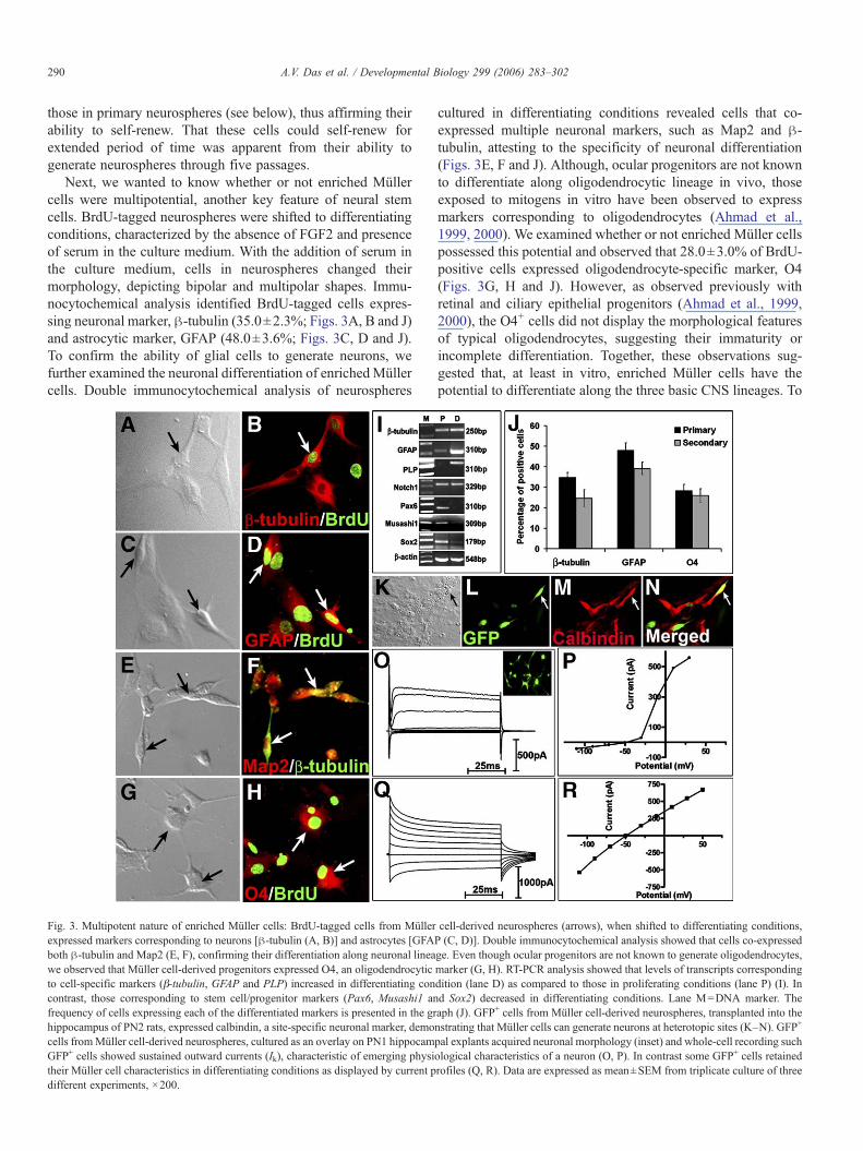
Next, we wanted to know whether or not enriched Müllercells were multipotential, another key feature of neural stemcells. BrdU-tagged neurospheres were shifted to differentiatingconditions, characterized by the absence of FGF2 and presenceof serum in the culture medium. With the addition of serum inthe culture medium, cells in neurospheres changed theirmorphology, depicting bipolar and multipolar shapes. Immu-nocytochemical analysis identified BrdU-tagged cells expres-sing neuronal marker, β-tubulin (35.0±2.3%; Figs. 3A, B and J)and astrocytic marker, GFAP (48.0±3.6%; Figs. 3C, D and J).To confirm the ability of glial cells to generate neurons, wefurther examined the neuronal differentiation of enrichedMüllercells. Double immunocytochemical analysis of neurospheres
Fig. 3. Multipotent nature of enriched Müller cells: BrdU-tagged cells from Müllerexpressed markers corresponding to neurons [β-tubulin (A, B)] and astrocytes [GFAboth β-tubulin and Map2 (E, F), confirming their differentiation along neuronal lineawe observed that Müller cell-derived progenitors expressed O4, an oligodendrocyticto cell-specific markers (β-tubulin, GFAP and PLP) increased in differentiating concontrast, those corresponding to stem cell/progenitor markers (Pax6, Musashi1 anfrequency of cells expressing each of the differentiated markers is presented in the grhippocampus of PN2 rats, expressed calbindin, a site-specific neuronal marker, democells fromMüller cell-derived neurospheres, cultured as an overlay on PN1 hippocamGFP+ cells showed sustained outward currents (Ik), characteristic of emerging physitheir Müller cell characteristics in differentiating conditions as displayed by current pdifferent experiments, ×200.
cultured in differentiating conditions revealed cells that co-expressed multiple neuronal markers, such as Map2 and β-tubulin, attesting to the specificity of neuronal differentiation(Figs. 3E, F and J). Although, ocular progenitors are not knownto differentiate along oligodendrocytic lineage in vivo, thoseexposed to mitogens in vitro have been observed to expressmarkers corresponding to oligodendrocytes (Ahmad et al.,1999, 2000). We examined whether or not enriched Müller cellspossessed this potential and observed that 28.0±3.0% of BrdU-positive cells expressed oligodendrocyte-specific marker, O4(Figs. 3G, H and J). However, as observed previously withretinal and ciliary epithelial progenitors (Ahmad et al., 1999,2000), the O4+ cells did not display the morphological featuresof typical oligodendrocytes, suggesting their immaturity orincomplete differentiation. Together, these observations sug-gested that, at least in vitro, enriched Müller cells have thepotential to differentiate along the three basic CNS lineages. To
cell-derived neurospheres (arrows), when shifted to differentiating conditions,P (C, D)]. Double immunocytochemical analysis showed that cells co-expressedge. Even though ocular progenitors are not known to generate oligodendrocytes,marker (G, H). RT-PCR analysis showed that levels of transcripts correspondingdition (lane D) as compared to those in proliferating conditions (lane P) (I). Ind Sox2) decreased in differentiating conditions. Lane M=DNA marker. Theaph (J). GFP+ cells from Müller cell-derived neurospheres, transplanted into thenstrating that Müller cells can generate neurons at heterotopic sites (K–N). GFP+
pal explants acquired neuronal morphology (inset) and whole-cell recording suchological characteristics of a neuron (O, P). In contrast some GFP+ cells retainedrofiles (Q, R). Data are expressed as mean±SEM from triplicate culture of three
291A.V. Das et al. / Developmental Biology 299 (2006) 283–302
further corroborate multipotentiality of Müller cells, weexamined the change in the expression of transcripts corre-sponding to progenitor, neuronal and glial markers whenneurospheres were shifted from proliferating to differentiatingconditions. RT-PCR analysis revealed an increase in levels oftranscripts corresponding to β-tubulin (neuron), GFAP (astro-cytes) and PLP (oligodendrocytes) in neurospheres in differ-entiating conditions, compared to those in proliferatingconditions (Fig. 3I). In contrast, levels of transcripts corre-sponding to neural progenitor markers such as Pax6, Musashi1and Sox2 decreased in differentiating conditions. The inverserelationship between the expression of progenitor- and the cell-type-specific transcripts suggested that cells in Müller cell-derived neurospheres down-regulated the expression of pro-genitor markers as they differentiate into neurons and glia. Todetermine the potential of self-renewing cells, secondaryneurospheres were cultured in differentiating conditions,followed by the examination of the expression of cell-type-specific markers. Just as observed in primary neurospheres,cells in secondary neurospheres, following the exposure todifferentiating conditions, expressed neuronal- and glial-specific markers. The proportion of cells expressing cell-type-specific markers was similar to those in the primary neuro-spheres (secondary neurospheres versus primary neurospheres;β-tubulin: 25.0±4.0 versus 35.0±2.3; GFAP: 39.0±3.0 versus48.0±3.6; O4: 26.0±3.3 versus 28±3.0), suggesting that self-renewing cells possess the multipotentiality of their parents(Fig. 3J).
To investigate the neurogenic potential of Müller cells in thecontext of the in vivo microenvironment, we transplanted GFP-expressing cells from Müller cell-derived neurospheres in thehippocampus of the PN2 rats. One week following transplanta-tion, immunohistochemical analysis of the brain sectionsrevealed GFP+ cells expressing calbindin, a marker expressedby hippocampal neurons, demonstrating their capacity forneuronal differentiation at heterologous sites (Figs. 3K–N). Itmust be mentioned that calbindin expression is not exclusive tohippocampus. However, its expression in cells, grafted inhippocampus, suggests that they may have acquired hippo-campal phenotype. To ascertain that cells that appearedneuronal by biochemical and molecular criteria possessedproperties of a functional neuron, we carried out electrophy-siological analysis of GFP+ cells from Müller cell-derivedneurospheres in a brain slice overlay culture. Within 2 days inculture, GFP+ cells were observed within and on brain slicesthat had elaborated multipolar structure resembling differen-tiated neurons (Fig. 3O, inset). GFP+ cells in the slice culturewere identified under epifluorescence and patched for electro-physiological analysis. Whole-cell recording of GFP+ cells(70%) revealed prominent outward currents activated bypotentials above −30 mV, similar to delayed rectifying currentsfound in retinal neurons (Figs. 3O and P). In contrast, few GFP+
cells exhibited large inward rectifying currents as observed inmatured Müller cells in vivo (Figs. 3Q and R) (Newman, 1993).Together, these observations underscored the multipotentialnature of enriched Müller cells and their capacity to generatefunctional neurons.
Enriched Müller cells can generate retinal neurons
Our observation that proliferating cells in neurosphereculture, besides expressing pan neural progenitor markers,expressed multiple retinal progenitor markers such as Pax6(Marquardt et al., 2001), Chx10 (Belecky-Adams et al., 1997)and Rx (Mathers et al., 1997) (Figs. 4A–G) raised the possibilitythat Müller cells, retrospectively enriched from the normalretina, can generate retinal neurons. To test this premise, weexamined their ability to generate both early (e.g., RGCs) andlate (e.g., photoreceptors and bipolar cells) born retinal neurons,when exposed to environment conducive for cell-type-specificdifferentiation. First we co-cultured BrdU-tagged Muller cell-derived neurospheres with chick embryonic day 3 (E3) retinalcells across a membrane. We have previously demonstrated thatE3 chick retinal cells elaborate diffusible and evolutionarilyconserved RGC-promoting activities and therefore, suchactivities may influence the differentiation of enriched Müllercells (James et al., 2003). Co-culture was carried out across amembrane to ensure that the result was not due to cell fusion(James et al., 2003). BrdU-tagged cells in neurospheres wereobserved expressing immunoreactivities corresponding toRPF1, a pou-domain factor expressed in developing RGCs(Yang et al., 2003; Zhou et al., 1996), suggesting theirdifferentiation along RGC lineage. The proportion of BrdU+
and RPF-1+ cells was ∼5-fold higher in the presence E3 chickretinal cells than in the FBS control (26.6±3.9% versus 4.6±1.2% and p<0.0001) (Figs. 4H–I and N). To further ascertainthe differentiation of Müller cells along RGC lineage, weexamined the expression of transcripts corresponding to knownregulators of RGC differentiation, Ath5 and Brn3b (Mu andKlein, 2004). Ath5 encodes a bHLH transcription factors thatregulate the competence of retinal progenitors for RGCdifferentiation and Brn3b encodes a pou-domain transcriptionfactor that is the downstream target of Ath5, regulating thedifferentiation and survival of committed RGC precursors (Muand Klein, 2004; Yang et al., 2003). There was an increase inlevels of both Ath5 and Brn3b transcripts when Müller cell-derived neurospheres were co-cultured with E3 chick retinalcells as compared to those in FBS control, suggesting that thedifferentiation of Müller cells along RGC lineage involvednormal regulatory mechanism (Fig. 4O). Second, we used asimilar paradigm to test the ability ofMüller cells to generate lateborn retinal neurons, rod photoreceptors and bipolar cells. BrdU-tagged Müller cell-derived neurospheres were co-cultured withPN1 rat retinal cells, known to induce the differentiation of lateborn retinal neurons (James et al., 2003). Immunocytochemicalexamination of cells after 5 days in co-culture revealed BrdU+
cells expressing a rod photoreceptor-specific marker, opsin(Figs. 4J, K and N) or a bipolar cell-specific marker, PKC (Figs.4L, M and N). The proportions of cells expressing these markerswere significantly higher in co-culture conditions than those inFBS controls (opsin: 21.0±5.2% versus 4.7±1.4%; p<0.0001,PKC: 25.0±7% versus 7.0±3%; p<0.0001), suggesting theirdifferentiation along rod photoreceptor and bipolar cell lineages,which could be enhanced in conducive environment. PKC isexpressed in neurons other than bipolar cells. Since cells are
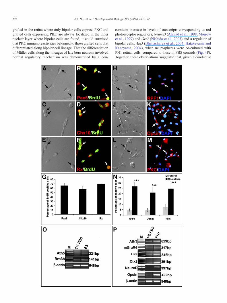
292 A.V. Das et al. / Developmental Biology 299 (2006) 283–302
grafted in the retina where only bipolar cells express PKC andgrafted cells expressing PKC are always localized in the innernuclear layer where bipolar cells are found, it could surmisedthat PKC immunoreactivities belonged to those grafted cells thatdifferentiated along bipolar cell lineage. That the differentiationof Müller cells along the lineages of late born neurons involvednormal regulatory mechanism was demonstrated by a con-
comitant increase in levels of transcripts corresponding to rodphotoreceptor regulators, NeuroD (Ahmad et al., 1998; Morrowet al., 1999) and Otx2 (Nishida et al., 2003) and a regulator ofbipolar cells, Ath3 (Bhattacharya et al., 2004; Hatakeyama andKageyama, 2004), when neurospheres were co-cultured withPN1 retinal cells, compared to those in FBS controls (Fig. 4P).Together, these observations suggested that, given a conducive
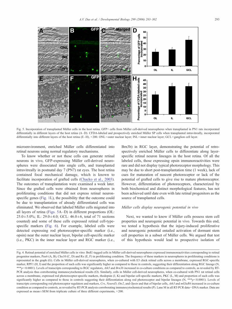
Fig. 5. Incorporation of transplanted Müller cells in the host retina. GFP+ cells from Müller cell-derived neurospheres when transplanted in PN1 rats incorporateddifferentially in different layers of the host retina (A–D). CFDA-labeled and prospectively enriched Müller SP cells when transplanted intravitreally, incorporateddifferentially into different layers of the host retina (E–H), ×200. ONL=outer nuclear layer; INL=inner nuclear layer; GCL=ganglion cell layer.
293A.V. Das et al. / Developmental Biology 299 (2006) 283–302
microenvironment, enriched Müller cells differentiated intoretinal neurons using normal regulatory mechanisms.
To know whether or not these cells can generate retinalneurons in vivo, GFP-expressing Müller cell-derived neuro-spheres were dissociated into single cells, and transplantedintravitreally in postnatal day 7 (PN7) rat eyes. The host retinacontained focal mechanical damage, which is known tofacilitate incorporation of grafted cells (Chacko et al., 2003).The outcomes of transplantation were examined a week later.Since the grafted cells were obtained from neurospheres inproliferating conditions that did not express retinal neuron-specific genes (Fig. 1L), the possibility that the outcome couldbe due to transplantation of already differentiated cells waseliminated. We observed that labeled Müller cells migrated intoall layers of retina (Figs. 5A–D) in different proportions (OL:25.0±5.0%; IL: 29.0±4.0; GCL: 46.0±6, total of 71 sectionscounted) and some of these cells expressed retinal cell-type-specific markers (Fig. 6). For example, labeled cells weredetected expressing rod photoreceptor-specific marker (i.e.,opsin) near the outer nuclear layer, bipolar cell-specific marker(i.e., PKC) in the inner nuclear layer and RGC marker (i.e.,
Fig. 4. Retinal potential of enriched Müller cells in vitro: BrdU-tagged cells in Müllerprogenitor markers, Pax6 (A, B), Chx10 (C, D) and Rx (E, F) in proliferating conditiorepresented in the graph (G). Cells in Müller cell-derived neurospheres, when co-cumarker, RPF1 (H, I) and the proportion of such cells was significantly higher as comp***p<0.0001). Levels of transcripts corresponding to RGC regulators, Ath5 and Brn3PCR analysis thus corroborating immunocytochemical results (O). Similarly, cells inacross a membrane, expressed rod photoreceptor-specific markers, rhodopsin (J, K)significantly higher as compared to those in controls suggesting their differentiatiotranscripts corresponding rod photoreceptor regulators and markers, Crx, NeuroD, Otcondition as compared to controls, as revealed by RT-PCR analysis corroborating immexpressed as mean±SEM from triplicate culture of three different experiments, ×20
Brn3b) in RGC layer, demonstrating the potential of retro-spectively enriched Müller cells to differentiate along layer-specific retinal neuron lineages in the host retina. Of all thelabeled cells, those expressing opsin immunoreactivities wererare and did not display typical photoreceptor morphology. Thismay be due to short post-transplantation time (1 week), lack ofcues for maturation of nascent photoreceptor or lack of thepotential of grafted cells to give rise to mature photoreceptor.However, differentiation of photoreceptors, characterized byboth biochemical and distinct morphological features, has notbeen achieved until date even with late retinal progenitors as thesource of transplanted cells.
Müller cells display neurogenic potential in vivo
Next, we wanted to know if Müller cells possess stem cellproperties and neurogenic potential in vivo. Towards this end,we tested a hypothesis that the injury-induced proliferativeand neurogenic potential entailed activation of dormant stemcell properties in a subset of Müller cells. We argued that testof this hypothesis would lead to prospective isolation of
cell-derived neurospheres expressed immunoreactivities corresponding to retinaln. The frequency of these markers in neurospheres in proliferating conditions isltured with E3 chick retinal cells across a membrane, expressed RGC-specificared to those in controls, suggesting their differentiation along RGC lineage (N,b increased in co-culture conditions as compared to controls, as revealed by RT-Müller cell-derived neurospheres, when co-cultured with PN1 rat retinal cells
and bipolar cell-specific markers, PKC (L, M) and proportion of such cells wasn along rod photoreceptor and bipolar lineages (N, ***p<0.0001). Levels ofx2, and Opsin and that of bipolar cells, Ath3 and mGluR6 increased in co-cultureunocytochemical results (P). LaneM in all RT-PCR data=DNAmarker. Data are0.
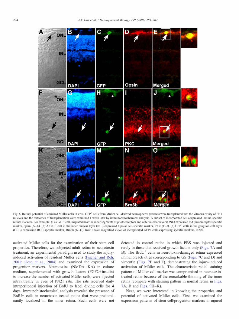
Fig. 6. Retinal potential of enriched Müller cells in vivo: GFP+ cells from Müller cell-derived neurospheres (arrows) were transplanted into the vitreous cavity of PN1rat eyes and the outcomes of transplantation were examined 1 week later by immunohistochemical analysis. A subset of incorporated cells expressed lamina-specificretinal markers. For example: (1) a GFP+ cell, migrated near the inner segments of photoreceptors and outer nuclear layer (ONL) expressed rod photoreceptor-specificmarker, opsin (A–E). (2) A GFP+ cell in the inner nuclear layer (INL) expressed bipolar cell-specific marker, PKC (F–J). (3) GFP+ cells in the ganglion cell layer(GCL) expression RGC-specific marker, Brn3b (K–O). Inset shows magnified views of incorporated GFP+ cells expressing specific markers, ×200.
294 A.V. Das et al. / Developmental Biology 299 (2006) 283–302
activated Müller cells for the examination of their stem cellproperties. Therefore, we subjected adult retina to neurotoxintreatment, an experimental paradigm used to study the injury-induced activation of resident Müller cells (Fischer and Reh,2001; Ooto et al., 2004) and examined the expression ofprogenitor markers. Neurotoxins (NMDA+KA) in culturemedium, supplemented with growth factors (FGF2+insulin)to increase the number of activated Müller cells, were injectedintravitreally in eyes of PN21 rats. The rats received dailyintraperitoneal injection of BrdU to label diving cells for 4days. Immunohistochemical analysis revealed the presence ofBrdU+ cells in neurotoxin-treated retina that were predomi-nantly localized in the inner retina. Such cells were not
detected in control retina in which PBS was injected andrarely in those that received growth factors only (Figs. 7A andB). The BrdU+ cells in neurotoxin-damaged retina expressedimmunoreactivities corresponding to GS (Figs. 7C and D) andvimentin (Figs. 7E and F), demonstrating the injury-inducedactivation of Müller cells. The characteristic radial stainingpattern of Müller cell marker was compromised in neurotoxin-treated retina because of the remarkable thinning of the innerretina (compare with staining pattern in normal retina in Figs.7A, B and Figs. 9H–K).
Next, we were interested in knowing the properties andpotential of activated Müller cells. First, we examined theexpression patterns of stem cell/progenitor markers in injured
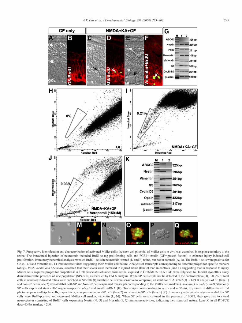
Fig. 7. Prospective identification and characterization of activated Müller cells: the stem cell potential of Müller cells in vivo was examined in response to injury to theretina. The intravitreal injection of neurotoxin included BrdU to tag proliferating cells and FGF2+insulin (GF=growth factors) to enhance injury-induced cellproliferation. Immunocytochemical analysis revealed BrdU+ cells in neurotoxin-treated (D and F) retina, but not in controls (A, B). The BrdU+ cells were positive forGS (C, D) and vimentin (E, F) immunoreactivities suggesting their Müller cell nature. Analysis of transcripts corresponding to different progenitor-specific markers(abcg2, Pax6, Nestin and Musashi1) revealed that their levels were increased in injured retina (lane 2) than in controls (lane 1), suggesting that in response to injuryMüller cells acquired progenitor properties (G). Cell dissociates obtained from retina, exposed to GF/NMDA+KA+GF, were subjected to Hoechst dye efflux assaydemonstrated the presence of side population (SP) cells, as revealed by FACS analysis. While SP cells could not be detected in the control retina (H), ∼0.2% of totalcells in neurotoxin-treated retina were enriched as SP cells (I) and these cells were sensitive to verapamil, an inhibitor of ABCG2 (J). RT-PCR analysis of SP (lane 1)and non-SP cells (lane 2) revealed that both SP and Non-SP cells expressed transcripts corresponding to the Müller cell markers (Vimentin, GS and CyclinD3) but onlySP cells expressed stem cell-/progenitor-specific abcg2 and Nestin mRNA (K). Transcripts corresponding to opsin and mGluR6, expressed in differentiated rodphotoreceptors and bipolar cells, respectively, were present in non-SP cells (lane 2) and absent in SP cells (lane 1) (K). Immunocytochemical analysis revealed that SPcells were BrdU-positive and expressed Müller cell marker, vimentin (L, M). When SP cells were cultured in the presence of FGF2, they gave rise to clonalneurospheres consisting of BrdU+ cells expressing Nestin (N, O) and Musashi (P, Q) immunoreactivities, indicating their stem cell nature. Lane M in all RT-PCRdata=DNA marker, ×200.
295A.V. Das et al. / Developmental Biology 299 (2006) 283–302
296 A.V. Das et al. / Developmental Biology 299 (2006) 283–302
and control retina, which led to the second approach, i.e.,prospective enrichment and characterization of activated Müllercells. An examination of the expression of stem cell-/progenitor-specific transcripts (abcg2, Pax6, Nestin andMusashi1) revealed that their levels were higher in injuredretina than in controls (Fig. 7G). Interestingly, one of the geneswhose transcripts appeared to be injury induced was abcg2 thatencodes a protein belonging to the ABC membrane transporterfamily, which includes drug efflux pumps (Krishnamurthy andSchuetz, 2006). Expression of ABCG2 enables cells topreferentially exclude a fluorescent DNA intercalating dyecalled Hoechst 33342, thus allowing their enrichment as sidepopulation (SP) cells, using fluorescent activated cell sorting(FACS) (Bhattacharya et al., 2003; Goodell et al., 1996;Krishnamurthy and Schuetz, 2005). The SP cell phenotype hasemerged as a universal feature of stem cells including thosefrom blood (Goodell et al., 1996), heart (Martin et al., 2004),gonads (Lassalle et al., 2004) and brain (Ahmad et al., 2004;Hulspas and Quesenberry, 2000). We sought to prospectivelyenrich activated Müller cells as SP cells using the Hoechst dyeefflux assay. The assay showed that SP cells were not detectedin the control retina (Fig. 7H). In contrast, when the assay wasperformed on cell dissociates obtained from neurotoxin-injuredretina, ∼0.2% of the total cells were enriched as SP cells (Fig.7I). Treatment of cell dissociates with verapamil, an inhibitor ofABCG2 transporter, led to a dercrease in the proportion of SPcells, demonstrating the specificity of SP cell phenotype (Fig.7J). Together, these observations demonstrated that the injury-induced activation of abcg2 was accompanied by an increase inthe proportion SP cells in the injured retina.
The next question was whether or not the SP cellsrepresented activated Müller cells and if they had the abilityto generate neurospheres. We addressed this question, first, byexamining the expression of transcripts corresponding to Müllercell- and stem cell-specific markers in SP and non-SP (NSP)cells. We observed that SP cells expressed both Müller cell(Vimentin, GS and CyclinD3)- and stem cell (i.e., Nestin andabcg2)-specific markers, attesting to their Müller cell identityand yet revealing their progenitor nature, a proof of injury-induced activation (Fig. 7J). In contrast, NSP cells expressedMüller cell (Vimentin, GS and CyclinD3)-, rod photoreceptor(Opsin)- and bipolar cell (mGluR6)-specific transcripts suggest-ing their post-mitotic and terminally differentiated nature (Fig.7K). Second, we carried out immunocytochemical analysis onSP cells, which revealed that they were BrdU-positive andexpressed vimentin, thus corroborating their proliferative natureand Müller cell identity (Figs. 7L and M). When SP cells werecultured in the presence FGF2, neurospheres were generatedwhich, like those generated by enriched Müller cells, consistedof proliferating cells expressing stem cell markers, Nestin (Figs.7N and O) and Musashi1 (Figs. 7P and Q).
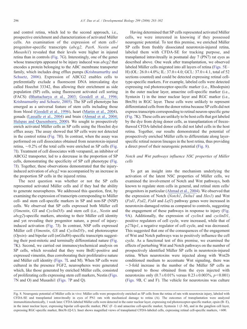
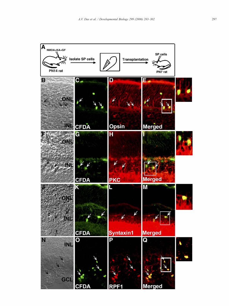
Fig. 8. Neurogenic potential of Müller cells in vivo: Müller cells were prospectivelyCFDA-SE and transplanted intravitreally in eyes of PN1 rats with mechanicimmunohistochemically, 1 week later. CFDA-labeled Müller cells were detected in thin the inner nuclear layer, expressing bipolar cell-specific marker, PKC (F–I) and aexpressing RGC-specific marker, Brn3b (Q-U). Inset shows magnified views of tran
Having determined that SP cells represented activated Müllercells, we were interested in knowing if they possessedneurogenic potential. To test this premise, we enriched MüllerSP cells from freshly dissociated neurotoxin-injured retina,labeled them with CFDA-SE for tracking purpose, andtransplanted intravitreally in postnatal day 7 (PN7) rat eyes asdescribed above. One week after transplantation, we observedlabeled Müller cells migrated into all layers of retina (Figs. 5E–H) (OL: 26.0±4.0%; IL: 37.0±4.0; GCL: 37.0±4.1, total of 52sections counted) and could be detected expressing retinal cell-type-specific markers. For example, labeled cells were detectedexpressing rod photoreceptor-specific marker (i.e., Rhodopsin)in the outer nuclear layer, amacrine cell-specific marker (i.e.,Syntaxin 1) in the inner nuclear layer and RGC marker (i.e.,Brn3b) in RGC layer. These cells were unlikely to representdifferentiated cells from the donor retina because SP cells did notexpress transcripts corresponding to retinal neuron-specific gene(Fig. 7K). These cells are unlikely to be host cells that got labeledby the dye from dying donor cells, as transplantation of freeze-thawed CFDA-labeled donor cells did not label cells in the hostretina. Together, our results demonstrated the potential ofprospectively enriched Müller cells to differentiate along layer-specific retinal neuron lineages in the host retina, thus providinga direct proof of their neurogenic potential (Fig. 8).
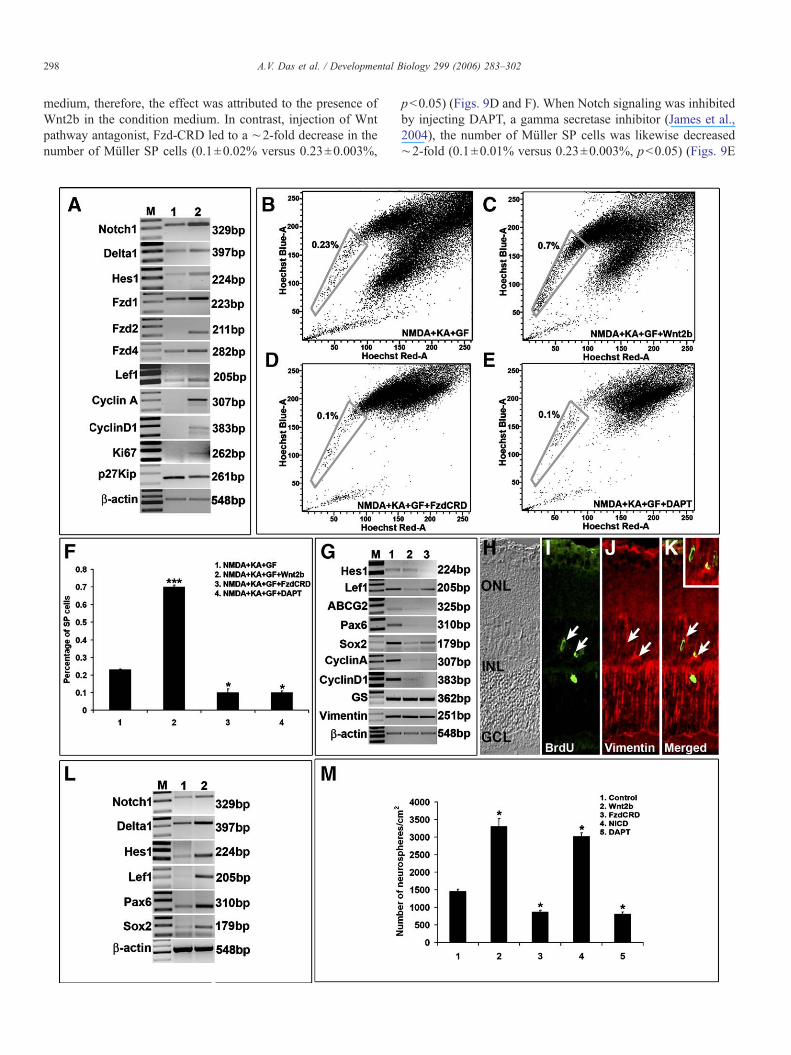
Notch and Wnt pathways influence NSC properties of Müllercells
To get an insight into the mechanism underlying theactivation of the latent NSC properties of Müller cells, weexamined the involvement of Notch and Wnt pathways, bothknown to regulate stem cells in general, and retinal stem cells/progenitors in particular (Ahmad et al., 2004). We observed thatthe expression of Notch (Notch1, Delta1 and Hes1) and Wnt(Fzd1, Fzd2, Fzd4 and Lef1) pathway genes were increased inneurotoxin-damaged retina as compared to controls, suggestingthe engagement of these pathways in response to injury (Fig.9A). Additionally, the expression of cyclinA and cyclinD1,positive regulators of cell cycle, were increased, while that ofp27kip1, a negative regulator of cell cycle, and was decreased.This suggested that one of the consequences of the engagementof Wnt and Notch pathways was to positively influence the cellcycle. As a functional test of this premise, we examined theeffects of perturbing Wnt and Notch pathways on the number ofprospectively identified Müller SP cells in neurotoxin-injuredretina. When neurotoxins were injected along with Wnt2bconditioned medium to accentuate Wnt signaling, there was∼3-fold increase in the number of the Müller SP cells ascompared to those obtained from the eyes injected withneurotoxins only (0.7±0.01% versus 0.23±0.003%, p<0.001)(Figs. 9B, C and F). The vehicle for neurotoxins was culture
enriched as SP cells from the retina of rats with neurotoxin injury, labeled withal damage to retina (A). The outcomes of transplantation were analyzede outer nuclear layer, expressing rod photoreceptor-specific marker, opsin (B–E),macrine cell-specific marker, Syntaxin 1 (J–M), and in the ganglion cell layer,splanted CFDA-labeled cells, expressing retinal cell-specific markers, ×600.
297A.V. Das et al. / Developmental Biology 299 (2006) 283–302
298 A.V. Das et al. / Developmental Biology 299 (2006) 283–302
medium, therefore, the effect was attributed to the presence ofWnt2b in the condition medium. In contrast, injection of Wntpathway antagonist, Fzd-CRD led to a ∼2-fold decrease in thenumber of Müller SP cells (0.1±0.02% versus 0.23±0.003%,
p<0.05) (Figs. 9D and F). When Notch signaling was inhibitedby injecting DAPT, a gamma secretase inhibitor (James et al.,2004), the number of Müller SP cells was likewise decreased∼2-fold (0.1±0.01% versus 0.23±0.003%, p<0.05) (Figs. 9E
299A.V. Das et al. / Developmental Biology 299 (2006) 283–302
and F). Taken together, these observations suggested that bothWnt and Notch pathways were involved in the emergence ofMüller SP cell phenotype in injured retina. The observations thatthe number of Müller SP cells in the presence of antagonists ofWnt (Fzd-CRD) and Notch (DAPT) signaling was lower thanthat in controls suggested the involvement of endogenous Wntand Notch signaling in the maintenance of stem cell properties ofMüller cells in response to injury (Figs. 9B, D–F). To test thespecificity and intracellular response to perturbation in Wnt andNotch signaling, cells from neurotoxin-injured retina (controls)and similarly injured retina in which Wnt and Notch signalingpathways were attenuated by Fzd-CRD and DAPT injection,respectively, were collected and subjected to RT-PCR analysis(Fig. 9G). In all groups, constitutive expression of vimentin andGS was observed, attesting to the fact that the Müller cellphenotype remained unchanged in response to injury. Tran-scripts corresponding to effectors of Notch (Hes1) and Wnt(Lef1) and those encoding progenitor regulators and markers(abcg2, Pax6 and Sox2) could be detected in cells in controlgroups, but their levels decreased significantly in groups inwhich Wnt and Notch signaling had been attenuated. Similarly,levels of cyclinA and cyclinD1 transcripts, present in the controlsgroups, were decreased after the attenuation of Wnt and Notchsignaling. Together, these observations suggested that both Wntand Notch signaling were involved in injury-induced activationof stem cell properties ofMüller cells in vivo. Since the exposureof injured retina to Wnt2b conditioned medium caused aremarkable increase in Müller SP cells, we wanted to know ifaccentuation of Wnt signaling could activate Müller cells inuninjured retina. The identification of BrdU+ cells expressingvimentin in the retina injected with Wnt2b (Figs. 9H–K)demonstrated that Wnt signaling might regulate Müller cells invivo in the normal retina.
Do Wnt and Notch pathways regulate stem cell properties ofMüller cells enriched from normal adult retina? Levels ofNotch 1, Delta1, Hes1 and Lef1 transcripts were increased inenriched Müller cells in proliferating conditions relative tothose not exposed to FGF2, suggesting that Wnt and Notchpathway are operational in vitro in sustaining stem cell pro-
Fig. 9. Involvement of Notch and Wnt pathways in stem cell potential of Müller cellsand Hes1) and Wnt (Fzd1, Fzd2, Fzd4 and Lef1) pathways by RT-PCR revealed that tto controls (A, lane 2), suggesting a role for Notch and Wnt signaling in Müller cell acyclinD1) and an indicator (Ki67), and a decrease in cell cycle inhibitor (p27Kip1)suggesting a shift in cell cycle stages in response to neurotoxin injury (A, lane 1). Towas influenced by Wnt and Notch signaling, neurotoxin injury was done in the prepathway antagonist, DAPT. The number of Müller SP cells increased in the presence onumber decreased in the presence of Fzd-CRD (D and F) and DAPT (E and F) asphenotype of Müller cells was sensitive to Notch and Wnt signaling. RT-PCR analysiwere inhibited by Fzd-CRD and DAPT, respectively, revealed that transcripts correspoof cell cycle regulators (cyclinA and cyclinD1), neuronal regulators (Sox2 and Pax6)(lane1) (G). Levels of vimentin and GS transcripts remained unchanged, suggesting tMüller cell phenotype were affected through these pathways during injury. There wasretina (H–K and inset) suggesting that Wnt signaling regulates proliferation of Müllthat of neural stem cell regulators, Sox2 and Pax6 were increased in enriched Mülleexposed to mitogens (L, lane 1), suggesting that Notch and Wnt pathways influencgeneration of clonal neurospheres by enriched Müller cells was enhanced and attenu(M). A similar increase and decrease in the number of neurospheres was observed wheobservations suggested that both Wnt and Notch signaling regulated stem cell propeculture of three different experiments. Lane M in all RT-PCR data=DNA marker. *
perties in enriched Müller cells (Fig. 9L). There was asignificant increase in the number of neurospheres generatedwhen enriched Müller cells were cultured in the presence ofWnt2b to accentuate Wnt signaling and a significant decreasewhen cultured in the presence of Fzd-CRD to attenuate it ascompared to controls (Wnt2b; 3302±221 versus 1460±55,p<0.05; Fzd-CRD; 869±50 versus 1460±55, p<0.05) (Fig.9M). Similarly, there was a significant increase in the numberof neurospheres when enriched Müller cells were transfectedwith NICD to constitutively activate Notch signaling and adecrease in the presence of DAPT to attenuate it as compared tocontrols (NICD, 3025±101 versus 1460±55, p<0.05; 811±58versus 1460±55, p<0.05) (Fig. 9M). These observations thatperturbation of Wnt and Notch pathways affected generation ofneurospheres suggest their roles in regulating the stem cellproperties of the Müller cells enriched from the normal retina.The observations that the number of neurospheres generated inthe presence of antagonists of Wnt (Fzd-CRD) and Notch(DAPT) signaling was lower than that in controls suggested theinvolvement of endogenous Wnt and Notch signaling in themaintenance of stem cell properties of enriched Müller cells(Fig. 9M).
Discussion
Müller cells morphologically and biochemically resembleradial glia. For example, Müller cells elaborate processesradiating towards the outer and inner surfaces of the retina andexpress markers such as Vimentin (Dahl et al., 1981; Schnitzer,1988), GLAST (Hartfuss et al., 2001; Lehre et al., 1997) andNestin (Lendahl et al., 1990; Walcott and Provis, 2003), whichare also expressed by radial glia. We have tested the hypothesisthat, like radial glia, Müller cells possess NSC potential, whichremains dormant under the constraints imposed by the non-neurogenic nature of the niche in the adult retina. Their NSCproperties become apparent only when the Müller cellenvironment is altered, either by removing them from theirniche or perturbing the niche. This hypothesis was confirmed bythe following observations.
: analysis of transcripts corresponding to components of Notch (Notch1, Delta1,heir levels increased in the retina with neurotoxin injury (A, lane 1) as comparedctivation in vivo. There was an increase in the cell cycle regulators (cyclinA andin the neurotoxin-treated retina (A, lane 2) when compared to uninjured retina,determine whether or not the emergence of the Müller SP cells in injured retinassence of Wnt pathway agonist (Wnt2b) and antagonist (Fzd-CRD), and Notchf Wnt2b (C and F) as compared to controls (B and F). In contrast, Müller SP cellcompared to controls (B and F). These observations suggested that the SP cells of neurotoxin-injured retina in which Wnt (lane 2) and Notch (lane 3) pathwaysnding to intercellular regulators of these pathways, Lef1 andHes1 including thatand stem cell regulator/marker (abcg2) were decreased, as compared to controlshat stem cell properties such as cell proliferation and SP cell phenotype and notan increase in the vimentin-expressing BrdU+ cells in the Wnt2b treated normaler cells in vivo. Levels of Notch1, Delta1, Hes1 and Lef1 transcripts along withr cells in proliferating conditions (L, lane 2) as compared to those that were noted the proliferative and neurogenic state of enriched Müller cells in vitro. Theated in the presence of Wnt2b and FzdCRD, respectively, compared to controlsn NICDwas over-expressed or in the presence of DAPT, respectively (M). Theserties of enriched Müller cells. Data are expressed as mean±SEM from triplicate**p<0.001, *p<0.05, ×200.

300 A.V. Das et al. / Developmental Biology 299 (2006) 283–302
First, Müller cells, enriched from the normal retina, generateclonal neurospheres. These neurospheres consist of self-renewing and multipotent cells, thus demonstrating the cardinalfeatures of stem cells in vitro. A similar approach has beenpreviously used to confirm the stem cell nature of radial glia andSVZ astrocytes, enriched from the adult brain (Doetsch et al.,1999; Laywell et al., 2000; Merkle et al., 2004). Furthermore,cells in the clonal neurospheres demonstrate the capability ofdifferentiating into functional neurons in vitro, and ability togenerate site-specific neurons upon homotopic and heterotopictransplantation. Second, changes in the milieu of the adultretina, due to neurotoxin injury, confer SP cell phenotype onMüller cells. The SP cell phenotype has emerged as a universalfeature of stem cells (Das et al., 2005a; Pevny and Rao, 2003).Evidence suggests that the molecular determinant of SP cellphenotype in a wide variety of stem cells, including those fromblood (Goodell et al., 1996), heart (Martin et al., 2004), gonads(Lassalle et al., 2004) and brain (Ahmad et al., 2004; Hulspasand Quesenberry, 2000), is the expression of ABCG2, a proteinbelonging to the ABC membrane transporter family whichinclude drug efflux pumps (Krishnamurthy and Schuetz, 2006).It is thought that ABCG2 may constitute a core group ofproteins expressed in stem cells to maintain their homeostasisand protect their genome from damage by excluding toxins andharmful chemicals (Das et al., 2005a) and maintain theiruncommitted state by excluding differentiation promotingfactor(s) (Krishnamurthy and Schuetz, 2006). The expressionof abcg2 and the associated SP cell phenotypes is a prominentfeature of retinal stem cells/progenitors throughout retinalhistogenesis (Das et al., 2005a). The expression of abcg2declines precipitously in the adult retina (data not shown) andthus few cells emerge as SP cells when the Hoechst dye effluxassay was carried out on uninjured retina. The emergence of SPcells in neurotoxin-injured retina, expressing neural progenitorregulators/markers, Pax6, Sox2 and Musashi1, is an indicationof the activation of NSC potential. This notion is furthersupported by the observation that the Müller SP cells cangenerate clonal neurospheres, consisting of self-renewing cellswith neural properties and potential, demonstrated both in vitroand in vivo.
Several studies have suggested that Müller cells can acquireneurogenic potential in response to injury to the retina. Forexample, it is thought that proliferating and apically migratingMüller cells in goldfish retina, injured by laser lesion, may bethe source of observed photoreceptor regeneration (Braisted etal., 1994). Studies in rats suggested mitogenic effects in theretina in response excitatory amino acids which was mediatedthrough action on glial cationic channels (Sahel et al., 1991).Müller cells have been observed to proliferate and expressprogenitor properties in neurotoxin-injured retina of postnatalchicken (Fischer and Reh, 2001) and adult mouse (Ooto et al.,2004). In both cases, the evidence of neurogenic potential wasindirect and based on the observation that BrdU-positive cellsin injured retina expressed markers corresponding to differ-entiated neurons or specific retinal cell types. Due to the lackof direct evidence, it remained possible that neurons formedafter injury were derived from a dormant population of non-
Müller stem cells within the postnatal retina, similar to thosefound in fish (Fischer and Reh, 2001). To test the neurogenicpotential of Müller cells, we demonstrated that enrichedMüller cells generated cells with biochemical, molecular andphysiological characteristics of a generic neuron in vitro andalso had the capacity to differentiate into specific retinalneurons both in vitro and in vivo. As a direct evidence ofneurogenic potential in vivo, we demonstrated that Müllercells, prospectively enriched as SP cells, integrated andgenerated lamina-specific retinal neurons after transplantationinto mechanically injured retina. Our observations, therefore,suggest that the neurogenic potential of Müller cells is aninherent, but dormant, feature. These cells express multiplestem cell/progenitor-specific genes whose expression isenvironment-sensitive. The dual phenotype of Müller cells isreminiscent of radial glia and suggestive of an ambivalentnature that allows them to carry out dual roles of supportingneuronal functions and participating in neurogenesis, when themicroenvironment allows.
The mechanism of activation of the dormant NSC propertiesand potential of Müller cells are likely to involve signal(s) thatintegrate cell-intrinsic and cell-extrinsic information. Ourobservations suggest that Notch and Wnt pathway may mediatethe integration of such signal(s). For example, the activities ofthese pathways are enhanced in Müller cells in response to injuryand that the stem cell properties of Müller cells are sensitive toperturbation of these pathways. Both Notch and Wnt pathways,like elsewhere in theCNS, regulate themaintenance of stem cells/progenitors in the developing retina (Ahmad et al., 2004; Kubo etal., 2003; Liu et al., 2003). Similar to its context-dependentinvolvement in radial glia development, the Notch pathway isutilized for the differentiation of Müller cells (Furukawa et al.,2000; James et al., 2004). Although the involvement of Wntsignaling in Müller cell differentiation has not been previouslyreported, its role in progenitor properties of radial glia hasrecently emerged (Zhou et al., 2004). Therefore, it is quite likelythat both radial glia andMüller cells retain their ability to respondto Notch and Wnt signaling long after their differentiation iscompleted as a part of their malleable nature. Therefore, injury-induced extrinsic cues, such as cytokines and growth factors maylead to the activation ofNotch (Faux et al., 2001; Liao et al., 2004)and Wnt (Viti et al., 2003; Zhou et al., 2004) pathways in Müllercells, prompting them to shift from a maintenance to a stem cellmode. These pathways act cooperatively towards the activation ofMüller cells (to be published elsewhere). Based on previousobservations that p27kip1 keeps Müller cells from entering cellcycle (Dyer and Cepko, 2000), and the recent evidence that Notchand Wnt signaling adversely affect the activities of p27kip1(Castelo-Branco et al., 2003; Murata et al., 2005; Sarmento et al.,2005), it is reasonable to postulate that Notch and Wnt signaling-dependent inhibition of p27kip1 may constitute one of the firststeps towards the shift of Müller cells to the stem cell mode. Theprocess is furthered by accentuating the expression of cell cycleregulators such as cyclins, neurogenic genes such as Sox2(Graham et al., 2003) and Pax6 (Heins et al., 2002; Kohwi et al.,2005) and homeostasis-promoting genes such as abcg2. Takentogether, our study suggests that the Müller cells are the NSCs of
301A.V. Das et al. / Developmental Biology 299 (2006) 283–302
the adult retina. These cells keep their stem cell potential dormantunder the constraints of non-neurogenic niche of the adult retina.When the environment changes demanding stem cell activity,they are capable of functioning asNSCs and as such can serve as atarget of therapy for regenerative medicine of the retina.
Acknowledgments
We would like to thank Dr. Spyros Artavanis-Tsakonas forthe Notch1 antibody, Dr. Shinichi Nakagawa for the Wnt2bconstruct, Dr. Jeremy Nathans for the FzdCRD constructs andDr. Anuja Ghorpade for the Iba-1 antibody. Thanks are due toDr. Jim Rogers for his valuable advice and guidance on LDA,Drs. Graham Sharp and Gregory Bennett for critiquing themanuscript and helpful suggestions, Dr. Kenneth Cowan for theadenovirus vectors and Gaurav Sahay for the technicalassistance. This work was supported by the Nebraska TobaccoSettlement Biomedical Research Development, NIH andResearch to Prevent Blindness.
References
Ahmad, I., Acharya, H.R., Rogers, J.A., Shibata, A., Smithgall, T.E., Dooley,C.M., 1998. The role of NeuroD as a differentiation factor in themammalian retina. J. Mol. Neurosci. 11, 165–178.
Ahmad, I., Dooley, C.M., Thoreson, W.B., Rogers, J.A., Afiat, S., 1999. In vitroanalysis of a mammalian retinal progenitor that gives rise to neurons andglia. Brain Res. 831, 1–10.
Ahmad, I., Tang, L., Pham, H., 2000. Identification of neural progenitors in theadult mammalian eye. Biochem. Biophys. Res. Commun. 270, 517–521.
Ahmad, I., Das, A.V., James, J., Bhattacharya, S., Zhao, X., 2004. Neural stemcells in the mammalian eye: types and regulation. Semin. Cell Biol. 15,53–62.
Alvarez-Buylla, A., Lim, D.A., 2004. For the long run: maintaining germinalniches in the adult brain. Neuron 41, 683–686.
Alvarez-Buylla, A., Garcia-Verdugo, J.M., Tramontin, A.D., 2001. A unifiedhypothesis on the lineage of neural stem cells. Nat. Rev., Neurosci. 2,287–293.
Barres, B.A., Silverstein, B.E., Corey, D.P., Chun, L.L., 1988. Immunological,morphological, and electrophysiological variation among retinal ganglioncells purified by panning. Neuron 1, 791–803.
Belecky-Adams, T., Tomarev, S., Li, H.S., Ploder, L., McInnes, R.R., Sundin,O., Adler, R., 1997. Pax-6, Prox 1, and Chx10 homeobox gene expressioncorrelates with phenotypic fate of retinal precursor cells. Invest. Ophthalmol.Visual Sci. 38, 1293–1303.
Benninger, F., Beck, H., Wernig, M., Tucker, K.L., Brustle, O., Scheffler, B.,2003. Functional integration of embryonic stem cell-derived neurons inhippocampal slice cultures. J. Neurosci. 23, 7075–7083.
Bhattacharya, S., Jackson, J.D., Das, A.V., Thoreson, W.B., Kuszynski, C.,James, J., Joshi, S., Ahmad, I., 2003. Direct identification and enrichment ofretinal stem cells/progenitors using Hoechst dye efflux assay. IOVS 44,2764–2773.
Bhattacharya, S., Dooley, C., Soto, F., Madson, J., Das, A.V., Ahmad, I., 2004.Involvement of Ath3 in CNTF-mediated differentiation of the late retinalprogenitors. Mol. Cell. Neurosci. 27, 32–43.
Blackshaw, S., Harpavat, S., Trimarchi, J., Cai, L., Huang, H., Kuo, W.P.,Weber, G., Lee, K., Fraioli, R.E., Cho, S.H., Yung, R., Asch, E.,Ohno-Machado, L., Wong, W.H., Cepko, C.L., 2004. Genomic analysis ofmouse retinal development. PLoS Biol. 2, E247.
Braisted, J.E., Essman, T.F., Raymond, P.A., 1994. Selective regeneration ofphotoreceptors in goldfish retina. Development 120, 2409–2419.
Castelo-Branco, G., Wagner, J., Rodriguez, F.J., Kele, J., Sousa, K., Rawal, N.,Pasolli, H.A., Fuchs, E., Kitajewski, J., Arenas, E., 2003. Differential
regulation of midbrain dopaminergic neuron development by Wnt-1, Wnt-3a, and Wnt-5a. Proc. Natl. Acad. Sci. U. S. A. 100, 12747–12752.
Chacko, D.M., Das, A.V., Zhao, X., James, J., Bhattacharya, S., Ahmad, I.,2003. Transplantation of ocular stem cells: the role of injury inincorporation and differentiation of grafted cells in the retina. Vision Res.43, 937–946.
Dahl, D., Rueger, D.C., Bignami, A., Weber, K., Osborn, M., 1981. Vimentin,the 57,000 molecular weight protein of fibroblast filaments, is the majorcytoskeletal component in immature glia. Eur. J. Cell Biol. 24, 191–196.
Das, A., James, J., Zhao, X., Bhattacharya, S., Ahmad, I., 2004. Involvement ofc-Kit receptor tyrosine kinase in the maintenance of ciliary epithelial neuralstem cells; interaction with notch signaling. Dev. Biol. 273, 87–105.
Das, A.V., Edakkot, S., Thoreson, W.B., James, J., Bhattacharya, S., Ahmad, I.,2005a. Membrane properties of retinal stem cells/progenitors. Prog. RetinalEye Res. 24, 663–681.
Das, A., James, J., Rahnenführer, J., Thoreson, W.B., Bhattacharya, S., Zhao,X., Ahmad, I., 2005b. Retinal properties and potential of the adultmammalian ciliary epithelium stem cells. Vision. Res. 45, 1653–1666.
Das, A.V., Zhao, X., James, J., Kim, M., Cowan, K.H., Ahmad, I., 2006. Neuralstem cells in the adult ciliary epithelium express GFAP and are regulated byWnt signaling. Biochem. Biophys. Res. Commun. 339, 708–716.
Doetsch, F., 2003. The glial identity of neural stem cells. Nat. Neurosci. 6,1127–1134.
Doetsch, F., Caille, I., Lim, D.A., Garcia-Verdugo, J.M., Alvarez-Buylla, A.,1999. Subventricular zone astrocytes are neural stem cells in the adultmammalian brain. Cell 97, 703–716.
Dyer, M.A., Cepko, C.L., 2000. Control of Muller glial cell proliferation andactivation following retinal injury. Nat. Neurosci. 3, 873–880.
Faux, C.H., Turnley, A.M., Epa, R., Cappai, R., Bartlett, P.F., 2001. Interactionsbetween fibroblast growth factors and Notch regulate neuronal differentia-tion. J. Neurosci. 21, 5587–5596.
Fischer, A.J., Reh, T.A., 2001. Muller glia are a potential source of neuralregeneration in the postnatal chicken retina. Nat. Neurosci. 4, 247–252.
Furukawa, T., Mukherjee, S., Bao, Z.Z., Morrow, E.M., Cepko, C.L., 2000. rax,Hes1, and notch1 promote the formation of Muller glia by postnatal retinalprogenitor cells. Neuron 26, 383–394.
Goodell, M.A., Brose, K., Paradis, G., Conner, A.S., Mulligan, R.C., 1996.Isolation and functional properties of murine hematopoietic stem cells thatare replicating in vivo. J. Exp. Med. 183, 1797–1806.
Gotz, M., Barde, Y.A., 2005. Radial glial cells defined and major intermediatesbetween embryonic stem cells and CNS neurons. Neuron 46, 369–372.
Graham, V., Khudyakov, J., Ellis, P., Pevny, L., 2003. SOX2 functions tomaintain neural progenitor identity. Neuron 39, 749–765.
Hamburger, V., Hamilton, H.L., 1951. A series of normal stages in thedevelopment of the chick embryo. J. Morphol. 88, 49–92.
Hartfuss, E., Galli, R., Heins, N., Gotz, M., 2001. Characterization of CNSprecursor subtypes and radial glia. Dev. Biol. 229, 15–30.
Hatakeyama, J., Kageyama, R., 2004. Retinal cell fate determination and bHLHfactors. Semin. Cell Biol. 15, 83–89.
Heins, N., Malatesta, P., Cecconi, F., Nakafuku, M., Tucker, K.L., Hack, M.A.,Chapouton, P., Barde, Y.A., Gotz, M., 2002. Glial cells generate neurons: therole of the transcription factor Pax6. Nat. Neurosci. 5, 308–315.
Hicks, D., Courtois, Y., 1990. The growth and behaviour of rat retinal Mullercells in vitro: I. An improved method for isolation and culture. Exp. Eye Res.51, 119–129.
Hsieh, J.C., Rattner, A., Smallwood, P.M., Nathans, J., 1999. Biochemicalcharacterization of Wnt-frizzled interactions using a soluble, biologicallyactive vertebrate Wnt protein. Proc. Natl. Acad. Sci. U. S. A. 96,3546–3551.
Hulspas, R., Quesenberry, P.J., 2000. Characterization of neurosphere cellphenotypes by flow cytometry. Cytometry 40, 245–250.
James, J., Das, A.V., Bhattacharya, S., Chacko, D.M., Zhao, X., Ahmad, I.,2003. In vitro generation of early-born neurons from late retinal progenitors.J. Neurosci. 23, 8193–8203.
James, J., Das, A.V., Rahnenfuhrer, J., Ahmad, I., 2004. Cellular and molecularcharacterization of early and late retinal stem cells/progenitors: differentialregulation of proliferation and context dependent role of Notch signaling.J. Neurobiol. 61, 359–376.
302 A.V. Das et al. / Developmental Biology 299 (2006) 283–302
Kohwi, M., Osumi, N., Rubenstein, J.L., Alvarez-Buylla, A., 2005. Pax6 isrequired for making specific subpopulations of granule and periglomerularneurons in the olfactory bulb. J. Neurosci. 25, 6997–7003.
Krishnamurthy, P., Schuetz, J.D., 2006. Role of ABCG2/BCRP in biology andmedicine. Annu. Rev. Pharmacol. Toxicol. 46, 381–410.
Kubo, F., Takeichi, M., Nakagawa, S., 2003. Wnt2b controls retinal celldifferentiation at the ciliary marginal zone. Development 130,587–598.
Lassalle, B., Bastos, H., Louis, J.P., Riou, L., Testart, J., Dutrillaux, B., Fouchet,P., Allemand, I., 2004. ‘Side Population’ cells in adult mouse testis expressBcrp1 gene and are enriched in spermatogonia and germinal stem cells.Development 131, 479–487.
Laywell, E.D., Rakic, P., Kukekov, V.G., Holland, E.C., Steindler, D.A., 2000.Identification of a multipotent astrocytic stem cell in the immature and adultmouse brain. Proc. Natl. Acad. Sci. U. S. A. 97, 13883–13888.
Lehre, K.P., Davanger, S., Danbolt, N.C., 1997. Localization of the glutamatetransporter protein GLAST in rat retina. Brain Res. 744, 129–137.
Lendahl, U., Zimmerman, L.B., McKay, R.D., 1990. CNS stem cells express anew class of intermediate filament protein. Cell 60, 585–595.
Liao, Y.F., Wang, B.J., Cheng, H.T., Kuo, L.H., Wolfe, M.S., 2004. Tumornecrosis factor-alpha, interleukin-1beta, and interferon-gamma stimulategamma-secretase-mediated cleavage of amyloid precursor proteinthrough a JNK-dependent MAPK pathway. J. Biol. Chem. 279,49523–49532.
Liu, H., Mohamed, O., Dufort, D., Wallace, V.A., 2003. Characterization of Wntsignaling components and activation of the Wnt canonical pathway in themurine retina. Dev. Dyn. 227, 323–334.
Malatesta, P., Hack, M.A., Hartfuss, E., Kettenmann, H., Klinkert, W.,Kirchhoff, F., Gotz, M., 2003. Neuronal or glial progeny: regionaldifferences in radial glia fate. Neuron 37, 751–764.
Marquardt, T., Ashery-Padan, R., Andrejewski, N., Scardigli, R., Guillemot, F.,Gruss, P., 2001. Pax6 is required for the multipotent state of retinalprogenitor cells. Cell 105, 43–55.
Martin, C.M., Meeson, A.P., Robertson, S.M., Hawke, T.J., Richardson, J.A.,Bates, S., Goetsch, S.C., Gallardo, T.D., Garry, D.J., 2004. Persistentexpression of the ATP-binding cassette transporter, Abcg2, identifies cardiacSP cells in the developing and adult heart. Dev. Biol. 265, 262–275.
Mathers, P.H., Grinberg, A., Mahon, K.A., Jamrich, M., 1997. The Rxhomeobox gene is essential for vertebrate eye development. Nature 387,603–607.
Merkle, F.T., Tramontin, A.D., Garcia-Verdugo, J.M., Alvarez-Buylla, A., 2004.Radial glia give rise to adult neural stem cells in the subventricular zone.Proc. Natl. Acad. Sci. U. S. A. 101, 17528–17532.
Meucci, O., Fatatis, A., Simen, A.A., Bushell, T.J., Gray, P.W., Miller, R.J.,1998. Chemokines regulate hippocampal neuronal signaling and gp120neurotoxicity. Proc. Natl. Acad. Sci. U. S. A. 95, 14500–14505.
Morrow, E.M., Furukawa, T., Lee, J.E., Cepko, C.L., 1999. NeuroD regulatesmultiple functions in the developing neural retina in rodent. Development126, 23–36.
Morshead, C.M., Garcia, A.D., Sofroniew, M.V., van Der Kooy, D., 2003. Theablation of glial fibrillary acidic protein-positive cells from the adult centralnervous system results in the loss of forebrain neural stem cells but notretinal stem cells. Eur. J. Neurosci. 18, 76–84.
Mu, X., Klein, W.H., 2004. A gene regulatory hierarchy for retinal ganglion cellspecification and differentiation. Semin. Cell Dev. Biol. 15, 115–123.
Murata, K., Hattori, M., Hirai, N., Shinozuka, Y., Hirata, H., Kageyama, R.,Sakai, T., Minato, N., 2005. Hes1 directly controls cell proliferation throughthe transcriptional repression of p27Kip1. Mol. Cell. Biol. 25, 4262–4271.
Newman, E.A., 1993. Inward-rectifying potassium channels in retinal glial(Muller) cells. J. Neurosci. 13, 3333–3345.
Nishida, A., Furukawa, A., Koike, C., Tano, Y., Aizawa, S., Matsuo, I.,Furukawa, T., 2003. Otx2 homeobox gene controls retinal photoreceptor cellfate and pineal gland development. Nat. Neurosci. 6, 1255–1263.
Noctor, S.C., Flint, A.C., Weissman, T.A., Dammerman, R.S., Kriegstein, A.R.,2001. Neurons derived from radial glial cells establish radial units inneocortex. Nature 409, 714–720.
Ooto, S., Akagi, T., Kageyama, R., Akita, J., Mandai, M., Honda, Y., Takahashi,M., 2004. Potential for neural regeneration after neurotoxic injury in theadult mammalian retina. Proc. Natl. Acad. Sci. U. S. A. 101, 13654–13659.
Pevny, L., Rao, M.S., 2003. The stem-cell menageriec. Trends Neurosci. 26,351–359.
Polleux, F., Giger, R.J., Ginty, D.D., Kolodkin, A.L., Ghosh, A., 1998.Patterning of cortical efferent projections by semaphorin–neuropilininteractions. Science 282, 1904–1906.
Reh, T.A., Fischer, A.J., 2001. Stem cells in the vertebrate retina. Brain Behav.Evol. 58, 296–305.
Reynolds, B.A., Rietze, R.L., 2005. Neural stem cells and neurospheres–re-evaluating the relationship. Nat. Methods 2, 333–336.
Roque, R.S., Caldwell, R.B., 1993. Isolation and culture of retinal microglia.Curr. Eye Res. 12, 285–290.
Sahel, J.A., Albert, D.M., Lessell, S., Adler, H., McGee, T.L., Konrad-Rastegar,J., 1991. Mitogenic effects of excitatory amino acids in the adult rat retina.Exp. Eye Res. 53, 657–664.
Sarmento, L.M., Huang,H., Limon,A., Gordon,W., Fernandes, J., Tavares,M.J.,Miele, L., Cardoso, A.A., Classon,M., Carlesso, N., 2005. Notch1modulatestiming of G1-S progression by inducing SKP2 transcription and p27 Kip1degradation. J. Exp. Med. 202, 157–168.
Schnitzer, J., 1988. Immunocytochemical studies on the development ofastrocytes, Muller (glial) cells, and oligodendrocytes in the rabbit retina.Brain Res. Dev. Brain Res. 44, 59–72.
Seri, B., Garcia-Verdugo, J.M., McEwen, B.S., Alvarez-Buylla, A., 2001.Astrocytes give rise to new neurons in the adult mammalian hippocampus.J. Neurosci. 21, 7153–7160.
Vandier, D., Rixe, O., Besnard, F., Kim, M., Rikiyama, T., Goldsmith, M.,Brenner, M., Gouyette, A., Cowan, K.H., 2000. Inhibition of glioma cells invitro and in vivo using a recombinant adenoviral vector containing anastrocyte-specific promoter. Cancer Gene Ther. 7, 1120–1126.
Vicario-Abejon, C., Cunningham, M.G., McKay, R.D., 1995. Cerebellarprecursors transplanted to the neonatal dentate gyrus express featurescharacteristic of hippocampal neurons. J. Neurosci. 15, 6351–6363.
Viti, J., Gulacsi, A., Lillien, L., 2003. Wnt regulation of progenitor maturation inthe cortex depends on Shh or fibroblast growth factor 2. J. Neurosci. 23,5919–5927.
Walcott, J.C., Provis, J.M., 2003. Muller cells express the neuronal progenitorcell marker nestin in both differentiated and undifferentiated human foetalretina. Clin. Exp. Ophthalmol. 31, 246–249.
Yang, Z., Ding, K., Pan, L., Deng, M., Gan, L., 2003. Math5 determines thecompetence state of retinal ganglion cell progenitors. Dev. Biol. 264,240–254.
Zhao, X., Das, A., Thoreson, W.B., Rodriguez-Sierra, J.F., Ahmad, I., 2002.Adult corneal limbal epithelium; a model for studying neural potential ofnon-neural stem cells. Dev. Biol. 250, 317–331.
Zhao, X., Das, A.V., Soto-Leon, F., Ahmad, I., 2005. Growth factor-responsiveprogenitors in the postnatal mammalian retina. Dev. Dyn. 232, 349–358.
Zhou, H., Yoshioka, T., Nathans, J., 1996. Retina-derived POU-domain factor-1:a complex POU-domain gene implicated in the development of retinalganglion and amacrine cells. J. Neurosci. 16, 2261–2274.
Zhou, C.J., Zhao, C., Pleasure, S.J., 2004. Wnt signaling mutants havedecreased dentate granule cell production and radial glial scaffoldingabnormalities. J. Neurosci. 24, 121–126.