Nicotine Uses Neuron-Glia Communication to Enhance Hippocampal Synaptic Transmission and Long-term...
12
Nicotine Uses Neuron-Glia Communication to Enhance Hippocampal Synaptic Transmission and Long-term Memory Mo ´ nica Lo ´ pez-Hidalgo 1 , Karla Salgado-Puga 1 , Reynaldo Alvarado-Martı´nez 1 , Andrea Cristina Medina 2 , Roberto A. Prado-Alcala ´ 2 , Jesu ´ s Garcı ´a-Colunga 1 * 1 Departamento de Neurobiologı ´a Celular y Molecular, Instituto de Neurobiologı ´a, Universidad Nacional Auto ´noma de Me ´ xico, Campus Juriquilla, Quere ´taro, Me ´ xico, 2 Departamento de Neurobiologı ´a Conductual y Cognitiva, Instituto de Neurobiologı ´a, Universidad Nacional Auto ´ noma de Me ´xico, Campus Juriquilla, Quere ´taro, Me ´xico Abstract Nicotine enhances synaptic transmission and facilitates long-term memory. Now it is known that bi-directional glia-neuron interactions play important roles in the physiology of the brain. However, the involvement of glial cells in the effects of nicotine has not been considered until now. In particular, the gliotransmitter D-serine, an endogenous co-agonist of NMDA receptors, enables different types of synaptic plasticity and memory in the hippocampus. Here, we report that hippocampal long-term synaptic plasticity induced by nicotine was annulled by an enzyme that degrades endogenous D-serine, or by an NMDA receptor antagonist that acts at the D-serine binding site. Accordingly, both effects of nicotine: the enhancement of synaptic transmission and facilitation of long-term memory were eliminated by impairing glial cells with fluoroacetate, and were restored with exogenous D-serine. Together, these results show that glial D-serine is essential for the long-term effects of nicotine on synaptic plasticity and memory, and they highlight the roles of glial cells as key participants in brain functions. Citation: Lo ´ pez-Hidalgo M, Salgado-Puga K, Alvarado-Martı ´nez R, Medina AC, Prado-Alcala ´ RA, et al. (2012) Nicotine Uses Neuron-Glia Communication to Enhance Hippocampal Synaptic Transmission and Long-term Memory. PLoS ONE 7(11): e49998. doi:10.1371/journal.pone.0049998 Editor: Jean-Pierre Mothet, CNRS - Universite ´ Aix Marseille, France Received June 8, 2012; Accepted October 19, 2012; Published November 21, 2012 Copyright: ß 2012 Lo ´ pez-Hidalgo et al. This is an open-access article distributed under the terms of the Creative Commons Attribution License, which permits unrestricted use, distribution, and reproduction in any medium, provided the original author and source are credited. Funding: This work was supported by Grants from Consejo Nacional de Ciencia y Tecnologı ´a Me ´xico (81911 and 128259) and by a Grant from Direccio ´ n General de Asuntos del Personal Acade ´ mico, U.N.A.M. (IN204809). The funders had no role in study design, data collection and analysis, decision to publish, or preparation of the manuscript. Competing Interests: The authors have declared that no competing interests exist. * E-mail: [email protected] Introduction The role of astrocytes in the central nervous system has been redefined, and they are now accepted as the third element in the synapses, alongside the pre- and postsynaptic neurons, astrocytes sense neuronal activity and respond with Ca 2+ elevations which, in turn, can induce the release of gliotransmitters such as ATP, glutamate, and D-serine [1,2]. For instance, astrocytes surround a substantial portion of synapses which are largely glutamatergic [3]. In the hippocampus almost 60% of synapses have an apposed astrocytic process [4,5]. The release of the gliotransmitter D- serine, an endogenous co-agonist of N-methyl-D-aspartate (NMDA) receptors [6,7], has been implicated in different types of activity-dependent synaptic plasticity including long-term potentiation and depression [8,9,10,11]. On the other hand, nicotine is considered to be the main addictive substance of tobacco, and it affects several brain functions due to its affinity for nicotinic acetylcholine receptors (nAChRs) [12]. Nicotine has effects on mnemonic functions; for example, nicotine improves long-term spatial memory, as mea- sured in the Morris Water Maze [13,14] and procedural learning, evaluated in the one-trial step-through inhibitory-avoidance task [15,16]. Administration of nicotine also enhances cognitive functions in pathological conditions such as Alzheimer’s disease [13,17,18], and it reverses memory deficits caused by a lesion of the cholinergic system [19,20,21,22]. The effect of nicotine on hippocampal synaptic plasticity has been widely documented [20,22] and is known to involve the activation and desensitization of nAChRs present on interneurons and pyramidal neurons [23,24,25,26,27]. Furthermore, the fact that hippocampal astrocytes express nAChRs [28,29] and respond to nAChR agonists by increasing their intracellular Ca 2+ concentration [30], raises the possibility that astrocytes could also be mediating nicotine effects in the hippocampus. Although there is no evidence of causality between long-term synaptic plasticity and memory, glial cells are critical for some types of memory [11,31,32]. Thus, glial cells could also mediate nicotine facilitation of long-term memory and the enhancement of synaptic transmis- sion. In the present work, we show that glial cells, very likely through the release of D-serine, are essential for nicotine potentiation of synaptic transmission and nicotine facilitation of long-term memory. Materials and Methods Ethics Statement All procedures were carried out in strict accordance with the recommendations of the National Institutes of Health Guide for the Care and Use of Experimental Animals and were approved by the local PLOS ONE | www.plosone.org 1 November 2012 | Volume 7 | Issue 11 | e49998
Transcript of Nicotine Uses Neuron-Glia Communication to Enhance Hippocampal Synaptic Transmission and Long-term...

Nicotine Uses Neuron-Glia Communication to EnhanceHippocampal Synaptic Transmission and Long-termMemoryMonica Lopez-Hidalgo1, Karla Salgado-Puga1, Reynaldo Alvarado-Martınez1, Andrea Cristina Medina2,
Roberto A. Prado-Alcala2, Jesus Garcıa-Colunga1*
1 Departamento de Neurobiologıa Celular y Molecular, Instituto de Neurobiologıa, Universidad Nacional Autonoma de Mexico, Campus Juriquilla, Queretaro, Mexico,
2 Departamento de Neurobiologıa Conductual y Cognitiva, Instituto de Neurobiologıa, Universidad Nacional Autonoma de Mexico, Campus Juriquilla, Queretaro, Mexico
Abstract
Nicotine enhances synaptic transmission and facilitates long-term memory. Now it is known that bi-directional glia-neuroninteractions play important roles in the physiology of the brain. However, the involvement of glial cells in the effects ofnicotine has not been considered until now. In particular, the gliotransmitter D-serine, an endogenous co-agonist of NMDAreceptors, enables different types of synaptic plasticity and memory in the hippocampus. Here, we report that hippocampallong-term synaptic plasticity induced by nicotine was annulled by an enzyme that degrades endogenous D-serine, or by anNMDA receptor antagonist that acts at the D-serine binding site. Accordingly, both effects of nicotine: the enhancement ofsynaptic transmission and facilitation of long-term memory were eliminated by impairing glial cells with fluoroacetate, andwere restored with exogenous D-serine. Together, these results show that glial D-serine is essential for the long-term effectsof nicotine on synaptic plasticity and memory, and they highlight the roles of glial cells as key participants in brainfunctions.
Citation: Lopez-Hidalgo M, Salgado-Puga K, Alvarado-Martınez R, Medina AC, Prado-Alcala RA, et al. (2012) Nicotine Uses Neuron-Glia Communication toEnhance Hippocampal Synaptic Transmission and Long-term Memory. PLoS ONE 7(11): e49998. doi:10.1371/journal.pone.0049998
Editor: Jean-Pierre Mothet, CNRS - Universite Aix Marseille, France
Received June 8, 2012; Accepted October 19, 2012; Published November 21, 2012
Copyright: � 2012 Lopez-Hidalgo et al. This is an open-access article distributed under the terms of the Creative Commons Attribution License, which permitsunrestricted use, distribution, and reproduction in any medium, provided the original author and source are credited.
Funding: This work was supported by Grants from Consejo Nacional de Ciencia y Tecnologıa Mexico (81911 and 128259) and by a Grant from Direccion Generalde Asuntos del Personal Academico, U.N.A.M. (IN204809). The funders had no role in study design, data collection and analysis, decision to publish, or preparationof the manuscript.
Competing Interests: The authors have declared that no competing interests exist.
* E-mail: [email protected]
Introduction
The role of astrocytes in the central nervous system has been
redefined, and they are now accepted as the third element in the
synapses, alongside the pre- and postsynaptic neurons, astrocytes
sense neuronal activity and respond with Ca2+ elevations which, in
turn, can induce the release of gliotransmitters such as ATP,
glutamate, and D-serine [1,2]. For instance, astrocytes surround a
substantial portion of synapses which are largely glutamatergic [3].
In the hippocampus almost 60% of synapses have an apposed
astrocytic process [4,5]. The release of the gliotransmitter D-
serine, an endogenous co-agonist of N-methyl-D-aspartate
(NMDA) receptors [6,7], has been implicated in different types
of activity-dependent synaptic plasticity including long-term
potentiation and depression [8,9,10,11].
On the other hand, nicotine is considered to be the main
addictive substance of tobacco, and it affects several brain
functions due to its affinity for nicotinic acetylcholine receptors
(nAChRs) [12]. Nicotine has effects on mnemonic functions; for
example, nicotine improves long-term spatial memory, as mea-
sured in the Morris Water Maze [13,14] and procedural learning,
evaluated in the one-trial step-through inhibitory-avoidance task
[15,16]. Administration of nicotine also enhances cognitive
functions in pathological conditions such as Alzheimer’s disease
[13,17,18], and it reverses memory deficits caused by a lesion of
the cholinergic system [19,20,21,22].
The effect of nicotine on hippocampal synaptic plasticity has
been widely documented [20,22] and is known to involve the
activation and desensitization of nAChRs present on interneurons
and pyramidal neurons [23,24,25,26,27]. Furthermore, the fact
that hippocampal astrocytes express nAChRs [28,29] and respond
to nAChR agonists by increasing their intracellular Ca2+
concentration [30], raises the possibility that astrocytes could also
be mediating nicotine effects in the hippocampus. Although there
is no evidence of causality between long-term synaptic plasticity
and memory, glial cells are critical for some types of memory
[11,31,32]. Thus, glial cells could also mediate nicotine facilitation
of long-term memory and the enhancement of synaptic transmis-
sion. In the present work, we show that glial cells, very likely
through the release of D-serine, are essential for nicotine
potentiation of synaptic transmission and nicotine facilitation of
long-term memory.
Materials and Methods
Ethics StatementAll procedures were carried out in strict accordance with the
recommendations of the National Institutes of Health Guide for the
Care and Use of Experimental Animals and were approved by the local
PLOS ONE | www.plosone.org 1 November 2012 | Volume 7 | Issue 11 | e49998

Animal Research Committee of the Instituto de Neurobiologıa at
Universidad Nacional Autonoma de Mexico.
Hippocampal SlicesTransverse hippocampal slices (400 mm thick) from 120–130 g
male Wistar rats were obtained in an ice-cold solution containing
(in mM): sucrose 238, KCl 3, NaHCO3 25, MgCl2 2.5, and
glucose 30 (pH 7.4). The slices were then transferred to a
submersion chamber for storage in artificial cerebrospinal fluid
(ACSF) containing (in mM): NaCl 126, KCl 3, NaHCO3 25,
MgCl2 1, CaCl2 2, and glucose 11 (pH 7.4). The slices were
allowed to stabilize for at least 1 h before starting the recordings. A
single slice was then gently transferred to the recording chamber
where it was superfused throughout the experiment with ACSF at
a flow rate of 2 ml/min. All solutions were continuously bubbled
with 95% O2/5% CO2 and maintained at room temperature (22–
25uC).
Electrophysiological RecordingsA concentric bipolar platinum electrode (25 mm diameter; FHC
Inc., Bowdoin ME, USA) was placed in the stratum radiatum of the
CA1 hippocampal region to stimulate Schaffer collaterals. Evoked
field potentials (EFP) were recorded in the stratum radiatum using
glass electrodes filled with 2 M NaCl and with a resistance of 1–
2 MV. When required, the paired-pulse protocol was applied to
induce synaptic responses. The test stimulus duration was 150 ms
with an interpulse interval of 60 ms (Fig. 1A). The stimulus
intensity was adjusted in each experiment to elicit 60–70% of the
maximum response. A paired-pulse was delivered every 15 s. The
paired-pulse ratio (PPR) was defined as the quotient between the
slope of the second response (P2) and the slope of the first response
(P1), i.e., P2/P1. This parameter was used to evaluate changes in
the probability of neurotransmitter release from Schaffer collat-
erals [33,34]. Each point on the time curse graphs corresponds to
the mean of 20 stimuli (5 min of recording). To induce long-term
potentiation of synaptic transmission, in the presence of 50 mM
picrotoxin (an antagonist of the GABAA receptors) we applied two,
1-s trains of high-frequency stimulation (HFS, 100 Hz) with a 5-s
inter-train interval. The EFP were recorded with an Axopatch-
200B amplifier (Axon Instruments, CA) and filtered at 5 KHz.
Data were acquired and stored for offline analysis with Digidata
1200, Clampfit 9.0 (Axon Instruments, CA) and Sigma plot 9.0
software.
Behavioral TestsAdult male Wistar rats (250–350 g at the time of surgery) were
housed individually in a temperature-controlled (24uC) colony
room and maintained on a 12-h/12-h light/dark cycle (lights on at
7:00 A.M.). Food and water were provided ad libitum throughout
the experiment.
Surgical procedure. The rats were adapted to the laboratory
vivarium for at least one week before surgery. They were
anesthetized with sodium pentobarbital (50 mg/kg, i.p.), received
atropine sulfate (1 mg/kg, i.p.), and were positioned in a
stereotaxic instrument (Stoelting Co., IL). Stainless steel guide
cannulae (23-gauge) were bilaterally implanted into the dorsal
hippocampus (AP = 24.0, L 62.6, V = 22.5); the nose bar was set
at 23.3 mm from the interaural line [35]. The cannulae were
affixed to the skull using two screws and dental acrylic, and a stylet
was inserted in each cannula and retained there at all times except
during the injections. The rats were allowed seven days to recover
from surgical procedures before the initiation of training.Training and memory tests. The rats were trained in a
one-trial step-through inhibitory-avoidance task. The training and
retention testing were carried out in an apparatus with two distinct
compartments, separated by a guillotine door. The safe compart-
ment (30630630 cm) had walls and lid of red-colored acrylic with
a floor of stainless steel bars (6 mm in diameter, separated by
9 mm). This compartment was illuminated by a 10-Watt light bulb
located in the center of its lid. The other, shock compartment had
front and back walls and floor made of stainless steel with end walls
and lid made of red-colored acrylic. The compartment was 30 cm
long and 25 cm deep. The walls and floor were shaped as a
trough, 20 cm wide at the top and 8 cm wide at the bottom. In the
middle of the floor, a 1.5-cm slot separated the two stainless steel
plates that made up the walls and floor. When in this
compartment, the rats were in contact with both plates that can
be electrified and, thereby, deliver aversive stimulation consistently
to every subject. A square-pulse stimulator (Grass S-48), in series
with a constant current unit (Grass CCU-1A), generated the
electric shock. The duration of shock and the measurement of
latencies to cross from one compartment to the other were
accomplished with automated equipment. The conditioning box
was located inside a dark, sound-proof room provided with
background masking noise.
Rats were placed in the safe compartment; 10 s later the
guillotine door was opened, and latency to enter was recorded
(training latency). When the rat was completely inside the dark
compartment the door was closed and a foot-shock (0.7 mA) was
delivered. After 5 s, the door was opened, allowing the animal to
escape into the first compartment (escape latency). After 30 s in
the safe compartment the animal was put back in its home cage.
Forty-eight hours later, during the retention test (long-term
memory), the same procedure was followed except that the foot-
shock was not delivered. The test was terminated either when the
rat entered the dark compartment or after 600 s without entry,
and a score of 600 was assigned to the retention latency.
Microinjection procedure. The bilateral infusions into the
hippocampus (0.5 ml/side) were made through 30-gauge injection
needles connected to a Hamilton microsyringe by polyethylene
tubing. The injection needles were inserted into the guide
cannulae and protruded 1 mm beyond the tip of the cannulae.
The infusion rate was 0.5 ml/min and was controlled by an
automated microinfusion pump (WPI, 220i). At the end of the
infusion, the injection needles remained inside the guide cannulae
for 60 s to insure diffusion away from the injector tip. The
injection procedure was carried out in a different room from that
in which training and testing took place.
DrugsFor electrophysiological experiments, the drugs were diluted in
ACSF and applied in the superfusion bath. In all experiments,
30 min of stable baseline was recorded before each drug
administration. Depending on the purpose of each experiment,
slices were incubated with D-(–)-2-amino-5-phosphonopentanoic
acid (AP5, 50 mM), D-serine (20 mM), 5,7-dichlorokynurenic acid
(DCKA, 200 nM), nicotine (1 mM), fluoroacetate (FAC, 5 mM),
mecamylamine (50 mM), or D-amino acid oxidase (DAAO,
0.1 U/ml). The DAAO enzyme was dialyzed for 8–10 h at 4uCagainst 20 mM sodium pyrophosphate pH 8–8.5 containing
10 mM flavin adenine dinucleotide and stored at 220uC until
use. For the behavioral tests, the drugs were diluted in 0.9% NaCl
and administrated at different times before training: FAC (5, 10,
20, or 40 mM at 40 min), D-serine (100 mM at 40 min) and AP5
(50 mM at 20 min) in the CA1 hippocampal region, or nicotine
(1 ml/kg at 15 min with doses of 0.2, 0.4, or 0.6 mg/kg)
subcutaneously. All the drugs were purchased from Sigma-Aldrich
(St. Louis MO).
Nicotine Effects Are Mediated by Glial Cells
PLOS ONE | www.plosone.org 2 November 2012 | Volume 7 | Issue 11 | e49998

Figure 1. Nicotine potentiation of synaptic transmission depends of glial cell activity. A, Experimental arrangement for recording theevoked field potential (EFP) using a stimulation electrode (Stim) located in Schaffer collaterals (SC). B, the EFP slope before, during, and after nicotineadministration (Nic; 1 mM, 7 min). The numerals in B (1, 2 and 3) indicate the time at which the representative traces (insets) were taken. The color ofthe numeral correlates with the color o the trace. The same code was used for the subsequent figures. C, The PPR and representative traces fromexperiments in B, before (Control, brown), during (black), and after nicotine administration (15 min, orange; 60 min, green). Columns represent themean 6 S.E.M. of results in each condition. Connected circles correspond to individual experiments. Effects on the EFP slope of nicotine combinedwith mecamylamine, a non-selective antagonist of nAChRs, (D, Mec; 50 mM); with AP5, an antagonist of NMDA receptors (E, F, AP5; 50 mM); or in thepresence of fluoroacetate (G, FAC; 5 mM). Insets, for this and subsequent figures, they show representative traces for EFP responses before (brown)and after nicotine administration (early, orange; late, green) in the absence and presence of the test drug. Horizontal bars indicate the timing of drugapplication. H, Summary of the experiments in B, D–G. For this and subsequent figures, the results are the mean 6 S.E.M. of the EFP slope expressedas percent of baseline.10–20 min (early, orange) and 50–60 min (late, green) after nicotine administration, in the absence or presence of the testdrug. The dashed line indicates the normalized basal level in each condition (*p,0.05, **p,0.01, one-way repeated-measures ANOVA, post hoc Fishertest).doi:10.1371/journal.pone.0049998.g001
Nicotine Effects Are Mediated by Glial Cells
PLOS ONE | www.plosone.org 3 November 2012 | Volume 7 | Issue 11 | e49998

Statistical AnalysisIn each of the electrophysiological experiments, data were
obtained from 5 to 11 slices. For the time course plots, the results
are shown as means 6 S.E.M. of the EFP slope, relative to
baseline (the mean of the first 30 min of recording). The early and
late effects correspond to the mean measured from 10 to 20 and 50
to 60 min, respectively, after electrical stimulation, nicotine, or
FAC administration. These two components were compared to
the baseline EFP slope (control) using the repeated-measures one-
way ANOVA. Due to changes produced by FAC, when nicotine
or HFS was preceded by FAC, the synaptic responses previous to
nicotine or HFS were used as another group to compare. To
determine if there were significant differences between measure-
ments, the post hoc Fisher test was used. When only two groups
were compared a Student’s t-test was used. If the measurements
were done in the same slices, then paired Student’s t-test was
utilized.
In the behavioral experiments, only the rats in which the
cannulae were located in the CA1 hippocampal region were
included in the analyses. The final sample size for each group was
between 6 and 11 rats. Because the measurement of retention was
truncated at 600 s, nonparametric statistics were used. Indepen-
dent Kruskal-Wallis analyses of variance were computed to
compare training, escape, and retention latencies among groups.
When appropriate, the Mann-Whitney U test was used to make
comparisons between any two groups.
Results
Glial Cells Are Required for Long-term Nicotine Effects inSynaptic Transmission
It is well known that nicotine modulates synaptic plasticity in the
hippocampus [20,36]. Thus, the effects of nicotine on synaptic
transmission were evaluated by measuring the EFP slope and the
PPR produced in the CA1 hippocampal region by stimulating
Schaffer collaterals (Fig. 1A).
First, we applied a brief administration (7 min) of a low
concentration of nicotine (1 mM), close to the brain nicotine
concentration after smoke one cigarette: 520–770 nM [37]. Thus,
nicotine by itself increased the EFP slope of P1 in hippocampal
slices (Fig. 1B). Maximal potentiation was observed 15 min after
nicotine administration (204645% relative to baseline). Later, the
responses of synaptic transmission diminished but remained above
baseline after 60 min of nicotine administration (135619%
relative to baseline; Fig. 1B).
The statistical analysis (ANOVA) of the early and late effects of
nicotine showed a significant treatment effect (F2,20 = 8.36;
p = 0.002). The Fisher test indicated that both early and late
components (see Materials and Methods) were significantly
different from the control (p,0.001, p = 0.03, respectively;
Fig. 1H). The analysis of the PPR showed a significant effect of
nicotine (F3,30 = 4.327; p = 0.012). The Fisher test indicated a
decrease of the PPR during the nicotine administration with
respect to the control (p = 0.007; Fig. 1C, right panel), without an
effect on the early and late components (p = 0.74, p = 0.78,
respectively; Fig. 1C). The role of nAChRs was revealed when
slices were preincubated with mecamylamine, a non-selective
nAChR antagonist; in this condition, nicotine did not modify the
EFP slope (F2,10 = 0.33, p = 0.72; Fig. 1D, H).
The involvement of NMDA receptors in nicotine potentiation of
synaptic transmission is controversial [27,36]. To determine if
these receptors were participating, nicotine was applied in the
continuous presence of an antagonist of the NMDA receptors,
AP5. In this condition, a small enhancement of the early
component was observed when nicotine was administrated,
without affecting the responses of the late component
(p = 0.028 p = 0.22, respectively; Fig. 1E, H). Additionally, once
established the nicotine potentiation of synaptic transmission (late
component), AP5 was applied and the EFP slope returned to the
baseline (p = 0.495 with respect to baseline; Fig. 1F, H), indicating
that nicotine induced a NMDA receptor-dependent long-lasting
synaptic plasticity. The administration of AP5 alone to hippo-
campal slices did not affect the EFP slope with respect to the
control (paired t-test, p = 0.89; Fig. S1A, C).
Because hippocampal astrocytes express functional nAChRs
[29,30,38] we asked whether glial cells were mediating long-term
enhancement of synaptic transmission by nicotine. To determine
the role of glial cells in this synaptic plasticity, we studied the
effects of FAC, a selective inhibitor of glial cell metabolism [39,40].
Perfusion of the hippocampal slices with FAC led to an increase in
the EFP slope relative to baseline (late, 150617%; paired t-test,
p = 0.02; Fig. S1B, C). To determine whether this increase was
also mediated by NMDA receptors, AP5 was applied after the
onset of FAC administration. Under this condition FAC still
increased the EFP slope (paired t-test, p = 0.02; Fig. S1B, C), which
suggest that the effect of FAC was not dependent on NMDA
receptors.
Interestingly, when nicotine was administrated in the presence
of FAC, a significant reduction of the synaptic responses was
observed in the early component (p = 0.048), without any effect on
the late component (p = 0. 326) Because FAC administration
increased synaptic transmission, a depotentiation of the responses
rather than a depression would better describe this effect [41].
Glial D-serine Is Necessary for the Long-term Effects ofNicotine on Synaptic Transmission
Because NMDA receptors are required for the enhancement of
synaptic transmission by nicotine, and the gliotransmitter D-serine
is involved in synaptic plasticity [7,10,11], we hypothesized that D-
serine could also mediate the long-term effects of nicotine on
synaptic transmission. To address this point, D-serine and DCKA
(an agonist and an antagonist of the NMDA receptors, respec-
tively, both acting at the glycine-binding site) were administered.
The application of these drugs by themselves did not induce
changes in synaptic transmission with respect to their controls
(paired t-test, D-serine: p = 0.95; DCKA: p = 0.47; Fig. S2A–C). In
the presence of D-serine, nicotine potentiated synaptic transmis-
sion (F2,16 = 8.14, p = 0.004) of both the early and late components
(p = 0.001 and 0.01, respectively; Fig. 2A, E), and the maximal
potentiation was similar to that with nicotine alone (paired t-test
p = 0.28). Interestingly, in the presence of DCKA, nicotine
potentiation of synaptic transmission was not observed. Moreover,
after nicotine administration, a significant depression (F2,8 = 14.24,
p = 0.002) of synaptic responses was observed in the late
component (early p = 0.24; late p#0.008; Fig. 2B, E).
When hippocampal slices were superfused before (60 min),
during, and after nicotine application with DAAO, an enzyme that
specifically degrades endogenous D-serine [11], nicotine again
failed to potentiate synaptic transmission (F2,12 = 0.35, p = 0.5;
Fig. 2C, E). Together, these results strongly suggest that
endogenous D-serine is provided by glial cells and that it is
required for the long-lasting enhancement of synaptic transmission
by nicotine. If this is true, exogenous D-serine in the presence of
FAC should restore nicotine effects. Indeed, when nicotine was
administrated in the presence of FAC and D-serine, a significant
effect on EFP was found (F3,21 = 7.195, p = 0.002). The post hoc
analyses showed a significant increase in the early and late
components (p = 0.027, p#0.001; Fig. 2D, E).
Nicotine Effects Are Mediated by Glial Cells
PLOS ONE | www.plosone.org 4 November 2012 | Volume 7 | Issue 11 | e49998

Furthermore, another type of synaptic plasticity, electrically
induced long-term potentiation, which has been suggested as the
cellular basis of the learning and memory processes [42,43]
depends also on glial D-serine [9,44]. Thus, we tested whether
FAC and exogenous D-serine affected long-term potentiation
induced by high-frequency electrical stimulation (HFS). Accord-
ingly, the EFP slope in hippocampal slices immediately
increased after HFS and was sustained for at least 60 min
(p = 0.003, paired t-test; Fig. S3A, B). In the presence of FAC
this long-term potentiation of synaptic responses was not
observed (p = 0.47; Fig. S3A, B). The potentiation of synaptic
responses blocked by FAC was completely restored when
exogenous D-serine was administrated (p = 0.005; Fig. S3A, B).
The HFS alone or in the presence of FAC, or FAC plus D-
serine did not change the PPR (t-test, p = 0.76, 0.67, and 0.193,
respectively; Fig. S3C).
Nicotine Facilitates Long-term MemoryNicotine is known to improve long-term hippocampal-depen-
dent memory in both laboratory animals and humans
[13,14,20,45,46,47]. Here, the long-term memory was evaluated
with the one-trial step-through inhibitory-avoidance task by
measuring the retention score.
When a single, acute dose of nicotine (0.2, 0.4, and 0.6 mg/
kg) was systemically administrated 15 min before the training,
no significant differences among the groups were found when
training (H [3] = 7.15, p = 0.07) and escape (H [3] = 0.34,
p = 0.95) latencies were analyzed (Fig. 3A). Unless otherwise
stated, the same results regarding training and escape latencies
were found in the remaining behavioral experiments (Fig. 4A,
C; Fig. 5A).
The fact that there were no significant differences in training
latencies among the groups indicates that nicotine and the other
drugs that were administered in the remaining experiments of this
study did not interfere with the motor and perceptual mechanisms
necessary for performance during training. Furthermore, the lack
of differences in escape latencies argues against deficiencies in
sensitivity to the foot-shock. Therefore, changes in the retention
performance in the various experiments can be attributed to the
differential effects of the treatments.
Regarding retention latencies (long-term memory), a significant
treatment effect became evident (H [3] = 8.92, p = 0.03). Pair-wise
comparisons revealed that the group that had been treated with
0.4 mg/kg had a significantly higher retention score than each of
the other groups (p,0.05 for each comparison), while no
differences were found among the other groups (Fig. 3B) consistent
with previous works [16].
Nicotine Facilitation of Long-term Memory Depends onNMDA Receptors
Because nicotine facilitation of long-term memory requires
NMDA receptors [15,47,48] and because glial cells are the main
source of D-serine [6], we reasoned that glial D-serine could be
mediating the nicotine effects on long-term memory. Firstly, to
Figure 2. Glial D-serine is necessary for nicotine potentiation of synaptic transmission. The EFP slope as a function of time before andafter administration of nicotine (Nic, 1 mM) in combination with: D-serine (A, D-ser, 20 mM); DCKA (B, 200 nM), an antagonist of NMDA receptors at theglycine-binding site; DAAO (C, 0.1 U/ml), a specific enzyme that degrades D-serine; or D-serine in the presence of FAC (D, 5 mM). Insets,representative traces with the indicated drugs (see Fig. 1 for details). E, Summary of the experiments in A–D; data represent the mean 6 S.E.M. of theEFP slope (as percent of control), after nicotine administration (see Fig. 1 for details), (*p,0.05, **p,0.01, one-way repeated-measures ANOVA, posthoc Fisher test).doi:10.1371/journal.pone.0049998.g002
Nicotine Effects Are Mediated by Glial Cells
PLOS ONE | www.plosone.org 5 November 2012 | Volume 7 | Issue 11 | e49998

evaluate the role of NMDA receptors in both normal and nicotine-
facilitated long-term memory, 50 mM AP5 was bilaterally
administered in the CA1 hippocampal region before the training,
either alone or combined with 0.4 mg/kg of nicotine.
Escape latencies differed significantly among the groups (H
[3] = 8.47, p = 0.04); the group that received combined adminis-
tration of nicotine and AP5 showed higher escape latencies than
the control, nicotine, and AP5 groups (p = 0.02 for each
comparison; Fig. 3C). Regarding retention scores, a highly
significant treatment effect was found (H [3] = 21.24, p,0.0001).
The post hoc U test indicated that the nicotine group had higher
retention scores than the other groups (p values ranging between
0.05 and 0.005); on the other hand, the AP5 and AP5+nicotine
groups had significantly lower latencies than the other groups (p
values ranging between 0.03 and 0.0003); lastly, the AP5 and
AP5+nicotine groups did not differ significantly from each other
(Fig. 3D).
Taken together, these results show that NMDA receptors are
required both for the formation of normal memory and for the
facilitatory effect of nicotine on memory. Consequently, the
activation of nAChRs is not sufficient to improve memory because
improvement was annulled by blocking NMDA receptors.
Long-term Memory Depends on Glial D-serineThe roles of glial cells in long-term memory have been little
studied [11,31]. Consequently, to explore if glial cells are
implicated in memory formation, the effects of FAC (an inhibitor
of glial cell metabolism) were analyzed. Vehicle, 5, 10, 20, or
40 mM FAC was applied bilaterally in the CA1 hippocampal
region before training. The Kruskal-Wallis ANOVA showed
significant differences in retention among the groups (H
[4] = 10.20, p = 0.04). The post hoc U test indicated that there
were no significant differences among the FAC groups, and that
the three higher doses of FAC produced significantly lower
retention scores (amnesia) than the vehicle control (p values
ranging between 0.04 and 0.01; Fig. 4B). These results indicated
that the activity of glial cells is critically involved in long-term
memory formation.
To determine if glial D-serine was also involved in long-term
memory, the effect of D-serine alone and in the presence of FAC
was analyzed. The statistical analysis showed significant differences
among the groups (H [3] = 11.66, p = 0.01), and pair-wise
comparisons indicated that D-serine by itself did not change
long-term memory, but it completely restored the long-term
memory that had been blocked by FAC (p values ranging between
0.05 and 0.01; Fig. 4D). These results confirmed that the activity
Figure 3. Nicotine facilitation of long-term memory depends on NMDA receptors. Training (gray columns) and escape (black columns)latencies of rats that had been treated with: A, different doses of nicotine (Nic); or C, Nic (0.4 mg/kg, same data as in A), AP5 (50 mM), or acombination of both drugs. Retention latency in the inhibitory avoidance task: B, at different doses of Nic; or D, with Nic (0.4 mg/kg, same data as inA) and 50 mM AP5 alone or in combination. For this and Figs. 4, 5, median and interquartile ranges of latency scores are depicted. *p#0.05 vs.Control, Kruskal-Wallis, post hoc U-Mann-Whitnney.doi:10.1371/journal.pone.0049998.g003
Nicotine Effects Are Mediated by Glial Cells
PLOS ONE | www.plosone.org 6 November 2012 | Volume 7 | Issue 11 | e49998

Figure 4. Long-term memory depends on glial D-serine Training (gray columns) and escape (black columns) latencies at: A,different concentrations of FAC; or C, with FAC (10 mM, same data as in A), D-serine alone (D-ser, 100 mM), or in combination.Retention latency in the inhibitory-avoidance task at: B, different concentrations of FAC; or D, with FAC (10 mM, same data as in A), D-serine alone,and D-serine in combination with 10 mM FAC. *p#0.05, Kruskal-Wallis, post hoc U-Mann-Whitnney.doi:10.1371/journal.pone.0049998.g004
Figure 5. Long-term memory facilitated by nicotine depends on glial D-serine. A, Training (gray columns) and escape (black columns)latencies with nicotine (Nic, 0.4 mg/kg, same data as in Fig. 3A), Nic and FAC (10 mM), D-serine (D-ser, 100 mM), and Nic, FAC and D-ser. B, Retentionlatency in the inhibitory-avoidance task in rats subjected to the same experimental conditions as in A. *p#0.05, Kruskal-Wallis, post hoc U-Mann-Whitnney.doi:10.1371/journal.pone.0049998.g005
Nicotine Effects Are Mediated by Glial Cells
PLOS ONE | www.plosone.org 7 November 2012 | Volume 7 | Issue 11 | e49998

of glial cells is required for the formation of memory, as shown
above, very likely through the release of D-serine.
Glial D-serine is Necessary for Long-term MemoryFacilitated by Nicotine
Because glial D-serine mediates nicotine potentiation of synaptic
transmission in hippocampal slices (see Fig. 2B–D) and because
NMDA receptors are also required for facilitation of long-term
memory by nicotine (see Fig. 3D), it might be that the
gliotransmitter D-serine is also involved in long-term memory
facilitated by nicotine. To examine this possibility, we tested the
effects of nicotine, D-serine, or the combined administration of
FAC+nicotine, or FAC+D-serine+nicotine on the inhibitory-
avoidance task. From the retention scores, a highly significant
treatment effect became evident (H [4] = 21.71, p = 0.001). When
compared with the control group, nicotine (0.4 mg/kg) produced
a highly significant improvement of memory (p = 0.03; Fig. 5B).
When FAC (10 mM) was infused bilaterally into the CA1 region of
hippocampus before nicotine, it produced not only a complete
blockade of the nicotine effects, but also an amnesic state (p = 0.05;
Fig. 5B). By itself, D-serine (100 mM) did not modify retention of
the task. To determine if the gliotransmitter D-serine was also
mediating the facilitatory effects of nicotine on long-term memory,
FAC was bilaterally co-administrated with D-serine in the CA1
hippocampal region prior to nicotine administration. As expected,
the blocking effects of FAC on nicotine facilitation of long-term
memory were completely abolished by adding D-serine (p = 0.05;
Fig. 5B). These results clearly indicate that glial D-serine is
involved in the facilitating effect of nicotine on long-term memory.
The endogenous D-serine is sufficient for memory formation
because exogenous D-serine has no effect on retention. Thus, to
facilitate memory, the activation of both nAChRs by nicotine and
NMDA receptors, possibly by D-serine released from glial cells, is
required.
Discussion
Although considerable information has accumulated on the
roles of glial cells in synaptic plasticity, the possibility that they
mediate the actions of drugs, such as nicotine, has not been
examined. Here we show that the effects of nicotine on plasticity
and memory depend on the proper function of glial cells.
Moreover, our results show that glial D-serine is required for
nicotine to potentiate synaptic transmission in the hippocampus
and facilitate long-term memory.
Glial D-serine Mediates Long-term Effects of NicotineNicotine modulates hippocampal synaptic transmission and
facilitates long-term potentiation through activating nAChRs
[12,26,27]. Furthermore, activation of nAChRs per se enhances
synaptic transmission in the hippocampus and other cerebral
regions [27,49,50,51]. Besides the relevant role of neurons, here
we showed that the impairment of glial function with FAC is
enough to completely block nicotine effects on synaptic transmis-
sion and memory. In the hippocampus, several types of synaptic
plasticity depend on the availability of the D-serine [9,11,44,52],
and the nicotine potentiation of synaptic transmission depends of
NMDA receptor activity [27,53] and present work; therefore, it
seemed very likely that D-serine was involved in nicotine effects.
The results presented here indicate that astrocytes, probably
through the release of D-serine, govern the NMDA receptor-
dependent synaptic plasticity induced by nicotine. This is based on
our finding that exogenous D-serine completely restored the effects
of nicotine on synaptic plasticity that had been blocked with FAC
which is congruent with previous reports where FAC decreases the
level of D-serine but not of glycine in the prefrontal cortex [8,53].
Although D-serine and the synthesizing enzyme (serine race-
mase) have been found in neurons [54,55,56,57], neuronal D-
serine was not able to maintain nicotine effects when glial cells
were arrested with FAC, which reinforces the idea that astrocytes
are the main source of this D-amino acid [7,52]. We also
demonstrated that nicotine did not facilitate synaptic transmission
when DAAO was added. This result corroborates the requirement
for D-serine and also, because DAAO does not degrade glycine
[11], it excludes any significant role for endogenous glycine in
mediating the nicotine effects.
It has been reported that, depending on the source of the
enzyme, the DAAO might have impurities or low activity [6,58].
In our experimental conditions the basal synaptic transmission was
not modified in the presence of DAAO for 90 min. In this basal
activity AMPA receptors were involved, suggesting that DAAO
does not interfere neither with glutamate nor AMPA receptor
activity [6,59]. In this sense, although endogenous glutamate is
available to potentiate synaptic activity by nicotine, the depletion
of endogenous D-serine by DAAO avoids the activation of NMDA
receptors, and then nicotine potentiation of synaptic transmission.
Moreover, two other approaches were directed toward the D-
serine pathway to eliminate their actions: a) the impairment of the
release of D-serine by FAC [8,53] b) the antagonism of the D-
serine binding site at NMDA receptors by DCKA [9,44],
reinforcing that D-serine is involving in the enhancement of
synaptic transmission by nicotine.
FAC has been widely used to impair glial functions [8,11,60,61].
Some of the consequences of using FAC are the reduction of ATP
concentrations in glial but not neuronal elements [62], and
inhibition of the stimulated efflux of glutamine, ATP, adenosine,
and glutamate from glial cells [63,64]. Thus, several authors refer
a reduction in synaptic transmission by FAC [65,66,67,68].
However, it has also been reported that FAC does not affect the
spontaneous postsynaptic potentials [69], and that FAC increases
glutamatergic transmission mediated by AMPA receptors [9] but
not by NMDA receptors (present work). This potentiation of
synaptic transmission induced by FAC might be explained by a
reduction of the capability of astrocytes to reuptake glutamate
from the synaptic cleft [70] and/or by a decrease of the inhibitory
synaptic transmission mediated by astrocytes [71].
Glial cells express nAChRs and responds to different agonists as
acetylcholine, choline and nicotine, resulting in increases of
intracellular Ca2+ concentration [30,38,72,73,74] that is required
for the exocytosis of D-serine [75]. On the other hand, FAC is able
to reduce the intracellular Ca2+ responses in astrocytes induced by
activation of G-protein-coupled receptors [76,77]. Thus, it is
possible that nicotine is acting directly on astrocytes to induce the
release of D-serine and then, when FAC is administrated, a
reduction of the nicotine-evoked Ca2+ increases in glial cells with
the subsequent decrease in the level of this gliotransmitter would
affect the NMDA receptor function that is required for the long-
term enhancement of synaptic transmission by nicotine [27,78] as
well as on the plasticity induced by HFS.
Most of the changes in synaptic transmission in the CA1
hippocampal region require the activation of NMDA receptors
[79,80]. These receptors serve as coincidence detectors of
presynaptic and postsynaptic activity [81] because they require,
besides the presence of glutamate and the co-agonist, a membrane
depolarization to remove the Mg2+ blocking from their ion
channel [82].
In the present work we observed a decrease of the PPR during
the administration of nicotine, suggesting an increase of glutamate
Nicotine Effects Are Mediated by Glial Cells
PLOS ONE | www.plosone.org 8 November 2012 | Volume 7 | Issue 11 | e49998
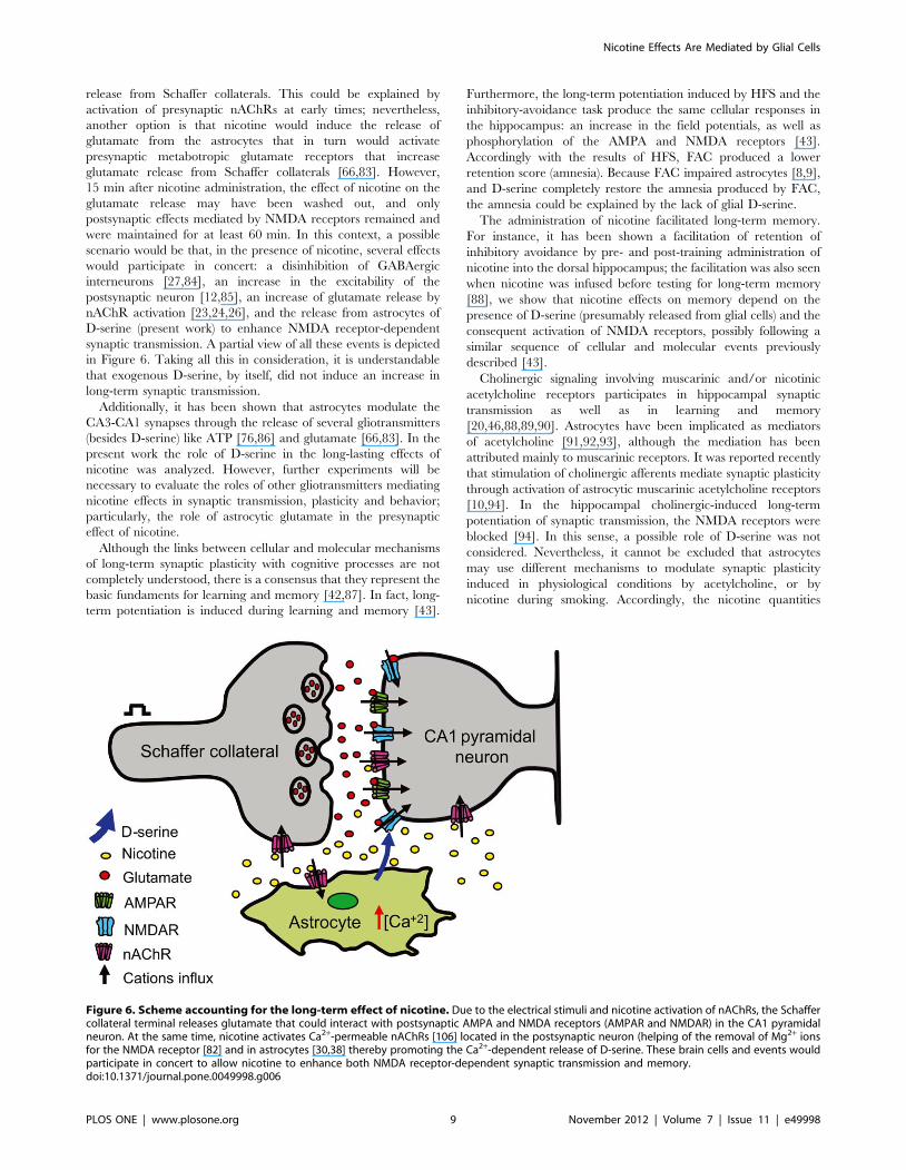
release from Schaffer collaterals. This could be explained by
activation of presynaptic nAChRs at early times; nevertheless,
another option is that nicotine would induce the release of
glutamate from the astrocytes that in turn would activate
presynaptic metabotropic glutamate receptors that increase
glutamate release from Schaffer collaterals [66,83]. However,
15 min after nicotine administration, the effect of nicotine on the
glutamate release may have been washed out, and only
postsynaptic effects mediated by NMDA receptors remained and
were maintained for at least 60 min. In this context, a possible
scenario would be that, in the presence of nicotine, several effects
would participate in concert: a disinhibition of GABAergic
interneurons [27,84], an increase in the excitability of the
postsynaptic neuron [12,85], an increase of glutamate release by
nAChR activation [23,24,26], and the release from astrocytes of
D-serine (present work) to enhance NMDA receptor-dependent
synaptic transmission. A partial view of all these events is depicted
in Figure 6. Taking all this in consideration, it is understandable
that exogenous D-serine, by itself, did not induce an increase in
long-term synaptic transmission.
Additionally, it has been shown that astrocytes modulate the
CA3-CA1 synapses through the release of several gliotransmitters
(besides D-serine) like ATP [76,86] and glutamate [66,83]. In the
present work the role of D-serine in the long-lasting effects of
nicotine was analyzed. However, further experiments will be
necessary to evaluate the roles of other gliotransmitters mediating
nicotine effects in synaptic transmission, plasticity and behavior;
particularly, the role of astrocytic glutamate in the presynaptic
effect of nicotine.
Although the links between cellular and molecular mechanisms
of long-term synaptic plasticity with cognitive processes are not
completely understood, there is a consensus that they represent the
basic fundaments for learning and memory [42,87]. In fact, long-
term potentiation is induced during learning and memory [43].
Furthermore, the long-term potentiation induced by HFS and the
inhibitory-avoidance task produce the same cellular responses in
the hippocampus: an increase in the field potentials, as well as
phosphorylation of the AMPA and NMDA receptors [43].
Accordingly with the results of HFS, FAC produced a lower
retention score (amnesia). Because FAC impaired astrocytes [8,9],
and D-serine completely restore the amnesia produced by FAC,
the amnesia could be explained by the lack of glial D-serine.
The administration of nicotine facilitated long-term memory.
For instance, it has been shown a facilitation of retention of
inhibitory avoidance by pre- and post-training administration of
nicotine into the dorsal hippocampus; the facilitation was also seen
when nicotine was infused before testing for long-term memory
[88], we show that nicotine effects on memory depend on the
presence of D-serine (presumably released from glial cells) and the
consequent activation of NMDA receptors, possibly following a
similar sequence of cellular and molecular events previously
described [43].
Cholinergic signaling involving muscarinic and/or nicotinic
acetylcholine receptors participates in hippocampal synaptic
transmission as well as in learning and memory
[20,46,88,89,90]. Astrocytes have been implicated as mediators
of acetylcholine [91,92,93], although the mediation has been
attributed mainly to muscarinic receptors. It was reported recently
that stimulation of cholinergic afferents mediate synaptic plasticity
through activation of astrocytic muscarinic acetylcholine receptors
[10,94]. In the hippocampal cholinergic-induced long-term
potentiation of synaptic transmission, the NMDA receptors were
blocked [94]. In this sense, a possible role of D-serine was not
considered. Nevertheless, it cannot be excluded that astrocytes
may use different mechanisms to modulate synaptic plasticity
induced in physiological conditions by acetylcholine, or by
nicotine during smoking. Accordingly, the nicotine quantities
Figure 6. Scheme accounting for the long-term effect of nicotine. Due to the electrical stimuli and nicotine activation of nAChRs, the Schaffercollateral terminal releases glutamate that could interact with postsynaptic AMPA and NMDA receptors (AMPAR and NMDAR) in the CA1 pyramidalneuron. At the same time, nicotine activates Ca2+-permeable nAChRs [106] located in the postsynaptic neuron (helping of the removal of Mg2+ ionsfor the NMDA receptor [82] and in astrocytes [30,38] thereby promoting the Ca2+-dependent release of D-serine. These brain cells and events wouldparticipate in concert to allow nicotine to enhance both NMDA receptor-dependent synaptic transmission and memory.doi:10.1371/journal.pone.0049998.g006
Nicotine Effects Are Mediated by Glial Cells
PLOS ONE | www.plosone.org 9 November 2012 | Volume 7 | Issue 11 | e49998

used in the current work (1 mM and 0.4 mg/kg) are very likely to
be reached in humans after smoking one cigarette [37,95].
Perspectives for Pathological Conditions andTherapeutics
The drug addiction process shares many characteristics with
normal learning and memory. In fact, it has been previously
proposed that nicotine modifies the same cellular mechanisms
used during learning and memory to induce addiction
[22,96,97,98,99,100]. Because astrocytes mediate nicotine effects
in long-term memory (present work), and there is an enhancement
of NMDA receptor-mediated responses induced by chronic
nicotine administration [27], we speculate that astrocytes (prob-
ably through the release of D-serine) could be involved in nicotine
addiction. It will be interesting to determine whether the physical
and chemical interactions between hippocampal astrocytes and
neurons are modified by nicotine addiction, and if these
interactions play a role in establishing the addiction.
Furthermore, cholinergic signaling due to nAChR activation is
also implicated in Alzheimer’s and Parkinson’s diseases [101,102],
schizophrenia [103], and depression [104], among other patho-
logical conditions. The roles of astrocytes as mediators of nicotine
effects in these pathologies need to be clarified, and some questions
arise: Is the neuroprotective effect of nicotine against loss of
nigrostriatal dopamine neurons [101] mediated by astrocytes? Do
astrocytes mediate the long-term potentiation induced by nicotine
in the mesolimbic dopaminergic system that accounts for reward
and drug addictions [105]? Additional studies to answer these
questions will further clarify the roles of glial cells and nicotine in
the complex functioning of the brain and provide the opportunity
for new strategies and the development of drugs that would help to
improve patients’ quality of life.
Supporting Information
Figure S1 The increase in synaptic transmission byfluoroacetate is not mediated by NMDA receptors. The
EFP slope as a function of time in the presence of AP5 (A, 50 mM),
or fluoroacetate (B, FAC, 5 mM) alone or in combination with
AP5. Insets, sample records before (brown) and (A) 15 min after
AP5 administration (orange), and (B) 70 min after FAC admin-
istration in the absence (left, green) and presence (right, green) of
AP5. C, Summary of experiments in A and B, representing the
mean 6 S.E.M. of the EFP slope (as percent of control) 20–30 min
after AP5 alone (early, orange), and 60–70 min after FAC (late,
green) in the absence or presence of AP5 (*p,0.05, one-way
repeated-measures ANOVA, post hoc Fisher test).
(TIF)
Figure S2 Effect of Glial D-serine on synaptic transmis-sion. The EFP slope as a function of time before, during, and
after the application of the antagonist DCKA (A, 200 nM) and
agonist D-serine (B, D-ser, 20 mM) of NMDA receptors at the
glycine-binding site. C, Summary of experiments in A and B of the
EFP slope (as percent of control) after DCKA and D-serine
administration (15 min; early, orange) (*p,0.05, one-way repeat-
ed-measures ANOVA, post hoc Fisher test).
(TIF)
Figure S3 Long-term potentiation evoked by electricalstimulation depends on glial D-serine. A, Changes of EFP
slope of P1 by high-frequency electrical stimulation (HFS, arrow)
in control conditions, in the presence of FAC (5 mM), and FAC
plus D-serine (D-ser, 20 mM). Insets, sample traces before (brown)
and 60 min after HFS (green) under these three conditions. B,
Summary of the experiments in A, representing the mean 6
S.E.M. for the EFP slope of P1 (as a percentage of baseline) 50–
60 min after HFS (late, green) alone, in the presence of
fluoroacetate (FAC), or FAC plus D-serine (**p,0.01 one-way
repeated-measures ANOVA, post hoc Fisher test). C, the paired-
pulse ratio (P2/P1) from experiments in A, before (Control) and
60 min after HFS stimulation under the three conditions. Insets,
representative traces of responses to the first (P1) and second (P2)
stimuli, before (Control, brown) and 60 min after HFS (green).
(TIF)
Acknowledgments
We are grateful to Martın Garcıa Servın and Angel Mendez for technical
assistance. We express our gratitude to Drs. Hugo Merchant, Fernando
Pena, and Dorothy Pless for critically reviewing the manuscript. MLH
received a scholarship from CONACyT (213708). This paper was part of
the Ph.D. thesis of MLH in Doctorado en Ciencias Biomedicas, UNAM.
Author Contributions
Conceived and designed the experiments: JGC MLH RAPA. Performed
the experiments: MLH KSP RAM ACM. Analyzed the data: MLH KSP
RAM ACM RAPA JGC. Contributed reagents/materials/analysis tools:
RAPA JGC. Wrote the paper: MLH RAPA JGC. Supervised the project:
RAPA JGC. Gave technical assistance: ACM.
References
1. Ben Achour S, Pascual O (2010) Glia: the many ways to modulate synaptic
plasticity. Neurochem Int 57: 440–445.
2. Perea G, Araque A (2010) GLIA modulates synaptic transmission. Brain Res
Rev 63: 93–102.
3. Magistretti PJ (2006) Neuron-glia metabolic coupling and plasticity. J Exp Biol209: 2304–2311.
4. Ventura R, Harris KM (1999) Three-dimensional relationships betweenhippocampal synapses and astrocytes. J Neurosci 19: 6897–6906.
5. Bushong EA, Martone ME, Jones YZ, Ellisman MH (2002) Protoplasmic
astrocytes in CA1 stratum radiatum occupy separate anatomical domains.
J Neurosci 22: 183–192.
6. Mothet JP, Parent AT, Wolosker H, Brady RO, Jr., Linden DJ, et al. (2000) D-serine is an endogenous ligand for the glycine site of the N-methyl-D-aspartate
receptor. Proc Natl Acad Sci U S A 97: 4926–4931.
7. Oliet SH, Mothet JP (2009) Regulation of N-methyl-D-aspartate receptors by
astrocytic D-serine. Neuroscience 158: 275–283.
8. Fossat P, Turpin FR, Sacchi S, Dulong J, Shi T, et al. (2011) Glial D-serine
gates NMDA receptors at excitatory synapses in prefrontal cortex. CerebCortex 22: 595–606.
9. Henneberger C, Papouin T, Oliet SH, Rusakov DA (2010) Long-term
potentiation depends on release of D-serine from astrocytes. Nature 463: 232–
236.
10. Takata N, Mishima T, Hisatsune C, Nagai T, Ebisui E, et al. (2011) Astrocyte
calcium signaling transforms cholinergic modulation to cortical plasticity
in vivo. J Neurosci 31: 18155–18165.
11. Zhang Z, Gong N, Wang W, Xu L, Xu TL (2008) Bell-shaped D-serine actionson hippocampal long-term depression and spatial memory retrieval. Cereb
Cortex 18: 2391–2401.
12. Albuquerque EX, Pereira EF, Alkondon M, Rogers SW (2009) Mammalian
nicotinic acetylcholine receptors: from structure to function. Physiol Rev 89:73–120.
13. Rangani RJ, Upadhya MA, Nakhate KT, Kokare DM, Subhedar NK (2012)Nicotine evoked improvement in learning and memory is mediated through
NPY Y1 receptors in rat model of Alzheimer’s disease. Peptides 33: 317–328.
14. Socci DJ, Sanberg PR, Arendash GW (1995) Nicotine enhances Morris water
maze performance of young and aged rats. Neurobiol Aging 16: 857–860.
15. Rezayof A, Shirazi-Zand Z, Zarrindast MR, Nayer-Nouri T (2010) Nicotine
improves ethanol-induced memory impairment: the role of dorsal hippocampalNMDA receptors. Life Sci 86: 260–266.
16. Sansone M, Castellano C, Battaglia M, Ammassari-Teule M (1991) Effects of
oxiracetam-nicotine combinations on active and passive avoidance learning in
mice. Pharmacol Biochem Behav 39: 197–200.
17. Jones GM, Sahakian BJ, Levy R, Warburton DM, Gray JA (1992) Effects of
acute subcutaneous nicotine on attention, information processing and short-
Nicotine Effects Are Mediated by Glial Cells
PLOS ONE | www.plosone.org 10 November 2012 | Volume 7 | Issue 11 | e49998

term memory in Alzheimer’s disease. Psychopharmacology (Berl) 108: 485–
494.
18. Newhouse P, Tatro A, Naylor M, Quealey K, Delgado P (2002) Alzheimer
disease, serotonin systems, and tryptophan depletion. Am J Geriatr Psychiatry
10: 483–484.
19. Decker MW, Majchrzak MJ, Anderson DJ (1992) Effects of nicotine on spatial
memory deficits in rats with septal lesions. Brain Res 572: 281–285.
20. Kenney JW, Gould TJ (2008) Modulation of hippocampus-dependent learning
and synaptic plasticity by nicotine. Mol Neurobiol 38: 101–121.
21. Levin ED, Christopher NC, Briggs SJ, Rose JE (1993) Chronic nicotine
reverses working memory deficits caused by lesions of the fimbria or medial
basalocortical projection. Brain Res Cogn Brain Res 1: 137–143.
22. Placzek AN, Zhang TA, Dani JA (2009) Nicotinic mechanisms influencing
synaptic plasticity in the hippocampus. Acta Pharmacol Sin 30: 752–760.
23. Jia Y, Yamazaki Y, Nakauchi S, Sumikawa K (2009) a2 nicotine receptors
function as a molecular switch to continuously excite a subset of interneurons in
rat hippocampal circuits. Eur J Neurosci 29: 1588–1603.
24. Le Magueresse C, Cherubini E (2007) Presynaptic calcium stores contribute to
nicotine-elicited potentiation of evoked synaptic transmission at CA3-CA1
connections in the neonatal rat hippocampus. Hippocampus 17: 316–325.
25. Matsuyama S, Matsumoto A (2003) Epibatidine induces long-term potentiation
(LTP) via activation of a4b2 nicotinic acetylcholine receptors (nAChRs) in vivo
in the intact mouse dentate gyrus: both a7 and a4b2 nAChRs essential to
nicotinic LTP. J Pharmacol Sci 93: 180–187.
26. Nakauchi S, Brennan RJ, Boulter J, Sumikawa K (2007) Nicotine gates long-
term potentiation in the hippocampal CA1 region via the activation of a2*
nicotinic ACh receptors. Eur J Neurosci 25: 2666–2681.
27. Yamazaki Y, Jia Y, Niu R, Sumikawa K (2006) Nicotine exposure in vivo
induces long-lasting enhancement of NMDA receptor-mediated currents in the
hippocampus. Eur J Neurosci 23: 1819–1828.
28. Gahring LC, Persiyanov K, Rogers SW (2004) Neuronal and astrocyte
expression of nicotinic receptor subunit b4 in the adult mouse brain. J Comp
Neurol 468: 322–333.
29. Hernandez-Morales M, Garcia-Colunga J (2009) Effects of nicotine on K+
currents and nicotinic receptors in astrocytes of the hippocampal CA1 region.
Neuropharmacology 56: 975–983.
30. Sharma G, Vijayaraghavan S (2001) Nicotinic cholinergic signaling in
hippocampal astrocytes involves calcium-induced calcium release from
intracellular stores. Proc Natl Acad Sci U S A 98: 4148–4153.
31. Ben Menachem-Zidon O, Avital A, Ben-Menahem Y, Goshen I, Kreisel T, et
al. (2011) Astrocytes support hippocampal-dependent memory and long-term
potentiation via interleukin-1 signaling. Brain Behav Immun 25: 1008–1016.
32. Newman LA, Korol DL, Gold PE (2011) Lactate produced by glycogenolysis in
astrocytes regulates memory processing. PLoS One 6: e28427.
33. Zucker RS, Regehr WG (2002) Short-term synaptic plasticity. Annu Rev
Physiol 64: 355–405.
34. Zucker RS (1989) Short-term synaptic plasticity. Annu Rev Neurosci 12: 13–
31.
35. Paxinos G, Watson C (2005) The rat brain in stereotaxic coordinates.; Press
EA, editor.
36. He J, Deng CY, Zhu XN, Yu JP, Chen RZ (2003) Different synaptic
mechanisms of long-term potentiation induced by nicotine and tetanic
stimulation in hippocampal CA1 region of rats. Acta Pharmacol Sin 24:
398–402.
37. Rose JE, Mukhin AG, Lokitz SJ, Turkington TG, Herskovic J, et al. (2010)
Kinetics of brain nicotine accumulation in dependent and nondependent
smokers assessed with PET and cigarettes containing 11C-nicotine. Proc Natl
Acad Sci U S A 107: 5190–5195.
38. Shen JX, Yakel JL (2012) Functional a7 Nicotinic ACh Receptors on
Astrocytes in Rat Hippocampal CA1 Slices. J Mol Neurosci 48: 14–21.
39. Fonnum F, Johnsen A, Hassel B (1997) Use of fluorocitrate and fluoroacetate in
the study of brain metabolism. Glia 21: 106–113.
40. Waniewski RA, Martin DL (1998) Preferential utilization of acetate by
astrocytes is attributable to transport. J Neurosci 18: 5225–5233.
41. O’Mara SM, Rowan MJ, Anwyl R (1995) Metabotropic glutamate receptor-
induced homosynaptic long-term depression and depotentiation in the dentate
gyrus of the rat hippocampus in vitro. Neuropharmacology 34: 983–989.
42. Lynch MA (2004) Long-term potentiation and memory. Physiol Rev 84: 87–
136.
43. Whitlock JR, Heynen AJ, Shuler MG, Bear MF (2006) Learning induces long-
term potentiation in the hippocampus. Science 313: 1093–1097.
44. Yang Y, Ge W, Chen Y, Zhang Z, Shen W, et al. (2003) Contribution of
astrocytes to hippocampal long-term potentiation through release of D-serine.
Proc Natl Acad Sci U S A 100: 15194–15199.
45. Le Houezec J, Halliday R, Benowitz NL, Callaway E, Naylor H, et al. (1994) A
low dose of subcutaneous nicotine improves information processing in non-
smokers. Psychopharmacology (Berl) 114: 628–634.
46. Levin ED, McClernon FJ, Rezvani AH (2006) Nicotinic effects on cognitive
function: behavioral characterization, pharmacological specification, and
anatomic localization. Psychopharmacology (Berl) 184: 523–539.
47. Levin ED, Simon BB (1998) Nicotinic acetylcholine involvement in cognitive
function in animals. Psychopharmacology (Berl) 138: 217–230.
48. Ciamei A, Aversano M, Cestari V, Castellano C (2001) Effects of MK-801 and
nicotine combinations on memory consolidation in CD1 mice. Psychophar-
macology (Berl) 154: 126–130.
49. Matsuyama S, Matsumoto A, Enomoto T, Nishizaki T (2000) Activation of
nicotinic acetylcholine receptors induces long-term potentiation in vivo in the
intact mouse dentate gyrus. Eur J Neurosci 12: 3741–3747.
50. Tang J, Dani JA (2009) Dopamine enables in vivo synaptic plasticity associated
with the addictive drug nicotine. Neuron 63: 673–682.
51. Wang J, Chen YB, Zhu XN, Chen RZ (2001) Activation of p42/44 mitogen-
activated protein kinase pathway in long-term potentiation induced by nicotine
in hippocampal CA1 region in rats. Acta Pharmacol Sin 22: 685–690.
52. Mothet JP, Rouaud E, Sinet PM, Potier B, Jouvenceau A, et al. (2006) A critical
role for the glial-derived neuromodulator D-serine in the age-related deficits of
cellular mechanisms of learning and memory. Aging Cell 5: 267–274.
53. Kanematsu S, Ishii S, Umino A, Fujihira T, Kashiwa A, et al. (2006) Evidence
for involvement of glial cell activity in the control of extracellular D-serine
contents in the rat brain. J Neural Transm 113: 1717–1721.
54. Miya K, Inoue R, Takata Y, Abe M, Natsume R, et al. (2008) Serine racemase
is predominantly localized in neurons in mouse brain. J Comp Neurol 510:
641–654.
55. Rosenberg D, Kartvelishvily E, Shleper M, Klinker CM, Bowser MT, et al.
(2010) Neuronal release of D-serine: a physiological pathway controlling
extracellular D-serine concentration. FASEB J 24: 2951–2961.
56. Williams SM, Diaz CM, Macnab LT, Sullivan RK, Pow DV (2006)
Immunocytochemical analysis of D-serine distribution in the mammalian
brain reveals novel anatomical compartmentalizations in glia and neurons. Glia
53: 401–411.
57. Wolosker H (2011) Serine racemase and the serine shuttle between neurons
and astrocytes. Biochim Biophys Acta 1814: 1558–1566.
58. Shleper M, Kartvelishvily E, Wolosker H (2005) D-serine is the dominant
endogenous coagonist for NMDA receptor neurotoxicity in organotypic
hippocampal slices. J Neurosci 25: 9413–9417.
59. Turpin F, Dallerac G, Mothet JP (2012) Electrophysiological analysis of the
modulation of NMDA-receptors function by D-serine and glycine in the central
nervous system. Methods Mol Biol 794: 299–312.
60. Ikeda H, Murase K (2004) Glial nitric oxide-mediated long-term presynaptic
facilitation revealed by optical imaging in rat spinal dorsal horn. J Neurosci 24:
9888–9896.
61. Muir D, Berl S, Clarke DD (1986) Acetate and fluoroacetate as possible
markers for glial metabolism in vivo. Brain Res 380: 336–340.
62. Keyser DO, Pellmar TC (1994) Synaptic transmission in the hippocampus:
critical role for glial cells. Glia 10: 237–243.
63. Heinrich A, Ando R, Turi G, Rozsa B, Sperlagh B (2012) K+ depolarization
evokes ATP, adenosine and glutamate release from glia in rat hippocampus: a
microelectrode biosensor study. Br J Pharmacol 167: 1003–1020.
64. Uwechue NM, Marx MC, Chevy Q, Billups B (2012) Activation of glutamate
transport evokes rapid glutamine release from perisynaptic astrocytes. J Physiol
590: 2317–2331.
65. Berg-Johnsen J, Paulsen RE, Fonnum F, Langmoen IA (1993) Changes in
evoked potentials and amino acid content during fluorocitrate action studied in
rat hippocampal cortex. Exp Brain Res 96: 241–246.
66. Bonansco C, Couve A, Perea G, Ferradas CA, Roncagliolo M, et al. (2011)
Glutamate released spontaneously from astrocytes sets the threshold for
synaptic plasticity. Eur J Neurosci 33: 1483–1492.
67. Canals S, Larrosa B, Pintor J, Mena MA, Herreras O (2008) Metabolic
challenge to glia activates an adenosine-mediated safety mechanism that
promotes neuronal survival by delaying the onset of spreading depression
waves. J Cereb Blood Flow Metab 28: 1835–1844.
68. Larrosa B, Pastor J, Lopez-Aguado L, Herreras O (2006) A role for glutamate
and glia in the fast network oscillations preceding spreading depression.
Neuroscience 141: 1057–1068.
69. Bacci A, Sancini G, Verderio C, Armano S, Pravettoni E, et al. (2002) Block of
glutamate-glutamine cycle between astrocytes and neurons inhibits epileptiform
activity in hippocampus. J Neurophysiol 88: 2302–2310.
70. Swanson RA, Graham SH (1994) Fluorocitrate and fluoroacetate effects on
astrocyte metabolism in vitro. Brain Res 664: 94–100.
71. Kang J, Jiang L, Goldman SA, Nedergaard M (1998) Astrocyte-mediated
potentiation of inhibitory synaptic transmission. Nat Neurosci 1: 683–692.
72. Delbro D, Westerlund A, Bjorklund U, Hansson E (2009) In inflammatory
reactive astrocytes co-cultured with brain endothelial cells nicotine-evoked
Ca2+ transients are attenuated due to interleukin-1b release and rearrangement
of actin filaments. Neuroscience 159: 770–779.
73. Oikawa H, Nakamichi N, Kambe Y, Ogura M, Yoneda Y (2005) An increase
in intracellular free calcium ions by nicotinic acetylcholine receptors in a single
cultured rat cortical astrocyte. J Neurosci Res 79: 535–544.
74. Velez-Fort M, Audinat E, Angulo MC (2009) Functional a7-containing
nicotinic receptors of NG2-expressing cells in the hippocampus. Glia 57: 1104–
1114.
75. Mothet JP, Pollegioni L, Ouanounou G, Martineau M, Fossier P, et al. (2005)
Glutamate receptor activation triggers a calcium-dependent and SNARE
protein-dependent release of the gliotransmitter D-serine. Proc Natl Acad
Sci U S A 102: 5606–5611.
Nicotine Effects Are Mediated by Glial Cells
PLOS ONE | www.plosone.org 11 November 2012 | Volume 7 | Issue 11 | e49998
76. Gibbs ME, Shleper M, Mustafa T, Burnstock G, Bowser DN (2012) ATP
derived from astrocytes modulates memory in the chick. Neuron Glia Biol: 1–
10.
77. Greenwood SM, Bushell TJ (2010) Astrocytic activation and an inhibition of
MAP kinases are required for proteinase-activated receptor-2-mediated
protection from neurotoxicity. J Neurochem 113: 1471–1480.
78. Yamazaki Y, Hamaue N, Sumikawa K (2002) Nicotine compensates for the
loss of cholinergic function to enhance long-term potentiation induction. Brain
Res 946: 148–152.
79. Citri A, Malenka RC (2008) Synaptic plasticity: multiple forms, functions, and
mechanisms. Neuropsychopharmacology 33: 18–41.
80. Malenka RC, Nicoll RA (1993) NMDA-receptor-dependent synaptic plasticity:
multiple forms and mechanisms. Trends Neurosci 16: 521–527.
81. Malenka RC, Bear MF (2004) LTP and LTD: an embarrassment of riches.
Neuron 44: 5–21.
82. Mayer ML, Westbrook GL, Guthrie PB (1984) Voltage-dependent block by
Mg2+ of NMDA responses in spinal cord neurones. Nature 309: 261–263.
83. Perea G, Araque A (2007) Astrocytes potentiate transmitter release at single
hippocampal synapses. Science 317: 1083–1086.
84. Rosato-Siri M, Cattaneo A, Cherubini E (2006) Nicotine-induced enhance-
ment of synaptic plasticity at CA3-CA1 synapses requires GABAergic
interneurons in adult anti-NGF mice. J Physiol 576: 361–377.
85. Szabo SI, Zelles T, Vizi ES, Lendvai B (2008) The effect of nicotine on spiking
activity and Ca2+ dynamics of dendritic spines in rat CA1 pyramidal neurons.
Hippocampus 18: 376–385.
86. Fellin T, Pascual O, Haydon PG (2006) Astrocytes coordinate synaptic
networks: balanced excitation and inhibition. Physiology (Bethesda) 21: 208–
215.
87. Bliss TV, Collingridge GL (1993) A synaptic model of memory: long-term
potentiation in the hippocampus. Nature 361: 31–39.
88. Marti Barros D, Ramirez MR, Dos Reis EA, Izquierdo I (2004) Participation of
hippocampal nicotinic receptors in acquisition, consolidation and retrieval of
memory for one trial inhibitory avoidance in rats. Neuroscience 126: 651–656.
89. Fernandez de Sevilla D, Buno W (2010) The muscarinic long-term
enhancement of NMDA and AMPA receptor-mediated transmission at
Schaffer collateral synapses develop through different intracellular mecha-
nisms. J Neurosci 30: 11032–11042.
90. Hasselmo ME (2006) The role of acetylcholine in learning and memory. Curr
Opin Neurobiol 16: 710–715.
91. Araque A, Martin ED, Perea G, Arellano JI, Buno W (2002) Synaptically
released acetylcholine evokes Ca2+ elevations in astrocytes in hippocampalslices. J Neurosci 22: 2443–2450.
92. Guizzetti M, Moore NH, Giordano G, Costa LG (2008) Modulation of
neuritogenesis by astrocyte muscarinic receptors. J Biol Chem 283: 31884–31897.
93. Perea G, Araque A (2005) Properties of synaptically evoked astrocyte calciumsignal reveal synaptic information processing by astrocytes. J Neurosci 25:
2192–2203.
94. Navarrete M, Perea G, Fernandez de Sevilla D, Gomez-Gonzalo M, Nunez A,et al. (2012) Astrocytes mediate in vivo cholinergic-induced synaptic plasticity.
PLoS Biol 10: e1001259.95. Reagan-Shaw S, Nihal M, Ahmad N (2008) Dose translation from animal to
human studies revisited. FASEB J 22: 659–661.96. Dani JA, Ji D, Zhou FM (2001) Synaptic plasticity and nicotine addiction.
Neuron 31: 349–352.
97. Kelley AE (2004) Memory and addiction: shared neural circuitry andmolecular mechanisms. Neuron 44: 161–179.
98. Nestler EJ (2002) Common molecular and cellular substrates of addiction andmemory. Neurobiol Learn Mem 78: 637–647.
99. Robbins TW, Everitt BJ (2002) Limbic-striatal memory systems and drug
addiction. Neurobiol Learn Mem 78: 625–636.100. Zhang TA, Tang J, Pidoplichko VI, Dani JA (2010) Addictive nicotine alters
local circuit inhibition during the induction of in vivo hippocampal synapticpotentiation. J Neurosci 30: 6443–6453.
101. Quik M, O’Neill M, Perez XA (2007) Nicotine neuroprotection againstnigrostriatal damage: importance of the animal model. Trends Pharmacol Sci
28: 229–235.
102. Srivareerat M, Tran TT, Salim S, Aleisa AM, Alkadhi KA (2011) Chronicnicotine restores normal Ab levels and prevents short-term memory and E-LTP
impairment in Ab rat model of Alzheimer’s disease. Neurobiol Aging 32: 834–844.
103. Brunzell DH, McIntosh JM (2012) a7 nicotinic acetylcholine receptors
modulate motivation to self-administer nicotine: implications for smoking andschizophrenia. Neuropsychopharmacology 37: 1134–1143.
104. Mineur YS, Picciotto MR (2010) Nicotine receptors and depression: revisitingand revising the cholinergic hypothesis. Trends Pharmacol Sci 31: 580–586.
105. Mansvelder HD, McGehee DS (2000) Long-term potentiation of excitatoryinputs to brain reward areas by nicotine. Neuron 27: 349–357.
106. Fucile S (2004) Ca2+ permeability of nicotinic acetylcholine receptors. Cell
Calcium 35: 1–8.
Nicotine Effects Are Mediated by Glial Cells
PLOS ONE | www.plosone.org 12 November 2012 | Volume 7 | Issue 11 | e49998