The effects of nicotine and cigarette smoking on cardiac ...
214
University of Louisville University of Louisville ThinkIR: The University of Louisville's Institutional Repository ThinkIR: The University of Louisville's Institutional Repository Electronic Theses and Dissertations 12-2019 The effects of nicotine and cigarette smoking on cardiac The effects of nicotine and cigarette smoking on cardiac electrophysiology. electrophysiology. Affan B. Irfan University of Louisville Follow this and additional works at: https://ir.library.louisville.edu/etd Part of the Cardiology Commons, Cardiovascular Diseases Commons, Medical Physiology Commons, and the Preventive Medicine Commons Recommended Citation Recommended Citation Irfan, Affan B., "The effects of nicotine and cigarette smoking on cardiac electrophysiology." (2019). Electronic Theses and Dissertations. Paper 3319. https://doi.org/10.18297/etd/3319 This Doctoral Dissertation is brought to you for free and open access by ThinkIR: The University of Louisville's Institutional Repository. It has been accepted for inclusion in Electronic Theses and Dissertations by an authorized administrator of ThinkIR: The University of Louisville's Institutional Repository. This title appears here courtesy of the author, who has retained all other copyrights. For more information, please contact [email protected].
-
Upload
khangminh22 -
Category
Documents
-
view
5 -
download
0
Transcript of The effects of nicotine and cigarette smoking on cardiac ...

University of Louisville University of Louisville
ThinkIR: The University of Louisville's Institutional Repository ThinkIR: The University of Louisville's Institutional Repository
Electronic Theses and Dissertations
12-2019
The effects of nicotine and cigarette smoking on cardiac The effects of nicotine and cigarette smoking on cardiac
electrophysiology. electrophysiology.
Affan B. Irfan University of Louisville
Follow this and additional works at: https://ir.library.louisville.edu/etd
Part of the Cardiology Commons, Cardiovascular Diseases Commons, Medical Physiology Commons,
and the Preventive Medicine Commons
Recommended Citation Recommended Citation Irfan, Affan B., "The effects of nicotine and cigarette smoking on cardiac electrophysiology." (2019). Electronic Theses and Dissertations. Paper 3319. https://doi.org/10.18297/etd/3319
This Doctoral Dissertation is brought to you for free and open access by ThinkIR: The University of Louisville's Institutional Repository. It has been accepted for inclusion in Electronic Theses and Dissertations by an authorized administrator of ThinkIR: The University of Louisville's Institutional Repository. This title appears here courtesy of the author, who has retained all other copyrights. For more information, please contact [email protected].
THE EFFECTS OF NICOTINE AND CIGARETTE SMOKING ON CARDIAC
ELECTROPHYSIOLOGY
By
Affan B. Irfan
M,B.B,S Aga Khan University, 2008
A Dissertation
Submitted to the Faculty of the
School of Medicine of the University of Louisville
In Partial Fulfillment of the Requirements
for the Degree of
Doctor of Philosophy in Physiology and Biophysics
Department of Physiology
University of Louisville
Louisville, Kentucky
December 2019


ii
THE EFFECTS OF NICOTINE AND CIGARETTE SMOKING ON CARDIAC
ELECTROPHYSIOLOGY
By
Affan B. Irfan
M,B.B,S Aga Khan University, 2008
A Dissertation Approved on
November 27, 2019
by the following Dissertation Committee:
Dr Aruni Bhatnagar (Dissertation Director)
Dr Alex Carll (Co-Director)
Dr. Andrew P. DeFilippis
Dr. Irving G. Joshua
Dr. Dale A. Schuschke

iii
DEDICATION
This dissertation is dedicated to my parents, Irfan Uddin and Farzana Irfan, (without whom
I would not have been here), my brother, Furqan Irfan, and sister Bismah Irfan, (without
whom I would not have been the middle child), and my wife, Sommaya Naveed, and our
daughter, Aiza Affan (without whom this dissertation would have been completed a year
ago).
iv
ACKNOWLEDGEMENTS
Firstly, I am indebted to Dr. Aruni Bhatnagar, for giving me the opportunity to
complete my Ph.D dissertation under his supervision, and also to Dr. Glenn Hirsch and the
University of Louisville Cardiovascular Fellowship Program, for allowing me to
participate in the Ph.D graduate studies during my fellowship training. I particularly would
like to thank my mentors, Dr. Bhatnagar and Dr. Alex P. Carll, for their tireless guidance
and “tough love” that pushed me out of my comfort zone (of just being a physician), and
onto the path of a successful (and humble) scientist. I would like to thank the rest of my
dissertation committee members (Dr. Andrew P. DeFilippis, Dr. Irving G. Joshua, and Dr.
Dale A. Schuschke) for their support and invaluable advice. I am also especially grateful
to Dr. Andrew P. DeFilippis for his expertise in clinical research studies that contributed
greatly to this research. Moreover, I am thankful to Dr. Elsayed Soliman for his advice,
collaboration, and contribution to projects related to this dissertation. This work would not
have been possible without the administrative and financial support of Marshall
University’s Departments of Cardiology and Clinical Translational Services. I am
especially grateful to research coordinators, Melissa Marcum and Scott Wiley, for their
perseverance and unwavering commitment to this research study. I am greatly indebted to,
and honored by, the encouragement and guidance from Dr. Christian Mueller, who
introduced me to cardiovascular research and helped take the first steps of my scientific
pursuits. Lastly, I thank my dear family, especially my mother, father, brother, sister, and
wife for their enduring love, support, and sacrifice.

v
ABSTRACT
THE EFFECTS OF NICOTINE AND CIGARETTE SMOKING ON CARDIAC
ELECTROPHYSIOLOGY
Affan B. Irfan
November 27, 2019
Cigarette smoking is a leading cause of preventable disease and premature death
worldwide. The adverse effects of cigarette smoking, including proarrhythmia, are related
to the mixture of chemicals, including nicotine (which sustains tobacco addiction).
However, it remains unclear which individual tobacco smoke constituents and biological
pathways mediate this increased risk. The purpose of this research was to explore the
chronic effects of cigarette smoking, as well as compare the acute effects of nicotine and
cigarette smoking, and the possible role of β-adrenoreceptors, on human cardiac
electrophysiology. Chapter 1 is a comprehensive literature review of (a) the ex vivo and in
vivo effects of nicotine and non-nicotine constituents of cigarette smoking on cardiac ion
channels, (b) the direct and indirect effects of the autonomic nervous system on cardiac
electrophysiology, and (c) studies of acute and chronic effects of cigarette smoking in
humans. Chapter 2 consists of two studies in which we used cotinine levels to investigate
the differences in baseline cardiac electrocardiogram between chronic smokers and non-
smokers, and to define smoking status and its burden. We also explored the relationship
between urinary catecholamines, cotinine, and electrocardiographic changes. Chapter 3
features the 2 x 2 factorial experimental study designed to compare
vi
the acute effects of cigarette smoking and nicotine, with and without a β-blocker
(propranolol). We found that chronic cigarette smoking was associated with a shortened
PR segment at baseline, and that dopamine possibly mediates this effect. There was also
(corrected) QT interval shortening with increased cotinine levels. This experimental study
revealed that the non-nicotine constituents in cigarette smoking were mainly responsible
for PR segment shortening, through β-adrenoreceptors. Other evidence revealed that,
although nicotine in cigarette smoke is primarily responsible for sympathetic activation
and (corrected) QT interval shortening, it is the non-nicotine constituents that depress the
ST segment. Collectively, acute and chronic exposure studies indicate that smoking may
promote cardiac arrhythmia, primarily via β-adrenoreceptors, causing acceleration of
dromotropy and ischemia (non-nicotine mediated), and ventricular repolarization
(nicotine-mediated). This research elucidated a major physiological mechanism driving the
effect of cigarette smoking and nicotine on cardiac electrophysiology. Consequently, these
findings will inform U.S. Food and Drug Administration of tobacco and nicotine-
containing products’ impact on the human cardiac electrical system, and potentially help
regulate alternative forms of nicotine delivery and protect public health.

vii
TABLE OF CONTENTS
PAGE
ACKNOWLEDGEMENTS ............................................................................................ iv
ABSTRACT ..................................................................................................................... v
LIST OF TABLES .......................................................................................................... ix
LIST OF FIGURES .......................................................................................................... x
CHAPTER I. INTRODUCTION ..................................................................................... 1
Background and context ............................................................................................... 1
Clinical significance of cigarette smoking, nicotine, and cardiac
electrophysiology ...................................................................................................... 1 Effects of cardiac autonomic system and cigarette smoke on cardiac
action potential .......................................................................................................... 7 Literature review of effects of autonomic nervous system and smoking on
electrocardiogram .................................................................................................... 32
Hypothesis ................................................................................................................... 44
Ethical considerations ............................................................................................. 45
CHAPTER II CHRONIC EFFECTS OF CIGARETTE SMOKING ON
ELECTROCARDIOGRAM ........................................................................................... 46
Cigarette smoking, ECG and interaction with Atrioventricular
nodal blockers ............................................................................................................. 46
Participants from NHANES database ..................................................................... 45 Results ..................................................................................................................... 52 Discussion ............................................................................................................... 61 Conclusions ............................................................................................................. 64
Effects of cigarette smoking on electrocardiogram mediated
via catecholamines ...................................................................................................... 65
Participants from Louisville Healthy Heart Study .................................................. 65 Results ..................................................................................................................... 69 Discussion ............................................................................................................... 79 Conclusions ............................................................................................................. 85
viii
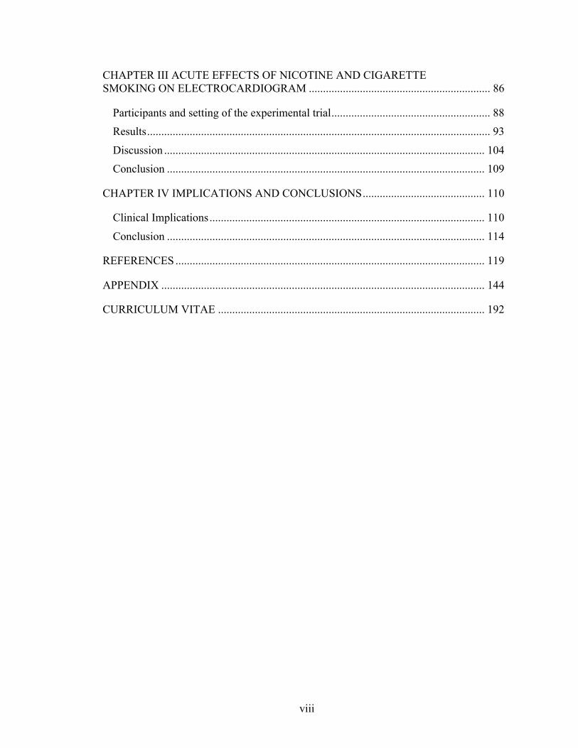
CHAPTER III ACUTE EFFECTS OF NICOTINE AND CIGARETTE
SMOKING ON ELECTROCARDIOGRAM ................................................................ 86
Participants and setting of the experimental trial ........................................................ 88
Results ......................................................................................................................... 93
Discussion ................................................................................................................. 104
Conclusion ................................................................................................................ 109
CHAPTER IV IMPLICATIONS AND CONCLUSIONS ........................................... 110
Clinical Implications ................................................................................................. 110
Conclusion ................................................................................................................ 114
REFERENCES ............................................................................................................. 119
APPENDIX .................................................................................................................. 144
CURRICULUM VITAE .............................................................................................. 192
ix
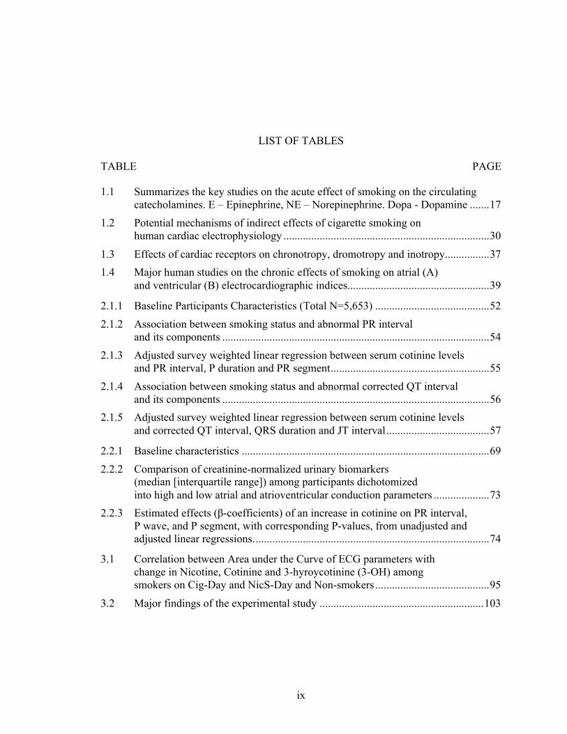
LIST OF TABLES
TABLE PAGE
1.1 Summarizes the key studies on the acute effect of smoking on the circulating
catecholamines. E – Epinephrine, NE – Norepinephrine. Dopa - Dopamine ....... 17
1.2 Potential mechanisms of indirect effects of cigarette smoking on
human cardiac electrophysiology .......................................................................... 30
1.3 Effects of cardiac receptors on chronotropy, dromotropy and inotropy ................ 37
1.4 Major human studies on the chronic effects of smoking on atrial (A)
and ventricular (B) electrocardiographic indices ................................................... 39
2.1.1 Baseline Participants Characteristics (Total N=5,653) ......................................... 52
2.1.2 Association between smoking status and abnormal PR interval
and its components ................................................................................................ 54
2.1.3 Adjusted survey weighted linear regression between serum cotinine levels
and PR interval, P duration and PR segment ......................................................... 55
2.1.4 Association between smoking status and abnormal corrected QT interval
and its components ................................................................................................ 56
2.1.5 Adjusted survey weighted linear regression between serum cotinine levels
and corrected QT interval, QRS duration and JT interval ..................................... 57
2.2.1 Baseline characteristics ......................................................................................... 69
2.2.2 Comparison of creatinine-normalized urinary biomarkers
(median [interquartile range]) among participants dichotomized
into high and low atrial and atrioventricular conduction parameters .................... 73
2.2.3 Estimated effects (β-coefficients) of an increase in cotinine on PR interval,
P wave, and P segment, with corresponding P-values, from unadjusted and
adjusted linear regressions. .................................................................................... 74
3.1 Correlation between Area under the Curve of ECG parameters with
change in Nicotine, Cotinine and 3-hyroycotinine (3-OH) among
smokers on Cig-Day and NicS-Day and Non-smokers ......................................... 95
3.2 Major findings of the experimental study ........................................................... 103
x
LIST OF FIGURES
FIGURE PAGE
1.1 Effect of Sympathetic nervous activation on human cardiac
electrophysiology .................................................................................................. 11
1.2 Schematic diagram to illustrate the different direct sites of action
for nicotine on the pathways of the cardiovascular autonomic
nervous system ...................................................................................................... 13
1.3 Direct effects of nicotine and non-nicotine constituents on cardiac ion
channels and action potential. SNS – Sympathetic Nervous Activation ............... 23
2.1.1 The effect of smoking status on baseline ECG ..................................................... 58
2.1.2 Panel A shows the distribution of PR segment (ms) and
QTc interval (ms) with cotinine levels .................................................................. 59
2.2.1 Scatterplot and linear relationships of (A) PR interval, (B) P wave
and (C) PR segment with log transformed urinary cotinine levels ....................... 75
2.2.2 Mediation analyses of cotinine, dopamine, and PR interval and
PR segment ............................................................................................................ 77
3.1 The Area Under the Curve for the raw changes (Δ) in ECG parameters
from baseline for each visit day among smokers .................................................. 98
3.2 ECG morphology in a typical participant during exposure on
different days ......................................................................................................... 99
3.3 The best fitting line to predict changes in Heart Rate, P amplitude,
and QTc upon Cigarette and Nicotine exposures, and additional lines
for PR segment and ST height upon Cigarette exposure ..................................... 101
3.4 Mediation analyses of changes in Nicotine, root mean squared of successive
differences (RMSSD) and Heart Rate and ST-height among smokers on
Cig-Day and NicS-Day and Non-smokers .......................................................... 102
4.1 Central illustration summarizing the impacts of nicotine and non-nicotine
constituents of cigarette smoke on cardiac electrophysiology, and
the pathways likely involved ............................................................................... 116
1
CHAPTER I
INTRODUCTION
Background and context
Clinical significance of cigarette smoking, nicotine, and cardiac electrophysiology
Cigarette smoke
Cigarette smoking is the leading preventable cause of premature death. It is
responsible for at least 480,000 premature deaths every year in the United States, with more
than 41,000 of these deaths from exposure to secondhand smoke [1, 2], out of which one-
third are secondary to cardiovascular disease (CVD) [3]. The mortality of current smokers
among both sexes is three times that of non-smokers [4, 5]. Diseases attributable to
smoking accounts for about 60% of smokers' deaths and the benefits of quitting smoking
are dramatic across all age groups, with substantial gains in life expectancy, as compared
to those who continue to smoke [5]. Furthermore, elevated CVD mortality has been seen
even among patients smoking fewer than five cigarettes per day, suggesting a nonlinear
dose-effect relationship [6]. Apart from the 21 common diseases formally attributed to
cigarette smoking, there are diseases that have not been formally established as being
caused by smoking (such as infections, hypertensive heart disease, renal failure, intestinal
ischemia, and other respiratory diseases), and account for a significant excess in mortality
[4]. The estimated annual smoking-related economic
2
burden in the United States is between $289–332.5 billion in direct medical care and other
economic costs [1, 7]. Smoking alsoappears to have a multiplicative interaction with other
major CVD risk factors (elevated serum lipid levels, untreated hypertension, and diabetes)
[2]. In 2016, 15.5% (37.8 million) of U.S. adults were cigarette smokers. The prevalence
of cigarette smoking was higher among adults who were male, 25–64 years old, of lower
socio-economic status, of American Indian/Alaska Native descent or multiracial, and
suffering from psychological distress [8].
Nicotine and addiction
Addiction to the nicotine in tobacco is the proximate cause of these diseases,
because it sustains smoking behavior by acting on nicotinic cholinergic receptors in the
brain to trigger the release of dopamine and other neurotransmitters [9]. Release of
dopamine, glutamate, and gamma-Aminobutyric acid (GABA) [10] is particularly
important in the development of nicotine dependence, and the extrahypothalamic
corticotropin-releasing factor may play a key role in withdrawal [11, 12]. Nicotine
addiction occurs when smokers rely on smoking to modulate mood and arousal, relieve
withdrawal symptoms, or both. Therefore, the magnitude of public health harm caused by
tobacco is inextricably linked to its addictive nature [9]. There is a continuum of risk for
products that deliver nicotine, ranging from the most harmful combusted products (e.g.,
cigarettes) and electronic nicotine delivery systems (ENDS), to medicinal nicotine
products.
Although significant research has been conducted to understand the
pathophysiology of smoking-associated CVD, new research in this era has slowed in recent

3
years. This lag in research is particularly harmful, as tobacco products have continued to
evolve, and new products, such as ENDS, are a popular source of nicotine, especially
among children and young adults [13]. In contrast with cigarettes, however, e-cigarettes
generate an aerosol by heating a liquid, usually consisting of propylene glycol or vegetable
glycerin, nicotine, and flavoring agents, without any combustion [14]. Despite almost
complete lack of knowledge regarding the biological effects of these new tobacco products,
ENDS have been implied to help conventional cigarette smokers quit or curtail smoking.
ENDS manufacturers are making claims that these products pose fewer health risks and are
much safer than smoking conventional cigarettes. This postulation is concerning because
such health claims are likely to lead to increased ENDS use, and may even lead to an
increase in nicotine dependence in the population at large. Although e-cigarettes deliver
lower levels of carcinogens than do conventional cigarettes, they still expose users to high
levels of ultrafine particles and other toxins, such as volatile organic compounds (VOCs)
[15], that may substantially increase risks for cardiovascular and noncancerous lung
disease, which account for more than half of all smoking-related deaths, at rates similar to
conventional cigarettes [16].
The primary concern for nicotine in cigarette smoking is an addiction. Tobacco
combustion products cause most of the adverse health effects from smoking, but some
health concerns are related to nicotine. Many of these concerns are related to the ability of
nicotine to release catecholamines, including hemodynamic effects (increase in heart rate,
transient increases in blood pressure, vasoconstriction of coronary and other vascular beds),
adverse effects on lipids, and induction of insulin resistance [17]. Both in vitro and in vivo
4
animal studies suggest that nicotine may inhibit apoptosis, enhance angiogenesis,
exacerbate atherosclerotic disease [18, 19], and produce endothelial dysfunction [20].
Cardiac electrophysiology
Basic cardiac electrophysiology is fundamental in understanding rhythmic cardiac
function and electrical conduction, as well as changes in electrical activity associated with
cardiac disease. The primary clinical tool for assessing cardiac electrical events is the
electrocardiogram (ECG) [21]. The 12-lead ECG remains the most widely available,
inexpensive, non-invasive, an indispensable tool for the diagnosis and prompt initiation of
therapy in patients with acute coronary syndromes. It provides the most accurate means of
diagnosing intraventricular conduction disturbances and arrhythmias, assessing
cardiovascular risk and screening individuals in high-risk occupations and, in some cases,
for participation in sports. As a research tool, it is used in long-term population-based
surveillance studies and experimental trials of drugs with recognized or potential cardiac
effects [22]. Therefore, ECG is used in clinical trials, as a valid, reliable, repeatable,
quantitative method that is inexpensive and unbiased by clinical information.
Most ECGs used clinically are produced from digital signaling and interpreted by
software using algorithms to assess cardiac rhythmicity, heart rate, heart rate variability
(HRV), and intervals between conventional ECG landmarks, especially the PR, QRS, and
QT intervals. These metrics provide essential insight into the autonomic function (HRV),
atrioventricular conduction (PR interval and segment), ventricular depolarization (QRS),
and repolarization (QT and JT). There are several major and minor ECG variables
associated with cardiovascular mortality; namely P wave (duration, interatrial block, and

5
deep terminal negativity of the P wave in V1), QT and Tpeak-Tend (Tp-Te) intervals, QRS
duration and fragmentation, bundle branch block, ST segment depression and elevation, T
waves (inverted, T wave axes), spatial angles between QRS and T vectors, premature
ventricular contractions, and ECG hypertrophy criteria [23-25]. Apart from these
traditional ECG markers, there is evidence for several other ECG intervals that have also
been shown to be associated with total all-cause and cardiovascular-related mortality, such
as PR interval [26-30], JT interval [31, 32] and Tp-Te [33-35].
Given its low cost, ubiquity, and safety, ECG is a useful candidate tool for screening
and risk stratification of asymptomatic participants [36]. Deaths due to smoking-related
CVD are generally preceded by a subclinical cardiovascular injury that may be detected
early in the disease process [37]. To improve risk identification and stratification among
asymptomatic smokers, and to aid in preventative measures, health professionals need
more sensitive smoking-related markers of early cardiovascular damage. This research
focus is particularly imperative in the new era of ENDs, as it remains unclear how ENDS
might compare to cigarette smoke in severity and temporality of associated adverse
outcomes.
Implications for public health practice
Some tobacco control researchers and advocates emphasize the need for strong
policies that would protect future and current generations from new products that lead to
nicotine addiction or serve as a gateway to cigarette smoking [16]. Others emphasize the
different risks for disease associated with different tobacco and nicotine products, and
argue that policies must prioritize reducing disease risk even if that means allowing for

6
new products that may have high addiction potential [38]. As part of their framework
announced in 2017, the Food and Drug Administration (FDA)—based on their recognition
that nicotine makes tobacco products addictive, but that it does not directly cause smokers’
cancer, lung disease, or heart disease—has proposed a regulation strategy designed to limit
nicotine in cigarettes to a minimal or nonaddictive level [39]. The premise is that the mode
of nicotine delivery, rather than the drug nicotine itself, is the key to reducing harm at a
population level. This action has the promise of helping current users quit while preventing
potential future smokers—youths, in particular—from becoming addicted via escalation
from experimentation to regular smoking [40]. Modeling estimates that, by appropriate
nicotine regulation, about 5 million adult smokers could quit within a year, and most youths
and young adults—could avoid becoming regular smokers [41]. Whether this is the right
approach is unclear, as through it, the FDA Center for Drug Evaluation Research has
primarily been conducted on targeting smoking cessation rather than dependence on
nicotine and alternate tobacco products [42]. As smokers find it difficult to achieve desired
nicotine levels from low-nicotine cigarettes, they may seek to replace cigarettes with other
tobacco products that deliver nicotine. The FDA expects that making cigarettes minimally
addictive or nonaddictive would reduce tobacco-related harm by promoting smoking
cessation or a complete migration to alternative, uncombusted products, and by reducing
initiation. Although concrete evidence is lacking, there are concerns that ENDs use may
renormalize smoking behavior, sustain dual-use, and initiate or maintain nicotine addiction
[13]. ENDS use also could serve as a gateway to the initiation of smoking by ex-smokers.
Unregulated e-cigarette use also has the potential to erode gains in smoking cessation and
smoke-free laws [13]. Furthermore, the health effects of nicotine and e-cigarettes have not
7
been well studied, and the potential harm incurred by long-term use of these devices
remains completely unknown.
To complement Center for Tobacco Products (CTP) and CTP-funded scientific
investigations to determine each product’s risks, benefits, and net public health impact, the
FDA encourages submission of information from additional rigorous research (e.g., outside
research institutions, or a manufacturer in an application for FDA marketing authorization
of a new product [40]). Hence, extensive new research is required to assess the health
effects of nicotine in order to develop appropriate regulatory policies.
Effects of cardiac autonomic system and cigarette smoke on cardiac action potential
Direct effects
Cardiac autonomic nervous system
Sympathetic and parasympathetic control: Superimposed on the intrinsic cardiac
control system are the major extrinsic factors—autonomic efferent postganglionic nerve
terminals— which affect the secretion of hormones into circulation, and the release of
chemicals directly onto the cardiomyocyte membrane [43]. The autonomic nervous system
(ANS) plays a vital role in the genesis of several cardiac arrhythmias, both in the atria and
in the ventricles. Modulation of the autonomic response is a complex process, in which the
final effect is the product of interactions among central, peripheral, and intracardiac
components. Autonomic activation alters not only the heart rate, conduction, and
hemodynamics, but also the cellular and subcellular properties of individual myocytes.
The cardiac ANS is divided into extrinsic (fibers that mediate connections between
the heart and the nervous system) and intrinsic (fibers facilitating function within the

8
pericardial sac) components. The extrinsic cardiac ANS is divided into sympathetic and
parasympathetic components. The fibers of the sympathetic nervous system (SNS) are
largely derived from major autonomic ganglia along the cervical and thoracic spinal cord.
The parasympathetic nervous system (PNS) originates predominantly in the nucleus
ambiguus of the medulla oblongata and is carried almost entirely within the vagus nerve.
In addition to the extrinsic cardiac ANS, the heart is also innervated by an exquisitely
complex intrinsic cardiac ANS, with the vast majority of these ganglia organized into
ganglionated plexi (GP) on the surface of the atria and ventricles, particularly at the sinus
node, atrioventricular node and pulmonary vein–left atrium junction [43, 44]. There is a
group of complex extracardiac and intrinsic cardiac neurons that comprise a local
distributive network, process (both centripetal and centrifugal) information in cardiac
control, and imply the presence of local information processing [45]. Furthermore, the
influences of sympathetic and parasympathetic stimulation exert not only different effects
on atrial and ventricular myocytes, but also during normal and diseased states [44].
Generally speaking, increased cardiac sympathetic efferent neuronal tone increases cardiac
chronotropy, dromotropy, and inotropy; the reverse holds for the effects exerted by
medullary (parasympathetic) efferent preganglionic neurons [43].
Sympathetic nerve stimulation results in well-defined changes in
electrophysiological properties at the cellular and tissue levels, including enhanced
conduction in working myocardium and shortening of the action potential duration and
refractory periods. Sympathetic control of cardiac electrical activity is mediated by the
activation of β-adrenergic receptors that regulate the activity of select ion channel proteins
via cyclic adenosine monophosphate (cAMP)-dependent protein kinase A (PKA), or by

9
direct binding of cAMP to channel subunits. The activation of β-receptors regulates the
function of many ion channels in the heart, including Na+, K+, and Ca2+ channels [46].
Sodium and Calcium channels: β-adrenergic receptors’ regulation of cardiac Na+
channels may occur via several distinct mechanisms. PKA-dependent and -independent (a
more recent and poorer defined pathway) signaling pathways impact cardiac Na+ channel
function. The indirect pathway engages canonical signaling, including PKA
phosphorylation of the Na+ channel α-subunit. PKA-independent regulation (direct
regulation) involves ion channels, such as NaV1.5, CaV1.2, and KV1.5, which are enriched
in caveolae and colocalized with Cav3. Caveolae are ready reservoirs of select cell
membrane proteins; β-adrenergic receptor stimulation opens caveolae through a Gαs-
involved, PKA-independent pathway, and increases membrane density of resident ion
channels [47]. In addition to caveolin-associated augmentation of surface expression and
channel phosphorylation, sympathetic activation increases L-type Ca2+ channel activity and
intracellular Ca2+. Phosphorylation of the NaV1.5 channel results in the alteration of the
voltage-dependent kinetics and whole-cell INa amplitude [48]. There is limited evidence
for direct regulation of cardiac Na+ currents by parasympathetic activity, but the reversal
of the effects of β-adrenergic receptor stimulation by acetylcholine has been described [49],
likely via pre-synaptic inhibition upon muscarinic receptor activation [50]. β-adrenergic
receptor stimulation appears to alter the voltage-gated calcium current via a dual
mechanism, perhaps similar to that demonstrated previously for sodium channels [51]. The
L-type cardiac calcium channel, CaV1.2a, is phosphorylated by PKA, which increases
open channel probability, and subsequently the overall cellular calcium current [47].
10
Potassium channels: Sympathetic activation can directly affect all potassium
currents, including Ito (responsible for transient outward current and level of the plateau in
action potential). The delayed rectifier K currents IKur, IKr, and IKs are slowly activating
outward currents that play major roles in the control of repolarization. β-adrenergic
stimulation regulates IKr through the activation of PKA (an inhibitory effect) and elevation
of c-AMP (a stimulatory effect through binding to the cyclic nucleotide binding domain of
the channel), whereas α-adrenergic stimulation is inhibitory. β-adrenergic stimulation also
accelerates repolarization by augmenting IKs via PKA-dependent phosphorylation of
Kv7.1 (also termed KvLQT1, encoded by the Kcnq1 gene) [52-54], and β-blockers prolong
transmural dispersion of repolarization and action potential duration [55]. However, there
is a potential inhomogeneity of effects of β-adrenergic stimulation on potassium channels
across different species, which also vary by stimulus and disease states [50]. Vagal
stimulation produces the opposite effects. Vagal stimulation releases acetylcholine, which
then activates a potassium current and an inward-rectifying K+ current (IKAch), following
stimulation of muscarinic (M2) receptors that hyperpolarizes the membrane potential and
abbreviates the action potential, slowing the Phase 4 depolarization of pacemaker cells [52,
56]. In contrast, Liang et al. tested acetylcholine shortened action potential duration in ex
vivo rat ventricular tissue, and the effect was inhibited by a G-protein-coupled inward
rectifier potassium (GIRK) channel blocker [57]. Furthermore, the muscarinic stimulation
can also partially reduce the amplitude of the L-type Ca2+ current by inhibiting adenylate
cyclase. Therefore, vagal stimulation’s effect on cardiac repolarization is complex and may
differ by species, phenotype, or stimulus.

11
As illustrated in Figure 1.1, sympathetic dominance in humans produces an
increase in upstroke velocity, amplitude, and conduction velocity, as well as a decrease in
the effective refractory period. These effects are mainly due to stimulation of β-adrenergic
receptors and the resulting augmentation of INa, ICa, IKr, and IKs. Therefore, acute
sympathetic neural activation results in the shortening of RR interval, P wave, PR interval,
PR segment, QRS duration, QT, and Tp-Te.

12
Figure 1.1 Effect of Sympathetic nervous activation on human cardiac electrophysiology
References Effects of SNS activation ECG intervals
[58] Short P wave / P-P wave
[59, 60] Short PR interval
[61] Short PR segment
[62, 63] Short QRS duration
[64-66] Short QT interval

13
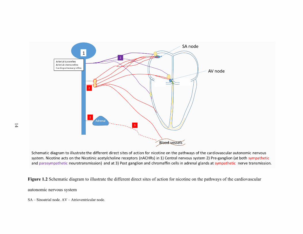
Nicotine
Cardiac autonomic nervous system: Nicotinic acetylcholinergic receptors
(nAchRs) mediate the neural transmission at the ganglia of both branches of the ANS.
However, different nicotinic receptors at the ganglia play distinct roles in sympathetic and
parasympathetic cardiovascular responses (Figure 1.2). Specifically, activation of α4β2
nAchRs elicits a parasympathetic cardiovascular response, and activation of α7 nAchRs
elicits a sympathetic cardiovascular response [67, 68]. Neff et al. used the perforated patch-
clamp technique in a visualized rat brain stem slice to identify three potential sites of action
by which nicotine increases the activity of cardiac vagal neurons: (a) direct activation of
postsynaptic ligand-gated nicotinic channels in cardiac vagal neurons, (b) different
presynaptic terminals, and (c) postsynaptic glutamatergic terminals [69]. Moreover, in
various animal models and species, nicotine has been shown to evoke norepinephrine
release from both the peripheral postganglionic sympathetic nerve endings and adrenal
medulla [70, 71]. Another potentially sympathoexcitatory mechanism of nicotine involves
the inhibition of neuronal nitric oxide synthase, which decreases central nitric oxide
availability, thereby removing its tonic inhibitory effect on central sympathetic outflow
[72].
14
Figure 1.2 Schematic diagram to illustrate the different direct sites of action for nicotine on the pathways of the cardiovascular
autonomic nervous system
SA – Sinoatrial node. AV – Atrioventricular node.
15
Cardiac sodium and calcium channels: The binding of nicotine to the extracellular
binding site of the nicotinic acetylcholine receptor leads to a conformational change of the
central pore, which results in the influx of sodium and calcium ions [73, 74]. In addition to
the increase in intracellular calcium concentration facilitated by sodium influx through the
nicotine receptor [73], nicotine also evokes calcium influx by direct activation of voltage-
dependent calcium channels [75-77]. More specifically, the L-type Ca2+ channels—Cav
1.2 channel, in particular—have been implicated in nicotine addiction, and are controlled
by the SNS and stimulated by nicotine [78, 79]. Therefore, several studies have consistently
shown that nicotine can both indirectly (catecholamine-mediated) and directly activate
voltage-gated sodium and calcium ion channels, depolarize membrane potential, and
increase cardiac contractility [80-83].
Cardiac potassium channels: On the other hand, nicotine directly blocks multiple
types of potassium currents, (A-type K+ currents (Ito current/Kv4.3 channel), delayed
rectifier K+ currents (IKr/HERG) and inward rectifier K+ currents (Ik1/Kir2.1),
independent of nicotinic receptor stimulation or catecholamine release [84-86]. However,
the ex vivo studies on the effects of nicotine on the duration of action potential have been
inconsistent. Some found that nicotine shortens the action potential duration (particularly
phase 2) [83], whereas others found that nicotine prolongs it [87]. These discrepancies
could be related to the time from drug administration [82], drug concentration [80, 88] or
the concomitant increase in force [89]. The shorter phase 2 could also reflect nicotine
stimulation of L-type Ca2+ channel, which, together with potassium channels, dictate the
duration of this phase of the action potential. Similar dose-dependent effects of nicotine
16
are seen in the sinoatrial node, where low doses of nicotine reduce [81, 88]—and large
doses increase [88]—the spontaneous cycle length of sinoatrial node pacemaker cells.
At physiological doses, nicotine can indirectly (through the sympathetic nervous
system) and directly (via immediate effects on cardiac ion channels) stimulate sodium and
calcium channels and block potassium channels. Per the pathways mentioned above,
nicotine is expected to induce multiple alterations in the surface ECG, including shortening
of the P wave, PR interval, PR segment, and QRS duration, and prolongation of QT
corrected (QTc) and Tp-Te. However, very few researchers have attempted to explore the
effects of nicotine on all ECG intervals in humans.
Cigarette smoking and its non-nicotine constituents
Cardiac autonomic nervous system: Cigarette smoke-induced cardiovascular
effects are at least partly due to stimulation of sympathetic neurotransmission, and can
theoretically manifest at four different sites of the sympathetic nervous system: the brain,
pre-ganglionic and post-ganglionic sympathetic nerves, and the adrenal medulla [71].
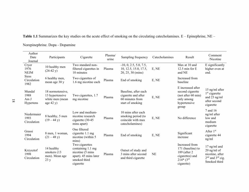
Acute cigarette smoking increases efferent sympathetic nerve activity, primarily via the
release of the catecholamines norepinephrine, and epinephrine (Table 1.1). This
catecholamine release increases myocardial work and oxygen consumption through an
increase in blood pressure, heart rate, and myocardial contractility [19]. However, several
studies suggest that the primary effect is from direct pharmacologic stimulation of nicotinic
acetylcholine receptors, as well as the catecholamine release from localized peripheral
postganglionic sympathetic nerve endings and the adrenal medulla [90, 91]. Despite
evidence that sympathetic tone is increased during smoking, either by increased release or
17
decreased clearance of catecholamines at neuroeffector junctions via inhibition of
monoamine oxidase (MOA) [92], Grassi et al. revealed that the central sympathetic activity
is inhibited, presumably via counteracting baroreceptor reflexes [90].
18
Table 1.1 Summarizes the key studies on the acute effect of smoking on the circulating catecholamines. E – Epinephrine, NE –
Norepinephrine. Dopa - Dopamine
Author Date
Journal Participants Cigarette Plasma/
urine Sampling frequency Catecholamines Result Comment /Nicotine
Cryer 1976 NEJM
10 healthy men (24-42 y)
Two standard non-filtered cigarettes in 10 minutes
Plasma -10, 0, 2.5, 5.0, 7.5, 10, 12.5, 15.0, 17.5, 20, 25, 30 (mins)
E, NE Max at 10 and 12.5 min for E and NE
E significantly higher even at end.
Siess Circulation 1982
6 healthy men, mean age 30 y
Two cigarettes of1.6 mg nicotine each Plasma End of smoking E, NE Increased from
baseline
Mundal 1988 Am J Hypertens
18 normotensive, 13 hypertensive white men (mean age 42 y)
Two cigarettes, 1.7 mg nicotine Plasma
Baseline, after each cigarette and after 60 minutes from start of smoking
E, NE
E increased after second cigarette (not after 60 min) only among hypertensive group
15 ng/ml after 1st cigarette and 23 ng/ml after second cigarette
Niedermaier 1993 Circulation
8 healthy, 5 men (19 – 44 y)
Low and medium-nicotine research cigarette (30-45 mins apart)
Plasma
10 mins after each smoking period (to coincide with max catecholamines)
E, NE No difference
7 and 16 ng/ml after low and medium cigarette
Grassi 1994 Circulation
8 men, 1 woman, (21 – 48 y)
One filtered cigarette 1.1 mg nicotine (within 5 mins)
Plasma End of smoking E, NE Significant increase
After 1st cigarette 44 ng/ml
Krzysztof 1998 Circulation
14 healthy smokers (13 men). Mean age 23 y
Two cigarettes containing 1.1 mg nicotine (5 mins apart). 45 mins later smoked third cigarette
Plasma Outset of study and 3 mins after second and third cigarette
NE
Increased from 171 (baseline) to 189 (after 2 cigarettes) and 214* (3rd cigarette)
17 ng/ml and 20 ng/ml of nicotine, after 2nd and 3rd cig. Smoked third

19
Gourlay 1997 Clin Pharmacol Ther
12 healthy male smokers, mean age 38 y
6 got nicotine nasal spray (1 x 0.5 mg in each nostril) and other 6 did smoking (usual brand)
Plasma 0, 2, 4, 6, 8, 10, 15, 20, 25, 30, 45, and 60 mins
E, NE Venous E and NE remain unchanged
Bragg 1956 J Appl Physi
11 healthy young men
Their choice of standard cigarettes during 2 / 8 hour (4 and 10 cigs)
Urine 8-hour urine collection E, NE E increased, NE
unchanged
Benowitz 1993 JACC
12 healthy male smokers, 31 – 65 y
cigarette smoking (own choice), transdermal nicotine (21 mg/d) and placebo
Urine 24 hr Urine collected day 5 Dopa, E, NE
Significantly higher with smoking
Benowitz 1989 Ann Intern Med
8 healthy male smokers
oral snuff, chewing tobacco, and cigarettes
Urine 24-hour urine after 3- or 4-day blocks Dopa, E, NE
Significantly higher with smoking

20
Non-nicotine constituents:
In addition to nicotine, several other cigarette components have been implicated in
inducing proarrhythmia, such as VOCs (aldehydes [acrolein, formaldehyde, acetaldehyde,
crotonaldehyde], benzene, toluene), particulate matter, gases (carbon monoxide), and
polycyclic aromatic hydrocarbons. It is estimated that about 90% of non-cancer mortality
from tobacco smoke is due to aldehydes (acrolein, formaldehyde, and acetaldehyde) [93].
In animal models, a single exposure to acrolein significantly increased HRV and
arrhythmia independent of heart rate, possibly through activation of the transient-receptor
potential ankyrin-1 channel (TRPA1), an irritant receptor channel found in the airways [94,
95]. Treatment with atenolol reduced this response, whereas atropine enhanced it,
suggesting parasympathetic dominance and sympathetic modulation [95]. Non-nicotinic
alkaloids, such as nornicotine or anabasine (which represent 8–12% of total alkaloid
content in tobacco), also exert an agonistic activity on nicotinic receptors [96]. Acute
exposure to toluene, the most abundant aromatic compound in mainstream smoke from
full-flavored cigarettes, enhances heart rate and blood pressure at baseline conditions,
primarily due to systemic increases in circulating catecholamines [97, 98], and the
enhanced protein expression of β1 adrenergic receptors [97]. Additionally, intravenous
acetylaldehyde in anesthetized cats has been found to increase systemic blood pressure and
heart rate (presumably from release of endogenous catecholamines from cardiac tissue)
[99]. Formaldehyde has been shown to induce significant bradycardia and negative
inotropic responses in both in situ preparations of guinea pig and rabbit hearts, and in vitro
cardiac preparations [100]. Notably, the negative chronotropic effect of formaldehyde in
21
animals seems to be caused mainly by the inhibition of sympathetic nervous activity
through the central nervous system [100].
Several non-nicotine cigarette ingredients have long been recognized as cardio-
toxic and linked with cardiac electrical activity disturbance and arrhythmias, such as
aromatic compounds [101], carbon monoxide [102], and aldehydes [99]. However,
literature on their individual drug concentration effects on cardiac ion channels and the
action potential is scarce.
Cardiac sodium and calcium channels: Toluene inhibits activated currents through
ligand and voltage-gated sodium and calcium channels [97, 103-106]. Phenol has been
shown to exert a dose-dependent negative inotropic effect in an isolated mammalian
cardiac muscle, possibly via blocking calcium channels [107]. In a patch-clamp
electrophysiology and confocal imaging experiment with isolated ventricular myocytes,
carbon monoxide activated nitric oxide synthase. This led to the nitric-oxide-mediated
nitrosylation of Nav1.5, as well as increased the sustained (late) component of the inward
Na(+) current, and inhibited peak Nav1.5 current amplitude, ultimately resulting in
prolonged action potential and associated intracellular Ca(2+) transient [102]. In addition,
carbon monoxide inhibits native rat cardiomyocyte L-type Ca2+ currents and the
recombinant α1C subunit of the human cardiac L-type Ca2+ channel [108, 109].
Formaldehyde and other aldehydes have previously been shown to cause dramatic
deceleration of sodium inactivation, depress Ina, and prolong action potential [110-113].
On the other hand, acetaldehyde has also been shown to increase the ICa, and thereby
increase the contractile force [114], Its effects can be potentiated by the additional
stimulation of α-adrenergic receptors [115]. Several other cigarette constituents have also

22
been found to, at minute quantities, significantly inhibit sodium and calcium channels in
isolated cardiac cells, such as cyanide [116, 117], lead [118], and cadmium [119].
Particulate matter, encountered during cigarette smoking, is also a significant cause of
cardiovascular morbidity and mortality. Particulate matter has been shown to dysregulate
prominent Na+ and K+ channel pathway genes [120], and carotid body sensitivity [120].
Cardiac potassium channels: Aldehydes have been proven to considerably inhibit
IK1 and Ito in animal atrial and ventricular myocytes [121-123]. In one study, ethanol and
acetaldehyde inhibited the (Na+ + K+)-activated ATPase activity of plasma membranes
prepared from a guinea-pig heart in a dose-dependent manner [124]. In addition, several
volatile agents found in cigarette smoke, commonly used as anesthetics, have been shown
to inhibit G-protein-coupled inwardly rectifying potassium channels [125]. Benzene
derivatives inhibit delayed rectifier K+ currents [126], specifically IKr2.1, in a voltage-
independent manner [127]. Carbon monoxide also inhibits inward-rectifying potassium
(Kir) channels, and prolongs the action potential duration [128, 129]. In their study, Ficker
et al. demonstrated that, despite there being an increase in cardiac calcium current, there is
reduced trafficking of cardiac potassium channel (hERG channels) to the cell surface
among patients treated with arsenic trioxide, resulting in QT prolongation [130]. Another
study revealed that arsenic trioxide blocks both IKr and IKs at clinically relevant
concentrations. However, it also activates IK-ATP [131]. Graff et al. discovered that
exposing rat cardiac myocytes to noncytotoxic concentrations of zinc and vanadium slowed
the spontaneous beating rate [132].
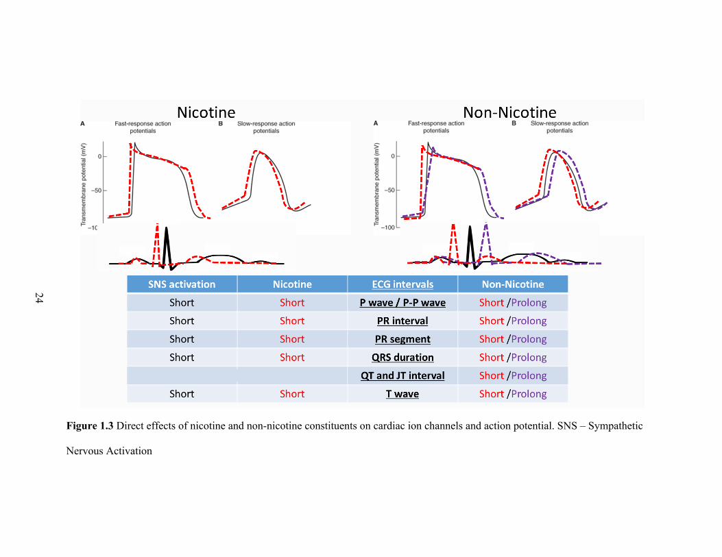
In summary, the studies on the several components of cigarette smoke with known
or suspected cardiotoxicity consistently show that they depress sodium and potassium
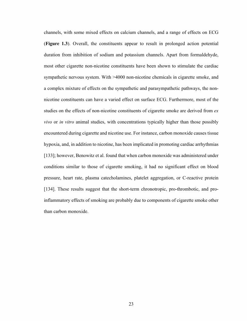
23
channels, with some mixed effects on calcium channels, and a range of effects on ECG
(Figure 1.3). Overall, the constituents appear to result in prolonged action potential
duration from inhibition of sodium and potassium channels. Apart from formaldehyde,
most other cigarette non-nicotine constituents have been shown to stimulate the cardiac
sympathetic nervous system. With >4000 non-nicotine chemicals in cigarette smoke, and
a complex mixture of effects on the sympathetic and parasympathetic pathways, the non-
nicotine constituents can have a varied effect on surface ECG. Furthermore, most of the
studies on the effects of non-nicotine constituents of cigarette smoke are derived from ex
vivo or in vitro animal studies, with concentrations typically higher than those possibly
encountered during cigarette and nicotine use. For instance, carbon monoxide causes tissue
hypoxia, and, in addition to nicotine, has been implicated in promoting cardiac arrhythmias
[133]; however, Benowitz et al. found that when carbon monoxide was administered under
conditions similar to those of cigarette smoking, it had no significant effect on blood
pressure, heart rate, plasma catecholamines, platelet aggregation, or C-reactive protein
[134]. These results suggest that the short-term chronotropic, pro-thrombotic, and pro-
inflammatory effects of smoking are probably due to components of cigarette smoke other
than carbon monoxide.
24
Figure 1.3 Direct effects of nicotine and non-nicotine constituents on cardiac ion channels and action potential. SNS – Sympathetic
Nervous Activation
25
Indirect (systemic) effects
There are several short- and long-term homeostatic mechanisms to ensure adequate
blood flow, pressure, distribution, and perfusion, and are categorized into three groups:
neural, humoral, and autoregulatory mechanisms. These mechanisms can also indirectly
alter cardiac autonomic output and electrophysiology. Table 1.2 summarizes the stimuli,
receptors, and physiologic effects most pertinent to cigarette smoking and nicotine-related
changes in human autonomic reflexes.
Neural reflexes to tobacco exposure: The baroreflex feedback loop is one of the
most important mechanisms controlling arterial pressure on a beat-to-beat basis. It achieves
this through arterial baroreceptors located in the carotid sinus and aortic arch. These
receptors are mechano-sensitive, and the distension of the vessels that occurs at each heart
beat leads to action potential generation on peripheral nerves that transmit to the central
nervous system, buffering arterial pressure fluctuations through changes in sympathetic
and parasympathetic activity. Therefore, when blood pressure rises, the baroreceptor
afferent tone increases, leading to increased vagal efferent activity and diminished
sympathetic outflow. These effects will lead to a decrease in cardiac output by decreasing
heart rate and cardiac contractility. Additionally, the fall in sympathetic tone to blood
vessels, as well as increased vagal effect activity (through increased guanylyl cyclase and
cGMP activity) leads to vasodilation and diminished vascular resistance. There is strong
evidence that this crucial inhibitory role of the baroreflex arc is blunted in habitual smokers,
and also impaired during acute exposure to smoking [72, 135, 136]. Grassi et al.’s study
showed that there is sympathetic activation induced by smoking via increased release or a
reduced clearance of catecholamines at the neuroeffector junctions. However, the central
26
sympathetic activity is inhibited by smoking, presumably via baroreceptor stimulation
triggered by a pressor response to smoking [90]. There is also evidence that nicotine
possibly decreases the baroreceptor sensitivity [137]. Besides nicotine, there are several
other cigarette components, mainly PM 2.5, that directly alter baroreflex responsiveness in
smokers [72, 138]. In one study, a one-time exposure to acrolein caused a decrease in the
sensitivity of baroreflex and increased incidence of arrhythmia in rats [139].
The peripheral arterial chemoreceptors in the carotid and aortic bodies are
stimulated by decreased arterial PO2, increased PCO2, and increased H+ concentration.
Their stimulation causes hyperventilation, as well as increases in sympathetic neural
activity and the rate and volume of breathing; chronic arterial chemoreflex sensitization in
smokers could also lead to sustained sympathetic activation. Arterial chemoreceptors are
activated by hypoxia, and chronic smokers may be at risk to toxic effects of carbon
monoxide in tobacco smoke. Perez et al. showed that acrolein-exposure-induced
cardiovascular effects in rats (i.e., an increase in systolic, diastolic and mean arterial blood
pressure during exposure, and a decrease in cardiac contractility one day after exposure)
were prevented after a blockade of carotid body signal transduction. This suggests that
acrolein-induced cardiovascular responses may be mediated by carotid body-triggered
changes in autonomic tone [140]. However, to date, there is no evidence for augmented
arterial chemoreceptor sensitivity in habitual smokers. Instead, nicotine does not affect
chemoreflex sensitivity, as evidenced by unchanged minute ventilation, apnea duration,
and oxygen saturation after nicotine and placebo in normoxia [141, 142].
Airways are lined with vagal afferent nerve fibers, including non-myelinated
afferent C-fibers sensitive to noxious chemicals. A subset of these vagal C-fibers expresses
27
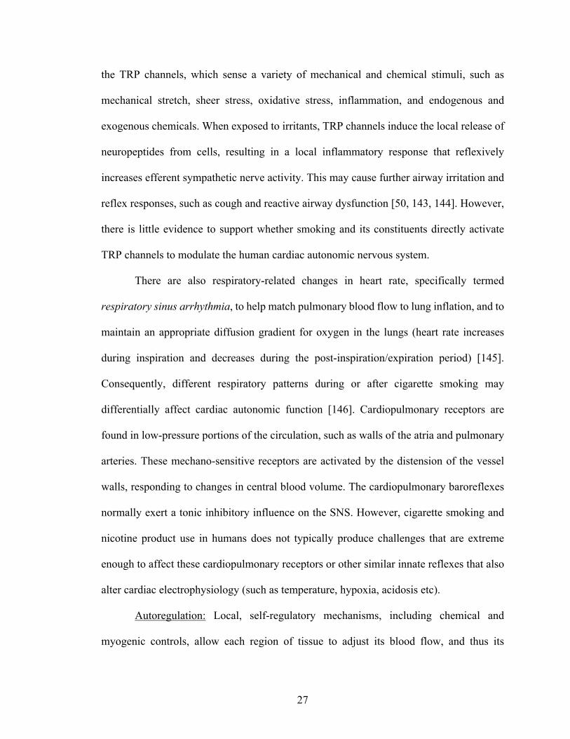
the TRP channels, which sense a variety of mechanical and chemical stimuli, such as
mechanical stretch, sheer stress, oxidative stress, inflammation, and endogenous and
exogenous chemicals. When exposed to irritants, TRP channels induce the local release of
neuropeptides from cells, resulting in a local inflammatory response that reflexively
increases efferent sympathetic nerve activity. This may cause further airway irritation and
reflex responses, such as cough and reactive airway dysfunction [50, 143, 144]. However,
there is little evidence to support whether smoking and its constituents directly activate
TRP channels to modulate the human cardiac autonomic nervous system.
There are also respiratory-related changes in heart rate, specifically termed
respiratory sinus arrhythmia, to help match pulmonary blood flow to lung inflation, and to
maintain an appropriate diffusion gradient for oxygen in the lungs (heart rate increases
during inspiration and decreases during the post-inspiration/expiration period) [145].
Consequently, different respiratory patterns during or after cigarette smoking may
differentially affect cardiac autonomic function [146]. Cardiopulmonary receptors are
found in low-pressure portions of the circulation, such as walls of the atria and pulmonary
arteries. These mechano-sensitive receptors are activated by the distension of the vessel
walls, responding to changes in central blood volume. The cardiopulmonary baroreflexes
normally exert a tonic inhibitory influence on the SNS. However, cigarette smoking and
nicotine product use in humans does not typically produce challenges that are extreme
enough to affect these cardiopulmonary receptors or other similar innate reflexes that also
alter cardiac electrophysiology (such as temperature, hypoxia, acidosis etc).
Autoregulation: Local, self-regulatory mechanisms, including chemical and
myogenic controls, allow each region of tissue to adjust its blood flow, and thus its

28
perfusion. Chemosensitive nerve endings are also found throughout the cardiovascular and
respiratory systems, and are stimulated by several exogenous chemicals and endogenous
chemicals formed and released in response to conditions such as hypoxia, ischemia, certain
mechanical demands, inflammation, or toxin exposures. The efferent pathways of the
reflex involve inhibition of sympathetic outflow to peripheral vessels and increased activity
in efferent vagal fibers to the heart [50]. The myogenic response is a reflex to the stretching
of the smooth muscle of the arteriolar walls as changes in blood flow occur through the
vessel (e.g., vasoconstriction in response to increased intraluminal pressure). Increased
peripheral vascular resistance, cigarette smoking, and nicotine also have detrimental effects
on coronary microvascular function (e.g., increases in coronary flow velocity and
resistance, and decreases in flow reserve) and can cause vascular dysfunction [147-151],
possibly via β-adrenergic receptor [152].
Humoral to tobacco exposure: Several studies have suggested that humoral systems
play a vital role in maintaining cardiac electric activity, and changes in their production or
action pathways may contribute to various cardiac diseases. Beyond the neurotransmitters
acetylcholine and norepinephrine, there is a local presence of peptidergic and nitrergic
neurons along with their associated neurotransmitters, such as neuropeptide-Y, vasostatin,
galactin, vasoactive intestinal peptide, nitric oxide synthase, and angiotensin-II [153, 154].
Neuropeptide-Y, coreleased by prolonged sympathetic activation, reduces acetylcholine
release from the nearby vagal nerve ending, and it is an excellent example of
sympathovagal cross-talk. These non-cholinergic, non-adrenergic neurotransmitters often
exert effects similar to cholinergic or adrenergic agonists or antagonists [153]. The release
of these neurotransmitters/modulators is often highly dependent on the level of neuronal
29
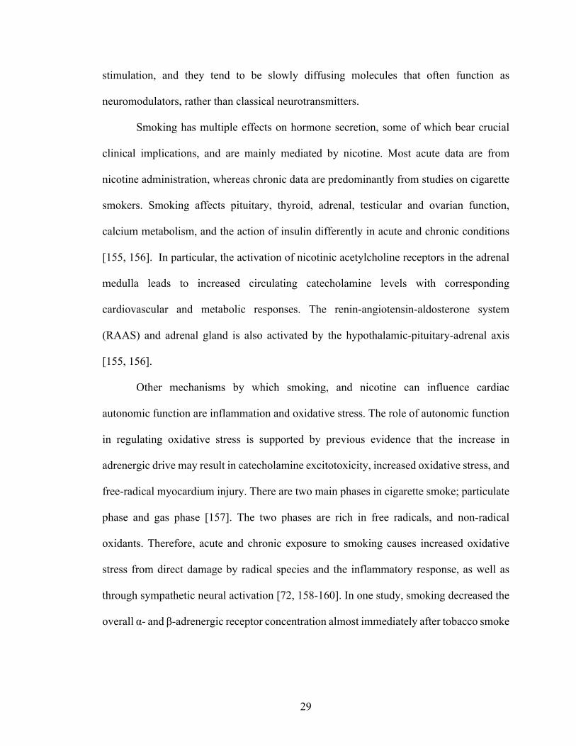
stimulation, and they tend to be slowly diffusing molecules that often function as
neuromodulators, rather than classical neurotransmitters.
Smoking has multiple effects on hormone secretion, some of which bear crucial
clinical implications, and are mainly mediated by nicotine. Most acute data are from
nicotine administration, whereas chronic data are predominantly from studies on cigarette
smokers. Smoking affects pituitary, thyroid, adrenal, testicular and ovarian function,
calcium metabolism, and the action of insulin differently in acute and chronic conditions
[155, 156]. In particular, the activation of nicotinic acetylcholine receptors in the adrenal
medulla leads to increased circulating catecholamine levels with corresponding
cardiovascular and metabolic responses. The renin-angiotensin-aldosterone system
(RAAS) and adrenal gland is also activated by the hypothalamic-pituitary-adrenal axis
[155, 156].
Other mechanisms by which smoking, and nicotine can influence cardiac
autonomic function are inflammation and oxidative stress. The role of autonomic function
in regulating oxidative stress is supported by previous evidence that the increase in
adrenergic drive may result in catecholamine excitotoxicity, increased oxidative stress, and
free-radical myocardium injury. There are two main phases in cigarette smoke; particulate
phase and gas phase [157]. The two phases are rich in free radicals, and non-radical
oxidants. Therefore, acute and chronic exposure to smoking causes increased oxidative
stress from direct damage by radical species and the inflammatory response, as well as
through sympathetic neural activation [72, 158-160]. In one study, smoking decreased the
overall α- and β-adrenergic receptor concentration almost immediately after tobacco smoke

30
exposure in rats—perhaps through receptor desensitization resulting from a release of
catecholamines—but was rapidly reversible after the termination of the exposure [161].

31
Table 1.2 Potential mechanisms of indirect effects of cigarette smoking on human cardiac electrophysiology
System Receptors Stimulus Effect on SNS
Effect on PNS Studies on the effects of smoking / nicotine
Baroreflex Carotid artery / Aorta
High Blood Pressure
Decrease Increase Activates the system by increasing blood pressure through release of catecholamines from end terminals. Smoking also impairs the baroreflex system
Chemoreflex Carotid / aortic (peripheral), medulla (central)
Low PaO2, High PaCO2/pH
Increase (increase pulmonary ventilation)
- Nicotine does not increase chemoreflex sensitivity to hypoxia.
Temperature CNS High Temp Increases - Temperature increases in lungs; decreases in skin temp
Inflammation / Oxidative stress
Lung afferent C fibers, vascular, myocardial
Direct / Reactive Oxygen Species
Increase Decrease Increases inflammation and oxidative stress during acute and chronic exposure
Endocrine hypothalamic–pituitary axis
Increase - Chronic: increases thyroid hormones, cortisol, possibly testosterone and estradiol. Decreases prolactin and growth hormone Acute: Increases prolactin, cortisol, growth hormone, vasopressin, endorphin, neuropeptide Y
Cardiopulmonary Ventricles High SNS (low vol LV)
Decrease Increase -
Pulmonary Pulmonary chemical stimuli, inflammation
? Increase -
Bainbridge Atria High blood vol Increase Decrease -
32
Literature review of effects of autonomic nervous system
and smoking on electrocardiogram
Autonomic nervous system
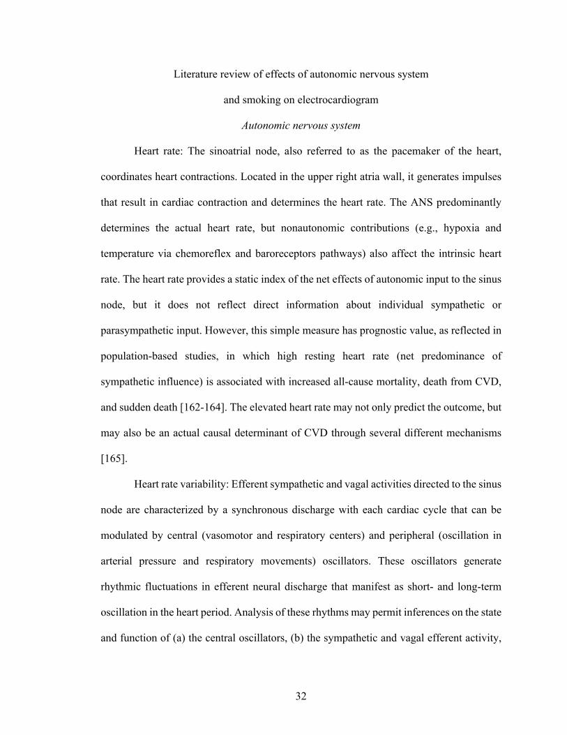
Heart rate: The sinoatrial node, also referred to as the pacemaker of the heart,
coordinates heart contractions. Located in the upper right atria wall, it generates impulses
that result in cardiac contraction and determines the heart rate. The ANS predominantly
determines the actual heart rate, but nonautonomic contributions (e.g., hypoxia and
temperature via chemoreflex and baroreceptors pathways) also affect the intrinsic heart
rate. The heart rate provides a static index of the net effects of autonomic input to the sinus
node, but it does not reflect direct information about individual sympathetic or
parasympathetic input. However, this simple measure has prognostic value, as reflected in
population-based studies, in which high resting heart rate (net predominance of
sympathetic influence) is associated with increased all-cause mortality, death from CVD,
and sudden death [162-164]. The elevated heart rate may not only predict the outcome, but
may also be an actual causal determinant of CVD through several different mechanisms
[165].
Heart rate variability: Efferent sympathetic and vagal activities directed to the sinus
node are characterized by a synchronous discharge with each cardiac cycle that can be
modulated by central (vasomotor and respiratory centers) and peripheral (oscillation in
arterial pressure and respiratory movements) oscillators. These oscillators generate
rhythmic fluctuations in efferent neural discharge that manifest as short- and long-term
oscillation in the heart period. Analysis of these rhythms may permit inferences on the state
and function of (a) the central oscillators, (b) the sympathetic and vagal efferent activity,

33
(c) humoral factors, and (d) the sinus node [166]. The oscillation in the intervals between
consecutive heartbeats can be measured for indices of HRV, using either time domain
approaches (based on statistical analysis of R-R intervals) or frequency domain approaches
(spectral analysis of a sequence of R-R intervals) [166]. Large population studies have
shown a higher risk of coronary artery disease, death, and cardiac mortality in individuals
with decreased HRV (in both healthy populations and patients with cardiac disease) [167].
P wave: The P wave is the expression of atrial depolarization and intraatrial
conduction. Electrocardiographic P wave indices consist of the P wave duration,
morphology, and amplitude, and provide information about the atrial structure and
function. A prolonged P-wave duration (>120 ms) is considered a marker of atrial
cardiopathy, which, in chronic cardiac disease, is usually reflective if reduced atrial
conduction related to architectural changes of atrial walls. P-wave duration is affected by
autonomic tone. In general, both sympathetic stimulation and parasympathetic blockade
shorten P-wave duration, whereas sympathetic blockade prolongs it [58]. P-wave terminal
force in lead V1 (PTFV1) is defined as the value of the amplitude multiplied by the duration
of the terminal’s negative deflection of the P wave in lead V1 of a standard 12-lead ECG.
P-wave area (PWA) is the total geometric area under the P wave in the 12-lead ECG. It is
usually represented by the product of the duration and peak amplitude of the P wave, and
is measured in microvolt × milliseconds. Both of these P-wave indices, together with P-
wave axis (net direction of electrical forces within the atria), are also markers of atrial
cardiopathy [168, 169]. Therefore, acute sympathetic activation may shorten P-wave
duration, whereas chronic sympathetic activation may prolong P-wave duration from long-
term structural atrial remodeling. The P amplitude, which is mostly governed by atrial

34
pressures, may be increased in height (and in depth of PTFV1) in acute and chronic
sympathetic stimulation.
PR interval: PR interval is the period of time from the start of the P wave (atrial
depolarization) until the start of the QRS complex (ventricular depolarization). Therefore,
the determinants of PR interval are atrial depolarization and the conduction time from the
sinus node to the atrioventricular node, His bundle, and Purkinje fibers [170]. The duration
of PR interval is normally between 120 and 200 ms. In most cases, a prolonged PR interval
(>200 ms) is determined by conduction delay in the atrioventricular node. The ANS’s acute
effects on PR intervals are well known, considering that autonomic innervation influences
the conduction through the atrioventricular node junction by modulating the refractory
period [61, 171]. However, in the chronic state, autonomic-imbalance-induced atrial
fibrosis may also cause PR interval prolongation by slowing atrial depolarization and
dromotropy. Both short [26, 27, 29, 30] and prolonged [30, 172-174] PR intervals have
been associated with adverse clinical outcomes (stroke, atrial fibrillation, and all-cause
mortality).
QRS complex: The QRS complex represents the electrical impulse as it spreads
through the ventricles, and depicts ventricular depolarization. Ventricular depolarization is
also influenced by autonomic modulation of the heart. In humans, increased sympathetic
tone by β-adrenergic stimulation shortens overall QRS duration [175], heterogeneity of
ventricular activation during disease states, and bundle branch blocks (mechanical and
electrical dyssynchrony) [176]. The QRS complex voltage reflects the viable left
ventricular mass and can be increased (e.g., athlete’s heart, hypertensive heart disease
[177]) or decreased (post-myocardial infarction, infiltrative cardiomyopathies, etc. [178])
35
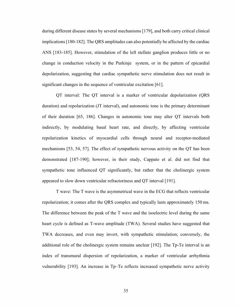
during different disease states by several mechanisms [179], and both carry critical clinical
implications [180-182]. The QRS amplitudes can also potentially be affected by the cardiac
ANS [183-185]. However, stimulation of the left stellate ganglion produces little or no
change in conduction velocity in the Purkinje system, or in the pattern of epicardial
depolarization, suggesting that cardiac sympathetic nerve stimulation does not result in
significant changes in the sequence of ventricular excitation [61].
QT interval: The QT interval is a marker of ventricular depolarization (QRS
duration) and repolarization (JT interval), and autonomic tone is the primary determinant
of their duration [65, 186]. Changes in autonomic tone may alter QT intervals both
indirectly, by modulating basal heart rate, and directly, by affecting ventricular
repolarization kinetics of myocardial cells through neural and receptor-mediated
mechanisms [53, 54, 57]. The effect of sympathetic nervous activity on the QT has been
demonstrated [187-190]; however, in their study, Cappato et al. did not find that
sympathetic tone influenced QT significantly, but rather that the cholinergic system
appeared to slow down ventricular refractoriness and QT interval [191].
T wave: The T wave is the asymmetrical wave in the ECG that reflects ventricular
repolarization; it comes after the QRS complex and typically lasts approximately 150 ms.
The difference between the peak of the T wave and the isoelectric level during the same
heart cycle is defined as T-wave amplitude (TWA). Several studies have suggested that
TWA decreases, and even may invert, with sympathetic stimulation; conversely, the
additional role of the cholinergic system remains unclear [192]. The Tp-Te interval is an
index of transmural dispersion of repolarization, a marker of ventricular arrhythmia
vulnerability [193]. An increase in Tp–Te reflects increased sympathetic nerve activity
36
[194], rather than the release of circulating norepinephrine [194]. However, more recently,
the influence of the ANS on the TWA and Tp-Te interval has fallen under heavy scrutiny,
and appears to be an unreliable index of myocardial sympathetic activity [195-197].
In summary, acute and chronic cardiac autonomic imbalance varies by phenotype
(normal and diseased states), and influences several ECG parameters differently.
Role of β-adrenergic receptors
Physiologic doses of epinephrine alter electrophysiology through β-receptor
activation, and manifest as an acceleration of atrioventricular nodal conduction and
shortening of refractoriness in the atrium and ventricle [198]. In contrast, norepinephrine
slows atrioventricular nodal conduction and lengthens the atrial and ventricular effective
refractory periods [199]. Therefore, norepinephrine may counteract several of the
electrophysiological effects of circulating epinephrine during physiologic degrees of stress.
Furthermore, a-adrenergic stimulation by epinephrine in the presence of propranolol was
found to prolong atrial and ventricular refractoriness [199].
Most studies on the effect of β-adrenergic receptors on cardiac electrophysiology
have been performed with either propranolol, a non-selective β-blocker, or isoproterenol,
a non-selective β-adrenoreceptor agonist. Propranolol decreases heart rate and prolongs
atrioventricular nodal conduction [200], and has been shown to have no significant effect
on intra-ventricular conduction. Shortening of action potential plateau was also not evident
after treatment with propranolol in normal cat ventricular muscles [89]. In the sinoatrial
node and left atrial appendage cells of the guinea-pig heart, propranolol antagonized the
positive chronotropism of nicotine and norepinephrine [88]. Studies conducted on five

37
human volunteers showed that intravenous propranolol followed by smoking significantly
decreased cardiac output, and significantly increased blood pressure and systemic vascular
resistance compared to smoking alone [201]. Similar effects were observed in another
study among 10 participants [202]. Smoking alone increased cardiac output, mean arterial
blood pressure, and decreased the calculated systemic vascular resistance [201]. The
cardiac output increases were due to a fall in systemic vascular resistance. The effect is
exaggerated after propranolol, potentially via the inhibition of β-adrenergic receptors,
either by preventing the vasodilatory effect of β-2 adrenergic stimulation, or as increased
availability of α-receptors may result in unopposed pronounced effects of vasoconstriction
from epinephrine. However, another study was conducted using 80 mg of oral propranolol
on six volunteers, and there was no significant difference from the placebo in blood
pressure or forearm hemodynamics, and no prevention of the acute vascular effects of
cigarette smoking with β-blocker pre-treatment [203]. β-blockers are first line therapy for
patients with Long QT syndrome (LQTS)—a genetic disorder that can potentially cause
life-threatening cardiac arrhythmia, is characterized by delayed myocardial repolarization,
and manifests as QT prolongation [204]. The response to β-blockers and epinephrine
depends on the genotype [204, 205]. Therefore, the differences in β-adrenergic receptor-
mediated effects on ventricular repolarization are likely related to the stimulus (e.g.,
frequency of cigarette smoking), dose (route) of propranolol use, and the underlying
genotype. Furthermore, the plasma propranolol steady-state concentration is lower among
smokers compared to non-smokers, possibly via an increased rate of drug metabolism
[206].
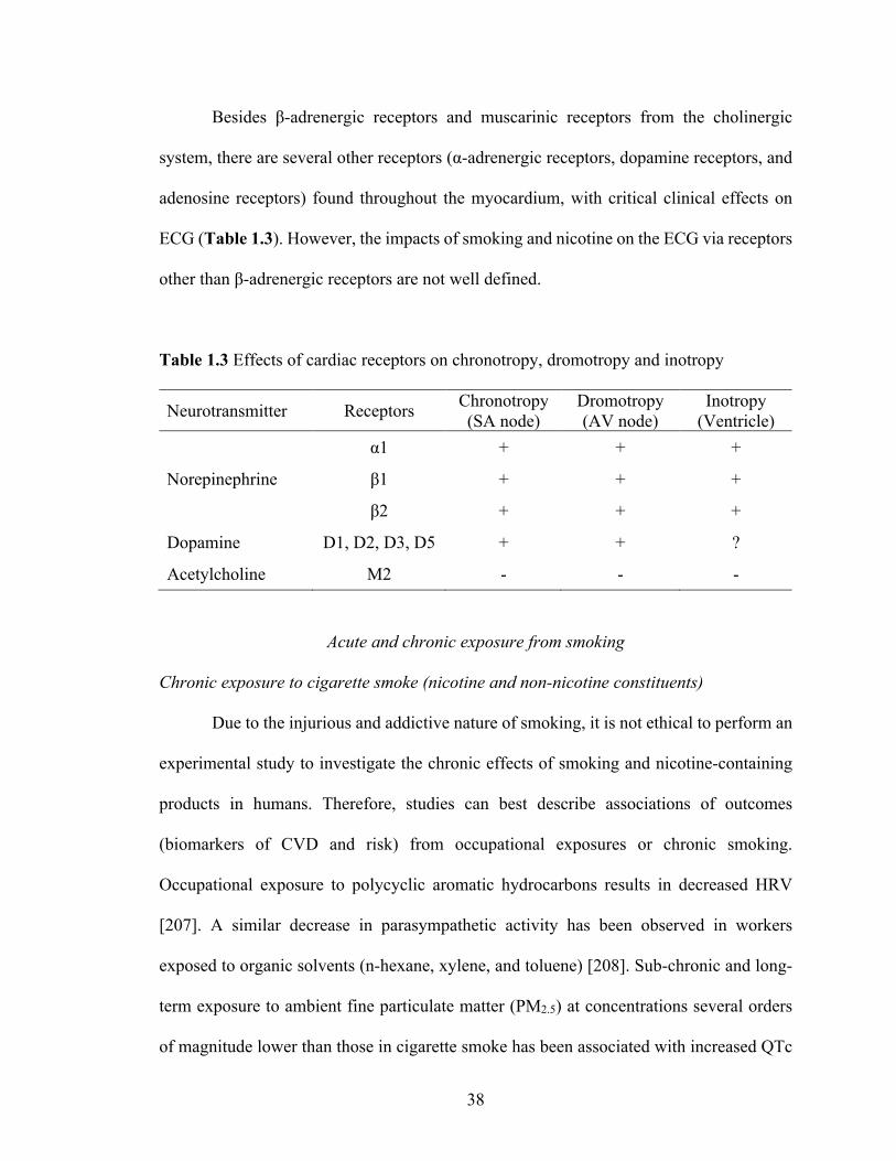
38
Besides β-adrenergic receptors and muscarinic receptors from the cholinergic
system, there are several other receptors (α-adrenergic receptors, dopamine receptors, and
adenosine receptors) found throughout the myocardium, with critical clinical effects on
ECG (Table 1.3). However, the impacts of smoking and nicotine on the ECG via receptors
other than β-adrenergic receptors are not well defined.
Table 1.3 Effects of cardiac receptors on chronotropy, dromotropy and inotropy
Neurotransmitter Receptors Chronotropy (SA node)
Dromotropy (AV node)
Inotropy (Ventricle)
Norepinephrine
α1 + + +
β1 + + +
β2 + + +
Dopamine D1, D2, D3, D5 + + ?
Acetylcholine M2 - - -
Acute and chronic exposure from smoking
Chronic exposure to cigarette smoke (nicotine and non-nicotine constituents)
Due to the injurious and addictive nature of smoking, it is not ethical to perform an
experimental study to investigate the chronic effects of smoking and nicotine-containing
products in humans. Therefore, studies can best describe associations of outcomes
(biomarkers of CVD and risk) from occupational exposures or chronic smoking.
Occupational exposure to polycyclic aromatic hydrocarbons results in decreased HRV
[207]. A similar decrease in parasympathetic activity has been observed in workers
exposed to organic solvents (n-hexane, xylene, and toluene) [208]. Sub-chronic and long-
term exposure to ambient fine particulate matter (PM2.5) at concentrations several orders
of magnitude lower than those in cigarette smoke has been associated with increased QTc
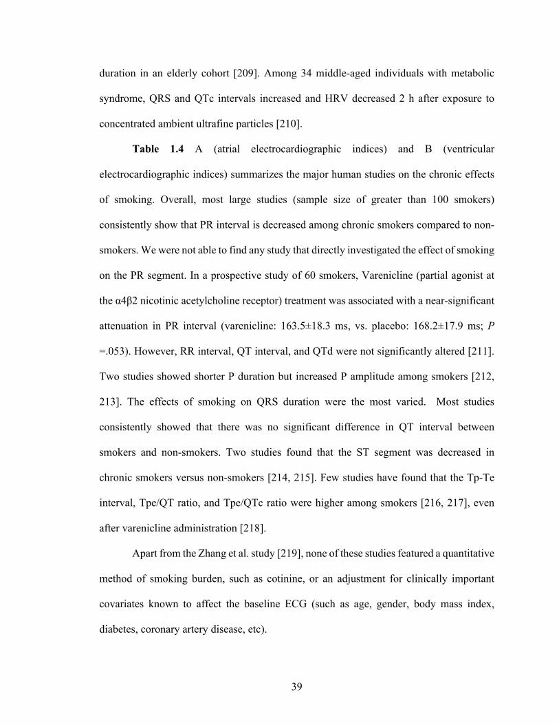
39
duration in an elderly cohort [209]. Among 34 middle-aged individuals with metabolic
syndrome, QRS and QTc intervals increased and HRV decreased 2 h after exposure to
concentrated ambient ultrafine particles [210].
Table 1.4 A (atrial electrocardiographic indices) and B (ventricular
electrocardiographic indices) summarizes the major human studies on the chronic effects
of smoking. Overall, most large studies (sample size of greater than 100 smokers)
consistently show that PR interval is decreased among chronic smokers compared to non-
smokers. We were not able to find any study that directly investigated the effect of smoking
on the PR segment. In a prospective study of 60 smokers, Varenicline (partial agonist at
the α4β2 nicotinic acetylcholine receptor) treatment was associated with a near-significant
attenuation in PR interval (varenicline: 163.5±18.3 ms, vs. placebo: 168.2±17.9 ms; P
=.053). However, RR interval, QT interval, and QTd were not significantly altered [211].
Two studies showed shorter P duration but increased P amplitude among smokers [212,
213]. The effects of smoking on QRS duration were the most varied. Most studies
consistently showed that there was no significant difference in QT interval between
smokers and non-smokers. Two studies found that the ST segment was decreased in
chronic smokers versus non-smokers [214, 215]. Few studies have found that the Tp-Te
interval, Tpe/QT ratio, and Tpe/QTc ratio were higher among smokers [216, 217], even
after varenicline administration [218].
Apart from the Zhang et al. study [219], none of these studies featured a quantitative
method of smoking burden, such as cotinine, or an adjustment for clinically important
covariates known to affect the baseline ECG (such as age, gender, body mass index,
diabetes, coronary artery disease, etc).
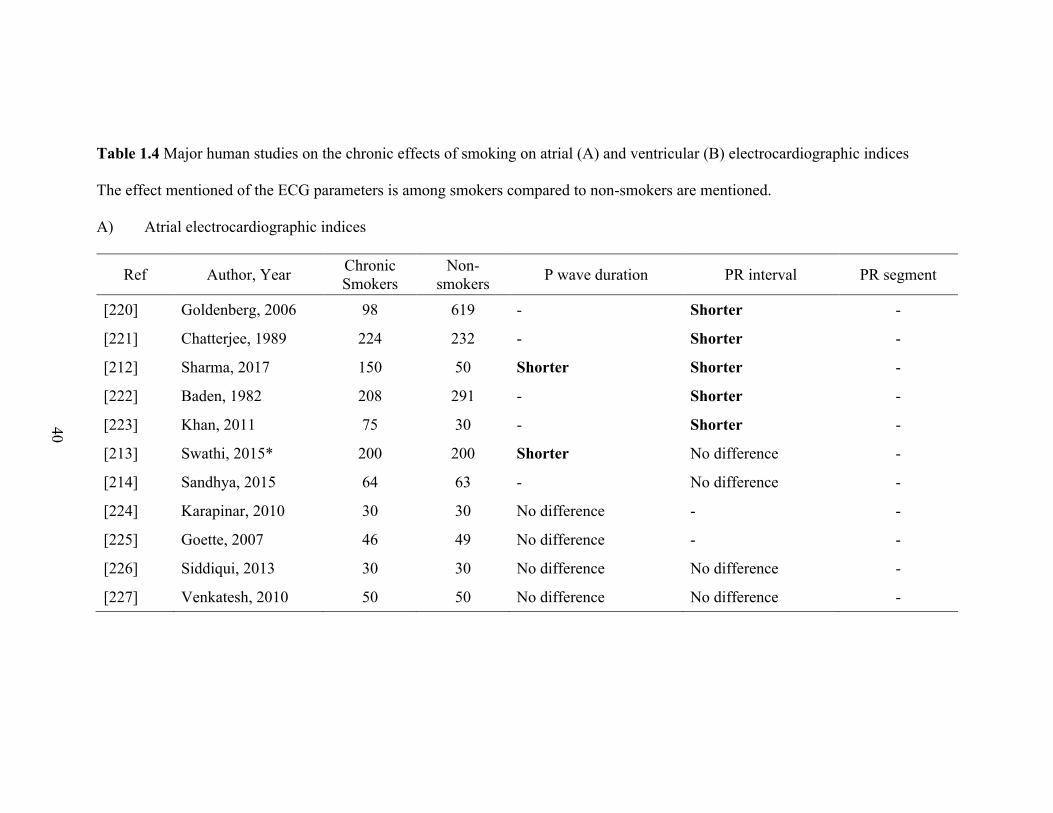
40
Table 1.4 Major human studies on the chronic effects of smoking on atrial (A) and ventricular (B) electrocardiographic indices
The effect mentioned of the ECG parameters is among smokers compared to non-smokers are mentioned.
A) Atrial electrocardiographic indices
Ref Author, Year Chronic Smokers
Non-smokers P wave duration PR interval PR segment
[220] Goldenberg, 2006 98 619 - Shorter -
[221] Chatterjee, 1989 224 232 - Shorter -
[212] Sharma, 2017 150 50 Shorter Shorter -
[222] Baden, 1982 208 291 - Shorter -
[223] Khan, 2011 75 30 - Shorter -
[213] Swathi, 2015* 200 200 Shorter No difference -
[214] Sandhya, 2015 64 63 - No difference -
[224] Karapinar, 2010 30 30 No difference - -
[225] Goette, 2007 46 49 No difference - -
[226] Siddiqui, 2013 30 30 No difference No difference -
[227] Venkatesh, 2010 50 50 No difference No difference -
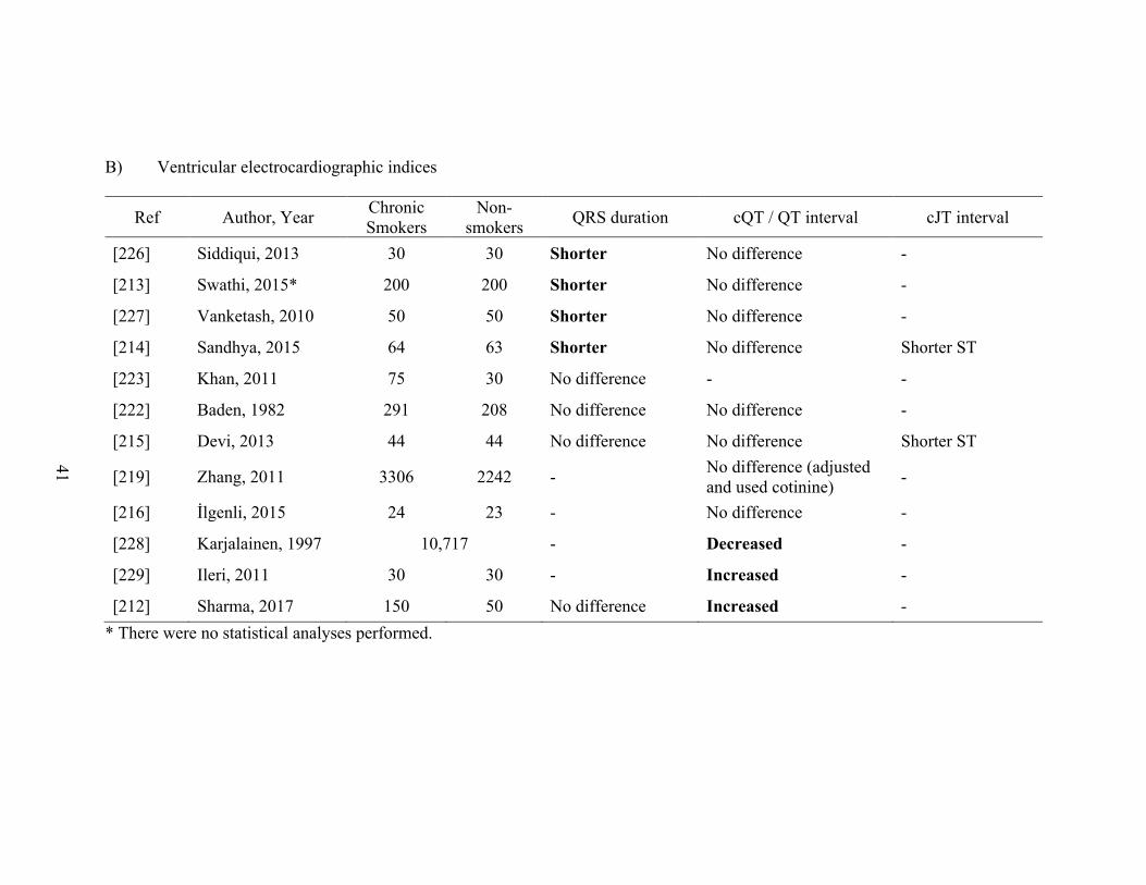
41
B) Ventricular electrocardiographic indices
Ref Author, Year Chronic Smokers
Non-smokers QRS duration cQT / QT interval cJT interval
[226] Siddiqui, 2013 30 30 Shorter No difference -
[213] Swathi, 2015* 200 200 Shorter No difference -
[227] Vanketash, 2010 50 50 Shorter No difference -
[214] Sandhya, 2015 64 63 Shorter No difference Shorter ST
[223] Khan, 2011 75 30 No difference - -
[222] Baden, 1982 291 208 No difference No difference -
[215] Devi, 2013 44 44 No difference No difference Shorter ST
[219] Zhang, 2011 3306 2242 - No difference (adjusted and used cotinine) -
[216] İlgenli, 2015 24 23 - No difference -
[228] Karjalainen, 1997 10,717 - Decreased -
[229] Ileri, 2011 30 30 - Increased -
[212] Sharma, 2017 150 50 No difference Increased - * There were no statistical analyses performed.

42
Acute exposure to smoking (nicotine and non-nicotine constituents)
Apart from the case reports, the scarce literature on the acute effects of individual
constituents of cigarette smoking on human in vivo ECG has mainly investigated nicotine
and carbon monoxide. Acute carbon monoxide intoxication in children impaired
ventricular repolarization with higher mean heart rate, QT dispersion, QTc dispersion, P
dispersion, Tp-Te, QTc durations, Tp-Te dispersion, and Tp-Te/QT ratio compared to
controls [230, 231]. However, it increased heart rate, ST-T wave changes, QTc dispersion,
and TpTe dispersion in adults [232, 233]. There is also long-term increased risk of acute
myocardial infarction in carbon monoxide poisoning [234]. Among 16 smokers
undergoing electrophysiological study, exposure to cigarette smoke produced a statistically
significant shortening of the sinus cycle length after chemical denervation of the sinus node
with β-blockers and atropine [235]. In a single case report of mild nicotine toxicity, a young
smoker presented with a typ1 Brugada pattern, raising the possibility of sodium channel
blockade [236]. Nicotine has proved to be arrhythmogenic in animals and cell preparations;
however, despite some case reports linking nicotine to atrial fibrillation in humans, these
findings have not been confirmed in pharmacological studies of nicotine administered to
human subjects [133].
There are very few human experimental studies that have investigated the acute
effects of smoking on the ECG. In their analysis of 40 healthy male participants (20
smokers), Akbarzadeh et al. discovered that heart rate, mean QT, and QTc dispersion
increased 10 minutes after smoking a single cigarette [237]. In another study, 31 male
smokers with atypical chest pain were screened with a treadmill exercise test, which
revealed that the heart rate increased within 10 minutes and returned to baseline after 30

43
minutes. Smoking was also associated with increased ectopic beats in these patients on the
24-hour Holter monitor [238]. On a signal averaged ECG, there was a minor lengthening
of the filtered QRS duration among 15 long-term smokers after smoking two cigarettes
[239].
To date, the most comprehensive assessment of acute smoking’s impact on
myocardial conduction was performed among 28 habitual smokers by Soad Bekheit and
Evan Fletcher in 1976 using His bundle electrograms [240]. The following measurements
were made from His bundle recordings in the control and “nicotine tracings” (immediately
after the first puff, sequential atrial pacing at identical rates to the control tracings): intra-
atrial conduction time (P1A), A-V nodal conduction time (AH), His-Purkinje-system
conduction time (HV), and total intraventricular conduction time (HS). They found that a
few puffs of a cigarette increase the velocity of conduction and shortens the effective
refractory period of the A-V node, whereas the conduction velocity in the His-Purkinje
system is not affected [240]. However, these effects were not studied against those from
nicotine exposure.
Acute cigarette smoking increases efferent sympathetic nerve activity, primarily
via the release of the catecholamines norepinephrine and epinephrine (Table 4). This
catecholamine release increases myocardial work and oxygen consumption through an
increase in blood pressure, heart rate, and myocardial contractility [19]. Overall, the vast
majority of published evidence suggests that acute and chronic active and passive exposure
to cigarette smoke generates marked disruptions in normal autonomic function
characterized by increased SNS drive, reduced PNS modulation, and overall HRV. This
phenomenon is partly attributable to a nicotine-induced up-regulation of catecholamine
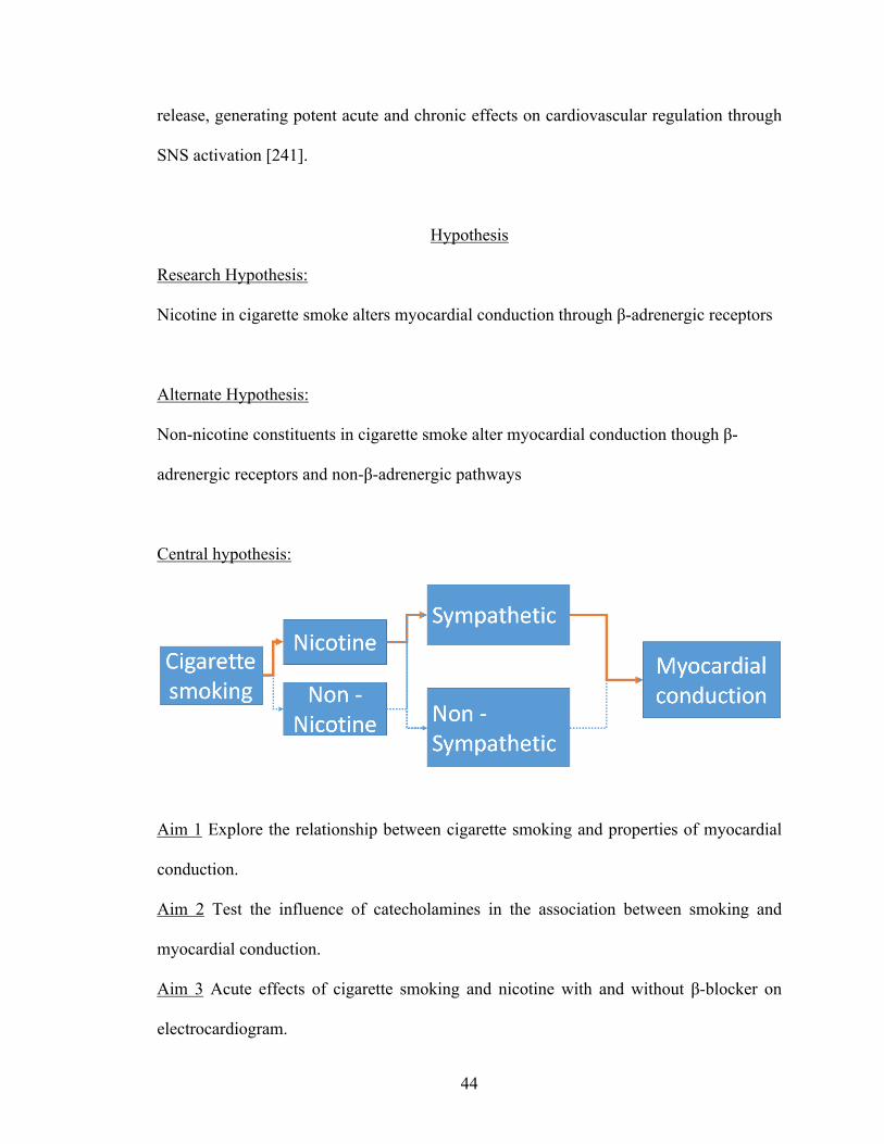
44
release, generating potent acute and chronic effects on cardiovascular regulation through
SNS activation [241].
Hypothesis
Research Hypothesis:
Nicotine in cigarette smoke alters myocardial conduction through β-adrenergic receptors
Alternate Hypothesis:
Non-nicotine constituents in cigarette smoke alter myocardial conduction though β-
adrenergic receptors and non-β-adrenergic pathways
Central hypothesis:
Aim 1 Explore the relationship between cigarette smoking and properties of myocardial
conduction.
Aim 2 Test the influence of catecholamines in the association between smoking and
myocardial conduction.
Aim 3 Acute effects of cigarette smoking and nicotine with and without β-blocker on
electrocardiogram.

45
Ethical considerations
The primary therapeutic use of nicotine is in treating nicotine dependence.
Controlled levels of nicotine are given to patients through gum, dermal patches, lozenges,
inhalers, or nasal and oral sprays to wean them off their dependence, increasing quitting
success by 50%–70% [242]. In contrast to recreational nicotine products, which have been
designed to maximize the likelihood of addiction, nicotine replacement products are
designed to minimize addictiveness. Hence, physiological studies on the effects of chronic
use of pure nicotine on humans are accompanied by the concern for potential addiction
among non-smokers, or the tolerance or modulation of neural plasticity among habitual
smokers. It is for this reason that there are no robust human physiological studies
investigating the cause–effect relationship of prolonged exposure to pure nicotine. There
are some studies on prolonged nicotine replacement therapies in smokers who have quit
smoking, and in these studies, no adverse effects have been found when nicotine
medication was administered for months to several years. Given the highly addictive nature
of nicotine, these studies are more appropriately termed as acute or chronic exposure of
nicotine in chronic tobacco (nicotine) users. Therefore, to investigate the chronic effects
of tobacco use on the electrocardiogram, we analyzed plasma/urine cotinine, a highly
sensitive and specific marker of chronic nicotine exposure, as well as urinary
catecholamines, in order to investigate the relationship between the sympathetic nervous
system, smoking, and the electrocardiogram. Furthermore, to study the acute effects of
smoking and nicotine with and without a β-blocker, we designed an open-label 2 x 2
factorial experimental trial.
46
CHAPTER II
CHRONIC EFFECTS OF CIGARETTE SMOKING ON ELECTROCARDIOGRAM
Cigarette smoking, ECG and interaction with Atrioventricular nodal blockers
Aim 1 Explore the relationship between cigarette smoking and properties of myocardial
conduction
Participants from NHANES database
The availability of serum cotinine levels and digital ECG in the Third National
Health and Nutrition Examination Survey (NHANES-III) provides a unique opportunity to
examine the association between tobacco exposure and ECG parameters, in a large
nationally representative human population. The NHANES is a program of studies
designed to assess the health and nutritional status of adults and children in the United
States. The survey is unique in that it combines interviews and physical examinations. The
survey examines a nationally representative sample of about 5,000 persons each year.
These persons are located in counties across the country, 15 of which are visited each year.
The NHANES interview includes demographic, socioeconomic, dietary, and health-related
questions. The examination component consists of medical, dental, and physiological
measurements, as well as laboratory tests administered by highly trained medical
personnel. To produce reliable statistics, NHANES over-samples persons 60 and older,
African Americans, and Hispanics. All participants visit the physician and all (but the very
young) have a blood sample taken. Health interviews are conducted in respondents’ homes.
Health measurements are performed in specially-designed and equipped mobile centers,
which travel to locations throughout the country [243].

47
Complete details about NHANES survey components, survey methodology, and sampling
procedures are available from the Centers for Disease Control NHANES website [244].
Briefly, NHANES data are not obtained using a simple random sample. Rather, a complex,
multistage, probability sampling design is used to select participants representative of the
civilian, non-institutionalized US population. The sample does not include persons residing
in nursing homes, members of the armed forces, institutionalized persons, or U.S. nationals
living abroad. A sample weight is assigned to each sample person. It is a measure of the
number of people in the population represented by that sample person in NHANES,
reflecting the unequal probability of selection, nonresponse adjustment, and adjustment to
independent population controls. NHANES is designed to sample larger numbers of certain
subgroups of particular public health interest. Oversampling is done to increase the
reliability and precision of estimates of health status indicators for these population
subgroups.
Measures
During the NHANES, individuals are asked questions related to smoking status,
duration, and smoking-related behaviors. Smoking status was assessed in the home, by
trained interviewers using the Computer-Assisted Personal Interviewing System (CAPI).
Participants responded to whether they currently smoke cigarettes daily, some days, or not
at all. Participants were categorized as never-smokers (individuals who have smoked
<100cig/lifetime), former smokers (having smoked >100cig/lifetime but do not currently
smoke), and current smokers. Current smokers are further classified as daily smokers
48
(smoking cigarettes every day) and nondaily smokers (identifies as a smoker, but does not
smoke cigarettes every day).
Cotinine
Measures such as cigarettes per day are imprecise indicators of tobacco smoke
exposure because of variability in how smokers smoke their cigarettes. There is
considerable individual variability in smoke intake, even by people smoking the same
brand of cigarettes, and the cigarette design and how the cigarette is smoked influence toxic
exposures. Therefore, the optimal assessment of exposure to tobacco smoke is the analysis
of biomarkers for quantifying the systemic exposure of smokers to toxic constituents of
smoke derived from tobacco use [245]. Nicotine measurement is highly specific for
tobacco use or exposure (in the absence of nicotine medication use), but because of
nicotine’s short half-life (2 h) the method is not recommended for general use. Nicotine is
extensively metabolized to a number of metabolites by the liver, of which quantitatively,
the most important metabolite of nicotine is the lactam derivative, cotinine. In humans,
about 70–80% of nicotine is converted to cotinine. Cotinine is a highly specific and
sensitive marker for tobacco use (in the absence nicotine medication use) and has the
advantages of a fairly long half-life (16 h) [245]. Measuring cotinine in people’s blood is
the most reliable way to determine exposure to nicotine for a marker for both active
smoking, and as an index to Environmental Tobacco Smoke (ETS) exposure, or "passive
smoking". There is a high correlation among cotinine concentrations measured in plasma,
saliva, and urine, and measurements in any one of these fluids can be used as a marker of
nicotine intake. Cotinine concentrations tend to be higher (3–8x) in urine than in serum;
49
however, for studies requiring a quantitative assessment of exposure, plasma or serum is
regarded as the fluid of choice. Therefore, serum is used for cotinine measurement in
NHANES [243].
Serum cotinine is measured by an isotope dilution-high performance liquid
chromatography / atmospheric pressure chemical ionization tandem mass spectrometry (ID
HPLC-APCI MS/MS). Briefly, the serum sample is spiked with methyl-D3 cotinine as an
internal standard, and after an equilibration period, the sample is applied to a basified solid-
phase extraction column. Cotinine is extracted off the column with methylene chloride, the
organic extract is concentrated, and the residue is injected onto a short, C18 HPLC column.
The eluant from these injections is monitored by APCI-MS/MS, and the m/z 80 daughter
ion from the m/z 177 quasi-molecular ion is quantitated, along with additional ions for the
internal standard, external standard, and for confirmation. Cotinine concentrations are
derived from the ratio of native to labeled cotinine in the sample, by comparisons to a
standard curve [243].
Electrocardiogram: Participants aged ≧ 40 years who attended the medical
examination received a resting 12-lead ECG with a Marquette MAC 12-unit (Marquette
Electronics, Inc., USA), and analyzed using the NOVACODE ECG program, which
classified the ECGs as per the Minnesota Coding (MC) System. Details of the ECG
examination have been published previously [244]. Computerized automated analysis of
the electrocardiographic data was performed with visual inspection of outlier values by a
trained technician in a central ECG core laboratory (EPICARE Center at the Wake Forest
School of Medicine, Winston Salem, NC). PR interval and P duration in lead II and global
QRS duration and QT interval were automatically measured. PR segment was calculated

50
as the difference between PR interval and P duration. QT interval was Heart rate corrected
using the Framingham formula (QTc), calculated as QT + 154 × (1 −60/HR) [246]. To
measure the association between cotinine and the ECG intervals (PR interval, P duration,
PR segment, QRS, QTc and JT) we divided them into three groups; >95th percentile (long),
5 – 95th percentile (reference) and <5th percentile (short). For the purpose of this analysis,
we only included NHANES III participants who had good quality ECG recording and with
no major ECG abnormalities including electrocardiographic evidence of myocardial
infarction or ischemia as defined by Minnesota Electrocardiogram Classification, and
available serum cotinine data, medical history, medication use, and anthropometric
measurements.
Covariates definitions: Since several ECG characteristics significantly differ by
demographics, anthropometric features and cardiovascular risk factors and cardiac
medications [247-252], we identified clinically important covariates to adjust for their
effect on ECG parameters. Diabetes was defined as a fasting plasma glucose level of ≥126
mg/dl, glycosylated hemoglobin A1C values ≥6.5, or a history of glucose-lowering
medications. Hypertension was defined as systolic blood pressure of ≥130 mm Hg,
diastolic blood pressure of ≥80 mm Hg, or use of blood pressure-lowering medications.
Body mass index was computed as the weight in kilogram divided by the square of the
height in meter, and obesity was defined as a body mass index of >30 kg/m2. Age, gender,
race/ ethnicity, and smoking status were self-reported. Chronic Obstructive Pulmonary
Disease (COPD) was defined as patients with a combination of asthma and emphysema.
Alcohol consumption was assessed by the food frequency questionnaire. Participants
reported the number of times that they drank beer, wine, and hard liquor in the past month,
51
and we categorized total alcohol consumption into 4 groups (0, 1–4, 5–13, ≥14
drinks/month). Serum cotinine levels >15 ng/ml were used to categorize the participants as
smokers, while those ≤15 ng/ml were categorized as non-smokers .
Statistical analyses
The ECG sampling weights were used in the analysis to account for the complex
sampling design [253]. Categorical variables were reported as frequency and population
percents, whereas continuous variables were recorded as geometric mean ± standard error
for all demographic tables. Statistical significance in demographic tables was tested using
survey weighted analysis; for continuous variables, t test or ANOVA, whereas Rao-Scott
chi-square was used for categorical variables. Survey weighted Multinomial regression
was used to calculate the odds ratios and 95% confidence intervals for the association
between serum cotinine levels(> 15 ng/ml) and ECG intervals using the group 5 – 95th
percentile as reference. A sensitivity analyses was carried out, and the overall results did
not differ after excluding those participants who had serum cotinine > 15 ng/ml, and
identified themselves as never-smokers (n=134) or ex-smokers (n=216). Multivariable
adjusted models were constructed with incremental adjustments as follows: model 1
adjusted for age, sex and race-ethnicity; and model 2 adjusted for model 1 covariates and
heart rate, obesity, diabetes, hypertension, dyslipidemia, previous CVD, congestive heart
failure, COPD and alcohol intake; and model 3 adjusted for model 2 covariates and β-
blockers, calcium channel blockers and anti-arrhythmic drugs. Model 4 was created based
only on the baseline characteristics associated with the ECG intervals with p<0.10
(Supplementary 2.1.2-2.1.7). The heart rate was not included in the model for QTc. We

52
also performed fully adjusted survey weighted linear regression between continuous
cotinine levels and ECG intervals as continuous variables. We conducted subgroup
analyses stratified by age (cut-off point by median - 59 years) and gender. A 2-sided p
value of ≤0.05 was considered significant for main effects and for interactions. Data were
analyzed using the survey procedures in SAS, version 9.4 (SAS Institute, North Carolina).
Results
A total of 5,633 study participants (mean age 59±13 years, 53% women, 48% non-
Hispanic white) were included in this analysis. A total of 1,580 (28%) participants were
identified as smokers (serum cotinine levels > 15 ng/ml). Smokers were more likely to be
younger in age, men, with lower prevalence of dyslipidemia and β-blocker use and higher
prevalence of COPD, higher resting heart rate and higher alcohol intake (Table 2.1.1). The
5th and 95th percentiles of ECG variables are shown in Supplementary Table 2.1.1. and
Supplementary Tables 2.1.2-2.1.7 represent the geometric mean and standard error for
ECG variables, and represent the population percentages for categorical baseline
characteristics.

53
Table 2.1.1 Baseline Participants Characteristics (Total N=5,653)
Characteristic
Smoker
p-value No
≤ 15 ng/ml n=4073, 72%
Yes > 15 ng/ml
n=1580, 28% Age (years)* 56.4 ± 0.44 53.4 ± 0.45 <0.001
Women 2371 (59.1%) 629 (43.7%) <0.001
Non-Hispanic White 2122 (82.2%) 738 (80.8%) 0.291
Smoking status
<0.001 Never 2381 (56.1%) 134 (5.8%)
Current 67 (1.6%) 1230 (78.4%)
Past 1625 (42.3%) 216 (15.8%)
Diabetes mellitus 479 (7.5%) 143 (7.0%) 0.630
Hypertension 1427 (31.8%) 489 (28.8%) 0.167
Dyslipidemia 1066 (29.3%) 308 (23.1%) 0.004
Obesity 853 (18.0%) 233 (15.3%) 0.120
COPD 263 (6.8%) 160 (10.8%) <0.001
Heart rate (beats/minute) 67.6 ± 0.3 68.9 ± .04 0.012
Prior cardiovascular disease 172 (3.2%) 67 (3.5%) 0.698
Congestive Heart Failure 131 (1.8%) 54 (1.7%) 0.872
Alcohol drinks per month
0 2415 (50.7%) 762 (45.1%)
0.003 1-4 670 (18.0%) 253 (17.2%)
5-13 440 (13.9%) 200 (13.3%)
>13 539 (17.4%) 359 (24.4%)
Β blockers 320 (8.2%) 87 (5.9%) 0.009
Calcium channel blockers 333 (6.7%) 103 (6.0%) 0.453
Antiarrhythmic drugs 34 (0.6%) 17 (1.5%) 0.129 *Except for age (which is represented by geometric mean and standard deviation), all other variables arerepresented as frequency and column percentages.
54
PR interval, P wave and PR segment
In multinomial logistic regression models adjusted for demographics, smoking was
associated with increased odds of short PR interval and short PR segment (p-value < 0.01)
(Table 1.2). This association was not attenuated after further adjustment for CVD risk
factors and AV nodal blockers, and was consistent in subgroups stratified by age and sex
(Supplementary Table 2.1.8). No significant association was observed with smoking
status and P wave duration. Follow-up linear regression analyses revealed a significant
negative association between continuous cotinine levels and PR segment, but not with P
wave and PR interval. According to model 4, the PR segment shortened by 0.554 ms per
100 ng/ml increase in cotinine levels (Table 2.1.3).
QTC interval, JT interval and QRS duration
In fully adjusted multinomial regression models, serum cotinine was associated
with short QRS and long JT (p-value <0.01), but not with abnormal QTc (Table 2.1.4). The
association between smoking status and short QRS duration (<5th percentile) and long JT
interval (>95th percentile) was consistent in subgroups stratified by age and sex
(Supplementary Table 2.1.8). However, linear regression showed a significant
association overall between continuous cotinine levels and QTc, but not with QRS duration
and JT interval (Table 2.1.5). According to model 4, QTc shortened by -1.345 ms with
every 100 ng/ml increase in cotinine.
The effect of smoking status on baseline ECG (results from logistic regression) is
shown in Figure 2.1.1, and the relationship between cotinine and ECG (results from linear
regression) is shown in Figure 2.1.2.
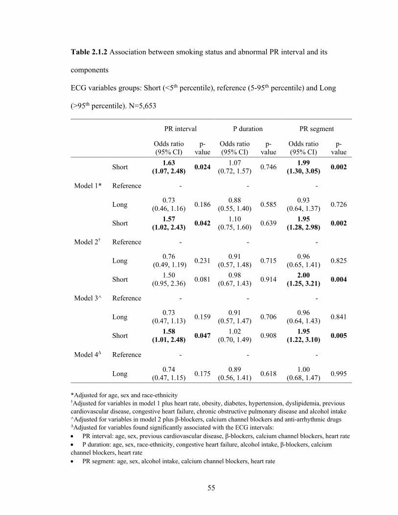
55
Table 2.1.2 Association between smoking status and abnormal PR interval and its
components
ECG variables groups: Short (<5th percentile), reference (5-95th percentile) and Long
(>95th percentile). N=5,653
PR interval P duration PR segment
Odds ratio (95% CI)
p-value
Odds ratio (95% CI)
p-value
Odds ratio (95% CI)
p-value
Model 1*
Short 1.63 (1.07, 2.48) 0.024 1.07
(0.72, 1.57) 0.746 1.99 (1.30, 3.05) 0.002
Reference - - -
Long 0.73 (0.46, 1.16) 0.186 0.88
(0.55, 1.40) 0.585 0.93 (0.64, 1.37) 0.726
Model 2†
Short 1.57 (1.02, 2.43) 0.042 1.10
(0.75, 1.60) 0.639 1.95 (1.28, 2.98) 0.002
Reference - - -
Long 0.76 (0.49, 1.19) 0.231 0.91
(0.57, 1.48) 0.715 0.96 (0.65, 1.41) 0.825
Model 3^
Short 1.50 (0.95, 2.36) 0.081 0.98
(0.67, 1.43) 0.914 2.00 (1.25, 3.21) 0.004
Reference - - -
Long 0.73 (0.47, 1.13) 0.159 0.91
(0.57, 1.47) 0.706 0.96 (0.64, 1.43) 0.841
Model 4Δ
Short 1.58 (1.01, 2.48) 0.047 1.02
(0.70, 1.49) 0.908 1.95 (1.22, 3.10) 0.005
Reference - - -
Long 0.74 (0.47, 1.15) 0.175 0.89
(0.56, 1.41) 0.618 1.00 (0.68, 1.47) 0.995
*Adjusted for age, sex and race-ethnicity†Adjusted for variables in model 1 plus heart rate, obesity, diabetes, hypertension, dyslipidemia, previous cardiovascular disease, congestive heart failure, chronic obstructive pulmonary disease and alcohol intake ^Adjusted for variables in model 2 plus β-blockers, calcium channel blockers and anti-arrhythmic drugs ΔAdjusted for variables found significantly associated with the ECG intervals: • PR interval: age, sex, previous cardiovascular disease, β-blockers, calcium channel blockers, heart rate• P duration: age, sex, race-ethnicity, congestive heart failure, alcohol intake, β-blockers, calciumchannel blockers, heart rate • PR segment: age, sex, alcohol intake, calcium channel blockers, heart rate
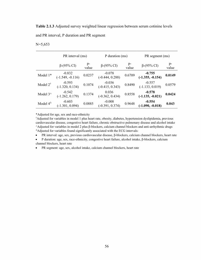
56
Table 2.1.3 Adjusted survey weighted linear regression between serum cotinine levels
and PR interval, P duration and PR segment
N=5,653
PR interval (ms) P duration (ms) PR segment (ms)
β-(95% CI) p-value β-(95% CI) p-
value β-(95% CI) p-value
Model 1* -0.832(-1.549, -0.116) 0.0237 -0.078
(-0.444, 0.288) 0.6709 -0.755(-1.355, -0.154) 0.0149
Model 2† -0.593(-1.320, 0.134) 0.1074 -0.036
(-0.415, 0.343) 0.8490 -0.557(-1.133, 0.019) 0.0579
Model 3^ -0.542(-1.262, 0.179) 0.1374 0.036
(-0.362, 0.434) 0.8558 -0.578(-1.135, -0.021) 0.0424
Model 4Δ -0.603(-1.301, 0.094) 0.0885 -0.008
(-0.391, 0.374) 0.9648 -0.554(-1.090, -0.018) 0.043
*Adjusted for age, sex and race-ethnicity†Adjusted for variables in model 1 plus heart rate, obesity, diabetes, hypertension dyslipidemia, previous cardiovascular disease, congestive heart failure, chronic obstructive pulmonary disease and alcohol intake ^Adjusted for variables in model 2 plus β-blockers, calcium channel blockers and anti-arrhythmic drugs ΔAdjusted for variables found significantly associated with the ECG intervals: • PR interval: age, sex, previous cardiovascular disease, β-blockers, calcium channel blockers, heart rate• P duration: age, sex, race-ethnicity, congestive heart failure, alcohol intake, β-blockers, calciumchannel blockers, heart rate • PR segment: age, sex, alcohol intake, calcium channel blockers, heart rate
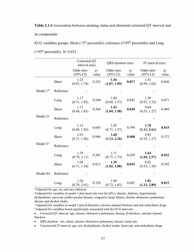
57
Table 2.1.4 Association between smoking status and abnormal corrected QT interval and
its components
ECG variables groups: Short (<5th percentile), reference (5-95th percentile) and Long
(>95th percentile). N=5,653
Corrected QT interval (ms) QRS duration (ms) JT interval (ms)
Odds ratio (95% CI)
p-value
Odds ratio (95% CI)
p-value
Odds ratio (95% CI)
p-value
Model 1*
Short 1.23(0.87, 1.74) 0.243 1.46
(1.07, 1.99) 0.017 1.41(0.99, 2.02) 0.060
Reference - - -
Long 1.17 (0.71, 1.92) 0.544 1.04
(0.69, 1.57) 0.842 1.50 (0.97, 2.32) 0.071
Model 2†
Short 1.17 (0.84, 1.63) 0.364 1.43
(1.04, 1.98) 0.030 0.84 (0.55, 1.27) 0.409
Reference - - -
Long 1.15 (0.68, 1.95) 0.603 1.05
(0.71, 1.57) 0.799 1.70 (1.11, 2.61) 0.015
Model 3^
Short 1.01 (0.71, 1.46) 0.946 1.60
(1.13, 2.26) 0.008 0.82 (0.52, 1.27) 0.372
Reference - - -
Long 1.29 (0.79, 2.11) 0.301 1.11
(0.71, 1.73) 0.659 1.64 (1.04, 2.57) 0.032
Model 4Δ
Short 1.05 (0.71, 1.54) 0.813 1.39
(1.02, 1.88) 0.035 0.82 (0.53, 1.29) 0.392
Reference - - -
Long 1.26 (0.79, 2.01) 0.328 1.09
(0.73, 1.63) 0.682 1.81 (1.13, 2.89) 0.013
*Adjusted for age, sex and race-ethnicity†Adjusted for variables in model 1 plus heart rate (not for QTc), obesity, diabetes, hypertension, dyslipidemia, previous cardiovascular disease, congestive heart failure, chronic obstructive pulmonary disease and alcohol intake ^Adjusted for variables in model 2 plus β-blockers, calcium channel blockers and anti-arrhythmic drugs ΔAdjusted for variables found significantly associated with the ECG intervals: • Corrected QT interval: age, chronic obstructive pulmonary disease, β-blockers, calcium channelblockers • QRS duration: sex, obese, chronic obstructive pulmonary disease, heart rate• Uncorrected JT interval: age, sex, dyslipidemia, alcohol intake, heart rate, anti-arrhythmic drugs
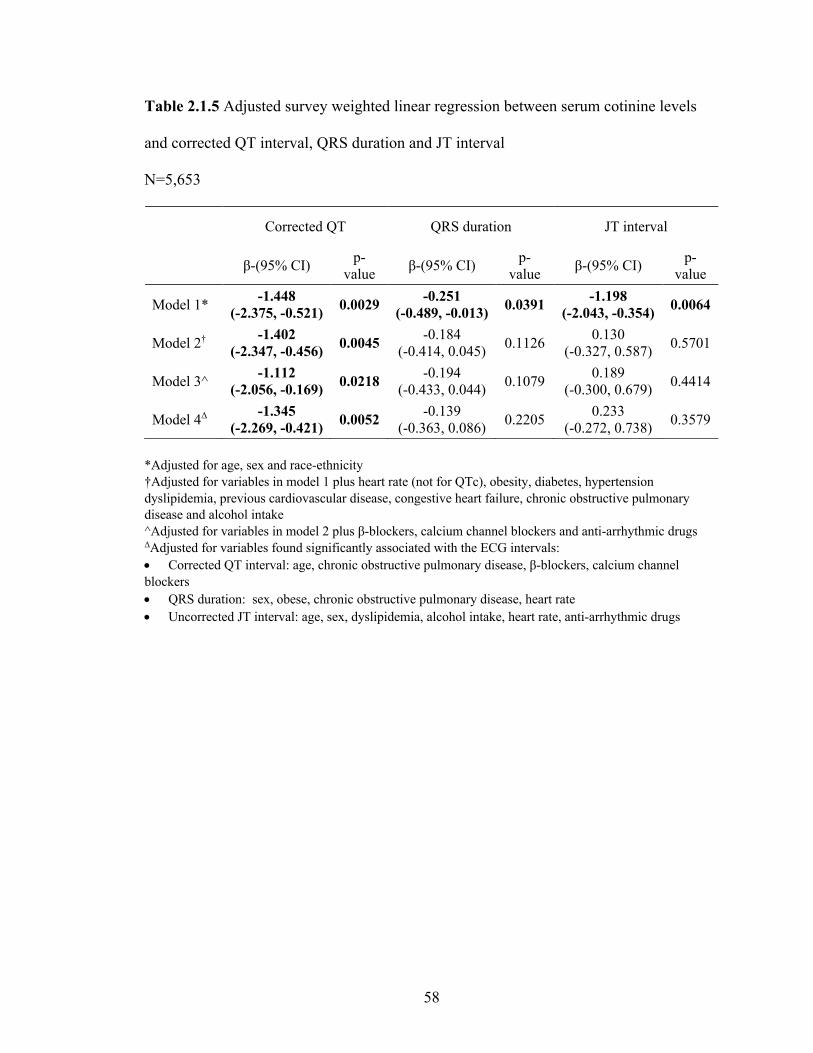
58
Table 2.1.5 Adjusted survey weighted linear regression between serum cotinine levels
and corrected QT interval, QRS duration and JT interval
N=5,653
Corrected QT QRS duration JT interval
β-(95% CI) p-value β-(95% CI) p-
value β-(95% CI) p-value
Model 1* -1.448(-2.375, -0.521) 0.0029 -0.251
(-0.489, -0.013) 0.0391 -1.198(-2.043, -0.354) 0.0064
Model 2† -1.402(-2.347, -0.456) 0.0045 -0.184
(-0.414, 0.045) 0.1126 0.130 (-0.327, 0.587) 0.5701
Model 3^ -1.112(-2.056, -0.169) 0.0218 -0.194
(-0.433, 0.044) 0.1079 0.189 (-0.300, 0.679) 0.4414
Model 4Δ -1.345(-2.269, -0.421) 0.0052 -0.139
(-0.363, 0.086) 0.2205 0.233 (-0.272, 0.738) 0.3579
*Adjusted for age, sex and race-ethnicity†Adjusted for variables in model 1 plus heart rate (not for QTc), obesity, diabetes, hypertension dyslipidemia, previous cardiovascular disease, congestive heart failure, chronic obstructive pulmonary disease and alcohol intake ^Adjusted for variables in model 2 plus β-blockers, calcium channel blockers and anti-arrhythmic drugs ΔAdjusted for variables found significantly associated with the ECG intervals: • Corrected QT interval: age, chronic obstructive pulmonary disease, β-blockers, calcium channelblockers • QRS duration: sex, obese, chronic obstructive pulmonary disease, heart rate• Uncorrected JT interval: age, sex, dyslipidemia, alcohol intake, heart rate, anti-arrhythmic drugs

59
Figure 2.1.1 The effect of smoking status on baseline ECG
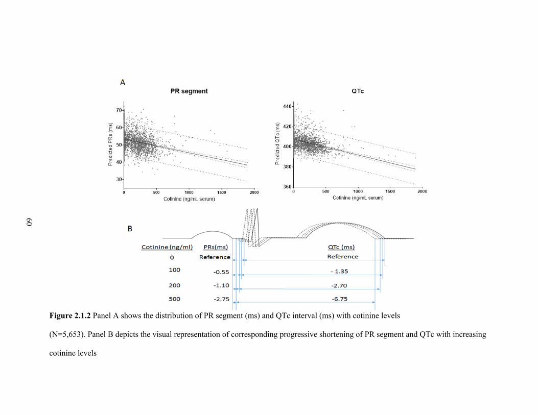
60
Figure 2.1.2 Panel A shows the distribution of PR segment (ms) and QTc interval (ms) with cotinine levels
(N=5,653). Panel B depicts the visual representation of corresponding progressive shortening of PR segment and QTc with increasing
cotinine levels

61
Discussion
In a large sample representative of the general US population, we found that higher
levels of serum cotinine, a measure of tobacco exposure, was associated with extremely
shortened PR interval, PR segment, QRS duration and extremely prolonged JT interval.
We also found that, although cotinine-confirmed smoking did not associate with extreme
changes in QTc, smoking severity as measured by serum cotinine had a significant negative
linear relationship with QTc in the overall cohort.
PR interval, P wave and PR segment
There is a wide variation in the results of prior studies on the effects of tobacco
exposure and PR interval and its components: PR segment and P duration [227, 254, 255].
However, studies with greater sample size consistently show smoking associated with
shorter PR interval [212, 221, 223, 256-258]. Few studies showed increased P wave
duration among smokers [222, 259] while other studies did not show any significant
difference, [225, 227] or found a trend towards decreasing P wave duration with smoking
[212]. We did not find any significant association between P wave duration and cotinine
levels, suggesting less overt effects of tobacco exposure on atrial activity and atrial
conduction. In a smaller cohort we recently revealed an inverse association between
cotinine and PR interval, with evidence that this relationship was mediated via increased
circulating dopamine[260]. In the present study, our observations spanned a large
nationally representative cohort and further corroborate these findings that smoking
accelerates atrioventricular conduction.

62
These findings are particularly important in light of emerging data on the prognostic
value of short PR interval to predict atrial fibrillation [27] and cardiovascular mortality [26,
29, 261]. Therefore, exposure to cigarette smoke (and perhaps nicotine specifically)
accelerates atrioventricular conduction (as exhibited by short PR interval) and may
possibly be a mechanism for increased risk of stroke and atrial fibrillation.
QTc interval, JT interval and QRS duration
Similar to the results of studies on PR interval and its components, there are mixed
results on the effects of smoking on QTc and QRS duration [212, 228, 229, 262-265]. We
were not able to find any study that explored the relationship between JT interval and
smoking exposure. Only one study has investigated the effect of smoking, measured
specifically by cotinine, on JT interval. Like us, Zhang et al [219] used the NHANES III
database, and among 7795 men and women found that in fully adjusted models QTc was
not associated with smoking and cotinine levels. At first this appears to conflict with the
results of our study, but ancillary analyses (not shown) revealed that this discrepancy stems
from their use of quartiles instead of continuous QTc, and their restricting analyses to
current smokers only. In addition, we also studied the association of cotinine with the
components of QTc interval, and found that higher cotinine levels are associated with
abnormally short QRS duration and long JT interval, but without any significant linear
relationship with either subcomponents.
The QT interval encompasses the time from the beginning of ventricular
depolarization (QRS duration) and ventricular repolarization (JT interval) [170]. Prolonged
QRS duration [182] and JT interval [31, 32] are well known predictors of mortality in the
63
general population, but the clinical implications for short QRS duration are not known.
Sympathetic neural stimulation and systemic catecholamine release may underlie smoking-
induced acceleration of AV nodal conduction, while also explaining the shortening of QRS
(ventricular depolarization)[63, 266, 267] and prolongation of JT (ventricular
repolarization) [194, 268, 269]. Nicotine has been found to directly inhibit IKr and Ito
repolarizing currents [270]. Notably, nicotine also directly stimulates release of
catecholamines from nerve terminals and the adrenal medulla [71, 271], effects that acutely
augment IKs current and directly cause phosphorylation of its corresponding potassium
channel to acutely accelerate repolarization via β-adrenergic receptors [272]. In addition to
increased sympathetic tone and direct effects on ion channels, nicotine may also cause a
pro-fibrotic state [273] and endothelial cell injury [274, 275]. Thus, nicotine is a
particularly plausible culprit of these effects. Nevertheless, particulate matter and
aldehydes within tobacco smoke may be key constituents and irritant reflexes or ischemia
may be critical pathways through which smoking disrupts electrophysiologic homeostasis
[276]
Limitations
It has not escaped our attention that a single measurement of serum cotinine may
not fully reflect chronic smoking intensity. Moreover, because cotinine only reflects
nicotine exposure, and by an imperfect proxy, smoking intensity, we are unable to assess
relationships between other cigarette smoke constituents and cardiac electrophysiology.
Although we have adjusted for several potential confounders, we recognize the possibility
of residual confounding that is similar to other studies with a cross-sectional design. Also,

64
though the automated measurement of ECG intervals and segments, are routinely reported
and can easily be calculated by the ECG systems, we did not perform manual
measurements. Despite these limitations, this is the first study examining the association
between serum cotinine, an objective measure of tobacco exposure, with PR and QT
interval and their components.
Conclusions
We found in a large racially diverse sample of the US population, that elevated
serum cotinine levels are independently associated with abnormally short PR interval, short
PR segment, short QRS duration, and long JT interval. Additionally, increases in cotinine
were associated with progressive shortening of QTc across all individuals. Collectively,
our findings indicate that exposure to cigarette smoke increases risk for abnormally fast
atrioventricular conduction and ventricular depolarization and abnormally long ventricular
repolarization. However, smoking induces a progressive prolongation of repolarization
when also evaluating individuals within normal QTc ranges. These observations enhance
our understanding of the relationship between smoking and cardiac arrhythmias and
implicate specific pathophysiological mechanisms by which smoking increases
cardiovascular morbidity and mortality, including for stroke and sudden cardiac death.
More research is warranted to examine the specificity and selectivity of these effects and
to delineate the direct contribution of specific tobacco constituents such as nicotine.
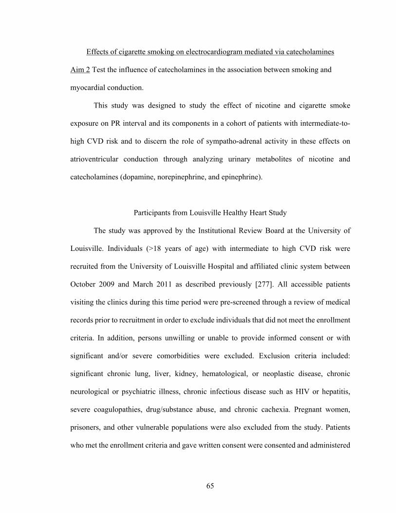
65
Effects of cigarette smoking on electrocardiogram mediated via catecholamines
Aim 2 Test the influence of catecholamines in the association between smoking and
myocardial conduction.
This study was designed to study the effect of nicotine and cigarette smoke
exposure on PR interval and its components in a cohort of patients with intermediate-to-
high CVD risk and to discern the role of sympatho-adrenal activity in these effects on
atrioventricular conduction through analyzing urinary metabolites of nicotine and
catecholamines (dopamine, norepinephrine, and epinephrine).
Participants from Louisville Healthy Heart Study
The study was approved by the Institutional Review Board at the University of
Louisville. Individuals (>18 years of age) with intermediate to high CVD risk were
recruited from the University of Louisville Hospital and affiliated clinic system between
October 2009 and March 2011 as described previously [277]. All accessible patients
visiting the clinics during this time period were pre-screened through a review of medical
records prior to recruitment in order to exclude individuals that did not meet the enrollment
criteria. In addition, persons unwilling or unable to provide informed consent or with
significant and/or severe comorbidities were excluded. Exclusion criteria included:
significant chronic lung, liver, kidney, hematological, or neoplastic disease, chronic
neurological or psychiatric illness, chronic infectious disease such as HIV or hepatitis,
severe coagulopathies, drug/substance abuse, and chronic cachexia. Pregnant women,
prisoners, and other vulnerable populations were also excluded from the study. Patients
who met the enrollment criteria and gave written consent were consented and administered

66
a questionnaire to provide demographic information and baseline characteristics. Medical
records were reviewed for past medical history, vital signs and medication history. To
reduce selection bias, all consecutive participants who were eligible for this study were
recruited. For our analyses, only those patients with complete urinary biomarkers and ECG
with normal sinus rhythm were included (Supplementary Figure 2.2.1).
Electrocardiogram (ECG) measurement protocol
Standard 12-lead ECGs with 2.5 seconds of each lead and 10 seconds of rhythm
strip (lead II) from medical records were used for ECG analyses. The following ECG
intervals were measured: P wave duration (from beginning to end of P) and PR interval
from lead II (from beginning of P to beginning of Q) [26], QRS duration from lead V6
(from beginning of Q to end of S) [278], and QT interval from V5 (from beginning of Q to
end of T) [279]. The PR segment was calculated as the difference between PR interval and
P wave duration. QT was also corrected using the Framingham formula [246] (Appendix
A). All intervals were measured with electronic calipers and adjusted to scale by reported
automated measures of RR (or heart rate). Two trained analysts (CA and AI) independently
and manually measured each ECG interval (except RR) from the first 3 sinus beats. HRV
parameters were derived from digital caliper measurements of all RRs in the rhythm strip.
When average of any ECG interval for a given patient differed between the two analysts
by >10%, both investigators re-measured that ECG interval independently. If the parameter
remained >10% different between analysts, the analysts reviewed the ECG together and
reached consensus on the appropriate measure.
Urinary measurements

67
A spot urine sample was collected on the day of study enrollment. Urinary cotinine
is a well-established metabolite for cigarette smoke exposure [245], and was measured by
Ultra performance liquid chromatography - tandem mass spectrometer (UPLC-MS/MS)
using D3-cotinine as an internal standard [280]. For UPLC-MS/MS analysis of dopamine,
norepinephrine, epinephrine, and their metabolites (metanephrine, normetanephrine,
vanillylmandelic acid, 3-methoxytyramine, homovanillic acid), urine samples were thawed
on ice, vortexed and diluted 1:50 with 0.2% formic acid containing isotopic labeled internal
standards. 1 µL of the mixture was analyzed on an UPLC-MS/MS instrument (ACQUITY
UPLC H-Class system and Xevo TQ-S micro triple quadrupole mass spectrometer, all from
Waters Inc., MA). Separation was performed on an Acquity UPLC HSS PFP (150 mm ×
2.1 mm, 1.8 μm) column (Waters Inc., MA) with a binary gradient comprised of 0.2%
formic acid (Solvent A) and methanol (Solvent B). Three multiple reaction monitoring
(MRM) transitions were set up for each sample: one for quantification, one for
confirmation, and one for labeled internal standard. At least 12 data points were collected
for each peak. Analytes were quantified using peak area ratio based on 8 point-standard
curves run before and after the urine samples. The concentration values of analytes were
normalized to creatinine level which was measured on a COBAS MIRA-plus analyzer
(Roche, NJ) with Infinity Creatinine Reagent (Thermo Fisher Scientific, MA).

68
Statistical Analysis
Baseline subject characteristics were summarized by smoking status. Categorical
characteristics, frequencies and percentages are reported along with Chi-square test p-
values, which were used to compare distributions across study groups. In addition to visual
inspection of histograms, Shapiro-Wilk tests were conducted for continuous characteristics
to determine if the characteristics were approximately normally distributed. Mean and
standard deviation are reported for continuous characteristics with a normal distribution,
whereas median and interquartile range are reported for continuous characteristics with a
skewed distribution, and the study groups were compared by the appropriate statistical test
based on normality. P-values were derived from Student’s t-tests for study group
comparisons of normally-distributed variables, whereas Mann-Whitney tests were used for
variables lacking a normal distribution. The urinary metabolites (cotinine, dopamine,
norepinephrine and epinephrine and their daughter metabolites) were log-transformed
because their distribution was skewed. We tested the associations between ECG parameters
and urinary metabolites by linear regression. ECG parameters were dichotomized by their
median levels into high- and low-value groups. Baseline characteristics associated with
ECG variables in bivariate analyses (with p<0.01) were used to build fully adjusted models
using linear regression analyses. Smoking status was determined as reported by the
participant (active smoker, former smoker or non-smoker) and by urinary cotinine levels
of 50 ng/ml [245]. Finally, mediation was assessed by the bootstrapping technique and
macro put forth by Preacher and Hayes [281]. Statistical analyses were performed using
Statistical Package for Social Sciences (SPSS) software (version 24, SPSS, Inc, Chicago,
IL, USA).
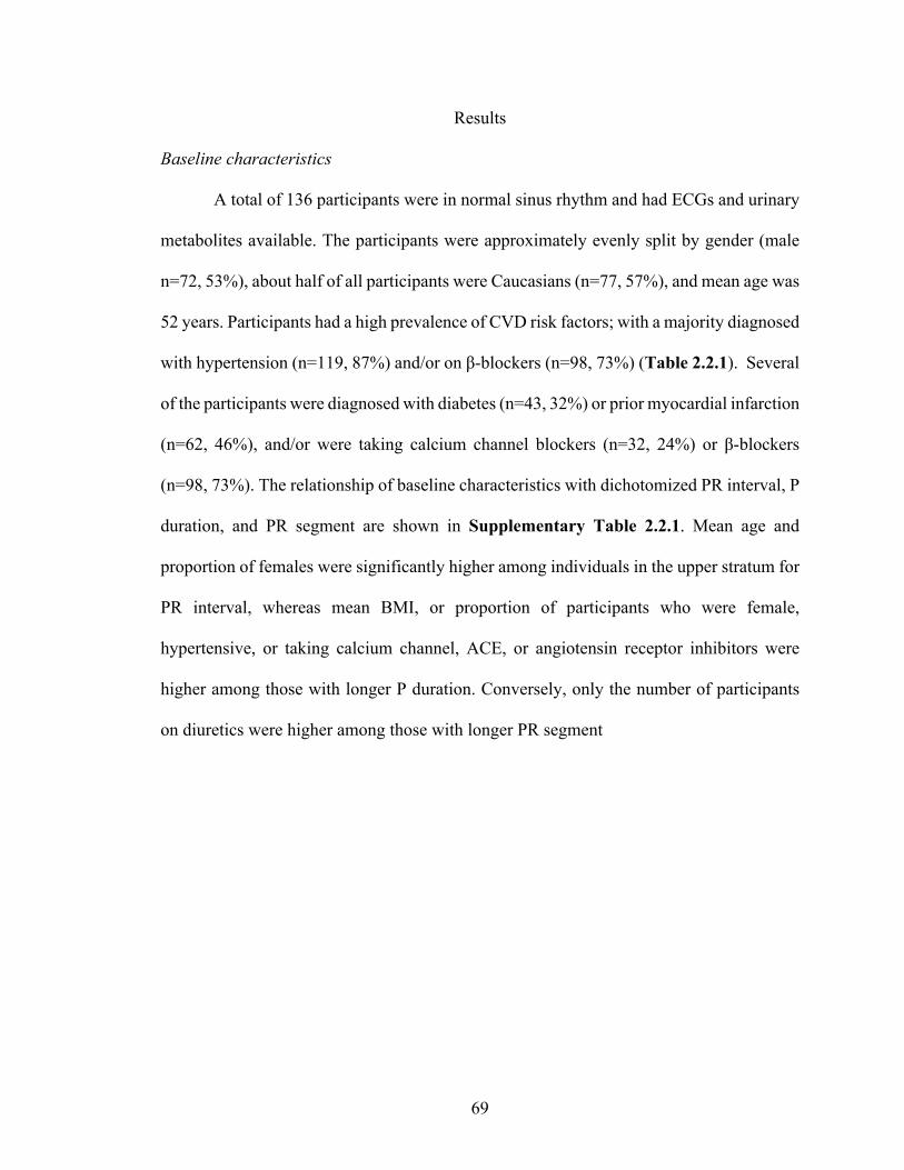
69
Results
Baseline characteristics
A total of 136 participants were in normal sinus rhythm and had ECGs and urinary
metabolites available. The participants were approximately evenly split by gender (male
n=72, 53%), about half of all participants were Caucasians (n=77, 57%), and mean age was
52 years. Participants had a high prevalence of CVD risk factors; with a majority diagnosed
with hypertension (n=119, 87%) and/or on β-blockers (n=98, 73%) (Table 2.2.1). Several
of the participants were diagnosed with diabetes (n=43, 32%) or prior myocardial infarction
(n=62, 46%), and/or were taking calcium channel blockers (n=32, 24%) or β-blockers
(n=98, 73%). The relationship of baseline characteristics with dichotomized PR interval, P
duration, and PR segment are shown in Supplementary Table 2.2.1. Mean age and
proportion of females were significantly higher among individuals in the upper stratum for
PR interval, whereas mean BMI, or proportion of participants who were female,
hypertensive, or taking calcium channel, ACE, or angiotensin receptor inhibitors were
higher among those with longer P duration. Conversely, only the number of participants
on diuretics were higher among those with longer PR segment

70
Table 2.2.1 Baseline characteristics
All patients Current smoker
N=136 Yes
(N=53, 39%)
No (N=83, 61%)
p value
Age (years)* 52, 10 53, 10 50, 9 0.05
BMI (kg/m2)* 33, 8 34, 8 31, 7 0.06
SBP (mm Hg)* 133, 23 132, 22 136, 24 0.15
DBP (mm Hg)* 81, 13 80, 11 81, 16 0.37
Heart rate (beats/min)* 73, 15 74, 15 73, 15 0.99
Male gender 72, 53% 43, 52% 30, 57% 0.58
Caucasian 77, 57% 46, 55% 31, 59% 0.73
Hypertension 118, 87% 74, 89% 45, 85% 0.47
Prior MI 62, 46% 40, 48% 23, 43% 0.58
Diabetes 43, 32% 28, 34% 15, 28% 0.51
Stroke 23, 17% 14, 17% 10, 19% 0.77
Arrhythmia 45, 33% 28, 34% 17, 32% 0.84
Β blocker 98, 73% 65, 78% 34, 64% 0.07
CCB 32, 24% 24, 29% 8, 15% 0.06
ACEI or ARB 89, 66% 54, 65% 35, 66% 0.91
Statin 84, 62% 55, 66% 30, 57% 0.26
Aspirin 81, 60% 49, 59% 33, 62% 0.71
Diuretics 58, 43% 38, 46% 20, 38% 0.35
Abbreviations: BMI - Body mass index, SBP- Systolic Blood Pressure, DBP – Diastolic Blood Pressure, MI – Myocardial Infarction, CCB – Calcium Channel Blocker, ACEI - Angiotensin-Converting Enzyme Inhibitor, ARB - Angiotensin II Receptor Blocker For each variable counts and column percentage has been reported except for where indicated * where the mean and standard deviation (SD) are reported
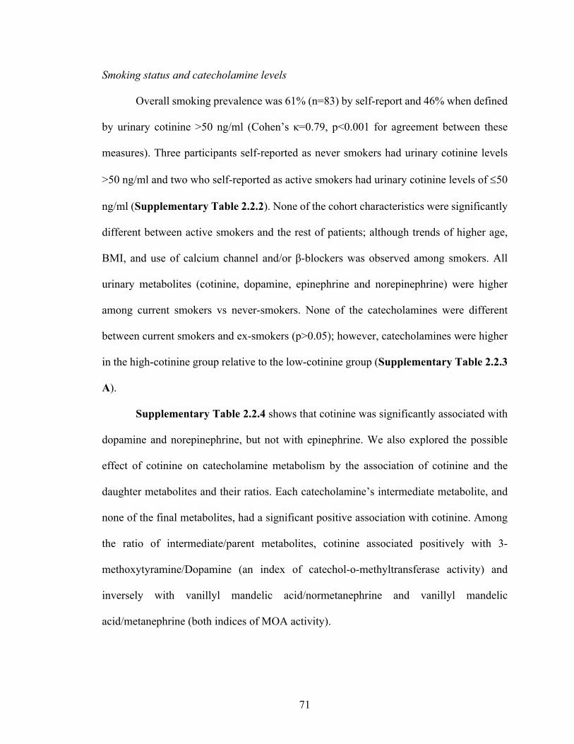
71
Smoking status and catecholamine levels
Overall smoking prevalence was 61% (n=83) by self-report and 46% when defined
by urinary cotinine >50 ng/ml (Cohen’s κ=0.79, p<0.001 for agreement between these
measures). Three participants self-reported as never smokers had urinary cotinine levels
>50 ng/ml and two who self-reported as active smokers had urinary cotinine levels of £50
ng/ml (Supplementary Table 2.2.2). None of the cohort characteristics were significantly
different between active smokers and the rest of patients; although trends of higher age,
BMI, and use of calcium channel and/or β-blockers was observed among smokers. All
urinary metabolites (cotinine, dopamine, epinephrine and norepinephrine) were higher
among current smokers vs never-smokers. None of the catecholamines were different
between current smokers and ex-smokers (p>0.05); however, catecholamines were higher
in the high-cotinine group relative to the low-cotinine group (Supplementary Table 2.2.3
A).
Supplementary Table 2.2.4 shows that cotinine was significantly associated with
dopamine and norepinephrine, but not with epinephrine. We also explored the possible
effect of cotinine on catecholamine metabolism by the association of cotinine and the
daughter metabolites and their ratios. Each catecholamine’s intermediate metabolite, and
none of the final metabolites, had a significant positive association with cotinine. Among
the ratio of intermediate/parent metabolites, cotinine associated positively with 3-
methoxytyramine/Dopamine (an index of catechol-o-methyltransferase activity) and
inversely with vanillyl mandelic acid/normetanephrine and vanillyl mandelic
acid/metanephrine (both indices of MOA activity).
72
Association of cotinine with P- and PR-parameters
Smokers did not differ from non-smokers in proportion of participants within strata
of long or short P duration, PR interval, or PR segment. Distribution of participants into
high- or low-PR or P-duration parameters also did not differ by cotinine dichotomization
(Supplementary Table 2.2.3 B). When analyzed as a continuous variable, cotinine was
significantly higher among participants with short PR segment compared to those with long
PR segment (p=0.03). PR interval also showed a similar trend (p=0.06), whereas cotinine
did not differ between participants with long vs. short P duration (p=0.25) (Table 2.2.2).
Among all participants, cotinine had a significant negative association with PR interval and
PR segment (but not with P duration) in linear regressions, even after adjusting for age,
gender and heart rate (Table 2.2.3). Figure 2.2.1 shows the scatterplot distributions of PR
interval, P duration, and PR segment across log-transformed urinary cotinine levels
normalized by creatinine. Cotinine was not significantly associated with other ECG
parameters, including QRS duration, QT interval and corrected QT, in unadjusted and
adjusted models (Supplementary Table 2.2.5).
Catecholamines and PR interval
All three catecholamines were significantly elevated among participants with short
P duration relative to those with long P duration (Table 2.2.2). Dopamine was also elevated
among those with shorter PR-interval relative to those with longer PR-interval (p=0.01).
Dopamine showed a trend of being higher among patients with short vs. those with long
PR-segment. The adjusted linear regression showed all three catecholamines inversely
associated with PR interval (p<0.05), none of the catecholamines associated with P
73
duration, and only dopamine inversely associated with PR segment (Supplementary
Table 2.2.6).
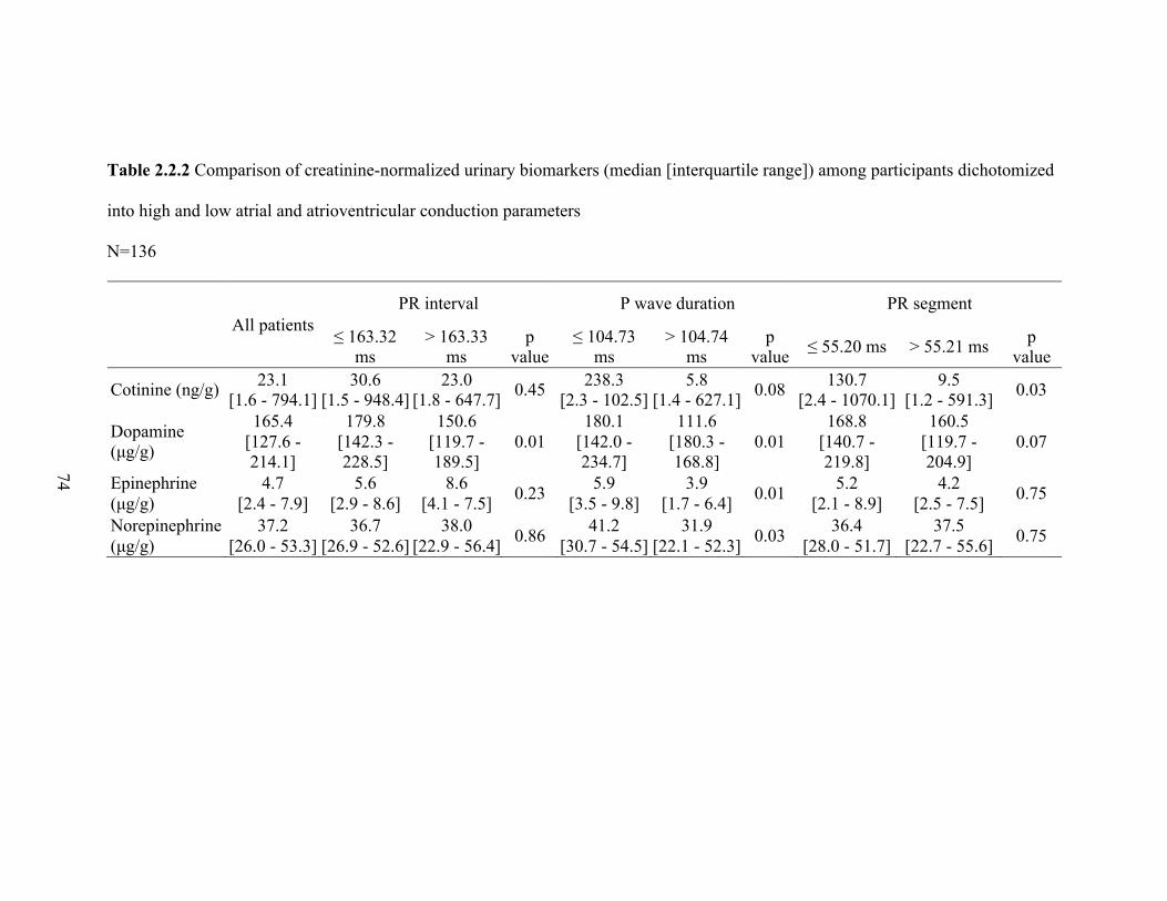
74
Table 2.2.2 Comparison of creatinine-normalized urinary biomarkers (median [interquartile range]) among participants dichotomized
into high and low atrial and atrioventricular conduction parameters
N=136
All patients PR interval P wave duration PR segment
≤ 163.32 ms
> 163.33 ms
p value
≤ 104.73 ms
> 104.74 ms
p value ≤ 55.20 ms > 55.21 ms p
value
Cotinine (ng/g) 23.1 [1.6 - 794.1]
30.6 [1.5 - 948.4]
23.0 [1.8 - 647.7] 0.45 238.3
[2.3 - 102.5] 5.8
[1.4 - 627.1] 0.08 130.7 [2.4 - 1070.1]
9.5 [1.2 - 591.3] 0.03
Dopamine (μg/g)
165.4 [127.6 - 214.1]
179.8 [142.3 - 228.5]
150.6 [119.7 - 189.5]
0.01 180.1
[142.0 - 234.7]
111.6 [180.3 - 168.8]
0.01 168.8
[140.7 - 219.8]
160.5 [119.7 - 204.9]
0.07
Epinephrine (μg/g)
4.7 [2.4 - 7.9]
5.6 [2.9 - 8.6]
8.6 [4.1 - 7.5] 0.23 5.9
[3.5 - 9.8] 3.9
[1.7 - 6.4] 0.01 5.2 [2.1 - 8.9]
4.2 [2.5 - 7.5] 0.75
Norepinephrine (μg/g)
37.2 [26.0 - 53.3]
36.7 [26.9 - 52.6]
38.0 [22.9 - 56.4] 0.86 41.2
[30.7 - 54.5] 31.9
[22.1 - 52.3] 0.03 36.4 [28.0 - 51.7]
37.5 [22.7 - 55.6] 0.75

75
Table 2.2.3 Estimated effects (β-coefficients) of an increase in cotinine on PR interval, P wave, and P segment, with corresponding P-
values, from unadjusted and adjusted linear regressions.
Urinary cotinine was log-transformed. N=136
PR interval P duration PR segment
b p value b p value b p value
Unadjusted Cotinine -2.38 0.04 0.33 0.73 -2.70 0.03
*Adjusted Cotinine -2.67 0.05 0.87 0.32 -2.10 0.04
* model adjusted for age, body mass index, and gender (PR interval); BMI, SBP, DBP, gender, hypertension, CCB, and ACEI, and ARB (P duration); or BMI, β-blocker, and diuretics (PR segment).
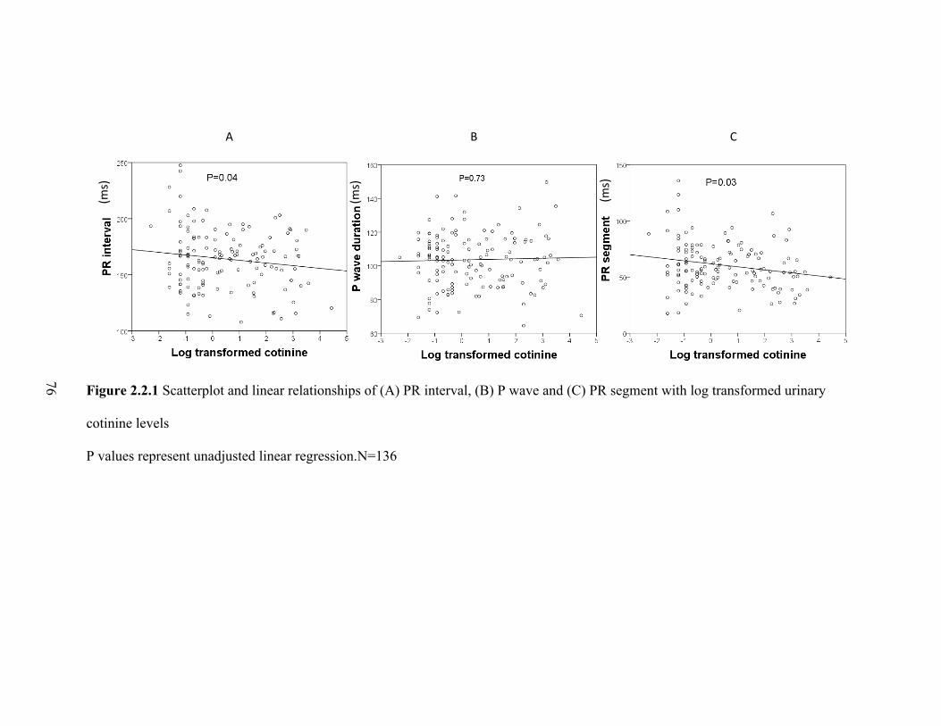
76
A B C
Figure 2.2.1 Scatterplot and linear relationships of (A) PR interval, (B) P wave and (C) PR segment with log transformed urinary
cotinine levels
P values represent unadjusted linear regression.N=136
(ms)
(ms)
(ms)
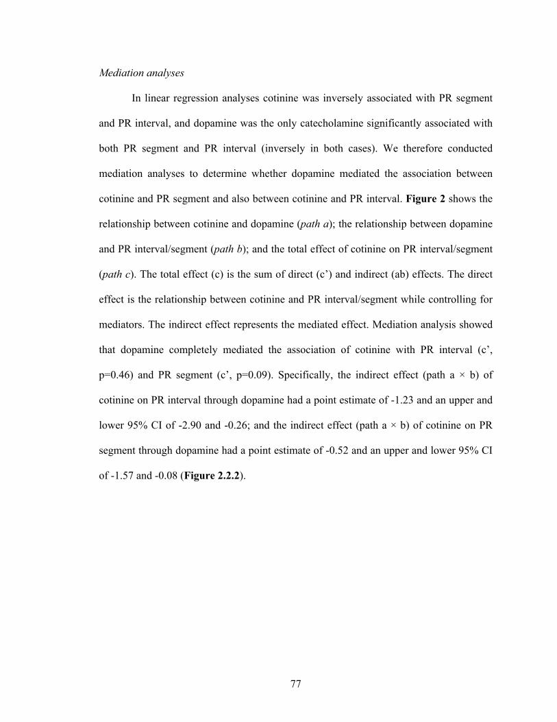
77
Mediation analyses
In linear regression analyses cotinine was inversely associated with PR segment
and PR interval, and dopamine was the only catecholamine significantly associated with
both PR segment and PR interval (inversely in both cases). We therefore conducted
mediation analyses to determine whether dopamine mediated the association between
cotinine and PR segment and also between cotinine and PR interval. Figure 2 shows the
relationship between cotinine and dopamine (path a); the relationship between dopamine
and PR interval/segment (path b); and the total effect of cotinine on PR interval/segment
(path c). The total effect (c) is the sum of direct (c’) and indirect (ab) effects. The direct
effect is the relationship between cotinine and PR interval/segment while controlling for
mediators. The indirect effect represents the mediated effect. Mediation analysis showed
that dopamine completely mediated the association of cotinine with PR interval (c’,
p=0.46) and PR segment (c’, p=0.09). Specifically, the indirect effect (path a × b) of
cotinine on PR interval through dopamine had a point estimate of -1.23 and an upper and
lower 95% CI of -2.90 and -0.26; and the indirect effect (path a × b) of cotinine on PR
segment through dopamine had a point estimate of -0.52 and an upper and lower 95% CI
of -1.57 and -0.08 (Figure 2.2.2).

78
Figure 2.2.2 Mediation analyses of cotinine, dopamine, and PR interval and PR segment
N=136

79
Discussion
The effects of smoking on atrial and atrioventricular nodal conduction velocity and
their underlying mechanisms are currently unknown. To address this gap in knowledge, we
measured PR interval, P wave duration, and PR segment from lead II of a 12-lead EKG,
urinary catecholamines, and nicotine and its metabolites in a cohort of 136 participants
with intermediate-high cardiovascular risk. We report three major findings from this study.
First, cotinine had a significant inverse association with PR interval and PR segment, and
not with P wave duration in adjusted linear regression models. Second, urinary dopamine
was inversely associated with PR interval and both its components (PR segment and P
duration), while epinephrine and norepinephrine did not associate with PR segment. Third,
mediation analyses indicate that dopamine may primarily account for the association
between cotinine and shortening of both PR interval and segment. Together, these findings
suggest smoking may increase cardiovascular morbidity and mortality through nicotine-
associated increases in catecholamine release and downstream modulation of
atrioventricular conduction.
Nicotine and PR interval and its components
Previous work on the chronic effects of smoking on PR interval has produced mixed
results. Some studies found short PR interval at baseline among chronic smokers vs non-
smokers [212, 221, 257], while others found no difference [254, 282]. Similarly, a few
studies showed increased P wave duration among smokers [222, 259], while other studies
did not show any significant difference [225, 227] or found a trend towards decreasing P
wave duration with smoking [212]. There are several reasons for the wide variation in the

80
results of prior studies; 1) small sample size, 2) insufficient consideration for the role of
the components of PR interval, and 3) inadequate assessment of nicotine exposure and its
potential impact. To the best of our knowledge, to date, no previous study investigated the
effects of nicotine exposure on PR interval and its components in humans. This is
particularly important in light of recent findings that short PR interval associates with
increased risk for atrial fibrillation and cardiovascular mortality [26, 29, 261, 283]. Beyond
overall cardiovascular mortality, smoking also associates with increased risk of atrial
fibrillation through unknown mechanisms [284]. Thus, by demonstrating that exposure to
cigarette smoke (and perhaps nicotine specifically) accelerates atrioventricular conduction,
our findings provide further insight into how smoking may confer cardiovascular risk,
including risk for atrial fibrillation.
In the present study, cotinine inversely associated with PR interval and PR segment,
indicating that increased cigarette smoke exposure accelerates atrioventricular conduction.
PR interval is mainly influenced by the sum of atrial activity and atrioventricular nodal
conduction [170]. Atrial pathology usually results in prolonging (rather than shortening) of
PR interval, whereas shortening of PR interval is likely a result of accelerated
atrioventricular nodal conduction. Hence, it is plausible from our results that chronic
nicotine exposure expedites atrioventricular nodal conduction. Moreover, because P wave
duration was not associated with cotinine, our findings suggest nicotine has limited effects
on atrial conduction. Using our linear regression coefficient, we found that each 100 ng/ml
cotinine is associated with a 12.3-ms decrease in PR interval and 9.7-ms decrease in PR
segment.

81
Cotinine and catecholamines
Cigarette smoking and nicotine result in increased central and peripheral sympatho-
adrenal activation. The activation of nicotinic acetylcholine receptors in the adrenal
medulla leads to increased catecholamine levels [285]. We found urinary dopamine and
norepinephrine (but not epinephrine) were significantly higher among smokers and those
with higher cotinine, suggesting smokers have chronically increased sympathetic neuronal
activity (norepinephrine), but similar adrenal medullary hormone secretion (epinephrine).
Interestingly, we also found that the intermediate metabolites of all three catecholamines
significantly associated with cotinine, but not with the final product, and there was a
negative association between cotinine and the ratio of the final and intermediate metabolite.
This suggests cigarette smoking is associated with increased synthesis of dopamine and
norepinephrine and at the same time decreases the catabolism of the intermediate
metabolites of all three catecholamines. This corroborates the findings of several other
studies that showed cigarette smoking inhibits the activity of MOA [286, 287]; an enzyme
primarily responsible for catabolism of the intermediate metabolites of norepinephrine and
epinephrine to their final metabolites.
There are several mechanisms through which nicotine can possibly increase
atrioventricular nodal conduction velocity. Nicotine stimulates sympathetic
neurotransmission via activation of nicotinic acetylcholine receptors localized on
peripheral postganglionic sympathetic nerve endings and the adrenal medulla which in turn
cause catecholamine release [91, 285]. Catecholamines mediate positive chronotropic,
inotropic, dromotropic, and bathmotropic effects (i.e., increased rate, force conductivity
and excitability) [288]. Furthermore, evidence from animal models and ex-vivo studies

82
suggests nicotine may also directly cause endothelial cell injury [275], a pro-fibrotic state
[273], and inhibition of cardiac A type potassium channels [85].
The mediation analysis is a novel component to our study that reveals, among
catecholamines, dopamine fully mediates the effect of cotinine and shortening PR
interval/segment. The positive dromotropic effects of dopamine accelerate atrioventricular
nodal conduction [289, 290], thereby expected to shorten the PR interval/segment without
affecting P wave duration, as seen in our study.
Cotinine was even associated with altered metabolism of the three catecholamines
(ratio of metabolite/parent). The positive association of cotinine with 3-
methoxytyramine/Dopamine ratio suggests smoking increases dopamine synthesis and/or
systemic secretion and, in compensation, also increases catechol-O-methyltransferase
(COMT) activity. Also, cotinine’s inverse association with Vanillylmandelic
acid/Normetanephrine and Vanillylmandelic acid/Metanephrine suggest that smoking
decreases MAO activity, consistent with observations of decreased MAO-B in the
amygdalae of smokers and the antidepressant effects of nicotine [291, 292]
Catecholamines and PR interval
Interestingly, in our study only dopamine and not epinephrine and norepinephrine
were associated with PR interval and PR segment. As all catecholamines are known to
exert a dromotropic effect, plausible reasons for our finding include: 1) short and long term
stress (smoking) disproportionately affect circulating dopamine relative to norepinephrine
and epinephrine [293], perhaps secondary to nicotine-mediated declines in dopamine
uptake [294]; 2) cigarette smoking and/or nicotine cause an immediate surge in dopamine
83
from neuronal [9], that may cross the blood brain barrier to increase circulating dopamine;
3) as the precursor of both epinephrine and norepinephrine, dopamine may have less
temporal variability, making it a more stable marker of chronic SNS activation; and 4) at
low pathophysiological stress levels, dopamine may have higher affinity than epinephrine
and norepinephrine for β-1- and β-2- adrenergic receptors, which modulate dromotropy in
the atrioventricular node [295] .
Other ECG parameters
We did not observe any association of smoking status or cotinine with QRS, QT or
cQT and. Previous studies in this area have yielded mixed results, including QT
prolongation [229, 262, 263], QT shortening [228], or no relationship [219] with smoking.
Limitations
While this is the first known investigation of the influence of catecholamines in
nicotine-mediated alterations in cardiac conduction, the sample size is limited and
participants were drawn at random from the outpatient clinic setting with intermediate to
high cardiovascular risk. Thus, we might have missed significant associations between
cotinine and other parameters (e.g., P-wave duration) due to insufficient statistical power.
Additionally, ECGs were obtained retrospectively from medical records and not on the day
of enrollment. The median and interquartile range of number of days from study enrollment
(urine sample collection) and ECG was 79 [20 – 320] days. We did not collect data on time
of last cigarette/nicotine exposure and states that may affect catecholamines (stress, noise,
discomfort, body position, consumption of food, caffeinated beverages and drugs).

84
Nevertheless, we used urinary analytes (cotinine and catecholamines) that are established
markers for chronic nicotine and sympatho-adrenal activation and are unlikely to exhibit
large acute variation in the outpatient clinic setting from which this cohort was derived.
We did not collect 24-hour urine to account for diurnal and intra-individual variation in
catecholamines [296]; however, summative analysis of 24-h catecholamine production
may mask elevations due to dilution. Importantly, a HRV parameter (RMSSD: square root
of the mean of squared differences of successive NN intervals), when dichotomized, tended
to inversely associate with urinary norepinephrine and dopamine (r= -0.13, p=0.10 for
both), suggesting concordance between measures of sympathetic activity in both ECG and
subsequent urine samples. Although most participants were on β-blockers, prevalence did
not significantly differ by cotinine strata, and PR and P wave durations did not differ by β-
blocker use. This latter point accords with observations that β-blockers do not alter resting
PR interval whereas they partially attenuate PR and RR interval shortening during exercise-
induced sympatho-excitation [297, 298]. Additionally, we were unable to assess
associations between other cigarette components or smoking habits (e.g., frequency) and
cardiac electrophysiology. Other constituents within tobacco smoke (e.g., particulate
matter and aldehydes) have been shown to alter autonomic balance and may thus plausibly
alter catecholamine synthesis, secretion, and metabolism. Finally, smoking and nicotine
can affect myocardial conduction velocity through induction of cardiac remodeling,
oxidative stress, and/or ion channel dysfunction, which were not assessed in this study and
may occur independent of sympathetic activation. Nevertheless, sympathetic activation can
induce all three of these pathogenic processes [299] and may thereby indirectly mediate
atrioventricular conduction defects.
85
Conclusions
Collectively, our findings suggest that exposure to cigarette smoke accelerates
atrioventricular conduction and that dopamine mediates these effects. More research is
warranted to examine the specificity and selectivity of these effects and to delineate the
direct contribution of nicotine. These observations identify a pathway by which smoking
may increase risk for cardiovascular morbidity and mortality.
86
CHAPTER III
ACUTE EFFECTS OF NICOTINE AND CIGARETTE SMOKING ON
ELECTROCARDIOGRAM
Aim 3 Acute effects of cigarette smoking and nicotine with and without β-blocker on
electrocardiogram.
Cigarette smoking has acute deleterious effects on the cardiovascular system,
resulting in increased incidence of coronary artery disease, atrial and ventricular
arrhythmias and sudden cardiac death among smokers [300, 301]. Despite, a strong dose-
response relationship between cigarette smoking and CVD, it remains unclear which
tobacco smoke constituents and biological pathways mediate this increased risk. The main
addictive component in cigarette smoke, nicotine, has been implicated as possibly the
major mediator of acute cigarette smoke induced ANS dysregulation of the heart via
increased sympathetic nerve activity and multiple downstream mechanisms [133].
Imbalance within the ANS is increasingly recognized as a major culprit of CVD. Indeed,
both acute and chronic activation of the sympatho-adrenal system, promotes
cardiovascular dysfunction and disease, including arrhythmia, hypertension, heart failure,
myocardial infarction, and ischemic stroke [302, 303]. The rising popularity of ENDS, and
the enhanced nicotine dose of some ENDS devices relative to conventional smoking [304],
have created an imperative to determine the cardiovascular effects of nicotine and the role
of autonomic imbalance within them.

87
The ECG is a widely available, inexpensive, non-invasive, routine method of
measuring electrical activity of the heart, and can be used to screen high risk populations
and identify ad predict CVD. Several ECG parameters have also been used clinically to
assess cardiovascular autonomic dysfunction as a diagnostic and prognostic factor. There
is now abundant evidence that acute ANS imbalance has profound effects on
electrophysiology [305, 306]. Further, acute variations in hemodynamics [307] and
conventional ECG parameters, including dispersion of ECG intervals and amplitudes, may
result from acute ANS imbalance and, when frequent, carry worse long term prognosis
[308-310]. Additional parameters have recently been added to the repertoire of ECG
indices that reliably predict cardiovascular morbidity and mortality (i.e., P wave amplitude,
PR interval, and JT interval) [26, 30, 283, 311-313].
Despite known cigarette induced acute ECG changes and powerfully predictive
value of ECG changes for future CVD events, no prior study has systematically 1)
evaluated the acute temporality of the acute electrocardiographic effects of smoking, and
2) compared these with nicotine alone, and 3) explored the potential underlying
mechanisms. To the best of our knowledge, this is the first report on the immediate effects
of cigarette smoking and nicotine on ECG morphologic endpoints. We therefore conducted
an open label 2 x 2 factorial experimental trial to study the acute effects of smoking and
nicotine, with and without β-blocker.

88
Participants and setting of the experimental trial
Study design
Participants were recruited via online and flyer advertisements in the public areas
city of Huntington, WV. The study enrolled healthy adult male and female smokers, aged
18–65 years, who had smoked at least 10 cigarettes/day for at least one year prior to the
trial. Eligible participants had to weigh at least 55 kg and have a body mass index (BMI)
within the range 17.5–30.0 kg/m2. Females who were pregnant or breast feeding,
vulnerable population (such as patients with mental illness, prisoners etc), and participants
with any medical illness requiring routine medications were excluded from the trial.
Participants were screened over the phone call before entering the trial. The study design
is shown in Supplementary Figure 3.1 A
Participants
The study was approved by the Institutional Review Board at Marshall University.
Each participant signed an informed consent before inclusion in the study. Twenty healthy
smokers (65% men; 37±13 years of age; body mass index, 28±5 kg/m2) and ten healthy
non-smokers smokers (70% men; 28±5 years of age; body mass index, 26±3 kg/m2) were
included in the study. All of the smokers were regular habitual cigarette smokers (22±8
cigarettes per day for 21±12 years). The baseline characteristics along with details of
smoking history and cigarette are provided in Supplementary Table 3.1. Except for one
smoker (who used rescue inhaler occasionally for well controlled mild asthma), no other
participant had any known medical illness nor routine medication use (including birth
control pills). All subjects were at least high school graduates.

89
Study visits and exposures
All participants had abstained from food, coffee, and tobacco overnight before each
study visit. Each smoker completed two-day visits, where on their first day they smoked
single preferred brand of Combustible Cigarette (Cig) in their usual manner, and second
day they were administered 4 mg of nicotine from Nicotine mouth Spray device (NicS)
designed to mimic the acute nicotine delivery of cigarettes. Ten of the twenty smokers were
randomly invited for two additional visits, where the exposures remained the same (i.e.
preferred single combustible cigarette smoke on day 3 and 4 mg nicotine spray on day 4),
but were pre-treated with 80 mg oral propranolol (non-selective b-blocker) for two hours
prior to the start of the study (BB-Cig and BB-NicS). The oral propranolol was
administered by physician, and participant’s vitals were closely monitored during the day
3 and 4. Ten of the twenty smokers also completed a fifth visit to serve as controls, where
they simulated smoking by inhaling through their unlit cigarette (“Sham”), for the similar
duration of smoking period on Cig.
The nicotine mouth spray device (Nicorette oromucosal nicotine spray, 1 mg/spray,
McNeil AB) consists of a plastic bottle with 13.2 ml of a clear to slightly opalescent liquid
with a mint flavor. After priming, one depression of the spray nozzle delivers a metered
dose of 1 mg of nicotine in 0.073 ml of a 10% ethanol-in-water solution. The spray liquid
also contains very small amounts of levomenthol (0.7 mg/spray dose) and other flavorings
(less than 0.3 mg/spray dose) [314]. The 4 mg of nicotine was administered by study
personnel by pressing the nozzle four times in rapid succession, directed straight into the
participant’s mouth. Participants were advised to avoid respiring during, and swallowing

90
immediately after, administration of the spray. The dose-concentration curve from the oral
spray (Cmax 9.1 ng/ml and Tmax, 10 minutes, T ½ 2.6 hr) [314], mimics that of smoking
cigarette with about 1 mg of nicotine content (Cmax 11.9 ng/ml and Tmax, 8 minutes, T
½ 2.5 hr) [315].
Procedures
All study visits were separated by washout periods of at least 24 hours, and were
started between 8 AM and 10:00 AM. After arrival to the research clinic, and obtaining
informed consent, each participant completed a detailed health and tobacco use
questionnaire. Height and weight were measured. There was a rest of 5 minutes before each
blood pressure measurement and ECG recording session. Pre and post exposure blood
pressure were measured in sitting position using a validated automated monitor, in
accordance with the guidelines [316]. Pre and post exposure serum and plasma were also
collected, and stored at −20 °C until analysis (except for on Sham smoking day). A standard
continuous 12 lead ECG was recorded (Cardio Card, Nasiff Associates, Central Square,
NY), using as per guidelines [317], for 5 mins indoor while supine (pre-exposure), 20 mins
outside while seated (during exposure) and 5 mins indoors while supine (post-exposure).
The exposures were always performed while seated in the same spot; which was outside in
an open public area, (200 feet away from the research building); which was not a designated
public smoking place and away from main road traffic area. Supplementary Figure 3.1 B
and Appendix B show the schematic timeline of the study day protocol.

91
ECG and HRV analyses
Data from Lead II was extracted and each beat was analyzed using the
commercially available software LabChart/ECG and HRV Analysis Add-On (version 8.0;
ADInstruments, Colorado Springs, CO, USA) (Appendix C). The artifacts were identified
and removed from the analysis using the ECG Beat Classifier; which measures the Activity
(sample-to-sample voltage differences), isoelectric noise, form factor (shape and time
course of QRS complex) and RR interval. Supplementary Figure 3.2 depicts a typical
ECG waveform, with the intervals and amplitudes that were measured automatically by the
software. The data was further cleaned by excluding variables with > 20% difference from
the median of 6 surrounding beats. To decrease variance, the ECG waveforms were
averaged over three-minute time intervals during the 20 minutes exposure, and were
averaged over 5 minutes during the pre- and post-exposure. For HRV, Time and frequency
domain analysis were performed, at 5 min intervals and the entire 20 minutes during
exposure, to record root mean square of successive differences (RMSSD) and standard
deviation of normal beat intervals (SDNN) and LF/HF (ratio of LF to HF power), using the
spectral settings as per the international guidelines [166].
Plasma measurements
The collected blood samples were immediately centrifuged and the plasma fraction
were frozen until the end of study for batch analysis. The plasma Nicotine, Cotinine and
Trans-3'-Hydroxy Cotinine in plasma samples were measured by UPLC-MS/MS using a
Xevo TQ-S micro quadrupole mass spectrometer with an ESI ionization source, interfaced
92
with Waters Acquity Class-H UPLC equipped with a quaternary pump system (Waters,
MA). Details about the procedure is given in Appendix D.
Statistical analyses
Each participant’s absolute change (Δ) for ECG waveforms (intervals and
amplitudes), and HRV variables were calculated from the pre-exposure (baseline). The
QTc was calculated by using Bazett formula (QT Interval / √ (RR interval). The Area Under
the Curve (AUC) for the ECG parameter changes from baseline during exposure (20
minutes) were calculated using the trapezoidal rule [318] for each participant and as
average for all participants per study visit. Time series physiologic data are presented as
change from baseline to allow consistent comparison to reported effects of cigarette,
nicotine, with and without propranolol. We analyzed time-series deltas (each participant’s
change during exposure from their own value at baseline) with linear mixed effects models
(PROC MIXED) for different exposure effects using SAS 9.3 (SAS Systems; Cary, NC).
A trendline function was also derived for ECG parameter that had significant change from
Sham, and utilized LINEST function without ‘forced-intercept’ [319], along with six
different regression types, to find the best fit curve, while maximizing the R-square and
minimizing the variance to avoid under- and over-fitting. Spearman correlation was used
to assess the relationship between the Δ in nicotine and its metabolites with the change in
ECG parameters and Blood Pressure. The potential mediation of effects on ECG
morphology by acute changes in autonomic balance (measured by RMSSD) was assessed
by the bootstrapping technique and a macro put forth by Preacher and Hayes [281].
93
A paired t-test was used to calculate the difference in blood pressure pre and post
exposure. All statistics with P < 0.05 were considered significant, and were performed in
SAS 9.3 and Statistical Package for Social Sciences (SPSS) software (version 24, SPSS,
Inc, Chicago, IL, USA). We also created a typical one-minute ECG waveforms, from pre
and during exposure, to represent the ECG changes in a Figure, between different
exposures, using ecgAuto, v3.3 (Emka Technologies, Paris, France).
Results
On Cig, NicS and Sham days, the baseline (pre-exposure) ECG parameters, except
for QTc and JTc, were similar. Pre-treatment with propranolol, decreased the Heart Rate,
and increased the RMSSD and SDNN and the ST height. There was no significant
difference in any baseline ECG parameter between BB-Cig and BB-NicS days
(Supplementary Figure 3.3).
The AUC for each exposure and study visit along with Sham is shown in Figure
3.1, while the raw values are shown in Supplementary Figure 3.4.
Heart Rate and HRV: Cig and NicS both immediately increased heart rate, which
peaked at minutes 3 to 6 (+20 beats/minute and +14 beats/minute, respectively). After both
exposures, the heart rate remained elevated up-to 20 minutes and returned to baseline post
exposure. However, the AUC of heart rate for Cig was higher than that for NicS (p<0.05).
Pre-treatment with propranolol had a significant impact on HR, with much lower AUC
observed with BB-Cig vs Cig alone; while there was a non-significant decrease in AUC
for BB-NicS vs NicS day. Cig immediately decreased RMSSD (within first 5 minutes and
lasting for 10 minutes), while NicS had delayed albeit significant and similar decrease in
94
HRV measurements at 10 minutes. The overall significant decrease (vs Sham) in AUC for
RMSSD was similar between Cig and NicS (P>0.05). Despite RMSSD increased after
treatment with propranolol on both days, cigarette smoke decreased the RMSSD ( -50 ms)
vs Cig day ( -20 ms) (p<0.05); while the AUC for NicS and BB-NicS was similar. Similar
patterns were observed with SDNN and LF/HF.
Atrial indices: The smoking and NicS related changes from baseline in P duration
were not different from those of Sham. However, cigarette use induced immediate and
significant shortening of PR interval (-14 ms at 9th minute) and PR segment ( -10 ms at 9th
minute) (P<0.05 vs Sham), and this effect was abolished with pre-treatment with
propranolol (P > 0.05 vs. Sham), while NicS had no significant impact on PR segment.
Both, Cig and NicS significantly and similarly increased the P amplitude at 3-6 minutes of
exposure compared to Sham (+21 µV and +28 µV, respectively), and interestingly this
effect remained unaltered by propranolol. The AUC of P amplitude for Cig was small
because, this effect reversed directionality to become negative (though not significantly) at
9-12 minutes of exposure (i.e. the above zero baseline AUC was similar between Cig and
NicS).
Ventricular indices: Cig caused greater shortening of QTc ( -11 ms) relative to NicS
( -4 ms), and both Cig and Nic significantly shortened QTc relative to Sham (P < 0.05 vs.
Sham). On BB days, this effect was abolished and the AUC for BB-Cig and BB-NicS were
similar to Sham, and higher than that of Cig and NicS, respectively. Cig immediately
shortened QTc in the first 3 min, with a subsequent plateau effect for the remainder of the
exposure and returning to baseline in the post-exposure period. Cig and NicS similarly
deepened Q wave amplitude ( -59 µV and -72 µV, respectively, P < 0.05 vs. BL or Sham),
95
with propranolol blocking that effect. There was no significant effect of Cig or NicS on
QRS and Tp-Te durations. The ST height gradually decreased after Cig and significantly
differed from Sham minutes 3-6 ( -27 µV), with maximum effect at 9-12 minutes ( -41 µV)
and slowly subsiding thereafter (remaining significantly decreased at minute 18-21) to
baseline levels in the post exposure period. NicS had no such effect, and propranolol
abolished the Cig-induced ST depression.
The typical 1-minute average ECG morphology during six exposures are shown in
Figure 3.2. Figure 3.3 shows the best fitting equation for the trendline of the changes in
Heart Rate, P amplitude, QTc from Cig and NicS and PR segment and ST height from Cig.
Supplementary Figure 3.4 show the raw and relative of the important ECG parameters
on different days among smokes.
Nicotine and its metabolites: The raw and relative changes in nicotine, cotinine and
3OH were similar among different visit days of smokers; and higher compared to non-
smokers (Supplementary Figure 3.5; p<0.05 for each biomarker). There was significant
correlation between change in nicotine levels with that of heart rate, ST-height, PR interval,
PR segment, Q amplitude, SDNN and RMSSD among smokers (Cig and NicS days) and
non-smokers (Table 3.1).
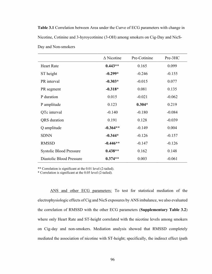
96
Table 3.1 Correlation between Area under the Curve of ECG parameters with change in
Nicotine, Cotinine and 3-hyroycotinine (3-OH) among smokers on Cig-Day and NicS-
Day and Non-smokers
Δ Nicotine Pre-Cotinine Pre-3HC
Heart Rate 0.443** 0.165 0.099
ST height -0.299* -0.246 -0.155
PR interval -0.303* -0.015 0.077
PR segment -0.318* 0.081 0.135
P duration 0.015 -0.021 -0.062
P amplitude 0.123 0.304* 0.219
QTc interval -0.140 -0.180 -0.084
QRS duration 0.191 0.128 -0.039
Q amplitude -0.364** -0.149 0.004
SDNN -0.344* -0.126 -0.157
RMSSD -0.446** -0.147 -0.126
Systolic Blood Pressure 0.438** 0.162 0.148
Diastolic Blood Pressure 0.374** 0.003 -0.061
** Correlation is significant at the 0.01 level (2-tailed). * Correlation is significant at the 0.05 level (2-tailed).
ANS and other ECG parameters: To test for statistical mediation of the
electrophysiologic effects of Cig and NicS exposures by ANS imbalance, we also evaluated
the correlation of RMSSD with the other ECG parameters (Supplementary Table 3.2)
where only Heart Rate and ST-height correlated with the nicotine levels among smokers
on Cig-day and non-smokers. Mediation analysis showed that RMSSD completely
mediated the association of nicotine with ST-height; specifically, the indirect effect (path
97
a x b) of Δ nicotine on ST-height through RMSSD had a point effect estimate (β) of -26.57
and an upper and lower 95% CI of -82.80 and -0.90. While change in nicotine directly
affected Heart Rate (c’ path; p=0.036), and this effect was independent of changes in
RMSSD (Figure 3.4).
There was no effect of baseline cotinine on the relationship between changes in
nicotine and that of ECG parameters (Heart Rate, PR segment, ST height and root mean
squared of successive differences (RMSSD)), among smokers on Cig-Day and NicS-Day
and Non-smokers (Supplementary Figure 3.6).
Blood pressure: Both Cig and NicS similarly and significantly increased post-
exposure systolic and diastolic blood pressures relative to Sham and non-smokers
(Supplementary Figure 3.7). At pre-exposure, blood pressures were similar between Cig,
NicS and Sham. There was no significant change in blood pressure on the BB days post
Cig and post NicS. The change in nicotine levels also significantly correlated with the
changes in blood pressure (Table 3.1), which were not observed on BB days (p>0.05).

98
«: <0.05 vs Sham
#: <0.05 vs Nicotine
β: <0.05 vs non- b-blocker

99
«: <0.05 vs Sham
#: <0.05 vs Nicotine
β: <0.05 vs non- b-blocker
Figure 3.1 The Area Under the Curve for the raw changes (Δ) in ECG parameters from
baseline for each visit day among smokers
N=20 on Cigarette and NicSpray days. N=10 on sham days

100
Sham exposure
Cigarette exposure Nicotine Spray exposure
Figure 3.2 ECG morphology in a typical participant during exposure on different days
Each ECG individual waveform is a 1-minute average before or during exposure to unlit cigarette (sham), cigarette, or nicotine spray.
100 µv
20 ms
Waveforms:Black –Pre-exposureRed shades – Sham exposure
100 µv
20 ms
100 µv
20 ms
Pre-exposure : Minutes 1 - 5 ( )
Pre-exposure : Minutes 1 - 5 ( )During exposure : Minutes 1 and 2 ( ), 3 and 4 ( ), 5 and 6 ( ), 7-20 ( ), and 21 ( )Exposure + β-Blocker : Minutes 1 – 20 ( )

101
Cigarette Nicotine Spray Heart rate
P amplitude
QT c
y = -0.0014x4 + 0.0799x3 - 1.5878x2 + 12.109x - 12.945
R² = 0.90660
10
20
30
0 5 10 15 20
Beat
s /
min
ute
y = -0.0119x4 + 0.6146x3 - 11.139x2 + 79.524x - 143.64
R² = 0.95650
20
40
60
0 5 10 15 20
Mic
rovo
lts (µ
V)
y = 3E-05x3 - 0.001x2 + 0.0104x - 0.0475
R² = 0.4694-0.08
-0.04
0
0 5 10 15 20
Seco
nds
(s)
y = -0.0013x4 + 0.0676x3 - 1.2402x2 + 8.7277x - 8.5387
R² = 0.84370
10
20
30
0 5 10 15 20
Beat
s /
min
ute
y = -0.0137x4 + 0.6326x3 - 9.8718x2 + 58.53x - 70.331
R² = 0.90500
20
40
60
0 5 10 15 20
Mic
rovo
lts (µ
V)y = 4E-05x3 - 0.0014x2 + 0.0158x - 0.0605
R² = 0.7192-0.08
-0.04
0
0 5 10 15 20 25
Seco
nds
(s)
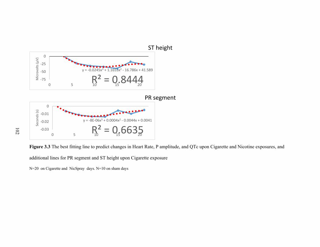
102
ST height
PR segment
Figure 3.3 The best fitting line to predict changes in Heart Rate, P amplitude, and QTc upon Cigarette and Nicotine exposures, and
additional lines for PR segment and ST height upon Cigarette exposure
N=20 on Cigarette and NicSpray days. N=10 on sham days
y = -0.0245x3 + 1.1653x2 - 16.786x + 41.589
R² = 0.8444-75
-50
-25
0
0 5 10 15 20
Mic
rovo
lts (µ
V)
y = -8E-06x3 + 0.0004x2 - 0.0044x + 0.0041
R² = 0.6635-0.03
-0.02
-0.01
0
0 5 10 15 20
Seco
nds
(s)
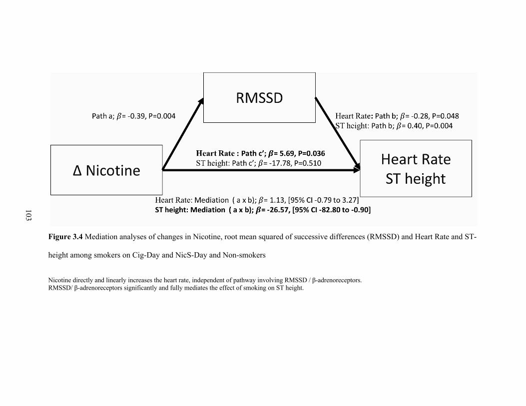
103
Figure 3.4 Mediation analyses of changes in Nicotine, root mean squared of successive differences (RMSSD) and Heart Rate and ST-
height among smokers on Cig-Day and NicS-Day and Non-smokers
Nicotine directly and linearly increases the heart rate, independent of pathway involving RMSSD / β-adrenoreceptors. RMSSD/ β-adrenoreceptors significantly and fully mediates the effect of smoking on ST height.
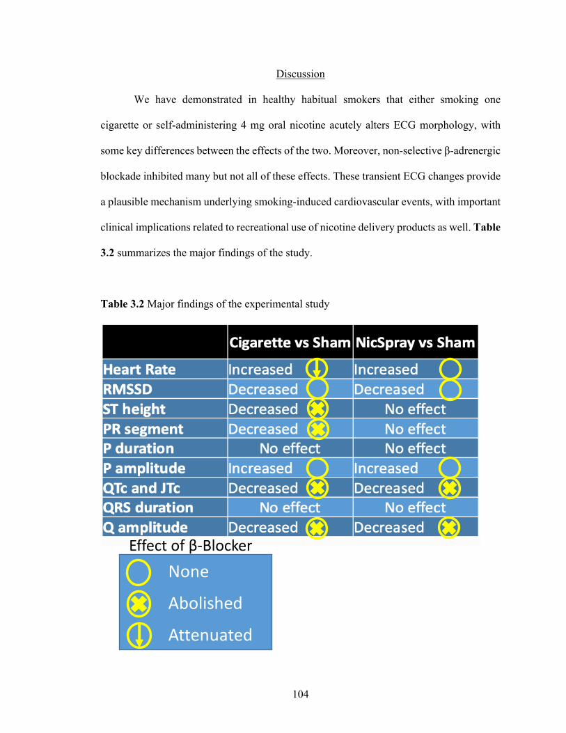
104
Discussion
We have demonstrated in healthy habitual smokers that either smoking one
cigarette or self-administering 4 mg oral nicotine acutely alters ECG morphology, with
some key differences between the effects of the two. Moreover, non-selective β-adrenergic
blockade inhibited many but not all of these effects. These transient ECG changes provide
a plausible mechanism underlying smoking-induced cardiovascular events, with important
clinical implications related to recreational use of nicotine delivery products as well. Table
3.2 summarizes the major findings of the study.
Table 3.2 Major findings of the experimental study
None
Abolished
Attenuated
Effect of β-Blocker
105
Sympathetic activation
Consistent with previous reports, our study found that smoking a single cigarette
acutely increased heart rate and blood pressure, and decreased HRV and PR segment [90,
240, 241, 320] – consistent with increased sympathetic activation. The duration of
increased heart rate up-to 20 minutes, dissipating after 30 minutes, recapitulates
observations by Ramakrishnan et al, among 31 male smokers with atypical chest pain and
24 h Holter monitor [238]. However, we showed for the first time that acute nicotine
exposure causes these changes, with dose-proportionate effects that were diminished by
propranolol. Nicotine is a known sympathomimetic that acts on nicotinic receptors to
increase sympathetic tone and catecholamine release. Thus, it is not surprising that we also
found a dose-proportionate increase in cardiac sympathetic activity from oral nicotine
administration. Our observations of nicotine-mediated sympatho-excitation add to recent
literature on the acute physiological effects of nicotine alone [321-323] or within e-
cigarette aerosols [321] and identify a plausible mechanism by which tobacco product use
increases sympathetic activity. Indeed, sympathetic dominance strongly predicts
cardiovascular morbidity and mortality [303, 324, 325], including for arrhythmia-related
events [326], and can acutely provoke arrhythmia through numerous pathways [327].
Importantly, our observations of acute sympathetic dominance with either nicotine oral
spray or conventional smoking identify nicotine as a plausible mediator of tobacco product-
induced cardiovascular morbidity and mortality.

106
Atria
P wave morphology is dependent on mainly four factors 1) intra-atrial conduction
abnormalities, 2) left atrial hypertension, 3) left atrial distention, and 4) chronicity disease
[328]. Another novel finding of the study was that cigarette smoking and nicotine spray,
within minutes, increased P wave amplitude, and propranolol prevented these effects. We
also found non-significant shortening of P duration with cigarette and nicotine use. Both
these observations are similar those of Goldberg et al., who noted that isoproterenol-
induced β-adrenergic stimulation significantly shortened the P wave in healthy subjects
[58]. Sympathetic stimulation increases peripheral vascular resistance, venous return and
cardiac pressures [329], effects also seen with cigarette smoking [330, 331]. Accordingly,
the significant increase in P amplitude induced by NicS and Cig, and inhibited by β-
blockade, indicate that nicotine increases atrial pressures through β-adrenergic receptors.
The acute increase in atrial pressures together with increased dromotropy, may represent
two distinct pathways with possible synergistic effects of tobacco products induced
increased atrial arrhythmia and thrombosis and stroke risk [283, 312].
Ventricles
To the best of our knowledge, this study demonstrates for the first time that smoking
acutely induces ST-depression. Smoking caused a gradually worsening ST-depression that
peaked at 9-12 minutes, abated by about 30 minutes post-exposure, and vanished with
propranolol pretreatment. Recently, Ramakrishnan noted gross alterations in ST-T
morphology with smoking (1-2 mm changes in ST segment or T wave), but only in one
tenth of men with prior atypical chest pain and with onset at 8-12 minutes after smoking

107
[238]. in [238]. The cardio-protective effects of β-blockers among patients with stable
angina is well described [332]. However, cigarette smoking induced ST-segment
depression at rest, via β-adrenergic receptors activation complements the propositions of
others that enhanced sympathetic tone mediates ST-segment depression in general [333-
335].
Interestingly, we also found that cigarette smoking caused significant albeit
minimal shortening of QTc. The relationship between QTc and the ANS is particularly
complex [299]. However, several studies have shown that acute adrenergic stimulation
shortens QTc in humans [64, 336], possibly by reducing transmural dispersion of
repolarization [337]. Chronic smoking is associated with higher potassium levels [338],
however, acutely it inhibits potassium channels [85] and/or may decrease potassium levels
via direct sympathetic activation [339]. The effects of potassium levels and/or ion channels
may be more pronounced in the ventricles than in atria, resulting in hypokalemia related
ECG changes and ST-segment depression [339]. Unlike the dose-dependent effects of
nicotine (in either NicS or Cig) on atrial and atrioventricular (AV) electrophysiology, there
were no significant effects of nicotine on ventricular depolarization and repolarization. We
speculate this may derive from disproportionate autonomic control of the atria, sinus node,
and AV node, which have a greater density of cardiac autonomic ganglia than the ventricles
[340, 341]. Alternatively, because of distinct atrial-specific ion-channel and interceullular
coupling properties and distribution, atrial myocardium may be more susceptible to
architectural or structural changes with a substantial impact on cardiac performance,
arrhythmia occurrence, and stroke risk [342].
108
Strengths and Limitations
It is well accepted that many of the effects of cigarette smoking, including addiction
and acute alterations in cardiovascular physiology, stem from the rapid delivery of nicotine.
However, there are very few studies that have directly compared the effects of nicotine vs
cigarette, and those studies found no significant effect of nicotine replacement therapy, in
contrast to cigarette exposure [343], we used a novel oral nicotine spray with
pharmacokinetic properties for nicotine similar to those of cigarettes [314, 315, 344, 345],
and also perhaps electronic nicotine delivery systems. We also recorded continuous
standard 12 lead ECG during exposure, and systematically analyzed the interval and
amplitudes of each beat with an automated software, to investigate the acute temporal
relationship of smoking and nicotine exposure on ECG.
One potential source of variability in this study was the outdoor setting during mid-
exposure ECG monitoring. However, to diminish the influence of outdoor conditions (e.g.,
temperature, humidity, air pollutants etc), most of the study visits were performed on
consecutive days. Similarly, we saw no differences in ECG variables between BB and
non-BB days during the first minute of outdoor recording. To facilitate detection of the
early acute effects under typical smoking conditions and the delayed acute effects under
conventional clinical conditions, participants were assessed for ECG changes while seated
at mid-exposure, but while supine at pre- and post-exposure. We also performed the same
protocol on 10 healthy non-smokers, where the exposure consisted of periodically inhaling
through an empty straw for 5 minutes to simulate smoking, and demonstrated that there
were minor changes related to position. We did not exclude any coronary artery disease
among the study participants, however, because of our eligibility criteria, participants had

109
no known cardiac medical condition, nor any active symptoms and were healthy. Another
limitation is that the oral nicotine spray may be pungent and taste unpleasant for some
participants, which may result in significant changes in heart rate. However, these changes
are usually transient and less potent than that seen during our study (i.e., increase in heart
rate of 7.1–13.6% for various taste stimuli, with maximum effect ∼25 s and returned to
pre-exposure after 80 and 100 seconds [346]. Lastly, since this is a human in vivo
experimental study, we were unable to differentiate direct vs indirect effects from nicotine
and cigarette smoking (e.g., heart rate may have increased due to direct effects of nicotine
and non-nicotine constituents on receptors in the sinoatrial node or central nervous system,
or instead via reflexes from increased cardiac pressures secondary to increased peripheral
vascular resistance and systemic pressures).
Conclusion
Smoking and nicotine alone acutely induce myriad changes in electrophysiology
that are known to be pro-arrhythmic. Smoking may acutely promote arrhythmia via
nicotine-mediated acceleration of heart rate and ventricular repolarization (QTc), with β-
adrenoceptors mediating only the latter. Unlike acute nicotine, cigarette smoke rapidly
depresses ST segment and shortens PR segment via β-adrenoceptor stimulation, indicating
smoking transiently induces ischemia and accelerates dromotropy by sympathetic
activation. Nicotine in cigarette smoke, via β-adrenoceptors, mostly seems responsible for
transient increase in atrial pressures, which may reflect possible mechanism for tobacco
induced atrial arrhythmia and stroke risk.
110
CHAPTER IV
IMPLICATIONS AND CONCLUSIONS
Clinical Implications
Cigarette smoking is the most significant modifiable risk factor for cardiovascular
morbidity and mortality. Although smoking rates in the United States have declined over
the past five decades to historic lows, efforts to protect public health are far from complete.
The burden of combustible tobacco use in the United States remains high, especially in
vulnerable populations. This persistence of cigarette smoking, coupled with the advent and
dramatic rise in the use of new tobacco products, such as electronic cigarettes, especially
among adolescents and young adults, are of significant concern [347]. These products may
benefit by helping some smokers quit or transition to a less harmful product; however, the
long-term health effects of these products and the net public health effect associated with
their use remain unclear and widely debated [348]. Evidence is mounting that the use of
these products may catalyze the transition to the use of other tobacco products or
recreational drugs, particularly in young adults [349]. The use of certain newly regulated
tobacco products has skyrocketed, particularly among youths. For example, from 2011 to
2018, the rate of e-cigarette use among high school students ballooned from 1.5% to 20.8%
[350], and then up to 27.5% in 2019, according to preliminary results from the Centers for
Disease Control and Prevention’s annual National Youth Tobacco Survey.
111
Nicotine is addictive and present alongside other chemicals in ENDS. Several
studies have revealed the existence of toxicants in both the liquid and aerosol of e-
cigarettes. Recent studies suggest that, although adverse experiences such as chest pain,
palpitations, coronary heart disease, lung injury [351], and increased risk of myocardial
infarction [352] are associated with e-cigarette use, further investigation is required to
evaluate whether any causal relationships exist. The FDA has included the discovery of
biomarkers to assess exposure and harm or toxicity of non-cigarette tobacco products,
including ENDS, as well the as short- and long-term health effects of tobacco products
among its research priorities [348]. The series of studies presented in this dissertation
directly relate to those scientific domains—to help establish sensitive biomarkers to detect
short-and long-term subclinical cardiovascular injury from tobacco and other nicotine-
related products, and potentially guide regulation of nicotine levels in recreational products
[347].
Most investigations into the pharmacokinetics of nicotine from ENDS have yielded
variable results, perhaps owing to the large variability of study protocols. Moreover,
several studies have lacked generalizability, as they incorporated a single standard e-
cigarette and nicotine concentrations with a protocol for vaping at fixed intervals [353].
Furthermore, they were unable to investigate the differential impacts of variations in
experimental parameters, such as battery output voltage or coil resistance, which are known
to impact delivery of nicotine and production of potentially toxic constituents [354-356].
Further complicating the relevance of past ENDS studies, e-cigarette devices have rapidly
evolved to more effectively deliver nicotine. A recent pharmacokinetic study revealed that
an ad-lib session with third-generation e-cigarettes is able to achieve the dose and speed of

112
nicotine delivery similar to conventional cigarettes [357]. Another study revealed that
newer generation e-cigarettes (“mod”-type cigarette) delivered nicotine at levels close to,
or even exceeding, those of combustible cigarettes (highest average Cmax - 43.6 ng/ml)
[358]. The parent company of JUUL, the relatively new pod device that has claimed 70%-
80% of the U.S. e-cigarette market [359], noted in their initial patent filing that their device
achieved 36% higher peak levels of plasma nicotine and higher increases in heart rate with
more rapid onset than full-flavored cigarettes. This is due in major part to the use of
nicotine benzoic acid salts, which further enhance nicotine delivery [359]. In our
experimental trial, the plasma levels were drawn about 45 minutes after the start of
exposure, and the nicotine levels increased an average of 7.83 ng/ml after cigarette use,
and 6.98 ng/ml after nicotine spray, which is similar to that achieved by newer e-cigarettes
[353]. The JUUL brand is one of the newer entries into the market and utilizes prefilled EC
fluid “pods,” being more similar to the “cig-a-like” products than to the recently available
tank/box mod styles. Nicotine concentrations (average 60.9 mg/mL) were significantly
higher in JUUL than those of any EC products previously analyzed by Omaiye et al. [360].
Such high nicotine levels are not observed after use of NRTs, with doses recommended to
reduce withdrawal symptoms and cigarette cravings [344]. Despite the large variability in
the pharmacokinetic profiles and levels of nicotine due to varying e-cigarette device
designs, e-liquid concentration, and use behavior, the results of our study provide valuable
insight into the likely cardiovascular effects and risks of e-cigarettes.
We conducted three studies that may elucidate the cardiac impacts of nicotine-
containing e-cigarettes. In our cross-sectional observational study, we found that chronic
nicotine or tobacco exposure leads to dose-dependent shortening of PR segment and QTc,
113
and that an extremely high burden of exposure (measured by the nicotine metabolite,
cotinine) leads to QRS shortening and JT prolongation. Although our analyses suggested a
role for nicotine in these effects, the design of this study made it impossible to delineate
between effects from exposure to cigarette smoke and exposure to nicotine. In our follow-
up acute exposure study, we found evidence that nicotine mediates increased sympathetic
activity (as measured by decreased HRV), shortened JTc, and increased P amplitude.
Taking these findings into consideration—and extending to the possible nicotine-related
effects from ENDS on cardiac electrophysiology—we suggest that e-cigarettes both
acutely and chronically increase sympathetic activity, as noted in recent observations [321,
361]. Other studies have also found that conventional smoking chronically increases
sympathetic modulation of the heart [72, 241]. Moreover, our results suggest that nicotine
in e-cigarettes may both acutely and chronically shorten ventricular repolarization while
also acutely increasing atrial pressures (indicated by P amplitude). Notably, a few studies
also observed an increased P amplitude among smokers at baseline [212, 213, 362, 363],
similar to our findings in which cotinine (proxy for tobacco use) was associated with P
amplitude (particularly in deep terminal negativity of the P wave in V1 [DTNPV1]) in the
chronic state [364]. However, the mechanisms for abnormal P wave indices during acute
and chronic exposure may differ while also carrying different prognostic value. As acute
increases in P amplitude may reflect increased cardiac filling pressure, chronic elevations
in P amplitude may reflect atrial hypertrophy/distention.
Such baseline pathophysiological changes in human cardiac electrophysiology
have clinically important implications. Increases in sympathetic activity [303], atrial
cardiopathy [365, 366], and shortened rate of ventricular repolarization [367-369] have

114
been associated with increased risk for adverse outcomes, such as all-cause mortality,
arrhythmia, and stroke. In this regard, a strategy formed by the FDA to limit nicotine levels
in ENDS might help reduce some of the long-term harmful cardiovascular effects. Our
study featuring oral nicotine spray indicated that the nicotine levels should be lower than
those currently found in newer generation e-cigarettes (e.g., JUUL and other pod devices).
However, more studies are required to establish whether the nicotine levels are directly
related to increased risk for short- or long-term adverse cardiovascular events.
Conclusion
The acute and chronic proarrhythmic effects of smoking and their underlying
mechanisms are currently unknown. To address this gap in the knowledge, we assessed the
effects of chronic cigarette exposure on ECG, using serum cotinine (as a sensitive and
specific marker of tobacco use) from the NHANES database, and urinary cotinine from
cardiovascular patients from an outpatient clinic. From the NHANES database, higher
serum cotinine levels were associated with considerably shortened PR intervals, PR
segments, and QRS duration, and considerably prolonged JT intervals. We also found a
negative linear relationship between cotinine and QTc, as well as between cotinine and PR
segment. A similar effect on atrial electrocardiographic indices (shortened PR interval and
PR segment) was also observed among the CVD patients. Interestingly, our statistical
mediation analyses indicated that dopamine fully mediated the association between
cotinine and the shortening of both PR intervals and segments. Collectively, the results
from the chronic exposure studies suggest that habitual smoking accelerates
atrioventricular conduction (possibly though dopamine) and ventricular depolarization, but
115
appears to exert a more complex effect on repolarization. Overall habitual smoking
modestly accelerates ventricular repolarization, and simultaneously increases the risk for
severe slowing of ventricular repolarization in a subset of patients.
An experimental trial to investigate β-adrenergic receptors’ role in the acute effects
of smoking versus nicotine spray on ECG was also performed. Nicotine, both with and
without a β-blocker, increases the sympathetic nervous activity to a similar extent as
observed with cigarette smoking in the presence of a β-blocker. Nicotine increases the heart
rate through receptors independent of the β-adrenergic receptors, suggesting its acute and
direct action on cardiomyocytes is independent of increased central sympathetic activation.
Cigarette smoking had a higher effect on heart rate; however, after treatment with a β-
blocker, the heart rate increase was similar to that from nicotine (with or without a β-
blocker). This suggests that the non-nicotine constituents of cigarette smoke that increase
heart rate act through β-adrenergic receptors. Cigarette smoking and nicotine comparably
increase P-wave amplitude without affecting P duration (which was also unaffected in
chronic exposure studies), and β-blockade did not alter this effect. This increase in P
amplitude may derive from increased peripheral vascular resistance with both exposures,
which increases cardiac filling pressures and cardiac output. Cigarette smoking, but not
nicotine, also shortened the PR segment and decreased the height of the ST segment, with
both being rescued by β-blockade. This suggests that non-nicotine constituents increase
dromotropy and ischemia through β-adrenergic receptors. Furthermore, the relationship
between cotinine and the PR segment found in the chronic exposure studies likely derived
from cotinine reflecting exposure to tobacco smoke, including non-nicotine constituents.
Indeed, the tar and nicotine levels in cigarettes correspond with one another, just as

116
exposures to each would be expected to correspond. Consistent with our observations of
the link between chronic smoking and QTc shortening, ventricular repolarization time was
more acutely shortened with cigarette smoking compared to nicotine, and was abolished
by the β-blockade. It is plausible that nicotine acutely accelerates repolarization via
adrenergic-related mechanisms (e.g., β-adrenergic induced phosphorylation of Kv7.1),
whereas chronic smoking, itself, impedes repolarization via myocardial injury (e.g.,
fibrosis). Alternatively, chronic smoking may impede repolarization by desensitizing
adrenergic pathways that would otherwise modulate the heart-rate-dependent shortening
of repolarization with sympathetic activation in healthy individuals.
The central illustration combines the findings of all three studies to demonstrate the
role of nicotine and non-nicotine constituents of cigarette smoke, and the pathways
involved in their effects on cardiac electrophysiology (Figure 4.1).
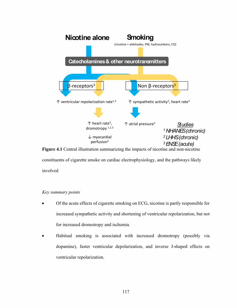
117
Figure 4.1 Central illustration summarizing the impacts of nicotine and non-nicotine
constituents of cigarette smoke on cardiac electrophysiology, and the pathways likely
involved
Key summary points
• Of the acute effects of cigarette smoking on ECG, nicotine is partly responsible for
increased sympathetic activity and shortening of ventricular repolarization, but not
for increased dromotropy and ischemia.
• Habitual smoking is associated with increased dromotropy (possibly via
dopamine), faster ventricular depolarization, and inverse J-shaped effects on
ventricular repolarization.
Nicotine alone
β-receptors3
Smoking(nicotine + aldehydes, PM, hydrocarbons, CO)
↑ heart rate3, dromotropy 1,2,3
↓ myocardial perfusion3
↑ sympathetic activity3, heart rate3↑ ventricular repolarization rate1,3
Non β-receptors3
Studies1 NHANES (chronic) 2 LHHS (chronic)3 ENSE (acute)
Catecholamines & other neurotransmitters
↑ atrial pressure3
118
• Such acute and chronic effects on ECG from cigarette smoking and nicotine may
promote cardiac arrhythmias or reflect a diseased state and an at-risk population.
• Future research is needed to (a) delineate the receptors used by nicotine to cause
sympathetic activation, (b) identify the non-nicotine constituents in cigarette smoke
that transiently increase dromotropy and myocardial ischemia, and (c) elucidate the
mechanisms involved in nicotine and non-nicotine constituents of cigarette-smoke-
induced complex effects on ventricular repolarization (shortening in most patients,
and prolonging in a small subset of patients).

119
REFERENCES
1. General., A.R.o.t.S., The health consequences of smoking — 50 years of progress:a report of the Surgeon General. . 2014: Washington, DC.
2. Burns, D.M., Epidemiology of smoking-induced cardiovascular disease. ProgCardiovasc Dis, 2003. 46(1): p. 11-29.
3. Centers for Disease, C. and Prevention, Smoking-attributable mortality, years ofpotential life lost, and productivity losses--United States, 2000-2004. MMWRMorb Mortal Wkly Rep, 2008. 57(45): p. 1226-8.
4. Smoking and Mortality - Beyond Established Causes. N Engl J Med, 2016.375(24): p. 2410.
5. Jha, P., et al., 21st-century hazards of smoking and benefits of cessation in theUnited States. N Engl J Med, 2013. 368(4): p. 341-50.
6. Bjartveit, K. and A. Tverdal, Health consequences of smoking 1-4 cigarettes perday: response to G F Cope (eletter to journal). Tob Control, 2006. 15(1): p. 71-2.
7. Xu, X., et al., Annual healthcare spending attributable to cigarette smoking: anupdate. Am J Prev Med, 2015. 48(3): p. 326-33.
8. Jamal, A., et al., Current Cigarette Smoking Among Adults - United States, 2016.MMWR Morb Mortal Wkly Rep, 2018. 67(2): p. 53-59.
9. Benowitz, N.L., Nicotine addiction. N Engl J Med, 2010. 362(24): p. 2295-303.10. Mansvelder, H.D. and D.S. McGehee, Long-term potentiation of excitatory inputs
to brain reward areas by nicotine. Neuron, 2000. 27(2): p. 349-57.11. George, O., et al., CRF-CRF1 system activation mediates withdrawal-induced
increases in nicotine self-administration in nicotine-dependent rats. Proc NatlAcad Sci U S A, 2007. 104(43): p. 17198-203.
12. Grunberg, N.E., A neurobiological basis for nicotine withdrawal. Proc Natl AcadSci U S A, 2007. 104(46): p. 17901-2.
13. Bhatnagar, A., et al., Electronic cigarettes: a policy statement from the AmericanHeart Association. Circulation, 2014. 130(16): p. 1418-36.
14. Grana, R., N. Benowitz, and S.A. Glantz, E-cigarettes: a scientific review.Circulation, 2014. 129(19): p. 1972-86.
15. Keith, R.J., et al., Characterization of Volatile Organic Compound (VOC)metabolites in Cigarette smokers, Electronic Nicotine Device Users, Dual Usersand Non- users of tobacco. Nicotine Tob Res, 2019.

120
16. Glantz, S.A. and D.W. Bareham, E-Cigarettes: Use, Effects on Smoking, Risks,and Policy Implications. Annu Rev Public Health, 2018. 39: p. 215-235.
17. in How Tobacco Smoke Causes Disease: The Biology and Behavioral Basis forSmoking-Attributable Disease: A Report of the Surgeon General. 2010: Atlanta(GA).
18. Arredondo, J., et al., Receptor-mediated tobacco toxicity: regulation of geneexpression through alpha3beta2 nicotinic receptor in oral epithelial cells. Am JPathol, 2005. 166(2): p. 597-613.
19. Ambrose, J.A. and R.S. Barua, The pathophysiology of cigarette smoking andcardiovascular disease: an update. J Am Coll Cardiol, 2004. 43(10): p. 1731-7.
20. Balakumar, P. and J. Kaur, Is nicotine a key player or spectator in the inductionand progression of cardiovascular disorders? Pharmacol Res, 2009. 60(5): p.361-8.
21. Klabunde, R.E., Cardiac electrophysiology: normal and ischemic ionic currentsand the ECG. Adv Physiol Educ, 2017. 41(1): p. 29-37.
22. Kligfield, P., et al., Recommendations for the standardization and interpretationof the electrocardiogram: part I: the electrocardiogram and its technology ascientific statement from the American Heart Association Electrocardiographyand Arrhythmias Committee, Council on Clinical Cardiology; the AmericanCollege of Cardiology Foundation; and the Heart Rhythm Society endorsed bythe International Society for Computerized Electrocardiology. J Am Coll Cardiol,2007. 49(10): p. 1109-27.
23. Mozos, I. and A. Caraba, Electrocardiographic Predictors of CardiovascularMortality. Dis Markers, 2015. 2015: p. 727401.
24. Denes, P., et al., Major and minor ECG abnormalities in asymptomatic womenand risk of cardiovascular events and mortality. JAMA, 2007. 297(9): p. 978-85.
25. Auer, R., et al., Association of major and minor ECG abnormalities withcoronary heart disease events. JAMA, 2012. 307(14): p. 1497-505.
26. Soliman, E.Z., M. Cammarata, and Y. Li, Explaining the inconsistent associationsof PR interval with mortality: the role of P-duration contribution to the length ofPR interval. Heart Rhythm, 2014. 11(1): p. 93-8.
27. Smith, J.W., et al., PR-Interval Components and Atrial Fibrillation Risk (from theAtherosclerosis Risk in Communities Study). Am J Cardiol, 2017. 119(3): p. 466-472.
28. Magnani, J.W., et al., P wave indices, obesity, and the metabolic syndrome: theatherosclerosis risk in communities study. Obesity (Silver Spring), 2012. 20(3): p.666-72.
29. Rasmussen, P.V., et al., Electrocardiographic PR Interval Duration andCardiovascular Risk: Results From the Copenhagen ECG Study. Can J Cardiol,2017. 33(5): p. 674-681.

121
30. Bang, C.N. and P.M. Okin, The long and short of the PR-interval: relation tocardiovascular outcome in patients with coronary heart disease. Europace, 2015.17(6): p. 838-9.
31. Crow, R.S., P.J. Hannan, and A.R. Folsom, Prognostic significance of correctedQT and corrected JT interval for incident coronary heart disease in a generalpopulation sample stratified by presence or absence of wide QRS complex: theARIC Study with 13 years of follow-up. Circulation, 2003. 108(16): p. 1985-9.
32. Zulqarnain, M.A., et al., Risk of Mortality Associated With QT and JT Intervals atDifferent Levels of QRS Duration (from the Third National Health and NutritionExamination Survey). Am J Cardiol, 2015. 116(1): p. 74-8.
33. Rosenthal, T.M., et al., T-peak to T-end interval for prediction of ventriculartachyarrhythmia and mortality in a primary prevention population with systoliccardiomyopathy. Heart Rhythm, 2015. 12(8): p. 1789-97.
34. Panikkath, R., et al., Prolonged Tpeak-to-tend interval on the resting ECG isassociated with increased risk of sudden cardiac death. Circ ArrhythmElectrophysiol, 2011. 4(4): p. 441-7.
35. Erikssen, G., et al., The terminal part of the QT interval (T peak to T end): apredictor of mortality after acute myocardial infarction. Ann NoninvasiveElectrocardiol, 2012. 17(2): p. 85-94.
36. Ashley, E.A., V. Raxwal, and V. Froelicher, An evidence-based review of theresting electrocardiogram as a screening technique for heart disease. ProgCardiovasc Dis, 2001. 44(1): p. 55-67.
37. Kianoush, S., et al., Associations of Cigarette Smoking With SubclinicalInflammation and Atherosclerosis: ELSA-Brasil (The Brazilian LongitudinalStudy of Adult Health). J Am Heart Assoc, 2017. 6(6).
38. Abrams, D.B., et al., Harm Minimization and Tobacco Control: ReframingSocietal Views of Nicotine Use to Rapidly Save Lives. Annu Rev Public Health,2018. 39: p. 193-213.
39. Administration, F.a.D., Tobacco product standard for nicotine level of combustedcigarettes: a proposed rule. 2018: Fed Reg. p. 11818–11843.
40. Zeller, M., The Future of Nicotine Regulation: Key Questions and Challenges.Nicotine Tob Res, 2019. 21(3): p. 331-332.
41. Apelberg, B.J., et al., Potential Public Health Effects of Reducing Nicotine Levelsin Cigarettes in the United States. N Engl J Med, 2018. 378(18): p. 1725-1733.
42. Hatsukami, D.K. and E.C. Donny, The Debate About Nicotine Addiction and theRole of Medicinal Products: Commentary on Zeller. Nicotine Tob Res, 2019.21(3): p. 338-339.
43. Armour, J.A., Potential clinical relevance of the 'little brain' on the mammalianheart. Exp Physiol, 2008. 93(2): p. 165-76.

122
44. Shen, M.J. and D.P. Zipes, Role of the autonomic nervous system in modulatingcardiac arrhythmias. Circ Res, 2014. 114(6): p. 1004-21.
45. Ali, I.M., et al., Modification of supraventricular tachyarrhythmias by stimulatingatrial neurons. Ann Thorac Surg, 1990. 50(2): p. 251-6.
46. Tomaselli, G.F., Mechanisms for Altered Autonomic and Oxidant Regulation ofCardiac Sodium Currents in Cardiac Electrophysiology: From Cell to Bedside,D.P.J. Zipes, José; Stevenson, William G.,, Editor. 2018, ELSEVIER. p. 362-369.
47. Shibata, E.F., et al., Autonomic regulation of voltage-gated cardiac ion channels.J Cardiovasc Electrophysiol, 2006. 17 Suppl 1: p. S34-S42.
48. Ono, K., H.A. Fozzard, and D.A. Hanck, Mechanism of cAMP-dependentmodulation of cardiac sodium channel current kinetics. Circ Res, 1993. 72(4): p.807-15.
49. Matsuda, J.J., H.C. Lee, and E.F. Shibata, Acetylcholine reversal of isoproterenol-stimulated sodium currents in rabbit ventricular myocytes. Circ Res, 1993. 72(3):p. 517-25.
50. Carll, A.F.A., Roberts AM, The Role of the Autonomic Nervous System inCardiovascular Toxicity, in Comprehensive Toxicology, C.A. McQueen, Editor.2018, Elsevier. p. 61-114.
51. Schubert, B., et al., Beta-adrenergic inhibition of cardiac sodium channels bydual G-protein pathways. Science, 1989. 245(4917): p. 516-9.
52. Grant, A.O., Cardiac ion channels. Circ Arrhythm Electrophysiol, 2009. 2(2): p.185-94.
53. Lundby, A. and J.V. Olsen, Phosphoproteomics taken to heart. Cell Cycle, 2013.12(17): p. 2707-8.
54. Dvir, M., et al., Long QT mutations at the interface between KCNQ1 helix C andKCNE1 disrupt I(KS) regulation by PKA and PIP(2). J Cell Sci, 2014. 127(Pt 18):p. 3943-55.
55. Kang, C., et al., beta-adrenergic stimulation augments transmural dispersion ofrepolarization via modulation of delayed rectifier currents IKs and IKr in thehuman ventricle. Sci Rep, 2017. 7(1): p. 15922.
56. Moreno-Galindo, E.G., et al., Pharmacological Conversion of a Cardiac InwardRectifier into an Outward Rectifier Potassium Channel. Mol Pharmacol, 2016.90(3): p. 334-40.
57. Liang, B., et al., G-protein-coupled inward rectifier potassium current contributesto ventricular repolarization. Cardiovasc Res, 2014. 101(1): p. 175-84.
58. Cheema, A.N., et al., Effects of autonomic stimulation and blockade on signal-averaged P wave duration. J Am Coll Cardiol, 1995. 26(2): p. 497-502.
59. Nollo, G., et al., Evidence of low- and high-frequency oscillations in human AVinterval variability: evaluation with spectral analysis. Am J Physiol, 1994. 267(4Pt 2): p. H1410-8.
123
60. Kowallik, P. and M. Meesmann, Independent autonomic modulation of the humansinus and AV nodes: evidence from beat-to-beat measurements of PR and PPintervals during sleep. J Cardiovasc Electrophysiol, 1995. 6(11): p. 993-1003.
61. Wallace, A.G. and S.J. Sarnoff, Effects of Cardiac Sympathetic Nerve Stimulationon Conduction in the Heart. Circ Res, 1964. 14: p. 86-92.
62. Goldberger, J.J., et al., Assessment of effects of autonomic stimulation andblockade on the signal-averaged electrocardiogram. Circulation, 1994. 89(4): p.1656-64.
63. Folino, A.F., et al., The effects of sympathetic stimulation induced by mentalstress on signal-averaged electrocardiogram. Int J Cardiol, 1995. 48(3): p. 279-85.
64. Algra, A., et al., Effect of beta-blockers on the relation between QT-interval andheart rate in exercise ECG. Eur Heart J, 1987. 8 Suppl D: p. 71-3.
65. Cuomo, S., et al., Influence of autonomic tone on QT interval duration.Cardiologia, 1997. 42(10): p. 1071-6.
66. Magnano, A.R., et al., Autonomic nervous system influences on QT interval innormal subjects. J Am Coll Cardiol, 2002. 39(11): p. 1820-6.
67. Li, Y.F., C. LaCroix, and J. Freeling, Specific subtypes of nicotinic cholinergicreceptors involved in sympathetic and parasympathetic cardiovascular responses.Neurosci Lett, 2009. 462(1): p. 20-3.
68. Li, Y.F., C. Lacroix, and J. Freeling, Cytisine induces autonomic cardiovascularresponses via activations of different nicotinic receptors. Auton Neurosci, 2010.154(1-2): p. 14-9.
69. Neff, R.A., et al., Nicotine enhances presynaptic and postsynaptic glutamatergicneurotransmission to activate cardiac parasympathetic neurons. Circ Res, 1998.83(12): p. 1241-7.
70. Kirpekar, S.M., A.G. Garcia, and J.C. Prat, Action of nicotine on sympatheticnerve terminals. J Pharmacol Exp Ther, 1980. 213(1): p. 133-8.
71. Haass, M. and W. Kubler, Nicotine and sympathetic neurotransmission.Cardiovasc Drugs Ther, 1997. 10(6): p. 657-65.
72. Middlekauff, H.R., J. Park, and R.S. Moheimani, Adverse effects of cigarette andnoncigarette smoke exposure on the autonomic nervous system: mechanisms andimplications for cardiovascular risk. J Am Coll Cardiol, 2014. 64(16): p. 1740-50.
73. Gerber, S.H., et al., Role of [Na+]i and [Ca2+]i in nicotine-inducednorepinephrine release from bovine adrenal chromaffin cells. Am J Physiol,1995. 269(3 Pt 1): p. C572-81.
74. Wada, A., et al., Influx of 22Na through acetylcholine receptor-associated Nachannels: relationship between 22Na influx, 45Ca influx and secretion of
124
catecholamines in cultured bovine adrenal medulla cells. Neuroscience, 1985. 15(1): p. 283-92.
75. Kruger, C., et al., Nicotine-induced exocytotic norepinephrine release in guinea-pig heart, human atrium and bovine adrenal chromaffin cells: modulation bysingle components of ischaemia. J Mol Cell Cardiol, 1995. 27(8): p. 1491-506.
76. Li, Q. and E.J. Forsberg, Catecholamine secretion induced by nicotine is due toCa++ channel but not Na+ channel activation in porcine adrenal chromaffincells. J Pharmacol Exp Ther, 1996. 277(3): p. 1209-14.
77. Abajo, F.J., et al., Sodium-dependent and sodium-independent nicotine-evokedcatecholamine release from cat adrenals. Neurosci Lett, 1989. 101(1): p. 101-6.
78. Wu, C.Y., et al., L-type calcium channels in sympathetic alpha3beta2-nAChR-mediated cerebral nitrergic neurogenic vasodilation. Acta Physiol (Oxf), 2014.211(4): p. 544-58.
79. Liu, Y., et al., Cav1.2, but not Cav1.3, L-type calcium channel subtype mediatesnicotine-induced conditioned place preference in miceo. ProgNeuropsychopharmacol Biol Psychiatry, 2017. 75: p. 176-182.
80. Pappano, A.J. and R.A. Rembish, Nicotine-induced restoration of actionpotentials to cardiac tissue depolarized by potassium. Life Sci I, 1970. 9(24): p.1381-8.
81. Satoh, H., Effects of nicotine on spontaneous activity and underlying ioniccurrents in rabbit sinoatrial nodal cells. Gen Pharmacol, 1997. 28(1): p. 39-44.
82. Carryl, O.R., A. Gallardo-Carpentier, and R.G. Carpentier, Effects of nicotine andethanol on rat atrial membrane potentials. Alcohol, 1992. 9(2): p. 87-92.
83. Greenspan, K., et al., Some effects of nicotine on cardiac automaticity,conduction, and inotropy. Arch Intern Med, 1969. 123(6): p. 707-12.
84. Wang, H., H. Shi, and Z. Wang, Nicotine depresses the functions of multiplecardiac potassium channels. Life Sci, 1999. 65(12): p. PL143-9.
85. Wang, H., et al., Nicotine is a potent blocker of the cardiac A-type K(+) channels.Effects on cloned Kv4.3 channels and native transient outward current.Circulation, 2000. 102(10): p. 1165-71.
86. Wang, H., et al., Direct block of inward rectifier potassium channels by nicotine.Toxicol Appl Pharmacol, 2000. 164(1): p. 97-101.
87. Brasch, H., H.B. Iven, and G. Zetler, Effects of simple indoles, noradrenaline,nicotine, and tyramine on action potential and contractility of the isolated guinea-pig papillary muscle. Naunyn Schmiedebergs Arch Pharmacol, 1977. 299(3): p.259-65.
88. Pappano, A.J., Biphasic effect of nicotine on action potential repolarization inelectrically driven guinea-pig atria. J Pharmacol Exp Ther, 1970. 172(2): p. 255-65.
125
89. Bassett, A.L. and H. Gelband, Nicotine and the action potential of cat ventricle. JPharmacol Exp Ther, 1974. 188(1): p. 157-65.
90. Grassi, G., et al., Mechanisms responsible for sympathetic activation by cigarettesmoking in humans. Circulation, 1994. 90(1): p. 248-53.
91. Cryer, P.E., et al., Norepinephrine and epinephrine release and adrenergicmediation of smoking-associated hemodynamic and metabolic events. N Engl JMed, 1976. 295(11): p. 573-7.
92. Berlin, I. and R.M. Anthenelli, Monoamine oxidases and tobacco smoking. Int JNeuropsychopharmacol, 2001. 4(1): p. 33-42.
93. Haussmann, H.J., Use of hazard indices for a theoretical evaluation of cigarettesmoke composition. Chem Res Toxicol, 2012. 25(4): p. 794-810.
94. Kurhanewicz, N., et al., TRPA1 mediates changes in heart rate variability andcardiac mechanical function in mice exposed to acrolein. Toxicol ApplPharmacol, 2017. 324: p. 51-60.
95. Kurhanewicz, N., et al., TRPA1 mediates the cardiac effects of acrolein throughparasympathetic dominance but also sympathetic modulation in mice. ToxicolAppl Pharmacol, 2018. 347: p. 104-114.
96. Clark, M.S., M.J. Rand, and S. Vanov, Comparison of pharmacological activity ofnicotine and related alkaloids occurring in cigarette smoke. Arch IntPharmacodyn Ther, 1965. 156(2): p. 363-79.
97. Carreon-Garciduenas, M., et al., Participation of voltage-gated sodium andcalcium channels in the acute cardiac effects of toluene. Toxicol Mech Methods,2018. 28(9): p. 670-677.
98. Gordon, C.J., et al., Cardiovascular effects of oral toluene exposure in the ratmonitored by radiotelemetry. Neurotoxicol Teratol, 2007. 29(2): p. 228-35.
99. Condouris, G.A. and D.M. Havelin, Acetaldehyde and cardiac arrhythmias. ArchInt Pharmacodyn Ther, 1987. 285(1): p. 50-9.
100. Tani, T. and Y. Horiguchi, Effects of formaldehyde on cardiac function. Jpn J Pharmacol, 1990. 52(4): p. 563-72.
101. Morvai, V., et al., ECG changes in benzene, toluene and xylene poisoned rats. Acta Med Acad Sci Hung, 1976. 33(3): p. 275-86.
102. Dallas, M.L., et al., Carbon monoxide induces cardiac arrhythmia via induction of the late Na+ current. Am J Respir Crit Care Med, 2012. 186(7): p. 648-56.
103. Tillar, R., T.J. Shafer, and J.J. Woodward, Toluene inhibits voltage-sensitive calcium channels expressed in pheochromocytoma cells. Neurochem Int, 2002. 41(6): p. 391-7.
104. Gauthereau, M.Y., E.M. Salinas-Stefanon, and S.L. Cruz, A mutation in the local anaesthetic binding site abolishes toluene effects in sodium channels. Eur J Pharmacol, 2005. 528(1-3): p. 17-26.
126
105. Cruz, S.L., et al., Inhibition of cardiac sodium currents by toluene exposure. Br J Pharmacol, 2003. 140(4): p. 653-60.
106. Cruz, S.L., M.T. Rivera-Garcia, and J.J. Woodward, Review of toluene action: clinical evidence, animal studies and molecular targets. J Drug Alcohol Res, 2014. 3.
107. Lee, J.C. and S.E. Downing, Negative inotropic effects of phenol on isolated cardiac muscle. Am J Pathol, 1981. 102(3): p. 367-72.
108. Scragg, J.L., et al., Carbon monoxide inhibits L-type Ca2+ channels via redox modulation of key cysteine residues by mitochondrial reactive oxygen species. J Biol Chem, 2008. 283(36): p. 24412-9.
109. Dallas, M.L., J.L. Scragg, and C. Peers, Inhibition of L-type Ca(2+) channels by carbon monoxide. Adv Exp Med Biol, 2009. 648: p. 89-95.
110. Shrager, P.G., A. Strickholm, and R.I. Macey, Chemical modification of crayfish axons by protein crosslinking aldehydes. J Cell Physiol, 1969. 74(1): p. 91-100.
111. Kohlhardt, M., Modifications of drug-induced cardiac slow inward current block by cooling, pH variations, and formaldehyde in mammalian ventricular myocardium. J Cardiovasc Pharmacol, 1983. 5(6): p. 968-77.
112. Nonner, W., B.C. Spalding, and B. Hille, Low intracellular pH and chemical agents slow inactivation gating in sodium channels of muscle. Nature, 1980. 284(5754): p. 360-3.
113. Williams, E.S., M.J. Mirro, and J.C. Bailey, Electrophysiological effects of ethanol, acetaldehyde, and acetate on cardiac tissues from dog and guinea pig. Circ Res, 1980. 47(3): p. 473-8.
114. Chen, F.S., et al., Effects of acetaldehyde on action potentials and Ca2+ currents in single atrial myocytes from the bullfrog. Pharmacology, 2012. 90(3-4): p. 216-22.
115. Martinez, J.L. and M. Penna, Influences of changes in calcium concentrations, cocaine and clonidine on the cardiac effect of acetaldehyde in rat isolated atria. Gen Pharmacol, 1997. 29(2): p. 281-4.
116. Ju, Y.K. and D.G. Allen, Cyanide inhibits the Na+/Ca2+ exchanger in isolated cardiac pacemaker cells of the cane toad. Pflugers Arch, 2005. 449(5): p. 442-8.
117. Elies, J., et al., Inhibition of the cardiac Na(+) channel Nav1.5 by carbon monoxide. J Biol Chem, 2014. 289(23): p. 16421-9.
118. Ferreira de Mattos, G., et al., Lead poisoning: acute exposure of the heart to lead ions promotes changes in cardiac function and Cav1.2 ion channels. Biophys Rev, 2017. 9(5): p. 807-825.
119. DiFrancesco, D., et al., Cadmium-induced blockade of the cardiac fast Na channels in calf Purkinje fibres. Proc R Soc Lond B Biol Sci, 1985. 223(1233): p. 475-84.
127
120. Wang, T., et al., Particulate matter induces cardiac arrhythmias via dysregulation of carotid body sensitivity and cardiac sodium channels. Am J Respir Cell Mol Biol, 2012. 46(4): p. 524-31.
121. Horakova, Z., et al., Effect of ethanol and acetaldehyde at clinically relevant concentrations on atrial inward rectifier potassium current IK1: separate and combined effect. J Physiol Pharmacol, 2016. 67(3): p. 339-51.
122. Bebarova, M., et al., Acetaldehyde at clinically relevant concentrations inhibits inward rectifier potassium current I(K1) in rat ventricular myocytes. Physiol Res, 2015. 64(6): p. 939-43.
123. Kawahara, H., et al., Effects of ethanol and acetaldehyde on the contraction of cultured Ito cells. Alcohol Alcohol Suppl, 1993. 1B: p. 9-14.
124. Williams, J.W., et al., Effect of ethanol and acetaldehyde on the (Na+ +K+)-activated adenosine triphosphatase activity of cardiac plasma membranes. Biochem Pharmacol, 1975. 24(1): p. 27-32.
125. Yamakura, T., J.M. Lewohl, and R.A. Harris, Differential effects of general anesthetics on G protein-coupled inwardly rectifying and other potassium channels. Anesthesiology, 2001. 95(1): p. 144-53.
126. Sanguinetti, M.C. and N.K. Jurkiewicz, Two components of cardiac delayed rectifier K+ current. Differential sensitivity to block by class III antiarrhythmic agents. J Gen Physiol, 1990. 96(1): p. 195-215.
127. Bhave, G., et al., Small-molecule modulators of inward rectifier K+ channels: recent advances and future possibilities. Future Med Chem, 2010. 2(5): p. 757-74.
128. Dallas, M.L., et al., Carbon monoxide protects against oxidant-induced apoptosis via inhibition of Kv2.1. FASEB J, 2011. 25(5): p. 1519-30.
129. Liang, S., et al., Carbon monoxide inhibits inward rectifier potassium channels in cardiomyocytes. Nat Commun, 2014. 5: p. 4676.
130. Ficker, E., et al., Mechanisms of arsenic-induced prolongation of cardiac repolarization. Mol Pharmacol, 2004. 66(1): p. 33-44.
131. Drolet, B., C. Simard, and D.M. Roden, Unusual effects of a QT-prolonging drug, arsenic trioxide, on cardiac potassium currents. Circulation, 2004. 109(1): p. 26-9.
132. Graff, D.W., et al., Metal particulate matter components affect gene expression and beat frequency of neonatal rat ventricular myocytes. Environ Health Perspect, 2004. 112(7): p. 792-8.
133. D'Alessandro, A., et al., Nicotine, cigarette smoking and cardiac arrhythmia: an overview. Eur J Prev Cardiol, 2012. 19(3): p. 297-305.
134. Zevin, S., et al., Cardiovascular effects of carbon monoxide and cigarette smoking. J Am Coll Cardiol, 2001. 38(6): p. 1633-8.
135. Mancia, G., et al., Smoking impairs baroreflex sensitivity in humans. Am J Physiol, 1997. 273(3 Pt 2): p. H1555-60.
128
136. Arosio, E., et al., Effects of smoking on cardiopulmonary baroreceptor activation and peripheral vascular resistance. Eur J Clin Invest, 2006. 36(5): p. 320-5.
137. Marie Hawkeswood, J., The effects of a nicotine nasal spray on baroreflex sensitivity and mood states over three days of smoking cessation. 2001, Thesis (M. Sc.)--Queen's University, Kingston, Ontario, Canada.
138. Perez, C.M., M.S. Hazari, and A.K. Farraj, Role of autonomic reflex arcs in cardiovascular responses to air pollution exposure. Cardiovasc Toxicol, 2015. 15(1): p. 69-78.
139. Hazari, M.S., et al., A single exposure to acrolein desensitizes baroreflex responsiveness and increases cardiac arrhythmias in normotensive and hypertensive rats. Cardiovasc Toxicol, 2014. 14(1): p. 52-63.
140. Perez, C.M., et al., Acrolein inhalation alters arterial blood gases and triggers carotid body-mediated cardiovascular responses in hypertensive rats. Inhal Toxicol, 2015. 27(1): p. 54-63.
141. Argacha, J.F., et al., Nicotine increases chemoreflex sensitivity to hypoxia in non-smokers. J Hypertens, 2008. 26(2): p. 284-94.
142. Najem, B., et al., Acute cardiovascular and sympathetic effects of nicotine replacement therapy. Hypertension, 2006. 47(6): p. 1162-7.
143. Yue, Z., et al., Role of TRP channels in the cardiovascular system. Am J Physiol Heart Circ Physiol, 2015. 308(3): p. H157-82.
144. Xing, J., J. Lu, and J. Li, TRPA1 mediates amplified sympathetic responsiveness to activation of metabolically sensitive muscle afferents in rats with femoral artery occlusion. Front Physiol, 2015. 6: p. 249.
145. Hayano, J., et al., Respiratory sinus arrhythmia. A phenomenon improving pulmonary gas exchange and circulatory efficiency. Circulation, 1996. 94(4): p. 842-7.
146. Lange, P., et al., Relationship of the type of tobacco and inhalation pattern to pulmonary and total mortality. Eur Respir J, 1992. 5(9): p. 1111-7.
147. Tanaka, T., et al., Acute effects of nicotine content in cigarettes on coronary flow velocity and coronary flow reserve in men. Am J Cardiol, 1998. 82(10): p. 1275-8, A9.
148. Gullu, H., et al., Light cigarette smoking impairs coronary microvascular functions as severely as smoking regular cigarettes. Heart, 2007. 93(10): p. 1274-7.
149. Klein, L.W., et al., Acute coronary hemodynamic response to cigarette smoking in patients with coronary artery disease. J Am Coll Cardiol, 1984. 3(4): p. 879-86.
150. Martin, J.L., et al., Acute coronary vasoconstrictive effects of cigarette smoking in coronary heart disease. Am J Cardiol, 1984. 54(1): p. 56-60.
151. Ciftci, O., et al., Acute effects of smoking light cigarettes on coronary microvascular functions. Clin Cardiol, 2009. 32(4): p. 210-4.
129
152. Nelson, M.D., et al., Acute Effect of Hookah Smoking on the Human Coronary Microcirculation. Am J Cardiol, 2016. 117(11): p. 1747-54.
153. Stavros Stavrakis, B.J.S., Paul Garabelli, Sunny S. Po, Role of the Autonomic Nervous System in Atrial Fibrillation, in Cardiac Electrophysiology: From Cell to Bedside,, J.J. Douglas P. Zipes, William G. Stevenson, Editor. 2018, ELSEVIER. p. 419-425.
154. Gordan, R., J.K. Gwathmey, and L.H. Xie, Autonomic and endocrine control of cardiovascular function. World J Cardiol, 2015. 7(4): p. 204-14.
155. Tweed, J.O., et al., The endocrine effects of nicotine and cigarette smoke. Trends Endocrinol Metab, 2012. 23(7): p. 334-42.
156. Kapoor, D. and T.H. Jones, Smoking and hormones in health and endocrine disorders. Eur J Endocrinol, 2005. 152(4): p. 491-9.
157. Smith, C.J. and T.H. Fischer, Particulate and vapor phase constituents of cigarette mainstream smoke and risk of myocardial infarction. Atherosclerosis, 2001. 158(2): p. 257-67.
158. Alberg, A., The influence of cigarette smoking on circulating concentrations of antioxidant micronutrients. Toxicology, 2002. 180(2): p. 121-37.
159. van der Vaart, H., et al., Acute effects of cigarette smoke on inflammation and oxidative stress: a review. Thorax, 2004. 59(8): p. 713-21.
160. Edirisinghe, I. and I. Rahman, Cigarette smoke-mediated oxidative stress, shear stress, and endothelial dysfunction: role of VEGFR2. Ann N Y Acad Sci, 2010. 1203: p. 66-72.
161. Larue, D. and G. Kato, Reduced number of alpha- and beta-adrenergic receptors in the myocardium of rats exposed to tobacco smoke. Eur J Pharmacol, 1981. 70(4): p. 519-29.
162. Kannel, W.B., et al., Heart rate and cardiovascular mortality: the Framingham Study. Am Heart J, 1987. 113(6): p. 1489-94.
163. Johansen, C.D., et al., Resting, night-time, and 24 h heart rate as markers of cardiovascular risk in middle-aged and elderly men and women with no apparent heart disease. Eur Heart J, 2013. 34(23): p. 1732-9.
164. Zhang, D., X. Shen, and X. Qi, Resting heart rate and all-cause and cardiovascular mortality in the general population: a meta-analysis. CMAJ, 2016. 188(3): p. E53-63.
165. Arnold, J.M., et al., Resting heart rate: a modifiable prognostic indicator of cardiovascular risk and outcomes? Can J Cardiol, 2008. 24 Suppl A: p. 3A-8A.
166. Heart rate variability: standards of measurement, physiological interpretation and clinical use. Task Force of the European Society of Cardiology and the North American Society of Pacing and Electrophysiology. Circulation, 1996. 93(5): p. 1043-65.
130
167. Tsuji, H., et al., Impact of reduced heart rate variability on risk for cardiac events. The Framingham Heart Study. Circulation, 1996. 94(11): p. 2850-5.
168. Acampa, M., P.E. Lazzerini, and G. Martini, Atrial Cardiopathy and Sympatho-Vagal Imbalance in Cryptogenic Stroke: Pathogenic Mechanisms and Effects on Electrocardiographic Markers. Front Neurol, 2018. 9: p. 469.
169. O'Neal, W.T., et al., The association between acute mental stress and abnormal left atrial electrophysiology. J Cardiovasc Electrophysiol, 2017. 28(10): p. 1151-1157.
170. Chen, G., et al., Electrophysiocardiogram: For the first time EPCG has been recorded on human body surface. Int J Cardiol, 2016. 222: p. 499-503.
171. Bagliani, G., et al., PR Interval and Junctional Zone. Card Electrophysiol Clin, 2017. 9(3): p. 411-433.
172. Thijs, V.N., et al., Predictors for atrial fibrillation detection after cryptogenic stroke: Results from CRYSTAL AF. Neurology, 2016. 86(3): p. 261-9.
173. Carrazco, C., et al., Prevalence and Risk Factors for Paroxysmal Atrial Fibrillation and Flutter Detection after Cryptogenic Ischemic Stroke. J Stroke Cerebrovasc Dis, 2018. 27(1): p. 203-209.
174. Montalvo, M., et al., PR Interval Prolongation and Cryptogenic Stroke: A Multicenter Retrospective Study. J Stroke Cerebrovasc Dis, 2017. 26(10): p. 2416-2420.
175. Salu, Y. and M.L. Marcus, Computer simulation of the precordial QRS complex: effects of simulated changes in ventricular wall thickness and volume. Am Heart J, 1976. 92(6): p. 758-66.
176. Kashani, A. and S.S. Barold, Significance of QRS complex duration in patients with heart failure. J Am Coll Cardiol, 2005. 46(12): p. 2183-92.
177. Peguero, J.G., et al., Electrocardiographic Criteria for the Diagnosis of Left Ventricular Hypertrophy. J Am Coll Cardiol, 2017. 69(13): p. 1694-1703.
178. Madias, J.E., Low QRS voltage and its causes. J Electrocardiol, 2008. 41(6): p. 498-500.
179. Madias, J.E., QRS Voltage Changes in Heart Failure: A 3-Compartment Mechanistic Model and its Implications. Indian Pacing Electrophysiol J, 2010. 10(10): p. 464-73.
180. Usoro, A.O., et al., Risk of mortality in individuals with low QRS voltage and free of cardiovascular disease. Am J Cardiol, 2014. 113(9): p. 1514-7.
181. Shenasa, M. and H. Shenasa, Hypertension, left ventricular hypertrophy, and sudden cardiac death. Int J Cardiol, 2017. 237: p. 60-63.
182. Desai, A.D., et al., Prognostic Significance of Quantitative QRS Duration. Am J Med, 2006. 119(7): p. 600-6.
183. Hosseini, S.M. and M. Jamshir, Valsalva Maneuver and Strain-Related ECG Changes. Res Cardiovasc Med, 2015. 4(4): p. e28136.
131
184. Saltykova, M.M., [Basic mechanisms of QRS voltage changes on ECG of healthy subjects during the exercise test]. Fiziol Cheloveka, 2015. 41(1): p. 74-82.
185. Vikram Kumar Yeragani, S.A., K. P. Seema, Rahul Kumar, Manuel Tancer, QRS Amplitude of ECG in Normal Humans: Effects of Orthostatic Challenge on Linear and Nonlinear Measures of Beat-to-Beat Variability. Cardiovascular Engineering, September 2005. 5(3): p. 135-40.
186. Karjalainen, J., et al., Relation between QT intervals and heart rates from 40 to 120 beats/min in rest electrocardiograms of men and a simple method to adjust QT interval values. J Am Coll Cardiol, 1994. 23(7): p. 1547-53.
187. Abildskov, J.A., Adrenergic effects of the QT interval of the electrocardiogram. Am Heart J, 1976. 92(2): p. 210-6.
188. Milne, J.R., et al., Effect of intravenous propranolol on QT interval. A new method of assessment. Br Heart J, 1980. 43(1): p. 1-6.
189. Browne, K.F., et al., Influence of the autonomic nervous system on the Q-T interval in man. Am J Cardiol, 1982. 50(5): p. 1099-103.
190. Bexton, R.S., H.O. Vallin, and A.J. Camm, Diurnal variation of the QT interval--influence of the autonomic nervous system. Br Heart J, 1986. 55(3): p. 253-8.
191. Cappato, R., et al., Sympathetic and vagal influences on rate-dependent changes of QT interval in healthy subjects. Am J Cardiol, 1991. 68(11): p. 1188-93.
192. van Lien, R., et al., Ambulatory measurement of the ECG T-wave amplitude. Psychophysiology, 2015. 52(2): p. 225-37.
193. Kanters, J.K., et al., T(peak)T(end) interval in long QT syndrome. J Electrocardiol, 2008. 41(6): p. 603-8.
194. Yagishita, D., et al., Sympathetic nerve stimulation, not circulating norepinephrine, modulates T-peak to T-end interval by increasing global dispersion of repolarization. Circ Arrhythm Electrophysiol, 2015. 8(1): p. 174-85.
195. Malik, M., et al., Is the Tpeak-Tend interval as a measure of repolarization heterogeneity dead or just seriously wounded? Heart Rhythm, 2019. 16(6): p. 952-953.
196. Srinivasan, N.T., et al., Differences in the upslope of the precordial body surface ECG T wave reflect right to left dispersion of repolarization in the intact human heart. Heart Rhythm, 2019. 16(6): p. 943-951.
197. Schwartz, P.J. and T. Weiss, T-wave amplitude as an index of cardiac sympathetic activity: a misleading concept. Psychophysiology, 1983. 20(6): p. 696-701.
198. Morady, F., et al., Electrophysiologic effects of epinephrine in humans. J Am Coll Cardiol, 1988. 11(6): p. 1235-44.
199. Weiss, R., et al., Cardiac electrophysiologic effects of norepinephrine in human beings. Am Heart J, 1998. 135(6 Pt 1): p. 945-51.

132
200. Stern, S. and S. Eisenberg, The effect of propranolol (Inderal) on the electrocardiogram of normal subjects. Am Heart J, 1969. 77(2): p. 192-5.
201. Frankl, W.S. and L.A. Soloff, The hemodynamic effects of propranolol hydrochloride after smoking. Am J Med Sci, 1967. 254(5): p. 623-8.
202. Coffman, J.D., Effect of propranolol on blood pressure and skin blood flow during cigarette smoking. J Clin Pharmacol J New Drugs, 1969. 9(1): p. 39-44.
203. Brunel, P., et al., Acute changes in forearm haemodynamics produced by cigarette smoking in healthy normotensive non-smokers are not influenced by propranolol or pindolol. Eur J Clin Pharmacol, 1992. 42(2): p. 143-6.
204. Writing Committee, M., et al., 2017 ACC/AHA/HRS guideline for the evaluation and management of patients with syncope: A report of the American College of Cardiology/American Heart Association Task Force on Clinical Practice Guidelines and the Heart Rhythm Society. Heart Rhythm, 2017. 14(8): p. e155-e217.
205. Shimizu, W., et al., Diagnostic value of epinephrine test for genotyping LQT1, LQT2, and LQT3 forms of congenital long QT syndrome. Heart Rhythm, 2004. 1(3): p. 276-83.
206. Feely, J., J. Crooks, and I.H. Stevenson, The influence of age, smoking and hyperthyroidism on plasma propranolol steady state concentration. Br J Clin Pharmacol, 1981. 12(1): p. 73-8.
207. Lee, M.S., S. Magari, and D.C. Christiani, Cardiac autonomic dysfunction from occupational exposure to polycyclic aromatic hydrocarbons. Occup Environ Med, 2011. 68(7): p. 474-8.
208. Murata, K., et al., Changes in autonomic function as determined by ECG R-R interval variability in sandal, shoe and leather workers exposed to n-hexane, xylene and toluene. Neurotoxicology, 1994. 15(4): p. 867-75.
209. Mordukhovich, I., et al., Association Between Particulate Air Pollution and QT Interval Duration in an Elderly Cohort. Epidemiology, 2016. 27(2): p. 284-90.
210. Devlin, R.B., et al., Controlled exposure of humans with metabolic syndrome to concentrated ultrafine ambient particulate matter causes cardiovascular effects. Toxicol Sci, 2014. 140(1): p. 61-72.
211. Yusuf Karavelioglu, H.K., Macit Kalçιk, Zekeriya Küçükdurmaz, Sultan Özkurt, Taner Sarak, Short-term effects of varenicline therapy on homogeneity of heart rate, atrioventricular conduction and ventricular repolarization. Curr Res Cardiol 2015. 2(2): p. 69 - 72.
212. Sharma, N.K., et al., ECG Changes in Young Healthy Smokers: A Simple and Cost-Effective Method to Assess Cardiovascular Risk According to Pack-Years of Smoking. J Assoc Physicians India, 2017. 65(6): p. 26-30.
213. Swathi K, S.G., Study of ECG Effects in Smokers and Normals. Journal of Pharmaceutical Sciences and Research, 2015. 7(3): p. 163 - 165.
133
214. Metta Sandhya, U.S., Deverashetty Joyarani, Study of ECG changes and left ventricular diastolic dysfunction as hemodynamic markers of myocardial stress in chronic smokers. International Journal of Research in Medical Sciences, 2015. 3(3): p. 588 - 92.
215. Renuka Devi, T.A., P. Sai Kumar, ECG Changes in Smokers and Non Smokers-A Comparative Study. Journal of Clinical and Diagnostic Research, 2013. 7(5): p. 824-826. .
216. Ilgenli, T.F., et al., The Effects of Cigarette Smoking on the Tp-e Interval, Tp-e/QT Ratio and Tp-e/QTc Ratio. Adv Clin Exp Med, 2015. 24(6): p. 973-8.
217. Tasolar, H., et al., Effect of smoking on Tp-e interval, Tp-e/QT and Tp-e/QTc ratios as indices of ventricular arrhythmogenesis. Heart Lung Circ, 2014. 23(9): p. 827-32.
218. Ari, H., et al., The effect of varenicline on Tp-e interval, Tp-e/QT ratio and Tp-e/QTc ratio in healthy smokers and nonsmokers. Cardiol J, 2015. 22(5): p. 551-6.
219. Zhang, Y., et al., Coffee, alcohol, smoking, physical activity and QT interval duration: results from the Third National Health and Nutrition Examination Survey. PLoS One, 2011. 6(2): p. e17584.
220. Goldenberg, I., et al., Cigarette smoking and the risk of supraventricular and ventricular tachyarrhythmias in high-risk cardiac patients with implantable cardioverter defibrillators. J Cardiovasc Electrophysiol, 2006. 17(9): p. 931-6.
221. Chatterjee, S., et al., Chronic effect of smoking on the electrocardiogram. Jpn Heart J, 1989. 30(6): p. 827-39.
222. Baden, L., et al., Smoking status and the electrocardiogram: a cross-sectional and longitudinal study. Arch Environ Health, 1982. 37(6): p. 365-9.
223. Khan IS, R.M., Amin R, Study of ECG Changes in Apparently Healthy Adult Male Smokers. Dinajpur Med Col J, 2011. 4(1): p. 7-14.
224. Hekim Karapinar, O.E., Mustafa Bulut, Selçuk Pala, Mustafa Akçakoyun,Ramazan Kargin, Irfan Barutcu, Ali Metin Esen, Effect of Habitual Cigarette Smoking on P Wave Duration and Dispersion. Kofluyolu Kalp Dergisi, 2010. 13(3): p. 10-13.
225. Goette, A., et al., Cigarette smoking induces atrial fibrosis in humans via nicotine. Heart, 2007. 93(9): p. 1056-63.
226. Salman S Siddiqui, S.N.H., Tanu Aggarwal, Deepankar, A comparison of 12 lead ECG status on tobacco smokers, tobacco chewers and nontobacco users. National Journal od Medical Research, 2013. 3(3): p. 203-5.
227. Venkatesh G and S. RM, A Study of Electrocardiographic changes in smokers compared to normal human beings. Biomedcal Research, 2010. 21(4): p. 389 - 392.
228. Karjalainen, J., et al., QT interval as a cardiac risk factor in a middle aged population. Heart, 1997. 77(6): p. 543-8.

134
229. Ileri, M., et al., Effect of habitual smoking on QT interval duration and dispersion. Am J Cardiol, 2001. 88(3): p. 322-5.
230. Ozyurt, A., et al., Effects of Acute Carbon Monoxide Poisoning on ECG and Echocardiographic Parameters in Children. Cardiovasc Toxicol, 2017. 17(3): p. 326-334.
231. Vural, C., et al., Evaluation of cardiac autonomic function using heart rate variability in children with acute carbon monoxide poisoning. Cardiol Young, 2017. 27(9): p. 1662-1669.
232. Eroglu, M., et al., Carbon monoxide poisoning increases Tpeak-Tend dispersion and QTc dispersion. Cardiovasc J Afr, 2014. 25(3): p. 106-9.
233. Satran, D., et al., Cardiovascular manifestations of moderate to severe carbon monoxide poisoning. J Am Coll Cardiol, 2005. 45(9): p. 1513-6.
234. Kaya, H., et al., COHgb levels predict the long-term development of acute myocardial infarction in CO poisoning. Am J Emerg Med, 2016. 34(5): p. 840-4.
235. Volosin, K.J., et al., Effect of cigarette smoke on sinus node automaticity. Am J Cardiol, 1990. 65(3): p. 243-5.
236. Ondrejka, J. and G. Giorgio, Type 1 Brugada Pattern Associated with Nicotine Toxicity. J Emerg Med, 2015. 49(6): p. e183-6.
237. Mohammad Ali Akbarzadeh, S.Y., Mohamad Esmail Ghaidari, Mohammad Asadpour-Piranfar, Negar Bahrololoumi-Bafruee, Allahyar Golabchi, Amirhossein Azhari, Acute effects of smoking on QT dispersion in healthy males. ARYA Atheroscler, 2014. 10(2): p. 89 - 93.
238. Ramakrishnan, S., et al., Acute electrocardiographic changes during smoking: an observational study. BMJ Open, 2013. 3(4).
239. Sperry, R.E., J.L. Vacek, and G.S. Smith, The effects of smoking on the signal-averaged electrocardiogram in normal subjects. Chest, 1991. 99(1): p. 121-2.
240. Bekheit, S. and E. Fletcher, The effects of smoking on myocardial conduction in the human heart. Am Heart J, 1976. 91(6): p. 712-20.
241. Dinas, P.C., Y. Koutedakis, and A.D. Flouris, Effects of active and passive tobacco cigarette smoking on heart rate variability. Int J Cardiol, 2013. 163(2): p. 109-15.
242. Stead, L.F., et al., Nicotine replacement therapy for smoking cessation. Cochrane Database Syst Rev, 2008(1): p. CD000146.
243. Survey, N.H.a.N.E. 2009-2010 Data Documentation, Codebook, and Frequencies. September 2011; Cotinine - Serum & Total NNAL - Urine (COTNAL_F) [Available from: https://wwwn.cdc.gov/nchs/nhanes/2009-2010/COTNAL_F.htm#References.
244. Health, U.D.o. and H. Services, Third national health and nutrition examination survey, 1988–1994. NHANES III laboratory data file. Hyattsville, MD: Centers for Disease Control and Prevention, 1996.
135
245. Benowitz, N.L., J. Hukkanen, and P. Jacob, 3rd, Nicotine chemistry, metabolism, kinetics and biomarkers. Handb Exp Pharmacol, 2009(192): p. 29-60.
246. Sagie, A., et al., An improved method for adjusting the QT interval for heart rate (the Framingham Heart Study). Am J Cardiol, 1992. 70(7): p. 797-801.
247. Soliman, E.Z., et al., Reference ranges of PR duration and P-wave indices in individuals free of cardiovascular disease: the Multi-Ethnic Study of Atherosclerosis (MESA). J Electrocardiol, 2013. 46(6): p. 702-6.
248. Mason, J.W., et al., Electrocardiographic reference ranges derived from 79,743 ambulatory subjects. J Electrocardiol, 2007. 40(3): p. 228-34.
249. Santini, M., et al., Electrocardiographic characteristics, anthropometric features, and cardiovascular risk factors in a large cohort of adolescents. Europace, 2018. 20(11): p. 1833-1840.
250. Nelson, M.R., et al., Associations between electrocardiographic interval durations and coronary artery calcium scores: the Diabetes Heart Study. Pacing Clin Electrophysiol, 2008. 31(3): p. 314-21.
251. Usama Boles, A.E., Wael Al Ghabra, Hoshiar Abdollah, Kevin A. Michael, Early changes on the electrocardiogram in hypertension ESC Council for Cardiology Practice 2015. 13(N° 30 - 08).
252. Vaughan Williams, E.M., A classification of antiarrhythmic actions reassessed after a decade of new drugs. J Clin Pharmacol, 1984. 24(4): p. 129-47.
253. Plan and operation of the Third National Health and Nutrition Examination Survey, 1988-94. Series 1: programs and collection procedures. Vital Health Stat 1, 1994(32): p. 1-407.
254. Amit Srivastava, et al., A Comparative Study of Electrocardiographic Changes between Non smokers and Smokers International Journal of Computer Science Engineering & Technology, 2012. 2(5): p. 1231.
255. M. Syamala Devi, D.T.L., Nazia Farah, Study of ECG changes among smokers. Journal of Evolution of Medical and Dental Sciences, 2014. 3(49): p. 11744 - 11748.
256. Deepmala J.Yadav, J.U.Y., A.A.Joshi, Study of Effects of Smoking on Electrocardiography in Smokers Compared to Non-smokers. International Journal of Medical and Health Sciences, 2015. 4(2): p. 190 - 195.
257. Mallikarjuna V, P.G., Arunkumar S, Prashanth KS, , A study of ECG changes in healthy smokers. International Journal Of Basic Medical Sciences, 2017. 7(5).
258. Renu Yadav, Subodh Kumar Yadav, and A. Karn, Electrocardiographic changes in young smokers: a cross-sectional study. Medicine Science International Medical Journal, 2016. 5(4): p. 945-947.
259. Yaniel Castro-Torresa, R.C.-P., Luis Castañeda-Carsarvilla, Increased maximum p wave duration in smoking patients with ST-elevation acute myocardial
136
infarction and its relationship with inflammatory markers. Cor et Vasa, 2017. 59(3): p. e246-e250.
260. Irfan, A., et al., Effects of chronic smoking on atrioventricular conduction and catecholamine production. Manuscript submitted for publication.
261. Holmqvist, F., et al., Clinical outcome as a function of the PR-interval-there is virtue in moderation: data from the Duke Databank for cardiovascular disease. Europace, 2015. 17(6): p. 978-85.
262. Dilaveris, P., et al., The effects of cigarette smoking on the heterogeneity of ventricular repolarization. Am Heart J, 2001. 142(5): p. 833-7.
263. Fauchier, L., et al., Association between heart rate-corrected QT interval and coronary risk factors in 2,894 healthy subjects (the DESIR Study). Data from an Epidemiological Study on the Insulin Resistance syndrome. Am J Cardiol, 2000. 86(5): p. 557-9, A9.
264. Thomakos, P., et al., Cigarette Smoking Is Associated with Prolongation of the QTc Interval Duration in Patients with Type 2 Diabetes Mellitus. Int J Endocrinol, 2013. 2013: p. 329189.
265. Romero Mestre, J.C., et al., [Studies of cardiovascular autonomic function and duration of QTc interval in smokers]. Rev Esp Cardiol, 1996. 49(4): p. 259-63.
266. Goldberger, A.L. and V. Bhargava, Effect of exercise on QRS duration in healthy men: a computer ECG analysis. J Appl Physiol Respir Environ Exerc Physiol, 1983. 54(4): p. 1083-8.
267. Frederiks, J., et al., Within-subject electrocardiographic differences at equal heart rates: role of the autonomic nervous system. Pflugers Arch, 2001. 441(5): p. 717-24.
268. Kozek, E., et al., The relation between time- and frequency-domain measures of heart rate variability, QT interval and dispersion, JT interval and corrected JT in patients with type 1 diabetes. Practical Diabetology, 2006. 7(6): p. 382-389.
269. Conrath, C.E. and T. Opthof, Ventricular repolarization: an overview of (patho)physiology, sympathetic effects and genetic aspects. Prog Biophys Mol Biol, 2006. 92(3): p. 269-307.
270. Bebarova, M., Z. Horakova, and R. Kula, Addictive drugs, arrhythmias, and cardiac inward rectifiers. Europace, 2017. 19(3): p. 346-355.
271. Mizobe, F. and B.G. Livett, Nicotine stimulates secretion of both catecholamines and acetylcholinesterase from cultured adrenal chromaffin cells. J Neurosci, 1983. 3(4): p. 871-6.
272. Lundby, A., et al., In vivo phosphoproteomics analysis reveals the cardiac targets of beta-adrenergic receptor signaling. Sci Signal, 2013. 6(278): p. rs11.
273. Goette, A., Nicotine, atrial fibrosis, and atrial fibrillation: do microRNAs help to clear the smoke? Cardiovasc Res, 2009. 83(3): p. 421-2.
137
274. Csonka, E., et al., The effect of nicotine on cultured cells of vascular origin. Virchows Arch A Pathol Anat Histopathol, 1985. 407(4): p. 441-7.
275. Zimmerman, M. and J. McGeachie, The effect of nicotine on aortic endothelium. A quantitative ultrastructural study. Atherosclerosis, 1987. 63(1): p. 33-41.
276. AP Carll, A.F., AM Roberts, The Role of the Autonomic Nervous System in Cardiovascular Toxicity, in Comprehensive Toxicology, C.A. McQueen, Editor. 2018, Elsevier. p. 61-114.
277. DeJarnett, N., et al., Acrolein exposure is associated with increased cardiovascular disease risk. J Am Heart Assoc, 2014. 3(4).
278. Goldberger, A.L., Electrocardiography in Harrison's Principles of Internal Medicine, K.D. F.A. Longo DL, Hauser SL, Jameons JL, Loscalzo J, Editor. 2012, New York: McGraw-Hill; 2012. .
279. Salles, G., et al., Prognostic value of QT interval parameters for mortality risk stratification in Chagas' disease: results of a long-term follow-up study. Circulation, 2003. 108(3): p. 305-12.
280. Man, C.N., et al., Simple, rapid and sensitive assay method for simultaneous quantification of urinary nicotine and cotinine using gas chromatography-mass spectrometry. J Chromatogr B Analyt Technol Biomed Life Sci, 2006. 844(2): p. 322-7.
281. Preacher, K.J. and A.F. Hayes, SPSS and SAS procedures for estimating indirect effects in simple mediation models. Behav Res Methods Instrum Comput, 2004. 36(4): p. 717-31.
282. Metta Sandhya, Uppala Satyanarayana, and D. Joyarani, Study of ECG changes and left ventricular diastolic dysfunction as hemodynamic markers of myocardial stress in chronic smokers. International Journal of Research in Medical Sciences, 2015. 3(3): p. 588 - 592.
283. Nielsen, J.B., et al., Risk of atrial fibrillation as a function of the electrocardiographic PR interval: results from the Copenhagen ECG Study. Heart Rhythm, 2013. 10(9): p. 1249-56.
284. Watanabe, I., Smoking and risk of atrial fibrillation. J Cardiol, 2018. 71(2): p. 111-112.
285. Hill, P. and E.L. Wynder, Smoking and cardiovascular disease. Effect of nicotine on the serum epinephrine and corticoids. Am Heart J, 1974. 87(4): p. 491-6.
286. Fowler, J.S., et al., Monoamine oxidase and cigarette smoking. Neurotoxicology, 2003. 24(1): p. 75-82.
287. Launay, J.M., et al., Smoking induces long-lasting effects through a monoamine-oxidase epigenetic regulation. PLoS One, 2009. 4(11): p. e7959.
288. Brodde, O.E. and M.C. Michel, Adrenergic and muscarinic receptors in the human heart. Pharmacol Rev, 1999. 51(4): p. 651-90.
138
289. Bianchi, C., et al., Effects of dobutamine on atrioventricular conduction. Am Heart J, 1975. 90(4): p. 474-8.
290. Nishikawa, K., et al., The effects of dobutamine and phenylephrine on atrioventricular conduction during combined use of halothane and thoracic epidural lidocaine. Anesth Analg, 1996. 82(3): p. 551-7.
291. Karolewicz, B., et al., Effects of depression, cigarette smoking, and age on monoamine oxidase B in amygdaloid nuclei. Brain Res, 2005. 1043(1-2): p. 57-64.
292. Picciotto, M.R., D.H. Brunzell, and B.J. Caldarone, Effect of nicotine and nicotinic receptors on anxiety and depression. Neuroreport, 2002. 13(9): p. 1097-106.
293. Snider, S.R. and O. Kuchel, Dopamine: an important neurohormone of the sympathoadrenal system. Significance of increased peripheral dopamine release for the human stress response and hypertension. Endocr Rev, 1983. 4(3): p. 291-309.
294. Danielson, K., et al., The effects of nicotine and tobacco particulate matter on dopamine uptake in the rat brain. Synapse, 2014. 68(2): p. 45-60.
295. Motomura, S. and K. Hashimoto, Beta 2-adrenoceptor-mediated positive dromotropic effects on atrioventricular node of dogs. Am J Physiol, 1992. 262(1 Pt 2): p. H123-9.
296. Townshend, M.M. and A.J. Smith, Factors influencing the urinary excretion of free catecholamines in man. Clin Sci, 1973. 44(3): p. 253-65.
297. Dorr, M., et al., beta-blocker therapy and heart rate control during exercise testing in the general population: role of a common G-protein beta-3 subunit variant. Pharmacogenomics, 2010. 11(9): p. 1209-21.
298. Luceri, R.M., et al., PR interval behavior during exercise: implications for physiological pacemakers. Pacing Clin Electrophysiol, 1990. 13(12 Pt 2): p. 1719-23.
299. Carll, A.P., A.K. Farraj, and A.M. Roberts, The Role of the Autonomic Nervous System in Cardiovascular Toxicity, in Comprehensive Toxicology, M.J. Campen, Editor. 2018, Elsevier, Ltd.: Oxford.
300. Centers for Disease, C. and Prevention, Annual smoking-attributable mortality, years of potential life lost, and economic costs--United States, 1995-1999. MMWR Morb Mortal Wkly Rep, 2002. 51(14): p. 300-3.
301. Mortality, G.B.D. and C. Causes of Death, Global, regional, and national life expectancy, all-cause mortality, and cause-specific mortality for 249 causes of death, 1980-2015: a systematic analysis for the Global Burden of Disease Study 2015. Lancet, 2016. 388(10053): p. 1459-1544.
302. Grassi, G., G. Seravalle, and G. Mancia, Sympathetic activation in cardiovascular disease: evidence, clinical impact and therapeutic implications. Eur J Clin Invest, 2015. 45(12): p. 1367-75.

139
303. Malpas, S.C., Sympathetic nervous system overactivity and its role in the development of cardiovascular disease. Physiol Rev, 2010. 90(2): p. 513-57.
304. Goniewicz, M.L., et al., High exposure to nicotine among adolescents who use Juul and other vape pod systems ('pods'). Tob Control, 2018.
305. Shusterman, V. and R. Lampert, Role of Stress in Cardiac Arrhythmias. J Atr Fibrillation, 2013. 5(6): p. 834.
306. Anderson, E.A., C.A. Sinkey, and A.L. Mark, Mental stress increases sympathetic nerve activity during sustained baroreceptor stimulation in humans. Hypertension, 1991. 17(4 Suppl): p. III43-9.
307. Mancia, G., Short- and long-term blood pressure variability: present and future. Hypertension, 2012. 60(2): p. 512-7.
308. Willich, S.N., et al., Sudden cardiac death. Support for a role of triggering in causation. Circulation, 1993. 87(5): p. 1442-50.
309. Mittleman, M.A., et al., Triggering of acute myocardial infarction onset by episodes of anger. Determinants of Myocardial Infarction Onset Study Investigators. Circulation, 1995. 92(7): p. 1720-5.
310. Porthan, K., et al., Predictive value of electrocardiographic QT interval and T-wave morphology parameters for all-cause and cardiovascular mortality in a general population sample. Heart Rhythm, 2009. 6(8): p. 1202-8, 1208 e1.
311. Alonso, A., et al., Simple risk model predicts incidence of atrial fibrillation in a racially and geographically diverse population: the CHARGE-AF consortium. J Am Heart Assoc, 2013. 2(2): p. e000102.
312. Nielsen, J.B., et al., P-wave duration and the risk of atrial fibrillation: Results from the Copenhagen ECG Study. Heart Rhythm, 2015. 12(9): p. 1887-95.
313. Nielsen, J.B., et al., J-shaped association between QTc interval duration and the risk of atrial fibrillation: results from the Copenhagen ECG study. J Am Coll Cardiol, 2013. 61(25): p. 2557-64.
314. Kraiczi, H., A. Hansson, and R. Perfekt, Single-dose pharmacokinetics of nicotine when given with a novel mouth spray for nicotine replacement therapy. Nicotine Tob Res, 2011. 13(12): p. 1176-82.
315. Picavet, P., et al., Comparison of the Pharmacokinetics of Nicotine Following Single and Ad Libitum Use of a Tobacco Heating System or Combustible Cigarettes. Nicotine Tob Res, 2016. 18(5): p. 557-63.
316. Muntner, P., et al., Measurement of Blood Pressure in Humans: A Scientific Statement From the American Heart Association. Hypertension, 2019. 73(5): p. e35-e66.
317. Kligfield, P., et al., Recommendations for the standardization and interpretation of the electrocardiogram: part I: The electrocardiogram and its technology: a scientific statement from the American Heart Association Electrocardiography and Arrhythmias Committee, Council on Clinical Cardiology; the American

140
College of Cardiology Foundation; and the Heart Rhythm Society: endorsed by the International Society for Computerized Electrocardiology. Circulation, 2007. 115(10): p. 1306-24.
318. Tai, M.M., A mathematical model for the determination of total area under glucose tolerance and other metabolic curves. Diabetes Care, 1994. 17(2): p. 152-4.
319. Bruce R.Hargreavesa, T.P.M., Polynomial Trendline function flaws in Microsoft Excel. Computational Statistics & Data Analysis, 2010. 54(4): p. 1190 -1196.
320. Narkiewicz, K., et al., Cigarette smoking increases sympathetic outflow in humans. Circulation, 1998. 98(6): p. 528-34.
321. Moheimani, R.S., et al., Sympathomimetic Effects of Acute E-Cigarette Use: Role of Nicotine and Non-Nicotine Constituents. J Am Heart Assoc, 2017. 6(9).
322. Cooke, W.H., et al., Acute inhalation of vaporized nicotine increases arterial pressure in young non-smokers: a pilot study. Clin Auton Res, 2015. 25(4): p. 267-70.
323. Sjoberg, N. and D.A. Saint, A single 4 mg dose of nicotine decreases heart rate variability in healthy nonsmokers: implications for smoking cessation programs. Nicotine Tob Res, 2011. 13(5): p. 369-72.
324. Shah, S.A., et al., Relation of short-term heart rate variability to incident heart failure (from the Multi-Ethnic Study of Atherosclerosis). Am J Cardiol, 2013. 112(4): p. 533-40.
325. La Rovere, M.T., et al., Short-term heart rate variability strongly predicts sudden cardiac death in chronic heart failure patients. Circulation, 2003. 107(4): p. 565-70.
326. La Rovere, M.T., et al., Baroreflex sensitivity and heart rate variability in the identification of patients at risk for life-threatening arrhythmias: implications for clinical trials. Circulation, 2001. 103(16): p. 2072-7.
327. Antzelevitch, C. and A. Burashnikov, Overview of Basic Mechanisms of Cardiac Arrhythmia. Card Electrophysiol Clin, 2011. 3(1): p. 23-45.
328. Kasser, I. and J.W. Kennedy, The relationship of increased left atrial volume and pressure to abnormal P waves on the electrocardiogram. Circulation, 1969. 39(3): p. 339-43.
329. Young, D.B., Control of cardiac output. Integrated systems physiology : from molecule to function ; lecture #6. 2010, United States: San Rafael, CA : Morgan & Claypool Life Sciences, c2010.
330. Kyriakides, Z.S., et al., Acute effects of cigarette smoking on left ventricular diastolic function. Eur Heart J, 1992. 13(6): p. 743-8.
331. Alam, M., et al., Acute effects of smoking on diastolic function in healthy participants: studies by conventional doppler echocardiography and doppler tissue imaging. J Am Soc Echocardiogr, 2002. 15(10 Pt 2): p. 1232-7.
141
332. Chierchia, S., et al., Role of the sympathetic nervous system in the pathogenesis of chronic stable angina. Implications for the mechanism of action of beta-blockers. Circulation, 1990. 82(3 Suppl): p. II71-81.
333. Sundkvist, G.M., et al., Mechanisms of exercise-induced ST-segment depression in patients without typical angina pectoris. J Intern Med, 2007. 261(2): p. 148-58.
334. Furberg, C., Adrenergic beta-blockade and electrocardiographical ST-T changes. Acta Med Scand, 1967. 181(1): p. 21-32.
335. Ponikowski, P., et al., Transient autonomic dysfunction precedes ST-segment depression in patients with syndrome X. Am J Cardiol, 1996. 77(11): p. 942-7.
336. Boudoulas, H., et al., Effect of increased adrenergic activity on the relationship between electrical and mechanical systole. Circulation, 1981. 64(1): p. 28-33.
337. Dukes, I.D. and E.M. Vaughan Williams, Effects of selective alpha 1-, alpha 2-, beta 1-and beta 2-adrenoceptor stimulation on potentials and contractions in the rabbit heart. J Physiol, 1984. 355: p. 523-46.
338. Wannamethee, S.G., et al., Serum potassium, cigarette smoking, and mortality in middle-aged men. Am J Epidemiol, 1997. 145(7): p. 598-606.
339. Kardalas, E., et al., Hypokalemia: a clinical update. Endocr Connect, 2018. 7(4): p. R135-R146.
340. Lachman, N., et al., Correlative anatomy for the electrophysiologist, part II: cardiac ganglia, phrenic nerve, coronary venous system. J Cardiovasc Electrophysiol, 2011. 22(1): p. 104-10.
341. Singh, S., et al., Topography of cardiac ganglia in the adult human heart. J Thorac Cardiovasc Surg, 1996. 112(4): p. 943-53.
342. Goette, A., et al., EHRA/HRS/APHRS/SOLAECE expert consensus on atrial cardiomyopathies: Definition, characterization, and clinical implication. Heart Rhythm, 2017. 14(1): p. e3-e40.
343. Gembala, M.I., et al., Acute changes in left ventricular diastolic function: cigarette smoking versus nicotine gum. Clin Cardiol, 2006. 29(2): p. 61-4.
344. Benowitz, N.L., et al., Nicotine absorption and cardiovascular effects with smokeless tobacco use: comparison with cigarettes and nicotine gum. Clin Pharmacol Ther, 1988. 44(1): p. 23-8.
345. Digard, H., et al., Determination of nicotine absorption from multiple tobacco products and nicotine gum. Nicotine Tob Res, 2013. 15(1): p. 255-61.
346. Horio, T., Effects of various taste stimuli on heart rate in humans. Chem Senses, 2000. 25(2): p. 149-53.
347. Bhatnagar, A., Cardiovascular Perspective of the Promises and Perils of E-Cigarettes. Circ Res, 2016. 118(12): p. 1872-5.
348. Bhatnagar, A., et al., New and Emerging Tobacco Products and the Nicotine Endgame: The Role of Robust Regulation and Comprehensive Tobacco Control
142
and Prevention: A Presidential Advisory From the American Heart Association. Circulation, 2019. 139(19): p. e937-e958.
349. Berry, K.M., et al., Association of Electronic Cigarette Use With Subsequent Initiation of Tobacco Cigarettes in US Youths. JAMA Netw Open, 2019. 2(2): p. e187794.
350. Cullen, K.A., et al., Notes from the Field: Use of Electronic Cigarettes and Any Tobacco Product Among Middle and High School Students - United States, 2011-2018. MMWR Morb Mortal Wkly Rep, 2018. 67(45): p. 1276-1277.
351. Wang, J.B., et al., Cigarette and e-cigarette dual use and risk of cardiopulmonary symptoms in the Health eHeart Study. PLoS One, 2018. 13(7): p. e0198681.
352. Bhatta, D.N. and S.A. Glantz, Electronic Cigarette Use and Myocardial Infarction Among Adults in the US Population Assessment of Tobacco and Health. J Am Heart Assoc, 2019. 8(12): p. e012317.
353. Fearon, I.M., et al., Nicotine pharmacokinetics of electronic cigarettes: A review of the literature. Regul Toxicol Pharmacol, 2018. 100: p. 25-34.
354. Ogunwale, M.A., et al., Aldehyde Detection in Electronic Cigarette Aerosols. ACS Omega, 2017. 2(3): p. 1207-1214.
355. Kosmider, L., et al., Carbonyl compounds in electronic cigarette vapors: effects of nicotine solvent and battery output voltage. Nicotine Tob Res, 2014. 16(10): p. 1319-26.
356. Farsalinos, K.E., V. Voudris, and K. Poulas, E-cigarettes generate high levels of aldehydes only in 'dry puff' conditions. Addiction, 2015. 110(8): p. 1352-6.
357. Wagener, T.L., et al., Have combustible cigarettes met their match? The nicotine delivery profiles and harmful constituent exposures of second-generation and third-generation electronic cigarette users. Tob Control, 2017. 26(e1): p. e23-e28.
358. Dawkins, L.E., et al., Self-titration by experienced e-cigarette users: blood nicotine delivery and subjective effects. Psychopharmacology (Berl), 2016. 233(15-16): p. 2933-41.
359. ; Available from: https://www.journalnow.com/business/juul-ends-with-percent-market-share/article_6f50f427-19ec-50be-8b0c-d3df18d08759.html.
360. Omaiye, E.E., et al., High-Nicotine Electronic Cigarette Products: Toxicity of JUUL Fluids and Aerosols Correlates Strongly with Nicotine and Some Flavor Chemical Concentrations. Chem Res Toxicol, 2019. 32(6): p. 1058-1069.
361. Moheimani, R.S., et al., Increased Cardiac Sympathetic Activity and Oxidative Stress in Habitual Electronic Cigarette Users: Implications for Cardiovascular Risk. JAMA Cardiol, 2017. 2(3): p. 278-284.
362. Von Ahn, B., The acute effect of tobacco-smoking and nicotine on the electrocardiogram especially during induced hypoxia; a clinical and experimental investigation. Acta Med Scand Suppl, 1954. 292: p. 1-112.
143
363. Lakhanpal A, K.M., Sultania S., A study of blood pressure and electrocardiography changes among smokers and non smokers. . International Journal of Contemporary Medical Research 2018. 5(1): p. 32-35.
364. Irfan, A., et al., Association between serum cotinine levels and electrocardiographic left atrial abnormality. Ann Noninvasive Electrocardiol, 2019. 24(1): p. e12586.
365. January, C.T., et al., 2014 AHA/ACC/HRS guideline for the management of patients with atrial fibrillation: a report of the American College of Cardiology/American Heart Association Task Force on Practice Guidelines and the Heart Rhythm Society. J Am Coll Cardiol, 2014. 64(21): p. e1-76.
366. Guichard, J.B. and S. Nattel, Atrial Cardiomyopathy: A Useful Notion in Cardiac Disease Management or a Passing Fad? J Am Coll Cardiol, 2017. 70(6): p. 756-765.
367. Algra, A., et al., QT interval variables from 24 hour electrocardiography and the two year risk of sudden death. Br Heart J, 1993. 70(1): p. 43-8.
368. Nielsen, J.B., et al., Risk prediction of cardiovascular death based on the QTc interval: evaluating age and gender differences in a large primary care population. Eur Heart J, 2014. 35(20): p. 1335-44.
369. Schimpf, R., M. Borggrefe, and C. Wolpert, Clinical and molecular genetics of the short QT syndrome. Curr Opin Cardiol, 2008. 23(3): p. 192-8.
144
APPENDIX
PAGE
Abbreviations ............................................................................................................... 146 Supplementary Tables 2.1.1 The 5th and 95th percentiles of PR interval, P duration, PR segment,
corrected QT interval (QTc), QRS durations and uncorrected JT interval ...... 146
2.1.2 Patient characteristics by PR interval short (<5th percentile), reference (5-95th percentile) and long (>95th percentile) groups ...................... 147
2.1.3 Patient characteristics by P duration short (<5th percentile), reference (5-95th percentile) and long (>95th percentile) groups ...................... 148
2.1.4 Patient characteristics by PR segment short (<5th percentile), reference (5-95th percentile) and long (>95th percentile) groups ...................... 149
2.1.5 Patient characteristics by corrected QT short (<5th percentile), reference (5-95th percentile) and long (>95th percentile) groups ...................... 150
2.1.6 Patient characteristics by QRS duration short (<5th percentile), reference (5-95th percentile) and long (>95th percentile) groups ...................... 151
2.1.7 Patient characteristics by (uncorrected) JT interval short (<5th percentile), reference (5-95th percentile) and long (>95th percentile) groups ...................... 152
2.1.8 Association between serum cotinine levels (dichotomous) and abnormal PR segment, QRS duration and JT interval vs the reference group among subgroups by age and sex ........................................................... 153
2.2.1 Baseline characteristics by high and low PR interval, P wave duration and PR segment ................................................................................................ 165
2.2.2 Distribution of urinary cotinine levels by self-reported active smoking status among all participants .............................................................. 166
2.2.3 Comparison among participants stratified by self-reported smoking status or by cotinine level of (A) urinary cotinine and catecholamines (median [interquartile range], creatinine-normalized), and (B) high and low PR interval, P wave duration and PR segment .......................................... 167
2.2.4 Association between creatinine-normalized log transformed cotinine and parent catecholamines, their intermediate and final metabolites, and their metabolism, denoted by the ratios of intermediate: parent and final: intermediate metabolites ....................................................... 169
145
2.2.5 Associations of QRS duration, QT interval and corrected QT with urinary cotinine in unadjusted and adjusted linear regressions ................ 170
2.2.6 Association between catecholamine and atrial or atrioventricular conduction determined by unadjusted and adjusted linear regressions ............ 171
3.1 Baseline characteristics .................................................................................... 172
3.2 Correlation between Area under the Curve of root mean squared of successive differences (RMSSD) and rest of ECG waveform parameters ....... 173
Supplementary Figures 2.2.1 Flowchart of selection of participants for study analysis ................................. 154
3.1 A) Flow chart to show the study design and enrollment B) Schematic Figureto illustrate the study visit procedures on different days ....................... 175
3.2 ECG morphological variables measured from Lead II ..................................... 176
3.3 The Area Under the Curve for the baseline ECG parameters for each visit day among smokers .................................................................... 178
3.4 The raw values of several ECG parameters in pre-post and during exposure days among smokers ......................................................................... 180
3.5 The raw (Δ) and relative (Δ%) changes with standard errors of nicotine, Cotinine and 3-hyroycotinine (3-OH) levels from baseline per study visit ..... 181
3.6 The relationship between Area Under the Curves (AUC) of ECG parameters (Heart Rate, PR segment, ST height and root mean squared of successive differences (RMSSD)), with changes in Nicotine and Baseline Cotinine levels .................................................................................................. 182
3.7 The changes in Blood Pressure post cigarette and Nicotine Spray use (A) and their correlation with change in nicotine levels (B) ............................ 183
Supplement text Appendix A .................................................................................................................. 155
Appendix B ................................................................................................................... 184
Appendix C ................................................................................................................... 186
Appendix D .................................................................................................................. 189
146
LIST OF ABBREVIATIONS
Cardiovascular disease (CVD)
Gamma-Aminobutyric Acid (GABA)
Electronic Nicotine Delivery Systems (ENDS)
Volatile Organic Compounds (VOCs)
Electrocardiogram (ECG)
Heart Rate Variability (HRV)
T peak – T end (Tp-Te)
Food and Drug Administration (FDA)
Autonomic Nervous System (ANS)
Sympathetic Nervous System (SNS)
Parasympathetic Nervous System (PNS)
Cyclic Adenosine monophosphate (cAMP)
Protein Kinase A (PKA)
G-protein-coupled inward rectifier potassium (GIRK)
Nicotinic Acetylcholinergic Receptors (nAchRs)
Third National Health and Nutrition Examination Survey (NHANES)
QT corrected (QTc)
Chronic Obstructive Pulmonary Disease (COPD)
Transient-Receptor Potential Ankyrin-1 channel (TRPA1)
Long QT syndrome (LQTS)
Fine Particulate Matter (PM2.5)
Monoamine oxidase (MOA)
Root of the Mean of Squared differences of Successive NN intervals (RMSSD)
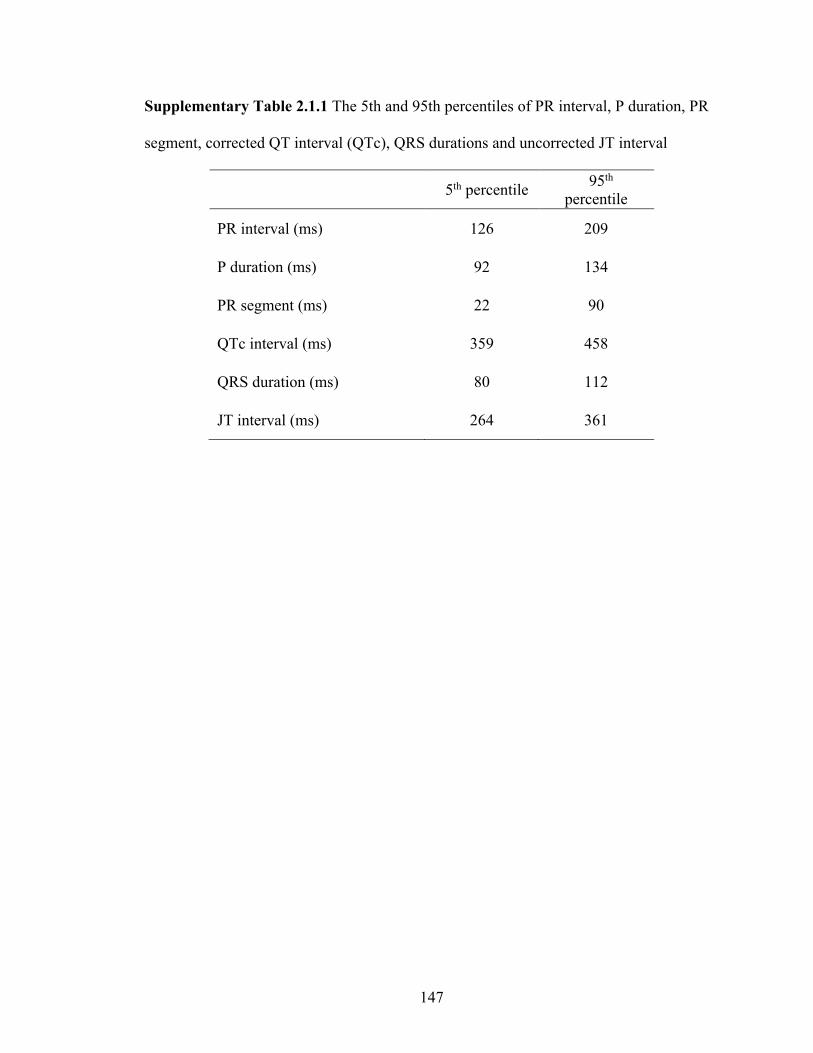
147
Supplementary Table 2.1.1 The 5th and 95th percentiles of PR interval, P duration, PR
segment, corrected QT interval (QTc), QRS durations and uncorrected JT interval
5th percentile 95th percentile
PR interval (ms) 126 209
P duration (ms) 92 134
PR segment (ms) 22 90
QTc interval (ms) 359 458
QRS duration (ms) 80 112
JT interval (ms) 264 361
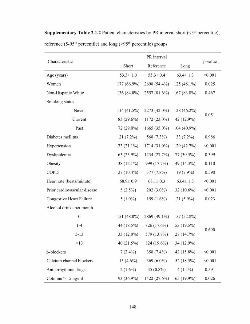
148
Supplementary Table 2.1.2 Patient characteristics by PR interval short (<5th percentile),
reference (5-95th percentile) and long (>95th percentile) groups
Characteristic PR interval
p-value Short Reference Long
Age (years) 53.3± 1.0 55.3± 0.4 63.4± 1.3 <0.001
Women 177 (66.9%) 2698 (54.4%) 125 (48.1%) 0.025
Non-Hispanic White 136 (84.0%) 2557 (81.6%) 167 (83.8%) 0.467
Smoking status
0.051 Never 114 (41.5%) 2273 (42.0%) 128 (46.2%)
Current 83 (29.6%) 1172 (23.0%) 42 (12.9%)
Past 72 (29.0%) 1665 (35.0%) 104 (40.9%)
Diabetes mellitus 21 (7.2%) 568 (7.3%) 33 (7.2%) 0.986
Hypertension 73 (21.1%) 1714 (31.0%) 129 (42.7%) <0.001
Dyslipidemia 63 (23.9%) 1234 (27.7%) 77 (30.5%) 0.399
Obesity 38 (12.1%) 999 (17.7%) 49 (14.3%) 0.110
COPD 27 (10.4%) 377 (7.8%) 19 (7.9%) 0.590
Heart rate (beats/minute) 68.9± 0.9 68.1± 0.3 63.4± 1.3 <0.001
Prior cardiovascular disease 5 (2.5%) 202 (3.0%) 32 (10.6%) <0.001
Congestive Heart Failure 5 (1.0%) 159 (1.6%) 21 (5.9%) 0.023
Alcohol drinks per month
0 151 (48.0%) 2869 (49.1%) 157 (52.8%)
0.690 1-4 44 (18.5%) 826 (17.6%) 53 (19.5%)
5-13 33 (12.0%) 579 (13.8%) 28 (14.7%)
>13 40 (21.5%) 824 (19.6%) 34 (12.9%)
b-blockers 7 (2.4%) 358 (7.4%) 42 (15.8%) <0.001
Calcium channel blockers 15 (4.6%) 369 (6.0%) 52 (18.5%) <0.001
Antiarrhythmic drugs 2 (1.6%) 45 (0.8%) 4 (1.4%) 0.591
Cotinine > 15 ng/ml 93 (36.9%) 1422 (27.6%) 65 (19.9%) 0.026

149
Supplementary Table 2.1.3 Patient characteristics by P duration short (<5th percentile),
reference (5-95th percentile) and long (>95th percentile) groups
Characteristic P duration
p-value Short Reference Long
Age (years) 52.5±0.9 55.5±0.4 61.8±1.2 <0.001
Women 187 (67.1%) 2714 (54.5%) 99 (43.0%) <0.001
Non-Hispanic White 173 (85.9%) 2547 (81.5%) 140 (82.3%) 0.257
Smoking status
0.684 Never 127 (43.2%) 2275 (42.1%) 113 (41.1%)
Current 67 (22.2%) 1187 (23.2%) 43 (17.9%)
Past 83 (34.6%) 1663 (34.7%) 95 (41.0%)
Diabetes mellitus 23 (5.2%) 568 (7.5%) 31 (7.2%) 0.460
Hypertension 68 (20.7%) 1732 (31.1%) 116 (45.0%) <0.001
Dyslipidemia 60 (21.6%) 1255 (27.9%) 59 (29.4%) 0.127
Obesity 38 (11.5%) 995 (17.5%) 53 (20.9%) 0.057
COPD 22 (6.6%) 384 (8.1%) 17 (5.4%) 0.346
Heart rate (beats/minute) 66.2±0.6 68.2±0.3 66.1±1.1 0.009
Prior cardiovascular disease 6 (2.0%) 209 (3.3%) 24 (5.7%) 0.114
Congestive Heart Failure 3 (0.2%) 162 (1.8%) 20 (3.9%) 0.021
Alcohol drinks per month
0 169 (54.3%) 2867 (48.7%) 141 (52.5%)
0.156 1-4 49 (21.4%) 838 (17.7%) 36 (12.4%)
5-13 26 (10.0%) 586 (14.0%) 28 (11.7%)
>13 33 (14.2%) 820 (19.5%) 45 (23.4%)
b-blockers 8 (2.6%) 361 (7.5%) 38 (16.2%) <0.001
Calcium channel blockers 12 (3.6%) 388 (6.4%) 36 (14.8%) <0.001
Antiarrhythmic drugs 3 (1.5%) 43 (0.8%) 5 (1.9%) 0.445
Cotinine > 15 ng/ml 79 (28.6%) 1436 (27.9%) 65 (24.2%) 0.718

150
Supplementary Table 2.1.4 Patient characteristics by PR segment short (<5th percentile),
reference (5-95th percentile) and long (>95th percentile) groups
Characteristic PR segment
p-value Short Reference Long
Age (years) 55.6±1.0 55.3± 0.4 60.2±1.4 0.003
Women 143 (60.8%) 2730 (54.7%) 127 (51.2%) 0.365
Non-Hispanic White 120 (82.2%) 2575 (81.7%) 165 (84.3%) 0.626
Smoking status
0.010 Never 99 (30.9%) 2299 (42.8%) 117 (40.8%)
Current 94 (35.6%) 1149 (22.5%) 54 (19.1%)
Past 79 (33.5%) 1664 (34.7%) 98 (40.1%)
Diabetes mellitus 27 (8.5%) 563 (7.3%) 32 (6.5%) 0.770
Hypertension 87 (29.2%) 1704 (30.6%) 125 (40.5%) 0.079
Dyslipidemia 64 (30.3%) 1241 (27.4%) 69 (28.9%) 0.744
Obesity 49 (12.1%) 989 (17.7%) 48 (14.2%) 0.139
COPD 19 (8.2%) 384 (7.8%) 20 (9.2%) 0.823
Heart rate (beats/minute) 70.5± 0.8 68.1± 0.3 63.6± 0.9 <0.001
Prior cardiovascular disease 6 (1.9%) 209 (3.1%) 24 (7.4%) 0.022
Congestive Heart Failure 6 (0.9%) 160 (1.7%) 19 (4.2%) 0.050
Alcohol drinks per month
0 140 (39.3%) 2883 (49.7%) 154 (48.7%)
0.690 1-4 39 (20.3%) 835 (17.6%) 49 (19.4%)
5-13 38 (15.6%) 574 (13.6%) 28 (13.5%)
>13 53 (24.7%) 809 (19.2%) 36 (18.4%)
b-blockers 15 (3.6%) 350 (7.3%) 42 (15.8%) 0.003
Calcium channel blockers 14 (4.2%) 375 (6.2%) 47 (15.5%) 0.002
Antiarrhythmic drugs 2 (0.8%) 47 (0.9%) 2 (0.8%) 0.999
Cotinine > 15 ng/ml 105 (41.2%) 1399 (27.3%) 76 (24.5%) 0.009
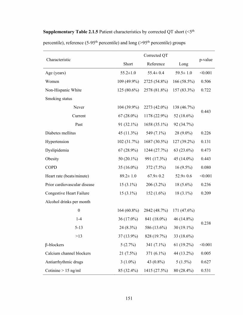
151
Supplementary Table 2.1.5 Patient characteristics by corrected QT short (<5th
percentile), reference (5-95th percentile) and long (>95th percentile) groups
Characteristic Corrected QT
p-value Short Reference Long
Age (years) 55.2±1.0 55.4± 0.4 59.5± 1.0 <0.001
Women 109 (49.9%) 2725 (54.8%) 166 (58.5%) 0.506
Non-Hispanic White 125 (80.6%) 2578 (81.8%) 157 (83.3%) 0.722
Smoking status
0.443 Never 104 (39.9%) 2273 (42.0%) 138 (46.7%)
Current 67 (28.0%) 1178 (22.9%) 52 (18.6%)
Past 91 (32.1%) 1658 (35.1%) 92 (34.7%)
Diabetes mellitus 45 (11.3%) 549 (7.1%) 28 (9.0%) 0.226
Hypertension 102 (31.7%) 1687 (30.5%) 127 (39.2%) 0.131
Dyslipidemia 67 (28.9%) 1244 (27.7%) 63 (23.6%) 0.473
Obesity 50 (20.1%) 991 (17.3%) 45 (14.0%) 0.443
COPD 35 (16.0%) 372 (7.5%) 16 (9.5%) 0.080
Heart rate (beats/minute) 89.2± 1.0 67.9± 0.2 52.9± 0.6 <0.001
Prior cardiovascular disease 15 (3.1%) 206 (3.2%) 18 (5.6%) 0.236
Congestive Heart Failure 15 (3.1%) 152 (1.6%) 18 (3.1%) 0.209
Alcohol drinks per month
0 164 (60.8%) 2842 (48.7%) 171 (47.6%)
0.238 1-4 36 (17.0%) 841 (18.0%) 46 (14.8%)
5-13 24 (8.3%) 586 (13.6%) 30 (19.1%)
>13 37 (13.9%) 828 (19.7%) 33 (18.6%)
b-blockers 5 (2.7%) 341 (7.1%) 61 (19.2%) <0.001
Calcium channel blockers 21 (7.5%) 371 (6.1%) 44 (13.2%) 0.005
Antiarrhythmic drugs 3 (1.0%) 43 (0.8%) 5 (1.5%) 0.627
Cotinine > 15 ng/ml 85 (32.4%) 1415 (27.5%) 80 (28.4%) 0.531

152
Supplementary Table 2.1.6 Patient characteristics by QRS duration short (<5th
percentile), reference (5-95th percentile) and long (>95th percentile) groups
Characteristic QRS duration
p-value Short Reference Long
Age (years) 56.8± 0.9 55.6± 0.4 53.6± 0.8 0.003
Women 204 (80.3%) 2726 (55.1%) 70 (22.7%) <0.001
Non-Hispanic White 144 (78.1%) 2599 (81.8%) 117 (86.1%) 0.223
Smoking status
0.197 Never 128 (44.5%) 2285 (42.1%) 102 (41.3%)
Current 65 (28.4%) 1163 (22.5%) 69 (25.3%)
Past 72 (27.0%) 1680 (35.4%) 89 (33.4%)
Diabetes mellitus 46 (11.5%) 548 (7.1%) 28 (7.3%) 0.205
Hypertension 91 (34.1%) 1730 (30.9%) 95 (29.9%) 0.704
Dyslipidemia 72 (30.7%) 1248 (27.5%) 54 (26.4%) 0.684
Obesity 44 (12.2%) 987 (17.6%) 55 (15.9%) 0.188
COPD 29 (10.6%) 372 (7.5%) 22 (13.6%) 0.293
Heart rate (beats/minute) 69.7± 0.9 68.0± 0.2 65.3± 1.1 0.003
Prior cardiovascular disease 8 (2.1%) 220 (3.3%) 11 (3.7%) 0.566
Congestive Heart Failure 14 (1.3%) 161 (1.8%) 10 (2.1%) 0.618
Alcohol drinks per month
0 176 (61.2%) 2876 (48.9%) 125 (41.2%)
0.032 1-4 37 (14.6%) 841 (17.9%) 45 (18.3%)
5-13 25 (10.2%) 583 (14.0%) 32 (11.0%)
>13 27 (14.0%) 814 (19.1%) 57 (29.5%)
b-blockers 12 (4.6%) 376 (7.6%) 19 (8.9%) 0.301
Calcium channel blockers 20 (5.6%) 396 (6.5%) 20 (7.9%) 0.584
Antiarrhythmic drugs 1 (0.1%) 45 (0.8%) 5 (2.2%) 0.137
Cotinine > 15 ng/ml 79 (31.2%) 1416 (27.3%) 85 (33.0%) 0.213
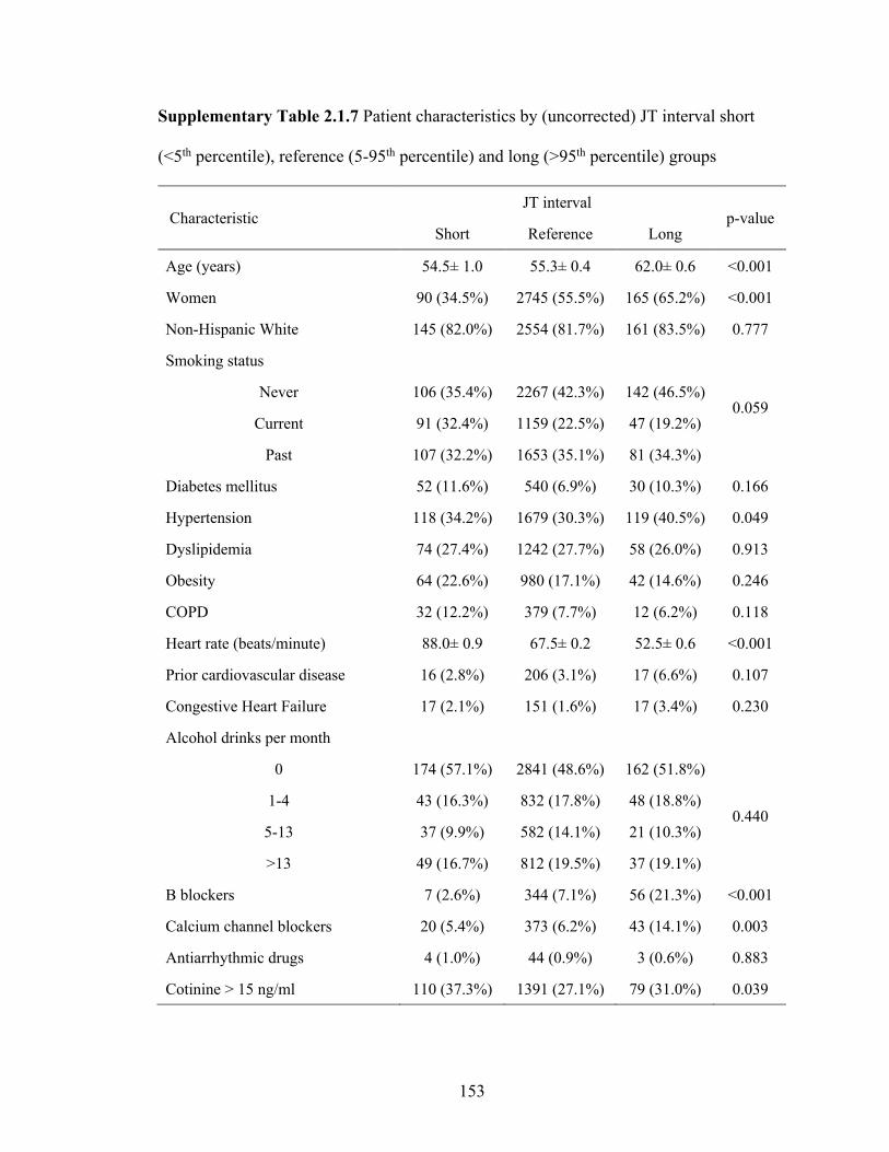
153
Supplementary Table 2.1.7 Patient characteristics by (uncorrected) JT interval short
(<5th percentile), reference (5-95th percentile) and long (>95th percentile) groups
Characteristic JT interval
p-value Short Reference Long
Age (years) 54.5± 1.0 55.3± 0.4 62.0± 0.6 <0.001
Women 90 (34.5%) 2745 (55.5%) 165 (65.2%) <0.001
Non-Hispanic White 145 (82.0%) 2554 (81.7%) 161 (83.5%) 0.777
Smoking status
0.059 Never 106 (35.4%) 2267 (42.3%) 142 (46.5%)
Current 91 (32.4%) 1159 (22.5%) 47 (19.2%)
Past 107 (32.2%) 1653 (35.1%) 81 (34.3%)
Diabetes mellitus 52 (11.6%) 540 (6.9%) 30 (10.3%) 0.166
Hypertension 118 (34.2%) 1679 (30.3%) 119 (40.5%) 0.049
Dyslipidemia 74 (27.4%) 1242 (27.7%) 58 (26.0%) 0.913
Obesity 64 (22.6%) 980 (17.1%) 42 (14.6%) 0.246
COPD 32 (12.2%) 379 (7.7%) 12 (6.2%) 0.118
Heart rate (beats/minute) 88.0± 0.9 67.5± 0.2 52.5± 0.6 <0.001
Prior cardiovascular disease 16 (2.8%) 206 (3.1%) 17 (6.6%) 0.107
Congestive Heart Failure 17 (2.1%) 151 (1.6%) 17 (3.4%) 0.230
Alcohol drinks per month
0 174 (57.1%) 2841 (48.6%) 162 (51.8%)
0.440 1-4 43 (16.3%) 832 (17.8%) 48 (18.8%)
5-13 37 (9.9%) 582 (14.1%) 21 (10.3%)
>13 49 (16.7%) 812 (19.5%) 37 (19.1%)
Β blockers 7 (2.6%) 344 (7.1%) 56 (21.3%) <0.001
Calcium channel blockers 20 (5.4%) 373 (6.2%) 43 (14.1%) 0.003
Antiarrhythmic drugs 4 (1.0%) 44 (0.9%) 3 (0.6%) 0.883
Cotinine > 15 ng/ml 110 (37.3%) 1391 (27.1%) 79 (31.0%) 0.039

154
Supplementary Table 2.1.8 Association between serum cotinine levels (dichotomous)
and abnormal PR segment, QRS duration and JT interval vs the reference group among
subgroups by age and sex
Subgroups
PR segment <5th percentile vs reference
QRS duration <5th percentile vs reference
JT interval >95th percentile vs
reference Odds ratio (95% CI)
Interaction p-value
Odds ratio (95% CI)
Interaction p-value
Odds ratio (95% CI)
Interaction p-value
Age < 59 years
1.92 (1.08, 3.42)
0.5661
1.82 (1.05, 3.15)
0.3070
1.69 (0.80, 3.58)
0.4007 ≥ 59 years
2.17 (1.14, 4.13)
1.22 (0.76, 1.95)
1.18 (0.64, 2.19)
Sex Male 2.26
(1.13, 4.50) 0.7550
0.76 (0.29, 1.96)
0.0589
1.49 (0.72, 3.11)
0.5171 Female 1.82
(1.03, 3.20) 2.03
(1.37, 3.00) 1.74
(0.92, 3.31)
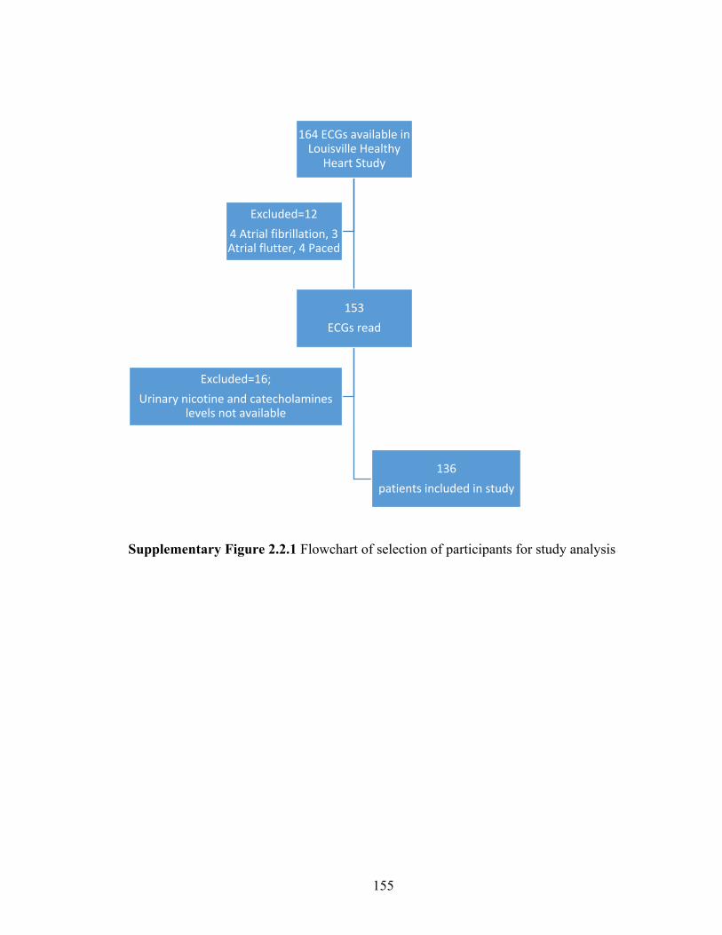
155
Supplementary Figure 2.2.1 Flowchart of selection of participants for study analysis
164 ECGs available in Louisville Healthy
Heart Study
153ECGs read
136 patients included in study
Excluded=16;Urinary nicotine and catecholamines
levels not available
Excluded=124 Atrial fibrillation, 3 Atrial flutter, 4 Paced
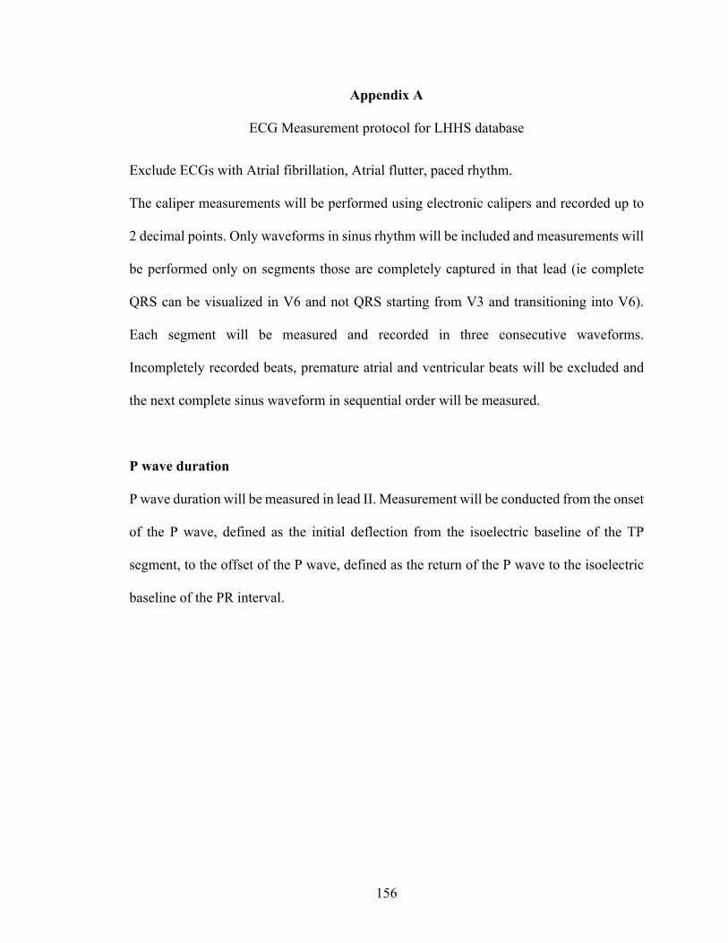
156
Appendix A
ECG Measurement protocol for LHHS database
Exclude ECGs with Atrial fibrillation, Atrial flutter, paced rhythm.
The caliper measurements will be performed using electronic calipers and recorded up to
2 decimal points. Only waveforms in sinus rhythm will be included and measurements will
be performed only on segments those are completely captured in that lead (ie complete
QRS can be visualized in V6 and not QRS starting from V3 and transitioning into V6).
Each segment will be measured and recorded in three consecutive waveforms.
Incompletely recorded beats, premature atrial and ventricular beats will be excluded and
the next complete sinus waveform in sequential order will be measured.
P wave duration
P wave duration will be measured in lead II. Measurement will be conducted from the onset
of the P wave, defined as the initial deflection from the isoelectric baseline of the TP
segment, to the offset of the P wave, defined as the return of the P wave to the isoelectric
baseline of the PR interval.
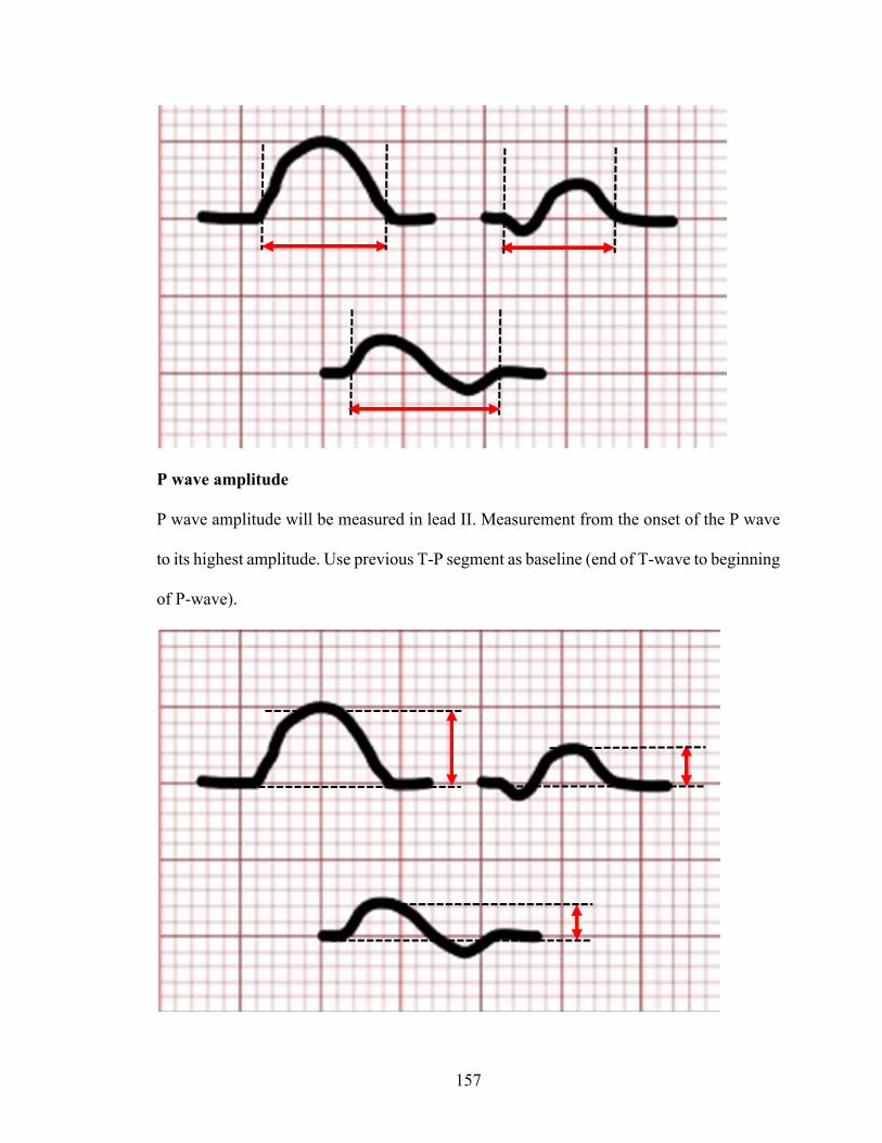
157
P wave amplitude
P wave amplitude will be measured in lead II. Measurement from the onset of the P wave
to its highest amplitude. Use previous T-P segment as baseline (end of T-wave to beginning
of P-wave).

158
PR wave duration
PR duration will be measured in lead II. The PR interval was measured from the onset of
the P wave to the onset of the QRS complex, defined as the initial deflection from the
baseline of the PR interval.

159
QT interval
The QT interval was measured in lead V5, and determined as the onset of the QRS complex
to the end of the T wave; defined as the return of the T wave to the isoelectric baseline of
the TP segment.
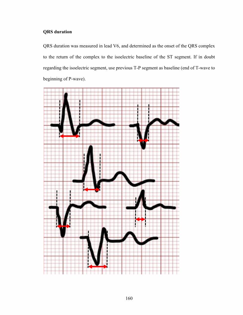
160
QRS duration
QRS duration was measured in lead V6, and determined as the onset of the QRS complex
to the return of the complex to the isoelectric baseline of the ST segment. If in doubt
regarding the isoelectric segment, use previous T-P segment as baseline (end of T-wave to
beginning of P-wave).
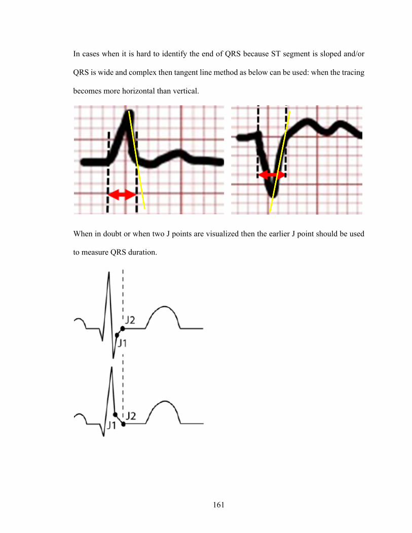
161
In cases when it is hard to identify the end of QRS because ST segment is sloped and/or
QRS is wide and complex then tangent line method as below can be used: when the tracing
becomes more horizontal than vertical.
When in doubt or when two J points are visualized then the earlier J point should be used
to measure QRS duration.
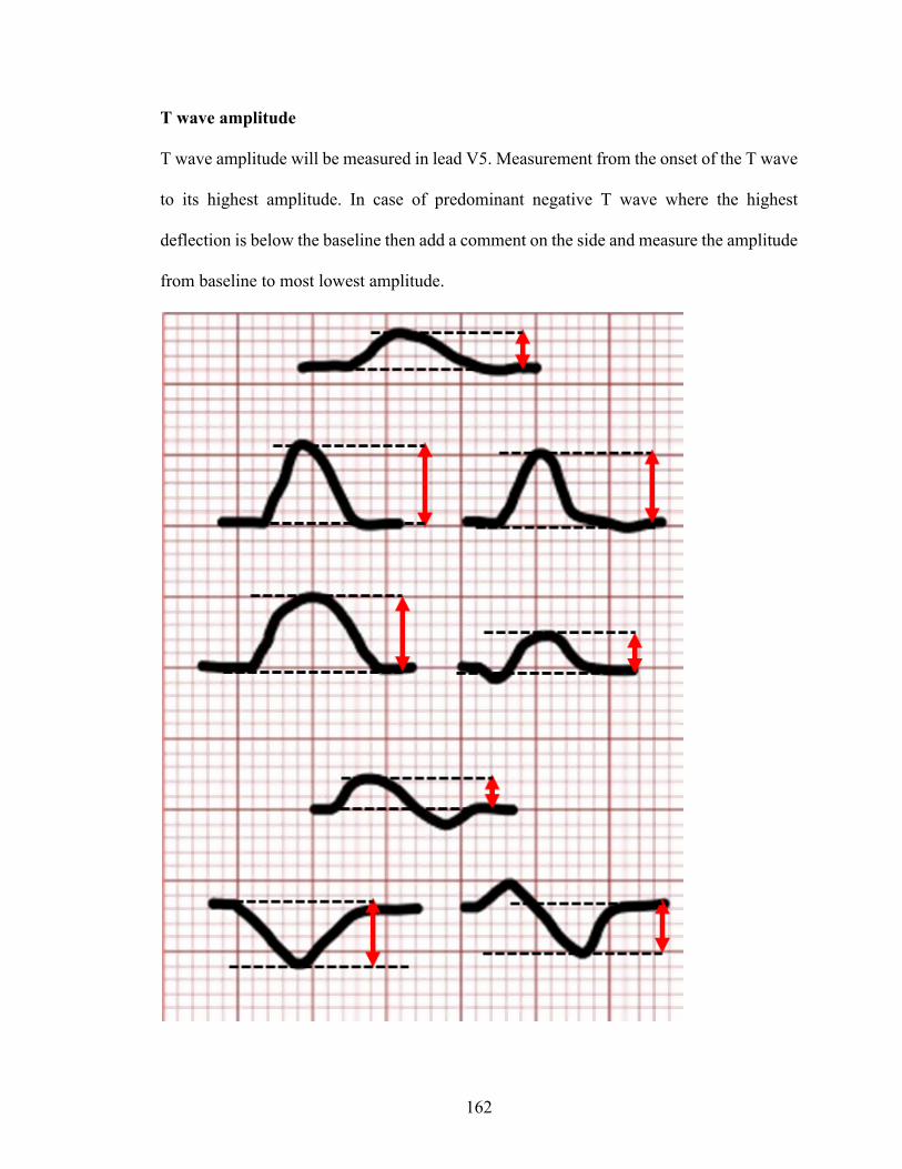
162
T wave amplitude
T wave amplitude will be measured in lead V5. Measurement from the onset of the T wave
to its highest amplitude. In case of predominant negative T wave where the highest
deflection is below the baseline then add a comment on the side and measure the amplitude
from baseline to most lowest amplitude.

163
T peak – T end
T wave amplitude will be measured in lead V5. Measurement will be conducted peak of
T-wave to the offset of the T wave, defined as the return of the T wave to the isoelectric
baseline (TP segment as described above).

164
In cases when it is hard to identify the end of T then tangent line method as below can be
used.
R-R intervals
Use the lead which has the longest continuous ECG recording, which is usually at the
bottom of the ECG sheet. Use preferably Lead II if available otherwise use V1. Measure
the duration between all R-R intervals. If R is not present then use any identical points on
consecutive EKG waveforms such as the Q-Q or P-P duration. Do not measure R-R
between sinus and a premature atrial or ventricular beat (defined as beat occurring 20%
earlier to previous interval) and measure the subsequent waveforms.

165
References for ECG Measurement protocol
Prineas RJ, Crow RS Zhang ZM. The Minnesota Code Manual of Electrocardiographic
Findings. 2nd Edition, London: Springer, 2010.
Jacob Melgaard et al. Automatic J-point Location in Subjects with Electrocardiographic
Early Repolarization Computing in Cardiology 2014; 41:585-588
Considerations about the polemic J point location
http://bscw.rediris.es/pub/bscw.cgi/d5046031/CONSIDERACIONES%20ACERCA%20
DE%20LA%20LOCALIZACI%C3%93N%20POL%C3%89MICA%20DEL%20
PUNTO%20J.pdf
Burke GM et al. J Electrocardiol. 2014 May-Jun;47(3):288-93. doi:
10.1016/j.jelectrocard.2014.01.004. Epub 2014 Jan 6. Assessment of
reproducibility--automated and digital caliper ECG measurement in the
Framingham Heart Study.
Tarek MohamedAbdelrahman The Egyptian Journal of Critical Care Medicine Volume 2,
Issue 1, April 2014, Pages 19-27. Prognostic value of T peak-to-end interval for
risk stratification after acute myocardial infarction
Erikssen G et al. Ann Noninvasive Electrocardiol. 2012 Apr;17(2):85-94. doi:
10.1111/j.1542-474X.2012.00493.x. The terminal part of the QT interval (T peak
to T end): a predictor of mortality after acute myocardial infarction.

166
Supplementary Table 2.2.1 Baseline characteristics by high and low PR interval, P wave duration and PR segment
PR interval P-wave duration PR segment ≤ 163.32
ms > 163.33+
ms p value ≤ 104.73ms
> 104.74 ms p value ≤ 55.20
ms > 55.21
ms p value
Age (years)* 49, 11 55, 8 0.01 51, 11 54, 8 0.11 51, 10 53, 9 0.18 BMI (kg/m2)* 31, 7 34, 9 0.07 31, 7 35, 8 0.01 32, 7 34, 9 0.10 SBP (mm Hg)* 133, 21 134, 25 0.95 129, 21 138, 25 0.07 135, 22 132, 24 0.13 DBP (mm Hg)* 80, 10 81, 15 0.52 78, 12 84, 14 0.05 82, 11 79, 15 0.16 Heart rate (beats/min)* 75, 17 71, 13 0.27 73, 16 73, 13 0.53 75, 16 72, 14 0.28 Male gender 27, 42% 45, 63% 0.01 31, 44% 41, 64% 0.02 33, 52% 39, 55% 0.70 Caucasian 37, 58% 40, 56% 0.86 44, 62% 33, 52% 0.22 39, 61% 38, 54% 0.39 Hypertension 54, 84% 64, 90% 0.31 57, 80% 61, 95% 0.01 55, 86% 63, 89% 0.63 Prior MI 26, 41% 36, 51% 0.24 31, 44% 31, 48% 0.58 32, 50% 30, 42% 0.37 Diabetes 18, 28% 25, 35% 0.38 20, 28% 23, 36% 0.33 17, 27% 26, 37% 0.21 Stroke 13, 20% 10, 14% 0.34 15, 21% 8, 13% 0.18 10, 16% 13, 18% 0.68 Arrhythmia 22, 34% 23, 32% 0.81 25, 35% 20, 31% 0.63 20, 31% 25, 35% 0.63 Β blocker 43, 67% 55, 78% 0.18 51, 72% 47, 73% 0.83 42, 66% 56, 79% 0.09 CCB 13, 20% 19, 27% 0.38 10, 14% 22, 34% 0.01 13, 20% 19, 27% 0.38 ACEI or ARB 38, 59% 51, 72% 0.13 39, 55% 50, 78% 0.01 44, 69% 45, 63% 0.51 Statin 41, 64% 43, 61% 0.68 45, 63% 39, 61% 0.77 41, 64% 43, 61% 0.68 Aspirin 35, 55% 46, 65% 0.23 43, 61% 38, 59% 0.89 34, 53% 47, 66% 0.12 Diuretics 24, 38% 34, 48% 0.22 26, 37% 32, 50% 0.12 21, 33% 37, 52% 0.02
Abbreviations: BMI - Body mass index, SBP- Systolic Blood Pressure, DBP – Diastolic Blood Pressure, MI – Myocardial Infarction, CCB – Calcium Channel Blocker, ACE - Angiotensin-Converting Enzyme Inhibitor, ARB - Angiotensin II Receptor Blocker. For each variable counts and column percentage has been reported except for where indicated * where the mean and standard deviation (SD) are reported
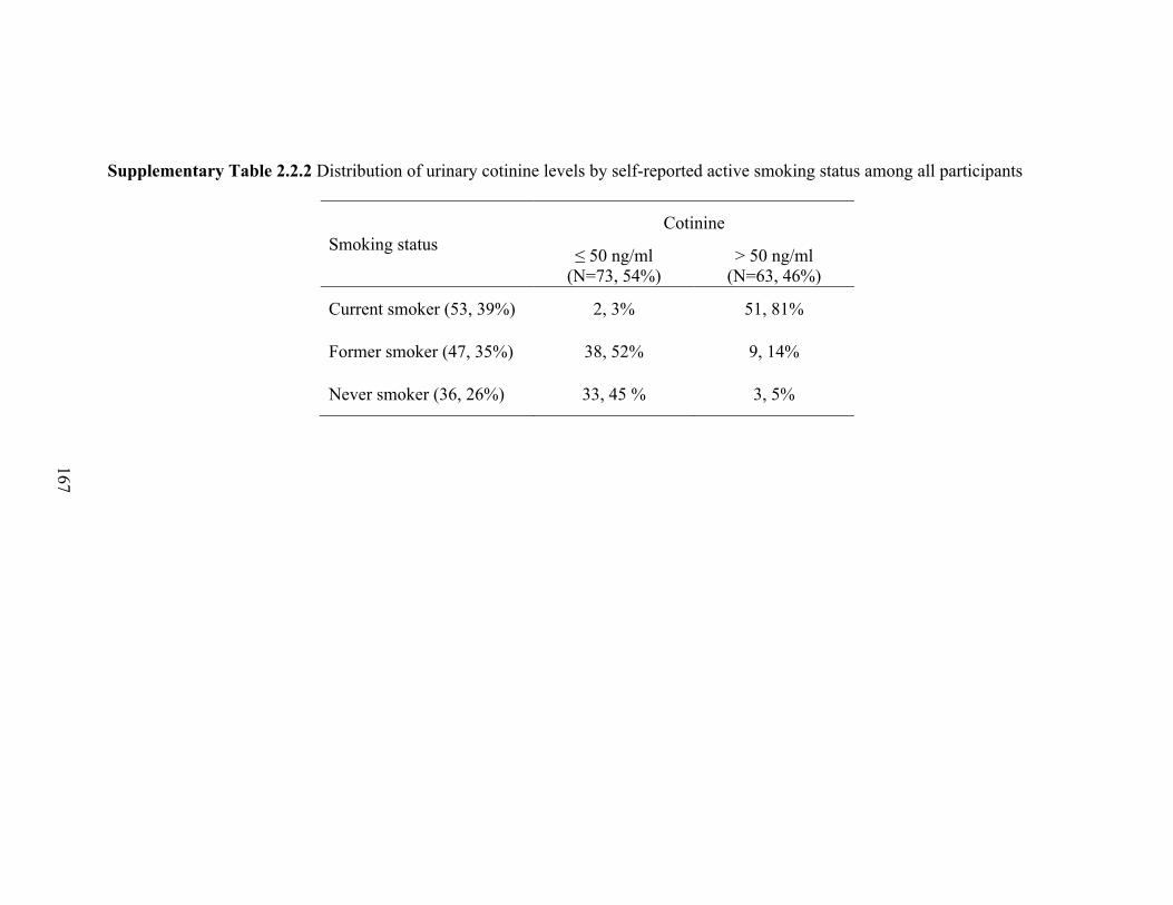
167
Supplementary Table 2.2.2 Distribution of urinary cotinine levels by self-reported active smoking status among all participants
Smoking status Cotinine
≤ 50 ng/ml (N=73, 54%)
> 50 ng/ml (N=63, 46%)
Current smoker (53, 39%) 2, 3% 51, 81%
Former smoker (47, 35%) 38, 52% 9, 14%
Never smoker (36, 26%) 33, 45 % 3, 5%

168
Supplementary Table 2.2.3 Comparison among participants stratified by self-reported smoking status or by cotinine level of (A)
urinary cotinine and catecholamines (median [interquartile range], creatinine-normalized), and (B) high and low PR interval, P wave
duration and PR segment
(A)
Cotinine (ng/g)
Dopamine (μg/g)
Epinephrine (μg/g)
Norepinephrine (μg/g)
p value
p value
p value
p value
Smoking status
Active smoker
827.1 [527.3 - 1445.3] <0.001 176.0
[146.5 - 225.8] 0.24 5.7 [3.3 - 10.8] 0.31 42.5
[30.7 - 60.3] 0.07
Former-smoker*
3.7 [1.3 - 18.9] - 167.1
[135.6 - 204.9] - 4.2 [2.6 - 8.5] - 36.2
[26.4 - 51.4] -
Never smoker
1.6 [0.9 - 4.0] <0.001 139.7
[114.3 - 169.7] 0.001 3.7 [1.5 - 6.5] 0.03 32.4
[19.0 - 49.2] 0.02
Cotinine ≤ 50 ng/ml 1.8
[1.0 - 4.2] <0.001
160.0 [ 112.4 - 189.1] 0.003
4.1 [2.1 - 6.9] 0.08
33.4 [22.7 - 50.6] 0.02
> 50 ng/ml 827.1 [522.8 - 1596.0]
175.8 [142.0 - 227.9]
5.9 [3.1 - 9.2]
42.5 [30.4 - 56.4]

169
(B)
PR interval P wave duration PR segment
≤ 163.32 ms > 163.33 ms p value ≤ 104.73 ms 104.74 ms p
value ≤ 55.20 ms 55.21 ms p value
Smoking status
Current smoker 25, 39% 28, 39%
0.76
34, 48% 19, 30%
0.08
26, 40% 27, 38%
0.58 Former smoker 24, 38% 23, 32% 20, 28% 27, 42% 24, 38% 23, 32%
Never smoker 15, 23% 20, 28% 17, 24% 18, 28% 14, 22% 21, 30%
Cotinine ≤ 50 ng/ml 34, 53% 38, 54%
0.96 34, 48% 38, 59%
0.18 32, 50% 40, 56%
0.49 > 50 ng/ml 30, 47% 33, 46% 37, 52% 26, 41% 32, 50% 31, 44%
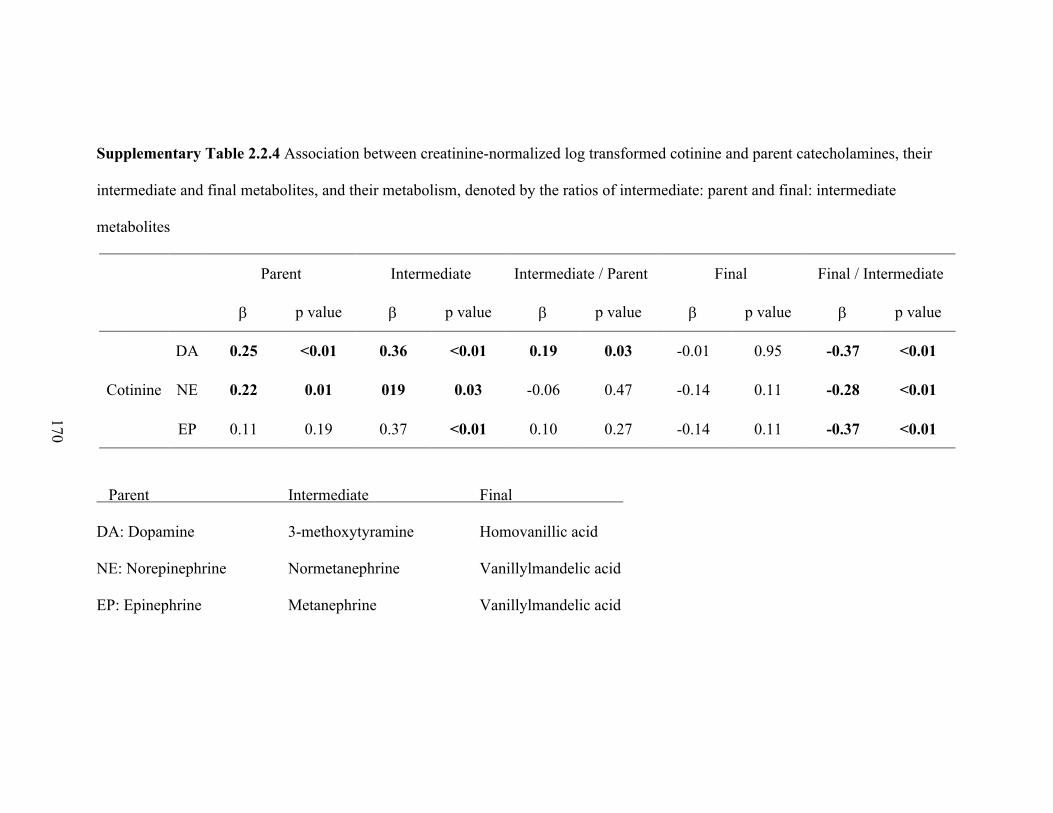
170
Supplementary Table 2.2.4 Association between creatinine-normalized log transformed cotinine and parent catecholamines, their
intermediate and final metabolites, and their metabolism, denoted by the ratios of intermediate: parent and final: intermediate
metabolites
Parent Intermediate Intermediate / Parent Final Final / Intermediate
b p value b p value b p value b p value b p value
Cotinine
DA 0.25 <0.01 0.36 <0.01 0.19 0.03 -0.01 0.95 -0.37 <0.01
NE 0.22 0.01 019 0.03 -0.06 0.47 -0.14 0.11 -0.28 <0.01
EP 0.11 0.19 0.37 <0.01 0.10 0.27 -0.14 0.11 -0.37 <0.01
Parent Intermediate Final
DA: Dopamine 3-methoxytyramine Homovanillic acid
NE: Norepinephrine Normetanephrine Vanillylmandelic acid
EP: Epinephrine Metanephrine Vanillylmandelic acid

171
Supplementary Table 2.2.5 Associations of QRS duration, QT interval and corrected QT with urinary cotinine in unadjusted and
adjusted linear regressions
Urinary cotinine was normalized by urine creatinine and log-transformed
QRS QT Corrected QT
b p value b p value b p value
Unadjusted Cotinine 0.32 0.80 4.71 0.11 3.48 0.14
*Adjusted Cotinine -0.34 0.78 3.33 0.78 0.11 0.13
* models adjusted for diastolic blood pressure and prior myocardial infarction (QRS); age, heart rate, systolic blood pressure, and calcium channel blocker (QT);age (QTc)
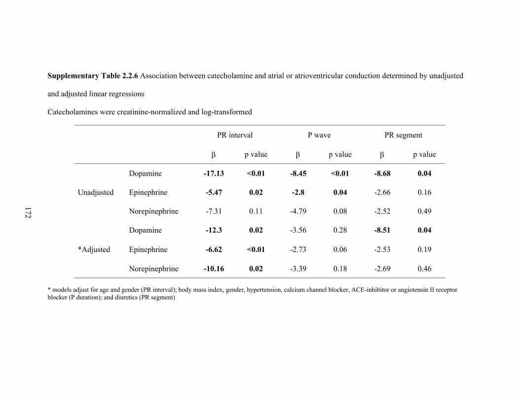
172
Supplementary Table 2.2.6 Association between catecholamine and atrial or atrioventricular conduction determined by unadjusted
and adjusted linear regressions
Catecholamines were creatinine-normalized and log-transformed
PR interval P wave PR segment
b p value b p value b p value
Unadjusted
Dopamine -17.13 <0.01 -8.45 <0.01 -8.68 0.04
Epinephrine -5.47 0.02 -2.8 0.04 -2.66 0.16
Norepinephrine -7.31 0.11 -4.79 0.08 -2.52 0.49
*Adjusted
Dopamine -12.3 0.02 -3.56 0.28 -8.51 0.04
Epinephrine -6.62 <0.01 -2.73 0.06 -2.53 0.19
Norepinephrine -10.16 0.02 -3.39 0.18 -2.69 0.46
* models adjust for age and gender (PR interval); body mass index, gender, hypertension, calcium channel blocker, ACE-inhibitor or angiotensin II receptorblocker (P duration); and diuretics (PR segment)

173
Supplementary Table 3.1 Baseline characteristics
Smoker ID Age Gender Race Body Mass Index
b-Blocker protocol
Sham- protocol
Pack-Years Cigarette brand Nicotine
content (mg) 1 26 Male White 31 Yes Yes 9 L&M 0.8 2 28 Male Black 29 Yes - 36 L&M 0.9 3 23 Female Black 29 Yes Yes 5 L&M 0.9 4 32 Male Black 23 - Yes 29 Newport 0.8 5 31 Male White 37 - - 15 Marlboro 1.1 6 39 Female White 25 - - 22 Pall Mall 0.9 7 23 Male White 24 Yes Yes 7 L&M 0.9 8 22 Male Black 22 - - 6 L&M 0.8 9 25 Male White 24 - - 7 Camel 0.8 10 25 Female White 30 - - 11 USA Gold Reds 0.8 11 52 Male White 25 Yes - 22 High Card 0.8 12 42 Female White 29 Yes Yes 60 High Card 0.8 13 53 Female White 34 Yes - 42 Pall Mall 1.1 14 43 Male White 30 Yes - 18 American Spirit 0.6 15 49 Female Other 21 Yes - 35 High Card 0.8 16 62 Male Black 28 Yes Yes 20 Newport 0.8 17 61 Male White 21 - Yes 48 Kool 0.6 18 39 Male White 26 - Yes 25 Marlboro 1.0 19 43 Female White 32 - Yes 29 Camel 1.0 20 28 Male Other 34 - Yes 14 Pall Mall 0.9
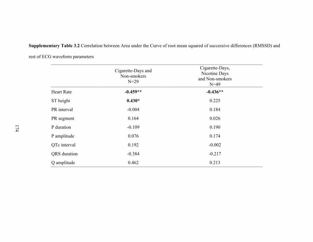
174
Supplementary Table 3.2 Correlation between Area under the Curve of root mean squared of successive differences (RMSSD) and
rest of ECG waveform parameters
Cigarette-Days and Non-smokers
N=29
Cigarette-Days, Nicotine Days
and Non-smokers N=49
Heart Rate -0.459** -0.436**
ST height 0.430* 0.225
PR interval -0.004 0.184
PR segment 0.164 0.026
P duration -0.109 0.190
P amplitude 0.076 0.174
QTc interval 0.192 -0.002
QRS duration -0.384 -0.217
Q amplitude 0.462 0.213

175
A)
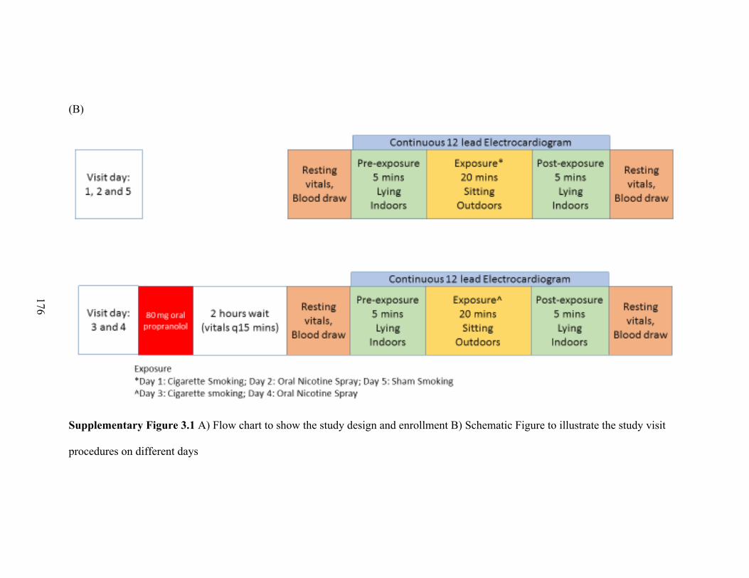
176
(B)
Supplementary Figure 3.1 A) Flow chart to show the study design and enrollment B) Schematic Figure to illustrate the study visit
procedures on different days
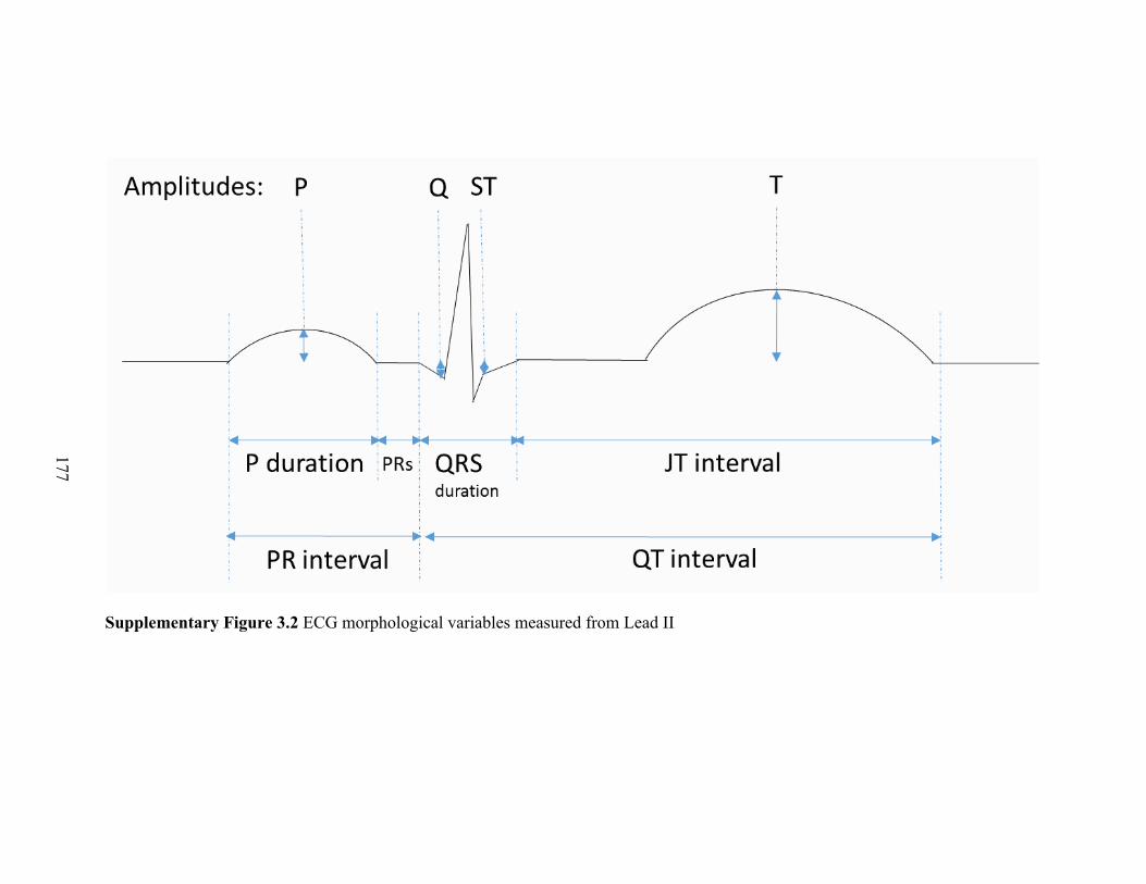
177
Supplementary Figure 3.2 ECG morphological variables measured from Lead II

178
«: <0.05 vs Sham#: <0.05 vs Nicotineβ: <0.05 vs non-Beta-blocker
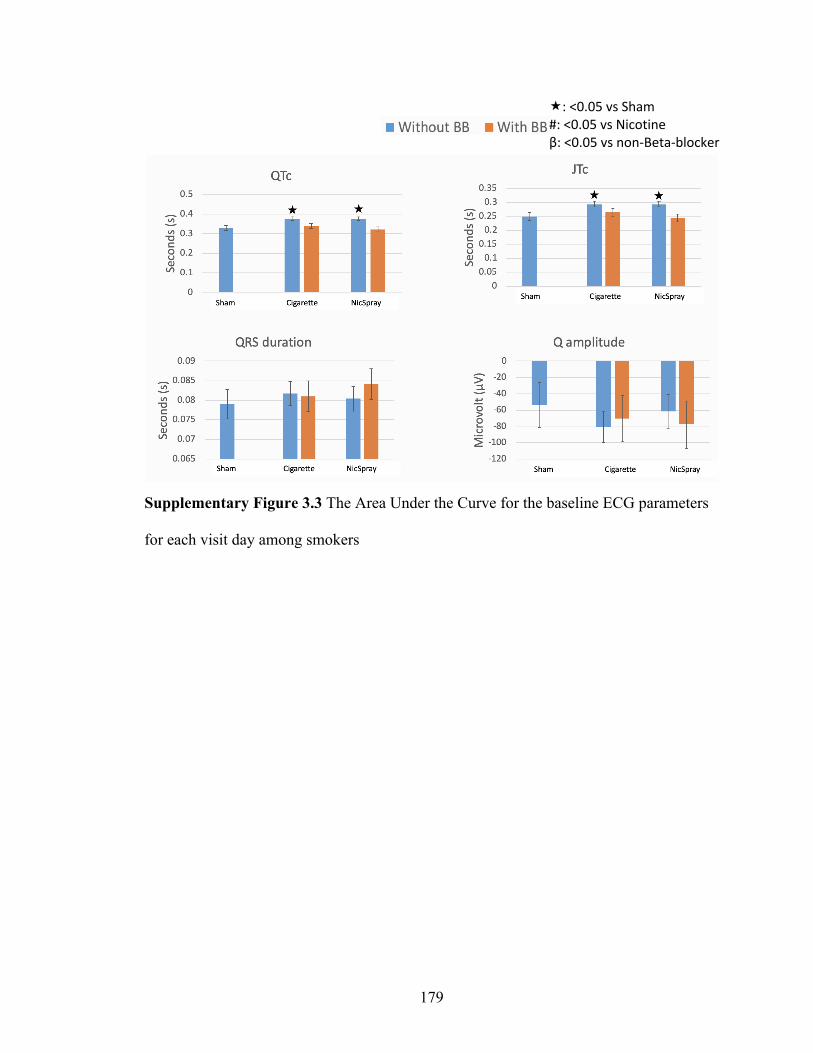
179
«: <0.05 vs Sham#: <0.05 vs Nicotineβ: <0.05 vs non-Beta-blocker
Supplementary Figure 3.3 The Area Under the Curve for the baseline ECG parameters
for each visit day among smokers
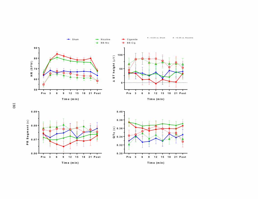
180
P re 3 6 9 1 2 1 5 1 8 2 1 P o s t5 0
6 0
7 0
8 0
9 0
T im e (m in )
HR
(BP
M)
P re 3 6 9 1 2 1 5 1 8 2 1 P o s t
0
5 0
1 0 0
T im e (m in )
D S
T h
eig
ht
(µV
)
P re 3 6 9 1 2 1 5 1 8 2 1 P o s t0 .0 6
0 .0 7
0 .0 8
0 .0 9
T im e (m in )
PR
Se
gm
en
t(s
)
P re 3 6 9 1 2 1 5 1 8 2 1 P o s t0 .3 0
0 .3 2
0 .3 4
0 .3 6
0 .3 8
0 .4 0
T im e (m in )
QT
c(s
)
S h am C ig a re tte« : < 0 .0 5 v s . S h a m # : < 0 .0 5 v s . N ic o t in e
N ic o t in eB B -N ic B B -C ig

181
Supplementary Figure 3.4 The raw values of several ECG parameters in pre-post and during exposure days among smokers

182
Δ Δ%
Supplementary Figure 3.5 The raw (Δ) and relative (Δ%) changes with standard errors of nicotine, Cotinine and 3-hyroycotinine (3-
OH) levels from baseline per study visit
Difference between pre and post exposure:
Nicotine Cotinine 3HC (3-hyoxycotinine)
Cig NicS BB-Cig BB-NicS Non-Smoker Cig NicS BB-Cig BB-NicS Non-Smoker

183
Supplementary Figure 3.6 The relationship between Area Under the Curves (AUC) of ECG parameters (Heart Rate, PR segment, ST
height and root mean squared of successive differences (RMSSD)), with changes in Nicotine and Baseline Cotinine levels
X axis - Δ Nicotine (ng/ml) Y axis – AUC of Δ ECG parameter Z axis – Relative size of bubble indicates baseline cotinine level
-200
0
200
400
600
-10 0 10 20 30
Heart Rate
-0.8
-0.6
-0.4
-0.2
0
0.2
0.4
0.6
-10 0 10 20 30
PR segment
-1500
-1000
-500
0
500
1000
-10 0 10 20 30
RMSSD
-6000
-4000
-2000
0
2000
4000
-10 0 10 20 30
ST height

184
(A)
Supplementary Figure 3.7 The changes in Blood Pressure post cigarette and Nicotine
Spray use (A) and their correlation with change in nicotine levels (B)
There is no significant difference in pre-exposure and post exposure Blood pressure
between Cig and NicS days (p>0.05).
(B)
20 smokers (Cig-day and NicS-day) and
9 non-smokers
β-Blocker Cigarette and β-Blocker Nicotine days
N=20
Systolic Blood Pressure 0.438 P = 0.001
0.180 P = 0.447
Diastolic Blood Pressure 0.374 P =0.008
-0.160 P =0.500
«: <0.05 vs Pre-exposure
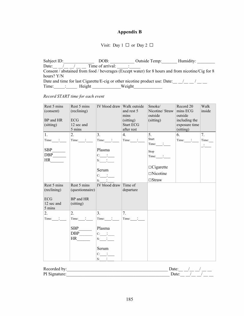
185
Appendix B
Visit: Day 1 □ or Day 2 □
Subject ID:______________ DOB: ___________ Outside Temp:_______ Humidity: ________ Date:__ __/__ __/ __ __ Time of arrival: _____:_____ Consent / abstained from food / beverages (Except water) for 8 hours and from nicotine/Cig for 8 hours? Y/N Date and time for last Cigarette/E-cig or other nicotine product use: Date:__ __/__ __ / __ __ Time:_____:_____ Height _____________Weight_____________
Record START time for each event
Rest 5 mins (consent)
BP and HR (sitting)
Rest 5 mins (reclining)
ECG 12 sec and 5 mins
IV blood draw Walk outside and rest 5 mins (sitting) Start ECG after rest
Smoke/ Nicotine/ Straw outside (sitting)
Record 20 mins ECG outside including the exposure time (sitting)
Walk inside
1. Time:___:___
SBP______ DBP______ HR______
2. Time:___:___
3. Time:___:___
Plasma C:___:___ S:___:___
Serum C:___:___ S:___:___
4. Time:___:___
5. Start Time:___:___
Stop Time:___:___
□Cigarette□Nicotine□Straw
6. Time:___:___
7. Time:___:___
Rest 5 mins (reclining)
ECG 12 sec and 5 mins
Rest 5 mins (questionnaire)
BP and HR (sitting)
IV blood draw Time of departure
2. Time:___:___
2. Time:___:___
SBP______ DBP______ HR______
3. Time:___:___
Plasma C:___:___ S:___:___
Serum C:___:___ S:___:___
7. Time:___:___
Recorded by:______________________________________________ Date:__ __/__ __/ __ __ PI Signature:_______________________________________________ Date:__ __/__ __/ __ __

186
Visit: Day 3 □ or Day 4 □Subject ID:______________ DOB: ___________ Outside Temp:_______ Humidity: ________ Date:__ __/__ __/ __ __ Time of arrival: _____:_____ Consent / abstained from food / beverages (Except water) for 8 hours and from nicotine/Cig for 8 hours? Y/N Date and time for last Cigarette/E-cig or other nicotine product use: Date:__ __/__ __ / __ __ Time:_____:_____ Height _____________Weight_____________ 100 mg propranolol ______:_______
BP and HR every 15 mins for 2 hours
Rest 5 mins (consent)
BP and HR (sitting)
Rest 5 mins (reclining)
ECG 12 sec and 5 mins
IV blood draw Walk outside and rest 5 mins (sitting) Start ECG after rest
Smoke/ Nicotine/ Straw outside (sitting)
Record 20 mins ECG outside including the exposure time (sitting)
Walk inside
1. Time:___:___
SBP______ DBP______ HR______
2. Time:___:___
3. Time:___:___
Plasma C:___:___ S:___:___
Serum C:___:___ S:___:___
4. Time:___:___
5. Start Time:___:___
Stop Time:___:___
□Cigarette□Nicotine□Straw
6. Time:___:___
7. Time:___:___
Rest 5 mins (reclining)
ECG 12 sec and 5 mins
Rest 5 mins (questionnaire)
BP and HR (sitting)
IV blood draw Time of departure
2. Time:___:___
2. Time:___:___
SBP______ DBP______ HR______
3. Time:___:___
Plasma C:___:___ S:___:___
Serum C:___:___ S:___:___
7. Time:___:___
Recorded by:_______________________________________________Date:__ __/__ __/ __ __ PI Signature:________________________________________________Date:__ __/__ __/ __ __
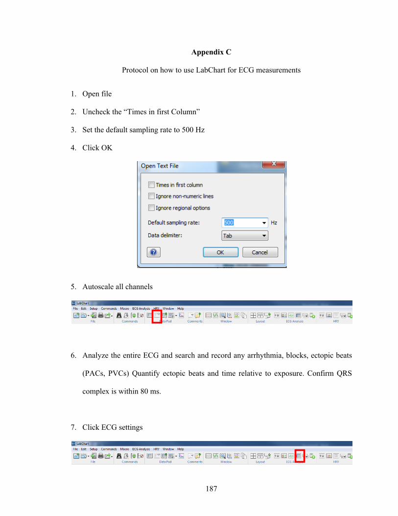
187
Appendix C
Protocol on how to use LabChart for ECG measurements
1. Open file
2. Uncheck the “Times in first Column”
3. Set the default sampling rate to 500 Hz
4. Click OK
5. Autoscale all channels
6. Analyze the entire ECG and search and record any arrhythmia, blocks, ectopic beats
(PACs, PVCs) Quantify ectopic beats and time relative to exposure. Confirm QRS
complex is within 80 ms.
7. Click ECG settings

188
8. Click ECG Beat Classifier View
Select Channel
Change to time and
select 60 s
Select Framingham
Other settings kept as default
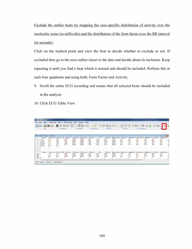
189
Exclude the outlier beats by mapping the case-specific distribution of activity over the
isoelectric noise (in millivolts) and the distribution of the form factor over the RR interval
(in seconds).
Click on the marked point and view the beat to decide whether to exclude or not. If
excluded then go to the next outlier closer to the data and decide about its inclusion. Keep
repeating it until you find a beat which is normal and should be included. Perform this at
each four quadrants and using both; Form Factor and Activity.
9. Scroll the entire ECG recording and ensure that all selected beats should be included
in the analysis
10. Click ECG Table View

190
Appendix D
Nicotine and metabolites determination in human plasma
2.1. Chemicals and reagents
(−)-Nicotine (NIC), (-)-Cotinine (COT), and LC-MS Ultra grade ammonium formate were
purchased from Sigma-Aldrich (St. Louis, MO). Trans-3'-Hydroxy Cotinine (3HC), and
trans-3'-Hydroxy Cotinine-d3 (3HC-d3) were purchased from Toronto Research Chemicals,
(Toronto, Canada). (±)-Nicotine-d3 (NIC-d3) and (±)-Cotinine-d3 (COT-d3), were
purchased from CDN Isotopes (Pointe-Claire, Quebec, Canada). UHPLC-MS grade water,
UHPLC-MS grade acetonitrile, and LC-MS grade formic acid were purchased from
Thermo Fisher Scientific Inc. (Waltham MA).
2.2. UPLC-MS/MS analysis
NIC, COT, and 3HC in human plasma samples were measured by UPLC-MS/MS using a
Xevo TQ-S micro quadrupole mass spectrometer with an ESI ionization source, interfaced
with Waters Acquity Class-H UPLC equipped with a quaternary pump system (Waters,
MA).
After the sample was thawed on ice, 20 μl of plasma was mixed with 480 μl of 75%
acetonitrile with isotopic labeled internal standard (COT-d3, NIC-d3, and 3HC-d3) in a 1.7
ml microcentrifuge tube. After incubation on ice for 30 min, the mixture was centrifuged
at 4°C for 30 min, and 2 μl of the supernatant was applied on the UPLC-MS/MS instrument.
The separation was performed on an Acquity UPLC BEH HILIC (50 mm × 2.1 mm, 1.7
μm) column (Waters, MA) with a binary gradient comprised of 95% water 5% ACN 10mM
ammonium formate pH 3 (Solvent A) and 0.1% formic acid in acetonitrile (Solvent B) at
a flow rate 0.4 ml/min. The gradient started at 96.5% for 1.2 min, ramped down to 30%
191
solvent B over a 3 min period. Then the gradient ramped down to 0% B in 0.1 min, held at
0% B for 0.3 min, return to 96.5% B in 0.1 min, and held for 5.3 min until next injection.
Chromatography was performed at 40 °C. The total chromatographic run time was 10 min.
The mass spectrometry detector worked in positive ion mode. Optimized cone voltage and
collision energy were used for each of the individual analytes. For each analyte, three
multiple reaction monitoring (MRM) transitions were set up: one for quantification, one
for confirmation, and one for labeled internal standard. Transitions for NIC are 163 > 130
for quantification, 163 > 117 for confirmation, and 166 > 130 for internal standard (NIC-
d3). Transitions for COT are 177 > 80 for quantification, 177 > 98 for confirmation, and
180 > 80 for internal standard (COT-d3). Transitions for 3HC are 193 > 80 for
quantification, 193 > 134 for confirmation, and 196 > 80 for internal standard (3HC-d3).
These MRMs were scheduled around the retention time of the analytes. No less than 15
data points were collected for each peak.
TargetLynx quantification application manager software (Waters, MA) was used for peak
integration, calibration, and quantification. Analytes in plasma samples were quantified
using peak area ratio based on a 7 point-standard curves which were run before and after
the plasma samples.
192
CURRICULUM VITAE
Affan B. Irfan
WORK EXPERIENCE
7/2017- Non-invasive Cardiologist Director, Echocardiography Laboratory, Cabell Huntington Hospital Site Director for Cardiology Fellowship, Cabell Huntington Hospital Assistant Professor Department Cardiology and Clinical Translational Services Marshall University, Huntington, WV, USA
2010 – 2011 Clinical Research Fellow University Hospital Basel, Switzerland
Research Supervisor: Professor Dr. Christian Mueller, MD, FESC Associate Director, Internal Medicine, University Hospital Basel.
2009 – 2010 Research Officer Indus Hospital Research Centre, Pakistan
Research Supervisor: Dr Aamir Javed Khan, M.D., PhD. International Health Johns Hopkins Bloomberg School of Public Health and Director, Center for Community Development, Karachi, Pakistan
2008 Research Officer Iberoamerican Cochrane Center, Hospital de Saint Pau, Spain Research Supervisor: Dr Pablo Alonso-Coello, M.D.PhD,
Universidad Autónoma de Barcelona, Spain
EDUCATION
2014-2017General Cardiology Fellow Level III Trained Echocardiography University of Louisville. KY, USA
2011-2014Internal Medicine Residency Program University of Illinois - Urbana Champaign, USA

193
2003-2008Bachelor of Medicine and Bachelor of Surgery (M.B.,B.S) Aga Khan University Medical College, Karachi, Pakistan
2001-2003General Certificate Examination Advanced Levels St. Mary’s Catholic High School, Dubai, U.A.E.
2001General Certificate Examination Ordinary Levels English Medium School Dubai, U.A.E.
CERTIFICATION
2019 Board Certified American Board of Cardiovascular Diseases
2018 Board Certified American Board of Echocardiography
2018 Board Certified American Society of Nuclear Cardiology
2014 Board Certified American Internal Medicine
2011 US ECFMG Certificate

194
PEER REVIEWED PUBLICATIONS (Total 34 with 14 as first or last authors)
Sympathetic Activation and Nicotine Mediate the Acute Proarrhythmic and Ischemic Effects of Smoking in Humans. Affan Irfan, George Koromia, Waiel Abusnina, Madison Crank, Lonnie Lucas, Meagen Carter, Nalini Santanam, Andrew DeFilippis, Mark Studeny, Zhengzhi, Pawel Lorkiewicz, Aruni Bhatnagar, Alex P. Carll Upcoming
Association between serum cotinine levels and PR and QT interval and their components Affan Irfan, Daniel Riggs, George A. Koromia, Andrew Paul DeFilippis, Elsayed Z. Soliman, Aruni Bhatnagar, Alex P. Carll Upcoming
Effects of chronic smoking on atrioventricular conduction and catecholamine production Affan Irfan, Claudia Arab, Andrew P. DeFilippis, Pawel Lorkiewicz, Zhengzhi Xie, Aruni Bhatnagar, Alex P. Carll Submitted
Clinical Determinants of Myocardial Injury, Detectable and Serial Troponin Levels among Patients with Hypertensive Crisis Giancarlo Acosta, Ahmed Amro, Rodrigo Aguilar, Waiel Abusnina, Niharika Bhardwaj, George Koromia, Mark Studeny, Affan Irfan Submitted
Irfan A, Li Y, Bhatnagar A, Soliman EZ. Association between serum cotinine levels and electrocardiographic left atrial abnormality. Ann Noninvasive Electrocardiol. 2019 Jan;24(1):e12586.
Irfan A. Letter by Irfan Regarding Article, "Assessment of the European Society of Cardiology 0-Hour/1-Hour Algorithm to Rule-Out and Rule-In Acute Myocardial Infarction". Circulation. 2017 Apr 18;135(16):e919-e920.
Alok R. Amraotkar; Patrick J. Trainor; Affan Irfan, Charles W. Hargis; Shesh N. Rai; Aruni Bhatnagar; Andrew P. DeFilippis. Presence of multiple coronary angiographic characteristics for the diagnosis of acute coronary thrombus. Cardiol J. 2017;24(1):25-34
Agarwal, A., B. C. Johnston, R. W. Vernooij, A. Carrasco-Labra, R. Brignardello-Petersen, I. Neumann, E. A. Akl, X. Sun, M. Briel, J. W. Busse, S. Ebrahim, C. E. Granados, A. Iorio, A. Irfan, L. Martinez Garcia, R. A. Mustafa, A. Ramirez-Morera, A. Selva, I. Sola, A. J. Sanabrai, K. A. Tikkinen, P. O. Vandvik, Y. Zhang, O. E. Zazueta, Q. Zhou, H. J. Schunemann, G. H. Guyatt and P. Alonso-Coello. "Authors Seldom Report the Most Patient-Important Outcomes and Absolute Effect Measures in Systematic Review Abstracts." J Clin Epidemiol, (2016). Aug 21. [Epub ahead of print].
195
Alonso-Coello P, Carrasco-Labra A, Brignardello-Petersen R, Neumann I, Akl EA, Vernooij RW, Johnston BC, Sun X, Briel M, Busse JW, Ebrahim S, Granados CE, Iorio A, Irfan A, Martínez García L, Mustafa RA, Ramírez-Morera A, Selva A, Solà I, Sanabria AJ, Tikkinen KA, Vandvik PO, Zazueta OE, Zhang Y, Zhou Q, Schünemann H, Guyatt GH. Systematic reviews experience major limitations in reporting absolute effects. J Clin Epidemiol. 2016 Apr;72:16-26.
Akl, E. A., A. Carrasco-Labra, R. Brignardello-Petersen, I. Neumann, B. C. Johnston, X. Sun, M. Briel, J. W. Busse, S. Ebrahim, C. E. Granados, A. Iorio, A. Irfan, L. Martinez Garcia, R. A. Mustafa, A. Ramirez-Morera, A. Selva, I. Sola, A. J. Sanabria, K. A. Tikkinen, P. O. Vandvik, R. W. Vernooij, O. E. Zazueta, Q. Zhou, G. H. Guyatt and P. Alonso-Coello. "Reporting, Handling and Assessing the Risk of Bias Associated with Missing Participant Data in Systematic Reviews: A Methodological Survey." BMJ Open 5, no. 9 (2015): e009368.
Irfan, A., T. Reichlin, R. Twerenbold, C. Fischer, P. Ballarino, B. Nelles, K. Wildi, C. Zellweger, M. Rubini Gimenez, M. Mueller, P. Haaf, A. Fischer, H. Freidank, S. Osswald and Mueller. "Cardiomyocyte Injury Induced by Hemodynamic Cardiac Stress: Differential Release of Cardiac Biomarkers." Clin Biochem 48, no. 18 (2015): 1225-9.
Cho, J. H., G. Kutti Sridharan, S. H. Kim, R. Kaw, T. Abburi, A. Irfan and A. G. Kocheril. "Right Ventricular Dysfunction as an Echocardiographic Prognostic Factor in Hemodynamically Stable Patients with Acute Pulmonary Embolism: A Meta-Analysis." BMC Cardiovasc Disord 14, (2014): 64.
Khan, M. B., R. Kumar, F. B. Irfan, A. B. Irfan and M. E. Bari. "Civilian Craniocerebral Gunshot Injuries in a Developing Country: Presentation, Injury Characteristics, Prognostic Indicators, and Complications." World Neurosurg 82, no. 1-2 (2014): 14-9.
Alonso-Coello, P., A. Carrasco-Labra, R. Brignardello-Petersen, I. Neumann, E. A. Akl, X. Sun, B. C. Johnston, M. Briel, J. W. Busse, D. Glujovsky, C. E. Granados, A. Iorio, A.
Irfan, L. Martinez Garcia, R. A. Mustafa, A. Ramirez-Morera, I. Sola, K. A. Tikkinen, S. Ebrahim, P. O. Vandvik, Y. Zhang, A. Selva, A. J. Sanabria, O. E. Zazueta, R. W. Vernooij, H. J. Schunemann and G. H. Guyatt. "A Methodological Survey of the Analysis, Reporting and Interpretation of Absolute Risk Reduction in Systematic Reviews (Arrow): A Study Protocol." Syst Rev 2, (2013): 113.
Irfan, A., S. Hublikar, J. H. Cho and J. Hill. "The Value of History and Goals of Care with Code Status; Even in an Emergency Setting." BMJ Case Rep 2013, (2013).
Haaf, P., R. Twerenbold, T. Reichlin, J. Faoro, M. Reiter, C. Meune, S. Steuer, S. Bassetti, R. Ziller, C. Balmelli, I. Campodarve, C. Zellweger, A. Kilchenmann, A. Irfan, J.

196
Papassotiriou, B. Drexler and C. Mueller. "Mid-Regional Pro-Adrenomedullin in the Early Evaluation of Acute Chest Pain Patients." Int J Cardiol 168, no. 2 (2013): 1048-55.
Irfan, A., T. Reichlin, R. Twerenbold, M. Meister, B. Moehring, K. Wildi, S. Bassetti, C. Zellweger, M. R. Gimenez, R. Hoeller, K. Murray, S. M. Sou, M. Mueller, T. Mosimann, M. Reiter, P. Haaf, R. Ziller, H. Freidank, S. Osswald and C. Mueller. "Early Diagnosis of Myocardial Infarction Using Absolute and Relative Changes in Cardiac Troponin Concentrations." Am J Med 126, no. 9 (2013): 781-788 e2.
Irfan, A., R. Twerenbold, M. Reiter, T. Reichlin, C. Stelzig, M. Freese, P. Haaf, W. Hochholzer, S. Steuer, S. Bassetti, C. Zellweger, H. Freidank, F. Peter, I. Campodarve, C. Meune and C. Mueller. "Determinants of High-Sensitivity Troponin T among Patients with a Noncardiac Cause of Chest Pain." Am J Med 125, no. 5 (2012): 491-498 e1.
Breidthardt, T*., A. Irfan*, T. Klima, B. Drexler, C. Balmelli, N. Arenja, T. Socrates, R. Ringger, C. Heinisch, R. Ziller, J. Schifferli, C. Meune and C. Mueller. "Pathophysiology of Lower Extremity Edema in Acute Heart Failure Revisited." Am J Med 125, no. 11 (2012): 1124 e1-1124 e8. (*co-first authors)
Meune, C., T. Reichlin, A. Irfan, N. Schaub, R. Twerenbold, J. Meissner, M. Reiter, A. Luthi, P. Haaf, C. Balmelli, B. Drexler, K. Winkler, W. Hochholzer, S. Osswald and C. Mueller. "How Safe Is the Outpatient Management of Patients with Acute Chest Pain and Mildly Increased Cardiac Troponin Concentrations?" Clin Chem 58, no. 5 (2012): 916-24.
Meune, C., R. Twerenbold, B. Drexler, C. Balmelli, C. Wolf, P. Haaf, T. Reichlin, A.
Irfan, M. Reiter, C. Zellweger, J. Meissner, C. Stelzig, M. Freese, I. Capodarve and C. Mueller. "Midregional Pro-a-Type Natriuretic Peptide for Diagnosis and Prognosis in Patients with Suspected Acute Myocardial Infarction." Am J Cardiol 109, no. 8 (2012): 1117-23.
Reichlin, T., A. Irfan, R. Twerenbold, M. Reiter, W. Hochholzer, H. Burkhalter, S. Bassetti, S. Steuer, K. Winkler, F. Peter, J. Meissner, P. Haaf, M. Potocki, B. Drexler, S. Osswald and C. Mueller. "Utility of Absolute and Relative Changes in Cardiac Troponin Concentrations in the Early Diagnosis of Acute Myocardial Infarction." Circulation 124, no. 2 (2011): 136-45.
Meissner, J., A. Irfan, R. Twerenbold, S. Mueller, M. Reiter, P. Haaf, T. Reichlin, N. Schaub, K. Winkler, O. Pfister, C. Heinisch and C. Mueller. "Use of Neutrophil Count in Early Diagnosis and Risk Stratification of Ami." Am J Med 124, no. 6 (2011): 534-42.
Meune, C., C. Balmelli, T. Marxer, J. Meissner, R. Twerenbold, M. Reiter, T. Reichlin, P. Haaf, B. Drexler, A. Irfan, C. Stelzig, M. Freese, K. Winkler and C. Mueller. "High-Sensitive Troponin, B-Type Natriuretic Peptide and Coronary Angiogram Findings in
197
Patients with Non St- Segment Elevation Acute Coronary Syndrome." Int J Cardiol 153, no. 3 (2011): 335-7.
Meune, C., C. Balmelli, R. Twerenbold, T. Reichlin, M. Reiter, P. Haaf, S. Steuer, S. Bassetti, K. Sakarikos, I. Campodarve, C. Zellweger, A. Irfan, B. Drexler and C. Mueller. "Patients with Acute Coronary Syndrome and Normal High-Sensitivity Troponin." Am J Med 124, no. 12 (2011): 1151-7.
Irfan, A., M. J. Zellweger and C. Mueller. ""Universal Definition" Methodology and Conclusions Are a Concern." J Am Coll Cardiol 58, no. 3 (2011): 313-4; author reply 314-5.
Irfan, A., P. Haaf, J. Meissner, R. Twerenbold, M. Reiter, T. Reichlin, N. Schaub, A. Zbinden, C. Heinisch, B. Drexler, K. Winkler and C. Mueller. "Systolic Blood Pressure at Emergency Department Presentation and 1-Year Mortality in Acute Chest Pain Patients." Eur J Intern Med 22, no. 5 (2011): 495-500.
Alonso-Coello, P., A. Irfan, I. Sola, I. Gich, M. Delgado-Noguera, D. Rigau, S. Tort, X. Bonfill, J. Burgers and H. Schunemann. "The Quality of Clinical Practice Guidelines over the Last Two Decades: A Systematic Review of Guideline Appraisal Studies." Qual Saf Health Care 19, no. 6 (2010): e58.
Bajaj, R. R., A. Mohammad, T. Hong, A. Irfan, W. Sharieff, A. Bagnall, J. A. Christie, M. J. Kutryk, R. J. Chisholm and A. N. Cheema. "Comparison of Abciximab and Eptifibatide on Angiographic and Clinical Outcomes in Rescue Percutaneous Coronary Intervention for Failed Fibrinolytic Therapy." J Invasive Cardiol 22, no. 8 (2010): 347-52.
Gallardo, C. R., D. Rigau, A. Irfan, A. Ferrer, J. A. Cayla, X. Bonfill and P. Alonso-Coello. "Quality of Tuberculosis Guidelines: Urgent Need for Improvement." Int J Tuberc Lung Dis 14, no. 8 (2010): 1045-51.
Aly, Z., K. Abbas, S. F. Kazim, F. Taj, F. Aziz, A. Irfan, R. Sheikh, M. Shakir, S. M. Javed and Z. Fatmi. "Awareness of Stroke Risk Factors, Signs and Treatment in a Pakistani Population." J Pak Med Assoc 59, no. 7 (2009): 495-9
Kazim, S. F., Z. Aly, H. K. Bangash, B. P. Harchandani, A. B. Irfan, S. M. Javed, R. Khalil- ur-Rehman, H. Naqvi and M. M. Khan. "Attitudes toward Suicide Bombing in Pakistan." Crisis 29, no. 2 (2008): 81-5.
Jaffe, R., A. Irfan, T. Hong, R. J. Chisholm and A. N. Cheema. "Intraluminal Filling Defects on Coronary Angiography: More Than Meets the Eye." Clin Cardiol 30, no. 9 (2007): 480-4.
198
ABSTRACTS / POSTERS (Total 28 with 17 as first or last authors)
Affan Irfan, Daniel Riggs, George Koromia, Andrew Defilippis, Elsayed Soliman, Aruni Bhatnagar, Alex Carl. Association between Serum Cotinine Levels and PR and QT Intervals and their components. American College of Cardiology Conference, 2019.
Ahmed Amro, Giancarlo Acosta, Kanaan Mansoor, George Koromia, Rodrigo Aguilar, Mehiar El-Hamdani, Affan Irfan. Elevated Diastolic Blood Pressure predicts short-term risk for Acute Coronary Syndrome in patients without prior Cardiovascular Disease. American College of Cardiology Conference, 2019.
Ahmed Amro, Giancarlo Acosta, George Koromia, Niharika Bhardwaj, Rodrigo Aguilar, Mehiar El-Hamdani, Affan Irfan Albumin Level As A Risk Marker And Predictor Of Peripartum Cardiomyopathy. American College of Cardiology Conference, 2019.
Affan Irfan, George Koromia, Waiel Abusnina, Mark Studney, Aruni Bhatnagar, Alex Carll. Comparison of the Acute Autonomic Effects of Mainstream Cigarette Smoke to Oral Nicotine Spray American Heart Association's EPI|Lifestyle 2019
Affan Irfan, Majd Kanbour, Giancarlo Acosta, Mark Studney. The additional cost of inpatient two-day myocardial perfusion scan in obese patients. ICNC (International Conference on Nuclear Cardiology and Cardiac CT. Lisbon 2019
Giancarlo Acosta, Ahmed Amro, Rodrigo Aguilar, Waiel Abusnina, Niharika Bhardwaj, George Koromia, Mark Studeny, Affan Irfan. Clinical Determinants of Myocardial Injury, Detectable and Serial Troponin Levels among Patients with Hypertensive Crisis. American Heart Association's Vascular Discovery 2019
Affan Irfan, George Koromia, Waiel Abusnina, Mark Studney, Aruni Bhatnagar, Alex Carll Comparison of the Acute Autonomic Effects of Mainstream Cigarette Smoke to Oral Nicotine Spray. AHA EPI/Lifestyle 2018
Daniel W. Riggs, Ray Yeager, Alex P. Carll, Affan Irfan, Natasha DeJarnett, Rachel J. Keith, Daniel J. Conklin, Sanjay Srivastava, Shesh N. Rai, Sumanth D. Prabhu, and Aruni Bhatnagar. Residential Greenspace is Associated with Arterial Stiffness and QT interval. University of Louisville 2016
Affan Irfan, Patrick Trainor, Schultz, Andrew DeFilippis. Acrolein, smoking exposure and acute myocardial infarction. University of Louisville 2016
Alok Amraotkar, Patrick Trainor, Charles Hargis, Affan Irfan, Andrew DeFilippis. Evaluation of multiple coronary angiographic characteristics for the diagnosis of acute coronary thrombus. Vascular Research Day poster session, Louisville, April 2015
199
Affan Irfan; Raphael Twerenbold; Karin Wildi; Rubini-Gimenez Maria; Cedric Jaeger; Michael Freese; Tobias Reichlin; Christian Mueller. Association of albumin and high sensitive cardiac troponin t and i levels among chest pain patients presenting to emergency department. J Am Coll Cardiol. 2015;65(10_S)
Affan Irfan, Krishan Kataria, Bisma Irfan , Sarwar Orakzai. A case of cardiac amyloidosis with a negative biopsy from a noncardiac site. Clinical & Experimental Cardiology. 2014
Affan Irfan; Tobias Reichlin; Rapahel Twerenbold; Karin Wildi; Christian Mueller. Hemodynamic cardiac stress induced myocardial ischemia as detected by the release of cardiac biomarkers: cardiac troponin t and i, ck-mb and myoglobin. J Am Coll Cardiol. 2013;61(10_S):. doi:10.1016/S0735-1097(13)60193-X
Affan Irfan; Tobias Reichlin; Raphael Twerenbold; Karin Wildi; Christian Mueller. Determinants of early changes in high-sensitive troponin levels among patients with non-acute myocardial infarction cause of chest pain. J Am Coll Cardiol. 2013;61(10_S):. doi:10.1016/S0735- 1097(13)60232-6
Affan Irfan; Tobias Reichlin; Raphael Twerenbold; Karin Wildi; Christian Mueller. The prognostic value of absolute and relative changes in cardiac troponin concentrations among non- acute myocardial infarction patients. J Am Coll Cardiol. 2013;61(10_S):. doi:10.1016/S0735- 1097(13)60236-3
Affan Irfan; Tobias Reichlin; Raphael Twerenbold; Karin Wildi; Christian Mueller. Combination of absolute and relative changes in cardiac troponin concentrations in the early diagnosis of acute myocardial infarction. J Am Coll Cardiol. 2013;61(10_S):. doi:10.1016/S0735- 1097(13)60233-8
A. Irfan TR, R. Twerenbold, K. Wildi, C. Mueller: The effect of Proton Pump Inhibitor on Blood pressure parameters among chest pain patients presenting to the Emergency Department. American College of Physicians (San Francisco Conference) April 2013.
A Irfan KN, J. Cho, C. Davies. Nausea and vomiting; Diagnosed by treatment. ACP Downstate conference, October, 2012.
JH Cho, S Kim, T Abburi, A Irfan, A. Kocheril. Right Ventricular Dysfunction as an Echocardiographic Prognostic Factor in Hemodynamically Stable Patients with Acute Pulmonary Embolism: a Meta-Analysis. ACP Downstate conference, October, 2012.
Johnston B, Alonso P, Neumann I, Carrasco A, Petersen RB, Sun X, Akl E, Briel M, Busse J, Dahm P, Granados CE, Glujovsky D, Iorio A, Irfan A, Martinez L, Mustafa R,

200
Ramírez A, Sola I, Sultan S, Tikkinen K, Vandvik P, Zhang M, Guyatt. G. Reporting of absolute estimates of effect of patient important benefits and harms in abstracts of Cochrane and non-Cochrane systematic reviews. 20th Cochrane Colloquium, Auckland, New Zealand October 2012.
Alonso P, Carrasco A, Petersen RB, Neumann I, Elie Akl XS, Johnston B, Briel M, Busse J, Dahm P, Granados CE, Glujovsky D, Iorio A, Irfan A, Martinez L, Mustafa R, Ramírez A, Sola I, Sultan S, Tikkinen K, Vandvik P, Zhang M, Guyatt G. Risk difference estimation and reporting in systematic reviews. 20th Cochrane Colloquium, Auckland, New Zealand October 2012.
Akl E, Alonso P, Neumann I, Petersen RB, Johnston B, Sun X, Carrasco A, Briel M, Busse J, Dahm P, Granados CE, Glujovsky D, Iorio A, Irfan A, Martinez L, Mustafa R, Ramírez A, Sola I, Sultan S, Tikkinen K, Vandvik P, Zhang M, Guyatt. G. Reporting, handling, and judging risk of bias associated with missing participant data in systematic reviews. 20th Cochrane Colloquium, Auckland, New Zealand October 2012.
Twerenbold R, Reiter M, Reichlin R, Balmelli C, Cattelan, Drexler B, Irfan A, Winkler K, Heinisch C, Osswald S, Mueller C. Utility of the ST-segment deviation score in the risk prediction of patients with suspected acute myocardial infarction. Congress of the European Society of Cardiology, Paris – France 2011.
Irfan A, Reichlin T, Twerenbold R, Reiter M, Balmelli C, Meissner J, Haaf P, Drexler B, Osswald S, Mueller C. Rule out acute myocardial infarction algorithm among patients with increased high sensitive troponin levels. Congress of the European Society of Cardiology, Paris – France 2011.
Irfan A, Reichlin T, Twerenbold R, Reiter M, Balmelli C, Meissner J, Haaf P, Drexler B, Osswald S, Mueller C. The significance of each traditional WHO criterion used to diagnose acute myocardial infarction. Congress of the European Society of Cardiology, Paris – France 2011.
Breidthardt T, Drexler B, Socrates T, Noveanu M, Haaf P, Cattelan CB, Irfan A, Reichlin T, Potocki M, Mueller CH. Plasma NGAL for the prediction of acute kidney injury in acute heart failure. Congress of the European Society of Cardiology, Paris – France 2011.
Balmelli C, Reiter M, Reichlin T, Twerenbold R, Haaf P, Irfan A, Drexler B, Meune C, Osswald S, Mueller C. Direct comparison of high-sensitive and sensitive cardiac troponin assays for risk stratification in patients with acute chest pain. Congress of the European Society of Cardiology, Paris – France 2011.
Agha SA, Georgiopoulou VV, Kalogeropoulos AP, Giamouzis G, Waheed S, Irfan
AB, Rashad MA, Bhatt DV, Laskar S, Smith AL, Butler J. Digoxin and Outcomes in Patients with Advanced Heart Failure on Contemporary Optimal Treatment Circulation 2008;118(S_1027-b).
201
BOOK CHAPTER
Affan Irfan, Wayne l. Miller, Allan Jaffe. “Using cardiac troponin to assist in the management of patients with congestive heart failure”. Heart Failure: The Expert’s Approach. Alan S. Maisel, Gerasimos Filippatos. Jaypee Brothers Medical Pub; 1 edition (July 2014)
ONLINE PUBLICATIONS
Irfan Affan, Hirsch GA. What Medication Would You Recommend for a 66-Year-Old Male Who Underwent PCI After MI? http://www.acc.org. April 14, 2015. Accessed Apr 14, 2015. http://www.acc.org/education-and-meetings/patient-case-quizzes/what-medication-would- you-recommend-for-a-66yo-male-who-underwent-pci-after-mi
Irfan Affan, Hirsch GA. Beta-Blocker Therapy in Patients With Stable Coronary Artery Disease. http://www.acc.org. April 14, 2015. Accessed Apr 14, 2015. http://www.acc.org/latest- in-cardiology/articles/2015/04/13/10/13/beta-blocker-therapy-in-patients-with-stable-coronary- artery-disease
HONORS / AWARDS
2019 International Conference on Nuclear and Cardiac CT Travel Award
2016-2019 University of Louisville Departmental Tuition Award
2015 First prize at DOC Student Research Proposal Competition
2015 American College of Cardiology Research Travel Award
2014 Best Research Scholar Award at University of Illinois
2005 Summer studentship at University of Toronto
2004 Summer studentship at University of Hamilton
GRANT
Marshall University Clinical Translational Services Role: Principal Investigator Project: Acute effects of Smoking and Nicotine on electrocardiogram Award: $50,000 State: Funded for 2017-2019
202
West Virginia Clinical & Translational Science Institute Role: Co-Principal Investigator Project: Development of a statewide integrated model for analysis of clinical & economic patterns of injection drug use-associated infective endocarditis Award: $49,938 State: Funded for 2019-2020