403170873 The Anorexia Nervosa Disorder that Reflected in ...
Upload
independentCategory
view
1download
0
Cell Metabolism
Article
Nesfatin-1-Regulated Oxytocinergic Signaling in theParaventricular Nucleus Causes Anorexia througha Leptin-Independent Melanocortin PathwayYuko Maejima,1,9 Udval Sedbazar,1,9 Shigetomo Suyama,1 Daisuke Kohno,1 Tatsushi Onaka,2 Eisuke Takano,1
Natsu Yoshida,1 Masato Koike,3 Yasuo Uchiyama,3 Ken Fujiwara,4 Takashi Yashiro,4 Tamas L. Horvath,5
Marcelo O. Dietrich,5,6 Shigeyasu Tanaka,7 Katsuya Dezaki,1 Shinsuke Oh-I,8 Koushi Hashimoto,8 Hiroyuki Shimizu,8
Masanori Nakata,1 Masatomo Mori,8 and Toshihiko Yada1,*1Division of Integrative Physiology2Division of Brain and Neurophysiology
Department of Physiology, Jichi Medical University School of Medicine, Shimotsuke, Tochigi 329-0498, Japan3Department of Cell Biology and Neurosciences, Juntendo University Graduate School of Medicine, Bunkyo-ku, Tokyo 113-8421, Japan4Division of Histology, Department of Anatomy, Jichi Medical University School of Medicine, Shimotsuke, Tochigi 329-0498, Japan5Program on Cell and Neurobiology of Energy Metabolism, Section of Comparative Medicine, Yale University School of Medicine, New Haven,
CT 06520, USA6Programa de Pos-graduacao em Bioquımica, Department of Biochemistry, Universidade Federal do Rio Grande do Sul,Porto Alegre RS 90035, Brazil7Department of Biology, Faculty of Science, Shizuoka University, Ohya 836, Shizuoka 422-8529, Japan8Department of Medicine and Molecular Science, Gunma University Graduate School of Medicine, Maebashi, Gunma 371-8511, Japan9These authors contributed equally to this work*Correspondence: [email protected]
DOI 10.1016/j.cmet.2009.09.002
SUMMARY
The hypothalamic paraventricular nucleus (PVN) func-tions as a center to integrate various neuronal activitiesfor regulating feeding behavior. Nesfatin-1, a recentlydiscovered anorectic molecule, is localized in thePVN. However, the anorectic neural pathway of nesfa-tin-1 remains unknown. Here we show that centralinjection of nesfatin-1 activates the PVN and brainstem nucleus tractus solitarius (NTS). In the PVN, nes-fatin-1 targets both magnocellular and parvocellularoxytocin neurons and nesfatin-1 neurons themselvesand stimulates oxytocin release. Immunoelectronmicrographs reveal nesfatin-1 specifically in the secre-tory vesicles of PVN neurons, and immunoneutraliza-tion against endogenous nesfatin-1 suppressesoxytocin release in the PVN, suggesting paracrine/autocrine actions of nesfatin-1. Nesfatin-1-inducedanorexia is abolished by an oxytocin receptor antago-nist. Moreover, oxytocin terminals are closely associ-ated with andoxytocin activates pro-opiomelanocortinneurons in the NTS. Oxytocin induces melanocortin-dependent anorexia in leptin-resistant Zucker-fattyrats. The present results reveal the nesfatin-1-opera-tive oxytocinergic signaling in the PVN that triggersleptin-independent melanocortin-mediated anorexia.
INTRODUCTION
Obesity and obesity-based metabolic syndrome are the major
risk factors for cardiovascular disease (Matsuzawa, 2006).
Cell M
Increasing incidence of obesity and metabolic syndrome has
become a serious health problem in the world (Matsuzawa,
2006). Obesity is basically caused by excessive food intake
and/or reduced energy expenditure (Schwartz et al., 2000).
Food intake is regulated by the feeding-regulating centers in
the hypothalamus and brain stem. Peripheral metabolic signals
are sensed by the brain stem including the nucleus tractus solitar-
ius (NTS) and by the hypothalamus including the arcuate nucleus
(ARC), and the processed information is sent to the hypothalamic
paraventricular nucleus (PVN). Inversely, neurons derived from
the PVN also extend their information to several brain areas to
determine feeding behavior. Thus, the PVN is considered an
integrative center for regulation of feeding (Balthasar et al.,
2005; Horvath and Bruning, 2006; Morton et al., 2006; Schwartz
et al., 2000). The PVN is equipped with neurons containing clas-
sical anorectic neuropeptides, corticotropin-releasing hormone
(CRH), oxytocin (Oxt), and thyrotropin-releasing hormone
(TRH). However, none of these neurons has been adequately
qualified to account for the pivotal anorectic function of the
PVN, in clear contrast to the ARC, where the neuron subset con-
taining orexigenic neuropeptide Y (NPY) and agouti-related
peptide (AgRP) and that containing anorectic pro-opiomelano-
cortin (POMC) and cocaine- and amphetamine-regulated tran-
script (CART) well explain the feeding and metabolic functions
of this nucleus (Morton et al., 2006; Schwartz et al., 2000).
Nesfatin-1 is a recently discovered anorectic peptide pro-
cessed from nesfatin or nucleobindin2 (NUCB2) (Oh-I et al.,
2006). Studies of injection of nesfatin-1 and NUCB2 antisense
RNA in rats indicated that nesfatin-1 can serve as both pharma-
cologic and physiologic regulator of feeding and body weight
(Oh-I et al., 2006). However, the neural pathway for the anorectic
action of nesfatin-1 remains to be clarified. Nesfatin-1 is local-
ized in the hypothalamic PVN, ARC, lateral hypothalamic area
(LHA), and supraoptic nucleus (SON) and in the brain stem
etabolism 10, 355–365, November 4, 2009 ª2009 Elsevier Inc. 355

Cell Metabolism
PVN Nesfatin-1-Oxytocin Pathway Causes Anorexia
NTS, the areas involved in the regulation of feeding (Oh-I et al.,
2006). Moreover, starvation decreases both NUCB2 mRNA
and nesfatin-1 peptide levels specifically in the PVN (Oh-I et al.,
2006), and refeeding induces c-Fos expression in nesfatin-1-
containing neurons in the PVN (Kohno et al., 2008). These obser-
vations suggest that the nesfatin-1 in the PVN could be impli-
cated in the physiological regulation of food intake.
Nesfatin-1 in the PVN colocalizes most extensively with Oxt
(Brailoiu et al., 2007; Foo et al., 2008; Kohno et al., 2008). To
date, several functions of Oxt have been established or proposed.
The PVN magnocellular Oxt neurons give rise to axons to the
posterior pituitary gland and release Oxt into circulation. Periph-
eral Oxt stimulates uterine smooth muscle contraction and milk
ejection (Kissand Mikkelsen, 2005). Oxt is also thought to function
in the central nervous system. Oxt is related to maternal nurturing
and social attachment (Donaldson and Young, 2008; Neumann,
2008). Furthermore, several lines of evidence have implicated
Oxt in the regulation of feeding and energy expenditure (Kublaoui
et al., 2008; Leng et al., 2008). Animals deficient in Oxt gene or
genes related to the differentiation of Oxt neurons show hyper-
phagia and obesity (Kublaoui et al., 2008; Takayanagi et al.,
2008). Patients with Prader-Willi syndrome, a human genetic
disorder characterized with severe obesity and hyperphagia,
display reduction in Oxt neurons (Swaab et al., 1995) and plasma
Oxt levels (Stock et al., 1989). Central administration of Oxt and an
agonist for Oxt receptor decrease food intake (Arletti et al., 1989).
Both centrally projecting parvocellular Oxt neurons of the PVN
(Gimpl and Fahrenholz, 2001) and dendritic release of Oxt from
magnocellular neurons (Ludwig and Leng, 2006) play a role in
regulating food intake (Blevins et al., 2003; Douglas et al., 2007;
Olson et al., 1991). The PVN Oxt is upregulated/depressed by
feeding/fasting (Tung et al., 2008). Taken together, Oxt is consid-
ered one of the important regulators of feeding and metabolism.
However, the neural pathways upstream and downstream of
the PVN Oxt neurons implicated in feeding remain unclear, which
hampers the full establishment of this peptide as a feeding regu-
lator. In line with this evidence, we hypothesized that nesfatin-1
could regulate and cooperate with Oxt neurons in the PVN to
evoke an anorectic signaling pathway.
It is particularly of interest to note that nesfatin-1 inhibits food
intake in Zucker-fatty rats in which leptin function is impaired due
to mutation of its receptor (Oh-I et al., 2006). On the other hand,
the satiety effect of nesfatin-1 is blocked by an antagonist for
melanocortin receptors, MC3/4R. These findings suggest that
nesfatin-1 suppresses feeding via leptin-independent melano-
cortin signaling (Oh-I et al., 2006; Shimizu et al., 2007). However,
underlying neural mechanisms absolutely remain unknown.
This study aimed to explore whether the PVN Oxt neuron is
targeted by nesfatin-1 and, if so, how nesfatin-1 controls Oxt
neurons in the PVN, whether the nesfatin-1 to Oxt pathway
drives melanocortin signaling, and whether it can operate under
leptin-resistant conditions.
RESULTS
Third Ventricle Injection of Nesfatin-1 Induces c-FosExpression in the PVN and NTSThird ventricle (3V) injection of nesfatin-1 induced significant
expression of c-Fos in the PVN and NTS, as compared to 3V injec-
356 Cell Metabolism 10, 355–365, November 4, 2009 ª2009 Elsevie
tion of vehicle in control experiments (Figures 1A–1E). In contrast,
nesfatin-1 did not significantly induce c-Fos expression in the
SON, LHA, ARC, dorsomedial hypothalamus (DMH), or ventrome-
dial hypothalamus (VMH) (Figure 1E), though a rising tendency
was observed in the SON, LHA, and DMH. The selective c-Fos
induction in the PVN and NTS by nesfatin-1 indicates possible
involvement of these nuclei in anorectic signaling for nesfatin-1.
Nesfatin-1 injection through a cannula placed focally into the
PVN significantly decreased cumulative food intake at 1 and 3 hr
(Figure 1F). These results indicate that at least one of the primary
effector sites for nesfatin-1 is the PVN, which might be connected
to the NTS. This notion is supported by the fact that PVN neurons
directly project to the NTS (McCann and Rogers, 1990).
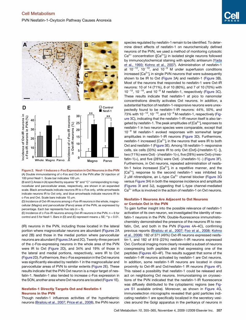
Nesfatin-1 Activates PVN Oxt NeuronsWe next explored the target neurons of nesfatin-1 in the PVN.
Double-labeling immunohistochemistry revealed that 3V injection
of nesfatin-1 induced c-Fos expression in Oxt-immunoreactive
Figure 1. Third Ventricle Injection of Nesfatin-1 Induces c-Fos Ex-
pression in the PVN and NTS, and Intra-PVN Injection of Nesfatin-1
Inhibits Feeding
(A–D) c-Fos expression in the PVN (A and B) and NTS (C and D) at 2 hr after 3V
injection of 5 ml vehicle as a control (A and C) or 100 pmol nesfatin-1 (Nesf-1)
(B and D). Scale bars indicate 50 mm. 3V, third ventricle; AP, area postrema.
(E) Number of c-Fos IR neurons in the feeding-related areas of the hypothal-
amus and NTS after injection of 100 pmol Nesf-1 (filled bars) or 5 ml vehicle
(open bars). n = 5 (rats) for Nesf-1 and for control.
(F) Cumulative food intake for 1, 3, and 6 hr after focal injection of 0.5 ml vehicle
or Nesf-1 (50 pmol) into PVN. n = 18 for control and 15 for Nesf-1. Bars in (E)
and (F) represent mean ± SE; **p < 0.01.
r Inc.
Cell Metabolism
PVN Nesfatin-1-Oxytocin Pathway Causes Anorexia
(IR) neurons in the PVN, including those located in the lateral
portion where magnocellular neurons are abundant (Figures 2A
and 2B) and those in the medial portion where parvocellular
neurons are abundant (Figures 2A and 2C). Twenty-three percent
of the c-Fos-expressing neurons in the whole area of the PVN
were IR to Oxt (Figure 2D), and 34% and 19% of those in
the lateral and medial portions, respectively, were IR to Oxt
(Figure 2D). Furthermore, the c-Fos expression in the Oxt neurons
was significantly elevated by nesfatin-1 in the magnocellular and
parvocellular areas of PVN to similar extents (Figure 2E). These
results indicate that the PVN Oxt neuron is a major target of nes-
fatin-1. Nesfatin-1 also tended to increase c-Fos expression in
the SON, another area where Oxt neurons are located (Figure 1E).
Nesfatin-1 Directly Targets Oxt and Nesfatin-1Neurons in the PVNThough nesfatin-1 influences activities of the hypothalamic
neurons (Brailoiu et al., 2007; Price et al., 2008), the PVN neuron
Figure 2. Nesf-1 Induces c-Fos Expression in Oxt Neurons in the PVN
(A) Double immunostaining of c-Fos and Oxt in the PVN after 3V injection of
100 pmol Nesf-1. Scale bar indicates 100 mm.
(B and C) Areas in (A) specified by squares ‘‘B’’ and ‘‘C’’ corresponding to mag-
nocellular and parvocellular areas, respectively, are shown in an expanded
scale. Black arrowheads indicate neurons IR to c-Fos only, white arrowheads
indicate neurons IR to Oxt only, and blue arrowheads indicate neurons IR to
c-Fos and Oxt. Scale bars indicate 10 mm.
(D) Incidence of Oxt-IR neurons among c-Fos-IR neurons in the whole, magno-
cellular (Magno) and parvocellular (Parvo) areas of the PVN, as expressed by
percentage. Each bar represents five rats (n = 5).
(E) Incidence of c-Fos-IR neurons among Oxt-IR neurons in the PVN. n = 6 for
control and 5 for Nesf-1. Bars in (D) and (E) represent means ± SE; **p < 0.01.
Cell M
species regulated by nesfatin-1 remain to be identified. To deter-
mine direct effects of nesfatin-1 on neurochemically defined
neurons of the PVN, we used a method of monitoring cytosolic
Ca2+ concentration ([Ca2+]i) in isolated single neurons followed
by immunocytochemical staining with specific antiserum (Yada
et al., 1993; Kohno et al., 2007). Administration of nesfatin-1
at 10�12, 10�10, and 10�9 M under superfusion conditions
increased [Ca2+]i in single PVN neurons that were subsequently
shown to be IR to Oxt (Figure 3A) and nesfatin-1 (Figure 3B).
Most of the neurons that responded to nesfatin-1 were Oxt-IR
neurons: 10 of 14 (71%), 8 of 10 (80%), and 7 of 10 (70%) with
10�12, 10�10, and 10�9 M nesfatin-1, respectively (Figure 3C).
These results indicate that nesfatin-1 at pico to nanomolar
concentrations directly activates Oxt neurons. In addition, a
substantial fraction of nesfatin-1-responsive neurons were unex-
pectedly found to be nesfatin-1-IR neurons: 44%, 50%, and
73% with 10�12, 10�10, and 10�9 M nesfatin-1, respectively (Fig-
ure 3C), indicating that the nesfatin-1-IR neuron itself is also tar-
geted by nesfatin-1. The peak amplitudes of [Ca2+]i responses to
nesfatin-1 in two neuron species were comparable, except that
10�9 M nesfatin-1 evoked responses with somewhat larger
amplitudes in nesfatin-1-IR neurons (Figure 3D). Furthermore,
nesfatin-1 increased [Ca2+]i in the neurons that were IR to both
Oxt and nesfatin-1 (Figure 3E). Among 18 nesfatin-1- responsive
cells, six cells (33%) were IR to only Oxt (Oxt[+]/nesfatin-1[�]),
two (11%) were Oxt(�)/nesfatin-1(+), five (28%) were Oxt(+)/nes-
fatin-1(+), and five (28%) were Oxt(�)/nesfatin-1(�) (Figure 3F).
Furthermore, in Oxt neurons, repeated administration of nesfa-
tin-1 twice increased [Ca2+]i in a repetitive manner, and the
[Ca2+]i response to the second nesfatin-1 was inhibited by
2 mM nitrendipine, an L-type Ca2+ channel blocker (Figure 3G
versus Figure 3H) in both the response incidence and amplitude
(Figures 3I and 3J), suggesting that L-type channel-mediated
Ca2+ influx is involved in the action of nesfatin-1 on Oxt neurons.
Nesfatin-1 Neurons Are Adjacent to Oxt Neuronsor Contain Oxt in the PVNTo gain further insight into the possible relevance of nesfatin-1
activation of its own neuron, we investigated the identity of nes-
fatin-1 neurons in the PVN. Double-fluorescence immunohisto-
chemistry demonstrated the presence of the neurons IR to nes-
fatin, Oxt, and both in the PVN (Figures 4A–4C), confirming
previous reports (Brailoiu et al., 2007; Foo et al., 2008; Kohno
et al., 2008): 182 of 371 (49%) Oxt-IR neurons expressed nesfa-
tin-1, and 182 of 819 (22%) nesfatin-1-IR neurons expressed
Oxt. Confocal imaging more clearly revealed a subset of neurons
coexpressing both peptides and that expressing one of the
peptides (Figures 4D–4F). The results suggest that some of the
nesfatin-1-IR neurons activated by nesfatin-1 are Oxt neurons.
In addition, some nesfatin-1-IR neurons are located in close
proximity to Oxt-IR and Oxt/nesfatin-1 IR neurons (Figure 4F).
This raised a possibility that nesfatin-1 could be released and
act on neighboring Oxt neurons. Immunostaining on cryosec-
tions of the PVN indicated that the nesfatin-1-IR fluorescence
was diffusely distributed to the cytoplasmic regions (see Fig-
ure S1 available online). Moreover, as shown in Figure 4G,
immunoelectron micrographs revealed that gold particles indi-
cating nesfatin-1 are specifically localized in the secretory vesi-
cles around the Golgi apparatus in the perikarya of neurons in
etabolism 10, 355–365, November 4, 2009 ª2009 Elsevier Inc. 357

Cell Metabolism
PVN Nesfatin-1-Oxytocin Pathway Causes Anorexia
Figure 3. Nesf-1 Increases [Ca2+]i in Oxt and
Nesf-1 Neurons in the PVN, and [Ca2+]iIncreases in Oxt Neurons Are Inhibited by
L-Type Ca2+ Channel Blocker
(A and B) Nesf-1 at 10�12 M increased [Ca2+]i (left
panel) in an isolated PVN neuron that was subse-
quently shown to be IR to Oxt (A) or Nesf-1 (B) (right
panel). Superfusates contain 1 mM glucose. Bars
above the tracings indicate the periods of adminis-
tration of agents. Scale bars indicate 10 mm.
(C) Incidence of Oxt-IR and Nesf-1-IR neurons in
the neurons that exhibited [Ca2+]i responses to
Nesf-1, expressed by percentage. Numbers above
each bar indicate the number of neurons IR to each
peptide over that responsive to Nesf-1.
(D) Amplitudes of [Ca2+]i increases in Nesf-1-IR and
Oxt-IR neurons. The number above each bar indi-
cates the number of Nesf-1-responsive neurons.
(E) Nesf-1 at 10�10 M increased [Ca2+]i (left panel) in
an isolated PVN neuron that was subsequently
shown to be IR to both Oxt and Nesf-1 (right panel).
(F) Incidence of neurons IR to Oxt, Nesf-1, Oxt+
Nesf-1, or none of them over those responded
to Nesf-1. Above each bar, the numbers of speci-
fied neurons over Nesf-1-responsive neurons are
indicated.
(G) Repetitive additions of Nesf-1 twice induced
repeated [Ca2+]i increases.
(H) Nesf-1-induced [Ca2+]i increase in Oxt neurons
was inhibited by an L-type Ca2+ channel blocker,
nitrendipine (NTD) (Yoshitomi Pharmaceuticals
industries; Osaka, Japan).
(I and J) Incidence (I) and amplitude (J) of [Ca2+]iresponses to the second Nesf-1 addition in the
absence and presence of NTD in Nesf-1-respon-
sive cells. Above each bar, the numbers of speci-
fied neurons over Nesf-1-responsive neurons are
indicated in (I), and the number of Nesf-1-respon-
sive neurons in (J). Bars in (D) and (J) represent
means ± SE; **p < 0.01.
the mouse PVN. It was confirmed in mice that nesfatin-1 also
suppressed food intake (Figure S2).
Endogenous Nesfatin-1 Promotes Oxt Releasein the PVNIn line with these histological observations, it was possible that
nesfatin-1 in the PVN might influence Oxt neurons via a para-
crine/autocrine route, dendritic release, and/or ultrashort-feed-
back projection, the mechanisms proposed in several brain
regions including the PVN (Ludwig and Leng, 2006). PVN slices
were used to examine this possibility. First, administration of
exogenous nesfatin-1 at 10�8 M stimulated the release of Oxt
from PVN slices (Figure 4H). Incubation of PVN slices with 50 mM
KCl, compared to control 3 mM KCl, also increased Oxt release,
and this increased Oxt release was partly and significantly sup-
pressed by the presence of an anti-nesfatin-1 IgG (Figure 4I). The
slices in 50 mM KCl solution responded to exogenous nesfatin-1
with further increase in Oxt release (Figure 4J). In addition,
50 mM KCl, nesfatin-1, and the combination failed to elevate
358 Cell Metabolism 10, 355–365, November 4, 2009 ª2009 Elsevie
the release of lactate dehydrogenase (LDH), a marker of cytotox-
icity (Figure S3). These results indicate that slices at high K+ are in
a sound state and able to respond to additional stimulation.
Collectively, our results suggest that endogenous nesfatin-1
acts on adjacent Oxt neurons to promote the release of Oxt in
the PVN.
Oxt Dependence of Nesfatin-1-Induced AnorexiaInhibition of cumulative food intake for 6 hr after 3V injection of
nesfatin-1 was blocked by H4928, a selective antagonist for
Oxt receptor (Figure 4K). This result indicates that nesfatin-1
suppresses feeding via stimulation of Oxt system, which may
include Oxt release in the PVN and activation of the PVN Oxt
neurons projecting to other brain areas.
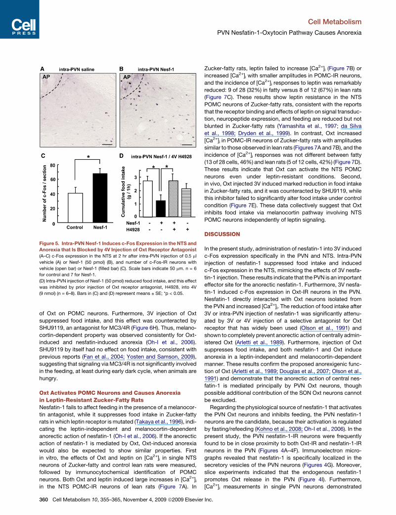
Intra-PVN Injection of Nesfatin-1 Suppresses Feedingvia Oxt Signaling to the NTSSince 3V nesfatin-1 induced c-Fos expression selectively in the
PVN and NTS (Figure 1), we examined whether the NTS is
r Inc.

Cell Metabolism
PVN Nesfatin-1-Oxytocin Pathway Causes Anorexia
activated by the signaling from the PVN. Focal injection of nesfa-
tin-1 into the PVN induced c-Fos expression in the NTS (Figures
5A–5C) and inhibited food intake (Figures 1F and 5D). Moreover,
this inhibition of feeding was significantly counteracted by injec-
tion of Oxt receptor antagonist H4928 into fourth ventricle (4V),
an area that provides an easy and effective access to the NTS
(Blevins et al., 2004) (Figure 5D). These results suggest that the
PVN oxytocinergic signaling to the NTS underlies the nesfatin-
1-induced anorexia.
Oxt Interacts with POMC Neurons in the NTSNesfatin-1 decreases food intake in a melanocortin-dependent
manner (Oh-I et al., 2006). Our data indicated that nesfatin-1
decreases food intake by activating Oxt neurons. Therefore,
a question arises as to whether Oxt stimulates melanocortin
signaling. POMC neurons form an essential element of melano-
cortin pathway and are exclusively localized to the NTS and ARC
Figure 4. Nesf-1 Is Localized in Secretory
Vesicles in the Neurons Located Close
to Oxt Neurons in the PVN, Promotes
Oxt Release, and Causes Oxt-Dependent
Anorexia
(A–F) Fluorescence (A–C) and confocal (D–F)
images of the PVN stained for Nesf-1 with Alexa
488 (green) (A and D), for Oxt with Alexa 594
(red) (B and E), and for both by merged images
(C and F). Arrowheads indicate Nesf-1-IR neurons,
arrows Oxt-IR neurons, and asterisks (*) double-IR
neurons. Scale bars represent 100 mm in (A) and
20 mm in (D).
(G) Immunoelectron micrographs show that gold
particles indicating Nesf-1 are specifically local-
ized in the secretory vesicles (arrows) around the
Golgi apparatus (g). Secretory granules in boxed
areas are shown in insets. n, nuclei. The scale
bars represent 200 nm.
(H–J) Oxt release from PVN slices following incu-
bation for 30 min with or without 10�8 M Nesf-1
in control 3 mM (n = 10) (H) and 50 mM KCl solu-
tions (n = 4–6) (J), and with Nesf-1 IgG (33 3
10�8 g/ml) or control IgG in 3 mM and 50 mM
KCl solutions (n = 5–11) (I). Data are expressed
as the amount of Oxt released per a rat.
(K) 3V injection of Nesf-1 (100 pmol) reduced food
intake, and this effect was blocked by prior injec-
tion of Oxt receptor antagonist, H4928 (9 nmol)
(n = 7–8). Bars in (H)–(K) represent means ± SE;
*p < 0.05; **p < 0.01.
(Appleyard et al., 2005; Fan et al., 2004;
Huo et al., 2006; Perello et al., 2007).
Based on the results that nesfatin-1
induced c-Fos expression in the NTS,
we examined whether Oxt could regulate
POMC neurons in the NTS. Using POMC-
green fluorescent protein (GFP) trans-
genic (Tg) mice, we performed double
immunostaining for GFP and Oxt and
obtained data suggesting that Oxt termi-
nals contact to POMC neurons in the
NTS (Figure 6A). Furthermore, [Ca2+]imeasurements in single neurons isolated from the NTS
combined with immunostaining indicated that Oxt evoked
[Ca2+]i increases in 7 of 18 POMC-IR neurons from the NTS
(39%) (Figures 6B and 6D), while it had no effect on [Ca2+]i in
the rest of high K+-responsive POMC-IR neurons (Figure 6C).
Conversely, 7 of 12 Oxt-responsive NTS neurons were POMC-
IR neurons (58%) (Figure 6E). These results indicate that Oxt
preferentially regulates POMC neurons in the NTS. The ampli-
tude of Oxt-induced [Ca2+]i increases was markedly reduced
by H4928 (Figures 6F and 6G), indicating that the Oxt receptor
mediates the responses. Furthermore, repeated administration
of Oxt twice increased [Ca2+]i in POMC neurons in a repetitive
manner, and the [Ca2+]i response to the second Oxt was sup-
pressed by nitrendipine and by thapsigargin, an inhibitor of the
endoplasmic reticulum (ER) Ca2+ pumps, in both the response
incidence and amplitude (Figures S4A–S4E), suggesting that
L-type channel and the ER Ca2+ release are involved in the action
Cell Metabolism 10, 355–365, November 4, 2009 ª2009 Elsevier Inc. 359
Cell Metabolism
PVN Nesfatin-1-Oxytocin Pathway Causes Anorexia
of Oxt on POMC neurons. Furthermore, 3V injection of Oxt
suppressed food intake, and this effect was counteracted by
SHU9119, an antagonist for MC3/4R (Figure 6H). Thus, melano-
cortin-dependent property was observed consistently for Oxt-
induced and nesfatin-induced anorexia (Oh-I et al., 2006).
SHU9119 by itself had no effect on food intake, consistent with
previous reports (Fan et al., 2004; Yosten and Samson, 2009),
suggesting that signaling via MC3/4R is not significantly involved
in the feeding, at least during early dark cycle, when animals are
hungry.
Oxt Activates POMC Neurons and Causes Anorexiain Leptin-Resistant Zucker-Fatty RatsNesfatin-1 fails to affect feeding in the presence of a melanocor-
tin antagonist, while it suppresses food intake in Zucker-fatty
rats in which leptin receptor is mutated (Takaya et al., 1996), indi-
cating the leptin-independent and melanocortin-dependent
anorectic action of nesfatin-1 (Oh-I et al., 2006). If the anorectic
action of nesfatin-1 is mediated by Oxt, Oxt-induced anorexia
would also be expected to show similar properties. First
in vitro, the effects of Oxt and leptin on [Ca2+]i in single NTS
neurons of Zucker-fatty and control lean rats were measured,
followed by immunocytochemical identification of POMC
neurons. Both Oxt and leptin induced large increases in [Ca2+]iin the NTS POMC-IR neurons of lean rats (Figure 7A). In
Figure 5. Intra-PVN Nesf-1 Induces c-Fos Expression in the NTS and
Anorexia that Is Blocked by 4V Injection of Oxt Receptor Antagonist
(A–C) c-Fos expression in the NTS at 2 hr after intra-PVN injection of 0.5 ml
vehicle (A) or Nesf-1 (50 pmol) (B), and number of c-Fos-IR neurons with
vehicle (open bar) or Nesf-1 (filled bar) (C). Scale bars indicate 50 mm. n = 6
for control and 7 for Nesf-1.
(D) Intra-PVN injection of Nesf-1 (50 pmol) reduced food intake, and this effect
was inhibited by prior injection of Oxt receptor antagonist, H4928, into 4V
(9 nmol) (n = 6–8). Bars in (C) and (D) represent means ± SE; *p < 0.05.
360 Cell Metabolism 10, 355–365, November 4, 2009 ª2009 Elsevie
Zucker-fatty rats, leptin failed to increase [Ca2+]i (Figure 7B) or
increased [Ca2+]i with smaller amplitudes in POMC-IR neurons,
and the incidence of [Ca2+]i responses to leptin was remarkably
reduced: 9 of 28 (32%) in fatty versus 8 of 12 (67%) in lean rats
(Figure 7C). These results show leptin resistance in the NTS
POMC neurons of Zucker-fatty rats, consistent with the reports
that the receptor binding and effects of leptin on signal transduc-
tion, neuropeptide expression, and feeding are reduced but not
blunted in Zucker-fatty rats (Yamashita et al., 1997; da Silva
et al., 1998; Dryden et al., 1999). In contrast, Oxt increased
[Ca2+]i in POMC-IR neurons of Zucker-fatty rats with amplitudes
similar to those observed in lean rats (Figures 7A and 7B), and the
incidence of [Ca2+]i responses was not different between fatty
(13 of 28 cells, 46%) and lean rats (5 of 12 cells, 42%) (Figure 7D).
These results indicate that Oxt can activate the NTS POMC
neurons even under leptin-resistant conditions. Second,
in vivo, Oxt injected 3V induced marked reduction in food intake
in Zucker-fatty rats, and it was counteracted by SHU9119, while
this inhibitor failed to significantly alter food intake under control
condition (Figure 7E). These data collectively suggest that Oxt
inhibits food intake via melanocortin pathway involving NTS
POMC neurons independently of leptin signaling.
DISCUSSION
In the present study, administration of nesfatin-1 into 3V induced
c-Fos expression specifically in the PVN and NTS. Intra-PVN
injection of nesfatin-1 suppressed food intake and induced
c-Fos expression in the NTS, mimicking the effects of 3V nesfa-
tin-1 injection. These results indicate that the PVN is an important
effector site for the anorectic nesfatin-1. Furthermore, 3V nesfa-
tin-1 induced c-Fos expression in Oxt-IR neurons in the PVN.
Nesfatin-1 directly interacted with Oxt neurons isolated from
the PVN and increased [Ca2+]i. The reduction of food intake after
3V or intra-PVN injection of nesfatin-1 was significantly attenu-
ated by 3V or 4V injection of a selective antagonist for Oxt
receptor that has widely been used (Olson et al., 1991) and
shown to completely prevent anorectic action of centrally admin-
istered Oxt (Arletti et al., 1989). Furthermore, injection of Oxt
suppresses food intake, and both nesfatin-1 and Oxt induce
anorexia in a leptin-independent and melanocortin-dependent
manner. These results confirm the proposed anorexigenic func-
tion of Oxt (Arletti et al., 1989; Douglas et al., 2007; Olson et al.,
1991) and demonstrate that the anorectic action of central nes-
fatin-1 is mediated principally by PVN Oxt neurons, though
possible additional contribution of the SON Oxt neurons cannot
be excluded.
Regarding the physiological source of nesfatin-1 that activates
the PVN Oxt neurons and inhibits feeding, the PVN nesfatin-1
neurons are the candidate, because their activation is regulated
by fasting/refeeding (Kohno et al., 2008; Oh-I et al., 2006). In the
present study, the PVN nesfatin-1-IR neurons were frequently
found to be in close proximity to both Oxt-IR and nesfatin-1-IR
neurons in the PVN (Figures 4A–4F). Immunoelectron micro-
graphs revealed that nesfatin-1 is specifically localized in the
secretory vesicles of the PVN neurons (Figures 4G). Moreover,
slice experiments indicated that the endogenous nesfatin-1
promotes Oxt release in the PVN (Figure 4I). Furthermore,
[Ca2+]i measurements in single PVN neurons demonstrated
r Inc.

Cell Metabolism
PVN Nesfatin-1-Oxytocin Pathway Causes Anorexia
that nesfatin-1 directly interacted with Oxt-IR and nesfatin-1-IR
neurons to increase [Ca2+]i (Figure 3). These results suggest
that nesfatin-1 is released and activates neighboring Oxt
neurons in the PVN in a paracrine/autocrine manner (Ludwig
and Leng, 2006) and/or via an ultrashort-feedback route (Cowley
et al., 2001). Alternatively, histological studies have shown the
projections from different brain areas to the PVN (Csaki et al.,
2000; Sawchenko et al., 1988). Therefore, under in vivo situa-
tions, nesfatin-1-containing neurons in several brain regions,
Figure 6. Oxt Neuron Terminals Contact to
NTS POMC Neurons, and Oxt Evokes
[Ca2+]i Increases in NTS POMC Neurons
and Melanocortin-Dependent Anorexia
(A) Confocal images of double immunofluores-
cence for POMC with Alexa 488 (green) and for
Oxt with Alexa 594 (red) in the NTS prepared
from POMC GFP-Tg mice. Oxt-IR terminals
contact to POMC-GFP-IR neuron (arrow heads).
Scale bar indicates 10 mm.
(B and C) Oxt at 10�10 M increased [Ca2+]i in an
isolated NTS neuron that was subsequently shown
to be IR to POMC (B), while it had no effect on
[Ca2+]i in another POMC-IR NTS neuron (C).
(D) Incidence of Oxt-responsive neurons among
POMC-IR neurons, expressed by percentage.
(E) Incidence of POMC-IR neurons among Oxt-
responsive neurons, expressed by percentage.
Numbers above each bar in (D) and (E) indicate
the number of neurons specified.
(F) Oxt at 10�10 M increased [Ca2+]i, and the [Ca2+]iincrease was inhibited by Oxt receptor antagonist
H4928 in a reversible manner.
(G) Amplitude of [Ca2+]i response to Oxt in the
presence of H4928 is expressed relatively to that
in the control by percentage. n = 9 (neurons).
(H) 3V injection of Oxt (4 nmol) reduced food
intake, and this effect was inhibited by prior injec-
tion of MC3/4R antagonist, SHU9119 (500 pmol)
(n = 7–9). Bars in (G) and (H) represent means ±
SE; *p < 0.05; **p < 0.01. In (B), (C), and (F), super-
fusates contain 1 mM glucose.
including NTS, ARC, and LHA, may send
projections to the PVN to regulate Oxt
neurons. Our study has identified nesfa-
tin-1 as the upstream regulator of the
PVN Oxt neurons implicated in regulation
of feeding (Figure S5). Another notable
finding is that of the nesfatin-1 activation
of its own neuron, which might reflect the
nesfatin-1-mediated autoamplification
system in the PVN.
It was previously shown that the
anorectic effect of central nesfatin-1 is
blocked by SHU9119, a melanocortin
antagonist (Oh-I et al., 2006). In the
present study, the anorectic effect of
central nesfatin-1 was blocked by an
antagonist for Oxt receptor, and Oxt-
induced anorexia was antagonized by
SHU9119. These results indicate that
the neural information conveyed via nesfatin-1-Oxt axis is linked
to the melanocortin pathway. POMC neurons, the essential
component of melanocortin pathway, are located exclusively in
the brain stem NTS and hypothalamic ARC (Fan et al., 2004; Li
et al., 2007; Valassi et al., 2008), where receptors or binding sites
for Oxt are expressed (Gimpl and Fahrenholz, 2001; Martins
et al., 2005; Quinones-Jenab et al., 1997). A tight link between
the PVN Oxt neurons and the brain stem NTS has been docu-
mented: the PVN parvocellular Oxt neurons densely innervate
Cell Metabolism 10, 355–365, November 4, 2009 ª2009 Elsevier Inc. 361

Cell Metabolism
PVN Nesfatin-1-Oxytocin Pathway Causes Anorexia
the NTS (Blevins et al., 2004; Rinaman, 1998; Sabatier, 2006;
McCann and Rogers, 1990), and oxytocinergic fibers in the
NTS originate solely from the PVN neurons, as demonstrated
by a retrograde transport of cholera toxin (Rinaman, 1998). In
the present study, 3V injection of nesfatin-1 induced c-Fos
expression in the medial PVN region that contains the parvocel-
lular Oxt neurons projecting to the NTS. Intra-PVN injection of
nesfatin-1 induced c-Fos expression in the NTS and evoked
anorexia that was significantly blocked by injection of an Oxt
receptor antagonist into 4V, the area that possesses an easy
access to the NTS. These data collectively suggest that the nes-
fatin-1-regulated Oxt neuron of the PVN is functionally, as well as
anatomically, linked to the NTS.
The Oxt injection (Arletti et al., 1989) and stimulation of mela-
nocortin system in the brain stem (Adan, 2006) share a common
property of inhibiting food consumption by reducing meal size.
The current immunohistochemical study using POMC GFP-Tg
mice suggested that Oxt terminals contacted to the NTS
POMC neurons. Moreover, Oxt directly interacted with and
Figure 7. Oxt Induces [Ca2+]i Increases in
NTS POMC Neurons and Melanocortin-
Dependent Anorexia in Leptin-Resistant
Zucker-Fatty Rats
(A) Leptin and Oxt at 10�12 M increased [Ca2+]i (left
panel) in an isolated NTS neuron of Zucker-lean
rats, and this neuron was subsequently shown to
be IR to POMC (right panel).
(B) Oxt induced a large increase while leptin
induced a little increase in [Ca2+]i (left panel) in an
isolated NTS neuron of Zucker-fatty rats that was
subsequently shown to be IR to POMC. In (A)
and (B), superfusates contain 1 mM glucose.
(C and D) Incidence of [Ca2+]i responses to leptin
(C) and Oxt (D) in NTS POMC neurons in Zucker-
lean (open bar) and Zucker-fatty (filled bar) rats,
expressed by percentage. Numbers above each
bar indicate the numbers of neurons that
responded to peptide over POMC-IR neurons.
(E) 3V injection of Oxt (4 nmol) induced anorexia
that was attenuated by prior injection of SHU9119
(500 pmol) in Zucker-fatty rats (n = 9–10). Bars
represent means ± SE; *p < 0.05; **p < 0.01.
increased [Ca2+]i in a substantial fraction
(39%) of NTS POMC neurons, and
POMC neurons constituted a major pop-
ulation (58%) of the NTS Oxt-responsive
neurons. Collectively, it is suggested
that the NTS POMC neuron is the target
for the PVN Oxt neurons, though ARC
POMC neurons could additionally be
involved. The PVN to NTS neural circuit
most likely underlies the Oxt- and mela-
nocortin-dependent anorectic action of
nesfatin-1. By now, it has been known
that a particular subpopulation of the
NTS neurons receive dense Oxt, but not
CRH, axon innervations from the PVN
(Blevins et al., 2003). However, the
upstream regulator of this PVN Oxt to
NTS circuit has remained unclear. Our study reveals nesfatin-1
as the upstream regulator of the PVN Oxt-NTS POMC route,
whereas the downstream effector site for this route remains to
be clarified.
It has been proposed that melanocortin signaling has both lep-
tin-dependent and -independent components (Shimizu et al.,
2007). It is well established that POMC neurons in the ARC are
regulated by leptin. In contrast, it is in controversy whether
POMC neurons in the NTS are insensitive (Huo et al., 2006; Per-
ello et al., 2007) or sensitive (Ellacott et al., 2006; Morton et al.,
2006) to leptin. In our study, POMC neurons isolated from the
NTS of normal rats responded to leptin with increases in
[Ca2+]i, clearly demonstrating that the NTS POMC neurons are
sensitive to leptin. Moreover, in Zucker-fatty rats in which the
leptin receptor is mutated at one amino acid (Glu269Pro muta-
tion), leptin-induced [Ca2+]i increases in the NTS POMC neurons
were markedly attenuated, in accordance with previous reports
of reduced, but not blunted, effects of leptin (Yamashita et al.,
1997; da Silva et al., 1998; Dryden et al., 1999), whereas
362 Cell Metabolism 10, 355–365, November 4, 2009 ª2009 Elsevier Inc.

Cell Metabolism
PVN Nesfatin-1-Oxytocin Pathway Causes Anorexia
Oxt-induced [Ca2+]i increases were intact (Figures 7C and 7D).
Thus, Oxt can fully activate the NTS POMC neurons under lep-
tin-resistant conditions. Furthermore, Oxt inhibited feeding in
Zucker-fatty rats in a melanocortin-dependent manner (Fig-
ure 7E). Thus, the leptin-independent property is now commonly
observed for nesfatin-1-induced anorexia (Oh-I et al., 2006) and
for Oxt-induced anorexia and [Ca2+]i increases in NTS POMC
neurons. A recent report has shown that PVN Oxt is upregulated
by leptin (Tung et al., 2008). It is therefore possible that the
activity of the PVN Oxt is attenuated under conditions of leptin
resistance and restored by nesfatin-1. We propose that the
neural circuit of the nesfatin-1-operated PVN Oxt neurons that
signal to the NTS POMC neurons represents an important
component of the leptin-independent melanocortin pathway,
which has been postulated but mechanistically unsolved (Shi-
mizu et al., 2007).
In conclusion, we propose an anorectic pathway operated by
the PVN and associated areas (Figure S5). Centrally adminis-
tered nesfatin-1 and feeding activate nesfatin-1 and Oxt neurons
in the PVN. The nesfatin-1 neurons, once activated, stimulate
Oxt neurons in the PVN. These processes drive oxytocinergic
signaling to the NTS POMC neuron, as at least one of the targets,
thereby causing melanocortin-dependent anorexia. This PVN
Oxt neuronal pathway operated by nesfatin-1 and relayed to
NTS POMC neurons can function independently of leptin
signaling and may provide a therapeutic target for treatment of
leptin-resistant obese humans showing hyperphagia. This study
also identifies nesfatin-1 as the upstream regulator and melano-
cortin pathway as the downstream effector of the PVN Oxt
neurons, which uncovers the neural pathway for anorexigenic
Oxt and thereby greatly substantiates the proposed status of
Oxt as a feeding regulator. This study provides Oxt with a general
function throughout the life beyond its established roles in the
female-specific physiology and maternal nurturing during peri-
natal and postnatal periods.
EXPERIMENTAL PROCEDURES
Animal Care
Male Wistar rats (200–250 g, Japan SLC, Japan), Zucker-fatty (370–400 g), and
Zucker-lean rats (260–290 g) (Clea, Osaka, Japan) were housed on 12 hr dark/
light cycle (19:30 lights off) and given standard food CE-2 and water ad libitum.
The animal protocols for this study were approved by the Jichi Medical Univer-
sity Institute of Animal Care and Use Committee.
Cannulation, Injection, and Analysis
A 26-gauge guide cannula was placed stereotaxically into 3V (2.5 mm caudal
to the bregma in the midline and 8 mm below the surface of the skull), 4V (12.3
mm caudal to the bregma in the midline and 7.4 mm below the surface of the
skull), or PVN (1.8 mm caudal to the bregma in the midline, 0.5 mm lateral, and
7.0 mm below the surface of the skull). The injector needle extended 1.2 mm
and 0.7 mm below the guide cannula for 3V and intra-PVN injection, respec-
tively, but not for 4V injection. Rats were allowed to recover from the operation
for 10 days while they were habituated to handling.
On the day of experiments, food was removed from cages at 15:00. At 19:00,
rats received an injection of antagonist, when combined, prior to the injection
of nesfatin-1 or Oxt at 19:30. Then food was returned to cages, and cumulative
food intake for the following 1, 3, and 6 hr was measured. The agents dissolved
in vehicle (sterile saline; 0.9% NaCl) were injected as described below: Oxt
receptor antagonist, H4928 ([d(CH2)51, Tyr(Me)2, Orn8]-Oxt) (Bachem, Buden-
dorf, Switzerland), 9 nmol/5 ml into 3V or 4V; nesfatin-1 (Yanaihara Institute Co.,
Shizuoka, Japan), 100 pmol/5 ml into 3V or 50 pmol/0.5 ml into PVN; Oxt
Cell M
(Peptides Institute Inc., Osaka, Japan), 4 nmol/5 ml into 3V; SHU9119 (Phoenix
Pharmaceuticals Inc., CA), 500 pmol/5 ml into 3V.
Measurements of c-Fos Expression
Animals were deprived of food at 15:00 and injected with 100 pmol nesfatin-1
(n = 5) or 5 ml saline (n = 5) into 3V or 50 pmol nesfatin-1 (n = 7) or 0.5 ml saline
(n = 6) into the PVN at 19:30–20:00. Two hours after injection, rats were trans-
cardially perfused, as described previously (Kohno et al., 2007). Coronal
sections at 200 mm interval between �0.92 and �3.3 mm and between �13.3
and�14.3 mm from the bregma were processed for c-Fos and Oxt immunore-
activity as previously reported (Kohno et al., 2008). Anti c-Fos antisera Ab-5
(Calbiochem, CA, dilution 1:40,000) and c-52 (Santa Cruz, CA, 1:5,000) were
used for immunohistochemistry post-intra-PVN and -intracerebroventricular
(i.c.v) injection of nesfatin-1, respectively. Anti-Oxt antibody (Chemicon, Teme-
cula, CA, 1:5,000) was used for Oxt staining. The number of c-Fos-positive cells
per a section was counted for PVN, SON, LHA, ARC, DMH, and VMH between
�0.92 and �3.3 or for NTS between �13.3 and �14.3 mm. Three to five
sections were averaged for PVN, SON, DMH, VMH, and NTS and six to eight
sections for LHA. The magnocellular and parvocellular subdivisions of the
PVN were determined by the rat brain map (Paxinos and Watson, 1998).
Immunofluorescence for Nesfatin-1 and Oxt
Double immunofluorescence for nesfatin-1 and Oxt was performed as
reported (Kohno et al., 2008). Briefly, sections were incubated with rabbit
anti-nesfatin-1 antibody, Ab24 (Oh-I et al., 2006) (1:1,000), or mouse anti-
Oxt monoclonal antibody (MAB5296; Chemicon, 1:600) and then with goat
Alexa 488 anti-rabbit or Alexa 594 anti-mouse IgG (Molecular Probes, Carls-
bad, CA; 1:500). Images were acquired with Olympus BX50 and Olympus
Fluoreview FV300-TO confocal laser-scanning microscope.
Immunofluorescence for Oxt and POMC-GFP
Mice expressing GFP under the transcriptional control of POMC genomic
sequence (Pinto et al., 2004) were from a mixed background, predominantly
C57BL/6 (BW; 22–29 g). Mice were perfused with 4% paraformaldehyde
(PFA) in 0.1 M PB containing 15% picric acid. Coronal sections (40 mm)
between �7.32 and �7.92 mm from the bregma were incubated with rabbit
anti-GFP antiserum (A11122, Molecular Probes, 1:4000) and mouse anti-Oxt
monoclonal antibody (1:5000) overnight at 4�C, and then with species-specific
Alexa 488 and 594 antibodies for 40 min. Confocal fluorescence images were
acquired with Olympus FV1000 confocal laser-scanning microscope.
Measurements of Oxt Release
The PVN area located at �0.92 to �2.12 mm from the bregma was dissected,
from which three 400 mm thickness slices were prepared using a vibrating
microtome in the buffer composed of (in mM) 229 mannitol, 3 KCl, 26 NaHCO3,
1 H3PO4, 0.5 CaCl2, 7 MgCl2, 7 glucose, and 1 kynurate at pH 7.4 with 95% O2
and 5% CO2 mixed gas. Six slices from two rats composed one sample.
The slices were first incubated at RT for 1 hr in artificial cerebrospinal fluid
(aCSF) composed of 126 NaCl, 3 KCl, 26 NaHCO3, 1 H3PO4, 2 CaCl2, 1
MgSO4, and 7 glucose at pH 7.4 with 95% O2 and 5% CO2 mixed gas, and
then for 30 min at 36�C in 200 ml aCSF under 3 mM and 50 mM K+ conditions
supplemented either with 10�8 M nesfatin-1 or with 0.1% anti-nesfatin-1 IgG.
KCl substituted for equimolar NaCl in 50 mM K+ conditions. Oxt in the super-
natant was determined by radioimmunoassay (RIA) with specific anti-Oxt anti-
serum (Higuchi et al., 1985) and [I-125]-labeled Oxt (Perkin Elmer, MA), as
described previously (Onaka and Yagi, 1990). Intra- and interassay variations
were within 3.6% and 10%, respectively. Oxt RIA kit (Phoenix) was used for
experiments with IgG.
Immunoelectron Micrograph
Ultrathin cryosections were prepared as reported elsewhere (Koike et al.,
2000; Mori et al., 2008). Briefly, adult male C57BL/6J mice (n = 4) were deeply
anesthetized with pentobarbital (25 mg/kg, i.p.) and fixed by cardiac perfusion
using 4% PFA buffered with 0.1 M PB (pH 7.2). Brain tissues containing PVN
were excised from the mice, further immersed in the same fixative at 4�C for
2 hr, and embedded in 12% gelatin in 0.1 M PB (pH 7.2). Small blocks were
rotated in 2.3 M sucrose in PB overnight at 4�C and quickly plunged into liquid
nitrogen. Sections approximately 60 nm thick were cut with a Leica UC6/FC6
etabolism 10, 355–365, November 4, 2009 ª2009 Elsevier Inc. 363
Cell Metabolism
PVN Nesfatin-1-Oxytocin Pathway Causes Anorexia
ultramicrotome and picked up with a 1:1 mixture of 2% methylcellulose and
2.3 M sucrose. The sections were reacted for 1 hr at RT with rabbit anti-nesfa-
tin-1 antiserum (1:10) and then for 1 hr at RT with goat anti-rabbit IgG conju-
gated with 10 nm colloidal gold particles (GE Healthcare) and examined
with a Hitachi H-7100 electron microscope. For control experiments, ultrathin
sections were reacted only with the gold particle-conjugated secondary
antibody.
Preparation of Single Neurons
Single neurons were prepared from the PVN and NTS as reported elsewhere
(Kohno et al., 2007), except that the entire PVN or the portion of NTS at the level
of the area postrema (AP) were punched out and that we used 1 mM glucose-
containing Krebs-Ringer bicarbonate buffer solution (KRB) composed of
(in mM) 129 NaCl, 5.0 NaHCO3, 4.7 KCl, 1.2 KH2PO4, 2.0 CaCl2, 1.2 MgSO4,
and 10.0 HEPES at pH 7.4.
Measurement of [Ca2+]i, Criteria for Responses, and Subsequent
Immunocytochemistry in Single Neurons
We used the method for analyzing species-specified single cells (Yada et al.,
1993), which is composed of ratiometric fura-2-[Ca2+]i imaging and subse-
quent immunostaining as reported elsewhere (Kohno et al., 2007). Criteria
for [Ca2+]i responses and their inhibition followed previous report (Kohno
et al., 2007). In addition, we excluded the cells with gradual elevation of basal
[Ca2+]i, large spontaneous fluctuations of [Ca2+]i, and insufficient recovery to
the baseline upon washing out agents. Post-[Ca2+]i immunostaining was per-
formed with rabbit antiserum against Oxt (Chemicon, 1:5000), nesfatin-1
(1:5000), or POMC (Tanaka and Kurosumi, 1992) (1:2000) and, for double
immunocytochemistry, with mouse anti-Oxt monoclonal antibody and rabbit
anti-nesfatin-1 antiserum (1:5000) followed by species-specific Alexa 488
and 594 antibodies.
Statistical Analysis
One-way ANOVA followed by Bonferroni multiple range tests was used to
compare multiple test groups and unpaired Student’s t tests for two groups.
Incidence of responses was analyzed by c2 test.
SUPPLEMENTAL DATA
Supplemental Data include Supplemental Experimental Procedures, fivefigures,
and Supplemental References and can be found with this article online at http://
www.cell.com/cell-metabolism/supplemental/S1550-4131(09)00267-8.
ACKNOWLEDGMENTS
We thank Drs. Motoshi Kikuchi and Kotaro Horiguchi at Jichi Medical Univer-
sity for technical assistance. This work was supported by Grant-in-Aid for
Scientific Research (B) (18390065, 20390061) from the Japan Society for the
Promotion of Science (JSPS) and a grant from the Smoking Research Founda-
tion to T. Yada, Grant-in-Aid for Young Scientists (B) (20790633) from JSPS
and Jichi Medical University Young Investigator Award to Y.M., and a National
Institutes of Health (NIH) grant (DK-060711) to T.L.H.
Received: March 8, 2009
Revised: July 7, 2009
Accepted: September 15, 2009
Published: November 3, 2009
REFERENCES
Adan, R.A. (2006). The MC4 receptor and control of appetite. Br. J. Pharmacol.
149, 815–827.
Appleyard, S.M., Bailey, T.W., Doyle, M.W., Jin, Y.H., Smart, J.L., Low, M.J.,
and Andresen, M.C. (2005). Proopiomelanocortin neurons in nucleus tractus
solitarius are activated by visceral afferents: regulation by cholecystokinin
and opioids. J. Neurosci. 25, 3578–3585.
Arletti, R., Benelli, A., and Bertolini, A. (1989). Influence of oxytocin on feeding
behavior in the rat. Peptides 10, 89–93.
364 Cell Metabolism 10, 355–365, November 4, 2009 ª2009 Elsevier
Balthasar, L., Dalgaard, T., Lee, C.E., Yu, J., Funahashi, H., Williams, T.,
Ferreira, M., Tang, V., McGovern, R.A., Kenny, C.D., et al. (2005). Divergence
of melanocortin pathways in the control of food intake and energy expenditure.
Cell 123, 493–505.
Blevins, J.E., Eakin, T.J., Murphy, J.A., Schwartz, M.W., and Baskin, D.G.
(2003). Oxytocin innervation of caudal brainstem nuclei activated by cholecys-
tokinin. Brain Res. 993, 30–41.
Blevins, J.E., Schwartz, M.W., and Baskin, D.G. (2004). Evidence that paraven-
tricular nucleus oxytocin neurons link hypothalamic leptin action to caudal
brain stem nuclei controlling meal size. Am. J. Physiol. Regul. Integr. Comp.
Physiol. 287, R87–R96.
Brailoiu, G.C., Dun, S.L., Brailoiu, E., Inan, S., Yang, J., Chang, J.K., and Dun,
N.J. (2007). Nesfatin-1: distribution and interaction with a G protein-coupled
receptor in the rat brain. Endocrinology 148, 5088–5094.
Cowley, M.A., Smart, J.L., Rubinstein, M., Cerdan, M.G., Diano, S., Horvath,
T.L., Cone, R.D., and Low, M.J. (2001). Leptin activates anorexigenic POMC
neurons through a neural network in the arcuate nucleus. Nature 411, 480–484.
Csaki, A.M., Kocsis, K., Halasz, B., and Kiss, J. (2000). Localization of glutama-
tergic/aspartatergic neurons projecting to the hypothalamic paraventricular
nucleus studied by retrograde transport of [3H] D-aspartate autoradiography.
Neuroscience 101, 637–655.
da Silva, B.A., Bjørbaek, C., Uotani, S., and Flier, J.S. (1998). Functional prop-
erties of leptin receptor isoforms containing the gln/pro extracellular domain
mutation of the fatty rat. Endocrinology 139, 3681–3690.
Donaldson, Z.R., and Young, L.J. (2008). Oxytocin, vasopressin, and the neu-
rogenetics of sociality. Science 322, 900–904.
Douglas, A.J., Johnstone, L.E., and Leng, G. (2007). Neuroendocrine mecha-
nisms of change in food intake during pregnancy: a potential role for brain
oxytocin. Physiol. Behav. 91, 352–365.
Dryden, S., King, P., Pickavance, L., Doyle, P., and Williams, G. (1999). Diver-
gent effects of intracerebroventricular and peripheral leptin administration of
feeding and hypothalamic neuropeptide Y in lean and obese (fa/fa) Zucker
rats. Clin. Sci. 96, 307–312.
Ellacott, K.L., Halatchev, I.G., and Cone, R.D. (2006). Characterization of
leptin-responsive neurons in the caudal brainstem. Endocrinology 147,
3190–3195.
Fan, W., Ellacott, K.L., Halatchev, I.G., Takahashi, K., Yu, P., and Cone, R.D.
(2004). Cholecystokinin-mediated suppression of feeding involves the brain-
stem melanocortin system. Nat. Neurosci. 7, 335–336.
Foo, K.S., Brismar, H., and Broberger, C. (2008). Distribution and neuropeptide
coexistence of nuclebindin-2 mRNA/nesfatin-like immunoreactivity in the rat
CNS. Neuroscience 156, 563–579.
Gimpl, G., and Fahrenholz, F. (2001). The oxytocin receptor system: structure,
function, and regulation. Physiol. Rev. 81, 629–683.
Higuchi, T., Honda, K., Fukuoka, T., Negoro, H., and Wakabayashi, K. (1985).
Release of oxytocin during suckling and parturition in the rat. J. Endocrinol.
105, 339–346.
Horvath, T.L., and Bruning, J.C. (2006). Development programming of the
hypothalamus: a matter of fat. Nat. Med. 12, 52–53.
Huo, L., Grill, H.J., and Bjørbæk, C. (2006). Divergent regulation of proopio-
melanocortin neurons by leptin in the nucleus of the solitary tract and in the
arcuate hypothalamic nucleus. Diabetes 55, 567–573.
Kiss, A., and Mikkelsen, J.D. (2005). Oxytocin-anatomy and functional assign-
ments: a minireview. Endocr. Regul. 39, 97–105.
Kohno, D., Nakata, M., Maekawa, F., Fujiwara, K., Maejima, Y., Kuramochi, M.,
Shimazaki, T., Okano, H., Onaka, T., and Yada, T. (2007). Leptin suppresses
ghrelin-induced activation of neuropeptide Y neurons in the arcuate nucleus
via phosphatidylinositol 3-kinase- and phosphodiesterase 3-mediated
pathway. Endocrinology 148, 2251–2263.
Kohno, D., Nakata, M., Maejima, Y., Shimizu, H., Sedbazar, U., Yoshida, N.,
Dezaki, K., Onaka, T., Mori, M., and Yada, T. (2008). Nesfatin-1 neurons in
paraventricular and supraoptic nuclei of the rat hypothalamus coexpress
oxytocin and vasopressin and are activated by refeeding. Endocrinology
149, 1295–1301.
Inc.

Cell Metabolism
PVN Nesfatin-1-Oxytocin Pathway Causes Anorexia
Koike, M., Nakanishi, H., Saftig, P., Ezaki, J., Isahara, K., Ohsawa, Y., Schulz-
Schaeffer, W., Watanabe, T., Waguri, S., Kametaka, S., et al. (2000). Cathepsin
D deficiency induces lysosomal storage with ceroid lipofuscin in mouse CNS
neurons. J. Neurosci. 20, 6898–6906.
Kublaoui, B.M., Gemelli, T., Tolson, P.K., Wang, Y., and Zinn, A.R. (2008).
Oxytocin deficiency mediates hyperphagic obesity of Sim1 haploinsufficient
mice. Mol. Endocrinol. 22, 1732–1734.
Leng, G., Onaka, T., Caquineau, C., Sabatier, N., Tobin, V.A., and Takayanagi,
Y. (2008). Oxytocin and appetite. Prog. Brain Res. 170, 137–150.
Li, G., Zhang, Y., Rodrigues, E., Zheng, D., Matheny, M., Cheng, K.Y., and
Scarpace, P.J. (2007). Melanocortin activation of nucleus of the solitary tract
avoids anorectic tachyphylaxis and induces prolonged weight loss. Am. J.
Physiol. Endocrinol. Metab. 293, E252–258.
Ludwig, M., and Leng, G. (2006). Dendritic peptide release and peptide-
dependent behaviors. Nat. Rev. Neurosci. 7, 126–136.
Martins, A.S., Crescenzi, A., Stern, J.E., Bordin, S., and Michelini, L.C. (2005).
Hypertension and exercise training differentially affect oxytocin and oxytocin
receptor expression in the brain. Hypertension 46, 1004–1009.
Matsuzawa, Y. (2006). Therapy insight: adipocytokines in metabolic syndrome
and related cardiovascular disease. Nat. Clin. Pract. Cardiovasc. Med. 3,
35–42.
McCann, M.J., and Rogers, R.C. (1990). Oxytocin excites gastric-related
neurones in rat dorsal vagal complex. J. Physiol. 428, 95–108.
Mori, Y., Koike, M., Moriishi, E., Kawabata, A., Tang, H., Oyaizu, H., Uchiyama,
Y., and Yamanishi, K. (2008). Human herpesvirus-6 induces MVB forma-
tion and virus egress occurs by an exosomal release pathway. Traffic 9,
1728–1742.
Morton, G.J., Cummings, D.E., Baskin, D.G., Barsh, G.S., and Schwartz, M.W.
(2006). Central nervous system control of food intake and body weight. Nature
443, 289–295.
Neumann, I.D. (2008). Brain oxytocin: a key regulator of emotional and social
behaviors in both females and males. J. Neuroendocrinol. 20, 858–865.
Oh-I, S., Shimizu, H., Satoh, T., Okada, S., Adachi, S., Inoue, K., Eguchi, H.,
Yamamoto, M., Imaki, T., Hashimoto, K., et al. (2006). Identification of nesfa-
tin-1 as a satiety molecule in the hypothalamus. Nature 443, 709–712.
Olson, B.R., Drutarosky, M.D., Stricker, E.M., and Verbalis, J.G. (1991). Brain
oxytocin receptor antagonism blunts the effects of anorexigenic treatments in
rats: evidence for central oxytocin inhibition of food intake. Endocrinology 129,
785–791.
Onaka, T., and Yagi, K. (1990). Differencial effects of naloxone on neuroendo-
crine responses to fear-related emotional stress. Exp. Brain Res. 81, 53–58.
Paxinos, G., and Watson, C. (1998). The Rat Brain in Stereotaxic Coordinates,
Fourth Edition (New York: Academic Press).
Perello, M., Stuart, R.C., and Nillni, E.A. (2007). Differential effects of fasting
and leptin on proopiomelanocortin peptides in the arcuate nucleus and in
the nucleus of the solitary tract. Am. J. Physiol. Endocrinol. Metab. 292,
E1348–E1357.
Pinto, S., Roseberry, A.G., Liu, H., Diano, S., Shanabrough, M., Cai, X., Fried-
man, J.M., and Horvath, T.L. (2004). Rapid rewiring of arcuate nucleus feeding
circuits by leptin. Science 304, 110–115.
Price, C.J., Hoyda, T.D., Samson, W.K., and Ferguson, A.V. (2008). Nesfatin-1
influences the excitability of paraventricular nucleus neurones. J. Neuroendoc-
rinol. 20, 245–250.
Cell M
Quinones-Jenab, V., Jenab, S., Ogawa, S., Adan, R.A.M., Burbach, J.P.H.,
and Pfaff, D.W. (1997). Effects of estrogen on oxytocin receptor messenger
ribonucleic acid expression in the uterus, pituitary, and forebrain of the female
rat. Neuroendocrinology 65, 9–17.
Rinaman, L. (1998). Oxytocinergic inputs to the nucleus of the solitary tract
and dorsal motor nucleus of the vagus in neonatal rats. J. Comp. Neurol.
399, 101–109.
Sabatier, N. (2006). a-melanocyte-stimulating hormone and oxytocin:
a peptide signaling cascade in the hypothalamus. J. Neuroendocrinol. 18,
703–710.
Sawchenko, P.E., Benoit, R., and Brown, M.R. (1988). Somatostatin 28-immu-
noreactive inputs to the paraventricular and supraoptic nuclei: principal origin
from non-aminergic neurons in the nucleus of the solitary tract. J. Chem.
Neuroanat. 1, 81–94.
Schwartz, M.W., Woods, S.C., Porte, D., Jr., Seeley, R.J., and Baskin, D.G.
(2000). Central nervous system control of food intake. Nature 404, 661–671.
Shimizu, H., Inoue, K., and Mori, M. (2007). The leptin-dependent and -inde-
pendent melanocortin signaling system: regulation of feeding and energy
expenditure. J. Endocrinol. 193, 1–9.
Stock, S., Granstrom, L., Backman, L., Matthiesen, A.S., and Uvnas-Moberg,
K. (1989). Elevated plasma levels of oxytocin in obese subjects before and
after gastric banding. Int. J. Obes. 13, 213–222.
Swaab, D.F., Purba, J.S., and Hofman, M.A. (1995). Alterations in the hypotha-
lamic paraventricular nucleus and its oxytocin neurons (putative satiety cell) in
Prader-Willi syndrome: a study of five cases. J. Clin. Endocrinol. Metab. 80,
573–579.
Takaya, K., Ogawa, Y., Isse, N., Okazaki, T., Satoh, N., Masuzaki, H., Mori, K.,
Tamura, N., Hosoda, K., and Nakao, K. (1996). Molecular cloning of rat leptin
receptor isoform complementary DNAs-identification of a missense mutation
in Zucker fatty (fa/fa) rats. Biochem. Biophys. Res. Commun. 225, 75–83.
Takayanagi, Y., Kasahara, Y., Onaka, T., Takahashi, N., Kawada, T., and Nishi-
mori, K. (2008). Oxytocin receptor-deficient mice developed late-onset
obesity. Neuroreport 19, 951–955.
Tanaka, S., and Kurosumi, K.A. (1992). Certain step of proteolytic processing
of proopiomelanocortin occurs during the transition between two distinct
stages of secretory granule maturation in rat anterior pituitary corticotrophs.
Endocrinology 131, 779–786.
Tung, Y.C., Ma, M., Piper, S., Coll, A., O’Rahilly, S., and Yeo, G.S.H. (2008).
Novel leptin-regulated genes revealed by transcriptional profiling of the hypo-
thalamic paraventricular nucleus. J. Neurosci. 19, 12419–12426.
Valassi, E., Scacchi, M., and Cavagnini, F. (2008). Neuroendocrine control of
food intake. Nutr. Metab. Cardiovasc. Dis. 18, 158–168.
Yada, T., Vigh, S., and Arimura, A. (1993). Pituitary adenylate cyclase activating
polypeptide (PACAP) increases cytosolic free Ca2+ concentration in folliculo-
stellate cells and somatotropes of rat pituitary. Peptides 14, 235–239.
Yamashita, T., Murakami, T., Iida, M., Kuwajima, M., and Shima, K. (1997).
Leptin receptor of Zucker-fatty rat performs reduced signal transduction.
Diabetes 46, 1077–1080.
Yosten, G.L., and Samson, W.K. (2009). Nesfatin-1 exerts cardiovascular
actions in brain: possible interaction with the central melanocortin system.
Am. J. Physiol. Regul. Integr. Comp. Physiol. 297, R330–R336.
etabolism 10, 355–365, November 4, 2009 ª2009 Elsevier Inc. 365
Copyright © 2022 FDOKUMEN