Regulation of constitutive and UVR-induced skin pigmentation by melanocortin 1 receptor isoforms
Upload
independentCategory
view
3download
0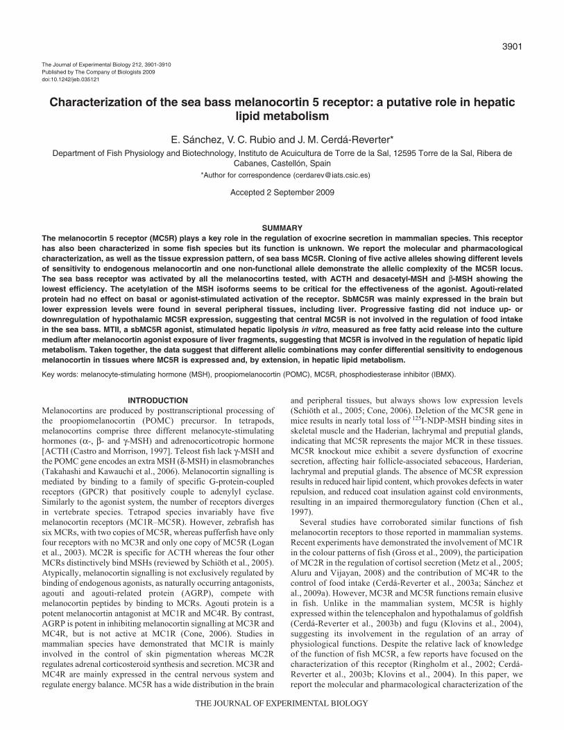
3901
INTRODUCTIONMelanocortins are produced by posttranscriptional processing ofthe proopiomelanocortin (POMC) precursor. In tetrapods,melanocortins comprise three different melanocyte-stimulatinghormones (-, - and -MSH) and adrenocorticotropic hormone[ACTH (Castro and Morrison, 1997]. Teleost fish lack -MSH andthe POMC gene encodes an extra MSH (-MSH) in elasmobranches(Takahashi and Kawauchi et al., 2006). Melanocortin signalling ismediated by binding to a family of specific G-protein-coupledreceptors (GPCR) that positively couple to adenylyl cyclase.Similarly to the agonist system, the number of receptors divergesin vertebrate species. Tetrapod species invariably have fivemelanocortin receptors (MC1R–MC5R). However, zebrafish hassix MCRs, with two copies of MC5R, whereas pufferfish have onlyfour receptors with no MC3R and only one copy of MC5R (Loganet al., 2003). MC2R is specific for ACTH whereas the four otherMCRs distinctively bind MSHs (reviewed by Schiöth et al., 2005).Atypically, melanocortin signalling is not exclusively regulated bybinding of endogenous agonists, as naturally occurring antagonists,agouti and agouti-related protein (AGRP), compete withmelanocortin peptides by binding to MCRs. Agouti protein is apotent melanocortin antagonist at MC1R and MC4R. By contrast,AGRP is potent in inhibiting melanocortin signalling at MC3R andMC4R, but is not active at MC1R (Cone, 2006). Studies inmammalian species have demonstrated that MC1R is mainlyinvolved in the control of skin pigmentation whereas MC2Rregulates adrenal corticosteroid synthesis and secretion. MC3R andMC4R are mainly expressed in the central nervous system andregulate energy balance. MC5R has a wide distribution in the brain
and peripheral tissues, but always shows low expression levels(Schiöth et al., 2005; Cone, 2006). Deletion of the MC5R gene inmice results in nearly total loss of 125I-NDP-MSH binding sites inskeletal muscle and the Haderian, lachrymal and preputial glands,indicating that MC5R represents the major MCR in these tissues.MC5R knockout mice exhibit a severe dysfunction of exocrinesecretion, affecting hair follicle-associated sebaceous, Harderian,lachrymal and preputial glands. The absence of MC5R expressionresults in reduced hair lipid content, which provokes defects in waterrepulsion, and reduced coat insulation against cold environments,resulting in an impaired thermoregulatory function (Chen et al.,1997).
Several studies have corroborated similar functions of fishmelanocortin receptors to those reported in mammalian systems.Recent experiments have demonstrated the involvement of MC1Rin the colour patterns of fish (Gross et al., 2009), the participationof MC2R in the regulation of cortisol secretion (Metz et al., 2005;Aluru and Vijayan, 2008) and the contribution of MC4R to thecontrol of food intake (Cerdá-Reverter et al., 2003a; Sánchez etal., 2009a). However, MC3R and MC5R functions remain elusivein fish. Unlike in the mammalian system, MC5R is highlyexpressed within the telencephalon and hypothalamus of goldfish(Cerdá-Reverter et al., 2003b) and fugu (Klovins et al., 2004),suggesting its involvement in the regulation of an array ofphysiological functions. Despite the relative lack of knowledgeof the function of fish MC5R, a few reports have focused on thecharacterization of this receptor (Ringholm et al., 2002; Cerdá-Reverter et al., 2003b; Klovins et al., 2004). In this paper, wereport the molecular and pharmacological characterization of the
The Journal of Experimental Biology 212, 3901-3910Published by The Company of Biologists 2009doi:10.1242/jeb.035121
Characterization of the sea bass melanocortin 5 receptor: a putative role in hepaticlipid metabolism
E. Sánchez, V. C. Rubio and J. M. Cerdá-Reverter*Department of Fish Physiology and Biotechnology, Instituto de Acuicultura de Torre de la Sal, 12595 Torre de la Sal, Ribera de
Cabanes, Castellón, Spain*Author for correspondence ([email protected])
Accepted 2 September 2009
SUMMARYThe melanocortin 5 receptor (MC5R) plays a key role in the regulation of exocrine secretion in mammalian species. This receptorhas also been characterized in some fish species but its function is unknown. We report the molecular and pharmacologicalcharacterization, as well as the tissue expression pattern, of sea bass MC5R. Cloning of five active alleles showing different levelsof sensitivity to endogenous melanocortin and one non-functional allele demonstrate the allelic complexity of the MC5R locus.The sea bass receptor was activated by all the melanocortins tested, with ACTH and desacetyl-MSH and -MSH showing thelowest efficiency. The acetylation of the MSH isoforms seems to be critical for the effectiveness of the agonist. Agouti-relatedprotein had no effect on basal or agonist-stimulated activation of the receptor. SbMC5R was mainly expressed in the brain butlower expression levels were found in several peripheral tissues, including liver. Progressive fasting did not induce up- ordownregulation of hypothalamic MC5R expression, suggesting that central MC5R is not involved in the regulation of food intakein the sea bass. MTII, a sbMC5R agonist, stimulated hepatic lipolysis in vitro, measured as free fatty acid release into the culturemedium after melanocortin agonist exposure of liver fragments, suggesting that MC5R is involved in the regulation of hepatic lipidmetabolism. Taken together, the data suggest that different allelic combinations may confer differential sensitivity to endogenousmelanocortin in tissues where MC5R is expressed and, by extension, in hepatic lipid metabolism.
Key words: melanocyte-stimulating hormone (MSH), proopiomelanocortin (POMC), MC5R, phosphodiesterase inhibitor (IBMX).
THE JOURNAL OF EXPERIMENTAL BIOLOGY

3902
sea bass MC5R (sbMC5R). The results provide the first evidenceof the complex allelic composition of the MC5R locus, as wellas the existence of null alleles producing nonfunctional proteinsand differences in the MC5R isoforms as derived form theirsensitivity to endogenous melanocortin agonists. In addition, ourdata provide the first evidence of the involvement of fish MC5Rin the control of hepatic lipid metabolism.
MATERIALS AND METHODSAnimals and reagents
Male and female sea bass (Dicentrarchus labrax L.) were kindlysupplied by Tinamenor (Santander, Spain). Prior to theexperiments, the animals were maintained in 500l tanks suppliedwith continuously aerated running sea water for 2months. Fishwere hand fed at 09:00h with a commercial diet (ProaquaNutrición, Palencia, Spain). Animals were anesthetized in 2-phenoxy-ethanol (0.1%) for 2min before any manipulation andkilled by rapid decapitation. All experiments were carried out inaccordance with the principles published in the European animaldirective (86/609/CEE) concerning the protection of experimentalanimals. Melanocortin peptides were from Bachem (Germany).Zebrafish agouti-related protein (zfAGRP), was kindly donatedby Dr Millhauser from Department of Chemistry, University ofCalifornia, Santa Cruz, USA (Sánchez et al., 2009a). Unlessotherwise indicated, all reagents were purchased from Sigma (StLouis, MO, USA).
Molecular cloning of sea bass MC5RGenomic DNA isolated from blood was used as template fortouchdown PCR reactions with Taq DNA polymerase (Invitrogen,Carlsbad, CA, USA) and degenerate primers designed againstconserved regions of the known MCR sequences. The 5� primer(MCR3Fw2) was a 20-mer with the sequence: 5�-TACCACAGYATCRTGACMGT-3�. The 3� primer (RevFish) hadthe sequence 5�-TSAGVGTGATGGCKCCCTT-3� (Fig.1). PCRproducts of about 274base pair (bp) were isolated from low meltingpoint (LMP) Nusieve GTG agarose gel (FMC) ligated into pGEM-T easy vector (Promega, Madison, WI, USA) and subsequentlytransformed into XLI-Blue E. coli. One clone that contained an insertof the expected size was sequenced. The 3� extreme was resolvedby 3� RACE PCR. Specific primers for RACE-PCR weresbMC5R_3RACE_1 (5�-GACAGTGAAGAGAGCCG-3�) andsbMC5R_RACE_2 (5�-TGCTATGCTGCTCATCATG-3�). Afragment of about 678bp was subcloned into pGEM-T easy vectorand sequenced. The 5� region was cloned by nested PCR using acDNA library as template (kindly supplied by Dr Bernardini formthe University of Insubria, Italy), sbMC5R-specific (MC5R_5�RACE_cDNA_synthesis: 5�-ACTCCTGAAGGCATAGAT-3� andMC5R-5WALKER2: 5�-GAACATTGAGACCAGGCAGATGA -TGACG-3�) and universal PBSSK-vector primers. A fragment ofabout 812bp was subcloned into pGEM-T easy vector andsequenced. Finally the coding region was amplified using specificprimers (Hind-sbMC5R-Forward 5�-TATAAGCTTATGAATG -CATCAGATGATCAGT-3�, Xho-sbMC5R-Reverse 5�-TTACT -CGAGTCATGTGGGCTTCTCCTCG-3� and brain cDNA,synthesized from total RNA pooled from several animals, astemplate. Fragments of about 990bp were subcloned into pGEM-T easy vector and sequenced on both strands. The nucleotidesequences of sbMC5Rs have been deposited in the EMBLNucleotide Sequence Database under accession numbers: P0:FN429774, P1: FN429769, P2: FN429770, P5: FN429772, P8:FN429773.
RT-PCR and Southern blot analysisTotal RNA was purified from fresh tissues (testis, ovary, intestine,fat, liver, white and red muscle, spleen, head kidney, posteriorkidney, gill, dorsal skin, ventral skin, eye, heart, pituitary and brain)and treated with RQ1-DNAse (Promega). Superscript II reversetranscriptase (Invitrogen) was used for cDNA synthesis by primingwith oligo(dT)12-18 (Invitrogen). The cDNA was subsequently usedas template for touchdown PCR reactions with Taq DNA polymerase(Invitrogen) and specific primers. The 5� primer was sbMC5R_3�RACE1 (5�-GACAGTGAAGAGAGCCG-3�) and the 3�-primerwas MC5R_5RACE_cDNA (see above). Subsequently, PCRfragments were separated on 1.2% agarose gel and transferred bycapillarity to Hybond-N nylon membrane (Amersham, Piscataway,NJ, USA). Membranes were prehybridized for at least 3h inhybridization solution (50% formamide, 6� SSPE, 0.5% SDS, 5�Denhardt’s solution and 10mgml–1 yeast tRNA type III; 1� SSPEcontained 150mmoll–1 NaCl, 1mmoll–1 EDTA, 9mmoll–1
NaH2PO4; pH7.4). Hybridization was carried out overnight in freshhybridization solution containing 0.5�106c.p.m.ml–1 [-32P]dCTPat 42°C. A probe containing the central region of the sbMC5R codingregion was used. Final washes were performed in 0.1� SSPE at65°C. After 2h and 3days of exposure at –80°C, films weredeveloped and scanned. Touchdown PCR for 18S RNA was carriedout as an internal control for the reverse transcription step. Primersequences were 18S_Fw 5�-GCATGCCGGAGTCTCGTT-3� and5� 18S_Rev 5�-TGCATGGCCGTTCTTAGTTG-3�.
Real-time quantitative PCRTo evaluate mRNA levels, total RNA from individual hypothalamusor pituitary was treated with RQ1-DNAse. One microgram was usedas template for cDNA synthesis, which was primed with randomhexaprimers (Invitrogen). Two microlitres of cDNA (sbMC5R) ordiluted cDNA (18S RNA) and primers (70nmoll–1) were added to7.5l of Sybr Green PCR master mix (ABgene, Thermo Scientific,Madrid, Spain) and the volume was adjusted to 15l with water.PCRs were carried out on an iCycler (Bio-Rad, Madrid, Spain).Data were analyzed with the Ct (cycle threshold) method. Asinternal control a fragment of the sea bass 18S RNA gene wasamplified, using primers 18S_Fw and 18S_Rev primers (see above).Expression of 18S RNA gene was demonstrated to be stable afterfasting in previous studies (Sánchez et al., 2009a). Specific geneprimers were as follows: qPCR_MC5R_Fw1 (5�-ATGAACACC -ACAGA GG CTCA-3�) and qPCR_MC5R_Rev1 (5�-ATAGCA -TCCTGTG GACGAGT3�).
Cell culture and transfectionHEK cells were transfected using a modified calcium phosphatetransfection method (Chen and Okayama, 1987) and grown inDMEM (Invitrogen) containing 10% foetal bovine serum(Invitrogen), penicillin (100i.u.ml–1) and streptomycin (100gml–1)in a humidified atmosphere of 5% CO2 at 37°C.
Galactosidase enzyme assayGalactosidase enzyme assays were performed as previouslydescribed (Sánchez et al., 2009a). Briefly, the medium was removedand 50l of lysis buffer containing 250mmoll–1 Tris-HCl pH8 and0.1% Triton X-100 were added. After one round of freezing(–80°C) and thawing, 10l of the lysate were preserved for proteinassays. Phosphate saline buffer (40l) containing 0.5% BSA and60l of substrate buffer (1mmoll–1 MgCl2, 10mmoll–1 KCl,5mmoll–1 -mercaptoethanol and 200mgml–1 o-nitrophenyl--D-galactopyranoside; ONPG) were added to the remaining lysate
E. Sánchez, V. C. Rubio, J. M. Cerdá-Reverter
THE JOURNAL OF EXPERIMENTAL BIOLOGY

3903Sea bass MC5R
volume. The plate was incubated at 37°C for 5h and the absorbancewas read at 405nm in a 96-well plate reader (Tecan). Measurementswere normalized for the protein content, which was determined usingthe BCA protein assay kit (Pierce, Rockford, IL, USA).
Pharmacological experimentsA HEK293 cell clone, stably expressing -galactosidase under thecontrol of vasoactive intestinal peptide promoter placeddownstream of tandem repeats of cAMP responsive elements(CRE), was generated by co-transfection (50:1) of pCRE/-galactosidase plasmid [kindly supplied by Dr R. Cone, VanderbiltUniversity Medical Center (Chen et al., 1995)] and thetgCMV/HyTK plasmid, which harbours a hygromycin resistancegene (Wellbrock et al., 1998). Cells were selected in mediumcontaining 400gml–1 hygromycin B (Invitrogen). -galactosidaseactivity was tested after incubating resistant clones in 96-wellplates (15,000 cells/well) with assay medium (DMEM medium +0.1mgml–1 bovine serum albumin, BSA + 0.1mmoll–1
isobutylmethylxanthine (IBMX), containing 10–6moll–1 forskolinfor 6h. The clone showing highest response to forskolin (Clon-Q) was selected for subsequent experiments (Sánchez et al.,2009a). The full coding region of the sbMC5R was released frompGEM-T easy vector (see above) and directionally subcloned intopcDNA3 (Invitrogen). Double stable clones expressing -galactosidase and sbMC5R were made by transfecting Clon-Q withthe latter construct using G-418 selection (800gml–1). Cloneswere tested by incubating cells with MTII 10–6moll–1 in the assaymedium. The clone Q/P5-23 was selected for followingcharacterization of the activation profiles in response to severalmelanocortins (monoacetyl-MSH or -MSH, diacetyl-MSH,desacetyl-MSH or MSH, human ACTH, monkey -MSH,zfAGRP, SHU9119 and HS024) in the absence of IBMX. Theeffect of zfAGRP on basal sbMC5R activity was studied in boththe presence and absence of the phosphodiesterase (PDE) inhibitor.
To test the activity of the different sbMC5R polymorphisms,Clon-Q was transiently transfected with sbMC5R-P0, -P1, -P2,-P5 or -P8 and a construct carrying the luciferase gene under thecontrol of a constitutive promoter (Muriach et al., 2008). After24h, cells were plated in 96-well plates (50,000 cells/well). Forty-eight hours post-transfection, cells were stimulated with severalendogenous melanocortins, including -MSH, diacetyl-MSH,desacetyl-MSH, human ACTH and monkey -MSH rangingfrom 10–6 to 10–8moll–1, as before. Measurements werenormalized for the protein content, the luciferase activity andforskolin-induced galactosidase activity. Receptor activationassays were always performed in quadruplicate wells and repeatedat least three times independently.
Effects of progressive fasting on melanocortin systemTo evaluate the effects of fasting on hypothalamic gene expression,10 groups of 10 fish each [body mass117±1.54g] wereacclimatized for 1week to individual 500l aquaria and fed adlibitum at 9:00h. After this acclimatization period, five groups werefed the same ratio, and the other five were fasted. One fish fromeach fed or fasted group was sampled at 12:00h (3h post-feedingin the case of the fed groups) at 1, 4, 8 and 15 and 29days,respectively. Anesthetized fish were weighed and blood sampleswere obtained by puncture of the caudal vessels. Subsequently thefish were decapitated and the whole hypothalamus dissected forimmediate total RNA extraction. RNA samples were kept at –80°Cin 75% ethanol until cDNA synthesis for quantitative PCRs (seeabove).
Effects of melanocortin agonist on hepatic lipolysisTo evaluate the effects of melanocortin agonist on hepaticlypolysis, the animals were sacrificed and their livers carefullyremoved. Small liver slices (50–80mg) were dissected andincubated in 1ml HB medium (136.9mmoll–1 NaCl, 5.4mmoll–1
KCl, 0.81nmoll–1 MgSO4, 0.44mmoll–1 KH2PO4, 0.33mmoll–1
Na2HPO4, 5mmoll–1 NaHCO3, pH7.6), containing 5mmoll–1
glucose, 1.5mmoll–1 CaCl2 and 2.5% fat-free BSA for 60min at25°C. Subsequently, the medium was removed and slices wereincubated with 1ml of HBS containing 10–6moll–l MTII. After 2and 4h at 25°C medium was removed for determination of non-esterified fatty acids (NEFA) using commercial kits (WAKODiagnostics, Neuss, Germany), following the supplier’srecommendations. Liver slices were mechanically homogenizedfor protein determination using the BCA protein assay kit (Pierce).Subsequently, dose–response studies were done by incubating liverslices with MTII in HB ranging from 10–6 to 10–9moll–1 for 4h.Experiments were always performed in quadruplicate wells andrepeated at least three times independently.
Data analysis and statisticsSequence comparisons and alignments were performed usingClustalX. A phylogenetic tree was derived using public domainClustalX, which uses the Neighbour-Joining method on a matrix ofdistances. The position of transmembrane domains was drawnaccording to secondary structure prediction for human MC1R(Haitina et al., 2007a). Receptor activation data were fitted usingSigmaPlot software. Differences in the sensitivity of sbMC5Risoforms and sbMC5R expression levels were analyzed by one-wayANOVA (P<0.05).
RESULTSMolecular cloning of sbMC5R
By means of RT-PCR and using degenerate primers designed againstconserved regions of fish melanocortin receptor sequences, wecloned a 274bp fragment showing high identity with the MC5Rsequences reported in other vertebrate species. The 5� and 3� regionswere resolved by regular PCR and 3� RACE PCR using a cDNAlibrary or brain cDNA as templates, respectively. The full-lengthcDNA sequence was amplified by PCR using specific primers andbrain cDNA as template. Fig.1 shows the full-length cDNAsequence of isoform 1 of the sbMC5R (P1), which was sequencedtwice on both strands. The cloned fragment contains an open readingframe of 1080bp that encodes a putative 360 amino acid proteinwith seven putative hydrophobic transmembrane domains (TMDs;Figs1, 2). Similar to other melanocortin receptors, the sbMC5Rorthologue has short extracellular (ECL) and intracellular (ICL)loops and has cysteine residues at positions 266, 279 and 285 (seabass numbering), which are fully conserved in all melanocortinreceptors (Fig.2 and http://www.gpcr.org). The deduced amino acidsequence shares 64–82% identity with other MC5R sequences butless than 59% with other MCR sequences. The identity is unequallydistributed, and the N-terminal extracellular domain has the lowestsimilarity to other MC5Rs, including pufferfish MC5R. More than50% of the divergence between sea bass and pufferfish MC5Rorthologues resides within the N-terminal domain, encoding only14% of the total protein length. A more detailed comparison amongcloned fish MC5R shows that the overall identity level of sea bassreceptor with other MC5Rs is highest (>96%) within TMD7 andlowest in TMD4 (<68%). The overall identity ranged between 80%and 92% within TMD1, TMD2, TMD3, TMD5 and TMD6. Unlikeother MC5Rs, the sea bass protein has an extended intracellular tail
THE JOURNAL OF EXPERIMENTAL BIOLOGY

3904
(ICL4). The deduced amino acid sequence has potential N-glycosylation sites within the N-terminal domain at positions 2, 17and 28 (Fig.1). Sea bass MC5R also has a PMY motif in the firstICL, which is conserved in most melanocortin receptors. The motifDRY of TM3, a consensus motif of the class A rodopsin-like Gprotein-coupled receptor, is also present. Consensus recognition sitesfor protein kinase C (PKC) and cAMP- and cGMP-dependentprotein kinases within ICLs were found at positions 170 in ICL2,248 in ICL3 and 320, 329 and 339 within ICL3 (Fig.1), suggestingregulation by phosphorylation.
We found five receptor isoforms (P0, P1, P2, P5, P8), which weresequenced twice on both strands, involving nine single nucleotidepolymorphisms (SNPs) at positions 42, 71, 274, 446, 652, 829, 866,937 and 1074 (Fig.1). These SNPs result in eight putative aminoacid substitutions at positions 14, 24 (ECL1), 92 (TM2), 149 (TM4),218 (TM6), 277 (ECL4), 289 (TM6) and 313 (ICL4, Fig.3). Otherthan P0, P5 was the most divergent isoform, exhibiting four uniquesubstitutions within the ECL1, TMD5, TMD7, ICL4. By contrast,P1 and P2 were found to have unique substitution within TMD2and ECL4, respectively, whereas P8 had no unique substitutions.There were no coincident substitutions between P2 and P5, differentfrom those observed in P1 and P8. The isoform P0 had an extrathymine base at position 551/552 (Fig.1), involving a change in thereading frame and resulting in a divergent and shortened C-terminalregion of the receptor (Fig.3). Analysis of the receptor structure
predicts the disruption of the transmembrane structure within theC-terminal region.
Peripheral and central distribution of sbMC5R mRNART-PCR using specific primers targeting sequences within TMD4and TMD7 of sbMC5R resulted in a band of the expected size ofabout 436bp (Fig.4A). The identity of the band was confirmed bySouthern blot hybridization with a sbMC5R probe including the fullcoding region. sbMC5R mRNA was easily detected in the retina,brain, liver, spleen, gill, testis and dorsal skin. Low levels were alsodistinguishable in the pituitary, posterior kidney, fat tissue, intestine,red muscle and ovary (Fig.3A). Finally residual levels were detectedin the head kidney, dorsal skin, white muscle and heart. Inversetranscriptions and cDNA quality were corroborated by PCRamplification of 18S RNA that yielded bands of expected size inall reactions (Fig.4B).
Activation by melanocortin analoguesFor pharmacological and functional characterization of sbMC5R,the coding region of the P5 isoform, was ligated into pcDNA3 andstably expressed in HEK293 cells already producing -galactosidaseunder the control of cAMP responsive elements. SbMC5R waspositively coupled to the cAMP-signalling pathway in response toall melanocortins tested (Fig.5). NDP-MSH was the most potentagonist, activating the receptor with a half-maximal effective
E. Sánchez, V. C. Rubio, J. M. Cerdá-Reverter
I S M N A S D Q S S Y Q E E V M L G N S T W A Y Y Y Y H Q 27 1 ATGAATGCAT CAGATCAGTC CTCCTACCAA GAGGAGGTGA TGCTGGGTAA CTCCACCTGG GCTTATTACT ACTATCACCA A(P5) C(P5;P8)
N Y T L S P P L L P D K T S T S K P A A C E Q V H I 53 81 AAACTACACC CTGTCCCCTC CACTGCTACC AGACAAGACC AGCACTTCCA AACCTGCAGC ATGCGAGCAG GTCCACATCG
A I E V F L I L G I I S L L E N I L V I T A I V K N K 80 161 CTATAGAGGT CTTTCTGATC CTGGGTATCA TCTCCCTGCT GGAGAACATC CTGGTCATCA CAGCCATAGT AAAGAACAAG
S N L H S P M Y F F V C N L A V A D M L V S V S N A W E 107 241 AACCTGCACT CACCCATGTA CTTCTTCGTT TGCAATCTGG CAGTAGCCGA CATGCTGGTG AGCGTGTCCA ACGCCTGGGA G(P2;P5;P8)
T I I I Y L L N N R Q L V V E D H F I R Q M D N V F 133 321 GACCATCATT ATTTACCTCC TCAACAACAG GCAGCTGGTT GTGGAGGACC ACTTCATCAG GCAGATGGAC AACGTGTTTG
P D S M I C I S V V A S M C S L L A I A V D R Y V T I F 160 401 ACTCCATGAT CTGCATCTCT GTGGTTGCGT CCATGTGTAG CCTGCTGGCC ATCGCTGTGG ATAGGTACGT CACTATCTTC C(P0)
Y A L R Y H N I M T V K R A G F I I A G I W T F C T G 187 481 TATGCGCTGA GGTATCACAA CATCATGACA GTGAAGAGAG CCGGCTTCAT CATCGCGGGA ATCTGGACCT TCTGCACGGG *(extra T;P0)
C G I V F I I Y S D T T P V I I C L V S M F F A M L 213 561 CTGCGGGATC GTCTTCATCA TCTACTCAGA CACCACCCCC GTCATCATCT GCCTGGTCTC AATGTTCTTT GCTATGCTGC
T L I M A S L Y S H M F M L A R S H V K R I A A L P G Y 240 641 TCATCATGGC CTCCCTCTAC AGCCACATGT TCATGCTGGC GCGCTCGCAC GTCAAGCGCA TCGCCGCCCT GCCTGGCTAC A(P5)
N S I H Q R A S M K G A I T L T I L L G I F I V C W A 267 721 AACTCCATCC ACCAGCGGGC CAGCATGAAG GGGGCCATCA CGCTGACCAT CCTGCTGGGG ATCTTCATCG TCTGCTGGGC
V T P F F L H L I L M I S C P R N L Y C V C F M S H F N 293 801 GCCCTTCTTC CTCCACCTGA TCCTGATGAT CTCCTGTCCG CGGAACCTCT ACTGCGTGTG CTTCATGTCC CACTTCAACA G(P2) C(P5) G M Y L I L I M C N S V I D P L I Y A F R S Q E M R K T 320 881 TGTACCTTAT CCTCATCATG TGCAACTCTG TGATTGACCC GCTCATCTAT GCCTTCAGGA GTCAGGAGAT GAGGAAGACT G(P5)
F K E I I C C Y S L R N A C T N I C T L T E T A A K T 347 961 TTTAAGGAGA TCATCTGCTG TTACAGCCTG AGAAACGCCT GCACCAACAT CTGCACCCTG ACAGAAACAG CAGCAAAAAC
L N V I R H R A E E K P T 3601041 ACTGAACGTG ATACGACATC GGGCCGAGGA GAAGCCCACA A(P2)
Fig.1. Nucleotide and deduced amino acidsequence of sea bass melanocortinreceptor 5 (sbMC5R), P1 isoform.Nucleotide and amino acid sequencenumbers are indicated on the left and rightsides, respectively. Position of differentpolymorphisms and the correspondingisoform are shown. Underlining indicatesputative transmembrane domains (Haitinaet al., 2007a). Boxes indicate putativeN-linked glycosylation sites; circles,possible phosphorylation sites. Sea bassMC5R_P1 sequence accession number isFN429769.
THE JOURNAL OF EXPERIMENTAL BIOLOGY

3905Sea bass MC5R
concentration (EC50) of 0.133nmoll–1. Activation by MTII andSHU9119 showed an EC50 of 1.82nmoll–1, and 3.72nmoll–1,respectively, whereas the effective concentration increased to22.4nmoll–1 and 93.3nmoll–1, and 107nmoll–1 and 167nmoll–1
when cells were incubated with HS024, diacetyl-MSH, -MSH andmonkey -MSH, respectively. Sigmoid curves cannot be fitted fordesacetyl-MSH and ACTH since receptor was only activated by thehighest melanocortin concentrations (1moll–1).
SbMC5R was also activated by the non-selective melanocortinagonist MTII (EC505.69nmoll–1). When a phosphodiesteraseinhibitor (IBMX) was added to the medium, the response of thereporter gene to incubation with MTII increased(EC500.167nmoll–1). The incubation of sbMC5R-expressing HEKcells with AGRP, in the presence or absence of IBMX, had no effectson receptor activation. Under these conditions, AGRP had no effecton MTII-stimulated cAMP production (EC5017nmoll–1 and
* 20 * 40 * 60 * 80 * 100 * 120Homo sapiens MC5R :-MNSSFHLHFLDLNLNATEG--------NLSGPN-------VKNKSSPCEDMG IAVEVFLTLGVISLLENILVIGA IVKNKNLHSPMY FFVCSLAVADMLVSMSSAWETITI YLLNNKHL:104Takifugu rubripes MC5R :-..T.HRSSDPQEGIMGNSTWNPLSYQP.FTLSPPLLP----.T.TAA..QLH..I.......I..........M...........................V.N.S...I.......Q.:115Dicentrarchus labrax MC5R P1:-..A.DQSSYQEEVMLGNSTWAYYYYHQ.YTLSPPLLPDKTSTS.PAA..QVH..I....I..I..........T.................N.........V.N.....I......RQ.:119Cyprinus carpio MC5RII :-..T.-EATLSLWAIYVNS--SPVSYLL.VTETP-------SHA.PKA..QLN..T....I..IV.........C...........................V.N.....I....T.RQ.:109Cyprinus carpio MC5RI :-..T.-EATLSLWAIYVNS--SPVSYLL.VTETP-------SHA.PKA..QLN..T....I..IV.........C...........................V.N.....V....T.RQ.:109Carassius auratus MC5R :M..T.-EATLSLWAIS.NS--SPVLDLL.TTETP-------SHA.PKA..QLN..T....I..I..........C...........................V.N.....V....T.RQ.:110Danio rerio MC5Ra :--------------MHVNS--SPASYIL.ATETP-------SH..PKA..QLN..T....I..IV.........C...........................V.N.....V....T.RQ.: 97Danio rerio MC5Rb :-....EWPTLSPNLSLSQAN------LSDE.SRP-------KTSA.AA..QVH..P.......L..........L...........................V.N.....V.H..A.RS.:106
* 140 * 160 * 180 * 200 * 220 * 240Homo sapiens MC5R :VIADA FVRHIDNVFDSMICISVVASMCSLLA IAVDRYVTIFYALRYHHIMTAR RSGAIIAGIWAFCTGCGIVFIL YSESTYVIL CLISMFFAMLFLLVSLYIHMF LLARTHVKRIAALP-:223Takifugu rubripes MC5R :IAE.HLI.QL.....................................N...V..A.C..G...T..........I..DT.P..I..VC......LIMA...S...M...S.........-:234Dicentrarchus labrax MC5R P1:.VE.H.I.QM.....................................N...VK.A.F......T..........I..DT.P..I..V.......LIMA...S...M...S.........-:238Cyprinus carpio MC5RII :.VE.H.I.QM.....................................N...V..AAL..G...T..........I..DN.S..V..V....I..A.MA...S...M...S.........-:228Cyprinus carpio MC5RI :.VE.H.I.QM.....................................N...V..AAL..G...T..........I..DN.S..V..V....I..A.MA...S...M...S.........-:228Carassius auratus MC5R :.VE.H.I.QM.....................................N...V..AAF..G...T...S......I..DN.S..V..V....I..A.MA...S...M...S.........-:229Danio rerio MC5Ra :.VE.H.I.QM.....................................N...V..AAL..G...T..........I..DN.S..V..V....I..A.MA...S...M...S.........-:216Danio rerio MC5Rb :..E.H.I.QM......L......G..W....................N...V..A.IL.GS..T.S.S...I..I..DTKP.VV..VA......LMMA...S.......S....M....-:225
* 260 * 280 * 300 * 320 * 340 * 360Homo sapiens MC5R :--GASS-ARQRTSM QGAVTVTMLLGVFTVCWAPFFLHLTLM LSCPQNLYCS RFMSHFNMYLILIMCNSVMDPLIYAF RSQEMRKTFKEIIC-CRGFRIACS----FPRRD----------:325Takifugu rubripes MC5R :--.SN.-IH..A..K..I.L.I...I.II.........I..I...R....MC.................I....................F-.YSL.NT..TICTL.GKY----------:340Dicentrarchus labrax MC5R P1:--.YN.-IH..A..K..I.L.I...I.I..........I..I...R....VC.................I.....................-.YSL.N..TNICTLTETAAKTLNVIRHR:354Cyprinus carpio MC5RII :--.YN.-IH..A..KA...L.I...I.I..........I..I...R....MC.................I...............L.....-.YSL.NVFG----MS.------------:328Cyprinus carpio MC5RI :--.YN.-IH..A..KA...L.I...I.I..........I..I...R....MC.................I...............L.....-.YSL.NVFG----MS.------------:328Carassius auratus MC5R :--.YN.-IH..A..KA...L.I...I.I..........I..I...R....MC.................I...............L.....-.YSL.NVFG----MS.------------:329Danio rerio MC5Ra :--.YN.-IH..A..KA...L.I...I.I..........I..I...R....MC.................I...............L.....-.YSL.NVFG----MS.------------:316Danio rerio MC5Rb :--.YNANI...A..K....L.I...I.I..........I..I...R.I..VC.................I......L............V.-.E.L.SF.N----MVSKY----------:328
Homo sapiens MC5R :------: -Takifugu rubripes MC5R :------: -Dicentrarchus labrax MC5R P1:AEEKPT:360Cyprinus carpio MC5RII :------: -Cyprinus carpio MC5RI :------: -Carassius auratus MC5R :------: -Danio rerio MC5Ra :------: -Danio rerio MC5Rb :------: -
Fig.2. Alignment of MC5R amino acid sequences. Sea bass MC5R sequence is in bold letters. Dots indicate amino acids identical to the top sequence.Dashes indicate spaces introduced to improve alignment. Grey boxes show putative transmembrane domains (see Fig.1).
* 20 * 40 * 60 * 80 * 100 MC5R_P1 : MNASDQSSYQEEVMLGNSTWAYYYYHQNYTLSPPLLPDKTSTSKPAACEQVHIAIEVFLILGIISLLENILVITAIVKNKNLHSPMYFFVCNLAVADMLV : 100 MC5R_P2 : ...........................................................................................S........ : 100 MC5R_P8 : .......................S...................................................................S........ : 100 MC5R_P5 : .............I.........S...................................................................S........ : 100 MC5R_P0 : .......................S...................................................................S........ * 120 * 140 * 160 * 180 * 200 MC5R_P1 : SVSNAWETIIIYLLNNRQLVVEDHFIRQMDNVFDSMICISVVASMCSLLAIAVDRYVTIFYALRYHNIMTVKRAGFIIAGIWTFCTGCGIVFIIYSDTTP : 200 MC5R_P2 : .................................................................................................... : 200 MC5R_P8 : .................................................................................................... : 200 MC5R_P5 : .................................................................................................... : 200 MC5R_P0 : ................................................P...................................LH.LRDRLHHLLRHH. : 200 * 220 * 240 * 260 * 280 * 300 MC5R_P1 : VIICLVSMFFAMLLIMASLYSHMFMLARSHVKRIAALPGYNSIHQRASMKGAITLTILLGIFIVCWAPFFLHLILMISCPRNLYCVCFMSHFNMYLILIM : 300 MC5R_P2 : ............................................................................V....................... : 300 MC5R_P8 : .................................................................................................... : 300 MC5R_P5 : .................T......................................................................T........... : 300 MC5R_P0 : RHHLPGLNVLCYAAHHGLPLQPHVHAGALARQAHRRPAWLQLHPPAGQHEGGHHADHPAGDLHRLLGALLPPPDPDDLLSAEPLLRVLHVPLQHVPYPHH : 300 * 320 * 340 * 360 MC5R_P1 : CNSVIDPLIYAFRSQEMRKTFKEIICCYSLRNACTNICTLTETAAKTLNVIRHRAEEKPT : 360 MC5R_P2 : ............................................................ : 360 MC5R_P8 : ............................................................ : 360 MC5R_P5 : ............G............................................... : 360 MC5R_P0 : VQLCD------------------------------------------------------- : 305
MC5R_P0
MC5R_P1
Fig.3. Alignment of sbMC5R isoforms. Dots indicate amino acids identical to the top sequence. Amino acid sequence numbers are indicated on the rightside. Underlined amino acids indicate putative transmembrane domains (see Fig.1). Amino acids that only change in one sequence are represented in red,whereas amino acids present in at least two sequences are shown in green. Inset shows sequencing chromatograms comparing DNA sequences ofisoforms P0 and P1. Note the extra thymine base in the P0 sequence, leading to a shift in the reading frame that results in a shortened and divergent C-terminal region in the P0 isoform. Sea bass MC5R sequence accession numbers: P0: FN429774, P1: FN429769, P2: FN429770, P5: FN429772, P8:FN429773.
THE JOURNAL OF EXPERIMENTAL BIOLOGY

3906
0.15nmoll–1, in the absence or presence of IBMX, respectively;Fig.6).
Subsequently, we used HEK293 cells stably expressing agalactosidase reporter gene to transiently transfect the differentsbMC5R isoforms (P0, P1, P2, P5, P8). The isoform P0 never
responded to any melanocortin peptide, whereas the binding ofendogenous melanocortin to the P1-P8 isoforms differentiallyactivated the galactosidase gene transcription. Isoforms P1 and P8activated the reporter gene more efficiently than P2 and P5 whenincubated with ACTH, -MSH, -MSH and desacetyl-MSH, butnot with diacetyl-MSH (Fig.7).
Effects of fasting on sbMC5R expressionQuantitative PCR did not yield significant differences inhypothalamic MC5R mRNA expression levels when fed and fastedanimals were compared after 1, 4, 8, 15 and 29 days of fasting (datanot shown).
Effects of melanocortin agonist on hepatic lipolysisThe incubation of liver slices with 10–6moll–1 MTII in HBSresulted in a significant increase in total NEFA concentration in theculture medium after 4h but not after 2h (Fig.8A). Only the highestMTII concentrations significantly stimulated NEFA production invitro (Fig.8B).
DISCUSSIONThe present study provides evidence of the allelic complexity ofthe MC5R locus in sea bass and substantiates the presence of severalisoforms that exhibit different sensitivity to endogenousmelanocortin agonist. We also describe a non-functional isoformof the sbMC5R with a divergent C-terminal region. Tissueexpression patterns revealed high expression levels in the centralnervous system with lower levels in several peripheral tissuesincluding liver. The function of MC5R in fish is unknown; however,we demonstrate that MTII, a potent agonist of the sbMC5R,
E. Sánchez, V. C. Rubio, J. M. Cerdá-ReverterR
etin
a
Hea
d ki
dney
Pitu
itary
Live
r
Bra
in
Pos
terio
r ki
dney
Spl
een
Gill
Fat t
issu
e
Inte
stin
e
Test
is
Dor
sal s
kin
Ven
tral
ski
n
Red
mus
cle
Whi
te m
uscl
e
Hea
rt
Ova
ry
Con
trol
A
B
A
B
Fig.4. Distribution of sea bass MCR5 mRNA expression in different tissues,as revealed by RT-PCR followed by Southern blot hybridization.(A)Phosphoimaging screen showing a Southern blot analysis of the seabass receptor following RT-PCR, and (B) ethidium bromide-stained agarosegel of RT-PCR amplifications of sea bass 18S RNA.
Dose (mol l–1)
10–13 10–12 10–11 10–10 10–9 10–8 10–7 10–6 10–5
Gal
acto
sida
se a
ctiv
ity (
% o
f bas
al v
alue
)
50
100
150
200
250
300
350
400
ACTH
α-MSH
β-MSH
Diacetyl-MSH
NDP-MSH
Desacetyl-MSH
MTII
SHU9119
HS024
Fig.5. Effects of synthetic (HS024, MTII, NDP-MSH, SHU9119), andendogenous (ACTH, -MSH, -MSH, diacetyl-MSH and desacetyl-MSH)melanocortin on galactosidase activity in HEK293 cells stably expressingboth sbMC5R-P5 and a cAMP-responsive -galactosidase reporter gene inthe absence of phosphodiesterase inhibitors (IBMX). Data were normalizedto protein levels and expressed as percentage of the basal levels.
50
100
150
200
250
300
350
400 MTII
MTII_IBMX
AGRP
AGRP_IBMX
MTII+AGRP
MTII+AGRP_IBMX
Dose (mol l–1)
10–13 10–12 10–11 10–10 10–9 10–8 10–7 10–6 10–5
Gal
acto
sida
se a
ctiv
ity (
% o
f bas
al v
alue
)
Fig.6. Effects of inverse agonist, agouti-related protein (AGRP), on basaland MTII-stimulated galactosidase activity in HEK293 cells stablyexpressing both sbMC5R-P5 and a cAMP-responsive -galactosidasereporter gene in the presence or absence of phosphodiesterase inhibitors(IBMX). Data were normalized to protein levels and expressed aspercentage of the basal levels.
THE JOURNAL OF EXPERIMENTAL BIOLOGY

3907Sea bass MC5R
stimulates hepatic lipolysis, suggesting that the different MC5Risoforms could play a role in hepatic metabolism.
Amino acid sequence comparisons show that the cloned receptorin sea bass displays high identity with other cloned MC5Rs. Mostof the divergence in sbMC5R is within the N-terminal extracellulardomain. However, this receptor domain is not likely to befunctionally important for agonist binding, since it has beendemonstrated that 28 amino acids from the N-terminal extracellulardomain can be deleted, including all potential N-glycosylation sites,without affecting ligand binding to human MC5R (Schiöth et al.,1997). Similarly, ECLs of the mouse MC5R do not participate inthe ligand binding of melanocortin agonist (Schiöth et al., 1998).Accordingly, sbMC5R has low sequence identity with other MC5Rs
within the ECLs but, in contrast, shares a high sequence identitywith other MC5Rs within the TMDs. Point mutagenesis of humanMC1R suggests that important determinants for agonist binding liewithin TMD2, TMD3 and TMD7 (Yang et al., 1997), wheresbMC5R has higher than 82%, 83% and 92% sequence identity,respectively, when compared with other MC5Rs.
To our knowledge, the MC5R locus, unlike MC1R, has not beenreported to be highly polymorphic in species in which the receptorhas been cloned (García-Borrón et al., 2005; Sanchez et al., 2009b).However, we found nine DNA polymorphisms during the cloningprocess. Eight mutations resulted in substitution of a different aminoacid and only one was a silent mutation. These polymorphismswere incorporated into five different isoforms. Furthermore the P0
0
50
100
150
200
250
Isoform P0
Isoform P1
Isoform P2
Isoform P5
Isoform P8
β-MSH
0
50
100
150
200
250 Desacetyl-MSH
Diacetyl-MSH
0
50
100
150
200
250
***
a
bb
cb,c
*
***
a a a
bb
b b
*
b bc c c
c
b
b
*bbbb
a a
c c
b
b,c
bb
c**
**
c*
*
* ** *
a
c c
b b
cc
b b
a
a aa
b b
* *
* *
a
bb
bb
bb
b
b b bb
aa
a,b
Basal
ACTH α-MSH
Dose (mol l–1)
10–810–710–6
Gal
acto
sida
se a
ctiv
ity (
% o
f bas
al v
alue
)
*
Basal 10–810–710–6 Basal 10–810–710–6
Basal 10–810–710–6 Basal 10–810–710–6
Dose (mol l–1)
Fig.7. Effects of endogenous (ACTH, -MSH, -MSH, diacetyl-MSH and desacetyl-MSH) melanocortin on galactosidase activity in HEK293 cells transientlyexpressing different sbMC5R isoforms and the luciferase gene under the control of a constitutive promoter and stably expressing a cAMP-responsive -galactosidase reporter gene. Experiments were done in the absence of phosphodiesterase inhibitors (IBMX). Data were normalized to protein levels,luciferase activity and forskolin-induced galactosidase activity and expressed as percentage of the common basal levels. Asterisks indicate differences fromthe basal level of that compound, after one-way ANOVA. Different letters indicate differences between isoforms at the specified hormone concentration, afterone-way ANOVA (P<0.05).
THE JOURNAL OF EXPERIMENTAL BIOLOGY
3908
isoform had an extra thymine that induces a change in the readingframe, resulting in a divergent and shortened C-terminal region.We initially selected the P5 isoform for pharmacologicalcharacterization of the sbMC5R. Activation experimentsdemonstrated that sbMC5R is activated by all melanocortinmimetics. NDP-MSH, MTII and SHU9119, antagonists of sbMC4R(Sánchez et al., 2009a), were the most potent agonists of thesbMC5R with ED50s within the nanomolar range. Similarly, NDP-MSH was also the most potent agonist of zebrafish (Ringholm etal., 2002) and pufferfish MC5Rs (Klovins et al., 2004). Bothdiacetyl-MSH and -MSH similarly stimulated cAMP-dependentgalactosidase activity but desacetyl-MSH-induced activation wasless efficient. Therefore, acetylation seems to be critical forsbMC5R activation but not the level of acetylation since monoacetyl(-MSH) and diacetyl-MSH were equipotent when activating thereceptor. All three MSH isoforms have been described in fishpituitary (Dores et al., 1993) and the regulation of MSH acetylationseems to be an important mechanism for background adaptationand pigment patterns (Arends et al., 2000; Kobayashi et al., 2009).SbMC5R was weakly activated by incubation with human ACTHand -MSH. Experiments in trout demonstrated that MC5R affinityfor human ACTH (1-24) was higher than that observed for -MSHand -MSH, both exhibiting equipotent binding capacities on troutMC5R (Haitina et al., 2004). Our data suggest that sbMC5R is notspecific for ACTH or -MSH, although no definitive conclusionscan be drawn since we used human and monkey peptides,respectively. Studies in lamprey have also demonstrated that thecloned MCR subtype exhibits higher affinity to the lamprey ACTHthan to the MSH-A suggesting that ACTH is the ancestral ligand(Haitina et al., 2007b).
Previous experiments in our laboratory demonstrated thatzebrafish AGRP (zfAGRP) is able to decrease the basal activity ofboth sbMC4R and sbMC1R when incubated with aphosphodiesterase inhibitor (IBMX), as occurs with an inverseagonist, and decrease the effects of the melanocortin agonists onthe sbMC4R activation (Sánchez et al., 2009a; Sánchez et al.,2009b), as occurs with an endogenous antagonist. We furtherdemonstrate here that this effect is specific for sbMC1R andsbMC4R, since zfAGRP had no effect on basal sbMC5R activityin the absence or presence of phosphodiesterase inhibitors. Inaddition, our data corroborate the role of the phosphodiesterasesystem in the AGRP inverse agonism. As expected, the addition ofIBMX decreased ED50 values of agonist-induced galactosidaseactivity, thus increasing the sensitivity of the assay. In addition,AGRP could not decrease the MTII-induced activation of thesbMC5R as a competitive antagonist does. When we further studiedthe activation of the different sbMC5R isoforms by the endogenousmelanocortin agonist, the P0 isoform, which has a shortened andhighly divergent C-terminal region, was ineffective in mediatingthe cAMP-induced galactosidase activity upon exposure to naturalligands. However, from our data it is not possible to discriminatewhether the binding properties of the receptor or its coupling to G-proteins were disrupted. However, they strongly suggest that P0 isa non-functional receptor isoform. The P1 and P8 isoforms displayedhigher sensitivity to endogenous melanocortins than the P2 and P5isoforms, suggesting that specific MC5R allelic combinations mayconfer differential sensitivity to endogenous melanocortin. It isdifficult to derive any conclusions concerning any structure–activityrelationship among active isoforms since no common substitutionpatterns between P2/P5 and P1/P8 were found.
MC5R has different expression patterns in different vertebratelineages. As in mammalian species, sbMC5R was expressed in avariety of peripheral tissues (Chen et al., 1997). Although noquantitative experiments were carried out, sbMC5R mRNA levelsin the CNS seem to be higher than in the peripheral tissues. Highexpression levels in the brain were also reported in other fish speciesincluding zebrafish (Ringholm et al., 2002), goldfish (Cerdá-Reverter et al., 2003b), pufferfish (Klovins et al., 2004), trout(Haitina et al., 2004) and carp (Metz et al., 2005). The CNSexpression of MC5R has also been reported in human (Chhajlaniet al., 1993), rat (Griffon et al., 1994) and chicken (Takeuchi andTakahashi, 1998) but very low levels were detected in mouse brain(Gantz et al., 1994). The central function of MC5R is unknown butexperiments in rodent have demonstrated that central administrationof melanocortin stimulates aggression. By contrast, MC5R-deficientmice exhibited reduced aggression and an elevated defence againstwild-type opponents (Morgan and Cone, 2006). The high expressionlevels of MC5R in fish brain suggest implemented central functionscompared with mammalian systems. Our previous experiments ingoldfish demonstrated that the melanocortin system is involved inthe control of food intake. Central administration of melanocortinanalogues inhibit food intake (Cerdá-Reverter et al., 2003a; Cerdá-Reverter et al., 2003b), whereas fasting dramatically increaseshypothalamic AGRP expression (Cerdá-Reverter and Peter, 2003).Similar to sbMC4R (Sánchez et al., 2009a), progressive fasting didnot induce changes in hypothalamic sbMC5R expression, suggestingthat food deprivation cannot induce a downregulation of receptorlevels in the hypothalamic structures. The sbMC4R exhibitsconstitutive activity and its actions are regulated by binding of theinverse agonist AGRP (Sánchez et al., 2009a). The above modelcannot account for the MC5R-regulated hypothalamic pathwayssince AGRP does not work as an inverse agonist or competitive
E. Sánchez, V. C. Rubio, J. M. Cerdá-Reverter
A
Time (h)2 40
0
10
20
30
40
50
Basal
MTII
Control
mg
NE
FA m
g–1 p
rote
in
0
10
20
30
40
50
aa
a
b
c
aa
aa
b
B
Dose (mol l–1)–9 –8 –7 –6
Fig.8. Effects of melanocortin agonist MTII on hepatic lipolysis measuredas release of non-esterified fatty acids (NEFA) after hormonal treatment ofthe culture medium. (A)Time course experiments using MTII at 10–6moll–1
after 2 and 4h incubation. (B)Dose–response assay after 4h incubation.
THE JOURNAL OF EXPERIMENTAL BIOLOGY

3909Sea bass MC5R
antagonist. POMC expression levels in the brain were also constantafter progressive fasting in sea bass (Sánchez et al., 2009a) andgoldfish (Cerdá-Reverter et al., 2003c). In the absence of modulationof POMC expression levels by fasting and of any effect of AGRPon basal or stimulated sbMC5R activity, an up- or downregulationof sbMC5R expression after fasting would seem likely if the receptoris involved in the control of food intake. Therefore, our data do notsupport the involvement of this receptor in the control of food intakein fish.
In mice, MC5R deficiency also causes severe dysfunction of theexocrine glands by mis-regulation of sebaceous and preputial lipidsynthesis (Chen et al., 1997). We and others have found high levelsof MC5R mRNA in the fish skin and gills (Cerdá-Reverter et al.,2003b; Metz et al., 2005) (present results), suggesting that MC5Rmay be involved in the mucus production by globet cells presentin the gills and skin, and/or additional secretory skin cells (Shepard,1994). RT-PCR experiments demonstrated expression of sbMC5Rin the ventral skin but no expression was found in the dorsal region.Similar to many other vertebrates, sea bass have a specific pigmentpattern with darker colour in the dorsal area and a progressivegradation towards white-yellow tones in the abdominal skin.Differential MC5R expression in the ventral skin suggests a role inpigment synthesis of the sea bass. Interestingly, sbMC5R wasexpressed in tissues containing a high number of macrophages, i.e.spleen, head kidney and gills, thus suggesting a role in the immunesystem. Accordingly -MSH has been demonstrated to modulatefish immune system (Harris and Bird, 2000). Moderate levels ofexpression were also found in the liver and adipose tissue.Involvement of the melanocortin system in the control of the fatbalance of fish is corroborated by the ‘cobalt’ phenotype of rainbowtrout. The absence of most of the pars intermedia of the pituitary,where -MSH is synthesized, seems to be responsible for thisphenotype. This ‘cobalt’ trout is hyperphagic and exhibits anenlarged liver and fat accumulation in the abdominal cavity,reflecting the absence of -MSH lipolytic activity. This suggeststhat the melanocortin system plays a role in the hepatic metabolismby stimulating peripheral lipolytic activity, thus enhancing energyexpenditure. Our results further demonstrate that MTII, an agonistof sbMC5R, is able to stimulate hepatic lipolysis in vitro, measuredas free fatty acid release into the culture medium after melanocortinagonist exposure of liver fragments. This suggests that differentallelic composition results in differential hepatic sensibility to fastingin sea bass. However, our experiments cannot fully exclude theparticipation of other melanocortin receptors expressed in the seabass liver. Previous experiments have demonstrated the presenceof residual levels of MC1R and MC4R in the sea bass liver (Sánchezet al., 2009a; Sánchez et al., 2009b). Our unpublished experimentsalso demonstrated the expression of MC2R in the sea bass liver butthis receptor is specific for ACTH peptides in sea bass (M. J.Agulleiro, E.S., V.C.R. and J.M.C.-R., unpublished data).
In summary, we report the molecular and pharmacologicalcharacterization of sbMC5R, and point to its potential participationin hepatic metabolism by promotion of lipolysis. sbMC5R is acomplex locus with at least five different alleles, resulting in fiveisoforms. One of these isoforms was non-functional and theremaining four exhibited different degrees of sensitivity tomelanocortin peptides. The results suggest that the different alleliccomposition of the sbMC5R locus might result in differentphenotypes exhibiting differential activity in the hepatic metabolism.
Funded by AGL2004-08137-C04-04; AGL2007-65744-C03-02; CSD 2007-00002from the Spanish Science and Innovation Ministry to J. M. C.-R. V. C. R. was
recipient of a ‘Juan de la Cierva’ research contract (2007) from the SpanishScience and Innovation Ministry. The authors wish to acknowledge TINAMENORfor supplying the experimental animals.
REFERENCESAluru, N. and Vijayan, M. M. (2008). Molecular characterization, tissue-specific
expression, and regulation of melanocortin 2 receptor in rainbow trout.Endocrinology. 149, 4577-4588.
Arends, R. J., Rotllant, J., Metz, J. R., Mancera, J. M., Wendelaar Bonga, S. E.and Flik, G. (2000). Alpha-MSH acetylation in the pituitary gland of the sea bream(Sparus aurata L.) in response to different backgrounds, confinement and airexposure. J. Endocrinology 166, 427-435.
Castro, M. G. and Morrison, E. (1997). Post-translational processing ofproopiomelanocortin in the pituitary and the brain. Crit. Rev. Neurobiol. 11, 35-57.
Cerdá-Reverter, J. M. and Peter, R. E. (2003). Endogenous melanocortin antagonistin fish: structure, brain mapping, and regulation by fasting of the goldfish agouti-related protein gene. Endocrinology 144, 4552-4561.
Cerdá-Reverter, J. M., Ringholm, A., Schiöth, H. B. and Peter, R. E. (2003a).Molecular cloning, pharmacological characterization, and brain mapping of themelanocortin 4 receptor in the goldfish: involvement in the control of food intake.Endocrinology 144, 2336-2349.
Cerdá-Reverter, J. M., Ling, M. K., Schiöth, H. B. and Peter, R. E. (2003b).Molecular cloning, characterization and brain mapping of the melanocortin 5 receptorin the goldfish. J. Neurochem. 87, 1354-1367.
Cerdá-Reverter, J. M., Schiöth, H. B. and Peter, R. E. (2003c). The centralmelanocortin system regulates food intake in goldfish. Regul. Pep. 115, 101-113.
Chen, C. and Okayama, H. (1987). High-efficiency transformation of mammalian cellsby plasmid DNA. Mol. Cell Biol. 7, 2745-2752.
Chen, W., Shields, T. S., Stork, P. J. S. and Cone, R. D. (1995). A colorimetricassay for measuring activation of Gs- and Gq-coupled signaling pathways. Anal.Biochem. 226, 349-354.
Chen, W., Kelly, M. A., Opitz-Araya, X., Thomas, R. E., Low, M. J. and Cone, R. D.(1997). Exocrine glands dysfunction in MC5-R-deficient mice: evidence forcoordinated regulation of exocrine gland function by melanocortin peptides. Cell 91,789-798.
Chhajlani, V., Muceniece, R. and Wikberg, J. E. (1993). Molecular cloning of a novelhuman melanocortin receptor. Biochem. Biophys. Res. Commun. 195, 866-873.
Cone, R. D. (2006). Studies on the physiological functions of the melanocortin system.Endocr. Rev. 27, 736-749.
Dores, R. M., Steveson, T. C. and Price, M. L. (1993). A view of the N-acetylation ofalpha-melanocyte-stimulating hormone and beta-endorphin from a phylogeneticperspective. Ann. NY Acad. Sci. 680, 161-174.
Gantz, I., Shimoto, Y., Konda, Y., Miwa, H., Dickinson, C. J. and Yamada, T.(1994). Molecular cloning, expression, and characterization of a fifth melanocortinreceptor. Biochem. Biophys. Res. Commun. 200, 1214-1220.
García-Borrón, J. C., Sánchez-Laorden, B. L. and Jiménez-Cervantes, C. (2005).Melanocortin-1 receptor structure and functional regulation. Pigment Cell Res. 18,393-410.
Griffon, N., Mignon, V., Facchinetti, P., Diaz, J., Schwartz, J. C. and Sokoloff, P.(1994). Molecular cloning and characterization of the rat fifth melanocortin receptor.Biochem. Biophys. Res. Commun. 200, 1007-1014.
Gross, J. B., Borowsky, R. and Tabin, C. J. (2009). A new role for Mc1r in theparallel evolution of depigmentation in independent populations of the cavefishAstyanax mexicanus. Plos Genetics 5, e1000326.
Haitina, T., Klovins, J., Andersson, J., Fredriksson, R., Lagerström, M. C.,Larhammar, D., Larson, E. T. and Schiöth, H. B. (2004). Cloning, tissuedistribution, pharmacology and three-dimensional modelling of melanocortinreceptors 4 and 5 in rainbow trout suggest close evolutionary relationship of thesesubtypes. Biochem. J. 380, 475-486.
Haitina, T., Klovins, J., Takahashi, A., Löwgren, M., Ringholm, A., Enberg, J.,Kawauchi, H., Larson, E. T., Fredriksson, R. and Schiöth, H. B. (2007a).Functional characterization of two melanocortin (MC) receptors in lamprey showingorthology to the MC1 and MC4 receptor subtypes. BMC Evol. Biol. 7,101.
Haitina, T., Ringholm, A., Kelly, J., Mundy, N. I. and Schiöth, H. B. (2007b). Highdiversity in functional properties of melanocortin 1 receptor (MC1R) in divergentprimate species is more strongly associated with phylogeny than coat color. Mol.Biol. Evol. 24, 2001-2008.
Harris, J. and Bird, D. J. (2000). Modulation of the fish immune system by hormones.Vet. Immunol. Immunopathol. 29, 63-76.
Klovins, J., Haitina, T., Fridmanis, D., Kilianova, Z., Kapa, I., Fredriksson, R.,Gallo-Payet, N. and Schiöth, H. B. (2004). The melanocortin system in Fugu:determination of POMC/AGRP/MCR gene repertoire and synteny, as well aspharmacology and anatomical distribution of the MCRs. Mol. Biol. Evol. 21, 563-579.
Kobayashi, Y., Mizusawa, K., Yamanome, T., Chiba, H. and Takahashi, A. (2009).Possible paracrine function of alpha-melanocyte-stimulating hormone and inhibitionof its melanin-dispersing activity by N-terminal acetylation in the skin of the barfinflounder, Verasper moseri. Gen. Comp. Endocrinol. 161, 419-424.
Logan, D. W., Bryson-Richardson, R. J., Pagán, K. E., Taylor, M. S., Currie, P. D.and Jackson, I. J. (2003). The structure and evolution of the melanocortin and MCHreceptors in fish and mammals. Genomics 81, 184-191.
Metz, J. R., Geven, E. J., van den Burg, E. H. and Flik, G. (2005). ACTH, alpha-MSH, and control of cortisol release: cloning, sequencing, and functional expressionof the melanocortin-2 and melanocortin-5 receptor in Cyprinus carpio. Am. J.Physiol. 289, R814-R826.
Morgan, C. and Cone, R. D. (2006). Melanocortin-5 receptor deficiency in mice blocksa novel pathway influencing pheromone-induced aggression. Behav. Genet. 36, 291-300.
THE JOURNAL OF EXPERIMENTAL BIOLOGY

3910
Muriach, B., Cerdá-Reverter, J. M., Gómez, A., Zanuy, S. and Carrillo, M. (2008).Molecular characterization and central distribution of the estradiol receptor alpha(ERalpha) in the sea bass (Dicentrarchus labrax). J. Chem. Neuroanat. 35, 33-48.
Ringholm, A., Fredriksson, R., Poliakova, N., Yan, Y. L., Postlethwait, J. H.,Larhammar, D. and Schiöth, H. B. (2002). One melanocortin 4 and twomelanocortin 5 receptors from zebrafish show remarkable conservation in structureand pharmacology. J. Neurochem. 82, 6-18.
Sánchez, E., Rubio, V. C., Thompson, D., Metz, J., Flik, G., Millhauser, G. L. andCerdá-Reverter, J. M. (2009a). Phosphodiesterase inhibitor-dependent inverseagonism of agouti-related protein on melanocortin 4 receptor in sea bass(Dicentrarchus labrax). Am. J. Physiol. Regul. Integr. Comp. Physiol. 296, R1293-R1306.
Sánchez, E., Rubio, V. C. and Cerdá-Reverter, J. M. (2009b). Molecular andpharmacological characterization of the melanocortin type 1 receptor in the seabass. Gen. Comp. Endocrinol. (in press).
Schiöth, H. B., Petersson, S., Muceniece, R., Szardenings, M. and Wikberg, J. E.(1997). Deletions of the N-terminal regions of the human melanocortin receptors.FEBS Lett. 410, 223-228.
Schiöth, H. B., Fredriksson, A., Carlsson, C., Yook, P., Muceniece, R. andWikberg, J. E. (1998). Evidence indicating that the extracellular loops of the mouseMC5 receptor do not participate in ligand binding. Mol. Cell Endocrinol. 139, 109-115.
Schiöth, H. B., Haitina, T., Ling, M. K., Ringholm, A., Fredriksson, R., Cerdá-Reverter, J. M. and Klovins, J. (2005). Evolutionary conservation of the structural,pharmacological, and genomic characteristics of the melanocortin receptor subtypes.Peptides 26, 1886-1900.
Shepard, K. L. (1994). Function for fish mucus. Rev. Fish Biol. Fish. 4, 401-429.Takahashi, A. and Kawauchi, H. (2006). Evolution of melanocortin systems in fish.
Gen. Comp. Endocrinol. 148, 85-94.Takeuchi, S. and Takahashi, S. (1998). Melanocortin receptor genes in the chicken
tissue distributions. Gen. Comp. Endocrinol. 112, 220-231.Wellbrock, C., Geissinger, E., Gomez, A., Fischer, P., Friedrich, K. and Schartl, M.
(1998). Signalling by the oncogenic receptor tyrosine kinase Xmrk leads to activationof STAT5 in Xiphophorus melanoma. Oncogene 16, 3047-3056.
Yang, Y., Dickinson, C., Haskell-Luevano, C. and Gantz, I. (1997). Molecular basisfor the interaction of [Nle4,D-Phe7] melanocyte stimulating hormone with the humanmelanocortin-1 receptor. J. Biol. Chem. 272, 23000-23010.
E. Sánchez, V. C. Rubio, J. M. Cerdá-Reverter
THE JOURNAL OF EXPERIMENTAL BIOLOGY
Copyright © 2022 FDOKUMEN