Nanoscale structure intercrystalline interactions in fat crystal networks
10
Nanoscale structure intercrystalline interactions in fat crystal networks ☆ Nuria C. Acevedo, Fernanda Peyronel, Alejandro G. Marangoni ⁎ Guelph-Waterloo Physics Institute, Centre for Food & Soft Materials Science, Dept. of Food Science, University of Guelph, Guelph, ON, Canada N1G2W1 abstract article info Article history: Received 3 August 2010 Accepted 31 May 2011 Available online 15 June 2011 Keywords: Triacylglycerols Crystals Crystals networks Colloidal gels Fats Van der Waals forces Nanoplatelets The functional attributes of fat-structured food products such as butter, margarine, chocolate, and ice cream are strongly influenced by the structure and physical properties of an underlying fat crystal network present in the material. Fat crystal networks are arranged in a hierarchical manner with characteristic and quantifiable nano and mesoscale structures. Recent studies carried out by our group have demonstrated that the formation of such a fat crystal network starts with the association of nanoplatelets at the lowest constitutional level. These nanoplatelets interact and aggregate via van der Waals's forces into larger fractal structures, which eventually form a 3-dimensional network responsible for the solid-like characteristics of the material. The purpose of this review is to summarize recent efforts in the characterization and quantification of these recently discovered crystalline nanoplatelets and to discuss the role of van der Waals interactions between them. In addition a brief discussion of previous fractal model will be presented. The new experimental findings on the nanostructural level will then be used to validate our fractal structural–mechanical model of fats (Marangoni, 2000). These new insights will contribute to our knowledge of the nature of fat crystal network in plastic fats at different length scales and the relationship of these structural characteristics to the function and properties of fats. © 2011 Elsevier Ltd. All rights reserved. 1. Introduction Common edible fats are responsible for many of the textural characteristics of popular fat-structured foods, including chocolate, butter and ice-cream. Fats are crystalline semisolid aliphatic materials which display a surprisingly complex structural hierarchy. The underlying structure of fat is a continuous fractal network of triacylglycerol (TAG) polycrystals (~1–3 μm) and crystal aggregates (~20–100 μm) stabilized by van der Waals' forces, with liquid oil trapped within [1–3,4 • ,5]. Thus, fat can be considered a unique case of a soft organic colloidal crystal, or polycrystalline particle organogel. From a rheological perspective, fats behave as elastic solids at low deformations, but also display a yield stress above which they flow. This plastic rheological behavior is a key characteristic of fats, influencing sensory perception and determining many of their uses. As is the case for different natural and man-made materials, understanding the effects of structure at different length scales on macroscopic properties remains a scientific challenge. Food materials are no different in this respect. The properties of fat-structured food products such as texture, appearance, flavor, as well as their thermal, mechanical and chemical stability are strongly linked to the structure of their underlying fat crystal network and the magnitude of the intercrystalline interactions present in that network. Humans enjoy consuming fat-rich foods since fats greatly contribute to palatability (mouth feel) and eating pleasure. The food industry incorporates fats into products to improve flavor, texture, color, tenderness, and moistness of the final product. However, over the last few decades, much attention has been focused on eliminating unhealthy fats from our diets. The removal of these fats from food products results in an important decrease in the solid fat content. Such large reduction in the solid crystalline structuring material has created a challenging problem for the food industry since it would compromise the physical structure which is essential for many important fat-structured food products. To engineer the mechanical properties of plastic fats it is essential to know the amount of solid crystal mass present, the solid state structure, the supramolecuar organization at different length scales and the strength of interactions between crystals. Thus, there is a pressing need to better understand fat structure at different length scales and intercrystalline interactions involved so as being able to engineer properties with significantly less crystalline fat, which is the structuring agent. The aim of the present work is to highlight the most important recent findings in the study of the different structural levels of fat crystal networks, particularly the nanoscale. This work includes a brief overview of the most recent contributions in the study of the microstructure in plastic fats. The main focus will be the study of the nanoscale, which has been recently Current Opinion in Colloid & Interface Science 16 (2011) 374–383 ☆ Major recent advances. New insights into the nature, organization and inter- molecular interactions involved at the nanoscale of fat crystal networks have been reported during the past year. This new information has improved our understanding of the structure of fats and opened up the possibility of nano-engineering material properties and to develop new products and processes. ⁎ Corresponding author at: Department of Food Science, University of Guelph, 50 Stone Road East, Guelph, Ontario, Canada N1G 2W1. Tel.: + 1 519 824 4120×54340; fax: +1 519 824 6631. E-mail addresses: [email protected] (N.C. Acevedo), [email protected] (F. Peyronel), [email protected] (A.G. Marangoni). 1359-0294/$ – see front matter © 2011 Elsevier Ltd. All rights reserved. doi:10.1016/j.cocis.2011.05.004 Contents lists available at ScienceDirect Current Opinion in Colloid & Interface Science journal homepage: www.elsevier.com/locate/cocis
Transcript of Nanoscale structure intercrystalline interactions in fat crystal networks
Current Opinion in Colloid & Interface Science 16 (2011) 374–383
Contents lists available at ScienceDirect
Current Opinion in Colloid & Interface Science
j ourna l homepage: www.e lsev ie r.com/ locate /coc is
Nanoscale structure intercrystalline interactions in fat crystal networks☆
Nuria C. Acevedo, Fernanda Peyronel, Alejandro G. Marangoni ⁎Guelph-Waterloo Physics Institute, Centre for Food & Soft Materials Science, Dept. of Food Science, University of Guelph, Guelph, ON, Canada N1G2W1
☆ Major recent advances. New insights into the namolecular interactions involved at the nanoscale of fareported during the past year. This new information hasthe structure of fats and opened up the possibilityproperties and to develop new products and processes.⁎ Corresponding author at: Department of Food S
50 Stone Road East, Guelph, Ontario, Canada N1G 2W1. Tfax: +1 519 824 6631.
E-mail addresses: [email protected] (N.C. Acev(F. Peyronel), [email protected] (A.G. Marangoni)
1359-0294/$ – see front matter © 2011 Elsevier Ltd. Aldoi:10.1016/j.cocis.2011.05.004
a b s t r a c t
a r t i c l e i n f oArticle history:Received 3 August 2010Accepted 31 May 2011Available online 15 June 2011
Keywords:TriacylglycerolsCrystalsCrystals networksColloidal gelsFatsVan der Waals forcesNanoplatelets
The functional attributes of fat-structured food products such as butter, margarine, chocolate, and ice creamare strongly influenced by the structure and physical properties of an underlying fat crystal network present inthe material. Fat crystal networks are arranged in a hierarchical manner with characteristic and quantifiablenano andmesoscale structures. Recent studies carried out by our group have demonstrated that the formationof such a fat crystal network startswith the association of nanoplatelets at the lowest constitutional level. Thesenanoplatelets interact and aggregate via van derWaals's forces into larger fractal structures, which eventuallyform a 3-dimensional network responsible for the solid-like characteristics of thematerial. The purpose of thisreview is to summarize recent efforts in the characterization and quantification of these recently discoveredcrystalline nanoplatelets and to discuss the role of van derWaals interactions between them. In addition a briefdiscussion of previous fractal model will be presented. The new experimental findings on the nanostructurallevel will then be used to validate our fractal structural–mechanical model of fats (Marangoni, 2000). Thesenew insights will contribute to our knowledge of the nature of fat crystal network in plastic fats at differentlength scales and the relationship of these structural characteristics to the function and properties of fats.
ture, organization and inter-t crystal networks have beenimproved our understanding ofof nano-engineering material
cience, University of Guelph,el.: +1 519 824 4120×54340;
edo), [email protected].
l rights reserved.
© 2011 Elsevier Ltd. All rights reserved.
1. Introduction
Common edible fats are responsible for many of the texturalcharacteristics of popular fat-structured foods, including chocolate,butter and ice-cream. Fats are crystalline semisolid aliphatic materialswhich display a surprisingly complex structural hierarchy. Theunderlying structure of fat is a continuous fractal network oftriacylglycerol (TAG) polycrystals (~1–3 μm) and crystal aggregates(~20–100 μm) stabilized by van der Waals' forces, with liquid oiltrapped within [1–3,4•,5]. Thus, fat can be considered a unique case ofa soft organic colloidal crystal, or polycrystalline particle organogel.From a rheological perspective, fats behave as elastic solids at lowdeformations, but also display a yield stress above which they flow.This plastic rheological behavior is a key characteristic of fats,influencing sensory perception and determining many of their uses.As is the case for different natural and man-made materials,understanding the effects of structure at different length scales onmacroscopic properties remains a scientific challenge. Food materials
are no different in this respect. The properties of fat-structured foodproducts such as texture, appearance, flavor, as well as their thermal,mechanical and chemical stability are strongly linked to the structureof their underlying fat crystal network and the magnitude of theintercrystalline interactions present in that network.
Humans enjoy consuming fat-rich foods since fats greatlycontribute to palatability (mouth feel) and eating pleasure. The foodindustry incorporates fats into products to improve flavor, texture,color, tenderness, and moistness of the final product. However, overthe last few decades, much attention has been focused on eliminatingunhealthy fats from our diets. The removal of these fats from foodproducts results in an important decrease in the solid fat content. Suchlarge reduction in the solid crystalline structuring material hascreated a challenging problem for the food industry since it wouldcompromise the physical structure which is essential for manyimportant fat-structured food products. To engineer the mechanicalproperties of plastic fats it is essential to know the amount of solidcrystal mass present, the solid state structure, the supramolecuarorganization at different length scales and the strength of interactionsbetween crystals. Thus, there is a pressing need to better understandfat structure at different length scales and intercrystalline interactionsinvolved so as being able to engineer properties with significantly lesscrystalline fat, which is the structuring agent. The aim of the presentwork is to highlight the most important recent findings in the study ofthe different structural levels of fat crystal networks, particularly thenanoscale. This work includes a brief overview of the most recentcontributions in the study of the microstructure in plastic fats. Themain focus will be the study of the nanoscale, which has been recently
375N.C. Acevedo et al. / Current Opinion in Colloid & Interface Science 16 (2011) 374–383
characterized [6••,7••] and the inclusion of new findings in this area. Inaddition, attention will be drawn to intercrystalline interactions andtheir role in network stabilization and mechanical properties.
2. The structure of plastic fats at different length scales
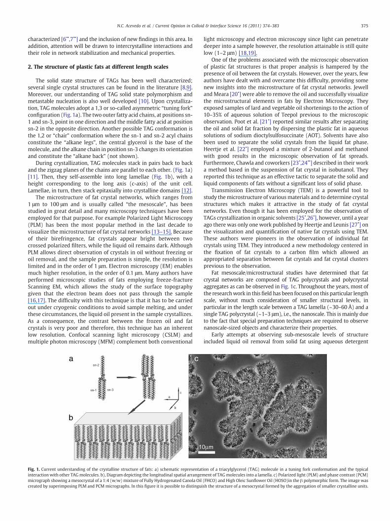
The solid state structure of TAGs has been well characterized;several single crystal structures can be found in the literature [8,9].Moreover, our understanding of TAG solid state polymorphism andmetastable nucleation is also well developed [10]. Upon crystalliza-tion, TAG molecules adopt a 1,3 or so-called asymmetric “tuning fork”configuration (Fig. 1a). The two outer fatty acid chains, at positions sn-1 and sn-3, point in one direction and the middle fatty acid at positionsn-2 in the opposite direction. Another possible TAG conformation isthe 1,2 or “chair” conformation where the sn-1 and sn-2 acyl chainsconstitute the “alkane legs”, the central glycerol is the base of themolecule, and the alkane chain in position sn-3 changes its orientationand constitute the “alkane back” (not shown).
During crystallization, TAG molecules stack in pairs back to backand the zigzag planes of the chains are parallel to each other. (Fig. 1a)[11]. Then, they self-assemble into long lamellae (Fig. 1b), with aheight corresponding to the long axis (c-axis) of the unit cell.Lamellae, in turn, then stack epitaxially into crystalline domains [12].
The microstructure of fat crystal networks, which ranges from1 μm to 100 μm and is usually called “the mesoscale”, has beenstudied in great detail and many microscopy techniques have beenemployed for that purpose. For example Polarized Light Microscopy(PLM) has been the most popular method in the last decade tovisualize the microstructure of fat crystal networks [13–15]. Becauseof their birefringence, fat crystals appear bright between twocrossed polarized filters, while the liquid oil remains dark. AlthoughPLM allows direct observation of crystals in oil without freezing oroil removal, and the sample preparation is simple, the resolution islimited and in the order of 1 μm. Electron microscopy (EM) enablesmuch higher resolution, in the order of 0.1 μm. Many authors haveperformed microscopic studies of fats employing freeze-fractureScanning EM, which allows the study of the surface topographygiven that the electron beam does not pass through the sample[16,17]. The difficulty with this technique is that it has to be carriedout under cryogenic conditions to avoid sample melting, and underthese circumstances, the liquid oil present in the sample crystallizes.As a consequence, the contrast between the frozen oil and fatcrystals is very poor and therefore, this technique has an inherentlow resolution. Confocal scanning light microscopy (CSLM) andmultiple photon microscopy (MFM) complement both conventional
Fig. 1. Current understanding of the crystalline structure of fats: a) schematic representatinteraction with other TAGmolecules. b), Diagram depicting the longitudinal spatial arrangemmicrograph showing a mesocrystal of a 1:4 (w/w) mixture of Fully Hydrogenated Canola Oilcreated by superimposing PLM and PCMmicrographs. In this figure it is possible to distingui
light microscopy and electron microscopy since light can penetratedeeper into a sample however, the resolution attainable is still quitelow (1–2 μm) [18,19].
One of the problems associated with the microscopic observationof plastic fat structures is that proper analysis is hampered by thepresence of oil between the fat crystals. However, over the years, fewauthors have dealt with and overcame this difficulty, providing somenew insights into the microstructure of fat crystal networks. Jewelland Meara [20•] were able to remove the oil and successfully visualizethe microstructural elements in fats by Electron Microscopy. Theyexposed samples of lard and vegetable oil shortenings to the action of10–35% of aqueous solution of Teepol previous to the microscopicobservation. Poot et al. [21•] reported similar results after separatingthe oil and solid fat fraction by dispersing the plastic fat in aqueoussolutions of sodium dioctylsulfosuccinate (AOT). Solvents have alsobeen used to separate the solid crystals from the liquid fat phase.Heertje et al. [22•] employed a mixture of 2-butanol and methanolwith good results in the microscopic observation of fat spreads.Furthermore, Chawla and coworkers [23•,24••] described in their worka method based in the suspension of fat crystal in isobutanol. Theyreported this technique as an effective tactic to separate the solid andliquid components of fats without a significant loss of solid phase.
Transmission Electron Microscopy (TEM) is a powerful tool tostudy themicrostructure of variousmaterials and to determine crystalstructures which makes it attractive in the study of fat crystalnetworks. Even though it has been employed for the observation ofTAGs crystallization in organic solvents [25•,26•], however, until a yearago there was only one work published by Heertje and Leunis [27•] onthe visualization and quantification of native fat crystals using TEM.These authors were pioneers in the observation of individual fatcrystals using TEM. They introduced a new methodology centered inthe fixation of fat crystals to a carbon film which allowed anappropriated separation between fat crystals and fat crystal clustersprevious to the observation.
Fat mesoscale/microstructural studies have determined that fatcrystal networks are composed of TAG polycrystals and polycrystalaggregates as can be observed in Fig. 1c. Throughout the years, most ofthe researchwork in this field has been focused on this particular lengthscale, without much consideration of smaller structural levels, inparticular in the length scale between a TAG lamella (~30–60 Å) and asingle TAG polycrystal (~1–3 μm), i.e., the nanoscale. This is mainly dueto the fact that special preparation techniques are required to observenanoscale-sized objects and characterize their properties.
Early attempts at observing sub-mesoscale levels of structureincluded liquid oil removal from solid fat using aqueous detergent
ion of a triacylglycerol (TAG) molecule in a tuning fork conformation and the typicalent of TAGmolecules into a lamella. c) Polarized light (PLM) and phase contrast (PCM)
(FHCO) and High Oleic Sunflower Oil (HOSO)in the β polymorphic form. The image wassh the structure of a mesocrystal formed by the aggregation of smaller crystalline units.
376 N.C. Acevedo et al. / Current Opinion in Colloid & Interface Science 16 (2011) 374–383
solutions, which did allow unique observations of near-single crystalsplatelets [20•,22•]. However, it was not until Acevedo andMarangoni'swork that the platelet structure and its place within the structuralhierarchy of a fat crystal network were identified. In addition, theyreported for the first time a systematic study in this area andconsidered as well the possibility of engineering the nanostructure ofsuch materials [6••,7••]. Their studies suggested that what waspreviously believed to be a fat “primary crystal” is in fact itself anagglomerate of well defined triglyceride nanoplatelets.
In their work, these authors developed a method based on coldsolvent extraction of the entrapped oil phase, disruption of the fatcrystalline network and extraction of crystal nanoplatelets wasdeveloped, which allowed the nanocrystals to be imaged usingCryo-TEM. Initially, several solvent, temperature and homogenizationtreatment combinations were assessed. In the end, it was determinedthat cold (10 °C) isobutanol in combination with mechanical matrixdisruption dissolved the oil without significantly dissolving thecrystalline matrix. The solvent and temperature chosen was verysimilar to that recommended by Chawla and deMan [24••] for theobservation of fat mesoscale surfaces by Scanning ElectronMicroscopy.
In this study we adopted the same treatment. Fat samples weresuspended in cold isobutanol in a ratio of ~1:25 (w/w). Subsequently,the mixtures were homogenized with a rotostator, and the crystalscollected by vacuum filtration. The recovered solid was re-suspendedin cold isobutanol and re-homogenized using the rotostator in orderto obtain a suitable dispersion of crystals. Finally, the mixtures weresonicated to complete the dispersion of the fat crystals before theobservation by Cryo-TEM.
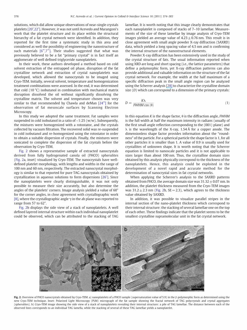
Fig. 2 shows a representative sample of extracted nanocrystalsderived from fully hydrogenated canola oil (FHCO) spherulites(Fig. 2a, inset) visualized by Cryo-TEM. The nanocrystals have well-defined platelet morphology, with lengths and widths in the range of100 nm and 60 nm, respectively. The extracted nanocrystal morphol-ogy is similar to that reported for pure TAG nanocrystals obtained bycrystallization in aqueous solutions to form dispersions [26•]. Sincethe nanoplatelets were clearly distinguishable, it was not onlypossible to measure their size accurately, but also determine theangles of the platelets' corners. Image analysis yielded a value of 60°for the corner angles, in close agreement with crystallographic work[8], where the crystallographic angle γ in the ab plane was reported torange from 57 to 62°.
Fig. 2b displays the side view of a stack of nanoplatelets. A welldefined layered internal structure within each individual nanoplateletcould be observed, which can be attributed to the stacking of TAG
Fig. 2. Overview of FHCO nanocrystals obtained by Cryo-TEM. a) nanoplatelets of a FHCO sanew Cryo-TEM technique. Inset: Polarized Light Microscopy (PLM) micrograph of the f(spherulites). b) Cryo-TEM image showing the side view of a stack of nanoplatelets reveaobserved lines corresponds to an individual TAG lamella, while the stacking of several of th
lamellae. It is worth noting that this image clearly demonstrates thateach nanoplatelet is composed of stacks of 7–10 lamellae. Measure-ments of the size of these lamellae by image analysis of Cryo-TEMimages yielded an average value of 4.23±0.76 nm. This result is inclose agreement with small angle powder X-ray diffraction (SAXRD)data, which yielded a long spacing value of 4.5 nm and is confirmingthe internal structure of the nanostructural elements.
Powder X-ray diffraction has been extensively used in the study ofthe crystal structure of fats. The usual information reported whenusing XRD are long and short spacing (i.e., the lattice parameters) thatdefine a polymorphic form, yet X-ray diffraction patterns can alsoprovide additional and valuable information on the structure of the fatcrystal network. For example, the width at the half maximum of aspecific diffraction peak in the small angle region can be analyzedusing the Scherrer analysis [28] to characterize the crystalline domainsize (D) which can correspond to a dimension of the primary crystals:
D =Kλ
FWHM cos θð Þ ð1Þ
In this equation K is the shape factor, θ is the diffraction angle, FWHMis the full width at half the maximum intensity in radians (usually ofthe first small angle reflection corresponding to the (001) plane) andλ is the wavelength of the X-ray, 1.54 Å for a copper anode. Thedimensionless shape factor provides information about the “round-ness” of the particle. For a spherical particle the shape factor is 1, for allother particles it is smaller than 1. A value of 0.9 is usually used forcrystallites of unknown shape. It is worth noting that the Scherrerequation is limited to nanoscale particles and it is not applicable tosizes larger than about 100 nm. Thus, the crystalline domain sizesobtained by this analysis physically correspond to the thickness of thenanoplatelets. Hence, this analysis could be exploited in thedevelopment of a novel rapid and accurate method for thedetermination of nanocrystal sizes in fat crystal networks.
When applying the Scherrer's analysis to the SAXRD patternsobtained from FHCO, the average domain size was 31.32±0.07 nm. Inaddition, the platelet thickness measured from the Cryo-TEM imageswas 31.2±2.3 nm (Fig. 2b, SE=2.3), which agrees to the thicknessvalue obtained by SAXRD.
In addition, it was possible to visualize parallel stripes in theinternal section of the nano-platelet thickness which correspond totheir internal structure: the stacking of several lamellae one on the topof each other. These findings indicate that the platelet seems to be thesmallest crystalline supramolecular unit in the fat crystal network.
mple (supersaturation value of 5.9) in the β polymorphic form as determined using theat sample showing the fractal network of TAG polycrystals and crystal aggregatesling their internal structure: a pile of TAG lamellae. The distance between each of theese TAG lamellae yields a nanoplatelet.
377N.C. Acevedo et al. / Current Opinion in Colloid & Interface Science 16 (2011) 374–383
Many research groups have studied the effects of composition andprocessing conditions on crystal size at the microstructural level.Their results have been explained by the effect exerted by thedifferent nucleation rates achieved from different processing condi-tions [29,30]. Some recent investigations have also demonstrated thatin general, the shape and size of fat crystal clusters are dependent onprocessing conditions and SFC, and are less dependent on the finalpolymorphism of the fat crystals [13,19]. In addition, changes inmatrix supersaturation induce alterations in the microstructure ofTAG networks which are manifested as a reduction in cluster size andnetwork density with the decrease in matrix supersaturation [31–33].
To correlate structural changes that may occur at the nanoscale inthe crystalline network with modification in supersaturation, FHCOsamples were blended with different amounts of HOSO. FHCO/HOSOblends were prepared in 10% increments (w/w) ranging from 20% to100% (w/w) FHCO.
The degree of supersaturation (lnβ) in the blends was calculatedas:
lnβ =ΔHm
RTTmTm− Tð Þ ð2Þ
where R is the gas constant, ΔHm is the enthalpy of melting, Tm is themelting temperature and T is the crystallization temperature. Theresults of supersaturation calculation along with the values of Tm,ΔHm are given in Table 1.
Aftermelting, the blendswere crystallized under conditionswherethe formation of the β polymorphic (triclinic) form is favored (seeSupplementary section S1). All the blends were analyzed employingSAXD (particularly, the Scherrer analysis), and were subjected to thesolvent treatment developed by Acevedo and Marangoni, previous tothe Cryo-TEM imaging. Platelet dimensions were obtained bysemiautomatic tools of image analysis [6••].
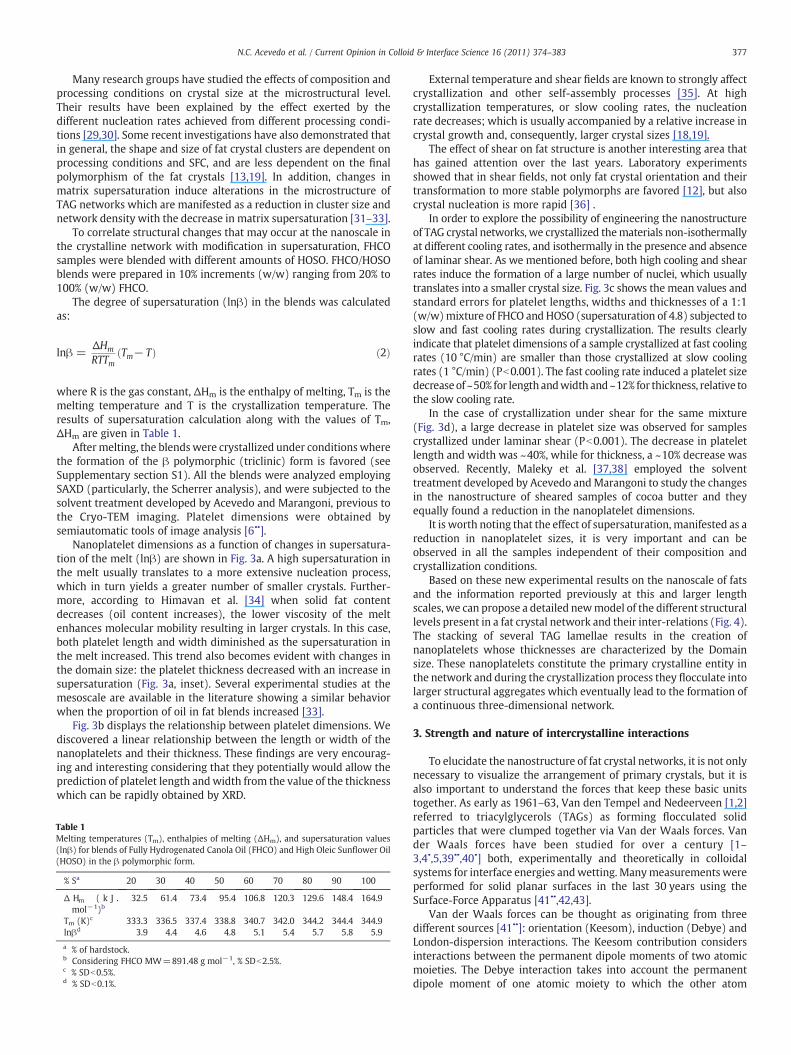
Nanoplatelet dimensions as a function of changes in supersatura-tion of the melt (lnβ) are shown in Fig. 3a. A high supersaturation inthe melt usually translates to a more extensive nucleation process,which in turn yields a greater number of smaller crystals. Further-more, according to Himavan et al. [34] when solid fat contentdecreases (oil content increases), the lower viscosity of the meltenhances molecular mobility resulting in larger crystals. In this case,both platelet length and width diminished as the supersaturation inthe melt increased. This trend also becomes evident with changes inthe domain size: the platelet thickness decreased with an increase insupersaturation (Fig. 3a, inset). Several experimental studies at themesoscale are available in the literature showing a similar behaviorwhen the proportion of oil in fat blends increased [33].
Fig. 3b displays the relationship between platelet dimensions. Wediscovered a linear relationship between the length or width of thenanoplatelets and their thickness. These findings are very encourag-ing and interesting considering that they potentially would allow theprediction of platelet length andwidth from the value of the thicknesswhich can be rapidly obtained by XRD.
Table 1Melting temperatures (Tm), enthalpies of melting (ΔHm), and supersaturation values(lnβ) for blends of Fully Hydrogenated Canola Oil (FHCO) and High Oleic Sunflower Oil(HOSO) in the β polymorphic form.
% Sa 20 30 40 50 60 70 80 90 100
Δ Hm ( k J .mol−1)b
32.5 61.4 73.4 95.4 106.8 120.3 129.6 148.4 164.9
Tm (K)c 333.3 336.5 337.4 338.8 340.7 342.0 344.2 344.4 344.9lnβd 3.9 4.4 4.6 4.8 5.1 5.4 5.7 5.8 5.9
a % of hardstock.b Considering FHCO MW=891.48 g mol−1, % SDb2.5%.c % SDb0.5%.d % SDb0.1%.
External temperature and shear fields are known to strongly affectcrystallization and other self-assembly processes [35]. At highcrystallization temperatures, or slow cooling rates, the nucleationrate decreases; which is usually accompanied by a relative increase incrystal growth and, consequently, larger crystal sizes [18,19].
The effect of shear on fat structure is another interesting area thathas gained attention over the last years. Laboratory experimentsshowed that in shear fields, not only fat crystal orientation and theirtransformation to more stable polymorphs are favored [12], but alsocrystal nucleation is more rapid [36] .
In order to explore the possibility of engineering the nanostructureof TAG crystal networks, we crystallized thematerials non-isothermallyat different cooling rates, and isothermally in the presence and absenceof laminar shear. As we mentioned before, both high cooling and shearrates induce the formation of a large number of nuclei, which usuallytranslates into a smaller crystal size. Fig. 3c shows the mean values andstandard errors for platelet lengths, widths and thicknesses of a 1:1(w/w)mixture of FHCO and HOSO (supersaturation of 4.8) subjected toslow and fast cooling rates during crystallization. The results clearlyindicate that platelet dimensions of a sample crystallized at fast coolingrates (10 °C/min) are smaller than those crystallized at slow coolingrates (1 °C/min) (Pb0.001). The fast cooling rate induced a platelet sizedecreaseof ~50% for length andwidth and~12% for thickness, relative tothe slow cooling rate.
In the case of crystallization under shear for the same mixture(Fig. 3d), a large decrease in platelet size was observed for samplescrystallized under laminar shear (Pb0.001). The decrease in plateletlength and width was ~40%, while for thickness, a ~10% decrease wasobserved. Recently, Maleky et al. [37,38] employed the solventtreatment developed by Acevedo andMarangoni to study the changesin the nanostructure of sheared samples of cocoa butter and theyequally found a reduction in the nanoplatelet dimensions.
It is worth noting that the effect of supersaturation, manifested as areduction in nanoplatelet sizes, it is very important and can beobserved in all the samples independent of their composition andcrystallization conditions.
Based on these new experimental results on the nanoscale of fatsand the information reported previously at this and larger lengthscales, we can propose a detailed newmodel of the different structurallevels present in a fat crystal network and their inter-relations (Fig. 4).The stacking of several TAG lamellae results in the creation ofnanoplatelets whose thicknesses are characterized by the Domainsize. These nanoplatelets constitute the primary crystalline entity inthe network and during the crystallization process they flocculate intolarger structural aggregates which eventually lead to the formation ofa continuous three-dimensional network.
3. Strength and nature of intercrystalline interactions
To elucidate the nanostructure of fat crystal networks, it is not onlynecessary to visualize the arrangement of primary crystals, but it isalso important to understand the forces that keep these basic unitstogether. As early as 1961–63, Van den Tempel and Nedeerveen [1,2]referred to triacylglycerols (TAGs) as forming flocculated solidparticles that were clumped together via Van der Waals forces. Vander Waals forces have been studied for over a century [1–3,4•,5,39••,40•] both, experimentally and theoretically in colloidalsystems for interface energies andwetting. Manymeasurements wereperformed for solid planar surfaces in the last 30 years using theSurface-Force Apparatus [41••,42,43].
Van der Waals forces can be thought as originating from threedifferent sources [41••]: orientation (Keesom), induction (Debye) andLondon-dispersion interactions. The Keesom contribution considersinteractions between the permanent dipole moments of two atomicmoieties. The Debye interaction takes into account the permanentdipole moment of one atomic moiety to which the other atom
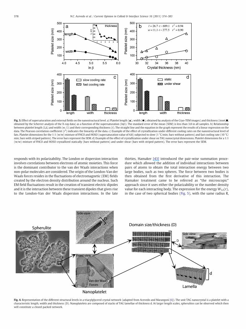
Fig. 3. Effect of supersaturation and external fields on the nanostructural level. a) Platelet length (▲), width (●), obtained by analysis of the Cryo-TEM images) and thickness (inset,■,obtained by the Scherrer analysis of the X-ray data) as a function of the supersaturation (lnβ). The standard error of the mean (SEM) is less than 3.8 in all samples. b) Relationshipbetween platelet length (l,Δ) andwidth (w,○) and their corresponding thickness (t). The straight line and the equation in the graph represent the results of a linear regression on thedata. The Pearson correlation coefficient (r2) indicates the linearity of the data. c) Example of the effect of crystallization under different cooling rates on the nanostructural level offats. Platelet dimensions for the 1:1 (w/w) mixture of FHCO and HOSO (supersaturation value of 4.8) subjected to slow (1 °C/min; bars without pattern) and fast cooling rate (10 °C/min; bars with striped pattern). The error bars represent the SEM. d) Example of the effect of crystallization under shear on TAG nanocrystal dimensions. Platelet dimensions for a 1:1(w/w) mixture of FHCO and HOSO crystallized statically (bars without pattern) and under shear (bars with striped pattern). The error bars represent the SEM.
378 N.C. Acevedo et al. / Current Opinion in Colloid & Interface Science 16 (2011) 374–383
responds with its polarizability. The London or dispersion interactioninvolves correlations between electrons of atomic moieties. This forceis the dominant contributor to the van der Waals interactions whennon-polarmolecules are considered. The origin of the London-Van derWaals forces resides in the fluctuations of electromagnetic (EM) fieldscreated by the electron density distribution around the nucleus. SuchEM field fluctuations result in the creation of transient electric dipolesand it is the interaction between these transient dipoles that gives riseto the London-Van der Waals dispersion interactions. In the late
Fig. 4. Representation of the different structural levels in a triacylglycerol crystal network (acharacteristic length, width and thickness (D). Nanoplatelets are composed of stacks of TAGwill constitute a closed packed network.
thirties, Hamaker [43] introduced the pair-wise summation proce-dure which allowed the addition of individual interactions betweenpairs of atoms to obtain the total interaction energy between twolarge bodies, such as two spheres. The force between two bodies isthen obtained from the first derivative of this interaction. TheHamaker treatment came to be referred as “the microscopic”approach since it uses either the polarizability or the number densityvalue for each interacting body. The expression for the energyW12(r),in the case of two spherical bodies (Fig. 5), with the same radius R,
dapted from Acevedo and Marangoni [6]). The unit TAG nanocrystal is a platelet with alamellae of thickness d. At larger length scales, spherulites can be observed which then
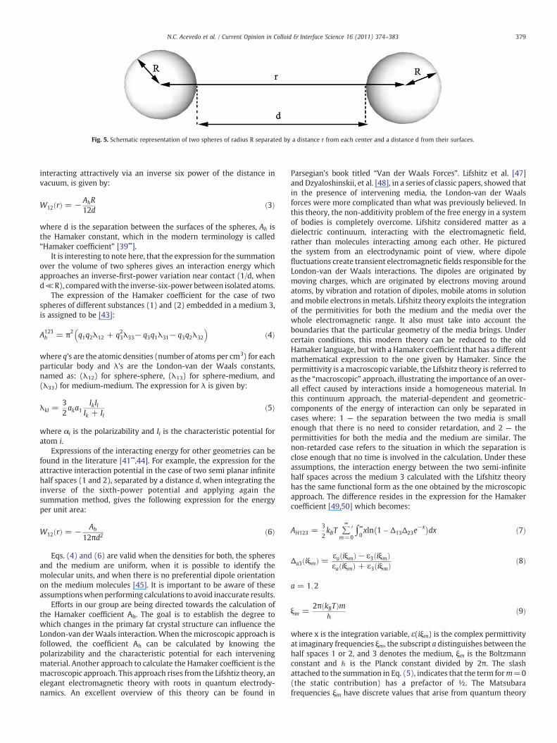
Fig. 5. Schematic representation of two spheres of radius R separated by a distance r from each center and a distance d from their surfaces.
379N.C. Acevedo et al. / Current Opinion in Colloid & Interface Science 16 (2011) 374–383
interacting attractively via an inverse six power of the distance invacuum, is given by:
W12 rð Þ = − AhR12d
ð3Þ
where d is the separation between the surfaces of the spheres, Ah isthe Hamaker constant, which in the modern terminology is called“Hamaker coefficient” [39••].
It is interesting to note here, that the expression for the summationover the volume of two spheres gives an interaction energy whichapproaches an inverse-first-power variation near contact (1/d, whend≪R), comparedwith the inverse-six-power between isolated atoms.
The expression of the Hamaker coefficient for the case of twospheres of different substances (1) and (2) embedded in a medium 3,is assigned to be [43]:
A123h = π2 q1q2λ12 + q23λ33− q3q1λ31− q3q2λ32
� �ð4Þ
where q's are the atomic densities (number of atoms per cm3) for eachparticular body and λ's are the London-van der Waals constants,named as: (λ12) for sphere-sphere, (λ13) for sphere-medium, and(λ33) for medium-medium. The expression for λ is given by:
λkl =32aka1
IkIlIk + Il
ð5Þ
where αi is the polarizability and Ii is the characteristic potential foratom i.
Expressions of the interacting energy for other geometries can befound in the literature [41••,44]. For example, the expression for theattractive interaction potential in the case of two semi planar infinitehalf spaces (1 and 2), separated by a distance d, when integrating theinverse of the sixth-power potential and applying again thesummation method, gives the following expression for the energyper unit area:
W12 rð Þ = − Ah
12πd2ð6Þ
Eqs. (4) and (6) are valid when the densities for both, the spheresand the medium are uniform, when it is possible to identify themolecular units, and when there is no preferential dipole orientationon the medium molecules [45]. It is important to be aware of theseassumptionswhenperforming calculations to avoid inaccurate results.
Efforts in our group are being directed towards the calculation ofthe Hamaker coefficient Ah. The goal is to establish the degree towhich changes in the primary fat crystal structure can influence theLondon-van der Waals interaction. When the microscopic approach isfollowed, the coefficient Ah can be calculated by knowing thepolarizability and the characteristic potential for each interveningmaterial. Another approach to calculate the Hamaker coefficient is themacroscopic approach. This approach rises from the Lifshtiz theory, anelegant electromagnetic theory with roots in quantum electrody-namics. An excellent overview of this theory can be found in
Parsegian's book titled “Van der Waals Forces”. Lifshitz et al. [47]and Dzyaloshinskii, et al. [48], in a series of classic papers, showed thatin the presence of intervening media, the London-van der Waalsforces were more complicated than what was previously believed. Inthis theory, the non-additivity problem of the free energy in a systemof bodies is completely overcome. Lifshitz considered matter as adielectric continuum, interacting with the electromagnetic field,rather than molecules interacting among each other. He picturedthe system from an electrodynamic point of view, where dipolefluctuations create transient electromagnetic fields responsible for theLondon-van der Waals interactions. The dipoles are originated bymoving charges, which are originated by electrons moving aroundatoms, by vibration and rotation of dipoles, mobile atoms in solutionandmobile electrons in metals. Lifshitz theory exploits the integrationof the permitivities for both the medium and the media over thewhole electromagnetic range. It also must take into account theboundaries that the particular geometry of the media brings. Undercertain conditions, this modern theory can be reduced to the oldHamaker language, but with a Hamaker coefficient that has a differentmathematical expression to the one given by Hamaker. Since thepermittivity is a macroscopic variable, the Lifshitz theory is referred toas the “macroscopic” approach, illustrating the importance of an over-all effect caused by interactions inside a homogeneous material. Inthis continuum approach, the material-dependent and geometric-components of the energy of interaction can only be separated incases where: 1 — the separation between the two media is smallenough that there is no need to consider retardation, and 2 — thepermittivities for both the media and the medium are similar. Thenon-retarded case refers to the situation in which the separation isclose enough that no time is involved in the calculation. Under theseassumptions, the interaction energy between the two semi-infinitehalf spaces across the medium 3 calculated with the Lifshitz theoryhas the same functional form as the one obtained by the microscopicapproach. The difference resides in the expression for the Hamakercoefficient [49,50] which becomes:
AH123 =3
2kBT ∑
∞
m=0
0 ∫∞0xln 1� Δ13Δ23e
−x� �dx ð7Þ
Δa3 iξmð Þ = εa iξmð Þ− ε3 iξmð Þεa iξmð Þ + ε3 iξmð Þ ð8Þ
a = 1;2
ξm =2π kBTð Þm
ℏð9Þ
where x is the integration variable, ε(iξm) is the complex permittivityat imaginary frequencies ξm, the subscript a distinguishes between thehalf spaces 1 or 2, and 3 denotes the medium, ξm is the Boltzmannconstant and ℏ is the Planck constant divided by 2π. The slashattached to the summation in Eq. (5), indicates that the term form=0(the static contribution) has a prefactor of ½. The Matsubarafrequencies ξm have discrete values that arise from quantum theory
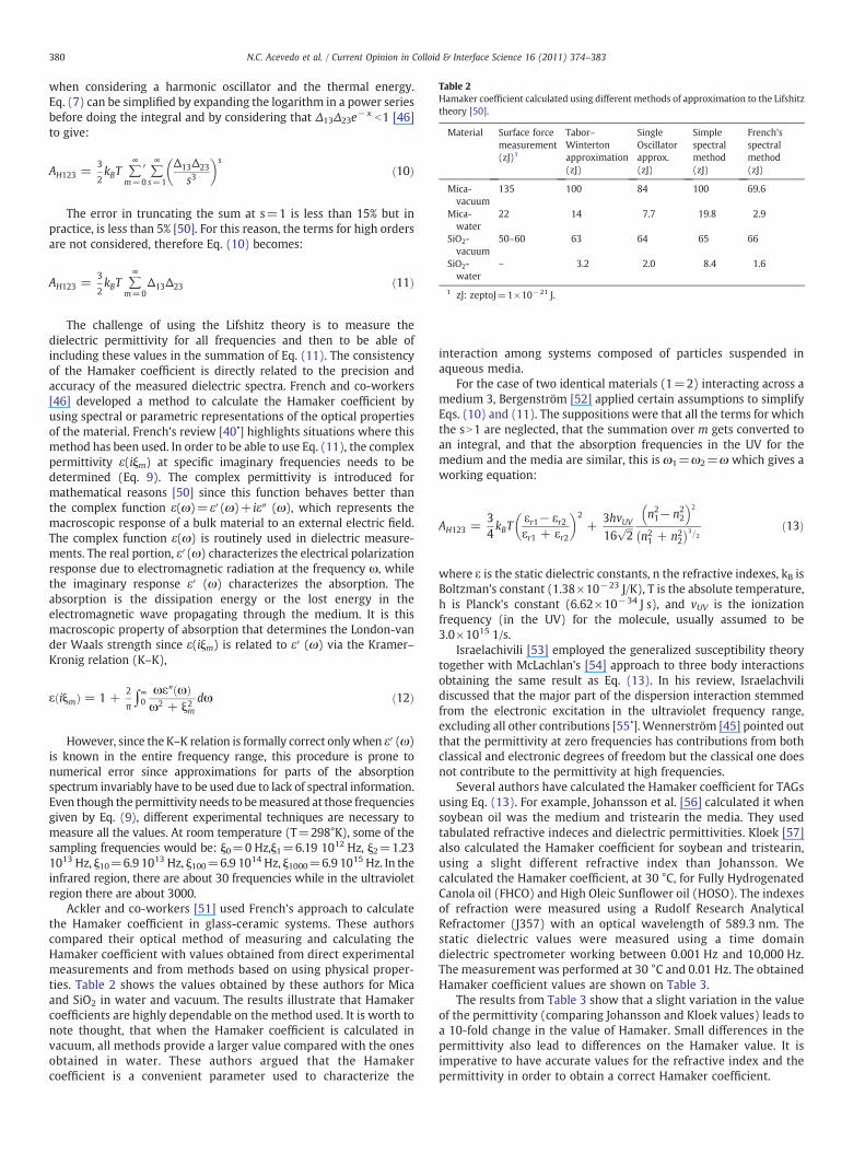
Table 2Hamaker coefficient calculated using different methods of approximation to the Lifshitztheory [50].
Material Surface forcemeasurement(zJ)1
Tabor–Wintertonapproximation(zJ)
SingleOscillatorapprox.(zJ)
Simplespectralmethod(zJ)
French'sspectralmethod(zJ)
Mica-vacuum
135 100 84 100 69.6
Mica-water
22 14 7.7 19.8 2.9
SiO2-vacuum
50–60 63 64 65 66
SiO2-water
– 3.2 2.0 8.4 1.6
1 zJ: zeptoJ=1×10⁻²¹ J.
380 N.C. Acevedo et al. / Current Opinion in Colloid & Interface Science 16 (2011) 374–383
when considering a harmonic oscillator and the thermal energy.Eq. (7) can be simplified by expanding the logarithm in a power seriesbefore doing the integral and by considering that Δ13Δ23e
− x b1 [46]to give:
AH123 =3
2kBT ∑
∞
m=0
0 ∑∞
s=1
Δ13Δ23
s3
� �s
ð10Þ
The error in truncating the sum at s=1 is less than 15% but inpractice, is less than 5% [50]. For this reason, the terms for high ordersare not considered, therefore Eq. (10) becomes:
AH123 =3
2kBT ∑
∞
m=0Δ13Δ23 ð11Þ
The challenge of using the Lifshitz theory is to measure thedielectric permittivity for all frequencies and then to be able ofincluding these values in the summation of Eq. (11). The consistencyof the Hamaker coefficient is directly related to the precision andaccuracy of the measured dielectric spectra. French and co-workers[46] developed a method to calculate the Hamaker coefficient byusing spectral or parametric representations of the optical propertiesof the material. French's review [40•] highlights situations where thismethod has been used. In order to be able to use Eq. (11), the complexpermittivity ε(iξm) at specific imaginary frequencies needs to bedetermined (Eq. 9). The complex permittivity is introduced formathematical reasons [50] since this function behaves better thanthe complex function ε(ω)=ε′(ω)+ iε″ (ω), which represents themacroscopic response of a bulk material to an external electric field.The complex function ε(ω) is routinely used in dielectric measure-ments. The real portion, ε′(ω) characterizes the electrical polarizationresponse due to electromagnetic radiation at the frequency ω, whilethe imaginary response ε′ (ω) characterizes the absorption. Theabsorption is the dissipation energy or the lost energy in theelectromagnetic wave propagating through the medium. It is thismacroscopic property of absorption that determines the London-vander Waals strength since ε(iξm) is related to ε′ (ω) via the Kramer–Kronig relation (K–K),
ε iξmð Þ = 1 +2
π∫∞0
ωε″ ωð Þω2 + ξ2m
dω ð12Þ
However, since the K–K relation is formally correct onlywhen ε′ (ω)is known in the entire frequency range, this procedure is prone tonumerical error since approximations for parts of the absorptionspectrum invariably have to be used due to lack of spectral information.Even though the permittivity needs to bemeasured at those frequenciesgiven by Eq. (9), different experimental techniques are necessary tomeasure all the values. At room temperature (T=298°K), some of thesampling frequencies would be: ξ0=0 Hz,ξ1=6.19 1012 Hz, ξ2=1.231013 Hz, ξ10=6.9 1013 Hz, ξ100=6.9 1014 Hz, ξ1000=6.9 1015 Hz. In theinfrared region, there are about 30 frequencies while in the ultravioletregion there are about 3000.
Ackler and co-workers [51] used French's approach to calculatethe Hamaker coefficient in glass-ceramic systems. These authorscompared their optical method of measuring and calculating theHamaker coefficient with values obtained from direct experimentalmeasurements and from methods based on using physical proper-ties. Table 2 shows the values obtained by these authors for Micaand SiO2 in water and vacuum. The results illustrate that Hamakercoefficients are highly dependable on the method used. It is worth tonote thought, that when the Hamaker coefficient is calculated invacuum, all methods provide a larger value compared with the onesobtained in water. These authors argued that the Hamakercoefficient is a convenient parameter used to characterize the
interaction among systems composed of particles suspended inaqueous media.
For the case of two identical materials (1=2) interacting across amedium 3, Bergenström [52] applied certain assumptions to simplifyEqs. (10) and (11). The suppositions were that all the terms for whichthe sN1 are neglected, that the summation over m gets converted toan integral, and that the absorption frequencies in the UV for themedium and the media are similar, this is ω1=ω2=ω which gives aworking equation:
AH123 =34kBT
εr1− εr2εr1 + εr2
� �2+
3hvUV16
ffiffiffi2
pn21− n2
2
� �2
n21 + n2
2
� �3=2 ð13Þ
where ε is the static dielectric constants, n the refractive indexes, kB isBoltzman's constant (1.38×10⁻²³ J/K), T is the absolute temperature,h is Planck's constant (6.62×10⁻³⁴ J s), and vUV is the ionizationfrequency (in the UV) for the molecule, usually assumed to be3.0×1015 1/s.
Israelachivili [53] employed the generalized susceptibility theorytogether with McLachlan's [54] approach to three body interactionsobtaining the same result as Eq. (13). In his review, Israelachvilidiscussed that the major part of the dispersion interaction stemmedfrom the electronic excitation in the ultraviolet frequency range,excluding all other contributions [55•]. Wennerström [45] pointed outthat the permittivity at zero frequencies has contributions from bothclassical and electronic degrees of freedom but the classical one doesnot contribute to the permittivity at high frequencies.
Several authors have calculated the Hamaker coefficient for TAGsusing Eq. (13). For example, Johansson et al. [56] calculated it whensoybean oil was the medium and tristearin the media. They usedtabulated refractive indeces and dielectric permittivities. Kloek [57]also calculated the Hamaker coefficient for soybean and tristearin,using a slight different refractive index than Johansson. Wecalculated the Hamaker coefficient, at 30 °C, for Fully HydrogenatedCanola oil (FHCO) and High Oleic Sunflower oil (HOSO). The indexesof refraction were measured using a Rudolf Research AnalyticalRefractomer (J357) with an optical wavelength of 589.3 nm. Thestatic dielectric values were measured using a time domaindielectric spectrometer working between 0.001 Hz and 10,000 Hz.The measurement was performed at 30 °C and 0.01 Hz. The obtainedHamaker coefficient values are shown on Table 3.
The results from Table 3 show that a slight variation in the valueof the permittivity (comparing Johansson and Kloek values) leads toa 10-fold change in the value of Hamaker. Small differences in thepermittivity also lead to differences on the Hamaker value. It isimperative to have accurate values for the refractive index and thepermittivity in order to obtain a correct Hamaker coefficient.
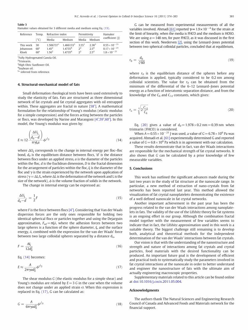
Table 3Hamaker values obtained for 3 different media and medium using Eq. (13).
Reference Temp. Refractive index Permittivity Hamakercoefficient (J)
(°C) Media Medium Media Medium
This work 30 1.5067371 1.4661333 3.551 2.383 0.55×10⁻²¹
Johansson 60* 1.452 1.47354 22 2.54 0.17×10⁻²¹
Kloek 60* 1.562 1.47354 22 2.54 1.8×10⁻²¹
1Fully Hydrogenated Canola Oil.2Tristearin.3High Oleic Sunflower Oil.4Soybean oil.5* inferred from reference.
381N.C. Acevedo et al. / Current Opinion in Colloid & Interface Science 16 (2011) 374–383
4. Structural-mechanical model of fats
Small deformation rheological tests have been used extensively tostudy the elasticity of fats. Fats are structured as three dimensionalnetwork of fat crystals and fat crystal aggregates with oil entrappedwithin. These aggregates are fractal in nature [58•]. A mathematicalformulation for the relationship of Young's modulus (elastic modulusfor a simple compression) and the forces acting between the particlesor flocs, was developed by Narine and Marangoni [4•,59•,60•]. In thismodel, the Young's modulus was given by:
E≈ 12
ΔUξ
d0−d0
!
πγξaϕ
1d−D ð14Þ
where ΔUξ corresponds to the change in internal energy per floc–flocbond, d0 is the equilibrium distance between flocs, ‘d’ is the distancebetween flocs under an applied stress, a is the diameter of the particleswithin the floc, d is the Euclidean dimension, D is the fractal dimensionfor the arrangement of particles within the floc, ξ is the diameter of thefloc and γ is the strain experienced by the network upon application ofstress (γ=ΔL/L,whereΔL is thedeformationof the network and L is thesize of the network), ϕ is the volume fraction of solids in the network.
The change in internal energy can be expressed as:
ΔUξ
d0−d0 =
12F ð15Þ
where F is the force between flocs [4•]. Considering that Van derWaalsdispersion forces are the only ones responsible for holding twoidentical spherical flocs or particles together and using the Dejarguinapproximation, Fad=πξδ, where the adhesion forces between twolarge spheres is a function of the sphere diameter, ξ, and the surfaceenergy, δ, combined with the expression for the van der Waals’ forcebetween two large colloidal spheres separated by a distance do,
F =Aξ
12d20ð16Þ
Eq. (14) becomes:
E≈ A2πγad20
ϕ1
d−D ð17Þ
The shear modulus G (the elastic modulus for a simple shear) andYoung's modulus are related by E=3 G in the case when the volumedoes not change under an applied strain σ. When this expression isreplaced in Eq. (17), G can be calculated as:
G =A
6πγad20ϕ
1d−D ð18Þ
G can be measured from experimental measurements of all thevariables involved. Ahmadi [61] reported γ=1.9×10⁻⁴ for the strain atthe limit of linearity, when the media is FHCO and the medium is HOSO.We are using a=148 nm, for pure FHCO, as it was discussed in the firstsection of this work. Neederven [2], using the Lennard–Jones potentialbetween two spherical colloidal particles, concluded that at equilibrium,
r0 =d0ffiffiffiffiffiffi606
p ð19Þ
where r0 is the equilibrium distance of the spheres before anydeformation is applied, typically considered to be 0.2 nm amongcolloidal scientists. The value for r0 can be obtained from theminimum of the differential of the 6–12 Lennard–Jones potentialenergy as a function of interatomic separation distance, and from theknowledge of the C6 and C12 constants, which gives:
ro =
ffiffiffiffiffiffiffiffiffiffiffi2C12
C6
6
sð20Þ
Eq. (20) gives a value of d0=1.978×0.2 nm=0.39 nm whentristearin (FHCO) is considered.
When A=0.55×10⁻²¹ J was used, a value of G=6.78×10⁶ Pa wasacquired. Ahmadi et al. [61] experimentally determined G and reporteda value of G=6.8×10⁶ Pa which is in agreement with our calculation.
These results demonstrate that in fact, van der Waals interactionsare responsible for the mechanical strength of fat crystal networks. Italso shows that G can be calculated by a prior knowledge of fewmeasurable variables.
5. Conclusions
This work has outlined the significant advances made during thelast two years in the study of fat structure at the nanoscale range. Inparticular, a new method of extraction of nano-crystals from fatnetworks has been reported last year. This method allowed thevisualization of fat crystal nanoplatelets demonstrating the existenceof a well defined nanoscale in fat crystal networks.
Another important achievement in the past year has been theprogress related to the van der Waals interactions among nanoplate-lets in fats. The validity of the use of the Lifshitz theory for fat systemsis an ongoing effort in our group. Although the combination fractalmodel together with the measurement of few variables seems toindicate that in fact, the Lifshitz approximation used in this work is asuitable theory. The biggest challenge still remaining is to developboth, analytical and theoretical methods for the independentdetermination of the van der Waals' interactions between fat crystals.
Our vision is that with the understanding of the nanostructure andstrength and nature of interactions among fat crystals and crystalparticles, food materials with the desired functionality can beproduced. An important future goal is the development of efficientand practical tools to systematically study the parameters involved infat crystal interactions at the nanoscale in order to better understandand engineer the nanostructure of fats with the ultimate aim ofactually engineering macroscopic properties.
Supplementarymaterials related to this article can be found onlineat doi:10.1016/j.cocis.2011.05.004.
Acknowledgments
The authors thank The Natural Sciences and Engineering ResearchCouncil of Canada and Advanced Foods and Materials network for thefinancial support.
382 N.C. Acevedo et al. / Current Opinion in Colloid & Interface Science 16 (2011) 374–383
References and recommended reading•,••
[1] Van den Tempel M.Mechanical properties of plastic disperse systems at very smalldeformations. J Colloid Sci 1961;16:284–96.
[2] Nederveen CJ. Dynamic mechanical behavior of suspensions of fat particles in oil. JColloid Sci 1963;18:276–91.
[3] Van den Tempel M. Rheology of concentrated suspensions. J Colloid Interface Sci1979;71:18–20.
•[4] Marangoni AG, Rogers M. Structural basis for the yield stress in plastic disperse
systems. Appl Phys Lett 2003;82:3239–41.This paper proposes a model thatrelates the morphological structure of fractal systems to its yield stress in colloidalsystems under the weak-link regime.
[5] Tang D, Marangoni AG. Fractal dimensions of simulated and real fat crystalnetworks in 3D Space. J Am Oil Chem Soc 2008;85:495–9.
•[6] Acevedo NC, Marangoni AG. Characterization of the nanoscale in triacylglycerol
crystal networks. Cryst Growth Des 2010;10(8):3327–33.The first systematic studyperformed at the nanoscale of the tridimensional fat crystal network is reported inthis paper. These authors found evidence suggesting that what was believed as a“primary crystal” is in fact an agglomerate of triglyceride nanoplatelets.
••[7] Acevedo NC, Marangoni AG. Towards nanoscale engineering of triacylglycerol
crystal networks. Cryst Growth Des 2010;10(8):3334–9.In this work the authorsshow for the first time, changes induced in the nanostructural elements byalterations produced in external fields. They proposed that it is possible toengineer the functional properties of fat systems by nano-manipulation via controlof crystallization conditions.
[8] Van Langevelde A, Peschar R, Schenk H. Structure of β-trimystirin and β-tristearinform high-resolution X-ray powder diffraction data. Acta Cryst 2001;B57:372–7.
[9] Sato K, Goto M, Yano J, Honda K, Kodali DR, Small DM. Atomic resolution structureanalysis of ß′ polymorph crystal of a triacylglycerol: 1,2-dipalmitoyl-3-myristoyl-sn-glycerol. J Lipid Res 2001;42:338–45.
[10] Garti N, Sato K. In: Garti N, Sato K, editors. Crystallization and polymorphism offats and fatty acids. New York: Marcel Dekker; 1988.
[11] Bennema P, Hollander FFA, Boerrigther SXM, Grimbergen RFP, Van de Streek J,Meekes H. Morphological connected Net-roughening transition theory. In: GartiN, Sato K, editors. Crystallization and Polymorphism of Fats and Fatty Acids. NewYork: Marcel Dekker; 1988. p. 99–150.
[12] Mazzanti G, Marangoni AG, Idziak SHJ. Modeling phase transitions during thecrystallization of a multicomponent fat under shear. Phys Rev E 2005;71:1–12.
[13] Campos R, Narine S, Marangoni AG. Effect of cooling rate on the structure andmechanical properties of milk fat and lard. Food Res Int 2002;35:971–81.
[14] Awad TS, Rogers MA, Marangoni AG. Scaling behavior of the elastic modulus incolloidal networks of fat crystals. J Phys Chem 2004;B108:171–9.
[15] Shi Y, Liang B, Hartel RW. Crystal morphology, microstructure, and texturalproperties of model lipid systems. J Am Oil Chem Soc 2005;82:399–408.
[16] Heertje I, Van Eendenburg J, Cornelissen JM, Juriaanse AC. The effect of processing onsomemicrostructural characteristics of fat spreads. FoodMicrostruct1988;7:189–93.
[17] Rousseau D, Hill AR, Marangoni AG. Restructuring butterfat through blending andchemical interestification. 2. Microstructure and polymorphism. J Am Oil ChemSoc 1996;73:973–81.
[18] Herrera ML, Hartel RW. Effect of processing conditions on physical properties of amilk fat model system: microstructure. J Am Oil Chem Soc 2000;77:1197–204.
[19] Wiking L, De Graef V, Rasmussen M, Dewettinck K. Relations between crystal-lisation mechanisms and microstructure of milk fat. Int Dairy J 2009;19:424–30.
•[20] Jewell GG, Meara ML. A new and rapid method for the electron microscopic
examination of fats. J Am Oil Chem Soc 1970;47:535–8.This paper and Ref. [21]provides a complete description of a new technique based in the use of detergentwhich was developed with the objective of eliminating oil from fat samplesprevious to the microscopic observation.
•[21] Poot C, Dijkshoorn W, Haighton AJ, Verburg CC. Laboratory separation of crystals from
plastic fats using detergent solution. J Am Oil Chem Soc 1975;70:69–72.See Ref. [20].
•[22] Heetje I, Leunis M, van Zeyl WJM, Berends E. Product morphology of fatty
products. Food Microstruct 1987;6:1–8.These workers developed an effective de-oiling process in plastic fats by the use of solvents and the construction of a specialsample holder to achieve a reproducible observation of the fat microstructure.
•[23] Chawla P, deMan JM, Smith AK. Crystal morphology of shortenings and
margarines. Food Struct 1990;9:329–36.These researchers claim that a solvent-treatment of fat networks previous tomicroscopic observation is not accompaniedby an appreciable loss of solid phase.
••[24] Chawla P, deMan JM.Measurement of the size distribution of fat crystals using a laser
particle counter. J Am Oil Chem Soc 1990;67:329–32.This manuscript served as thebasis for the development of de-oiling treatment in the nano-scale observation oftriacylglycerol networks performed by Acevedo and Marangoni [6, 7].
•[25] Unruh T,Westesen K, Bösecke P, Lindner P, KochMHJ. Self-assembly of triglyceride
nanocrystals in suspension. Langmuir 2002;18:1796–800.This paper and Ref. [26]reported the visualization by electron microscopy of solid-fat nanoparticles incolloidal suspension of triacylglycerides.
•[26] BunjesH, Steiniger F, RichterW.Visualizing the structure of triglyceridenanoparticles in
different crystal modifications. Langmuir 2007;23:4005–11.See Ref. [25].
•[27] Heertje I, Leunis M. Measurement of shape and size of fat crystals by electron
microscopy. Lebensm–Wiss U Technol 1997;30:141–6.The first published work
• of special interest.•• of outstanding interest.
reporting the study of fat crystals from native plastic fats by transmission electronmicroscopy.
[28] West AR. In: West AR, editor. Solid state chemistry and its applications. WestSussex, England: John Wiley & Sons, Chichester; 1984. p. 174–8.
[29] Martini S, Herrera ML, Hartel RW. Effect of cooling rate on crystallization behaviorof milk fat fraction/sunflower oil blends. J Am Oil Chem Soc 2002;79:1055–62.
[30] Singh AP, Bertoli C, Rousseau PR, Marangoni AG. Matching Avrami indices achievessimilar hardnesses in palm oil-based fats. J Agric Food Chem 2004;52:1551–7.
[31] Rodriguez A, Castro E, Salinas MC, Lopez R, MirandaM. Interesterification of tallowand sunflour oil. J Am Oil Chem Soc 2001;78:431–6.
[32] Ahmadi L, Wright AJ, Marangoni AG. Chemical and enzymatic interesterification oftristearin/triolein-rich blends: chemical composition, solid fat content andthermal properties. Eur J Lipid Sci Technol 2008;110:1014–24.
[33] Ribeiro APB, Grimaldi R, Gioielli LA, Oliveira dos Santos A, Cardoso LP, GoncalvesLAG. Thermal behavior, microstructure, polymorphism, and crystallizationproperties of zero trans fats from soybean oil and fully hydrogenated soybeanoil. Food Biophys 2009;4:106–18.
[34] Himavan C, Starov VM, Stapley AGF. Thermodynamic and kinetic aspects of fatcrystallization. Adv Colloid Interface Sci 2006;122:3–33.
[35] Mazzanti G, Guthrie SE, Sirota EB, Marangoni AG, Idziak SH. Novel shear-inducedphases in cocoa butter. J Cryst Growth Des 2004;4:409–11.
[36] Grzybowski BA, Wilmer CE, Kim J, Browne KP, Bishop KJM. Self-assembly: fromcrystals to cells. Soft Matter 2009;5:1110–28.
[37] Maleky F, Smith AK, Marangoni AG. Laminar shear effects on crystallinealignments and nanostructure of a triacylglycerol crystal network. Cryst GrowthDes 2010, doi:10.1021/cg200014w dx.doi.org/.
[38] Maleky F, Smith AK, Marangoni AG. Thermal and mechanical properties of cocoabutter crystallized under external laminar shear field. Cryst Growth Des 2010, doi:10.1021/cg200014w dx.doi.org/.
••[39] Parsegian VA. In: Parsegian VA, editor. Van Der Waals forces. A handbook for
biologists, chemists, engineers, and physicists. New York: Cambridge UniversityPress; 2006.This book covers van der Waals forces from the fundamentalprinciples to their calculation in many different systems. It shows themathematicson how to compute the van der Waals forces under virtually any physical orphysiological conditions.
•[40] French RH, Parsegian VA, Podgornik R, Rajter RF, Jagota A, Luo J, Asthagiri D,
Chaudhury MK, Chiang Y-M, Granick S, Kalinin S, et al. Long range interactionsin nanoscale science. Rev Mod Phys 2010;82:1887–944.A comprehensivereview that provides the latest research in regards of the Lifshitz theory andits applications.
••[41] Israelachvili J. In: Israelachvili J, editor. Intermolecular and surface forces. 2nd edn.
London: Academic Press; 1992.This book explains the role that some inter-molecular and interparticle forces play in the properties of different colloidal,polymeric and biological systems. It provides a thorough basis covering thetheories and concepts of intermolecular forces.
[42] Richetti P, Kékicheff P. Direct measurement of depletion and structural forces in amicellar system. Phys Rev Lett 1992;68:1951–4.
[43] Hamaker HC. The London–van der Waals attraction between spherical particles.Physica 1937;4:1058–72.
[44] Hiemenz PC, Rajagoplalan R. Principles of colloid and surface chemistry. Thirdedition. New York: Marcel Dekker Inc; 1997.
[45] Wennerström H. The van derWaals interaction between colloidal particles and itsmolecular interpretation. Colloids Surf A 2003;228:189–95.
[46] French RH. Origins and applications of London dispersion forces and Hamakerconstants in ceramics. J Am Ceram Soc 2000;83:2117–46.
[47] Lifshitz EM. The theory of molecular attractive forces between solids. Sov PhysJETP 1956;2:73–83.
[48] Dzyaloshinskii IE, Lifshitz EM, Pitaevskii LP. The general theory of van der Waalsforces. Adv Phys 1961;10:165–209.
[49] Hough DS, White LR. The calculation of Hamaker constants from Liftshitz theorywith applications to wetting phenomena. Adv Colloid Interface Sci 1980;14:3–41.
[50] Mahanty J, Ninham BW. In: Mahanty J, Ninham BW, editors. Dispersion forces.London: Academic Press; 1976.
[51] Ackler HD, French RH, Chiang Y-M. Comparisons of Hamaker constants for ceramicsystems with intervening vacuum or water: from force laws and physicalproperties. J Colloid Interface Sci 1996;460:179 460.
[52] Bergström L. Hamaker constant of inorganic materials. Adv Colloid Interface Sci1997;70:125–69.
[53] Israelachvili JN. The calculations of van der Waals dispersion forces betweenmacroscopic bodies. Proc R Soc London 1972;A331:39–55.
[54] McLachlan AD. Retarded dispersion forces in dielectrics at finite temperature. ProcRoy Soc 1963;A274:80–90.
•[55] Israelachvili JN. Van der Waals forces in biological systems. Q Rev Biophys 1974;6:
341–87.This is a complete review of van der Waals interaction. Although theemphasis is in biological systems, it is an exhaustive and valuable article aboutdispersion forces and calculation of the Hamaker coefficient.
[56] Johansoon D, Bergenståhl B. The influence of food emulsifiers on fat and sugardispersions in oils II. Rheology, colloidal forces. J Am Oil Chem Soc 1992;69:718–27.
[57] Kloek, W. Mechanical Properties of Fats in Relation to their Crystallization, Ph.Ddissertation. Wageningen Agric. Uni., Wageningen, The Netherlands, 1998.
•[58] Narine SS, Marangoni AG. Mechanical and structural model of fractal networks of
fat crystals at low deformations. Phys Rev E 1999;60:6991–7000.This workpresents a mechanical model that allows the shear elastic modulus of the systemto be correlated with the van der Waals forces acting within the fat crystalnetwork.
383N.C. Acevedo et al. / Current Opinion in Colloid & Interface Science 16 (2011) 374–383
•[59] Narine SS, Marangoni AG. Fractal nature of fat crystal networks. Phys Rev 1999;59:
1908–20.A particle counting method to measure mass fractal dimensions andchemical length exponents from in situ polarized light micrographs is presented.The obtained fractal dimension values for different fat systems are used toevaluate G′ following the weak-link regime for colloids.
•[60] Marangoni AG. Elasticity of high volume-fraction fractal aggregates networks: a
thermodynamic approach. Phys Rev B 2000;62:13951–5.This paper reports on an
exact relationship between Young's modulus and the volume fraction of solids in aweak-link regime in colloidal aggregates.
[61] Ahmadi L, Wright AJ, Marangoni AG. Structural and mechanical behavior oftristearin/triolein-rich mixtures and the modification achieved by interesterifica-tion. Food Biophys 2009;4:64–76.