Glycosylation at Asn91 of H1N1 haemagglutinin affects binding to glycan receptors
Upload
independentCategory
view
2download
0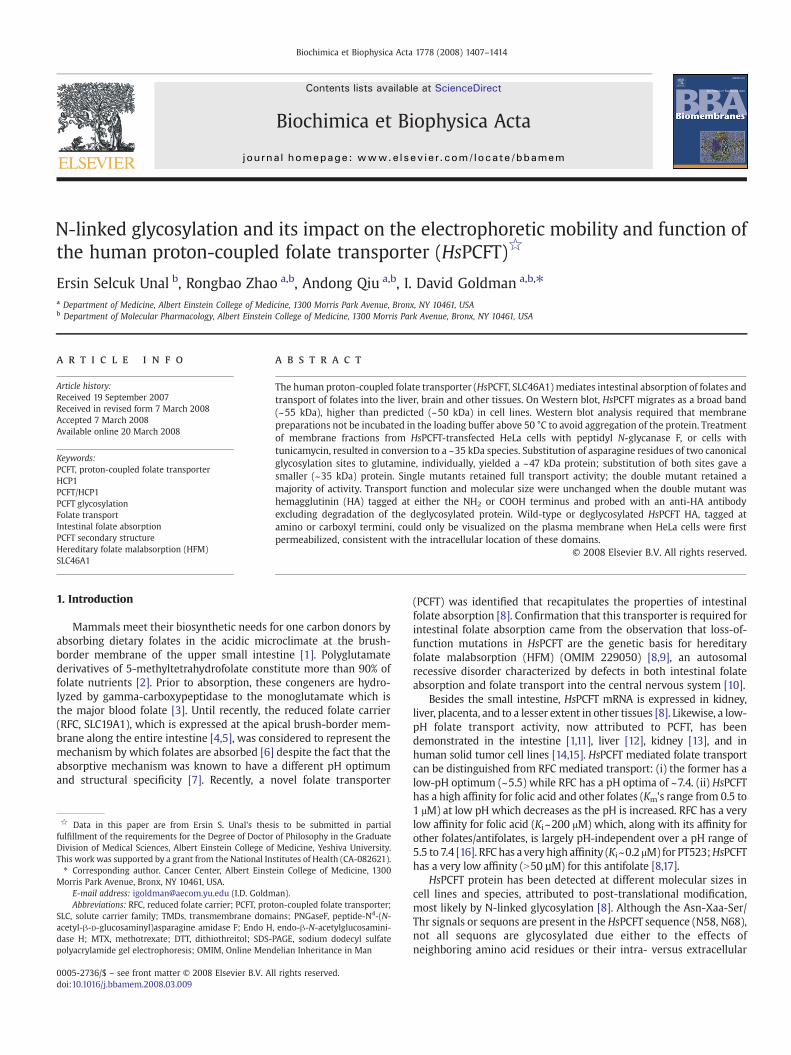
Biochimica et Biophysica Acta 1778 (2008) 1407–1414
Contents lists available at ScienceDirect
Biochimica et Biophysica Acta
j ourna l homepage: www.e lsev ie r.com/ locate /bbamem
N-linked glycosylation and its impact on the electrophoretic mobility and function ofthe human proton-coupled folate transporter (HsPCFT)☆
Ersin Selcuk Unal b, Rongbao Zhao a,b, Andong Qiu a,b, I. David Goldman a,b,⁎a Department of Medicine, Albert Einstein College of Medicine, 1300 Morris Park Avenue, Bronx, NY 10461, USAb Department of Molecular Pharmacology, Albert Einstein College of Medicine, 1300 Morris Park Avenue, Bronx, NY 10461, USA
a r t i c l e i n f o
☆ Data in this paper are from Ersin S. Unal's thesfulfillment of the requirements for the Degree of DoctorDivision of Medical Sciences, Albert Einstein College ofThis work was supported by a grant from the National In⁎ Corresponding author. Cancer Center, Albert Einst
Morris Park Avenue, Bronx, NY 10461, USA.E-mail address: [email protected] (I.D. GoldmAbbreviations: RFC, reduced folate carrier; PCFT, prot
SLC, solute carrier family; TMDs, transmembrane domacetyl-β-D-glucosaminyl)asparagine amidase F; Endo Hdase H; MTX, methotrexate; DTT, dithiothreitol; SDS-polyacrylamide gel electrophoresis; OMIM, Online Men
0005-2736/$ – see front matter © 2008 Elsevier B.V. Aldoi:10.1016/j.bbamem.2008.03.009
a b s t r a c t
Article history:Received 19 September 2007Received in revised form 7 March 2008Accepted 7 March 2008Available online 20 March 2008
The human proton-coupled folate transporter (HsPCFT, SLC46A1) mediates intestinal absorption of folates andtransport of folates into the liver, brain and other tissues. On Western blot, HsPCFT migrates as a broad band(~55 kDa), higher than predicted (~50 kDa) in cell lines. Western blot analysis required that membranepreparations not be incubated in the loading buffer above 50 °C to avoid aggregation of the protein. Treatmentof membrane fractions from HsPCFT-transfected HeLa cells with peptidyl N-glycanase F, or cells withtunicamycin, resulted in conversion to a ~35 kDa species. Substitution of asparagine residues of two canonicalglycosylation sites to glutamine, individually, yielded a ~47 kDa protein; substitution of both sites gave asmaller (~35 kDa) protein. Single mutants retained full transport activity; the double mutant retained amajority of activity. Transport function and molecular size were unchanged when the double mutant washemagglutinin (HA) tagged at either the NH2 or COOH terminus and probed with an anti-HA antibodyexcluding degradation of the deglycosylated protein. Wild-type or deglycosylated HsPCFT HA, tagged atamino or carboxyl termini, could only be visualized on the plasma membrane when HeLa cells were firstpermeabilized, consistent with the intracellular location of these domains.
© 2008 Elsevier B.V. All rights reserved.
Keywords:PCFT, proton-coupled folate transporterHCP1PCFT/HCP1PCFT glycosylationFolate transportIntestinal folate absorptionPCFT secondary structureHereditary folate malabsorption (HFM)SLC46A1
1. Introduction
Mammals meet their biosynthetic needs for one carbon donors byabsorbing dietary folates in the acidic microclimate at the brush-border membrane of the upper small intestine [1]. Polyglutamatederivatives of 5-methyltetrahydrofolate constitute more than 90% offolate nutrients [2]. Prior to absorption, these congeners are hydro-lyzed by gamma-carboxypeptidase to the monoglutamate which isthe major blood folate [3]. Until recently, the reduced folate carrier(RFC, SLC19A1), which is expressed at the apical brush-border mem-brane along the entire intestine [4,5], was considered to represent themechanism by which folates are absorbed [6] despite the fact that theabsorptive mechanism was known to have a different pH optimumand structural specificity [7]. Recently, a novel folate transporter
is to be submitted in partialof Philosophy in the GraduateMedicine, Yeshiva University.
stitutes of Health (CA-082621).ein College of Medicine, 1300
an).on-coupled folate transporter;ains; PNGaseF, peptide-N4-(N-, endo-β-N-acetylglucosamini-PAGE, sodium dodecyl sulfatedelian Inheritance in Man
l rights reserved.
(PCFT) was identified that recapitulates the properties of intestinalfolate absorption [8]. Confirmation that this transporter is required forintestinal folate absorption came from the observation that loss-of-function mutations in HsPCFT are the genetic basis for hereditaryfolate malabsorption (HFM) (OMIM 229050) [8,9], an autosomalrecessive disorder characterized by defects in both intestinal folateabsorption and folate transport into the central nervous system [10].
Besides the small intestine, HsPCFT mRNA is expressed in kidney,liver, placenta, and to a lesser extent in other tissues [8]. Likewise, a low-pH folate transport activity, now attributed to PCFT, has beendemonstrated in the intestine [1,11], liver [12], kidney [13], and inhuman solid tumor cell lines [14,15]. HsPCFT mediated folate transportcan be distinguished from RFC mediated transport: (i) the former has alow-pH optimum (~5.5) while RFC has a pH optima of ~7.4. (ii) HsPCFThas a high affinity for folic acid and other folates (Km's range from 0.5 to1 μM) at low pH which decreases as the pH is increased. RFC has a verylow affinity for folic acid (Ki~200 μM) which, along with its affinity forother folates/antifolates, is largely pH-independent over a pH range of5.5 to 7.4 [16]. RFC has a very high affinity (Ki~0.2 μM) for PT523;HsPCFThas a very low affinity (N50 µM) for this antifolate [8,17].
HsPCFT protein has been detected at different molecular sizes incell lines and species, attributed to post-translational modification,most likely by N-linked glycosylation [8]. Although the Asn-Xaa-Ser/Thr signals or sequons are present in the HsPCFT sequence (N58, N68),not all sequons are glycosylated due either to the effects ofneighboring amino acid residues or their intra- versus extracellular

1408 E.S. Unal et al. / Biochimica et Biophysica Acta 1778 (2008) 1407–1414
locations [18,19]. This study was designed to determine the extent towhich HsPCFT is, in fact, glycosylated and the impact of glycosylationon the electrophoretic properties of the protein and its transportfunction.
2. Materials and methods
2.1. Chemicals
Tritiated methotrexate (MTX-disodium salt, [3′, 5′, 7-[3H](N)]) (Catalog numberMT701) was obtained from Moravek Biochemicals Inc. (Brea, CA), purified by liquidchromatography, and maintained as previously described [20]. PNGaseF (peptide-N4-(N-acetyl-β-D-glucosaminyl)asparagine amidase F) (G5166) and tunicamycin fromStreptomyces species (T7765) were obtained from Sigma (St. Louis, MI). Proteaseinhibitor cocktail (Cat# 11836170001) was obtained from Roche Applied Science(Mannheim, Germany).
2.2. Site-directed mutagenesis
Site-directed mutagenesis was carried out according to the QuikChange II XLprotocol from Strategene (La Jolla, CA). Wild-type HsPCFT cDNA cloned into the BamHIsite of the mammalian expression vector pcDNA3.1(+) was used as the template [8].Two complementary forward and reverse primers, which carry the targeted nucleotidechanges in the middle region, were designed individually for introducing glutaminesinto HsPCFT primary sequence at positions 58, 68 and 58/68 [Table 1]. After initialdenaturation at 95 °C for 1min, the targeted vectors were generated for 18 cycles of 50 sat 95 °C for denaturation, 50 s at 60 °C for annealing, 16 min at 68 °C for extensionfollowed by a final extension period of 7 min at 68 °C. DpnI (10 U/µL) restriction enzymewas used to digest the parental dsDNA. The mixture (3 µL) was transformed into Top10ultra competent cells (Invitrogen). Plasmids carrying the desired mutations wereidentified by DNA automated sequencing in the Albert Einstein Cancer Center GenomicsShared Resource. The entire coding region of HsPCFT was sequenced to confirm theabsence of any other polymerase introduced mutations.
2.3. Epitope tagging
A hemagglutinin (HA) peptide epitope (YPYDVPDYA) was fused to the wild-typeand N58Q/N58Q-HsPCFT by PCR-based site-directed mutagenesis using the primerslisted in Table 1. A BglII restriction site was included in the upstream N-terminal HA tagprimer sequence for subcloning. The PCR product was purified, digested with BglII, andligated into BamHI-digested HsPCFT in pcDNA3.1(−) to generate the N-HA-HsPCFT andN-HA-N58Q/N68Q-HsPCFT. The downstream primer used for introducing the C-terminal HA epitope contained an XbaI restriction site. The upstream primer includeda HindIII restriction site. The PCR product was purified, digested with HindIII and XbaI,and ligated into HindIII–XbaI digested HsPCFT in pcDNA3.1(+) to generate the C-HA-HsPCFT and C-HA-N58Q/N68Q-HsPCFT. The ligation products were transformed intoTop10 ultra competent cells (Invitrogen). Plasmids carrying the HA tags were identifiedby DNA automated sequencing.
2.4. Cell culture and transfection
HeLa cells, originally obtained from the American Type Tissue Collection (Manassas,VA), have been maintained in this laboratory in RPMI-1640 medium supplementedwith 10% fetal bovine serum, 100 U/mL penicillin and 100 µg/mL streptomycin at 37 °Cin a humidified atmosphere of 5% CO2. Lipofectamine 2000 (Invitrogen) was usedaccording to the manufacturer's protocol for transient transfection of plasmid DNA intothese cells. To create stably transfected HeLa cell lines, transiently transfected cells were
Table 1Primers for site-directed mutagenesis and PCR primers for N- and C-terminal HA-epitope insertions for wild-type and N58Q/N68Q-HsPCFT
Primer sequence (5′ to 3′)
N58Q Forward GCCGACCTCGGCTACCAAGGCACCCGCCAAAGGReverse CCTTTGGCGGGTGCCTTGGTAGCCGAGGTCGGC
N68Q Forward AGGGGGGGCTGCAGCCAACGCAGCGCGGACCCCReverse GGGGTCCGCGCTGCGTTGGCTGCAGCCCCCCCT
N58Q/N68Q Forward GCCGACCTCGGCTACCAAGGCACCCGCCAAAGGGGGGGCTGCAGCCAACGCAGCGCGGACCCC
Reverse GGGGTCCGCGCTGCGTTGGCTGCAGCCCCCCCTTTGGCGGGTGCCTTGGTAGCCGAGGTCGGC
N-terminal HA tag Forward TCAAGATCTCACCATGTACCCATACGATGTTCCAGATTACGCTATGGAGGGGCGCGTGAGC
Reverse TATAGATCTCAGGGGCTCTGGGGAAACTGC-terminal HA tag Forward TATAAGCTTCACCATGGAGGGGAGCGCGAGC
Reverse TCATCTAGATTAAGCGTAATCTGGAACATCGTATGGGTAGGGCTCTGGGGAAACTG
trypsinized and passaged into 100 mm culture plates at appropriate densities andallowed to grow in RPMI-1640media containing 800 µg/mL of G418 sulfate (Mediatech,Herndon, VA). Individual clones were isolated, expanded and maintained in the samemedium supplemented with 800 µg/mL of G418. Several (N20) clones expressing thedesired N- and C-terminus tagged wild-type and N58Q/N68Q-HsPCFT were screenedand one clone of each construct with the highest activity was selected for furtheranalysis. HeLa cells were transfected with empty pcDNA3.1(+) vector (mock) or thesame vector containing wild-type or mutant HsPCFT. [3H]MTX influx was then assessedat pH 5.5.
2.5. PNGaseF and EndoH treatment
Enzymatic deglycosylation of total membrane protein (200 µg) prepared (seeWestern blot analysis below) from HsPCFT cDNA-transfected HeLa cells was performedwith PNGaseF enzyme (Sigma, G5166) following the manufacturer's protocol. Becauseheat denaturation was omitted, the incubation time of the protein sample in 250 mMphosphate buffer (pH 7.5) and 2.5 µL of 2% SDS with 1 M 2-mercaptoethanol wasincreased to 1 h at room temperature. Cleavage of the asparagine-N-acetyl glucosaminebond was monitored by SDS-PAGE. Membrane preparations were treated with EndoH(Roche Diagnostics, Mannheim, Germany) following the manufacturer's protocol.RNaseB (New England Biolabs) was treated under the same conditions as a control forEndoH activity.
2.6. Tunicamycin treatment
Tunicamycin (T7765)(Sigma, St. Louis, MI) was dissolved in DMSO and added togrowth medium to achieve concentrations of 0.25, 0.5, 1 µg/mL at the same time thecells were transfected with HsPCFT cDNA. Tunicamycin remained in the growthmedium for 48 h when the cells were assayed.
2.7. Membrane transport measurements
Cells were seeded in 17 mm glass scintillation vials to a final density of 4×10⁵ cells/mL. When cells reached mid-log phase growth ~72 h later, as determinedmicroscopically, uptake determinations were performed [21]. Briefly, the media wasaspirated and the cells incubated with HEPES Buffered Saline (HBS) (20 mM HEPES,5 mM dextrose, 140 mM NaCl, 5 mM KCl, 2 mM MgCl2, at pH 7.4) for 20 min. To initiateuptake, HBS was removed,MES buffered saline (MBS) (20mMMES,140mMNaCl, 5 mMKCl, 2 mM MgCl2, 5 mM dextrose, at pH 5.5) containing [3H]MTX (0.5 μM, specificactivity 1000–2000 dpm/pmol) was added to the cells. Uptake was stopped by placingthe vial in ice followed by rapid but careful addition of 6 mL of ice-cold HBS (pH 7.4).After three washes, cells were lysed with 0.5 mL of 0.2 N NaOH at 65 °C for 30 min;400 μL of lysate was used to determine radioactivity in a liquid scintillationspectrometer. Total protein for each sample was determined on a 20 μL portion of thelysate by the BCA protein assay reagent kit (Pierce, Rockford, IL). Data is expressed aspmol MTX/vial and normalized to total protein (pmol/mg protein).
2.8. Western blot analysis
Confluent HeLa cells in 17-mm glass scintillation vials were washed twice with ice-cold PBS and the cells were scraped off with PBS containing Roche protease inhibitor. Toobtain total membrane fractions, cells were incubated on ice for 30 min in hypotonicbuffer (200 µL/106 cells) (0.5 mMNa2HPO4, 0.1 mM EDTA at pH 7.4) containing proteaseinhibitor, following which the membrane fraction was pelleted by centrifugation at14,000 ×g and 4 °C for 2 min. The pellet was resuspended in lysis buffer (100 µL/106
cells; 20 mM TRIS-base, 150 mM NaCl, 1% Triton X-100, 0.1% SDS, 1 mM EDTA at pH 7.4)and briefly sonicated with the Microson XL 2000 ultrasonic liquid processor (MisonixIncorporated, Farmingdale, NY) and protein was determined as indicated above.Proteins were dissolved in SDS-PAGE loading buffer (0.225 M Tris.Cl [pH 6.8], 50%glycerol, 5% SDS, 0.05% bromphenol blue) with or without 0.25 M dithiothreitol (DTT).Portions of the preparation were then subjected to a 10 min incubation at roomtemperature, 50 °C, 75 °C or 95 °C followingwhich proteins were resolved on a 12% SDS-polyacrylamide gel. The proteins were then transferred to PVDF Transfer Membranes(Amersham Life Science). A polyclonal anti-HsPCFT antibody to the distal C-terminus ofHsPCFT (amino acids 446–459) was used to probe the plasma membrane as previouslydescribed [8]. A rabbit anti-HA antibody (#H6908) obtained from Sigma (St. Louis, MI)was used to detect HA-tagged proteins. Membranes were processed by the ECL PlusWestern Blotting Detection System (Amersham Life Science).
2.9. Immunofluorescence microscopy
A commercially available Alexa Fluor 488 conjugated anti-HA monoclonal antibody16B12 (1 mg/mL) obtained from Invitrogen (Carsbad, CA) was used to localize HA-tagged wild-type and N58Q/N68Q-HsPCFT proteins by direct immunofluorescence.Stably transfected HeLa cells grown on coverslips were washed five times with RPMI-1640 medium and the cells fixed with 1% paraformaldehyde for 30 min at roomtemperature. Cells were permeabilized with 0.2% Triton X-100 in PBS for 15 min at RT.The cells were blocked with 2% bovine serum albumin containing 5% donkey serum(blocking buffer) for 30 min, then 200 µL of Alexa Fluor 488 conjugated anti-HAmonoclonal antibody solution (1 µL of antibody in 8mL of blocking buffer) was added to

Fig. 2. The effect of heating onWestern blot analysis of HsPCFT. Equal amounts of lysate(30 µg) were incubated in the loading buffer with DTT at the indicated temperatures for10 min before loading onto the SDS-gel. The blots were probed with a polyclonalpeptide antibody directed to the C-terminus ofHsPCFT. The numbers on the left indicatethe molecular sizes of protein bands. The blot is representative of three separateexperiments.
1409E.S. Unal et al. / Biochimica et Biophysica Acta 1778 (2008) 1407–1414
each coverslip. After 1 h at room temperature, cells were washed 10 times with PBS,each for 5 min, and coverslips were placed on slides with mounting medium containingpropidium iodide to visualize DNA (Vector, CA). The slides were then sealed and viewedwith an Olympus IX70 Inverted Epifluorescence Microscope (Center Valley, PA) with488 nm excitation.
2.10. Data analysis
Data presented is the mean±standard error of the mean (SEM) of at least threeindependent experiments. Statistical comparisons were performed by the two-tailedStudent's paired t-test. Some experiments were assessed using a one-way repeatedmeasures analysis of variance (ANOVA) and the Tukey's post test. All statisticalanalyses were performed using GraphPad PRISM (version 3.0 for Windows, GraphPadSoftware).
3. Results
3.1. Computational analysis of the HsPCFT protein sequence
HsPCFT protein (NP_542400) consists of 459 amino acids with apredicted molecular weight (MW) of ~50 kDa (http://ca.expasy.org/tools/protparam.html). HsPCFT is a basic protein, with a predicted pIof 9.03, highly enriched with hydrophobic residues. The primarystructure of the protein has two putative N-linked glycosylationconsensus sites (http://www.cbs.dtu.dk/services/NetNGlyc) at posi-tions Asn58 (N-G-T) and Asn68 (N-R-S), as illustrated in Fig. 1. Thesesites are predicted to be within the extracellular loop between TMDs Iand II (HMMTOP, PredictProtein, TMpred, TopPred analyses). Theglycosylation consensus sites (N-X-S/T) are highly conserved amongspecies [Macaca mulatta (XP_001106954),Mus musculus (NP_081016),Bos taurus (NP_001073053), Xenopus laevis (AAH77859), Danio rerio(NP_956579), Monodelphis domestica (XP_001375817), Rattus norve-gicus (NP_001013991), Canis familiaris (XP_548286)] (http://www.ebi.ac.uk/clustalw/).
3.2. The effect of temperature on HsPCFT protein stability
When membrane samples were heated for 5–20 min at 95 °C todenature the protein in preparation for SDS-PAGE, as is the conven-tional method [22–24], no protein was detected likely due to HsPCFTaggregation and immobilization in the stacking gel. To establish con-
Fig. 1. A membrane topology model for HsPCFT predicted by online databases (http://wwwglycosylation consensus sites (arrows).
ditions that obviate aggregation, plasma membrane protein fractionswere prepared from HeLa cells 48 h after transfection with HsPCFTcDNA, then incubated in the loading buffer containing SDS with andwithout DTT at RT, 50 °C, 75 °C or 95 °C for 10 min before PAGE. Whenincubated in SDS solution at RT or 50 °C, protein migrated as threebands: (i) a broad band centered at ~55 kDa, (ii) a smaller band at~47.2 kDa, and (iii) the narrowest band at ~35.1 kDa. But there was nodetectable protein on the separating gel when it was heated above50 °C (Fig. 2). HsPCFT proteinwas detected in stacking gels but not theseparating gelwhen the samplewas heated at 75 °C and 95 °C (data notshown). Based on these observations, membrane fractions were main-tainedon iceor roomtemperature in the loadingbufferprior toSDS-PAGE.
3.3. Assessment of N-linked glycosylation with PNGaseF and EndoH
The molecular size of the HsPCFT protein detected in HeLa cells,~55 kDa, was higher than the predicted molecular weight of ~50 kDa[8]. As indicated above, there are two predicted N-linked glycosyla-tion sites in the first extracellular loop of HsPCFT. To determine if thissize discrepancy is due to post-translational glycosylation at thesesites, membranes from HeLa cells transiently transfected with HsPCFT
.expasy.org/tools/#topology) and the locations of asparagine residues in the N-linked

Fig. 4. The effect of tunicamycin treatment on the transport function (upper panel) andthe apparent molecular size (lower panel) of HsPCFT. Upper panel; [3H]MTX influx inHela cells transiently transfected with HsPCFT cDNA and treated with tunicamycin.Uptake of 0.5 µM [3H]MTX was assessed at pH 5.5 and 37 °C over 2 min in HeLa cellsgrown in the absence or presence of tunicamycin for 48 h. The transport activity incontrol cells is 214.0±30.2 pmol/mg/2 min. The p value reflects the comparison of thedifference between control cells and cells treated with 1 μg/mL tunicamycin. Data are themean±SEM from three independent experiments. Lower panel; Western blot analysis ofHsPCFT protein after tunicamycin treatment of HeLa cells. Equal amounts of lysate(30 µg) from HeLa cells, grown in the presence of increasing tunicamycin concentrationswere loaded and the blotswere probedwith a peptide antibody directed to the C-terminusofHsPCFT. The numbers on the left indicate themolecular sizes in the protein ladders. Theblot is representative of three separate experiments.
1410 E.S. Unal et al. / Biochimica et Biophysica Acta 1778 (2008) 1407–1414
cDNA were treated with PNGaseF, which cleaves the bond betweenthe first N-acetyl glucosamine of both complex and high mannoseoligosaccharides and the asparagine residue in the canonicalconsensus sequence [25]. Untreated protein displayed the above-noted three bands: a broad band centered at ~55 kDa and two othersmaller bands detected at ~47.2 kDa and ~35.1 kDa. When proteinsamples were treated with PNGaseF, virtually all protein migrated at amolecular weight of ~35.1 kDa with only very low levels of higherMW bands that likely represent incomplete PNGaseF reactionproducts (Fig. 3). Membrane protein fractions of HeLa cells transientlytransfected with HsPCFT were also treated with EndoH [26]. Therewas no detectable change in the Western blot. This was unlike theresults with RNaseB, an established substrate for EndoH, in whichthere was an expected gel-shift upon similar treatment with thisenzyme (data not shown).
3.4. Assessment of N-linked glycosylation in intact HeLa cells withtunicamycin
Another approach was utilized to assess glycosylation of HsPCFTbased on inhibition of de novo N-linked glycosylation by tunicamycin[27]. HeLa cells transiently transfected withHsPCFTcDNAwere exposedto a spectrum of tunicamycin concentrations for 48 h following whichcrude plasmamembranes were prepared and subjected toWestern blotanalysis. As indicated in Fig. 4 (lower panel), high molecular weightHsPCFTbands decrease as the tunicamycin concentrationwas increased;this was accompanied by an increase in the ~35 kDa band. By 1.0 µg/mLtunicamycin, the only band present was at 35.1 kDa. Studies were alsoundertaken to determine the impact of tunicamycin inhibition ofHsPCFT glycosylation on transport function in HeLa cells. There was nosignificant effect of low levels of tunicamycin on [3H]MTX influx (Fig. 4,upper panel) and the small decrease (~22%) observed at 1 μM drug,when glycosylation was completely inhibited, was not statisticallysignificant (p=0.09).
3.5. Site-directed mutagenesis of N-linked glycosylation sites
To further verify the N-linked glycosylation sites and theirfunctional importance, three different site-directed mutants ofHsPCFT were constructed at the putative N-glycosylation sites. Theasparagine residues at Asn58 and Asn68were replaced either singly ortogether by a glutamine residue to yield the Asn58Gln (N58Q-HsPCFT), Asn68Gln (N68Q-HsPCFT) single mutants and the N58Q/N68Q-HsPCF double mutant. To assess the functional consequences ofthese mutations, wild-type HsPCFT and the mutant constructs weretransiently expressed in HeLa cells and influx of 0.5 µM [3H]MTX was
Fig. 3. The effect of PNGaseF treatment on the apparent molecular size of HsPCFT.In vitro enzymatic deglycosylation was achieved by addition of PNGaseF (10 U/mL) to200 µg of membrane protein with incubation for 3 h at 37 °C. An equal amount ofPNGaseF treated (right lane) and control (left lane) lysate (30 µg) was loaded and theblots were probed with a peptide antibody directed to the C-terminus of HsPCFT.Numbers on the left indicate the molecular sizes in the protein ladders. The blot isrepresentative of three separate experiments.
assessed. As shown in Fig. 5 (upper panel), influx in the N58Q-HsPCFTand N68Q-HsPCFT single mutant transfectants was 79–83% of thetransport rate detected in wild-type HsPCFT cDNA-transfected cells(pN0.05). [3H]MTX influx was decreased by 40% in the double mutant(p=0.05).
Immunoblots of the crude plasmamembranes prepared fromHeLacells transiently transfected with HsPCFT cDNA, using anti-HsPCFTantibody, showed wild-type HsPCFT protein running at an apparentmolecular weight of ~55 kDa, Asn58Gln/Asn68Gln-HsPCFT proteinrunning at a molecular weight of ~35 kDa, and both N58Q-HsPCFT andN68Q-HsPCFT with a predominant band at an apparent molecularweight of ~47 kDa, a size intermediate between that of the wild-typeand the double mutant HsPCFT (Fig. 5, lower panel). Hence, bothAsn58 and Asn68 residues were glycosylated, and produced a similaralteration in the migration pattern of the protein on SDS-PAGE. Theseobservations confirm that the ~35 kDa band represents deglycosy-lated HsPCFT.
3.6. Analysis of the structural integrity of C- or N-terminus hemagglutinin(HA) tagged wild-type and deglycosylated (Asn58Gln/Asn68Gln) HsPCFTmutants in HeLa cells
The migration pattern observed by deglycosylation of HsPCFTin vitro with PNGaseF, after inhibition of N-linked glycosylation inintact cells with tunicamycin or by site-directed mutagenesis, all

1411E.S. Unal et al. / Biochimica et Biophysica Acta 1778 (2008) 1407–1414
produced a proteinwith amuch lowermolecular size thanpredicted. Toevaluate the possibility that this was due to proteolytic degradation,wild-type and N58Q/N68Q-HsPCFT, HA-tagged at either the N- or C-terminus, were transiently transfected into HeLa cells andWestern blotanalysis was performed with anti-HA antibody. The HA tag at eitherterminus did not affect the function of the wild-type HsPCFT or thedeglycosylated mutant (Fig. 6, upper panel). When probed with anti-HA antibody, both N- or C-terminus HA-tagged wild-type HsPCFTmigrated predominantly to a mean molecular size of ~55 kDa with asmaller band at ~47 kDa. Both N- or C-terminal HA-tagged N58Q/N68Q-HsPCFT migrated at a single band at a MW of ~35 kDa (Fig. 6,lower panel). These data exclude proteolytic degradation of wild-type or deglycosylated forms of HsPCFT as a basis for the lower thanpredicted molecular size observed on Western blot. Hence, theanomalous migration of the HsPCFT protein on SDS-PAGE appears tobe an intrinsic characteristic of the protein under these conditions.
3.7. Localization of the wild-type and deglycosylated HsPCFT proteins HAtagged at the amino or carboxyl terminus
Localization of the wild-type and N58Q/N68Q-HsPCFT, stable trans-fectants with high level expression of either the N- or C-terminusHA-epitope tag was assayed by immunofluorescence stainingwith or
Fig. 6. The effect of HA tagging on the transport function (upper panel) andmolecular size(lower panel) of thewild-type and deglycosylated (N58Q/N68Q) HsPCFT. Upper panel;[3H]MTX influx in HeLa cells transiently transfected with cDNAs of N- or C-terminus HA-taggedwild-type andN58Q/N68Q-HsPCFTmutant (degly-HsPCFT). Influxof 0.5µM[3H]MTXwas assessed at pH 5.5 and 37 °C over 2 min. Transport activity inwild-type transfectants is288.8±12.6 pmol/mg/2 min. The p value reflects the difference in activity between thewild-type and deglycosylated HsPCFT. Data are the mean±SEM from three indepen-dent experiments. Lower panel; Western blot analysis of membrane proteins fromHeLa cells transiently transfectedwith wild-type or deglycosylated HsPCFT fusedwithan HA epitope on either the N- or C-terminus. An equal amount of lysate (20 µg) wasloaded and the blots were probed with a monoclonal anti-HA antibody. The numberson the left indicate the molecular sizes in the protein ladders. Each blot isrepresentative of three separate experiments.
Fig. 5. The transport function (upper panel) and molecular size (lower panel) of N58Q-,N68Q- and N58Q/N68Q-HsPCFT mutants. Upper panel; [3H]MTX influx in HeLa cellstransiently transfected with wild-type HsPCFT or glycosylation mutants. Uptake of0.5 µM [3H]MTXwas assessed at pH 5.5 and 37 °C over 2 min. Transport activity inwild-type transfectants is 155.3±36.4 pmol/mg/2 min. The p value reflects the difference inactivities of the mutant as compared to the wild-type HsPCFT transfectants. Data arethe mean±SEM from three independent experiments. Lower panel; Western blotanalysis of wild-type and glycosylation mutant HsPCFT membrane proteins. An equalamount of lysate (20 µg) was loaded and the blots were probed with a peptide antibodydirected to the C-terminus of HsPCFT. The numbers shown on the left side indicate themolecular sizes in the protein ladders. The blot is representative of three separateexperiments.
without membrane permeabilization. As indicated in Fig. 7, fluores-cence was detected in all permeabilized cells with the exception ofmock transfected cells. No fluorescence could be detected in cells thatwere not permeabilized. Hence, both wild-type and deglycosylatedHsPCFT proteins localized to the plasma membrane and both N-and C-termini were accessible to the antibody only when cells werepermeabilized under these conditions. These data are consistentwith a cytosolic localization for both the N- and C-terminus andindicates that the lack of N-glycosylation has little or no effect onHsPCFT membrane targeting.
4. Discussion
HsPCFT is a recently discovered carrier protein that mediatesintestinal folate absorption and transport of folates into the centralnervous system [8]. HsPCFT is expressed in other tissues as well and alow-pH folate transport activity, presumably HsPCFT, is present inmost human solid tumors and likely contributes to transport of folatesin these settings [8,14]. The murine ortholog of HsPCFT was reportedto be a heme carrier protein, (HCP-1, SLC46A1), responsible forintestinal heme absorption that functions independent of pH (over a

Fig. 7. Immunofluorescence staining of wild-type and deglycosylated HsPCFT. HeLa cells were stably transfected with cDNA of wild-type or deglycosylated HsPCFT fused with the HAepitope on either the N- or C-terminus, then exposed to Alexa Fluor 488 conjugated anti-HA monoclonal antibody. Green fluorescence indicates localization of HsPCFT, while redfluorescence indicates nucleic acids counterstained by propidium iodide. The image is representative of at least three experiments. The left panel shows cells subjected topermeabilization with Triton X-100 before exposure to antibody. The right panel shows cells that were not permeabilized.
1412 E.S. Unal et al. / Biochimica et Biophysica Acta 1778 (2008) 1407–1414
pH range of from 6.5 to 8.0) and with a low affinity for heme(Km=125 μM for [55Fe]hemin) [28]. However, in studies from thislaboratory [8], and in a recent report from another laboratory [29], it isclear that this is a high-affinity folate transporter (influx Km's in therange of 0.2–1.0 µM)with a low-pH optimum. Additional confirmationthat the major, if not sole, function of this carrier is folate transportcomes from the observation that loss-of-function mutations inHsPCFT result in an autosomal recessive disorder, hereditary folatemalabsorption. Individuals with this disorder show no evidence ofiron deficiency and the metabolic consequences of the transportdefect can be corrected solely by the administration of pharmacolo-gical doses of folate [8,9].
The data indicate that HsPCFT is highly heat labile; at temperaturesabove 50 °C the protein aggregates, a condition that must beconsidered in the preparation of cell membranes for SDS-PAGE. Thebasis for this aggregation is not clear but may be due to the propertiesof specific hydrophobic regions of the carrier as reported for SARS-CoVmembrane protein [30]. On SDS-PAGE the wild-type protein ran asthree bands at ~55 kDa, ~47.2 kDa and ~35.1 kDa. The threeindependent approaches used to assess HsPCFT glycosylation status
yielded a deglycosylated protein with an apparent molecular size of~35 kDa, 15 kDa smaller than the predicted size for nonglycosylatedHsPCFT (~50 kDa). Further studies using HsPCFT HA tagged on the N-and C-termini excluded the possibility that this discrepancy is due todegradation of the protein. This anomalous electrophoretic behaviouris quite common among polytopic membrane proteins and is similarto what has been reported for the human norepinephrine [31] andglycine transporters [32]. This could be due to (i) the highlyhydrophobic nature and basic pI of the transporter [33] and/or, (ii)preservation of the native, compact conformation and decreased SDSbinding due to the heating limitation resulting in incompletedenaturation of the HsPCFT protein [34,35]. Decreased SDS bindingmay decrease the migration rate by reducing the net charge;alternatively, it may increase the migration rate by reducing themass and frictional drag. The intact disulfide bonds, which usuallyresult in less SDS binding of a more compact shaped protein, can alsocause faster migration rates [36]. However, there was no difference inthe HsPCFT migration pattern when membranes were prepared withor without DTT before loading onto the gel. Replacement of the sevennative cysteine residues with serines had no effect on HsPCFT function;

1413E.S. Unal et al. / Biochimica et Biophysica Acta 1778 (2008) 1407–1414
hence, disulfide bonds are not required for proper HsPCFT folding andfunction (Unal ES et al., unpublished).
Glycosylation on asparagine 58 and 68 confirms that the firstmethionine is the actual translation initiation site since the secondmethionine in the amino acid sequence ofHsPCFT is at the 75th positionafter the glycosylation consensus sites. The results suggest that anapproximately 20 kDa oligosaccharide chain is added to HsPCFT whenexpressed in HeLa cells based upon the size of glycosylated HsPCFT(~55 kDa) and deglycosylated transporters (~35 kDa). It is of interest thatHsPCFTmay be glycosylated to a different extent in different cells. Hence,~60 kDa and ~78 kDa sizeHsPCFT proteins were detected in HepG2 cellsandXenopusoocytes, respectively [8]. Further studies on theglycosylationstatus of HsPCFT from native tissues, and their effect on the migrationpattern relative to what is observed in Hela cells, will be of interest.
Similar to most membrane transporters that are N-linked glycosy-lated on a single large extracellular loop [37], post-translational modi-fication ofHsPCFTbyN-linked glycosylation atN58 andN68 requires theextracellular localization of this domain, since oligosaccharide chains aretransferred en bloc from a dolichol donor to the nascent polypeptidesimultaneously with translation only if the NXS/T consensus site facesthe endoplasmic reticulum lumen [38]. This is consistent with atopologicalmodel inwhich theN-terminus is localized to the cytoplasm.This configuration was supported by immunocytochemical analysis ofboth wild-type and deglycosylated HsPCFT constructs HA tagged at theamino terminus, visualized at the plasma membrane only when cellswere first permeabilized with Triton X100 [39–42]. This data alsosupports a model in which there is an even number of transmembranedomains, requiring that theC-terminus is also localized to the cytoplasm.Hence,wild-typeanddeglycosylatedHsPCFT,HAtaggedat theC-terminus,could only be detected after membrane permeabilization.
N-linked glycosylation is not essential for cell surface trafficking andfunctionofRFC [43] and the creatine transporter [44]. On theotherhand,N-linked glycosylation can play a role in proper folding of the poly-peptide chain, protection from proteolytic degradation [45], main-tenance of protein solubility [46] and targeting to subcellularcompartments and to the cell surface [47]. The latter roleof glycosylationhas been reported for many transporters such as the humannorepinephrine [31], glycine (GLYT1) [32], glucose (GLUT1) [48], organicanion (OAT4) [47] andorganic cation transporters [49]. In these cases thetransport proteins are heavily glycosylated on three or more asparagineresidues and the deglycosylated protein retains only a small percentageof wild-type activity. In contrast, HsPCFT is glycosylated only onasparagine 58 and 68 and the majority of HsPCFT transport functionwas preserved in the N58Q/N68Q-HsPCFT mutant. Indeed, the smalldecrease observed could be due to the amino acid changes per se ratherthan the absence of glycosyl moieties since, at a tunicamycin con-centration sufficient to completely abolish glycosylation, no significantdecrease in HsPCFT function could be detected.
N-linked glycosylation scanning mutagenesis is a useful techniqueto analyze the topology of various transporters [32,50]. The pre-requisite for this approach is a functional deglycosylated transporter[51]. The observation that the majority of function is preserved indeglycosylated HsPCFT indicates that glycosylation scanning muta-genesis can be used to further evaluate the secondary structure of thiscarrier and that bacterial systems can be used to produce largequantities of this transporter for structural studies.
Acknowledgement
This work was supported by a grant from the National Institutes ofHealth (CA-082621).
References
[1] J. Selhub, G.J. Dhar, I.H. Rosenberg, Gastrointestinal absorption of folates andantifolates, Pharmacol. Ther. 20 (1983) 397–418.
[2] C.H. Halsted, The intestinal absorption of folates, Am J. Clin Nutr. 32 (1979) 846–855.[3] A.M. Reisenauer, C.H. Halsted, Human jejunal brush border folate conjugase.
Characteristics and inhibition by salicylazosulfapyridine, Biochim. Biophys. Acta.659 (1981) 62–69.
[4] Y. Wang, R. Zhao, R.G. Russell, I.D. Goldman, Localization of the murine reducedfolate carrier as assessed by immunohistochemical analysis, Biochim. Biophys.Acta 1513 (2001) 49–54.
[5] H.M. Said, F.K. Ghishan, R. Redha, Folate transport by human intestinal brush-border membrane vesicles, Am. J. Physiol 252 (1987) G229–G236.
[6] C.K. Kumar, T.T. Nguyen, F.B. Gonzales, H.M. Said, Comparison of intestinal folatecarrier clone expressed in IEC-6 cells and inXenopus oocytes, Am. J. Physiol 274 (1998)C289–C294.
[7] L.H. Matherly, D.I. Goldman, Membrane transport of folates, Vitam. Horm. 66 (2003)403–456.
[8] A. Qiu,M. Jansen, A. Sakaris, S.H.Min, S. Chattopadhyay, E. Tsai, C. Sandoval, R. Zhao,M.H. Akabas, I.D. Goldman, Identification of an intestinal folate transporter and themolecular basis for hereditary folate malabsorption, Cell 127 (2006) 917–928.
[9] R. Zhao, S.H. Min, A. Qiu, A. Sakaris, G.L. Goldberg, C. Sandoval, J.J. Malatack, D.S.Rosenblatt, I.D. Goldman, The spectrum of mutations in the PCFT gene, coding foran intestinal folate transporter, that are the basis for hereditary folate malabsorp-tion, Blood. 110 (2007) 1147–1152.
[10] J. Geller, D. Kronn, S. Jayabose, C. Sandoval, Hereditary folatemalabsorption: familyreport and review of the literature, Medicine (Baltimore). 81 (2002) 51–68.
[11] J. Selhub, I.H. Rosenberg, Folate transport in isolated brush border membranevesicles from rat intestine, J. Biol. Chem. 256 (1981) 4489–4493.
[12] D.W. Horne, Transport of folates and antifolates in liver, Proc. Soc. Exp. Biol. Med.202 (1993) 385–391.
[13] S.D. Bhandari, S.K. Joshi, K.E. McMartin, Folate binding and transport by rat kidneybrush-border membrane vesicles, Biochim. Biophys. Acta 937 (1988) 211–218.
[14] R. Zhao, F. Gao, M. Hanscom, I.D. Goldman, A prominent low-pH methotrexatetransport activity in human solid tumor cells: contribution to the preservation ofmethotrexate pharmacological activity in HeLa cells lacking the reduced folatecarrier, Clin. Cancer Res. 10 (2004) 718–727.
[15] S. Chattopadhyay, R. Zhao, S.A. Krupenko, N. Krupenko, I.D. Goldman, The inverserelationship between reduced folate carrier function and pemetrexed activity in ahuman colon cancer cell line, Mol. Cancer Ther. 5 (2006) 438–449.
[16] R. Zhao, I.D. Goldman, The molecular identity and characterization of a proton-coupled folate transporter—PCFT; biological ramifications and impact on theactivity of pemetrexed, Cancer Metastasis Rev. 26 (2007) 129–139.
[17] Y. Wang, R. Zhao, I.D. Goldman, Characterization of a folate transporter in HeLacells with a low pH optimum and high affinity for pemetrexed distinct from thereduced folate carrier, Clin. Cancer Res. 10 (2004) 6256–6264.
[18] S. Ben Dor, N. Esterman, E. Rubin, N. Sharon, Biases and complex patterns in theresidues flanking protein N-glycosylation sites, Glycobiology. 14 (2004) 95–101.
[19] L. Kasturi, H. Chen, S.H. Shakin-Eshleman, Regulation of N-linked core glycosyla-tion: use of a site-directed mutagenesis approach to identify Asn-Xaa-Ser/Thrsequons that are poor oligosaccharide acceptors, Biochem. J. 323 (1997) 415–419.
[20] D.W. Fry, J.C. Yalowich, I.D. Goldman, Rapid formation of poly-gamma-glutamylderivatives of methotrexate and their association with dihydrofolate reductase asassessed by high pressure liquid chromatography in the Ehrlich ascites tumor cellin vitro, J. Biol. Chem. 257 (1982) 1890–1896.
[21] K.A. Sharif, I.D. Goldman, Rapid determination of membrane transport parametersin adherent cells, BioTechniques 28 (2000) 926–928, 930, 932.
[22] U.K. Laemmli, Cleavage of structural proteins during the assembly of the head ofbacteriophage T4, Nature 227 (1970) 680–685.
[23] N. Quandt, A. Stindl, U. Keller, Sodium dodecyl sulfate-polyacrylamide gelelectrophoresis for M(r) estimations of high-molecular-weight polypeptides,Anal. Biochem. 214 (1993) 490–494.
[24] L. Garcia-Ortega, l.R. De, V.A. Martinez-Ruiz, M. Onaderra, J. Lacadena, d.P.Martinez, J.G. Gavilanes, Anomalous electrophoretic behavior of a very acidicprotein: ribonuclease U2, Electrophoresis. 26 (2005) 3407–3413.
[25] G.D. Barsomian, T.L. Johnson, M. Borowski, J. Denman, J.F. Ollington, S. Hirani, D.S.McNeilly, J.R. Rasmussen, Cloning and expression of peptide-N4-(N-acetyl-beta-D-glucosaminyl)asparagine amidase F in Escherichia coli, J. Biol. Chem. 265 (1990)6967–6972.
[26] T. Tai, K. Yamashita, M. Ogata-Arakawa, N. Koide, T. Muramatsu, S. Iwashita, Y. Inoue,A. Kobata, Structural studies of two ovalbumin glycopeptides in relation to the endo-beta-N-acetylglucosaminidase specificity, J. Biol. Chem. 250 (1975) 8569–8575.
[27] A.D. Elbein, Inhibitors of the biosynthesis and processing of N-linked oligosac-charide chains, Annu. Rev. Biochem. 56 (497–534) (1987) 497–534.
[28] M. Shayeghi, G.O. Latunde-Dada, J.S. Oakhill, A.H. Laftah, K. Takeuchi, N. Halliday, Y.Khan, A. Warley, F.E. McCann, R.C. Hider, D.M. Frazer, G.J. Anderson, C.D. Vulpe, R.J.Simpson, A.T. McKie, Identification of an intestinal heme transporter, Cell.122 (2005)789–801.
[29] Y. Nakai, K. Inoue, N. Abe, M. Hatakeyama, K.Y. Ohta, M. Otagiri, Y. Hayashi, H.Yuasa, Functional characterization of human PCFT/HCP1 heterologously expressedin mammalian cells as a folate transporter, J. Pharmacol. Exp. Ther. (2007).
[30] Y.N. Lee, L.K. Chen, H.C. Ma, H.H. Yang, H.P. Li, S.Y. Lo, Thermal aggregation of SARS-CoV membrane protein, J. Virol. Methods. 129 (2005) 152–161.
[31] H.E. Melikian, J.K. McDonald, H. Gu, G. Rudnick, K.R. Moore, R.D. Blakely, Humannorepinephrine transporter. Biosynthetic studies using a site-directed polyclonalantibody, J. Biol. Chem. 269 (1994) 12290–12297.
[32] L. Olivares, C. Aragon, C. Gimenez, F. Zafra, Analysis of the transmembranetopology of the glycine transporter GLYT1, J. Biol. Chem. 272 (1997) 1211–1217.
[33] A.K. Dunker, R.R. Rueckert, Observations on molecular weight determinations onpolyacrylamide gel, J. Biol. Chem. 244 (1969) 5074–5080.

1414 E.S. Unal et al. / Biochimica et Biophysica Acta 1778 (2008) 1407–1414
[34] J.S. Tung, C.A. Knight, Relative importance of some factors affecting theelectrophoretic migration of proteins in sodium dodecyl sulfate-polyacrylamidegels, Anal. Biochem. 48 (1972) 153–163.
[35] H. Furthmayr, R. Timpl, Characterization of collagen peptides by sodiumdodecylsulfate-polyacrylamide electrophoresis, Anal. Biochem. 41 (1971) 510–516.
[36] J.A. Reynolds, C. Tanford, The gross conformation of protein–sodium dodecylsulfate complexes, J. Biol. Chem. 245 (1970) 5161–5165.
[37] C. Landolt-Marticorena, R.A. Reithmeier, Asparagine-linked oligosaccharides arelocalized to single extracytosolic segments in multi-span membrane glycopro-teins, Biochem. J. 302 (1994) 253–260.
[38] R. Kornfeld, S. Kornfeld, Assembly of asparagine-linked oligosaccharides, Annu.Rev. Biochem. 54 (631–64) (1985) 631–664.
[39] P.L. Ferguson, W.F. Flintoff, Topological and functional analysis of the humanreduced folate carrier by hemagglutinin epitope insertion, J. Biol. Chem. 274 (1999)16269–16278.
[40] J. Geyer, B. Doring, K. Meerkamp, B. Ugele, N. Bakhiya, C.F. Fernandes, J.R. Godoy, H.Glatt, E. Petzinger, Cloning and functional characterization of human sodium-dependent organic anion transporter (SLC10A6), J. Biol. Chem. 282 (2007)19728–19741.
[41] M. Hong, K. Tanaka, Z. Pan, J. Ma, G. You, Determination of the external loops andthe cellular orientation of the N- and the C-termini of the human organic aniontransporter hOAT1, Biochem. J. 401 (2007) 515–520.
[42] O. Levy, G. Dai, C. Riedel, C.S. Ginter, E.M. Paul, A.N. Lebowitz, N. Carrasco,Characterization of the thyroid Na+/I− symporter with an anti-COOH terminusantibody, Proc. Natl. Acad. Sci. U. S. A. 94 (1997) 5568–5573.
[43] S.C. Wong, L. Zhang, S.A. Proefke, L.H. Matherly, Effects of the loss of capacity forN-glycosylation on the transport activity and cellular localization of the humanreduced folate carrier, Biochim. Biophys. Acta 1375 (1998) 6–12.
[44] N. Straumann, A. Wind, T. Leuenberger, T. Wallimann, Effects of N-linkedglycosylation on the creatine transporter, Biochem. J. 393 (2006) 459–469.
[45] S.M. Hurtley, D.G. Bole, H. Hoover-Litty, A. Helenius, C.S. Copeland, Interactions ofmisfolded influenza virus hemagglutinin with binding protein (BiP), J. Cell Biol.108 (1989) 2117–2126.
[46] P.J. Gallagher, J.M. Henneberry, J.F. Sambrook, M.J. Gething, Glycosylationrequirements for intracellular transport and function of the hemagglutinin ofinfluenza virus, J. Virol. 66 (1992) 7136–7145.
[47] F. Zhou, W. Xu, M. Hong, Z. Pan, P.J. Sinko, J. Ma, G. You, The role of N-linkedglycosylation in protein folding, membrane targeting, and substrate binding ofhuman organic anion transporter hOAT4, Mol. Pharmacol. 67 (2005) 868–876.
[48] T. Asano, H. Katagiri, K. Takata, J.L. Lin, H. Ishihara, K. Inukai, K. Tsukuda, M. Kikuchi,H. Hirano, Y. Yazaki, The role of N-glycosylation of GLUT1 for glucose transportactivity, J. Biol. Chem. 266 (1991) 24632–24636.
[49] R.J. Ott, A.C. Hui, K.M. Giacomini, Inhibition of N-linked glycosylation affectsorganic cation transport across the brush border membrane of opossum kidney(OK) cells, J. Biol. Chem. 267 (1992) 133–139.
[50] X. Liu, L. Matherly, Analysis of membrane topology of the human reduced folatecarrier protein by hemagglutinin epitope insertion and scanning glycosylationinsertion mutagenesis, Biochim. Biophys. Acta 1564 (2002) 333–342.
[51] M. van Geest, J.S. Lolkema, Membrane topology and insertion of membraneproteins: search for topogenic signals, Microbiol. Mol. Biol. Rev. 64 (2000) 13–33.
Copyright © 2022 FDOKUMEN