Integration of a polarizable interface for electrophoretic ...
Upload
independentCategory
view
1download
0
Molecular Microbiology (1993) 7(4). 515-521
Electrophoretic karyotype and gene assignmentto resolved chromosomes of Trichoderma spp.
A. Herrera-Estrella/ G. H. Goldman,* M. VanMontagu=>̂ and R. A. GeremiaLaboratohum voor Genetica. Universiteit Gent, K. L.Ledeganckstraat 35, B-9000 Gent, Belgium.
Summary
A molecular karyotype for three different Tricho-derma species {T. harzianum, T. viride, and T. reesei)was determined by using two different systems:countour-clamped electric-field and rotating-elec-trode electrophoresis. Six chromosomal DNA bandswere observed in T. harzianum and T. reesei and fivein T. viride. The sizes of these molecules were esti-mated by their mobility relative to the Schizosaccha-romyces pombe chromosomes and ranged between2.2 and 7.4 megabase pairs (mbp). The estimatedgenome sizes range from 31 to 39 mbp, A number ofgenes were located in the different chromosomes bymeans of Southern analysis. The implications ofthese findings are discussed.
Introduction
The fungal genus Trichoderma contains species that areof economic importance. They are useful in the produc-tion of hydrolytic enzymes (as, for example, cellulasesand xylanases), biochemicals and antibiotics (Meyer,1991). Some of them have already been used for the pro-duction of heterologous proteins (Cheng et ai, 1990;Nevaiainen et ai, 1991) and as biocontrol agents againstphytopathogens (Chet, 1990), Despite its importance,there are still many problems concerning the taxonomicclassification of species of this genus. The taxonomicscheme most widely used is based on the revision of Rifai(1969). Teleomorphs, where known, belong to Hypocreaor related genera. The species of Trichoderma arereferred to as species aggregates because they aremade up of more than one genetic entity that cannot bedistinguished with the presently used morphological
Received 17 February, 1992; revised and accepted 1 October, 1992.fPresent address: CINVESTAV Irapuato, Depanment ot Molecular Biol-ogy, AP 629 Irapuato Gto,, Mexico, :tFuture address: Department of Phar-macology, UMDNJ-Robert Wood Johnson Medical School, Piscataway,New Jersey 08854, USA. 'For correspondence. Tel. (91) 645170; Fax(91) 645349; BITNET mamon @ gengen,rug,ac,be.
characters. Although other workers have tried to usemolecular techniques for the classification of Trichodermaspp. (Meyer, 1991; Meyer et ai, 1992), more studies ofmolecular taxonomy are needed in order to obtain a dearpicture of its dassification.
Traditionally, determining the number of chromosomespresent in a fungus has been done by direct microscopicexamination or by establishing genetic linkage relation-ships between known loci. These tvt/o procedures, whenpossible, are neither straightforward nor reliable. Thedevelopment of pulse-field gel electrophoresis (PFGE)has allowed electrophoretic karyotyping of several yeastsand filamentous fungi (for a review, see Skinner et ai,1991). The use of PFGE and molecular karyotyping tech-nology has led to the assignment of cloned genes to chro-mosomal locations. New possibilities can be offered bythe utilization of this technology, like, for example, thedetection of translocations and variations in chromosomenumber and to generate chromosome-specific subli-braries. Here we report the establishment of conditionsfor the preparation of intact chromosomal DNA from Tri-choderma spp. Resolution of six chromosomal bands ofup to 7,4 megabase pairs (mbp) has been achieved aswell as the localization of several genes in the corre-sponding chromosomes by Southern analysis.
Results
Pulsed-field gel electrophoresis was performed with thegenome of three of the most studied Trichoderma species:T. harzianum, T. reesei. and T. viride. We have preparedintact chromosomal DNA essentially as described forAspergillus nidulans (Brody and Carbon, 1989; see theExperimental procedures).
Initial experiments involving short runs (see the Experi-mental procedures — Electrophoresis test) with rotating-electrode electrophoresis and using DNA samples pre-pared as described (Experimental procedures) showedfour distinguishable bands for T harzianum, and a three-band pattern for T viride, all with varying intensities afterethidium bromide staining (data not shown). This indi-cated that the DNA was intact and. more importantly, thatthere could be several chromosomal bands co-migratingin the gel.
Using longer running times and different combinationsof angle ramping, agarose concentrations, and voltage,

516 A. Herrera-Estretta, G. H. Goldman, M Van Montagu and R. A. Geremia
Fig. 1. Trichoderma spp. molecular karyotype.Intact chromosomal DNA was prepared inagarose blocks (see the Experimentalprocedures); a piece large enough to fill a wellwas loaded into the gel and subjected to PFGE,A, Chromosomal DNAs from S, pombe (lane S,p,),T. harzianum {lane T.h,). and T. i';r/de(laneT,v,)were resolved into chromosomal bands using arotating-electrode system. The bands observed intbe lanes were separated under the conditionsdescribed as for smaller chromosomes in theExperimental procedures. The inserts show theresolution of tbe largest cbromosome into twobards by using different running conditions (seethe Experimental procedures). The arabic num-bers on the left indicate tbe sizes in megabasepairs (Mb) of tbe S. pombe chromosomes. Romannumbers on tbe right indicate tbe nomenclatureassigned to each band in order of increasing size,B. Chromosomal DNAs from S. pombe (S,p,) andT. reesei (T,r,) were resolved with the CHEF-Map-per (BioRad): for conditions, see Ihe Experimentalprocedures. Symbols are as in (A).
separation was achieved for the chromosomal DNAs of T.harzianum and of T. viride into five and four bands,respectively (Fig, 1 A; for conditions, seethe Experimentalprocedures). However, the two largest bands in the pat-tern presented by both species appeared more intense onethidium-bromide-stained gels. Further modifications ofthe parameters made it possible to obtain conditionswbich allovi/ed the resolution of the largest band into twoindependent chromosomes (see insert, Fig, 1 A) but it wasnot possible to resolve the second band. The sizes of thedifferent chromosomes were estimated by comparingtheir electrophoretic mobilities with those of the threechromosomes of Schizosaccharomyces pombe (3.5, 4.7,and 5.7 mbp) and that of the largest Saccharomycesoerevisiae chromosome (2.2 mbp). Because some of thebands observed are larger than the 5.7 mbp marker, theirsizes, as reported here, could have extrapolation errors.
By analysing the results obtained under the many dif-ferent conditions used during these studies we have beenable to distinguish, for T. harzianum, six bands corre-sponding to seven chromosomes ranging from 2.2to 7.3 mbp (Fig, 1), Numbers were assigned to thedifferent chromosomal bands in order of increasing size:I, 2.2mbp; II, 3.7mbp; III, 5.6mbp; IVa,b, 6,5mbp; V,7.0 mbp; and VI, 7.3 mbp (Fig. 1, lane T,h,), The electro-phoretic karyotype of T. viride is very similar to that of T.harzianum but the smallest chromosome is absent in theformer. The chromosome nomenclature assigned in thiscase is as follows: i, 4.2 mbp; II, 5.3 mbp; llla,b, 6,0 mbp;IV. 7.0 mbp; and V, 7.2 mbp (Fig, 1, lane T.v.).
These data suggest a close relationship between thesetwo species during evolution, although one of them mayhave suffered a major reorganization involving fusion orsplitting of a complete chromosome.
To obtain an initial indication of the variability within thegenus, another species lacking the ability to attack andcontrol phytopathogenic fungi was analysed. For this, wechose T. reesei because it does not have biocontrollingproperties (A. Herrera-Estrelia, unpublished) and It is alsoone of the most important cellulose-degrading micro-organisms.
When using the conditions established for 7,harzianum and T. viride, it was not possible to obtain fullysatisfactory results (data not shown). However, the avail-ability of contour-clamped homogenous field electro-phoresis (CHEF) allowed us to resolve the chromosomalDNA of T. reesei Mo six bands (Fig. IB). In this case andfollowing the same criteria, the chromosomes of T. reeseiare: I, 3.4mbp; li, 3.7mbp; ill, 4.6mbp; IV, 5.5mbp; V,6,6 mbp; and VI, 7.4 mbp. in this species, the sizes of theindividual chromosomes differ notabiy from those of thetwo biocontrolling species analysed. These observationsmight be useful for the study of the different evolutionarystages of this species since on the basis of isoenzymeidentification it has been proposed that T. harzianum isthe ancestral species from which T. reesei and T. viridewere derived (Stasz etai, 1989).
Assignment of genes to resolved chromosomes
Since it is currently impossible to construct a genetic mapbecause of the lack of stable genetic exchange in thesespecies, efforts have to be concentrated on the construc-tion of a molecular linkage map. For the localization ofgenes in specific chromosomes, we performed Southernblot analysis using as probes a series of genes of T.harzianum and T. w'r/de which have been cloned recently:the imidazoleglycerolphosphate dehydratase (igh) gene
Electrophoretic karotype of Trichoderma spp. 517
igh
T.h. Tv,
pyr4
prbi
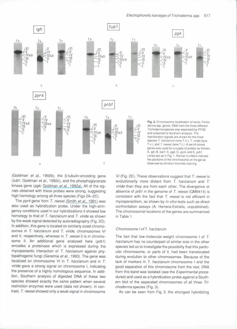
Fig. 2. Chromosomal localization of some Tricho-derma spp, genes DNA from the three differentTrichoderma species was separated by PFGEand subjected to Southern analysis. Thehybridization signals are shown for the threespecies T. harzianum (lane T.h.]. r, viride (laneT.V.), and T. reesei (lane T.r,). A set of clonedgenes was used for a supply of probes as follows:A. igh: B, iubV.C, pgk. D, pyr4, and E. prbT.Lanes are as in Fig. 1. Roman numbers indicatethe positions of the chromosome on the gel asobserved by ethidium bromide staining.
(Goldman ef at., 1992b), the [3-tubulin-encoding gene{tub^\ Goldman etal., 1992c), and the phosphoglyceratekinase gene {pgk; Goldman et at., 1992a). All of the sig-nals obtained with these probes were strong, suggestinghigh homology among all three species (Figs 2A-2C),
Tbe pyr4 gene from T. reesei (Smith ef al., 1991) wasalso used as hybridization probe. Under the high-strin-gency conditions used in our hybridizations it showed lowhomology to that of T. harzianum and T. viride as shownby the weak signal detected by autoradiography (Fig. 2D),In addition, this gene is located on similarly sized chromo-somes in T. harzianum and T. viride, chromosomes VIand V, respectively, whereas in T. reesei it is in chromo-some II. An additional gene analysed here {prbi)encodes a proteinase which is expressed during themycoparasitic interaction of T. harzianum against phy-topathogenic fungi (Geremia etat., 1993), The gene waslocalized on chromosome VI in T. harzianum and in T",viride gave a strong signal on chromosome I, indicatingthe presence of a highly homologous sequence. In addi-tion, Southern analysis of digested DNA of these twospecies showed exactly the same pattern when severalrestriction enzymes were used (data not shown). In con-trast, T. reesei showed only a weak signal in chromosome
VI (Fig. 2E). These observations suggest that T. reesei isevolutionarily more distant from 7. harzianum and 7.viride than they are from each other. The divergence orabsence of prb1 in the genome of 7 reesei (QM9414) isconsistent with the fact that 7 reesei is not efficient inmycoparasitism, as shown by in wfro tests such as directconfrontation assays (A, Herrera-Estrelia, unpublished).The chromosomal locations of the genes are summarizedin Table 1.
Chromosome t of T. harzianum
The fact that low-molecular-weight chromosome I of 7.harzianum has no counterpart of similar size in the otherspecies led us to investigate the possibility that this partic-ular chromosome, or parts of it, had been translocatedduring evolution to other chromosomes. Because of thelack of markers in 7. harzianum chromosome I and thegood separation of this chromosome from the rest, DNAfrom this band was isolated (see the Experimental proce-dures) and used as a hybridization probe against a South-ern blot of the separated chromosomes of all three Tri-choderma species (Fig. 3).
As can be seen from Fig. 3, the strongest hybridizing

518 A. Herrera-Esfrella. G. t-l. Goldman, M. Van Montagu and R. A. Geremia
Table 1. Chromosome gene assignment.T. harzianum
sizecbromosome (mbp)
VI
VIVa,bIII
II1
7.3
7,06.55,6
3,72,2
gene
ighprbitubipyr4
pg>^
25S rDNAleSrDNA
cbromosome
V
IVllla,b11
1
7", viride
size(mbp)
7,2
7,06,05,3
4.2
gene
tub)pyr4
pgi<
25S rDNA18SrDNAighprbi
cbromosome
VI
VIVIII
III*
T. reesei
size(mbp)
7,4
6,65.54,6
3,73.43,2
gene
ighprbt^tubi
P9l<
pyr4
25S rDNA18SrDNA
a. In this strain the homology with this gene is probably low, as predicted from the weak signalobserved upon hybridization.•Putative chromosome observed only upon hybridization.
band corresponds to 7. harzianum itself (lane 1); analmost as intense band was observed in the autoradio-graphy corresponding to the 7. viride chromosome I (lane2). In contrast, the lane corresponding to 7. reesei (lane3) showed only weak hybridization signals. These signalscorresponded to 7, /"eese/chromosomes I and III.
It is noteworthy that under the high-stringency condi-tions (washing at 65X with 0.5x SSC, 0.5% SDS} used inthis experiment, chromosome II from S. pombe aisohybridized to the probe and the signals observed were asintense as those shown by 7 reesei DNA, It has beenseen that, when total DNA is labelled, repetitivesequences such as ribosomal DNA have higher specificactivity than single-copy genes (Simoens et at., 1988), Totest for the presence of ribosomal DNA, two oligonu-deotides based on the 25S and 18S RNA were con-structed and used as probes to hybridize with the chromo-somai blots (Figs 4A and 4B). Both probes hybridized
Fig. 3. Analysis of 7". harzianum chromosome I. Chromosomal DNAs fromS. pombe, T. harzianum, T. viride, and T. reese^were resoived by PFGEas described m the Experimentai procedures. The DNA was then blottedonto Hybond-N* membranes (Amersham) and analysed by hybridization.The probe used in this expenment was DNA from chromosome I of T.harzianum which was recovered from the gel and ^^P-labelled (see theExperimentai procedures). Hybridization was carried out under high-strin-gency conditions (65"C. 5x SSC. 0,1 % SDS, 0,25% milk powder). Thelanes correspond lo Ihe chromosomes of the different organisms as fol-lows: iane i , T. harzianum: lane 2, T. viride: lane 3, T. reesei: and lane 4,S. pombe.
with chromosome III of 7 harzianum and chromosome IIot 7, viride, respectively. These data indicate that thehybridization pattern observed when DNA of chromo-some I is used as a probe is not due to the presence ofrDNA, In 7. reeseUhe hybridization signal correspondednot to any of the bands observed in Fig. 1, but to a posi-tion of slightly faster migration in the agarose gel. Thissuggests the presence of a lower-molecular-mass chro-mosome. This would be in agreement with the datarecently published by Mantyla et at. (1992) who reportedseven chromosomes for this particular strain. Further-more, in that report it was shown that the 25S rDNA islocated in a 3 mbp chromosome which we did not visual-ize by ethidium bromide staining.
Discussion and Conclusions
Based on the electrophoretic mobility of their chromo-somes under different conditions, we have estimated thegenome size of these three species to be in the31-39 mbp range. The sizes of the individual chromo-somes indicate significant variation between the biocon-trolling Trichoderma species and the cellulolytic one. Thishas also been observed in another filamentous fungalgenus even among different strains of the same species(Orbach et al., 1988), However, based on gene locationand DNA homology (as deduced from hybridization sig-nals), the data on the strains studied here partially sup-port the previously reported phylogenetic relationshipamong these species (Stasz ef at., 1989). Thus, 7.harzianum and 7 viride are closely related and couldhave evolved on the same phylogenetic branch, whereas7. reese/would probably have derived from an indepen-dent branch.
From hybridization experiments it was observed thatthe igh and prbi genes were present on chromosome VI

Eteotrophoretic karotype o^Trichoderma spp. 519
T,h, tv.
•VI
Fig. 4. Chromosomal location of 25S and 18SrDNAs. DNA from the three different species wasseparated by PFGE and subjected to Southernanalysis.A. Hybridization signals using a 23S rDNAsequence-derived oligonucleotide. Lane T,h,, T.harzianum, lane T,v.. T. viride: lane T,r,. T. reesei.B, Tbe same a (A), but for 18S rDNA.
of 7 ftarz/anum whereas in 7. viride they are on chromo-some I (see Figs 2A and 2E), in as much as other genesfound on chromosome VI of 7. harzianum [tubi and pyr4)are located on chromosome V of 7 viride (Figs 2B and2D). If 7 harzianum is considered as the ancestralspecies, as suggested by Stasz et at. (1989), our datawould imply that a translocation has occurred and thatduring evolution part of 7. harzianum chromosome VIrecombined with chromosome I to produce the actual 7.viride chromosome I, Data supporting this hypothesiswere obtained when using DNA from chromosome I of 7,harzianum as a probe (Fig. 3) and a strong hybridizationsignal was detected which corresponded to 7 viride chro-mosome I. However, more information is required to con-firm this hypothesis. With the availability of more genesand with the use of subgenomic restriction fragments, itwill be possible to establish molecular 'linkage groups'. Itwill also be useful to include other wild-type strains ofthese species to study chromosome number and sizeconservation within a given species. Our data allow thepossibility of using molecular karyotyping for further stud-ies on the taxonomy and evolution of these species.
Experimental procedures
Strains and hybridization probes
Intact chromosomes were prepared from the followingstrains; 7 harzianum IMI206040, 7 longibrachiatum (reesei)QM9414, and 7. viride T9, S. cerevisiae and S. pombe chro-mosomal DNA size markers were purchased from BioRad,
The following cloned genes were used as radiolabelledprobes: pgkirom T. wnde(Goldman etat., 1992a), pyr4from7 reese; (kindly provided by Dr Ward, Genencor), tubi from7 viride (Goldman et al., 1992c), prbi from 7. harzianum(Geremia et at., 1993), and igh from 7, harzianum (Goldmane/a/,, 1992b).
Preparation of intact chromosomat DNA
Agarose plugs containing S. pombe or S. cerevisiae intactchromosome DNAs were purchased from BioRad. Intact Tri-choderma spp, chromosomal DNA was prepared essentiallyas described by Brody and Carbon (1989), with the followingvariants.
(i) Approximately 20-30 potato dextrose agar plates wereoverlaid with cellophane sheets; 1x10'' conidia were inocu-lated and allowed to germinate (16-18h). The cellophanesheets were collected and the mycelium scraped off into30ml of the cell-wall digestion mix (1.2M MgS04, lOmMNaP04, pH 5.6, 5 mg ml"'' Novozyme 234), The mycelial cellswere incubated at 28"C with gentle agitation. Protoplast pro-duction was followed microscopically and after 2-3 h (about90% yield) the suspension was filtered through a 50 |iM nylonmesh. The filtrate was transferred to centrifuge tubes andcarefully overlaid with an equal volume of STE buffer (0.6 Msorbitol, lOmM Tris-HCI, pH 7.5, 1 mM EDTA, pH 8.0). Thegradient was then centrifuged at 4000xg in a swingingbucket rotor for 15 min at 4°C, The band of protoplasts wasremoved and carefully mixed with 1,5vols of S2TE buffer(1,2M sorbitol, lOmM Tris-HCI, pH 7.5, 1 mM EDTA). Theprotoplasts were pelleted by centrifugation at 5000xg for10 min at 4'C and washed twice with S2TE. The pellet wasresuspended in modified GMB buffer {0,125M EDTA,pH 8,0. 1.2 M sorbitol) at a concentration of 0,5-1 x 10^ cellsmrV The suspension was brought to 37''C and an equal vol-ume of 1.4% low-melting-point agarose (FMC) in GMB wasadded. The mixture was then poured into a plug mould andallowed to solidify for 30 min on ice. The plugs were thenimmersed in NDS buffer (0,5M EDTA, pH 8.0, lOmM Tris-HCI, pH 8,5, 1% sarkosyl) containing 2mgmr' proteinase K(Boehringer) at 5O'"C for at least 24 h. The chromosomeplugs were washed three times in 50 mM EDTA (pH 8.0) at5 0 ^ and stored at 4"C in the same solution. The chromo-somes appeared intact after 6 months of storage,
(ii) One litre of PBD medium was inoculated with 1 xiO''conidia ml"^ and incubated at 28°C for 16-18 h with vigorousshaking. Mycelium was collected by filtration through Mira-cloth (Calbiochem, USA), and rinsed with sterile, distilledwater. After removal of excess liquid, the mycelium was sus-pended in 70 ml of cell-wall digestion mix. The obtained pro-toplasts were then treated as described above.
Rotating-etectrodeetecfrophoresis
In this case the separation of chromosomal DNA wasachieved by using the Rotaphor System (Biometra), The run-ning conditions were as follows. For the resolution of thesmaller 7, harzianum and 7, viride chromosomes, durationwas 192h, switching intervals (SWIs) were 9000-500 s witha linear ramping, the angle was ramped in a linear fashionfrom 98-110°, the applied voltage was 46 V. the temperature

520 A. Herrera-Estretla, G. H. Goldman, M. Van Montagu and R, A, Geremia
was kept at 12''C, and the gel was 0.8% Megarose (Clon-tech). For the larger chromosomes, the conditions were asfollows: 168h running time, 7000-500s SWIs with logarith-mic ramping, angle from 120' to 98" changing in a logarith-mic way, 46 V applied voltage, 12''C constant temperature,and 0.7% Megarose gel.
second nucleotide corresponds to the 25S rDNA and itssequence is 5'-CGGCACATCTGTTAAACCATAACGCAGG-TGTCCTAAGGGGGGCTCATGGA-3'. This sequence wastaken from the T. reesei 25S rDNA (Vanhanen. 1991). Thecomputer program, termed OLIGO (Rychlik and Rhoads,1989), was used to select the optimal sequence.
Electrophoresis test
The following conditions were used in the initial experimentsand later to test the quality o( the DNA samples: 48 h run,4000-2000 s SWIs, logarithmic ramping, angle 95-105" log-arithmic ramping. 48 V, and 14"C.
CHEF gel conditions
CHEF analysis was performed with the CHEF mapperdesigned hy BioRad, This technique was successfullyapplied to the separation of T. reese/chromosomes with thefollowing conditions: 168h running time, 1800-5400 s SWIswith linear ramping, 120'- constant angle, 1.3 V cm "̂ voltage,12''C constant temperature, and 0,65% Megarose gel. In allcases the gels were stained for 30 min with a 0.5 ̂ gml"^etbidium bromide solution and destained overnight in water.
Southern blotting and hybridization conditions
Gels were successively soaked in 0.25 M HCl for two periodsof 15 min, in 0.5 M NaOH/I.OM NaCI twice for 20 min, in0.5 M Tris-HGI, pH 7.5/1.5 M NaCI for two 20-min periods andequilibrated with lOx SSC (1x SSC = 0,15M NaCI, 0,015sodium citrate, pH 7.0). The DNA was then transferred bycapillary blotting onto Hybond-N* membranes (Amersham)using 10x SSC for 12-16 h. The DNA was fixed on the mem-branes with 0.4 M NaOH for 20 min and washed with 5x SSC.DNA fragments (25 ng) containing the different genes wereusually labelled to a specific activity of at least 5x10^c.p.m.ng"'' using the Random Primed ONA Labelling Kit(Boehringer) and (a-^^P]-dCTP (3000Cimmor'; Amer-sbam). Hybridizations were carried out for 16-24 h at 65''C in6x SSC, 0.1% SDS, 100)jgmr' salmon-sperm DNA, 0.25%non-fat milk powder in a volume of 20-25 ml. The blots werewashed twice at 65"C for 30 min in 2x SSC, 0.5% SDS. andtwice for 15 min in 1x SSC, 0,1% SDS, and subjected toautoradiography at -70"C using two intensifying screens andXRM Kodak films. When the oligonucleotides were used theywere radioactively labelled by the kinase reaction with theenzyme polynucleotide kinase (Pharmacia) and [y-^^P]-ATP(Amersham Ltd). Hybridization and washing temperatureswere 55°C and washing buffer was 2x SSC, 0.5% SDS,
Design of oligonucleotide probes for rDNA
Two oligonucleotides (50-mers) were designed to detectrDNA repeats. One, corresponding to the 18S with thesequence 5'-TGGGCCGCACGCGCGCTACACTGACAGA-GCCAGCG-3', corresponds to nucleotides 1435-1484 of the18S rDNA of S. cerevisiae (Mankin et al., 1986) and is alsopresent in the 18S rDNA of T. reesei (Vanhanen, 1991). The
Acknowledgements
The authors wish to thank Drs Allan Caplan and Sergei Kush-nir for critical reading of the manuscript, Martine De Cock fortyping it, and Karel Spruyt, Vera Vermaercke, and StefaanVan Gijsegem for drawings and photographs. This work wascarried out with grants from the Services of the Prime Minis-ter (l.tJ.A.P. 12OC0187) and 'Vlaams ActieprogrammaBiotechnologie' (174KP490). R.A.G, and G.H.G. areindebted to the Commission of European Communities andthe CAPES-BRAZIL (812-88-2) for a post-doctoral and pre-doctoral fellowship, respectively.
References
Brody H,, and Carbon J. (1989) Electrophoretic karyotype ofAspergillus nidulans, Proc Natl Acad Sci USA 86: 6260-6263.
Cheng, C, Tsukagoshi, N., and Udaka, S. (1990) Transformationof Trichoderma viride using the Neurospora crassa pyr4 geneand its use in the expression of a Taka-amylase A gene fromAspergillus oryzae. Curr Genet ^8•. 453-456.
Chet, Y. (1990) Mycoparasitism - recognition, physiology andecology. In New Directioris in Biological Controt: Alternativesfor Suppressing Agricutturat Pests and Diseases (UCLA Sym-posia on Molecular and Cellular Biology, New Series, Vol.112). Baker, R.R., and Dunn, P.E. (eds). New York: Alan R.Liss, pp. 725-733.
Geremia, R., Goldman, G.H.. Jacobs, D., Ardiles, W., Vita, S.B.,Van Montagu, M,, and Herrera-Estrella, A. (1993) Molecularcharacterization of the proteinase-encoding gene, prbi.related to mycoparasitism by Trichoderma harzianum. Motl^icrobiol, in press.
Goldman, G.H., Geremia, R., Caplan, A., Vila, S,, Villarroel, R.,Van Montagu, M., and Herrera-Estrella, A. (1992a) Molecularcharacterization and regulation ot the phosphoglyceratekinase gene from Trichoderma viride. Moi Microbiot 6:1231-1242.
Goldman, G.H,, Demolder, J., Dewaele, S,, Herrera-Estrella. A.,Geremia, R.A-, Van Montagu. M., and Contreras, R. (1992b)Molecular cloning of the imidazoleglycerolphosphate dehy-dratase gene of Trichoderma harzianum by genetic comple-mentation in Saccharomyces cerevisiae using a direct expres-sion vector, h/tol Gen Gener 234: 481-488,
Goldman, G.H., Temmerman. W,, Jacobs, D., Contreras, R., VanMontagu, M.. and Herrera-Estrella, A. (1992c) A nucleotidesubstitution in one of the p-tubulin genes of Trictioderma virideconfers resistance to the antimitotic drug methyl benzimida-zole-2-yl-carbamate. Moi Gen Genet, in press.
Mankin, A.S., Skryabin, K.G., and Rubtsov, P.M. (1986) Identifi-cation of ten additional nucteotides in the primary structure ofyeast 18S rRNA. Gene 44:143-145,
Mantyla, A.L., Rossi, K.H., Vanhanen, S.A., Penttila, M.E.,Suominen, P,L,, and Nevalainen, K.M.H. (1992) Elec-trophoretic karyotyping of wild-type and mutant Trichodermalongibrachiatum (reesei) strains. Curr Genet 21: 471 -477.

Etectrophorefic karotype o^Trichoderma spp, 521
Meyer, R.J. (1991) Mitochondrial DNAs and plasmids as foxo-nomic characteristics in Trichoderma viride- Appl EnvironMicrobiot 57: 2269-2276.
Meyer, W,, Morawetz. R., Borner, T., and Kubicek, C. (1992) Theuse of DNA-fihgerprint analysts in the classification of somespecies of the Trichoderma aggregate. Curr Genet 21: 27-30.
Nevalainen, H,, Penttila, M,, Harkki, A., Teeri, T., and Knowies, J.(1991) The molecular bioiogy of Trichoderma and its applica-tion to the expression of both homologous and heterologousgenes. In Molecular tndustriat Mycotogy. Systems and Appli-cations for Filamentous Fungi. Leon, S.A., and Berka, R.M.(eds). New York: Marcel Dekker, pp. 129-148.
Orbach, M.J., Volirath, D,, Davis, R.W., and Yanofsky, C. (1988)An electrophoretic karyotype of Neurospora crassa. Mot CeltBiol8: 1469-1473.
Rifai, M.A, (1969) A revision ot the genus Trichoderma. Com-monw Mycol Inst Mycot Pap 116:1 -56.
Rychlik, W., and Rhoads, R.E, (1989) A computer program forchoosing optimal nucleotides for filter hybridization, sequenc-ing and in vitro amplification of DNA. Nuct Acids Res 17:8543-8551.
Simoens, C.R., Gielen, J.. Van Montagu, M., and Inze, D. (1988)Characterization of highly repetitive sequences of Arabidopsisthatiana. Nucl Acids Res 16: 6753-6766,
Skinner, D.Z., Budde, A.D., and Leong, S-A, (1991) Molecularkaryotype analysis of fungi. In More Gene Maniputations inFungi. Bennett, J,W., and Lasure, L,L. (eds). San Diego: Aca-demic Press, pp. 86-103.
Smith, J,L., Bayliss, F.T., and Ward, M. (1991) Sequence of thecloned pyr4 gene of Trichoderma reesei and its use as ahomologous selectable marker for transformation. Curr Genet19:27-33,
Stasz, T.E,, Nixon, K., Harman, G.E., Weeden, N.F., and Kuter,G.A. (1989) Evaluation of phenetic species and phylogeneticrelationships in the genus Trichoderma by cladistic analysis ofisozyme polymorphism. Mycotogia8^•. 391-403.
Vanhanen, S. (1991) Isolation and characterization of genesinvolved in basic metabolism of the filamentous fungus Tricho-derma reesei. Academic Dissertion, University of Helsinki, Fin-land (Technical Research Centre of Finland, Publication 75).

Copyright © 2022 FDOKUMEN