Interpretation of karyotype evolution should consider ...
10
Interpretation of karyotype evolution should consider chromosome structural constraints Ingo Schubert 1 and Martin A. Lysak 2 1 Leibniz Institute of Plant Genetics and Crop Plant Research (IPK), D-06466 Gatersleben, Germany 2 Department of Functional Genomics and Proteomics, and CEITEC, Masaryk University, CZ-625 00 Brno, Czech Republic Comparative genetics, genomics and cytogenetics pro- vide tools to trace the evolutionary history of extant genomes. Yet, the interpretation of rapidly increasing genomic data is not always done in agreement with constraints determined by chromosome structural fea- tures and by insights obtained from chromosome mutagenesis. The terms ‘non-reciprocal chromosome translocation’, ‘chromosome fusion’ and ‘centromere shift’ used to explain genomic differences among organ- isms are misleading and often do not correctly reflect the mechanisms of chromosome rearrangements underly- ing the evolutionary karyotypic variation. Here, we (re)interpret evolutionary genome alterations in a parsi- monious way and demonstrate that results of compara- tive genomics and comparative chromosome painting can be explained on the basis of known primary and secondary chromosome rearrangements. Therefore, some widespread terms used in comparative and evolu- tionary genomics should be either avoided (e.g. non- reciprocal translocation) or redefined (e.g. chromosome fusion and centromere shift). Interpretation of genomic data needs to be in agreement with chromosome constraints Nuclear genomes are contained within chromosomes representing genetic linkage groups. The number, size and shape of chromosomes, which constitute the karyotype (see Glossary) of an organism, vary considerably among groups of eukaryotes. The increasing number of sequenced genomes has revealed that most higher eukaryote genomes contain between approximately 14000 and 40000 protein coding genes. By contrast, nuclear genome sizes vary by more than four orders of magnitude from approximately 12 Mb in yeast [1] up to 400000 Mb in dinoflagellates [2]. Accordingly, the size of linear metaphase chromosomes spans a range from <1 mm to some dozen micrometers. The number of chromosome pairs can also vary widely, from 1 (in an ant [3]) to approximately 700 in the fern genus Ophioglossum [4]. Whereas genome size and chromosome numbers are rather stable in some phylogenetic clades, they vary considerably in others [5]. Several mechanisms responsible for the variation in size, shape and number of chromosomes, as well as in DNA content between organisms, are recognized. Chromo- somes change their size and shape by gain (i.e. insertion or duplication) or loss (i.e. deletion) of DNA, or through rearrangements within or between chromosomes. Chromo- some rearrangements can also increase or decrease the number of chromosomes, a phenomenon called ascending or descending dysploidy, respectively [6]. In addition, chro- mosome numbers can be altered by ploidy mutations in- volving the entire complement (polyploidy) or individual chromosomes (aneuploidy). The evolutionary history of a karyotype is often difficult to determine, especially for older events. With time, the accumulation of chromosome rearrangements will obscure the exact identity, number and order of events that have occurred along a lineage leading to extant karyotypes. There are, however, techniques to help reconstruct this blurred history. Comparative chromosome painting [7,8] has proved to be useful for tracing karyotype evolution in mammals (reviewed in [9]) and in plants (e.g. in Brassi- caceae [10–13]). Alternatively, karyotype evolution can be Opinion Glossary Acrocentric: bi-armed chromosome with asymmetric arm length, including one very short arm. Aneuploidy: single chromosome types are less (e.g. 2n – 1, hypoploid) or more frequent (e.g. 2n + 1, hyperploid) than chromosome pairs. Chromosome painting: fluorescent in situ hybridization (FISH) with chromo- some or chromosome region-specific probes to the chromosome complement of a given species or a closely related one. Dysploidy: decrease or increase of chromosome number in connection with chromosome rearrangements; not by addition or loss of single chromosomes as in aneuploidy. Therefore, dysploidy is also called pseudoaneuploidy. Homeologues: chromosomes of related extant species that share a similar gene content in the same (syntenic) order. Karyotype: entire chromosome complement of an organism. Linkage group: combination of sequences (in a defined order) that segregate together during meiosis because they are located on the same chromosome. Metacentric: chromosome with approximately equal-sized chromosome arms. Monocentric: chromosome with one centromere (attachment site of spindle fibres during nuclear divisions) that appears as a primary constriction. This is in contrast to dicentric chromosomes with two centromeres and poly- or holocentric chromosomes on which spindle fibres attach along almost the entire chromosome length. Palaeogenomics: comparison of genetic, genomic and cytogenetic data of extant species to interpret their evolutionary origin from tentative ancestral genomes. Robertsonian rearrangements (i.e. fusion–fission cycle): symmetric or asym- metric translocation between the centric ends of two telocentric or acrocentric chromosomes forming a metacentric chromosome (‘centric fusion’), or splitting of a metacentric chromosome into two telocentric chromosomes (‘centric fission’); see Figure 3d, main text. Telocentric: quasi one-armed chromosome with a short arm (presumably) consisting only of the telomere flanking the centromere on one side. Corresponding author: Schubert, I. ([email protected]). 0168-9525/$ – see front matter ß 2011 Published by Elsevier Ltd. doi:10.1016/j.tig.2011.03.004 Trends in Genetics, June 2011, Vol. 27, No. 6 207
-
Upload
khangminh22 -
Category
Documents
-
view
1 -
download
0
Transcript of Interpretation of karyotype evolution should consider ...

Opinion
Interpretation of karyotype evolutionshould consider chromosomestructural constraintsIngo Schubert1 and Martin A. Lysak2
1 Leibniz Institute of Plant Genetics and Crop Plant Research (IPK), D-06466 Gatersleben, Germany2 Department of Functional Genomics and Proteomics, and CEITEC, Masaryk University, CZ-625 00 Brno, Czech Republic
Glossary
Acrocentric: bi-armed chromosome with asymmetric arm length, including
one very short arm.
Aneuploidy: single chromosome types are less (e.g. 2n – 1, hypoploid) or more
frequent (e.g. 2n + 1, hyperploid) than chromosome pairs.
Chromosome painting: fluorescent in situ hybridization (FISH) with chromo-
some or chromosome region-specific probes to the chromosome complement
of a given species or a closely related one.
Dysploidy: decrease or increase of chromosome number in connection with
chromosome rearrangements; not by addition or loss of single chromosomes
as in aneuploidy. Therefore, dysploidy is also called pseudoaneuploidy.
Homeologues: chromosomes of related extant species that share a similar
gene content in the same (syntenic) order.
Karyotype: entire chromosome complement of an organism.
Linkage group: combination of sequences (in a defined order) that segregate
together during meiosis because they are located on the same chromosome.
Metacentric: chromosome with approximately equal-sized chromosome arms.
Monocentric: chromosome with one centromere (attachment site of spindle
fibres during nuclear divisions) that appears as a primary constriction. This is
in contrast to dicentric chromosomes with two centromeres and poly- or
holocentric chromosomes on which spindle fibres attach along almost the
entire chromosome length.
Palaeogenomics: comparison of genetic, genomic and cytogenetic data of
extant species to interpret their evolutionary origin from tentative ancestral
genomes.
Robertsonian rearrangements (i.e. fusion–fission cycle): symmetric or asym-
metric translocation between the centric ends of two telocentric or acrocentric
chromosomes forming a metacentric chromosome (‘centric fusion’), or
splitting of a metacentric chromosome into two telocentric chromosomes
(‘centric fission’); see Figure 3d, main text.
Telocentric: quasi one-armed chromosome with a short arm (presumably)
Comparative genetics, genomics and cytogenetics pro-vide tools to trace the evolutionary history of extantgenomes. Yet, the interpretation of rapidly increasinggenomic data is not always done in agreement withconstraints determined by chromosome structural fea-tures and by insights obtained from chromosomemutagenesis. The terms ‘non-reciprocal chromosometranslocation’, ‘chromosome fusion’ and ‘centromereshift’ used to explain genomic differences among organ-isms are misleading and often do not correctly reflect themechanisms of chromosome rearrangements underly-ing the evolutionary karyotypic variation. Here, we(re)interpret evolutionary genome alterations in a parsi-monious way and demonstrate that results of compara-tive genomics and comparative chromosome paintingcan be explained on the basis of known primary andsecondary chromosome rearrangements. Therefore,some widespread terms used in comparative and evolu-tionary genomics should be either avoided (e.g. non-reciprocal translocation) or redefined (e.g. chromosomefusion and centromere shift).
Interpretation of genomic data needs to be inagreement with chromosome constraintsNuclear genomes are contained within chromosomesrepresenting genetic linkage groups. The number, sizeand shape of chromosomes, which constitute the karyotype(see Glossary) of an organism, vary considerably amonggroups of eukaryotes. The increasing number of sequencedgenomes has revealed thatmost higher eukaryote genomescontain between approximately 14000 and 40000 proteincoding genes. By contrast, nuclear genome sizes vary bymore than four orders of magnitude from approximately 12Mb in yeast [1] up to 400000 Mb in dinoflagellates [2].Accordingly, the size of linear metaphase chromosomesspans a range from <1 mm to some dozen micrometers.The number of chromosome pairs can also vary widely,from 1 (in an ant [3]) to approximately 700 in the fern genusOphioglossum [4]. Whereas genome size and chromosomenumbers are rather stable in some phylogenetic clades,they vary considerably in others [5].
Several mechanisms responsible for the variation insize, shape and number of chromosomes, as well as inDNA content between organisms, are recognized. Chromo-
Corresponding author: Schubert, I. ([email protected]).
0168-9525/$ – see front matter � 2011 Published by Elsevier Ltd. doi:10.1016/j.tig.2011.03.004
somes change their size and shape by gain (i.e. insertion orduplication) or loss (i.e. deletion) of DNA, or throughrearrangements within or between chromosomes. Chromo-some rearrangements can also increase or decrease thenumber of chromosomes, a phenomenon called ascendingor descending dysploidy, respectively [6]. In addition, chro-mosome numbers can be altered by ploidy mutations in-volving the entire complement (polyploidy) or individualchromosomes (aneuploidy).
The evolutionary history of a karyotype is often difficultto determine, especially for older events. With time, theaccumulation of chromosome rearrangements will obscurethe exact identity, number and order of events that haveoccurred along a lineage leading to extant karyotypes.There are, however, techniques to help reconstruct thisblurred history. Comparative chromosome painting [7,8]has proved to be useful for tracing karyotype evolution inmammals (reviewed in [9]) and in plants (e.g. in Brassi-caceae [10–13]). Alternatively, karyotype evolution can be
consisting only of the telomere flanking the centromere on one side.
Trends in Genetics, June 2011, Vol. 27, No. 6 207
Opinion Trends in Genetics June 2011, Vol. 27, No. 6
studied by using comparative genetic and genomic analy-ses. Palaeogenomics, which is an integrated approachcombining genetic and cytogenetic maps, EST data sets[14,15] and whole-genome sequences [16–20], enablesresearchers to trace the evolutionary history of genomeswithin a phylogenetic framework. It also helps to elucidatewhole-genome duplications and genome reshuffling thathave led to extant chromosome complements (Box 1).
A re-interpretation is necessary in cases when compar-ative genomics neglected the mechanics of chromosomerearrangements; for example, by postulates that disagreewith observations from chromosomemutagenesis. Our aimhere is to bring genomic interpretation into accordancewith chromosome constraints. We focus on mechanismsthat underlie karyotypic changes as a basis for interpretingcomparative genomic data and for elucidating the evolu-tionary origin of extant karyotypes.
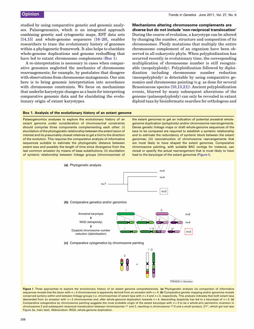
Box 1. Analysis of the evolutionary history of an extant genome
Palaeogenomics analyses to explore the evolutionary history of an
extant genome under consideration of chromosomal constraints
should comprise three components complementing each other: (i)
elucidation of the phylogenetic relationship between the extant taxon of
interest and its presumably closest relatives to get a hint to the direction
of the evolution. This requires the comparative analysis of informative
sequences suitable to estimate the phylogenetic distance between
extant taxa and possibly the length of time since divergence from the
last common ancestor by means of base substitutions; (ii) elucidation
of syntenic relationship between linkage groups (chromosomes) of
[()TD$FIG]
Figure I. Three approaches to explore the evolutionary history of an extant genom
sequences reveals that the taxon with n = 3 chromosomes is apparently derived from a
conserved synteny within and between linkage groups (i.e. chromosomes) of extant ta
descended from an ancestor with n = 2 chromosomes and, after whole-genome dup
Comparative cytogenetics by chromosome painting suggests the most probable orig
chromosome 2 and subsequent reciprocal translocation between chromosomes 1* an
Figure 3a, main text). Abbreviation: WGD, whole-genome duplication.
Ancestral karyotype
n=?
Dysploid chromosome numberreduction (diploidization)
WGD (tetraploidy)
1
1
1
2
(a) Phylogenetic analysis
(b) Comparative genetics and/or genomics
(c) Comparative cytogenetics by chromosome
208
Mechanisms altering chromosome complements arediverse but do not include ‘non-reciprocal translocation’During the course of evolution, a karyotype can be alteredby changing the number, structure and composition of itschromosomes. Ploidy mutations that multiply the entirechromosome complement of an organism have been ob-served in all eukaryotic phyla. When polyploidization hasoccurred recently in evolutionary time, the correspondingmultiplication of chromosome number is still recogniz-able (neopolyploidy). Polyploidization followed by diploi-dization including chromosome number reduction(mesopolyploidy) is detectable by using comparative ge-nomics and chromosome painting (e.g. as done for severalBrassicaceae species [10,13,21]). Ancient polyploidizationevents, blurred by many subsequent alterations of thegenome (palaeopolyploidy) can only be revealed in extantdiploid taxa by bioinformatic searches for orthologous and
the extant genomes to get an indication of potential ancestral whole-
genome duplication (polyploidy) and/or chromosome rearrangements.
Dense genetic linkage maps or draft whole-genome sequences of the
taxa to be compared are required to establish a syntenic relationship
and to estimate the redundancy of syntenic block between the extant
genomes; (iii) reconstruction of chromosome rearrangements that
are most likely to have shaped the extant genomes. Comparative
chromosome painting, with suitable BAC contigs for instance, can
reveal or specify the actual rearrangement that is most likely to have
lead to the karyotype of the extant genomes (Figure I).
e comprehensively. (a) Phylogenetic analysis via comparison of informative
n ancestor with n = 4. (b) Comparative genetic mapping and/or genomics reveals
xa with n = 4 and n = 3, respectively. This analysis indicates that both extant taxa
lication towards n = 4, descending dysploidy has led to a karyotype of n = 3. (c)
in of the extant karyotype with n = 3 to be a whole-arm pericentric inversion in
d 2, resulting in chromosome 1*/2 and a small product, 2/1*, which got lost (see
n=4
n=3
+
n=2
n=3
n=4
2
1*
1*/2
2 2*
2*
1*/2
n=4
painting
TRENDS in Genetics

Opinion Trends in Genetics June 2011, Vol. 27, No. 6
paralogous sequence markers (reviewed in [22]). Al-though polyploid genomes increase the cost of replication,they also provide evolutionary advantages, because theypresent a basis for speciation. Gene duplicates can ac-quire new functions and chromosome rearrangementsthat lead to deletions (see below) that are usually lethalin diploid genomes can be tolerated in neo- and mesopo-lyploid genomes.
Structural chromosome alterations are the result ofprimary or secondary rearrangements. Primary rearran-gements are the outcome of illegitimate recombinationduring double-strand break (DSB) repair, either via directjoining of ends between different DSBs, or through recom-bination with ectopic (instead of allelic) homologoussequences. The frequent use of ectopic homologoussequences as template for recombination repair explainswhy primary rearrangements have breakpoints preferen-tially within heterochromatic regions enriched in similarrepetitive sequences [11,23].
Primary chromosome rearrangements are: insertion,deletion or duplication, peri- or paracentric inversion, andintra- or interchromosomal reciprocal translocation(Figure 1). Deletions are tolerated only in polyploids orwhen dispensable sequences are involved [24,25]. Con-trary to reciprocal translocation, during which chromo-somes exchange segments mutually, in non-reciprocaltranslocation a chromosome segment is transferred in aunidirectional manner from one chromosome to another.Duplicated chromosome segments on different chromo-somes have also been hypothesized to result from non-reciprocal translocation. Non-reciprocal exchange of mi-croscopically detectable chromosome segments has, how-ever, never been demonstrated experimentally andsuspect cases can be interpreted as unbalanced segrega-tion after reciprocal translocation. Indeed, unbalancedsegregation from reciprocal translocations can yielddaughter nuclei with duplications and deletions, mimick-ing ‘non-reciprocal’ translocations (Figure 1d, bottomright). Cells with nuclei containing deletions are usuallycounter-selected, but can survive when deletions occur inpolyploid backgrounds or in some tumours [26].
Gene conversion [27–29], between homeologous chromo-somes of polyploid species, can transfer homeologoussequences in a non-reciprocal manner [27–29]. However,gene conversion should not be misinterpreted as non-re-ciprocal translocation. Gene conversion rather representsa variant of DSB repair during which, by transient inva-sion of break-ends into a sequence-related double helix andsubsequent replicative elongation of break-ends (to bridgethe DSB), relatively short regions are copied into thebroken double helix (similar to that shown in Figure 1b,bottom left).
Structural chromosome alterations can additionallyarise as secondary chromosome rearrangements(Figure 2; reviewed in [30]). Such rearrangements can arisein organisms that are doubly heterozygous for two primaryrearrangements (translocations and/or inversions), if onechromosome is involved in both. Meiotic crossing over be-tween homologous regions of rearranged chromosomes,which differ in the regions distal to the cross over, leadsto gametes with a new karyotype and to complementary
gametes displaying a re-established wild-type chromosomecomplement (Figure 2a). Depending on the type of primaryrearrangements involved, unbalanced gametes can alsoarise, harbouring duplications as well as deletions andmimicking non-reciprocal translocation (Figure 2b) [31,32].
In summary, experimentally observed ploidy muta-tions, as well as primary and secondary chromosomerearrangements, are sufficient to explain evolutionarykaryotype alterations. Even the interpretation of unbal-anced karyotypes does not require the assumption of non-reciprocal translocations (see also below).
‘Chromosome fusion’ is the result of reciprocaltranslocationGeneticists speak of chromosome fusion when all geneticmarkers belonging to two ancestral genetic linkage groupssegregate as a single linkage group in a derived species.Similarly, chromosome painting probes can cover two chro-mosomes in an ancestral karyotype and only one in a morederived species. Such cases do not necessarily representchromosome fusions. A simple fusion of intact chromosomesis unlikely because telomeres distinguish natural ends oflinear chromosomes from break-ends and prevent fusionof natural chromosome ends [33]. True fusion of chromo-some ends (e.g. forming ring chromosomes) can occur intelomerase mutants [34]; however, the karyotypes of telo-merase mutants are unstable unless the mutation becomescompensated by an alternative telomere elongation mecha-nism stabilizing the chromosome ends. Chromosome fusionsensu stricto further implies that no loss of chromatin occursand the process is reversible. Usually, however, the ‘fused’chromosomes have only one centromeric region and no largeinternal telomeric sequence arrays, indicating an irrevers-ible symmetric reciprocal translocation, rather than a fu-sion, as the causative process. Furthermore, in the firstmetaphase after exposure to a genotoxic compound or ion-izing irradiation, ligations between entire chromosomeshave so far not been reported to occur among the structuralchromosome mutations. Therefore, the term ‘chromosomefusion’ has to be understood as a substitute for reciprocaltranslocation combining two linkage groups within thelarger of two translocation products.
In so-called ‘end-to-end fusion’, a telo- or acrocentricchromosome (with no essential genes in its short arm)undergoes symmetric reciprocal translocation with anoth-er chromosome with breakpoints close to the centromere inthe long arm of the telo- or acrocentric chromosome, andclose to one arm end in the other chromosome (Figure 3a).The large translocation product therefore combinesmost ofboth chromosomes, whereas the second, small productcomprises the centromere of the telo- or acrocentric chro-mosome plus two telomeres. Such small chromosomes,when free of essential genes, are often lost by unstabletransmission through meiosis [35–37]. If two metacentricsare to be combined into one, at least one of them has tobecome telo- or acrocentric; for example, via a pericentricinversion (with one breakpoint close to the centromere andthe other close to the opposite arm end) before reciprocaltranslocation (Figure 3a). Such inversions are detectable indense genetic and/or cytogenetic maps [11,12,38]. Chromo-some colinearity can be restored by a second paracentric
209

[()TD$FIG]
(c) Inversion
Asymmetric
Intrachromosomal
(d) Reciprocal translocations
Interchromosomalsymmetric
Via erroneous DSB repair
or
Duplicative insertion into aDSB by break end elongationalong an ectopic template (red)
Deletion via exonucleolyticdigestion of break ends beforejoining ( exonuclease)
1.
2.
3.
4.
1.
2.
3.
+
(a) Insertion
+
Unbalanced mitoticsegregation
Balanced mitoticsegregation
Symmetric chromatidtranslocation
or
(b) Duplication/deletion
Via unequal sister chromatid exchange
Pericentric Paracentric
NOR
or
TRENDS in Genetics
Figure 1. Primary structural chromosome rearrangements. (a) Insertion of alien or endogenous sequences through integration of extrachromosomal circular DNA [59,60].
(b) Deletions or duplications via unequal sister chromatid exchange or unequal crossing over at meiosis (top), or via erroneous DSB repair (bottom) [61,62]. (c) Pericentric
inversion with breakpoints on either side of the centromere (black constricted area) and paracentric inversion with both breakpoints within the same chromosome arm. A
nucleolus-organizing region (NOR) is shown to illustrate the effect of paracentric inversion on its position. (d) Intrachromosomal reciprocal translocations resulting in a ring
chromosome and an acentric fragment, and interchromosomal translocations that can be either symmetric and yield monocentric products, or asymmetric, resulting in a
dicentric chromosome and an acentric fragment. The products of asymmetric reciprocal translocations are usually unstable: the acentric fragment gets lost because of the
lack of a centromere; the instability of dicentric fragments increases with the distance between the centromeres because of the increasing risk that sister chromatids twist
between centromeres, resulting in anaphase bridges (top right), which subsequently get disrupted. Symmetric chromatid translocations might be subject to balanced or
unbalanced mitotic segregation, the latter yielding nuclei with deletions and duplications, respectively (bottom right). Unbalanced segregation is usually lethal, but, if
survived, mimics non-reciprocal translocation.
Opinion Trends in Genetics June 2011, Vol. 27, No. 6
210

[()TD$FIG]
WT)b()a(
2 translocationsInversion Translocation
Meiotic cross over in a hexavalent of a double heterozygote
Re-established WT New karyotype New karyotypes each with duplications ( )that correspond to deletions in the reciprocal karyotype and vice versa
TRENDS in Genetics
Figure 2. Secondary chromosome rearrangements. (a) Two translocations involving three wild-type (WT) chromosomes in different individuals lead to hexavalents (in
brackets) during meiosis in double heterozygous individuals. Crossing over (red) involving homologous regions flanked by non-homologous regions generates a new
karyotype and re-establishes the WT chromosome complement. (b) If one chromosome is involved in an inversion in one individual and in a translocation in another
individual, meiotic crossing over involving homologous regions flanked by non-homologous regions generates two new karyotypes, each harbouring complementary
duplication and deletions, respectively (bottom).
Opinion Trends in Genetics June 2011, Vol. 27, No. 6
inversion involving the pericentrically inverted arm [11].Asymmetric reciprocal translocation between the ends oftwo metacentrics (mimicking end-to-end fusion) can alsoyield a large stable chromosome if one centromere becomeslost or inactivated [13]. Circumstantial evidence for allvariants that mimic an end-to-end chromosome fusionhas been obtained by comparative chromosome painting[11–13].
‘Insertional’ or ‘nested chromosome fusion’, where anancestral linkage group is on both ends flanked bymarkersof the chromosome arms of a second ancestral linkagegroup, also needs reinterpretation (Figure 3b). Such fusiontypes have been detected in several grass species [19,39–
43] and less frequently in Brassicaceae [11]. Instead of‘fusion’ of the ‘insertion’ chromosome into a single centro-meric DSB of the ‘recipient’ chromosome, simultaneousbreaks at both arm ends of the insertion chromosome
and around the centromere of the recipient chromosomecan, via mis-repair, result in an asymmetric insertionaltranslocation, adding the arms of the recipient chromo-some to the arms of the insertion chromosome. The centro-mere of the recipient chromosome (or separated parts ofthe recipient centromere) must be inactivated to stabilizethe new ‘fusion’ chromosome and the small acentric trans-location product gets lost. Inactivation of centromeres hasbeen observed [44,45]. When, however, the recipient chro-mosome has a break on either side of its centromere, asymmetric reciprocal translocation can yield two mono-centric products: the large one spanning both originallinkage groups and a smaller one comprising the centro-mere of the recipient chromosome and the telomeres of theinserted chromosome. The small product is prone to lossduring meiosis [29]. It is hard to prove whether the cen-tromere of the recipient chromosome (or its split portions)
211

[()TD$FIG]
+
+
+
+
‚End-to-end fusion‘ by symmetricreciprocal translocation (after pericentricinversion, rendering one chromosomeacrocentric)
‚Nested fusion‘ by reciprocal translocation
Asymmetric Symmetric
Asymmetric+ donor arm repositioning
(a)
(b)
Field bean metaphase cell (2n=12) witha multiple reciprocal translocationinvolving 5 acrocentric chromosomes
(c)
or
Split of a metacentric+ telomere gain
(+ )
‚Fusion-fission-cycle‘(d)
Tel
omer
ase
heal
s br
eak
ends
Ascending and descending dysploidy bymeiotic mis-segregation in a heterozygote for2 translocations involving 3 chromosomes
(I) Duplications in hyper- and deletions inhypoploid gametes
(e)
n=3
n=2
n=4
TRENDS in Genetics
Figure 3. Interpretation of dysploid alterations of chromosome number. (a) ‘End-to-end fusion’ by symmetric reciprocal translocation (after pericentromeric inversion
rendering one chromosome acrocentric) yields a large and a small monocentric product; the latter prone to getting lost during meiosis. (b) ‘Nested fusion’ by asymmetric
(top left), symmetric (top right) or asymmetric reciprocal translocation with donor arm re-positioning (bottom) [39] might appear depending on the number of interacting
DSBs; the small acrocentric fragments are lost during mitosis and the small centric fragment might get lost during meiosis. (c) Multiple chromatid translocation involving
five acrocentric chromosomes in a complete first metaphase of the field bean Vicia faba after treatment with an S phase-dependent mutagen. (d) ‘Fusion–fission cycle’
(Robertsonian rearrangements) can reversibly alter the chromosome number by asymmetric reciprocal translocation involving centric chromosome ends of telocentric
chromosomes and yielding a meta(di)centric chromosome and an acentric fragment consisting of telomeric sequence arrays (green). A DSB within the telomeric sequences
between the two centromeres of the large translocation product can re-establish two stable telocentric chromosomes (left), as proven for the field bean [46]. Alternatively,
DSB within a monocentric bi-armed chromosome can yield stable telocentric chromosomes when telomeric sequences are added to the break-ends (right). These
telocentric chromosomes could again ‘fuse’ by symmetric (irreversible) or asymmetric (reversible) reciprocal translocation. (e) Simultaneously ascending and descending
Opinion Trends in Genetics June 2011, Vol. 27, No. 6
212

Opinion Trends in Genetics June 2011, Vol. 27, No. 6
was deleted after inactivation, or whether the respectivechromosome was monocentric from the beginning owing tosymmetric translocation. In some cases of ‘nested’ chromo-somes of Brachypodium distachyon, centromeric (but nottelomeric; T.Wicker, pers. communication) sequences werefound to flank the insertion chromosome [19]. It remainselusive whether these centromeric sequences (much lesscopious than in active centromeres) represent remnants ofa gradually inactivated recipient centromere or were in-sufficient to establish a functional centromere immediatelyafter translocation. Theoretically, the two arms of therecipient chromosome could have been translocated tothe nested chromosome arm ends via two subsequentevents. However, it is hard to explain why the second eventagain involved the same two chromosomes in so manycases [19,39,40]. It is easier to assume that multiplebreak-ends interact when the territories of the involvedchromosomes are in close vicinity. Complex chromosomerearrangements resulting from multiple breaks that occursimultaneously can indeed be observed in the first nucleardivision after their origin (Figure 3c). A simultaneoustranslocation of broken insertion chromosome arm endsto break-ends of the same recipient chromosomes bears alower risk for the appearance of unbalanced gametes (to beselected against) during meiotic segregation, than does asimultaneous translocation to different recipient chromo-somes.
Summarizing, one can consider linkage groups to befused, but a simple fusion of chromosome ends is notcompatible with the end-protecting function of telomeres.Reciprocal translocation events therefore provide a moreprobable interpretation.
Translocation-based dysploid chromosome numberalterations are experimentally provenAsymmetric reciprocal translocation between the centricends of two telocentric chromosomes results in a dicentric‘fusion’ chromosome and a dispensable acentric fragmentconsisting of telomeric sequences (descending dysploidy).In the course of a chromosomal ‘fusion–fission cycle’(Robertsonian rearrangements [6]), stable centric fissionproducts (ascending dysploidy) can arise from a breakwithin the remaining telomere sequence array that sepa-rates the centromeres of the ‘fusion’ dicentric ([46],Figure 3d, left). By contrast, fission within a centromericregion of a ‘normal’ monocentric chromosome requires asplit of the original centromere into two functional frag-ments and the addition of telomeres for survival of theresulting telocentric chromosomes (Figure 3d, right). Al-though telomerase prefers pre-existing telomere repeats toprime the elongation of a telomere array, recombination-dependent ‘telomere capture’ [47] and de novo synthesis oftelomeres (e.g. inTetrahymena, [48], or inwheat, [49]) werereported to be involved in ‘chromosome healing’. Internaltelomere repeats that result from ancient inversions withone breakpoint in a telomeric array could also primetelomere elongation when positioned at a breakpoint.
dysploid karyotypes can be the result of mis-segregation from meiotic hexavalents (in b
chromosomes (one of them metacentric) when the two metacentric translocation chrom
(as proven for the field bean [50]). The hypoploid gametes harbour small deletions an
reciprocal translocations.
As an alternative to the ‘fusion–fission cycle’, the oc-currence of two translocations between three chromo-somes (one of the three involved in both translocations)with all breakpoints close to the centromeres can result insimultaneous ascending and descending dysploidy(Figure 3e). In individuals double-heterozygous for bothtranslocations, a hexavalent is formed during meioticchromosome pairing. At a low frequency, the two meta-centric translocation chromosomes of the hexavalent seg-regate to one pole and the four acrocentric chromosomesto the other. Consequently, gametes containing the acro-centric translocation chromosomes have one chromosomemore than do the parental lines and a duplication of atleast one centromere plus two terminal regions, whereasgametes with the metacentric translocation chromosomeshave one chromosome less and the corresponding dele-tions. When gametes of the same dysploid karyotype fuse,the chromosome number can increase or decrease simul-taneously in a homozygous fashion, provided the accom-panying duplications and deletions can be tolerated. Thisdysploidy mechanism has been experimentally proven inVicia faba [50]. Similar mechanisms have been suggestedelsewhere [37]. The increase in chromosome number fromn = 10 to n = 12 postulated for a common (tetraploid)ancestor of cereals [14] could be explained in the sameway. After whole-genome duplication events, a broadrange of chromosome rearrangements can be tolerated[13], and dysploidies, including deletions or duplications,are likely to become fixed but are difficult to interpretlater on as, for example, the descending dysploidy leadingto the extant maize genome [14,43,51,52]. The moredistant from the centromere the breakpoints in the trans-location chromosomes of the double heterozygous individ-uals are, the larger the duplicated (and the correspondingdeleted) regions in the dysploid progeny karyotypes are.The resulting segmental duplications or deletions canagain give the erroneous impression of ‘non-reciprocaltranslocations’.
Epigenetic de novo formation of a centromere withoutspecific sequence requirement (as described for humans[53], Drosophila [54] and barley [44]), could also result in adysploid increase in chromosome number when occurringin acentric fragments. Although several cases of de novoformation of a regular centromere have been described,such an event has so far not been observed in statu nas-cendi (i.e. in the first metaphase after genotoxin exposure).The reason could be that epigenetic de novo centromereformation is very rare and, therefore, most acentric frag-ments get lost.
Experimentally proven Robertsonian rearrangements[46] and numerical changes by meiotic mis-segregationin double heterozygous carriers of suitable reciprocaltranslocations [50] offer an adequate explanation for dys-ploid chromosome number alteration. Terms that denotehitherto unproven processes, such as chromosome fusionand non-reciprocal translocation, should therefore beavoided.
rackets) of individuals doubly heterozygous for two translocations involving three
osomes segregate to one pole and the four acrocentric chromosomes to the other
d the hyperploid gametes the corresponding duplications (bars), mimicking non-
213

[()TD$FIG]
(a)
Old centromereinactivated,new centromere established
Oldpericentromeredecayed,new pericentr.established
(b)
Subsequent peri-andparacentricinversion with breaks flanking the corecentromere (black) on either side
(c)
Mis-repair of three DSBs withre-insertion of the centric fragment
TRENDS in Genetics
Figure 4. Alternative interpretations of positional centromere shift. (a) A
centromere at the old position becomes inactivated and a new centromere gets
established at another position; subsequently, the old pericentromere (grey)
decays and a new pericentromeric sequence accumulates at the new centromere
position. (b) Subsequent peri- and paracentric inversions, each with a break
flanking the core centromere (black) on either side can be followed by decaying of
pericentromeric arrays at old and re-establishing at new centromeric positions. (c)
Opinion Trends in Genetics June 2011, Vol. 27, No. 6
214
Intrachromosomal centromere shifts can be explainedby known primary rearrangementsThe positional shift of a centromere means the loss at oneposition in favour of an appearance at a new position on thesame chromosome without changing the sequence colin-earity between the old and the new centromere position(Figure 4a). Such centromere repositioning can change thearm ratio and, thus, the shape of the corresponding chro-mosome; however, the underlying mechanisms haveremained obscure. In the case of a centromere shift in amonocentric chromosome, it is unclear how the correspond-ing chromosome resolves the problem of having either noactive or two centromeres during the transition phase (i.e.one no longer active and one just-arising centromere).Centromere shifts assuming a gradual decay at the oldand de novo appearance of a centromere at the new positionare claimed for primates [55], for the genus Equus [56] andfor homeologous chromosomes of cucumber andmelon [57].However, the observed positional shift of centromeresbetween cucumber chromosome 6 and melon chromosome1 is probably the result of a reciprocal translocation com-bining two melon chromosomes into one cucumber chro-mosome (see Figure 3a and [58]). The centromere shift inanother homeologue between cucumber and melon, couldbe explained as a result of two subsequent inversions, onepericentric and the other paracentric, each with a break-point flanking the ‘old’ core centromere but on oppositesides (Figure 4b). Alternatively, intrachromosomal ligationbetween the two distal break-ends of centromere-flankingDSBs and simultaneous insertion of the resulting centricfragment product into another DSB on the same chromo-some (Figure 4c) could explain centromere shifts. Thus,two subsequent inversions or a transposition-like re-inser-tion seem to be plausible and parsimonious explanationsfor centromere shifts in monocentric chromosomes, as longas identical or similar sequences occur at old and new corecentromere positions. Only if no centromeric sequencestypical for the given species are detectable at the newcentromere position, is a shift by epigenetic ‘de novo’centromere formation probable; however, it is still neces-sary to explain why and how the old centromere becameinactive or deleted.
Concluding remarksWe have shown that several abundant differences thatdiscriminate extant genomes can be explained on the basisof known and inducible primary and spontaneous second-ary chromosome rearrangements that had to pass mitoticand meiotic divisions, without assuming unproven pro-cesses. For segmental duplications or deletions, it is notnecessary to claim ‘non-reciprocal’ translocations becauseunbalanced segregation in progenies of heterozygoustranslocation carriers, or secondary chromosome rearran-gements offer reasonable explanations. The differenttypes of ‘chromosome fusion’ can all be explained by recip-rocal translocations based on different numbers of simul-taneously occurring DSBs and do not require unlikely
The distal ends of centromere-flanking DSBs undergo intrachromosomal ligation
and the centric fragment gets re-inserted into another DSB on the same
chromosome. (b) and (c) do not have to pass a potential dicentric or acentric
state, whereas as (a) does.

Opinion Trends in Genetics June 2011, Vol. 27, No. 6
interaction of telomeres with break-ends. In some cases,however, differences in genome structure are highly com-plex (caused by several subsequently occurring events)and, thus, can be explained either only tentatively orequally well by alternative mechanisms. Nevertheless,we expect that our approach will provide a satisfyinginterpretation for future findings of comparative genetics,genomics and cytogenetics.
AcknowledgementsWe thank Jorg Fuchs, Rigomar Rieger, Andreas Houben, EricJenczewski, Jirı Fajkus and Giang T.H.Vu for helpful discussions, andJorg Fuchs and Ursula Tiemann for artwork. MAL was supported by aresearch grant from the Grant Agency of the Czech Academy of Science(IAA601630902) and grant MSM0021622415.
References1 Mewes, H.W. et al. (1997) Overview of the yeast genome. Nature 387,
7–652 Sparrow, A.H. et al. (1972) A survey of DNA content per cell and
per chromosome of prokaryotic and eukaryotic organisms: someevolutionary considerations. Brookhaven Symp. Biol. 23, 451–494
3 Imai, H.T. and Taylor, R.W. (1989) Chromosomal polymorphismsinvolving telomere fusion, centromeric inactivation and centromereshift in the ant Myrmecia (pilosula) n=1. Chromosoma 98, 456–460
4 Khandelwal, S. (1990) Chromosome evolution in the genusOphioglossum L. Bot. J. Linn. Soc. 102, 205–217
5 Ohno, S. (1984) Conservation of linkage relationships between genes asthe underlying theme of karyological evolution in mammals. InChromosomes in Evolution of Eukaryotic Groups (Vol. 2) (Sharma,A.K., ed.), In pp. 1–11, CRC Press
6 Rieger, R. et al. (1991) Glossary of Genetics: Classical and Molecular,Springer-Verlag
7 Pinkel, D. et al. (1988) Fluorescence in situ hybridization with humanchromosome-specific libraries: detection of trisomy 21 andtranslocations of chromosome 4. Proc. Natl. Acad. Sci. U.S.A. 85,9138–9142
8 Lichter, P. et al. (1988) Rapid detection of human chromosome 21aberrations by in situ hybridization. Proc. Natl. Acad. Sci. U.S.A.85, 9664–9668
9 Ferguson-Smith, M.A. and Trifonov, V. (2007) Mammalian karyotypeevolution. Nat. Rev. Genet. 8, 950–962
10 Lysak, M.A. et al. (2005) Chromosome triplication found across thetribe Brassiceae. Genome Res. 15, 516–525
11 Lysak,M.A. et al. (2006)Mechanisms of chromosome number reductionin Arabidopsis thaliana and related Brassicaceae species. Proc. Natl.Acad. Sci. U.S.A. 103, 5224–5229
12 Mandakova, T. and Lysak, M.A. (2008) Chromosomal phylogeny andkaryotype evolution in x = 7 crucifer species (Brassicaceae). Plant Cell20, 2559–2570
13 Mandakova, T. et al. (2010) Fast diploidization in close mesopolyploidrelatives of Arabidopsis. Plant Cell 22, 2277–2290
14 Salse, J. et al. (2008) Identification and characterization of sharedduplications between rice and wheat provide new insight into grassgenome evolution. Plant Cell 20, 11–24
15 Schlueter, J.A. et al. (2004) Mining EST databases to resolveevolutionary events in major crop species. Genome 47, 868–876
16 Ming, R. et al. (2008) The draft genome of the transgenic tropical fruittree papaya (Carica papaya Linnaeus). Nature 452, 991–996
17 Arabidopsis Genome Initiative (2000) Analysis of the genomesequence of the flowering plant Arabidopsis thaliana. Nature 408,796–815
18 Cannon, S.B. et al. (2009) Three sequenced legume genomes and manycrop species: rich opportunities for translational genomics. Plant.Physiol. 151, 970–977
19 International Brachypodium Initiative (2010) Genome sequencing andanalysis of the model grass Brachypodium distachyon. Nature 463,763–768
20 Schmutz, J. et al. (2010) Genome sequence of the palaeopolyploidsoybean. Nature 463, 178–183
21 Lysak, M.A. et al. (2007) Ancestral chromosomal blocks are triplicatedin Brassiceae species with varying chromosome number and genomesize. Plant Physiol. 145, 402–410
22 Soltis, D.E. et al. (2009) Polyploidy and angiosperm diversification.Am.J. Bot. 96, 336–348
23 Schubert, I. et al. (1994) Sequence organization and the mechanism ofinterstitial deletion clustering in a plant genome (Vicia faba). Mutat.Res. 325, 1–5
24 Schubert, I. (1984) Mobile nucleolar organizing regions (NORs) inAllium (Liliaceae s. lat.) – inferences from the specificity of silverstaining. Plant Syst. Evol. 144, 291–305
25 Schubert, I. (2001) Alteration of chromosome numbers by generation ofminichromosomes – is there a lower limit of chromosome size for stablesegregation? Cytogenet. Cell Genet. 93, 175–181
26 Nussenzweig, A. and Nussenzweig, M.C. (2010) Origin of chromosomaltranslocations in lymphoid cancer. Cell 141, 27–38
27 Gaeta, R.T. and Pires, J.C. (2010) Homoeologous recombination inallopolyploids: the polyploid ratchet. New Phytol. 186, 18–28
28 Salmon, A. et al. (2010) Homoeologous nonreciprocal recombination inpolyploid cotton. New Phytol. 186, 123–134
29 Nicolas, S.D. et al. (2007) Homeologous recombination plays a majorrole in chromosome rearrangements that occur during meiosis ofBrassica napus haploids. Genetics 175, 487–503
30 Schubert, I. (2007) Chromosome evolution. Curr. Opin. Plant Biol. 10,109–115
31 Schubert, I. et al. (1982) Karyotype variability and evolution in Viciafaba L. Biol. Zentralbl. 101, 793–806
32 Schubert, I. et al. (1988) On the toleration of duplications and deletionsby the Vicia faba genome. Theor. Appl. Genet. 76, 64–70
33 Muller, H.J. (1940) An analysis of the process of structural change inchromosomes of Drosophila. J. Genet. 40, 1–66
34 Nakamura, T.M. et al. (1998) Two modes of survival of fission yeastwithout telomerase. Science 282, 493–496
35 Darlington, C.D. (1937) Recent Advances in Cytology, Blakiston, Sonand Co.
36 Tobgy, H.A. (1943) A cytological study of Crepis fuliginosa, C. neglecta,and their F1 hybrid, and its bearing on the mechanism of phylogeneticreduction in chromosome number. J. Genet. 45, 67–111
37 Stebbins, G.L. (1971) Chromosomal Evolution in Higher Plants,Edward Arnold
38 Koch, M.A. and Kiefer, M. (2005) Genome evolution among cruciferousplants: a lecture from the comparison of the genetic maps of threediploid species – Capsella rubella, Arabidopsis lyrata subsp. petraea,and A. thaliana. Am. J. Bot. 92, 761–767
39 Luo, M.C. et al. (2009) Genome comparisons reveal a dominantmechanism of chromosome number reduction in grasses andaccelerated genome evolution in Triticeae. Proc. Natl. Acad. Sci.U.S.A. 106, 15780–15785
40 Thiel, T. et al. (2009) Evidence and evolutionary analysis of ancientwhole-genome duplication in barley predating the divergence from rice.BMC Evol. Biol. 9, 209
41 Abrouk, M. et al. (2010) Palaeogenomics of plants: synteny-based modelling of extinct ancestors. Trends Plant Sci. 15, 479–
48742 Murat, F. et al. (2010) Ancestral grass karyotype reconstruction
unravels new mechanisms of genome shuffling as a source of plantevolution. Genome Res. 20, 1545–1557
43 Salse, J. et al. (2009) Reconstruction of monocotyledonous proto-chromosomes reveals faster evolution in plants than in animals.Proc. Natl. Acad. Sci. U.S.A. 106, 14908–14913
44 Nasuda, S. et al. (2005) Stable barley chromosomes withoutcentromeric repeats. Proc. Natl. Acad. Sci. U.S.A. 102, 9842–
984745 Han, F. et al. (2006) High frequency of centromere inactivation
resulting in stable dicentric chromosomes of maize. Proc. Natl.Acad. Sci. U.S.A. 103, 3238–3243
46 Schubert, I. et al. (1995) Alteration of basic chromosome number byfusion–fission cycles. Genome 38, 1289–1292
47 Kostiner, D.R. et al. (2002) Stabilization of a terminal inversionduplication of 8p by telomere capture from 18q. Cytogenet. GenomeRes. 98, 9–12
48 Harrington, L.A. and Greider, C.W. (1991) Telomerase primerspecificity and chromosome healing. Nature 353, 451–454
215
Opinion Trends in Genetics June 2011, Vol. 27, No. 6
49 Tsujimoto, H. et al. (1999) De novo synthesis of telomere sequences atthe healed breakpoints of wheat deletion chromosomes. Mol. Gen.Genet. 262, 851–856
50 Schubert, I. and Rieger, R. (1985) A new mechanism for alteringchromosome-number during karyotype evolution. Theor. Appl.Genet. 70, 213–221
51 Wei, F. et al. (2007) Physical and genetic structure of the maize genomereflects its complex evolutionary history. PLoS Genet. 3, e123
52 Wei, F. et al. (2009) The physical and genetic framework of the maizeB73 genome. PLoS Genet. 5, e1000715
53 Amor, D.J. and Choo, K.H. (2002) Neocentromeres: role in humandisease, evolution, and centromere study. Am. J. Hum. Genet. 71,695–714
54 Maggert, K.A. and Karpen, G.H. (2001) The activation of aneocentromere in Drosophila requires proximity to an endogenouscentromere. Genetics 158, 1615–1628
55 Ventura, M. et al. (2007) Evolutionary formation of new centromeres inmacaque. Science 316, 243–246
216
56 Piras, F.M. et al. (2010) Uncoupling of satellite DNA and centromericfunction in the genus Equus. PLoS Genet. 6, e1000845
57 Han, Y. et al. (2009) Centromere repositioning in cucurbitspecies: implication of the genomic impact from centromereactivation and inactivation. Proc. Natl. Acad. Sci. U.S.A. 106,14937–14941
58 Huang, S. et al. (2009) The genome of the cucumber.Cucumis sativus L.Nat. Genet. 41, 1275–1281
59 Cohen, S. et al. (2008) Extrachromosomal circular DNA derived fromtandemly repeated genomic sequences in plants. Plant J. 53, 1027–
103460 Navratilova, A. et al. (2008) Survey of extrachromosomal circular DNA
derived from plant satellite repeats. BMC Plant Biol. 8, 9061 Kirik, A. et al. (2000) Species-specific double-strand break repair and
genome evolution in plants. EMBO J. 19, 5562–556662 Wicker, T. et al. (2010) Patching gaps in plant genomes results
in gene movement and erosion of colinearity. Genome Res. 20,1229–1237