Cysteine S-glycosylation, a new post-translational modification found in glycopeptide bacteriocins
6
Cysteine S-glycosylation, a new post-translational modification found in glycopeptide bacteriocins Judith Stepper a , Shilpa Shastri a , Trevor S. Loo a , Joanne C. Preston a , Petr Novak b , Petr Man b , Christopher H. Moore a , Vladimír Havlíc ˇek b , Mark L. Patchett a , Gillian E. Norris a,⇑ a Institute of Molecular Biosciences, Massey University, Palmerston North 4442, New Zealand b Institute of Microbiology, v.v.i., Academy of Sciences of the Czech Republic, Videnska 1083, 142 20 Prague 4, Czech Republic article info Article history: Received 11 December 2010 Revised 13 January 2011 Accepted 13 January 2011 Available online 18 January 2011 Edited by Stuart Ferguson Keywords: Post-translational modification Glycosylation Bacteriocin Glycocin F S-linked glycopeptide Sublancin abstract O-glycosylation is a ubiquitous eukaryotic post-translational modification, whereas early reports of S-linked glycopeptides have never been verified. Prokaryotes also glycosylate proteins, but there are no confirmed examples of sidechain glycosylation in ribosomal antimicrobial polypeptides collec- tively known as bacteriocins. Here we show that glycocin F, a bacteriocin secreted by Lactobacillus plantarum KW30, is modified by an N-acetylglucosamine b-O-linked to Ser18, and an N-acetylhexos- amine S-linked to C-terminal Cys43. The O-linked N-acetylglucosamine is essential for bacteriostatic activity, and the C-terminus is required for full potency (IC 50 2 nM). Genomic context analysis iden- tified diverse putative glycopeptide bacteriocins in Firmicutes. One of these, the reputed lantibiotic sublancin, was shown to contain a hexose S-linked to Cys22. Ó 2011 Federation of European Biochemical Societies. Published by Elsevier B.V. All rights reserved. 1. Introduction Glycosylation is a ubiquitous eukaryotic post-translational modification, the glycans being typically linked to asparagine (N-linked) or serine/threonine (O-linked) sidechains and fulfilling physicochemical and/or molecular recognition roles [1]. There is also a growing appreciation of the extent and functional signifi- cance of bacterial glycoproteomes [2,3]. Prokaryotes secrete bacteriocins, ribosomally synthesised anti- microbial polypeptides that often have a narrow phylogenetic range of toxicity determined by specific interactions with receptor molecules and/or targets of inhibitory action [4]. Some bacteriocins exhibit unusual post-translational modifications [5,6], for example the C-terminal glycosyl ester linkage in microcin E492m [7], but there are no confirmed reports of bacteriocins with glycosylated sidechains. Glycocin F (GccF, formerly plantaricin KW30), secreted by Lacto- bacillus plantarum KW30, is stable between pH 2-10, and at 100 °C for 2 h, and is unaffected by incubation with a-amylase and lyso- zyme [8]. These properties, deduced from studies on culture super- natant, are typical of bacteriocins from L. plantarum [9] and other lactic acid bacteria. Here we present the molecular characterisation of purified GccF and show that it contains post-translational modifications not described previously in bacteriocins, including peptide cysteine S-glycosylation which is without verified biological precedent. Earlier accounts of naturally occurring cysteine (S-linked) glyco- peptides [10,11] are almost forty-years old and in doubt because the peptide sequences (GenBank accessions P02728, P02729) have not been found in the human proteome. Genomic context analysis indicated the sporadic occurrence of diverse putative glycopeptide bacteriocins (glycocins) in Firmi- cutes. We confirmed that one of these, the reputed lantibiotic sublancin 168 [12], is in fact an S-linked glycopeptide bacteriocin. These discoveries expand our awareness of the array of post- translational modifications available to confer antimicrobial and other properties on peptide scaffolds, and support the contention that bacteriocin diversity is greater than is currently recognised [13]. 0014-5793/$36.00 Ó 2011 Federation of European Biochemical Societies. Published by Elsevier B.V. All rights reserved. doi:10.1016/j.febslet.2011.01.023 Abbreviations: GccF, glycocin F; deOGlcNAc GccF, O-deglycosylated GccF; GccF 1–41 , peptide fragment 1–41 of GccF; GlcNAc, N-acetylglucosamine; HexNAc, N-acetyl- hexosamine; CD, circular dichroism; FT-ICR-MS, Fourier transform ion cyclotron resonance mass spectrometry; IC 50 , concentration that decreases growth rate by 50%; ECD, electron capture dissociation; TFA, trifluoroacetic acid ⇑ Corresponding author. Fax: +64 6 3505688. E-mail address: [email protected] (G.E. Norris). FEBS Letters 585 (2011) 645–650 journal homepage: www.FEBSLetters.org
Transcript of Cysteine S-glycosylation, a new post-translational modification found in glycopeptide bacteriocins

FEBS Letters 585 (2011) 645–650
journal homepage: www.FEBSLetters .org
Cysteine S-glycosylation, a new post-translational modification found inglycopeptide bacteriocins
Judith Stepper a, Shilpa Shastri a, Trevor S. Loo a, Joanne C. Preston a, Petr Novak b, Petr Man b,Christopher H. Moore a, Vladimír Havlícek b, Mark L. Patchett a, Gillian E. Norris a,⇑a Institute of Molecular Biosciences, Massey University, Palmerston North 4442, New Zealandb Institute of Microbiology, v.v.i., Academy of Sciences of the Czech Republic, Videnska 1083, 142 20 Prague 4, Czech Republic
a r t i c l e i n f o
Article history:Received 11 December 2010Revised 13 January 2011Accepted 13 January 2011Available online 18 January 2011
Edited by Stuart Ferguson
Keywords:Post-translational modificationGlycosylationBacteriocinGlycocin FS-linked glycopeptideSublancin
0014-5793/$36.00 � 2011 Federation of European Biodoi:10.1016/j.febslet.2011.01.023
Abbreviations: GccF, glycocin F; deOGlcNAcGccF, O-dpeptide fragment 1–41 of GccF; GlcNAc, N-acetylgluhexosamine; CD, circular dichroism; FT-ICR-MS, Fouresonance mass spectrometry; IC50, concentration th50%; ECD, electron capture dissociation; TFA, trifluoro⇑ Corresponding author. Fax: +64 6 3505688.
E-mail address: [email protected] (G.E. Norris
a b s t r a c t
O-glycosylation is a ubiquitous eukaryotic post-translational modification, whereas early reports ofS-linked glycopeptides have never been verified. Prokaryotes also glycosylate proteins, but there areno confirmed examples of sidechain glycosylation in ribosomal antimicrobial polypeptides collec-tively known as bacteriocins. Here we show that glycocin F, a bacteriocin secreted by Lactobacillusplantarum KW30, is modified by an N-acetylglucosamine b-O-linked to Ser18, and an N-acetylhexos-amine S-linked to C-terminal Cys43. The O-linked N-acetylglucosamine is essential for bacteriostaticactivity, and the C-terminus is required for full potency (IC50 2 nM). Genomic context analysis iden-tified diverse putative glycopeptide bacteriocins in Firmicutes. One of these, the reputed lantibioticsublancin, was shown to contain a hexose S-linked to Cys22.� 2011 Federation of European Biochemical Societies. Published by Elsevier B.V. All rights reserved.
1. Introduction
Glycosylation is a ubiquitous eukaryotic post-translationalmodification, the glycans being typically linked to asparagine(N-linked) or serine/threonine (O-linked) sidechains and fulfillingphysicochemical and/or molecular recognition roles [1]. There isalso a growing appreciation of the extent and functional signifi-cance of bacterial glycoproteomes [2,3].
Prokaryotes secrete bacteriocins, ribosomally synthesised anti-microbial polypeptides that often have a narrow phylogeneticrange of toxicity determined by specific interactions with receptormolecules and/or targets of inhibitory action [4]. Some bacteriocinsexhibit unusual post-translational modifications [5,6], for examplethe C-terminal glycosyl ester linkage in microcin E492m [7], butthere are no confirmed reports of bacteriocins with glycosylatedsidechains.
chemical Societies. Published by E
eglycosylated GccF; GccF1–41,cosamine; HexNAc, N-acetyl-rier transform ion cyclotronat decreases growth rate byacetic acid
).
Glycocin F (GccF, formerly plantaricin KW30), secreted by Lacto-bacillus plantarum KW30, is stable between pH 2-10, and at 100 �Cfor 2 h, and is unaffected by incubation with a-amylase and lyso-zyme [8]. These properties, deduced from studies on culture super-natant, are typical of bacteriocins from L. plantarum [9] and otherlactic acid bacteria.
Here we present the molecular characterisation of purified GccFand show that it contains post-translational modifications notdescribed previously in bacteriocins, including peptide cysteineS-glycosylation which is without verified biological precedent.Earlier accounts of naturally occurring cysteine (S-linked) glyco-peptides [10,11] are almost forty-years old and in doubt becausethe peptide sequences (GenBank accessions P02728, P02729) havenot been found in the human proteome.
Genomic context analysis indicated the sporadic occurrence ofdiverse putative glycopeptide bacteriocins (glycocins) in Firmi-cutes. We confirmed that one of these, the reputed lantibioticsublancin 168 [12], is in fact an S-linked glycopeptide bacteriocin.These discoveries expand our awareness of the array of post-translational modifications available to confer antimicrobial andother properties on peptide scaffolds, and support the contentionthat bacteriocin diversity is greater than is currently recognised[13].
lsevier B.V. All rights reserved.
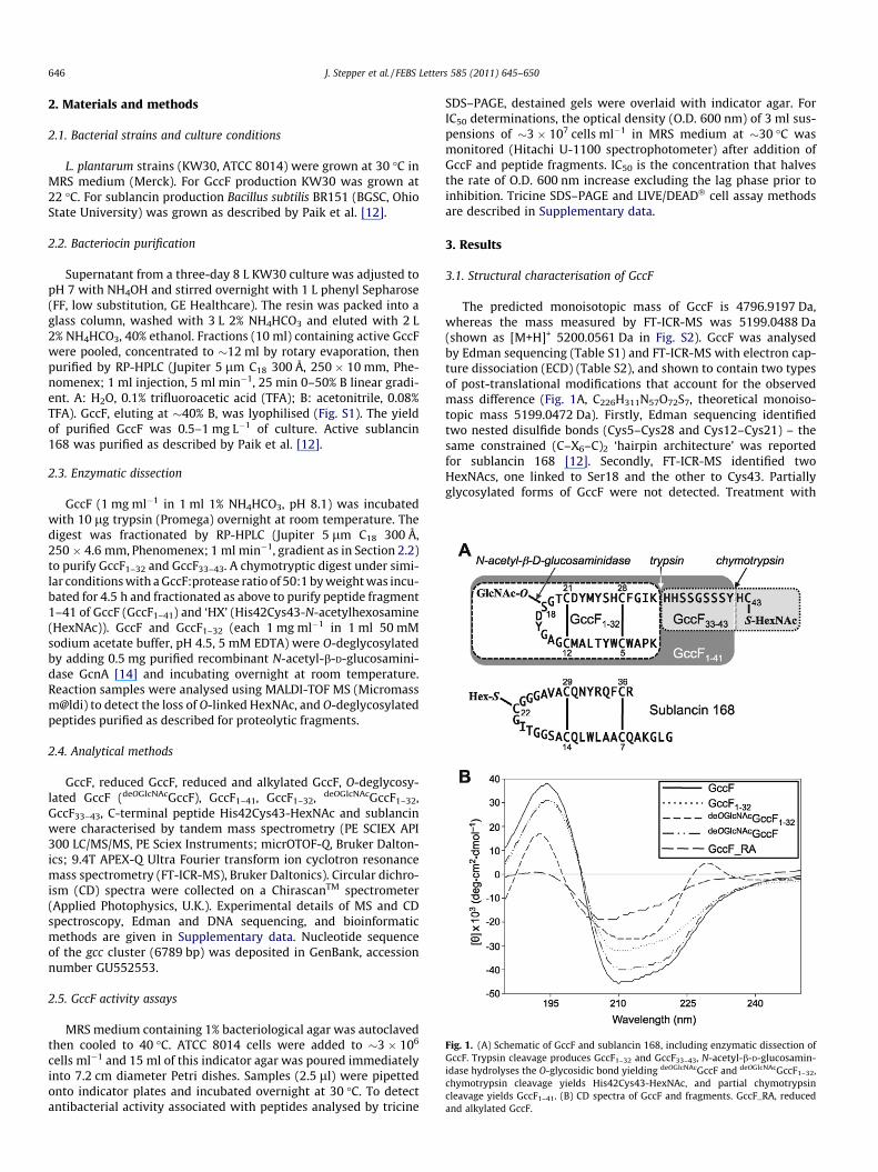
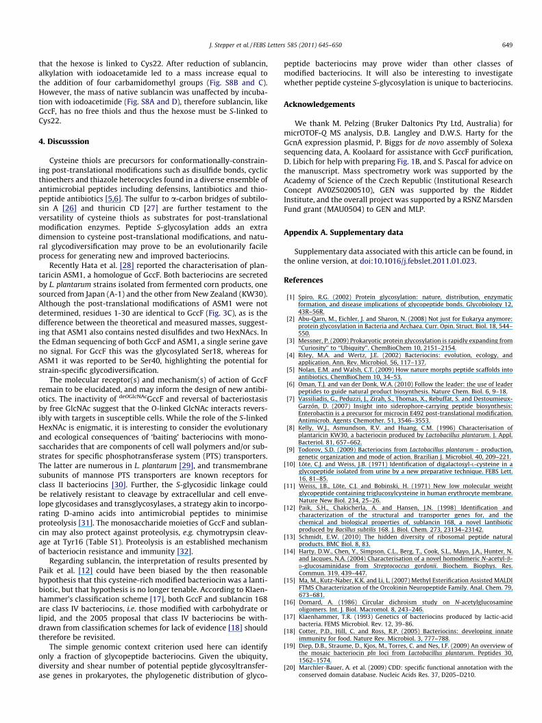
Fig. 1. (A) Schematic of GccF and sublancin 168, including enzymatic dissection ofGccF. Trypsin cleavage produces GccF1–32 and GccF33–43, N-acetyl-b-D-glucosamin-idase hydrolyses the O-glycosidic bond yielding deOGlcNAcGccF and deOGlcNAcGccF1–32,chymotrypsin cleavage yields His42Cys43-HexNAc, and partial chymotrypsincleavage yields GccF1–41. (B) CD spectra of GccF and fragments. GccF_RA, reducedand alkylated GccF.
646 J. Stepper et al. / FEBS Letters 585 (2011) 645–650
2. Materials and methods
2.1. Bacterial strains and culture conditions
L. plantarum strains (KW30, ATCC 8014) were grown at 30 �C inMRS medium (Merck). For GccF production KW30 was grown at22 �C. For sublancin production Bacillus subtilis BR151 (BGSC, OhioState University) was grown as described by Paik et al. [12].
2.2. Bacteriocin purification
Supernatant from a three-day 8 L KW30 culture was adjusted topH 7 with NH4OH and stirred overnight with 1 L phenyl Sepharose(FF, low substitution, GE Healthcare). The resin was packed into aglass column, washed with 3 L 2% NH4HCO3 and eluted with 2 L2% NH4HCO3, 40% ethanol. Fractions (10 ml) containing active GccFwere pooled, concentrated to �12 ml by rotary evaporation, thenpurified by RP-HPLC (Jupiter 5 lm C18 300 Å, 250 � 10 mm, Phe-nomenex; 1 ml injection, 5 ml min�1, 25 min 0–50% B linear gradi-ent. A: H2O, 0.1% trifluoroacetic acid (TFA); B: acetonitrile, 0.08%TFA). GccF, eluting at �40% B, was lyophilised (Fig. S1). The yieldof purified GccF was 0.5–1 mg L�1 of culture. Active sublancin168 was purified as described by Paik et al. [12].
2.3. Enzymatic dissection
GccF (1 mg ml�1 in 1 ml 1% NH4HCO3, pH 8.1) was incubatedwith 10 lg trypsin (Promega) overnight at room temperature. Thedigest was fractionated by RP-HPLC (Jupiter 5 lm C18 300 Å,250 � 4.6 mm, Phenomenex; 1 ml min�1, gradient as in Section 2.2)to purify GccF1–32 and GccF33–43. A chymotryptic digest under simi-lar conditions with a GccF:protease ratio of 50:1 by weight was incu-bated for 4.5 h and fractionated as above to purify peptide fragment1–41 of GccF (GccF1–41) and ‘HX’ (His42Cys43-N-acetylhexosamine(HexNAc)). GccF and GccF1–32 (each 1 mg ml�1 in 1 ml 50 mMsodium acetate buffer, pH 4.5, 5 mM EDTA) were O-deglycosylatedby adding 0.5 mg purified recombinant N-acetyl-b-D-glucosamini-dase GcnA [14] and incubating overnight at room temperature.Reaction samples were analysed using MALDI-TOF MS (Micromassm@ldi) to detect the loss of O-linked HexNAc, and O-deglycosylatedpeptides purified as described for proteolytic fragments.
2.4. Analytical methods
GccF, reduced GccF, reduced and alkylated GccF, O-deglycosy-lated GccF (deOGlcNAcGccF), GccF1–41, GccF1–32, deOGlcNAcGccF1–32,GccF33–43, C-terminal peptide His42Cys43-HexNAc and sublancinwere characterised by tandem mass spectrometry (PE SCIEX API300 LC/MS/MS, PE Sciex Instruments; micrOTOF-Q, Bruker Dalton-ics; 9.4T APEX-Q Ultra Fourier transform ion cyclotron resonancemass spectrometry (FT-ICR-MS), Bruker Daltonics). Circular dichro-ism (CD) spectra were collected on a ChirascanTM spectrometer(Applied Photophysics, U.K.). Experimental details of MS and CDspectroscopy, Edman and DNA sequencing, and bioinformaticmethods are given in Supplementary data. Nucleotide sequenceof the gcc cluster (6789 bp) was deposited in GenBank, accessionnumber GU552553.
2.5. GccF activity assays
MRS medium containing 1% bacteriological agar was autoclavedthen cooled to 40 �C. ATCC 8014 cells were added to �3 � 106
cells ml�1 and 15 ml of this indicator agar was poured immediatelyinto 7.2 cm diameter Petri dishes. Samples (2.5 ll) were pipettedonto indicator plates and incubated overnight at 30 �C. To detectantibacterial activity associated with peptides analysed by tricine
SDS–PAGE, destained gels were overlaid with indicator agar. ForIC50 determinations, the optical density (O.D. 600 nm) of 3 ml sus-pensions of �3 � 107 cells ml�1 in MRS medium at �30 �C wasmonitored (Hitachi U-1100 spectrophotometer) after addition ofGccF and peptide fragments. IC50 is the concentration that halvesthe rate of O.D. 600 nm increase excluding the lag phase prior toinhibition. Tricine SDS–PAGE and LIVE/DEAD� cell assay methodsare described in Supplementary data.
3. Results
3.1. Structural characterisation of GccF
The predicted monoisotopic mass of GccF is 4796.9197 Da,whereas the mass measured by FT-ICR-MS was 5199.0488 Da(shown as [M+H]+ 5200.0561 Da in Fig. S2). GccF was analysedby Edman sequencing (Table S1) and FT-ICR-MS with electron cap-ture dissociation (ECD) (Table S2), and shown to contain two typesof post-translational modifications that account for the observedmass difference (Fig. 1A, C226H311N57O72S7, theoretical monoiso-topic mass 5199.0472 Da). Firstly, Edman sequencing identifiedtwo nested disulfide bonds (Cys5–Cys28 and Cys12–Cys21) – thesame constrained (C–X6–C)2 ‘hairpin architecture’ was reportedfor sublancin 168 [12]. Secondly, FT-ICR-MS identified twoHexNAcs, one linked to Ser18 and the other to Cys43. Partiallyglycosylated forms of GccF were not detected. Treatment with
J. Stepper et al. / FEBS Letters 585 (2011) 645–650 647
the N-acetyl-b-D-glucosaminidase GcnA specifically released theSer18-linked HexNAc from GccF and GccF1-32 (Fig. S3), indicatingthat this HexNAc moiety is a b-O-linked N-acetylglucosamine(GlcNAc). Reduction of GccF CNBr peptides generated four freethiols that reacted with 4-vinyl pyridine (Table S1), whereas native(disulfide-bonded) GccF did not react with 4-vinyl pyridine oriodoacetamide. Edman sequencing, FT-ICR-MS and chemicalmodification results all supported the conclusion that GccF con-tains no free thiols; Cys5, Cys12, Cys21 and Cys28 thiols formdisulfide bonds, and the C-terminal HexNAc is S-linked to theCys43 thiol. Methyl esterification [15] of GccF provided evidencethat the Cys43 HexNAc is not linked via the C-terminal carboxylate,as Asp17, Asp22 and the C-terminus were esterified.
CD spectra of GccF were dominated by bands at �193 and�210 nm, characteristic of helical structure, which diminished onreduction and alkylation (Fig. 1B). Compared to GccF, some of thedecrease in 193 and 210 nm band intensity may be attributed tothe loss of one (deOGlcNAcGccF, GccF1–32) or two (deOGlcNAcGccF1–32)HexNAcs [16]. However, the magnitude of the differences suggeststhat changes in secondary structure occurred when GccF33–43 wasremoved, and that further changes occurred when GccF1–32 was O-deglycosylated (a 230 nm band appeared), despite the disulfidebonds remaining intact throughout (Fig. S3).
3.2. GccF activity
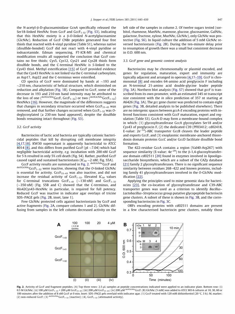
Bacteriocins of lactic acid bacteria are typically cationic bacteri-cidal peptides that kill by disrupting cell membrane integrity[4,17,18]. KW30 supernatant is apparently bactericidal to ATCC8014 [8], and this differs from purified GccF (pI �7.04) which hadnegligible bactericidal activity, e.g. incubation with 200 nM GccFfor 5 h resulted in only 5% cell death (Fig. S4). Rather, purified GccFcaused rapid and sustained bacteriostasis (IC50 �2 nM; Fig. S5A).
GccF activity results are summarised in Fig. 2. deOGlcNAcGccF anddeOGlcNAcGccF1–32 were inactive, showing that the O-linked GlcNAcis essential for activity. GccF33–43 was also inactive, and did notincrease the residual activity of GccF1–32. Elevated IC50 valuesfor C-terminal truncations GccF1–41 (�130 nM) and GccF1–32
(�350 nM) (Fig. S5B and C) showed that the C-terminus, andHis42Cys43-HexNAc in particular, is required for full potency.Reduced GccF was inactive in indicator agar overlays of tricineSDS–PAGE gels (Fig. 2B, inset, lane 1).
Free GlcNAc protected cells against bacteriostasis by GccF andactive fragments (Fig. 2A, compare columns 1 and 2). GlcNAc dif-fusing from samples in the left column decreased activity on the
Fig. 2. Activity of GccF and fragment peptides. (A) Top three rows: 2.5 lL samples at pe0.5 M GlcNAc; (ii) 100 lM GccF1–32 + 200 lM GccF33–43; (iii) 200 lM GccF33–43; (iv) 200 l100 minutes after the addition of 8 nM GccF at 0 min. Inset: SDS–PAGE gels overlaid with(2) non-reduced GccF; (3) deOGlcNAcGccF1–32 (inactive); (4), GccF1–32 (attenuated activity)
left side of the samples in column 2. Of twelve sugars tested (sor-bitol, rhamnose, ManNAc, mannose, glucose, glucosamine, GalNAc,galactose, fructose, xylose, MurNAc, GlcNAc), only GlcNAc was pro-tective (Fig. S6). In liquid culture the addition of 5 mM GlcNAc re-versed bacteriostasis (Fig. 2B). During the ten-minute delay priorto resumption of growth there was a small but consistent decreasein O.D. 600 nm.
3.3. GccF gene and genomic context analysis
Bacteriocins may be chromosomally or plasmid encoded, andgenes for regulation, maturation, export and immunity aretypically adjacent and arranged in operons [4,17–19]. GccF is chro-mosomal [8] and encodes 64-amino acid preglycocin F includingan N-terminal 21-amino acid double-glycine leader peptide(Fig. 3A). Northern blot analysis (Fig. S7) showed that gccF is tran-scribed from its own promoter, with an estimated 345 nt transcriptsize consistent with the in silico prediction of 357 nt starting atA6426 (Fig. 3A). The gcc gene cluster was predicted to contain eightgenes (Fig. 3B, detailed analysis to be published elsewhere). Thereare no intergenic spaces between gccA-E encoding proteins with in-ferred functions consistent with GccF maturation, export and reg-ulation (Table S3). GccA-D may form a membrane-bound complexin which: (1) glycosyltransferase GccA glycosylates Ser18 and/orCys43 of preglycocin F, (2) the dedicated C39 (PF03412; cd02418,E-value: 2e–14)-ABC transporter GccB cleaves the leader peptideand exports GccF, and (3) exoplasmic membrane-anchored thiore-doxin domain proteins GccC and/or GccD facilitate disulfide bondformation.
The 422-residue GccA contains a region (Val40-Arg267) withsequence similarity (E-value: 4e–29) to the b-1,4-glucosyltransfer-ase domain cd02511 [20] found in enzymes involved in lipooligo-saccharide biosynthesis, which are a subset of the CAZy database[21] family 2 glycosyltransferases. There is no significant sequencesimilarity between residues 268–422 and known proteins, includ-ing family 41 glycosyltransferases involved in the O-GlcNAc mod-ification [22].
Applying the principles used to mine genomic data for bacteri-ocins [23], the co-location of glycosyltransferase and C39-ABCtransporter genes was used as a criterion to identify Bacillus–Lactobacillus–Streptococcus group putative glycopeptide bacteriocingene clusters. A subset of these is shown in Fig. 3B, and the corre-sponding bacteriocins in Fig. 3C.
ORFs encoding proteins with cd02511 domains are presentin a few characterised bacteriocin gene clusters, notably those
ptide concentrations indicated were applied to an indicator plate. Bottom row: (i)M deOGlcNAcGccF. (B) GlcNAc (5 mM) was added to ATCC 8014 cultures at 10, 30, 60 orindicator agar. (1) GccF treated with 120 mM dithiothreitol (20 �C, 3 h); M, marker;
.

Fig. 3. The GccF gene, genomic context analysis, and putative glycopeptide bacteriocins. (A) DNA sequence of gccF. Numbering is from GenBank accession GU552553, and thetranslation to preglycocin F is shown. The TAG stop codon of the upstream gccE ORF lies within an imperfect repeat (underlined) with an 18 nt-spacing (AAG-N18-AAG)characteristic of promoter elements recognised by LytTR response regulators that control transcription of some bacteriocin genes. The imperfect repeat encoding Cys5 andCys28 is in bold type. A Rho-independent transcriptional terminator is highlighted (DG = –16.9 kcal/mol). (B) Genomic context of gccF and representative putativeglycopeptide bacteriocin genes. 1, L. plantarum KW30; 2, B. subtilis 168; 3, Enterococcus faecalis HIP11704; 4, E. faecalis TX0104; 5, Streptococcus suis 89-1591. Solid blackarrows are gccF and putative bacteriocin genes (AA sequences of corresponding preglycocins are shown in (C)); vertical lines, glycosyltransferase (GT); diagonal lines, C39-ABC transporter; grid lines, thioredoxin domain protein; dotted pattern, putative signal transduction/regulation; solid grey, putative immunity; white, unknown. In 4, thetriangle marks the position of a transposase in E. faecalis V583. DNA sequence for the enterocin 96 gene cluster of E. faecalis WHE 96 was not available; the E. faecalis TX0104gene cluster encoding an identical prebacteriocin (gi:227517439) is shown here. (C) AA sequences of preglycocin F and putative preglycocins, including ASM1 [28], sublancin[12] and enterocin 96 [24]. Leader peptides (lowercase italics), and known (for GccF and SunA) and hypothetical disulfide bonds and glycosylation sites (bold, underlined) areindicated.
648 J. Stepper et al. / FEBS Letters 585 (2011) 645–650
associated with the production of sublancin [12], PlnN [19] andenterocin 96 [24]. Although a role for peptide glycosyltransferasesin the maturation of bacteriocins from Gram-positive bacteria hasnot previously been proposed, the molecular weights of sublancinand enterocin 96 are greater than predicted, suggesting that theycould be glycopeptides.
3.4. Sublancin 168 is an S-linked glycopeptide bacteriocin
The initial report on sublancin 168 concluded that it containedlanthionine modifications [12], presumably introduced by Lan
family enzymes. However, blastp searches (default parameters)using LanB, LanM and N-terminal regions of the recently describedLanL proteins [25] as query sequences failed to detect Lan proteinsin the B. subtilis 168 proteome. Given the proximity of sunA to yolJ,which encodes a cd02511 glycosyltransferase, the alternativehypothesis that sublancin is a glycopeptide was tested. Thepredicted monoisotopic mass of sublancin is 3717.7207 Da,whereas the mass measured by FT-ICR-MS was 3875.7454 Da,consistent with two disulfide bonds and a covalently attachedhexose (Fig 1A; C162H254N50O51S5, theoretical monoisotopic mass3875.7422 Da). Subsequent ECD analysis (Table S2B) confirmed
J. Stepper et al. / FEBS Letters 585 (2011) 645–650 649
that the hexose is linked to Cys22. After reduction of sublancin,alkylation with iodoacetamide led to a mass increase equal tothe addition of four carbamidomethyl groups (Fig. S8B and C).However, the mass of native sublancin was unaffected by incuba-tion with iodoacetimide (Fig. S8A and D), therefore sublancin, likeGccF, has no free thiols and thus the hexose must be S-linked toCys22.
4. Discusssion
Cysteine thiols are precursors for conformationally-constrain-ing post-translational modifications such as disulfide bonds, cyclicthioethers and thiazole heterocycles found in a diverse ensemble ofantimicrobial peptides including defensins, lantibiotics and thio-peptide antibiotics [5,6]. The sulfur to a-carbon bridges of subtilo-sin A [26] and thuricin CD [27] are further testament to theversatility of cysteine thiols as substrates for post-translationalmodification enzymes. Peptide S-glycosylation adds an extradimension to cysteine post-translational modifications, and natu-ral glycodiversification may prove to be an evolutionarily facileprocess for generating new and improved bacteriocins.
Recently Hata et al. [28] reported the characterisation of plan-taricin ASM1, a homologue of GccF. Both bacteriocins are secretedby L. plantarum strains isolated from fermented corn products, onesourced from Japan (A-1) and the other from New Zealand (KW30).Although the post-translational modifications of ASM1 were notdetermined, residues 1-30 are identical to GccF (Fig. 3C), as is thedifference between the theoretical and measured masses, suggest-ing that ASM1 also contains nested disulfides and two HexNAcs. Inthe Edman sequencing of both GccF and ASM1, a single serine gaveno signal. For GccF this was the glycosylated Ser18, whereas forASM1 it was reported to be Ser40, highlighting the potential forstrain-specific glycodiversification.
The molecular receptor(s) and mechanism(s) of action of GccFremain to be elucidated, and may inform the design of new antibi-otics. The inactivity of deOGlcNAcGccF and reversal of bacteriostasisby free GlcNAc suggest that the O-linked GlcNAc interacts revers-ibly with targets in susceptible cells. While the role of the S-linkedHexNAc is enigmatic, it is interesting to consider the evolutionaryand ecological consequences of ‘baiting’ bacteriocins with mono-saccharides that are components of cell wall polymers and/or sub-strates for specific phosphotransferase system (PTS) transporters.The latter are numerous in L. plantarum [29], and transmembranesubunits of mannose PTS transporters are known receptors forclass II bacteriocins [30]. Further, the S-glycosidic linkage couldbe relatively resistant to cleavage by extracellular and cell enve-lope glycosidases and transglycosylases, a strategy akin to incorpo-rating D-amino acids into antimicrobial peptides to minimiseproteolysis [31]. The monosaccharide moieties of GccF and sublan-cin may also protect against proteolysis, e.g. chymotrypsin cleav-age at Tyr16 (Table S1). Proteolysis is an established mechanismof bacteriocin resistance and immunity [32].
Regarding sublancin, the interpretation of results presented byPaik et al. [12] could have been biased by the then reasonablehypothesis that this cysteine-rich modified bacteriocin was a lanti-biotic, but that hypothesis is no longer tenable. According to Klaen-hammer’s classification scheme [17], both GccF and sublancin 168are class IV bacteriocins, i.e. those modified with carbohydrate orlipid, and the 2005 proposal that class IV bacteriocins be with-drawn from classification schemes for lack of evidence [18] shouldtherefore be revisited.
The simple genomic context criterion used here can identifyonly a fraction of glycopeptide bacteriocins. Given the ubiquity,diversity and shear number of potential peptide glycosyltransfer-ase genes in prokaryotes, the phylogenetic distribution of glyco-
peptide bacteriocins may prove wider than other classes ofmodified bacteriocins. It will also be interesting to investigatewhether peptide cysteine S-glycosylation is unique to bacteriocins.
Acknowledgements
We thank M. Pelzing (Bruker Daltonics Pty Ltd, Australia) formicrOTOF-Q MS analysis, D.B. Langley and D.W.S. Harty for theGcnA expression plasmid, P. Biggs for de novo assembly of Solexasequencing data, A. Koolaard for assistance with GccF purification,D. Libich for help with preparing Fig. 1B, and S. Pascal for advice onthe manuscript. Mass spectrometry work was supported by theAcademy of Science of the Czech Republic (Institutional ResearchConcept AV0Z50200510), GEN was supported by the RiddetInstitute, and the overall project was supported by a RSNZ MarsdenFund grant (MAU0504) to GEN and MLP.
Appendix A. Supplementary data
Supplementary data associated with this article can be found, inthe online version, at doi:10.1016/j.febslet.2011.01.023.
References
[1] Spiro, R.G. (2002) Protein glycosylation: nature, distribution, enzymaticformation, and disease implications of glycopeptide bonds. Glycobiology 12,43R–56R.
[2] Abu-Qarn, M., Eichler, J. and Sharon, N. (2008) Not just for Eukarya anymore:protein glycosylation in Bacteria and Archaea. Curr. Opin. Struct. Biol. 18, 544–550.
[3] Messner, P. (2009) Prokaryotic protein glycosylation is rapidly expanding from‘‘Curiosity’’ to ‘‘Ubiquity’’. ChemBioChem 10, 2151–2154.
[4] Riley, M.A. and Wertz, J.E. (2002) Bacteriocins: evolution, ecology, andapplication. Ann. Rev. Microbiol. 56, 117–137.
[5] Nolan, E.M. and Walsh, C.T. (2009) How nature morphs peptide scaffolds intoantibiotics. ChemBioChem 10, 34–53.
[6] Oman, T.J. and van der Donk, W.A. (2010) Follow the leader: the use of leaderpeptides to guide natural product biosynthesis. Nature Chem. Biol. 6, 9–18.
[7] Vassiliadis, G., Peduzzi, J., Zirah, S., Thomas, X., Rebuffat, S. and Destoumieux-Garzón, D. (2007) Insight into siderophore-carrying peptide biosynthesis:Enterobactin is a precursor for microcin E492 post-translational modification.Antimicrob. Agents Chemother. 51, 3546–3553.
[8] Kelly, W.J., Asmundson, R.V. and Huang, C.M. (1996) Characterisation ofplantaricin KW30, a bacteriocin produced by Lactobacillus plantarum. J. Appl.Bacteriol. 81, 657–662.
[9] Todorov, S.D. (2009) Bacteriocins from Lactobacillus plantarum - production,genetic organization and mode of action. Brazilian J. Microbiol. 40, 209–221.
[10] Löte, C.J. and Weiss, J.B. (1971) Identification of digalactosyl-L-cysteine in aglycopeptide isolated from urine by a new preparative technique. FEBS Lett.16, 81–85.
[11] Weiss, J.B., Löte, C.J. and Bobinski, H. (1971) New low molecular weightglycopeptide containing triglucosylcysteine in human erythrocyte membrane.Nature New Biol. 234, 25–26.
[12] Paik, S.H., Chakicherla, A. and Hansen, J.N. (1998) Identification andcharacterization of the structural and transporter genes for, and thechemical and biological properties of, sublancin 168, a novel lantibioticproduced by Bacillus subtilis 168. J. Biol. Chem. 273, 23134–23142.
[13] Schmidt, E.W. (2010) The hidden diversity of ribosomal peptide naturalproducts. BMC Biol. 8, 83.
[14] Harty, D.W., Chen, Y., Simpson, C.L., Berg, T., Cook, S.L., Mayo, J.A., Hunter, N.and Jacques, N.A. (2004) Characterisation of a novel homodimeric N-acetyl-b-D-glucosaminidase from Streptococcus gordonii. Biochem. Biophys. Res.Commun. 319, 439–447.
[15] Ma, M., Kutz-Naber, K.K. and Li, L. (2007) Methyl Esterification Assisted MALDIFTMS Characterization of the Orcokinin Neuropeptide Family. Anal. Chem. 79,673–681.
[16] Domard, A. (1986) Circular dichroism study on N-acetylglucosamineoligomers. Int. J. Biol. Macromol. 8, 243–246.
[17] Klaenhammer, T.R. (1993) Genetics of bacteriocins produced by lactic-acidbacteria. FEMS Microbiol. Rev. 12, 39–86.
[18] Cotter, P.D., Hill, C. and Ross, R.P. (2005) Bacteriocins: developing innateimmunity for food. Nature Rev. Microbiol. 3, 777–788.
[19] Diep, D.B., Straume, D., Kjos, M., Torres, C. and Nes, I.F. (2009) An overview ofthe mosaic bacteriocin pln loci from Lactobacillus plantarum. Peptides 30,1562–1574.
[20] Marchler-Bauer, A. et al. (2009) CDD: specific functional annotation with theconserved domain database. Nucleic Acids Res. 37, D205–D210.

650 J. Stepper et al. / FEBS Letters 585 (2011) 645–650
[21] Cantarel, B.L., Coutinho, P.M., Rancurel, C., Bernard, T., Lombard, V. andHenrissat, B. (2009) The carbohydrate-active EnZymes database (CAZy): anexpert resource for glycogenomics. Nucleic Acids Res. 37, D233–D238.
[22] Martinez-Fleites, C., He, Y. and Davies, G.J. (2010) Structural analyses ofenzymes involved in the O-GlcNAc modification. Biochim. Biophys. Acta 1800,122–133.
[23] de Jong, A., van Heel, A.J., Kok, J. and Kuipers, O.P. (2010) BAGEL2: mining forbacteriocins in genomic data. Nucleic Acids Res. 38, W647–W651.
[24] Izquierdo, E., Wagner, C., Marchioni, E., Aoude-Werner, D. and Ennahar, S.(2009) Enterocin 96, a novel class II bacteriocin produced by Enterococcusfaecalis WHE 96, isolated from Munster cheese. Appl. Environ. Microbiol. 75,4273–4276.
[25] Goto, Y., Li, B., Claesen, J., Shi, Y., Bibb, M.J. and van der Donk, W.A. (2010)Discovery of unique lanthionine synthetases reveals new mechanistic andevolutionary insights. PLoS Biol. 8, e1000339.
[26] Kawulka, K.E., Sprules, T., Diaper, C.M., Whittal, R.M., McKay, R.T., Mercier, P.,Zuber, P. and Vederas, J.C. (2004) Structure of subtilosin A, a cyclicantimicrobial peptide from Bacillus subtilis with unusual sulfur to a-carboncross-links: formation and reduction of a-thio-a-amino acid derivatives.Biochemistry 43, 3385–3395.
[27] Rea, M.C., Sit, C.S., Claytona, E., O’Connora, P.M., Whittal, R.M., Zheng, J.,Vederas, J.C., Ross, R.P. and Hill, C. (2010) Thuricin CD, a posttranslationallymodified bacteriocin with a narrow spectrum of activity against Clostridiumdifficile. PNAS 107, 9352–9357.
[28] Hata, T., Tanaka, R. and Ohmomo, S. (2010) Isolation and characterization ofplantaricin ASM1: A new bacteriocin produced by Lactobacillus plantarum A-1.Int. J. Food Microbiol. 137, 94–99.
[29] Molenaar, D., Bringel, F., Schuren, F.H., de Vos, W.M., Siezen, R.J. andKleerebezem, M. (2005) Exploring Lactobacillus plantarum genome diversityby using microarrays. J. Bacteriol. 187, 6119–6127.
[30] Kjos, M., Salehian, Z., Nes, I.F. and Diep, D.B. (2010) An extracellular loop of themannose phosphotransferase system component IIC is responsible for specifictargeting by class IIa bacteriocins. J. Bacteriol. 192, 5906–5913.
[31] Zasloff, M. (2002) Antimicrobial peptides of multicellular organisms. Nature415, 389–395.
[32] Kjos, M., Snipen, L., Salehian, Z., Nes, I.F. and Diep, D.B. (2010) The Abi proteinsand their involvement in bacteriocin self-immunity. J. Bacteriol. 192, 2068–2076.