Glycosylation at Asn91 of H1N1 haemagglutinin affects binding to glycan receptors

ICANCER RESEARCH 56. 1682-1689. April 1. IW6]
a6/31-Integrin, a Major Cell Surface Carrier of /31-6-branched Oligosaccharides,
Mediates Migration of EJ-ras-transformed Fibroblasts on Laminili-1Independently of Its Glycosylation State1
Miriam G. Jasiulionis, Roger Chammas,2 Armando M. Ventura,3 Luis R. Travassos, and Ricardo R. Brentani
Ludwig Institute for Cancer Research, Sao Paulo, SP JM. G. J., R. C.. A. M. V., R. R. B.I. ami Disciplina tie BiologÃa Celular. Escola Paulisra tie Mctlicinti. Univt'rsitlatle
Federal tie Sao Paulo. Sao Paulo. SP ¡L R. T.], Brasil
ABSTRACT
EJ-ras oncogene-induced malignant transformation is characterized bya series of changes in cell surface carbohydrates and cell-cell and cell-matrix interactions. Here, we show that EJ-ra.v-transformed NIH-3T3fibroblasts acquired a migratory phenotype on laminin-1 surfaces. Such a
phenotype was accompanied by overexpression of: (a) functional ad/il.but not other laminin binding ßl-integrins; and (b ) glycoconjugates on the
cell surface bearing large Oligosaccharides recognized by leukoagglutininfrom Phaseolus vulgaris (L-PHA). The internal pool of pre-/31-integrinswas differently regulated in EJ-ras-transformed cells compared with non-transfected fibroblasts. Conversion of pre-ßl- into mature ßl-integrinswas faster in EJ-ra.v-transformed cells, a process associated with theoverexpression of the a6-chain. Overexpression of L-PHA-reactive Oligosaccharides is dependent on the activity of iV-acetylglucosaminyltrans-
ferase V, which is increased in transformed cells [J. W. Dennis et al.,Science (Washington DC), 236: 582-585,1987]. We show that 01-integrinswere the major carriers of L-PHA-reactive Oligosaccharides on the cell
surface. This glycosylation pattern, however, was not necessary for eitherthe cell surface expression of /31-integrins or their functional activity inthe migratory response to laminin-l. Moreover, EJ-ro.v-transformed II-
broblasts aggregated spontaneously. These effects were not observed inc-y'i/H-transfected fibroblasts, which were unable to migrate on laminin,
did not overexpress either ßl-integrins or L-PHA-reactive Oligosaccharides, and did not self-aggregate.
INTRODUCTION
ras proto-oncogenes are activated by point mutations in at least one
out of five human tumors ( I ). Mutations can activate ras genes,leading to malignant transformation and acquisition of the invasivephenotype (2). Upstream events may also lead to activation of Rasproteins. Nonetheless, downstream events to the expression of mutation or activation of Ras proteins leading to tumor progression are stillnot fully understood. Among the pleiotropic effects of ras oncogeneactivation are: (a) altered cell surface carbohydrates (3, 4); (b) alteredcell interactions with extracellular matrix proteins, such as fibronectinand laminin (5); and (<•)altered cell-cell interactions (6). These effects
converge to establish the malignant phenotype. although the criticaland temporal participation of each is not completely determined.Altered glycosylation of cell surface glycoconjugates represents notonly a useful feature for early diagnosis of cancer, but also a potentialtarget for pharmacological intervention (7, 8).
Altered cell-cell and cell-extracellular matrix interactions are im
portant in the acquisition of a migratory phenotype. leading to invasion. Among the different cell adhesion molecules that mediate cellular migration, integrins are the best characterized (9, 10). Integrins
Received 9/27/95; accepted 2/1/96.The costs of publication of this article were defrayed in part hy the payment of page
charges. This article must therefore he hereby marked advertisement in accordance with18 U.S.C. Section 1734 solely to indicate this fact.
1Supported by CNPq Brasil Grants 840178/95-6 (M. G. J.) and 200015/94-0 (R. C.).2 To whom requests for reprints should be addressed, at Glycobiology Program. Cancer
Center, 0687. University of California San Diego School of Medicine. La Jolla. CA92093-0687. Phone: (619) 534-1346; Kax: (619) 534-5611; E-mail: rchammasfe'ucsd.edu.
' Present address: Departamento de Microbiologia. Instituto de Ciencias Biomédicas,
Universidade de Sao Paulo. Sao Paulo. SP. Brasil.
are a/ß-heterodimers involved in either cell-cell interactions (e.g.,ß2-integrins)or cell-matrix interactions (e.g., ßl-integrins). ßl-inte-
grins constitute the very late antigen subfamily, composed of at least10 different molecules (al-a9- and avßl-integrins; Ref. 9). Here, wehave focused on laminin-binding integrins of the very late antigen
subfamily.Cancer cells frequently display altered patterns of glycosylation of
cell surface glycoconjugates (11). Large /V-linked Oligosaccharides
modify the glycoproteins of transformed cells. These Oligosaccharidesare usually complex, with triantennary or multiantennary structures(12). Although such alterations have been described in past decades,little is known about the role of these altered glycans in glycoproteinstructure and/or function, if any. Among altered patterns, the expression of tri- or tetra-antennary Oligosaccharides recognized by L-PHA4
(13) has been more extensively studied, because it was shown thatthese Oligosaccharides could be related to the metastatic phenotype.The biosynthesis of L-PHA-reactive Oligosaccharides depends on theactivity of GlcNAc-T V. a branching enzyme that catalyzes thesubstitution of the trimannosyl core by a ß(1 —¿�»6)-linked W-acetyl-glucosamine residue. This enzyme is up-regulated by activated onco-
genes that stimulate the Ras pathway (4, 14). In the present work, wehave generated EJ-ra.v-transformed NIH-3T3 cells. EJ-ra.s-transformed cells overexpressed aoßl-integrin on the cell surface,migrated actively on laminin substrata, and overexpressed L-PHA-reacting components on the cell surface, ßl-integrins were shown tobe suitable substrates for GlcNAc-T V and the major carriers of tri-and tetra-antennary structures among the cell surface glycoproteins.However, we showed that this glycosylation pattern was not directlyassociated with integrin-mediated migration on laminin.
MATERIALS AND METHODS
Reagents and Immunochemicals. Unless otherwise stated, all reagentswere purchased from Sigma Chemical Co. (St. Louis. MO). Rb3847. a rabbitanti-ßl-integriti serum, was a kind giti troni Dr. K. Yumuda (National Institute
for Dental Research. Bethesda. MD). Mouse monoclonal antibodies toLAMP-1 (clone 1D4B) and LAMP-2 (clone ABL-93) were obtained from the
Developmental Studies Hybridoma Bank (maintained by the Department ofPharmacology and Molecular Sciences. Johns Hopkins University School ofMedicine. Baltimore. MD. and the Department of Biological Sciences. University of Iowa. Iowa City. IA, under contract N01-HD-6-2915 from theNICHD). GoH3. a rat monoclonal antibody anti-«6-chain. was purchased from
Centraal Laboratorium van de Bloedtransfusiedienst (Amsterdam, the Netherlands). All other anti-integrin antibodies (anti-«2. «3,and «5)were purchasedfrom Telios (La Jolla. CA). Laminin-1 was purified from the Engelbreth-
Holm-Swarm tumor as described (15).Cells. NIH-3T3 cells, a gift from Dr. M. C. S. Armelin (Departamento de
BioquÃmica. Instituto de QuÃmica,Universidade de Sao Paulo), were grown inDMEM with 10% FCS. NIH 3T3 cells were transfected with pEJ-ra.v (16)using lipotectin (GIBCO-BRL. Gaithersburg. MD) according to the protocol of
4 The abbreviations used are: L-PHA. leukoagglutinin from Phaseolus vulgaris; dMN.deoxymannojirimycin; GlcNAc-T V. /V-acetylglucosaminyltransferase V; LAMP, lysoso-mal-associated membrane protein; SW. swainsonine: kDa. kilodalton; pi. isoelectric point:ER. endoplasmi^1 reticulum.
1682
on July 21, 2015. © 1996 American Association for Cancer Research. cancerres.aacrjournals.org Downloaded from
INTEGRIN EXPRESSION AND MIGRATION OF ros FIBROBLASTS
the manufacturer ( 1 fig plasmid/106 cells). After 48 h, transfected cells were
plated in soft agar (0.3<7ragarose ¡nDMEM with 10% PCS), and 2 weeks later,
individual anchorage-independent colonies were isolated and expanded. Sixdifferent clones were obtained, with similar levels of EJ-ras expression, as
determined by Nothern blot analysis. The clone CCR2 was used in all experiments. The results were routinely reconfirmed in other clones. c-j«n-trans-fected NIH-3T3 fibroblasts were kindly provided by Dr. V. R. Martins (Fun-daçà oAntonio Prudente. Sao Paulo). These cells were transfected with c-jun ina plasmid under the control of the Rous sarcoma virus promoter. Log phase-
growing cells were treated with SW (2 /ng/ml) or dMN (10 /ng/ml) for 16 h.Treatment efficiency was checked either by flow cytometry using lectins or byWestern blots to evaluate the presence of mature glycoforms of ßl-integrin
chains.Assay for Tumorigenicity. Three-week-old male and female BALB/c
mice or nude mice were used to assess tumorigenicity of EJ-raj-transformed
cells. Two million cells were injected s.c. in the flank of each animal.Nontransfected cells were injected as controls. Animals were maintained in anadequate vivarium, without any food or water restriction, and were routinelyexamined for the development of tumors. Tumor-bearing mice were sacrificed
by decapitation and examined either for local invasion or spontaneous metastasis in different organs. Tumors were excised en bloc, preserving adjacent
tissues for routine histológica! analysis, using H&E staining.Migration Assays. Migration assays were performed on modified
Boyden's chambers with an 8-ju.m-pore polycarbonate membrane, using 10
fig/ml laminin-1 as the haptotatic agent. The membrane was incubated with the
laminin solution in DMEM without PCS for 2 h. and it was then blocked with0.5% BSA. Nontransfected N1H-3T3 and c-jun-, and EJ-ras-transformed cellswere harvested by trypsinization, followed by trypsin inactivation with serum-
containing media. Cells were then washed with PBS. resuspended in DMEMwithout PCS. and added to the upper compartment of the chamber (2 X IO5cells/chamber). After 3 h at 37°Cand 5% CO2, membranes were washed with
PBS, fixed in 3.7% formaldehyde, further washed, and stained with toluidineblue for 2 min (17). Nonmigrating cells of the upper compartment wereremoved with a cotton swab. The membrane was then washed in distilled waterfor 1 h, air dried, and soaked in 1% SDS for dye solubilization for 1 h at 37°C.
Solutions were transferred to 96-well plates, and the A at 600 nm was measured
in an ELISA plate reader. For inhibition assays, cells were incubated withdifferent concentrations of the antibodies immediately before the assay. Cellstreated overnight with the mannosidase inhibitors were also tested in themigration assay.
Flow Cytometric Assays. Nontransfected and transformed cells were harvested with trypsin-EDTA. After trypsin inactivation, cells were washed inPBS-0.1% BSA and incubated with different anti-integrin antibodies (IO6
cells/ml at a final concentration of 10 jug/ml of each antibody or, in the caseof Rb3847, a 1:400 dilution) for 1 h at 4°C.After three washes with PBS-0.1 %
BSA, cells were incubated with FITC-conjugated antirabbit or antimouse IgG( 1:80) for 1 h at 4°C.After washing, cells were analyzed in a flow cytometer
(FACScan; Becton Dickinson, San Jose, CA). Alternatively, cells were eitherincubated with Datura stramonium agglutinin (for studying the presence ofß-galactosyl residues) or L-PHA-FITC (for tri- or tetra-antennary N-linked
oligosaccharides). Spontaneous cell aggregation was also evaluated by flowcytometry, using the light-scattering properties of the cells. After harvestingwith trypsin-EDTA. followed by trypsin inactivation, cells were washed withPBS and resuspended in Ca2 +- and Mg:+-free buffer. After 1 h under constant
agitation (~6() rpm) at 37°C.cells were fixed with 3.7% formaldehyde in PBS
and analyzed by flow cytometry and conventional microscopy.Analysis of 01-Integrin Biosynthesis. Cell extracts of normal and trans
formed cells were prepared in 1% Triton X-100, 50 min Tris (pH 7.4), 150 mM
NaCl. 5 mM EDTA, 1 mM phenylmethylsulfonyl fluoride, and 2 mg/mlaprotinin. Proteins were separated by either one- or two-dimensional SDS-PAGE (18. 19). The first dimension of two-dimensional SDS-PAGE consisted
of equilibrium isoelectrofocusing, using an ampholyte mixture that gave optimal separation in the range of 4-7 units of pH [Pharmalyte (pH 4-6.5 and3-10); Pharmacia, Uppsala, Sweden]. The second dimension, as for unidimen
sional gels, consisted of a discontinuous 7.5% SDS-polyacrylamide gel. After
separation, proteins were electrotransferred to nitrocellulose for conventionalWestern or lectin blots (20).
Pulse-Chase Experiments. Cells were cultured for 1 h in cysteine-freeDMEM supplemented with 10% PCS. Fifty ^iCi/ml [15S]cysteine (Amersham,
Buckinghamshire, United Kingdom) were then added for a 1-h pulse. Cells
were washed and incubated with nonradioactive medium, and. at differenttimes, whole-cell lysates were prepared. Proteins were then immunoprecipi-
tated using Rb3847 as the primary antibody. Immunoprecipitates were separated in SDS-polyacrylamide gels, embedded in Amplify (Amersham), dried,and exposed to X-OMAT films (Eastman Kodak Co., Rochester, NY).
RESULTS
a6ßl-Integrin Expression Mediates Migration of EJ-ras-transformed Cells on Laminin-1-coated Surfaces. NIH-3T3 cells
were transformed by transfection with a plasmid containing the activated oncogene ras (pEJ-ras). EJ-ra.v-transfected fibroblasts were
selected for their ability to form colonies in soft agar, a characteristicfeature of transformed cells in culture (21, 22). Six different clones oftransformed cells were tested for their ability to grow as tumors eitherin nude mice or in BALB/c mice and were analyzed for their invasivebehavior in all animals injected. Local tumors developed in 2-3
weeks. Nontransfected cells were injected as controls and were nevertumorigenic (observation periods, >6 months). Tumor formation atdistant sites was not detected.
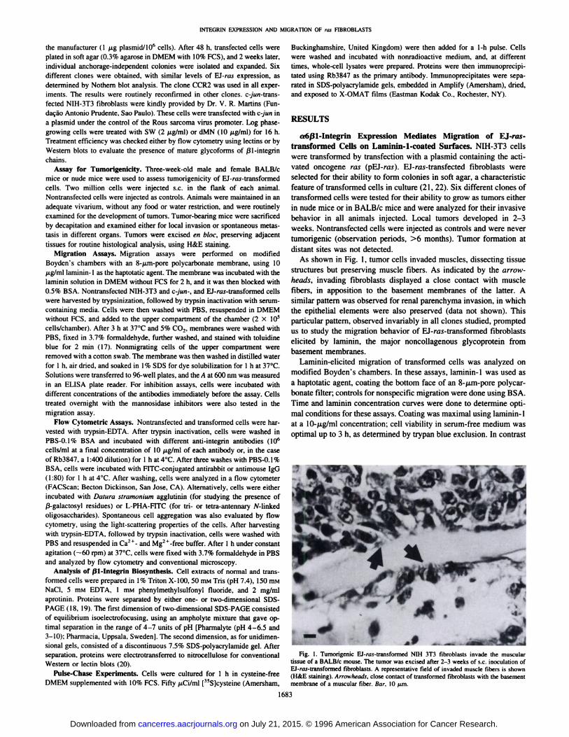
As shown in Fig. 1, tumor cells invaded muscles, dissecting tissuestructures but preserving muscle fibers. As indicated by the arrowheads, invading fibroblasts displayed a close contact with musclefibers, in apposition to the basement membranes of the latter. Asimilar pattern was observed for renal parenchyma invasion, in whichthe epithelial elements were also preserved (data not shown). Thisparticular pattern, observed invariably in all clones studied, promptedus to study the migration behavior of EJ-ras-transformed fibroblasts
elicited by laminin, the major noncollagenous glycoprotein frombasement membranes.
Laminin-elicited migration of transformed cells was analyzed onmodified Boyden's chambers. In these assays, laminin-1 was used as
a haptotatic agent, coating the bottom face of an 8-fim-pore polycar
bonate filter; controls for nonspecific migration were done using BSA.Time and laminin concentration curves were done to determine optimal conditions for these assays. Coating was maximal using laminin-1at a 10-/j.g/ml concentration; cell viability in serum-free medium was
optimal up to 3 h, as determined by trypan blue exclusion. In contrast
Fig. 1. Tumorigenic EJ-ra.v-transformed NIH 3T3 fibroblasts invade the musculartissue of a BALB/c mouse. The tumor was excised after 2-3 weeks of s.c. inoculation ofEJ-ruj-iransfomied fibroblasts. A representative field of invaded muscle fibers is shown
(H&E staining). Arrowheads, close contaci of transformed fibroblasts with the basementmembrane of a muscular fiber. Bar, 10 (im.
1683
on July 21, 2015. © 1996 American Association for Cancer Research. cancerres.aacrjournals.org Downloaded from

1NTF.ÃœR1NKXPRKSSION AND MIORATION OF ras FIBROBLASTS
with nontransformed cells, the EJ-raÃ-transformed fibroblasts migrated toward laminin-l (Fig. 2).
Integrins are the major laminin-binding proteins associated with the
migratory phenotype (9). We examined the expression of putativelaminin-binding integrins in nontransfected and transformed cells. Weanalyzed the expression of a2ßl-, a3ßl-, and a6ßl-integrins. whicharc the known laminin-binding integrins from fibroblasts (23). Fig. 3
shows that on transformation, there is an increase in the cell surfaceexpression of ß\-integrins, as analyzed by flow cytometry. No
changes were observed on either a2 or a3 cell surface expression(data not shown). On the other hand, the a6-subunit showed increased
cell surface expression on transformation (Fig. 3). We also analyzedcell surface expression of a5ßl, the major fibronectin-binding inte-
grin from fibroblasts; no significant differences were observed in itsexpression on transformation (data not shown).
To test whether the migratory phenotype of EJ-ra.î-transformedfibroblasts was associated with the increased expression of aoßl-
integrins on the cell surface, we performed migration assays byincubating cells with different anti-integrin antibodies. As shown inTable 1, antibodies against both integrin chains inhibited the migra-
8
NIH NIH-rasFig. 2. Migratory properties of EJ-rav-transformed fibroblasts toward laminin-1 -coated
surfaces, using a modified Boyden's chamber. A, scanning electron micrograph of
transformed cells on the top of the polycarbonate membrane (which is not coated withlaminin) ready to migrate through pores toward the laminin-coated surface underneath. B,migrating cells spread on the laminin-coated surface (i.e., the underside of the membrane).C, comparison of migratory capacity of nontransformed (NIH} and EJ-ra.ï-transformed(NIH-mx) fibroblasts, as described in "Materials and Methods." •¿�laminin-coated sur
face; D, BSA-coated surface.
P ""•¿�o •¿�m <o-
%-
o :i<r io 10* 10° 10
ßl-chain
10° 101 102 IO3 IO4
a6-chaln
10° IO1 102 IO3 10" 10° 101 102 IO3 IO4
Fig. 3. Increased expression of a6ßI-integrin in EJ-raj-traized by flow cytometry with antibodies specific for the a- and j
•¿�flj-transformed fibroblasts analyzed by flow cytometry with antibodies specific for the a- and ß-integrinchains. Dashed
lines, controls with normal rabbit serum (left) and irrelevant rat monoclonal antibody(right); solid lines, reactivity, measured as fluorescence intensities (l-IO4), of nontrans
formed (NIH-3T3) and EJ-ras-transformed (NIH-3T3 ras) fibroblasts (values are relativecell counts) with rabbit anti-ßlpolyclonal antibody and rat anti-«6monoclonal antibody.
Table I ras-transformed ßbroblast migration inward laminin-1-coated surfaces isblocked by anti-ao- and anli-ßl-integrili chain antibodies
Data are relative percent means of three determinations ±SD.
Relative migration (<#•)
Antiserum orantibodyNormal
rabbitAntiß-1polyclonalNormal
ratAnti-a6monoclonalTiler1/1001/2001/1001/501/201/401/201/10Experiment
1100nd"38
±2.224±2.1100nd58
±5.742±8.2Experiment
29610034
±2.934±9.110010053
±10.647±8.4
" nd. not determined.
tion of EJ-ra.ï-transformed fibroblasts toward laminin-l surfaces.
Controls were done using either normal rabbit or rat serum. In theseassays, nontransfected fibroblasts or c-^wn-transformed fibroblasts did
not migrate.Biosynthesis of ßl-Integrins Is Altered on Transformation. We
compared the ßl-integrins synthesized by nontransfected and Ei-ras-
transformed fibroblasts by Western blotting analysis using antiserumRb3847. Two polypeptides were consistently observed in these cells.These two ßl-polypeptides displayed apparent molecular masses of
105 and 120 kDa, as illustrated in Fig. 4. On transformation, the120-kDa polypeptide was always found, whereas the level of accumulation of the 105-kDa polypeptide varied in different assays. Non
transfected cells growing in the log phase tended to accumulate the105-kDa form relative to the 120-kDa form, different from the trans-fected cells (data not shown). As indicated in Fig. 4B. the 105-kDapolypetide had a less acidic pi. The 120-kDa form (pi, 5.0-5.4) had
a more complex glycosylation pattern, as inferred from its reactivitywith L-PHA, which recognizes tri- and tetra-antennary /V-linked structures. These data suggested that the 105-kDa form is a precursor of the120-kDa form, the latter representing the fully glycosylated form ofthe ßl-integrin chain (24. 25). Pulse-chase experiments, followed byimmunoprecipitation using anti-ßl antibodies, confirmed that the105-kDa form is a pre-ßl-chain, as shown in Fig. 4A. An unknownpolypeptide with a lower molecular mass (arrow) consistently immu-
1684
on July 21, 2015. © 1996 American Association for Cancer Research. cancerres.aacrjournals.org Downloaded from

INTEGRIN EXPRESSION AND MIGRATION OF raj FIBROBLASTS
NIH
Time (h) C 1
NIH-ras
Fig. 4. EJ-ra.s-transformed fibroblasls show a faster processing of theßl-integrin from the pre-ßlto the mature form. A. pulse-chase experiment with ['5S]cysteine showing the kinetics of ßl-integrin maturation:
the labeled extracts of nontransformed (NIH} and EJ-ra.s-transformed(NIH-ras) fibroblasts were immunoprecipitated with polyclonal anti-ßl
antiserum. In addition to the integrin, a noncharacterized polypeptidewas coprecipitated (arrow). B. Isoelectric focusing of protein extracts ofEJ-rai-transformed fibroblasts showing the mature form of ßl-integrin(pi, 5.0-5.4) recognized by both the anti-ßlantiserum and the L-PHA.The lectin also reacted with 90-l(K)-kDa components, which correspondto LAMP-1 and LAMP-2. Arrows, mature ßl-integrin polypeptides alsoidentified by L-PHA.
mature
pré
ßi-ßi-
itt **»
kDa
116 -98 -
116 -
98 -
Acid Basic B
LPHA
noprecipitated with Rb3847. This polypeptide was not studied furtherhere. Interestingly, processing of ßl-integrin chains in EJ-ra.?-trans-
formed cells is faster (<2 h) than that in normal cells, which tookabout 3 h for completion.
As indicated in Fig. 4fl, the mature ßl-chain comigrated with apolypeptide recognized by L-PHA. L-PHA reactivity indicates thatthe ßl-integrin chain contains tri- or tetra-antennary oligosaccharides,and thus it is a substrate for GlcNAc-T V (4, 14). Extracts of
nontransfected and transformed cells were also subjected to affinitychromatography in L-PHA agarose columns, and ßl-integrin chains
were observed in the eluate of the lectin column (data not shown).Only four major polypeptides reacted with L-PHA: apparent molecular masses, 90, 100, 120, and 205 kDa. The 90-, 100-, and 120-kDapolypeptides are indicated in the L-PHA blot in Fig. 4B. The 90- and100-kDa polypeptides were recognized by anti-LAMP antibodies, andthe 205-kDa polypeptide was recognized by antilaminin antibodies
(data not shown), consistent with the structural analysis of the glycanchains of LAMPs and laminins (26, 27). The 120-kDa band wasrecognized by anti-ßl-integrin antibodies, as shown in Fig. 4B
(arrows). Note that not all dots identified as ßl-integrin chains wereL-PHA reactive.
ßl-Integrins Are the Major Cell Surface Carriers of L-PHA-
reactive Oligosaccharides. It is well known that on transformationwith ras, there is an accumulation of L-PHA-reactive glycoproteins
(14). Here, we focused on cell surface glycoproteins identified assubstrates for GlcNAc-T V. As mentioned above, from the fourL-PHA-reactive polypeptides, three were transmembrane polypeptides: LAMP-1, LAMP-2, and the ßl-integrin chain. Laminin, as a
matrix glycoprotein, was not observed on the cell surface of harvestedcells analyzed by flow cytometry. Flow cytometric analyses werecarried out by comparing the expression of these glycoproteins on thesurfaces of normal and transformed cells, as shown in Fig. 5. Ontransformation, an accumulation of L-PHA-reactive glycoproteins onthe cell surface was observed. Neither LAMP-1 nor LAMP-2 was
observed at significant levels on the cell surface, as determined byreactivity with monoclonal antibodies 1D4B and ABL-93, respectively. Both antibodies recognize epitopes in the amino-terminal do
main of LAMPs (28): thus, they are suitable probes to assess LAMPs
Fig. 5. Differential expression of L-PHA-react-ing glycoproteins on the surface of nontransformedand EJ-r</.v-translormed tibroblasts. LAMP-1 andLAMP-2 are not surface expressed: thus, these molecules could not account for increased L-PHA reactivity on the cell surface. An increase in ßl-integrin chain expression is associated withincreased L-PHA reactivity. Dashed lines, untreated
cells or cells incubated with irrelevant antibodies.Values indicate relative cell counts (0-100 cells)and fluorescence intensity (1-104).
§.
pco S-i
10" 10' u>2L-PHA
10" 10° io1 io2 io3 io
LAMP 1(2)
10° IO1 IO2 IO3 1Ò4
10° IO1 IO2 103 104
ßl-chain
8.
10° 101 102 103 IO4
on July 21, 2015. © 1996 American Association for Cancer Research. cancerres.aacrjournals.org Downloaded from

INTEGRIN EXPRESSION AND MIGRATION OF raj FIBROBLASTS
Oo
8-
8-A10°
IO1 1C2 IO310L-PHA
g.s-9-O
'•O|•¿�O1lii\
A./\/\\/V)°IO1 102 10310ßl-chain
io" io110
Fig. 6. SW treatment of EJ-ra.s-transformed cells does not affect the surface expressionof j31-intcgrin. although the reactivity with L-PHA was abolished.
DISCUSSION
Cells transfected with activated E}-ras acquired common featuresof transformed cells: (a) anchorage-independent growth; (b) in vivo
tumorigenicity; (c) loss of extracellular matrix assembly; (d) increased cellular aggregation; and (e) expression of large oligosaccha-rides on the cell surface, as determined by L-PHA reactivity (11, 12.
21). Increased migration on laminin substrates in vitro was directlyassociated with the surface expression of aoßl-integrin, because thiseffect could be abrogated with antibodies against the a6- and ßl-integrin chains. Overexpression of aoßl-integrins in transformed
fibroblasts has been shown to be an important element of tumorprogression in fibrosarcomas (29) and melanomas (30). Conversely,in mammary epithelial tumors, the a6-chain is lost in cancer, sug
gesting that it is a putative tumor suppressor gene product (31 ). Thiscould be explained, at least in part, by a possible association of a6with ß4,another integrin chain associated with hemidesmosome formation, usually present in epithelial cells but absent in fibroblasts andmelanoma cells (32).
The ratio between pre-ßl- and ßl-chains was altered on EJ-ra.ctransformation. Differences in the maturation time of the ßl-polypep-
on the cell surface. On the other hand, expression of the ßl-integrin
chain on the cell surface increased on transformation. Thus, these dataindicate that, on the cell surface, the majority of oligosaccharidestructures processed by GlcNAc-T V is found on ßl-integrins.
L-PHA-reactive Oligosaccharides Present in the ßl-IntegritiChain Are Not Directly Related to EJ-ras-transformed Cell
Migration on Laminin. The model described thus far provided uswith a valuable system for studying the role of a particular glycosy-
lation pattern on the migratory phenotype of transformed fibroblasts.GlcNAc-T V activity can be indirectly blocked by inhibitors of
mannosidases I and II, dMN and SW, respectively. EJ-ra.c-trans-
formed fibroblasts were treated with SW or dMN and analyzed forL-PHA reactivity and ßlexpression on the cell surface as well as their
migratory activity toward laminin. Fig. 6 shows a representativeexample of treatment of cells with SW. The efficiency of the treatmentcould be ascertained by the loss of L-PHA reactivity on the cell
surface. Similar results were obtained for dMN treatment (data notshown). Although underglycosylated after mannosidase inhibitortreatment (Fig. 7), ßl-integrin chain expression was not altered on thecell surface. Thus, tri- and tetra-antennary complexes are not crucialfor ßl-integrin cell surface expression. Laminin-induced migrationwas also studied in control and SW- and dMN-treated EJ-ra.s-
transformed fibroblasts. The results, summarized in Table 2, showeda slight tendency for increased migration on laminin-coated surfaceswhen EJ-w.v-transformed fibroblasts were treated with mannosidase in
hibitors. Table 2 summarizes the results obtained with the clone CCR2(experiments 1 and 2), although similar results were obtained with otherclones of EJ-ras-transformed fibroblasts (e.g., experiment 3).
The increased expression of aoßl-integrin and the migratory properties of EJ-ra.i-transformed fibroblasts seem to be specific for thisoncogenic transformation, as determined with c-ywi-transfected fibroblasts. These cells, which do not overexpress either ß-galactosylresidues (Fig. 8A) or the ßl-integrin chain (Fig. 8ß),and which
display an insignificant increase of L-PHA-binding Oligosaccharideson the cell surface (Fig. 8C), were unable to migrate on laminin-coated surfaces. Compared with EJ-ra.v-transformed cells they dis
played an altered morphology and reduced adherence to the plate butdid not show spontaneous homotypic aggregation (Fig. 8D).
Ctl SW dMN
B1 —¿�pre-ß1—
Fig. 7. ßl -integrin chains from whole-cell lysates of fibroblasts grown in controlconditions (cil) and in the presence of either SW or dMN were analyzed in Western blots.Both prc-ßl- and mature ßl-integrin chains were observed in the control; with SWtreatment, an underglycosylated form and the pre-ßl-chains were observed; with dMNtreatment, only a polypeptide with the molecular mass of the pre-/3I-chain was observed.
Table 2 Effect of SW and dMN on migration of EJ-ras-îransfortned fibroblasts towardlaminin-Ì-coaled surfaces
Data are relative percent means of three determinations ±SD.
Relative Migration(%)conditionNIH-3T33T3-C-JH"3T3-TOJ3T3-ra\
+SW3T3-ra.s+ dMNL-PHAreactivityLowLowHighLowLowExperiment
100100118.0
±15.0106.0±15.9Experiment
200100149.0
±14.2133.0±10.1Experiment
3"00100nd"173.3
±2.35" A different clone of EJ-ra.v-transformed fibroblasts was used for this experiment.' nd, not determined.
1686
on July 21, 2015. © 1996 American Association for Cancer Research. cancerres.aacrjournals.org Downloaded from

INTEORIN EXPRESSION AND MIGRATION OF ras FIBROBLASTS
tide in EJ-ras-transformed cells could be due to a more efficienttransport of the ßl-chain from the ER to c/s-Golgi. In the ER,pre-ßI-integrin chains accumulate due to their association with cal-nexin, an ER-resident lectin with specificity for/V-linked glycans (33)./V-linked glycans are pivotal for proper folding and association of bothintegrin chains, as indicated by reassociation studies of de-W-glyco-sylated integrin chains (34). On expression of the a6-chain, theheterodimer is formed and translocated to c/s-Golgi in the bulk flow.
In the Golgi apparatus, glycoproteins are substrates for multipleglycosyltransferases, which are responsible for the final steps in
processing oligosaccharide chains, ras oncogene activation is associated with increased activity of branching enzymes (such as GlcNAc-TIII and IV) and elongating enzymes (/31,4-galactosyltransferase andßl,3-GlcNAc-T; Refs. 3, 35, and 36). In a recent report, transfectionof the GlcNAc-TV gene in immortalized mink lung epithelial cells wasaccompanied by phenotypic changes similar to those of Ei-ras trans
formation, involving reduced contact inhibition and substrate adhesion (37). Taken together, these reports would suggest that the increase in GlcNAc-T V activity is a downstream event of E)-rasactivation in cancer, rather than an epiphenomenon due to the pleio-
B
102 103 10°101 102 103
Fluorescence intensity
10° 101 102 103 104
0)
(Oo
0)T3
05
NIH-3T3
See ' ' 480 609
- 3T3-ras
(1)
ioon 400 600 000 1000
3T3-C-/ u n
S ". '•'•••'•'•¿�' •¿�'•'"i'.-^ÉaÉ^iiaÃ?".
3T3-ras(2)
A.-;.-r'-'O-vfi^C.
-toc tuo eoe looo 0 800 400 600 800 1000
Forward ScatterFig. S.y'un-transfected fibroblasls do not overexpress ß-galactosides,as determined by reactivity with Datura stramonium agglutinin (A ) or ß\-integrin (B). A slight, but insignificant
increase was observed for L-PHA-reacting components (C). Dashed lines, control reactions without lectins or with irrelevant antibodies. The reactivities of c-y'urc-transfected,EJ-ra.s-transformed, and nontransformed fibroblasts are shown as indicated. Dot plots indicating the light-scattering properties of NIH-3T3. 3T3-c-;'u/i. and two different clones of
EJ-rai-transformed fibroblasts are shown. Boxes, single cells (S) and cell aggregates (A). Note that only EJ-rai-transformed cells form spontaneous aggregates.
1687
on July 21, 2015. © 1996 American Association for Cancer Research. cancerres.aacrjournals.org Downloaded from

INTEORIN EXPRESSION AND MIGRATION OK ras KIBROBI.ASTS
tropic effects of Eì-rasoncogene products. Interestingly, only a smallnumber of glycoproteins are suitable substrates for GlcNAc-T Vactivity. Preferred substrates for GlcNAc-T V are lysosomal membrane proteins, such as LAMP-1 and LAMP-2 (26), laminin (27), andßl-integrins (37, 38; this report). Transference of the ¿V-acetylglu-
cosamine unit depends on the accessibility of the enzyme to theoligosaccharide acceptors in the glycoprotein rather than on specificpeptide sequences or conformation-specific determinants (39).
GlcNAc-T V activity is directly related to oligosaccharide reactivity with L-PHA (9). In turn. L-PHA reactivity is associated with the
metastatic potential in a number of rodent and human models, such asmelanomas (40), colon carcinoma cells (41), and ra.ï-transformed
fibroblasts (42), but not breast carcinomas (43). It is reasonable toargue that the role of GlcNAc-T V activity in malignant transformation would be related to the presence of tri- and tetra-antennary
complexes in the glycoproteins, as mentioned above. Two independent and nonexclusive effects of this glycosylation step, which allowsfurther elongation of complex oligosaccharide chains, could accountfor such a role: (a) intramolecularly, these glycans may affect theprimary function of the glycoproteins they modify; and (b) intermo-
lecularly. these glycans may generate accessory functions for theseglycoproteins (e.g., generating novel receptor-ligand systems).
Previous work of this laboratory had shown that different glycosylation forms of ßl-integrins were associated with murine melanomacell spreading, but not cell adhesion, on laminin-1 surfaces (38).
Likewise, Demetriou et al. (37) have recently shown that differentglycoforms of ßl-integrins, among other cell surface glycoproteins,
were also associated with cell motility. In the model we have presentlystudied, ßl-integrins from both normal and EJ-ra.y-transformed NIH-3T3 cells displayed tri- and tetra-antennary oligosaccharide complexes. It was clear that ßl-integrins were the major carriers of this
glycosylation pattern among cell surface glycoproteins, because theother L-PHA-reacting polypeptides were not expressed on the cellsurface of EJ-r«.s--transformedcells. It was not possible to determine
precisely the ratio of L-PHA-reactive and -nonreactive mature ßl-
integrin glycoforms in both normal and transformed cells, because thisobservation was made strictly by Western and lectin blots, which arenot quantitative. Regardless, treatment of transformed cells with eitherSW or dMN, drugs that inhibit tri- and tetra-antennary chain biosyn
thesis, did not affect either translocation of the underglycosylatedßl-integrin heterodimer to the cell surface or the migratory responseof transformed cells on laminin-1-coated surfaces. Thus, in the case ofEJ-ra.v-transformed fibroblasts. altered cell surface glycosylation doesnot seem to be involved in modulating the activity of aoßl-integrin incell-matrix interactions.
How could this altered pattern of glycosylation be related totumor progression of ras-transformed fibroblasts? An interestingworking hypothesis comes from the comparison of c-jun- andEJ-ra.v-transformed cells. EJ-ra.c-transformed cells were not onlymore migratory than c-y'wtt-transfected cells, but they also dis
played a greater density of ß-galactosides on the cell surface and
aggregated spontaneously. Several studies have indicated thattransformed fibroblasts also overexpress a ß-galactoside-bindinglectin, galectin-3 (44-46). Galectin-3 could elicit homotypic cell
adhesion, leading to the formation of aggregates (47). Most of theL-PHA-reactive glycoproteins are ligands for members of the
galectin family (48). Recently, integrins from macrophages wereidentified as ligands for galectin-3 (49). Further studies are necessary to determine whether ligation of integrins and other L-PHA-
reactive glycoproteins by galectins modulates the ligand functionand how this relates to the transformed phenotype.
ACKNOWLEDGMENTS
We thank Drs. K. Yamada. R. Colin Hughes. M. C. S. Armelin. and V.Martins tor providing reagents. Electronmicroscopy studies were done underthe supervision of Dr. E. Haapalainen (Centro de Mieroseopia Eletrônica.Universidade Federal de Sao Paulo). We thank N. A. Pinheiro and M. C. Q. B.Elias for their help in some experiments. We gratefully acknowledge Dr. D.Cooper for suggestions and Drs. J. W. Dennis. L. Powell, and A. Varki forcritical reading of the manuscript and useful suggestions.
REFERENCES
1. Khosravi-Far, R., and Der, C. J. The EJ-rax signaling Iransduciion pathway. CancerMetastasis Rev., 13: 67-89, 1994.
2. Muschel, R., and Liotta, L. A. Role of oncogenes in metaslases. Carcinogenesis(Lond.), V: 705-710, 1988.
3. Bolscher. J. G. M.. van der Bijl. M. M. W.. Neeljes. J. J., Hall. A.. Smets. L. A., andPloegh. H. L. EJ-rm (proio)oncogene induces /V-linked carbohydrale modification: temporal relationship with induction of invasive potential. EMBO J.. 7: 3361-3368. 1988.
4. Dennis, J. W.. Kosh, K., Bryce. D-M.. and Breilman. M. L. Oncogenes conferringmetastatic potential induce increased branching of Asn-linked oligosaccharides in rat2fibroblasts. Oncogene. 4: 853-860. 1989.
5. Chambers. A. F.. Hola. C., and Prince. C. W. Adhesion of metastatic. ra.v-transformcdNIH-3T3 cells to osteopontin. fibroneclin and laminin. Cancer Res.. J.?: 7(11-706.
1993.6. Birchmeier. W.. Weidner. K. M., Hulsken. J.. and Behrens. J. Molecular mechanisms
leading to cell junction (cadherin) deficiency in invasive carcinomas. Semin. CancerBiol., 4: 231-239, 1993.
7. Olden. K. Adhesion molecules and inhibitors of glycosylation in cancer. Semin.Cancer Biol., 4: 269-276. 1993.
8. Goss. P. E., Baker. M. A.. Carver. J. P.. and Dennis, J. W. Inhibitors of carbohydrateprocessing: a new class of anticancer agents. Clin. Cancer Res.. /: 935-944. 1995.
9. Albelda. S. M. Role of inlegrins and olher cell adhesion molecules in tumor progression and metastasis. Lab. Invest.. M: 4-17. 1993.
10. Zetter. B. R. Adhesion molecules in tumor metastasis. Semin. Cancer Biol.. 4:219-229. 1993.
11. Hakomori. S. Aberrant glycosylation in tumors and tumor-associated carbohydrateantigens. Adv. Cancer Res.. 52: 257-331, 1989.
12. Kobata, A. Structural changes induced in the sugar chains of glycoproteins bymalignant transformation of producing cells and their clinical application. Biochimie(Paris), 70: 1575-1585, 1988.
13. Cummings. R. D.. Trowbridge. I. S.. and Kornfeld. S. A mouse lymphoma cell lineresistant to the leukoagglutinating lectin from Phust'olus vulgaris is deficient inUDP-GlcNAC:a-r>-mannoside ßl.6/V-acetylglucosaminyltransferase. J. Biol. Chem..257: 13421-13427, 1982.
14. Dennis, J. W., Laferte, S.. Waghorne, C, Breitman. M. L.. and Kerbel, R. S. ßl-6branching of Asn-linked oligosaccharides is directly associated with metastasis.Science (Washington DC), 236: 582-585. 1987.
15. Paulsson, M.. Aumailley. M.. Deutzmann. R.. Timpl. R.. Beck. K., and Engel. J.Laminin-nidogen complex: extraction with chelating agents and structural characterization. Eur. J. Biochem.. 166: 11-19. 1987.
16. Tabin. C. J.. Bradley, S. M., Bargmann, C. I., Weinberg. R. A.. Papageorge. A. G..Scolnick. E. M., Dhar, R.. Lowy. D. R., and Chang. E. H. Mechanism of activationof a human oncogene. Nature (Lond.). 300: 143-149, 1982.
17. Muir. D.. Sukhu. L.. Johnson. J.. Lahorra. M. A., and Maria. B. L. Quantitativemethods for scoring cell migration and invasion in filter-based assays. Anal. Biochem., 215: 104-109, 1993.
18. Laemmli, U. K. Cleavage of structural proteins during the assembly of the head ofbacteriophage T4. Nature (Lond.), 227: 680-685, 1995.
19. O'Farrell, P. H. High resolution two-dimensional elcctrophoresis of proteins. J. Biol.
Chem.. 250: 4007-4021. 1975.20. Towbin. H.. Staehelin. T.. and Gordon, J. Electrophoretic transfer of proteins from
polyacrylanlide gels to nitrocellulose sheets: procedure and some applications. Proc.Nati. Acad. Sci. USA, 76: 4350-4354, 1979.
21. Freshney. R. I. The transformed phenotype. In: I. R. Freshney (ed.). Culture ofAnimal Cells, pp.. 197-206. New York: Alan R. Liss, Inc., 1987.
22. Rubin. H. Incipient and overt stages of neoplastic transformation. Proc. Nail. Acad.Sci. USA, VI: I2076-I2080, 1994.
23. Mercurio, A. M. Laminin receptors: achieving specificity through cooperation.Trends Cell Biol.. 5: 419-423. 1995.
24. Akiyama, S. K.. and Yamada, K. M. Biosynthesis and acquisition of biologicalactivity of the fibronectin receptor. J. Biol. Chem.. 262: 17536-17542. 1987.
25. Akiyama. S. K.. Yamada. S. S.. and Yamada, K. M. Analysis of the role ofglycosylation of the human fibronectin receptor. J. Biol. Chem.. 264: 18011-18018,
1989.26. Fukuda. M. Lysosomal membrane glycoproteins. Structure, biosynthesis, and intra-
eellular trafficking. J. Biol. Chem.. 266: 21327-21330. 1991.27. Jin, F., Chammas, R.. Engel. J.. and Reinhold. V. N. Structure and function of
Luminili glycans: glycan profiling. Glycobiology. 5: 157-158. 1995.28. Chen. J. W., Murphy. T. L.. Willingham. M. C, Pastan, I., and August. J. T.
Identification of two lysosomal membrane glycoproteins. J. Cell Biol.. 101: 85-95.
1985.29. Lin. C., Zhang. K.. and Kramer. R. «6-integrin is up-regulated in step increments
1688
on July 21, 2015. © 1996 American Association for Cancer Research. cancerres.aacrjournals.org Downloaded from

INTEGRIN EXPRESSION AND MIGRATION OF ras FIBROBLASTS
accompanying neoplastia transformation and tumorigenic conversion of human fi-broblasts. Cancer Res.. 53: 2950-2953, 1993.
30. Falcioni. R., Kennel, S. J., Giacomini, P., Zupi, G., and Sacchi, A. Expression oftumor antigen correlated with metastatic potential of Lewis lung carcinoma and B16melanoma clones in mice. Cancer Res., 46: 5772-5778, 1986.
31. Sager, R., Anisowicz, A., Neveu, M., Liang, P., and Sotiropoulou, G. Identificationby differential display of a6-integrin as a candidate tumor suppressor gene. FASEBJ., 7: 964-970, 1993.
32. Lee. E. C, Lotz, M. M., Steele, G. D., Jr. and Mercurio, A. M. The integrin a6ß4isa laminin receptor. J. Cell Biol., 117: 671-678, 1992.
33. Lenter, M., and Vestweber, D. The integrin chains ß\and «6associate with thechaperone calnexin prior to integrin assembly. J. Biol. Chem., 209.- 12263-12268, 1994.
34. Zheng, M., Fang. H., and Hakomori, S. Functional role of N-glycosylation in a5ßlintegrin receptor-de-A'-glycosylation induces dissociation or altered association of a5
and ßlsubunits and concomitant loss of nbronectin binding activity. J. Biol. Chem.,269: 12325-12331, 1994.
35. Easton, E. W., Bolscher, J. G. M., and van den Eijnden, D. H. Enzymatic amplification involving glycosyltransferases forms the basis for the increased size of aspar-agine-linked glycans at the surface of NIH3T3 cells expressing the N-ras protooncogene. J. Biol. Chem., 266.- 21674-21680, 1991.
36. Lu, Y., and Chancy, W. Induction of ¿V-acetylglucosaminyl-transferaseV by elevatedexpression of activated or proto-Ha-ras oncogenes. Mol. Cell. Biochem., 722: 85-92,
1993.37. Demetriou. M., Nabi, I. R., Coppolino, M., Dedhar, S.. and Dennis, J. W. Reduced
contact-inhibition and substratum adhesion in epithelial cells expressing GlcNAc-transferase V. J. Cell Biol., 130: 383-392, 1995.
38. Chammas, R., Veiga. S. S., Travassos, L. R., and Brentani, R. R. Functionally distinctroles for glycosylation of a and ßintegrin chain in cell-matrix interactions. Proc.Nati. Acad. Sci. USA, 90: 1795-1799, 1993.
39. Do, K., Fregien, N., Pierce, M.. and Cummings. R. D. Modification of glycoproteinsby /V-acetylglucosaminyltransferase V is greatly influenced by accessibility of theenzyme to oligosaccharide acceptors. J. Biol. Chem., 269: 23456-23464, 1994.
40. Dennis, J. W. Changes in glycosylation associated with malignant transformation andtumor progression. In: M. Fukuda (ed.), Cell surface carbohydrates and cell development, pp. 161-194. Boca Raton. FL: CRC Press, Inc., 1992.
41. Fernandes, B., Sagman, U., Auger, M.. Demetriou, M., and Dennis. J. W. 01-6
branched oligosaccharides as a marker of tumor progression in human breast andcolon neoplasia. Cancer Res., 57: 718-723, 1991.
42. Lu, Y., Felling, J. C., and Chancy, W. G. Tumor cell surface ßl-6 branchedoligosaccharides and lung metastasis. Clin. & Exp. Metastasis, 12: 47-54, 1994.
43. Chammas, R., Cella, N., Marques, L. A., Brentani, R. R., Hynes, N., and Franco,E. L. F. Correspondence re: B. Fernandes et al, ßl-6 branched oligosaccharides as
a marker of tumor progression in human breast and colon neoplasia. Cancer Res., 54:306-307, 1994.
44. Barondes, S. H., Cooper, D. N., Gitt, M. A., and Leffler, H. Galectins: structure andfunction of a large family of animal lectins. J. Biol. Chem., 269: 20807-20810. 1994.
45. Lotan, R., and Raz, A. Lectins in cancer cells. Ann. NY Acad. Sci., 557: 385-398,
1988.46. Hebert, E., and Monsigny, M. Galectin-3 mRNA level depends on transformation
phenotype in rai-transformed NIH 3T3 cells. Biol. Cell, 81: 73-76, 1994.47. Inohara, H., and Raz, A. Functional evidence that cell surface galectin-3 mediates
homotypic cell adhesion. Cancer Res., 55: 3267-3271, 1995.48. Ohannesian, D. W., Lotan. D., Thomas, P., Jessup, J. M., Fukuda, M., Gabius, H-J.,
and Lotan. R. Carcinoembryonic antigen and other glycoconjugates act as ligands forgalectin-3 in human colon carcinoma cells. Cancer Res., 55: 2191-2199. 1995.
49. Dong, S., and Hughes, R. C. Identification of galectin-3 receptors on macrophage cell
surface. Glycoconj. J., 12: 548, 1995.
1689
on July 21, 2015. © 1996 American Association for Cancer Research. cancerres.aacrjournals.org Downloaded from

1996;56:1682-1689. Cancer Res Miriam G. Jasiulionis, Roger Chammas, Armando M. Ventura, et al. StateFibroblasts on Laminin-1 Independently of Its Glycosylation
-transformedrasOligosaccharides, Mediates Migration of EJ-1-6-branchedβ1-Integrin, a Major Cell Surface Carrier of β6α
Updated version
http://cancerres.aacrjournals.org/content/56/7/1682
Access the most recent version of this article at:
E-mail alerts related to this article or journal.Sign up to receive free email-alerts
Subscriptions
Reprints and
To order reprints of this article or to subscribe to the journal, contact the AACR Publications
Permissions
To request permission to re-use all or part of this article, contact the AACR Publications
on July 21, 2015. © 1996 American Association for Cancer Research. cancerres.aacrjournals.org Downloaded from
Copyright © 2022 FDOKUMEN