Glycosyltransferase Function in Core 2Type Protein O Glycosylation
13
MOLECULAR AND CELLULAR BIOLOGY, July 2009, p. 3770–3782 Vol. 29, No. 13 0270-7306/09/$08.000 doi:10.1128/MCB.00204-09 Copyright © 2009, American Society for Microbiology. All Rights Reserved. Glycosyltransferase Function in Core 2-Type Protein O Glycosylation Erica L. Stone, 1 Mohd Nazri Ismail, 2 Seung Ho Lee, 3 Ying Luu, 4 Kevin Ramirez, 1 Stuart M. Haslam, 2 Samuel B. Ho, 4 Anne Dell, 2 Minoru Fukuda, 3 and Jamey D. Marth 1 * Howard Hughes Medical Institute and the Department of Cellular and Molecular Medicine, University of California, San Diego, La Jolla, California 92093 1 ; Division of Molecular Biosciences, Faculty of Natural Sciences, Imperial College London, London SW7 2AZ, United Kingdom 2 ; Burnham Institute for Medical Research, La Jolla, California 92037 3 ; and Department of Medicine, VA San Diego Healthcare System and the University of California, San Diego, La Jolla, California 92161 4 Received 13 February 2009/Accepted 25 March 2009 Three glycosyltransferases have been identified in mammals that can initiate core 2 protein O glycosylation. Core 2 O-glycans are abundant among glycoproteins but, to date, few functions for these structures have been identified. To investigate the biological roles of core 2 O-glycans, we produced and characterized mice deficient in one or more of the three known glycosyltransferases that generate core 2 O-glycans (C2GnT1, C2GnT2, and C2GnT3). A role for C2GnT1 in selectin ligand formation has been described. We now report that C2GnT2 deficiency impaired the mucosal barrier and increased susceptibility to colitis. C2GnT2 deficiency also reduced immunoglobulin abundance and resulted in the loss of all core 4 O-glycan biosynthetic activity. In contrast, the absence of C2GnT3 altered behavior linked to reduced thyroxine levels in circulation. Remarkably, elimination of all three C2GnTs was permissive of viability and fertility. Core 2 O-glycan structures were reduced among tissues from individual C2GnT deficiencies and completely absent from triply deficient mice. C2GnT deficiency also induced alterations in I- branching, core 1 O-glycan formation, and O mannosylation. Although the absence of C2GnT and C4GnT activities is tolerable in vivo, core 2 O glycosylation exerts a significant influence on O-glycan biosynthesis and is important in multiple physiological processes. Protein O glycosylation is a posttranslational modification implicated in a wide range of physiological processes, including cell adhesion and trafficking, T-cell apoptosis, cell signaling, endocytosis and pathogen-host interaction (1, 6, 27, 30, 54, 61, 71). Core-type protein O glycosylation is initiated in the secre- tory pathway by the covalent addition of a N-acetylgalac- tosamine (GalNAc) to the hydroxyl group of serine or threo- nine residues by one of multiple polypeptide GalNAc transferases (ppGalNAcTs) (20, 44, 57, 58). After linkage of the GalNAc monosaccharide to serine or threonine, other glycosyltransferases sequentially and sometimes competitively elaborate the repertoire of O-glycan structures to include dif- ferent core subtypes (31, 42, 48, 49). The core 2 1,6-N-acetylglucosaminyltransferases (C2GnTs) and the Core 2 O-glycans they generate are widely expressed among cells of mammalian species. The C2GnTs act after the core 1 -1,3-galactosyltransferase adds a galactose in a 1,3-link- age to the GalNAc-Ser/Thr generating the initial core 1 O-glycan disaccharide structure (26). Then, one of the three C2GnTs (C2GnT1, C2GnT2, and C2GnT3) can add an N-acetylglu- cosamine (GlcNAc) in a 1,6-linkage to the GalNAc to initiate what is known as the core 2 O-glycan branch (Fig. 1a) (7, 50, 51, 69). In a distinct pathway, core 3 -1,3-N-acetylglucosaminyltrans- ferase (C3GnT) can add a GlcNAc to the unmodified GalNAc to generate a core 3 O-glycan (24). In this case, C2GnT2 can add a GlcNAc in 1,6-linkage to the GalNAc of the core 3 O-glycan disaccharide to initiate the formation of a core 4 O-glycan (Fig. 1b) (50, 69). In addition, both C2GnT2 and the I -1,6-N-acetyl- glucosaminyltransferase (IGnT) are independently capable of forming branched polylactosamine structures (I-branches) from otherwise linear polylactosamine glycan chains (Fig. 1c) (69). C2GnT1-deficient mice have been shown to have an unex- pected phenotype first observed as leukocytosis reflecting neu- trophilia (14). This appears to be due to a severe but selective defect in selectin ligand biosynthesis among myeloid cells, leading to decreased recruitment of neutrophils that attenu- ates inflammation and vascular disease pathogenesis (14, 64). C2GnT1-deficient mice also exhibit a partial reduction in L- selectin ligand biosynthesis on high endothelial venules, result- ing in reduced B-cell homing and colonization of peripheral lymph nodes (18, 21). Furthermore, thymic progenitors from C2GnT1-deficient mice have a reduced ability to home to the thymus due to the loss of P-selectin ligands on these cells (46). However, as of yet, C2GnT2 and C2GnT3 have not been similarly investigated, and their biological functions remain to be elucidated. To further investigate why multiple glycosyl- transferases capable of core 2 O-glycan formation have been conserved, we have generated mice singly and multiply defi- cient in the three known C2GnTs and characterized the result- ing physiology and alterations to the glycome. MATERIALS AND METHODS Mice. Genomic clones isolated from the 129/SvJ mouse strain were used to construct targeting vectors for Gcnt3, encoding C2GnT2, and Gcnt4, encoding C2GnT3. To generate individual targeting vectors for each gene, the genomic clones and the pflox vector were digested with the appropriate restriction en- zymes as indicated (Fig. 2). Each genomic clone was then ligated into the pflox vector to generate the targeting vector for each gene. The targeting vectors were then individually electroporated into R1 embryonic stem (ES) cells (39). Ho- mologous recombination between the targeting vector and genomic DNA re- * Corresponding author. Mailing address: UCSD, 9500 Gilman Drive 0625, La Jolla, CA 92093. Phone: (858) 534-6526. Fax: (858) 534-6724. E-mail: [email protected]. Published ahead of print on 6 April 2009. 3770 on December 2, 2014 by guest http://mcb.asm.org/ Downloaded from
-
Upload
independent -
Category
Documents
-
view
1 -
download
0
Transcript of Glycosyltransferase Function in Core 2Type Protein O Glycosylation

MOLECULAR AND CELLULAR BIOLOGY, July 2009, p. 3770–3782 Vol. 29, No. 130270-7306/09/$08.00�0 doi:10.1128/MCB.00204-09Copyright © 2009, American Society for Microbiology. All Rights Reserved.
Glycosyltransferase Function in Core 2-Type Protein O Glycosylation�
Erica L. Stone,1 Mohd Nazri Ismail,2 Seung Ho Lee,3 Ying Luu,4 Kevin Ramirez,1 Stuart M. Haslam,2Samuel B. Ho,4 Anne Dell,2 Minoru Fukuda,3 and Jamey D. Marth1*
Howard Hughes Medical Institute and the Department of Cellular and Molecular Medicine, University of California, San Diego,La Jolla, California 920931; Division of Molecular Biosciences, Faculty of Natural Sciences, Imperial College London,
London SW7 2AZ, United Kingdom2; Burnham Institute for Medical Research, La Jolla, California 920373; andDepartment of Medicine, VA San Diego Healthcare System and the University of California, San Diego,
La Jolla, California 921614
Received 13 February 2009/Accepted 25 March 2009
Three glycosyltransferases have been identified in mammals that can initiate core 2 protein O glycosylation. Core2 O-glycans are abundant among glycoproteins but, to date, few functions for these structures have been identified.To investigate the biological roles of core 2 O-glycans, we produced and characterized mice deficient in one or moreof the three known glycosyltransferases that generate core 2 O-glycans (C2GnT1, C2GnT2, and C2GnT3). A role forC2GnT1 in selectin ligand formation has been described. We now report that C2GnT2 deficiency impaired themucosal barrier and increased susceptibility to colitis. C2GnT2 deficiency also reduced immunoglobulin abundanceand resulted in the loss of all core 4 O-glycan biosynthetic activity. In contrast, the absence of C2GnT3 alteredbehavior linked to reduced thyroxine levels in circulation. Remarkably, elimination of all three C2GnTs waspermissive of viability and fertility. Core 2 O-glycan structures were reduced among tissues from individual C2GnTdeficiencies and completely absent from triply deficient mice. C2GnT deficiency also induced alterations in I-branching, core 1 O-glycan formation, and O mannosylation. Although the absence of C2GnT and C4GnT activitiesis tolerable in vivo, core 2 O glycosylation exerts a significant influence on O-glycan biosynthesis and is importantin multiple physiological processes.
Protein O glycosylation is a posttranslational modificationimplicated in a wide range of physiological processes, includingcell adhesion and trafficking, T-cell apoptosis, cell signaling,endocytosis and pathogen-host interaction (1, 6, 27, 30, 54, 61,71). Core-type protein O glycosylation is initiated in the secre-tory pathway by the covalent addition of a N-acetylgalac-tosamine (GalNAc) to the hydroxyl group of serine or threo-nine residues by one of multiple polypeptide GalNActransferases (ppGalNAcTs) (20, 44, 57, 58). After linkage ofthe GalNAc monosaccharide to serine or threonine, otherglycosyltransferases sequentially and sometimes competitivelyelaborate the repertoire of O-glycan structures to include dif-ferent core subtypes (31, 42, 48, 49).
The core 2 �1,6-N-acetylglucosaminyltransferases (C2GnTs)and the Core 2 O-glycans they generate are widely expressedamong cells of mammalian species. The C2GnTs act after thecore 1 �-1,3-galactosyltransferase adds a galactose in a �1,3-link-age to the GalNAc-Ser/Thr generating the initial core 1 O-glycandisaccharide structure (26). Then, one of the three C2GnTs(C2GnT1, C2GnT2, and C2GnT3) can add an N-acetylglu-cosamine (GlcNAc) in a �1,6-linkage to the GalNAc to initiatewhat is known as the core 2 O-glycan branch (Fig. 1a) (7, 50, 51,69). In a distinct pathway, core 3 �-1,3-N-acetylglucosaminyltrans-ferase (C3GnT) can add a GlcNAc to the unmodified GalNAc togenerate a core 3 O-glycan (24). In this case, C2GnT2 can add aGlcNAc in �1,6-linkage to the GalNAc of the core 3 O-glycandisaccharide to initiate the formation of a core 4 O-glycan (Fig.
1b) (50, 69). In addition, both C2GnT2 and the I �-1,6-N-acetyl-glucosaminyltransferase (IGnT) are independently capable offorming branched polylactosamine structures (I-branches) fromotherwise linear polylactosamine glycan chains (Fig. 1c) (69).
C2GnT1-deficient mice have been shown to have an unex-pected phenotype first observed as leukocytosis reflecting neu-trophilia (14). This appears to be due to a severe but selectivedefect in selectin ligand biosynthesis among myeloid cells,leading to decreased recruitment of neutrophils that attenu-ates inflammation and vascular disease pathogenesis (14, 64).C2GnT1-deficient mice also exhibit a partial reduction in L-selectin ligand biosynthesis on high endothelial venules, result-ing in reduced B-cell homing and colonization of peripherallymph nodes (18, 21). Furthermore, thymic progenitors fromC2GnT1-deficient mice have a reduced ability to home to thethymus due to the loss of P-selectin ligands on these cells (46).However, as of yet, C2GnT2 and C2GnT3 have not beensimilarly investigated, and their biological functions remain tobe elucidated. To further investigate why multiple glycosyl-transferases capable of core 2 O-glycan formation have beenconserved, we have generated mice singly and multiply defi-cient in the three known C2GnTs and characterized the result-ing physiology and alterations to the glycome.
MATERIALS AND METHODS
Mice. Genomic clones isolated from the 129/SvJ mouse strain were used toconstruct targeting vectors for Gcnt3, encoding C2GnT2, and Gcnt4, encodingC2GnT3. To generate individual targeting vectors for each gene, the genomicclones and the pflox vector were digested with the appropriate restriction en-zymes as indicated (Fig. 2). Each genomic clone was then ligated into the pfloxvector to generate the targeting vector for each gene. The targeting vectors werethen individually electroporated into R1 embryonic stem (ES) cells (39). Ho-mologous recombination between the targeting vector and genomic DNA re-
* Corresponding author. Mailing address: UCSD, 9500 GilmanDrive 0625, La Jolla, CA 92093. Phone: (858) 534-6526. Fax: (858)534-6724. E-mail: [email protected].
� Published ahead of print on 6 April 2009.
3770
on Decem
ber 2, 2014 by guesthttp://m
cb.asm.org/
Dow
nloaded from

sulted in F[tk-neo] alleles. G418 (to select for neo gene expression) was used toselect for cells in which the targeting vectors had integrated. A Cre recombinase-expressing plasmid was electroporated into these cells. Ganciclovir was used toselect for colonies in which thymidine kinase (tk) was deleted by Cre recombi-nase activity. Southern blotting of genomic DNA confirmed the expected allelicstructures were present. Individual chimeric mice were obtained from C57BL/6NHsd blastocytes injected separately with ES cells containing the alleles inwhich the single coding exon of interest was flanked by loxP sites. Mice carryingthese alleles, Gcnt3F or Gcnt4F, in the germ line were crossed with femaleZP3-Cre mice to generate separate mice with systematic deletions, i.e., Gcnt3�/�
or Gcnt4�/� mice. Experimental mice were from a mixed background of 129Sv/Jand C57BL/6NHsd mice; for this reason, littermate control mice were usedwhenever possible.
Crossing of singly deficient strains generated mice deficient in multipleC2GnTs. C2GnT1-deficient mice, which have been previously described (14),were crossed to C2GnT3-deficient mice to generate mice heterozygous for bothalleles. These doubly heterozygous mice were bred to each other to generatemice doubly deficient for C2GnT1 and C2GnT3 (T1/T3). T1/T3 mice were thenbred to C2GnT2-deficient mice to generate mice heterozygous for all three genesencoding C2GnTs. Triply heterozygous mice were bred together to generateoffspring doubly deficient for C2GnT1 and C2GnT2 (T1/T2) and doubly defi-cient for C2GnT2 and C2GnT3 (T2/T3), as well as mice deficient for all threeC2GnTs (T1/T2/T3). Some T1/T2/T3 mice were used in additional breedings togenerate experimental mice. Animal studies were performed in accordance withthe Institution Animal Care and Use Committee of the University of California,San Diego.
qPCR. RNA was obtained from wild-type C57BL/6NHsd mice. Tissues wereharvested and stored at �80°C. To isolate the RNA, the tissue sample was placedin TRI-Reagent (Sigma, St. Louis, MO) and homogenized. After homogeniza-tion chloroform (Sigma) was added for extraction. RNA was pelleted by usingisopropanol (Sigma) and cleaned using 70% ethanol. RNA was dissolved in H2O
and treated with Turbo DNA-free (Ambion, Austin, TX) to remove DNA. RNAwas run on an agarose-formaldehyde gel to determine quality and stored at�80°C. RNA was quantified using an optical density at 260 nm and diluted to 0.5�g/�l. Quantitative PCR (qPCR) was done as previously described with slightmodifications (36). cDNA was generated by using 1 �g of RNA and SuperscriptIII First Strand (Invitrogen, Carlsbad, CA). cDNA product was diluted 1/10 inH2O, and 1 �l of diluted cDNA plus 0.5 �M of each primer was used withBrilliant SYBR green (Stratagene, Cedar Creek, TX) for the qPCR reaction.AGGCTCCTCTTCCCTCAAAG was used for the Gcnt4 forward primer, andACATCACCGTCCTCCAAGTC was used as the Gcnt4 reverse primer. Theresults were standardized by using �-actin.
Selectin ligand expression. Selectin ligand expression was analyzed as previ-ously described with slight modifications (14, 59). Chimeras consisting of thelectin domains of mouse E- or P-selectin and the Fc portion of human immu-noglobulin G (IgG; R&D Systems, Minneapolis, MN) were bound to fluoresceinisothiocyanate (FITC)-conjugated anti-human IgG antibody (Fc specific; Sigma)in binding medium consisting of Dulbecco modified Eagle medium (Gibco) plus2% IgG-free bovine serum albumin (BSA; Jackson Immunoresearch, WestGrove, PA) plus 2 mM CaCl2 or 5 mM EDTA and placed in the dark at 4°C for30 min. White blood cells were washed with binding medium and then stainedwith selectin-Fc chimeras prebound to anti-human Fc-FITC. Selectin chimerabinding to white blood cells was determined by flow cytometry using a FACSC-alibur (BD Biosciences, San Jose, CA).
To activate T cells, splenocytes were cultured in RPMI media plus 10% bovineserum in the presence of plate-bound anti-CD3 (BD Biosciences) and 20 ng ofinterleukin-2 (R&D Systems)/ml for 48 h. At the indicated time points, cells weresuspended in binding medium, and the expression of activation markers orselectin ligands was determined by flow cytometry.
Hematology. Hematological parameters were determined utilizing a Hemavet850FS (Drew Scientific, Wayne, PA) as previously described (59).
FIG. 1. Activity and expression of C2GnTs. (a to c) Monosaccharides are depicted as geometric shapes, with GalNAc as a yellow square,galactose as a yellow circle, and GlcNAc as a blue square. In addition, the vertical arrows indicate that each branch can be further elaborated byadditional saccharide linkages. (a) Biantennary core 2 O-glycans are generated when any of the three C2GnTs acts on the core 1 O-glycandisaccharide. (b) C2GnT2 can generate core 4 O-glycans from core 3 O-glycans by adding a GlcNAc to the initiating GalNAc. (c) C2GnT2, inaddition to IGnT, also has the ability to generate branched polylactosamine repeats from linear polylactosamine repeats. The figure depicts distalI-branching as the GlcNAc is transferred to the predistal galactose, the preferential I-branching activity of C2GnT2. However, IGnT preferentiallyhas central I-branching activity that adds GlcNAc on the internal galactose in Gal�134GlcNAc�133Gal-R (69). (d) RNA expression of murineGcnt3 (left panel) and Gcnt4 (right panel), which code for C2GnT2 and C2GnT3, respectively, as determined by qPCR. The data on single animalsare graphed relative to testes expression. All values are means � the standard errors of the mean (SEM).
VOL. 29, 2009 BIOLOGY AND STRUCTURE OF CORE 2 O-GLYCANS 3771
on Decem
ber 2, 2014 by guesthttp://m
cb.asm.org/
Dow
nloaded from

In vivo mucosal permeability assay. Mucosal barrier function was determinedas previously described (17). Dextran-FITC (Sigma) was administered via oralgavage (600 mg/kg), and blood was collected into Microtainer serum separatortubes (Becton Dickinson, Franklin Lakes, NJ) by retro-orbital bleeds at 4 h.
Blood was spun down in a tabletop centrifuge at 16.1 � g for 3 min to separatesera. The amount of FITC in each sample was measured in duplicate by usingSpectra Max Gemini EM fluorescent plate reader (Molecular Devices) at 490and 530 nm for the excitation and emission wavelengths, respectively.
FIG. 2. Generation of mice singly deficient for C2GnT2 or C2GnT3. (a) Gcnt3 genomic clone from 129/SvJ mouse strain was used to generatea targeting construct using the pflox vector as indicated. B, BglII; Ba, BamHI; E, EcoRI; S, SpeI; X, XbaI; Xh, XhoI. (b) Southern blotting ofgenomic DNA confirms the Gcnt3 allele structure present in ES cells using the genomic probe (top). Southern blotting with a loxP probe detectsthe location and number of loxP sites (bottom). (c) The targeting of the single coding exon of Gcnt4 using the pflox vector is depicted. A, AgeI;B, BamHI; E, EcoRV; S, SacI; Sa, SalI; St, StuI; X, XhoI. (d) Southern blots of genomic DNA with the genomic probe (top) or loxP probe (bottom)indicate the structure of the Gcnt4 alleles present. WT, wild type.
3772 STONE ET AL. MOL. CELL. BIOL.
on Decem
ber 2, 2014 by guesthttp://m
cb.asm.org/
Dow
nloaded from

DSS-induced colitis. Mice were administered drinking water containing 5%dextran sodium sulfate (DSS; molecular weight, 40,000 to 50,000; USB Corp.,Cleveland, OH) ad libitum and then returned to normal drinking water withoutDSS. DSS was administered for 5 or 6 days. The presence of occult or overt bloodin the stool, stool consistency, weight, and the activity level of each mouse weredetermined daily throughout the experiment and used to calculate disease ac-tivity index as previously described (22). Mice surviving to the end of the exper-iment were then sacrificed, and colons were fixed in 10% buffered formalin forhistological analyses. The amount of 5% DSS ingested was monitored daily anddid not differ between groups. Mice that did not drink enough to surpass aDSS-load of 30 mg/day were excluded from the study. Colon sections werestained with hematoxylin and eosin (H&E). The severity of mucosal injury wasgraded similarly to methods described previously (38, 40). Briefly, H&E-stainedsections were read in a blinded manner to determine length of colon ulcerationand the crypt damage score. The injury scale was graded from 0 to III as follows:grade 0, normal; grade I, distortion and/or destruction of the bottom third ofglands; grade II, erosions/destruction of all glands or the bottom two-thirds ofglands and inflammatory infiltrate with preserved surface epithelium; and gradeIII, loss of entire glands and surface epithelium. The results are reported as thetotal length of complete ulceration (grade III damage) and the total cryptdamage score as described previously (22).
Mucosal protein assays. A solution containing proteins from the mucosal layerwas produced as described earlier (13) with slight modification. Feces werecollected in a clean, empty cage for 1 h. The feces were weighed, diluted 20-foldweight to volume in phosphate-buffered saline (PBS), and vortexed to make afecal solution.
Relative Muc2 levels were determined by enzyme-linked immunosorbent assay(ELISA). Maxisorp 96-well plates (Nunc, Rochester, NY) were coated overnightat 4°C with fecal solution diluted 10-fold further with PBS. The plates werewashed with PBS, blocked with PBS plus 2% IgG-free bovine serum albumin(BSA; Jackson Immunoresearch) for 1 h at 22°C, and then washed with PBS plus0.05% Tween 20 (PBST; Fisher Scientific, Pittsburg, PA). Antibody to Muc2antigen H-300 (Santa Cruz Biotechnology, Santa Cruz, CA) diluted to 0.2 �g/mlin PBS plus 2% BSA was then allowed to bind overnight at 4°C. The plates werethen washed and coated with the secondary anti-rabbit antibody conjugated tohorseradish peroxidase (Vector Laboratories, Burlingame, CA) diluted 1/1,000in PBS plus 2% BSA. Tetramethylbenzidine (Sigma) was used as a substrate, andthe plates were analyzed at 650 nm by using a VersaMax plate reader (MolecularDevices, Sunnyvale, CA).
Immunoglobulin analyses. Flat-bottom Maxisorp 96-well plates were coatedwith 5 �g of anti-mouse isotype-specific antibodies (IgM, IgG1, IgG2a, IgG2b,IgG3, and IgA; BD Biosciences)/ml in PBS overnight at 4°C. Plates were blockedwith 2% BSA. Serum samples were diluted 1/2,000 (for IgG1, IgG2a, and IgA)or 1/10,000 (for IgG3, IgM, and IgG2b) in PBS plus 2% BSA and then incubatedfor 2 h at 22°C. Fecal solutions were diluted 1/100 in PBS plus 2% BSA. Plateswere washed with PBST. Alkaline phosphatase (AP)-conjugated antibodies tomouse IgG1, IgG2a, IgG2b, and IgG3 (BD Biosciences) were diluted 1/500 inPBS plus 2% BSA. AP-conjugated antibodies to mouse IgM (1/4,000) and IgA(1/1,000) were purchased from Sigma. Plates were incubated with these second-ary antibodies for 1 h at 22°C and washed with PBST, and then the AP substratepara-nitrophenyl phosphate (Sigma) was added. Signals were determined at 405nm on a VersaMax plate reader.
Behavioral testing. Behavioral screening included gross physical assessment,analysis of sensorimotor reflexes, and the following assays: acoustic startle, pre-pulse inhibition, hot plate, tail flick, conditioned fear, initiation of movement,rotarod, wire hang, grip strength, cage-top hang test, and pole test. These testswere accomplished as previously reported (3, 28). In the tube test for socialdominance, mice of different genotypes are put into opposite ends of a 30-cm-long tube (29). The mouse that stays in the tube and causes the other to back outis considered dominant and has “won” the challenge. If neither mouse backedout of the tube within 60 s, the challenge was deemed a “tie.” Each mouse waschallenged three times against three different mice, including littermates of theopposite genotype.
Determination of thyroid hormone levels. T4 levels were determined by T4enzyme immunoassays (Monobind, Inc., Lake Forest, CA). Thyroid-stimulatinghormone (TSH) levels were determined by a two-site chemiluminometric assayat the Hillcrest Medical Center of UC San Diego Medical Center.
Thyroid powder supplemented diet. Chow was supplemented with 0.025%porcine thyroid powder (Sigma) as previously described (34) with minor modi-fications. The Purina 5053 base diet supplemented with 0.025% porcine thyroidpowder was used (Purina, Richmond, IN). Thyroid powder-supplemented chowwas fed to wild-type and C2GnT3-deficient mice for 2 weeks in a conventionalvivarium. Mice were analyzed by the tube test for social dominance prior to and
after diet supplementation. In addition, sera were collected at both time pointsfollowing the tube test for social dominance. Sera were stored at �20°C untilused to determine T4 levels.
TRH stimulation assay. Mice were stimulated with thyrotropin-releasing hor-mone (TRH) as previously described (67) with minor modifications. Blood wasobtained at time zero, and then mice were immediately given, by intraperitonealinjection, 5 �g of TRH (Sigma)/kg dissolved in 100 �l of PBS. Blood was thencollected into serum separator tubes (Becton Dickinson) at 1 or 2 h and centri-fuged at 16.1 � g for 3 min to separate sera. Sera were stored in fresh microfugetubes at �20°C until analyzed.
T4 half-life assay. NHS-LC-biotin (10 mg/kg of body weight; Pierce, Rockland,IL) was injected intravenously into mice to biotinylate circulating proteins assimilarly described (19). Mice were then bled at time zero. Blood was collectedat additional time points in serum separator tubes. Sera were stored at �20°Cuntil assayed. Glycine was added to sera to quench any additional NHS activity.Biotinylated T4 remaining was determined by ELISA. A flat-bottom Maxisorpplate was coated overnight at 4°C with anti-T4 antibody (Santa Cruz Biotech-nology) diluted 1/1,000 in PBS. The plates were washed with PBS and thenblocked overnight at 4°C with PBS plus 2% BSA. Serum samples were incubatedon the blocked plates for 1 h at 22°C, and the plates were then washed withPBST. Streptavidin-horseradish peroxidase (BD Biosciences) diluted 1/5,000 inPBS plus 2% BSA was allowed to bind for 45 min at 22°C. The plates werewashed again, tetramethylbenzidine substrate was added, and the plates wereread at 650 nm by using a VersaMax plate reader.
Enzyme activity assays. Tissues from freshly sacrificed mice were immediatelyfrozen using dry ice and stored at �80°C until used. C2GnT and C4GnT activ-ities from tissue lysates were determined as previously described (69).
Sample preparation for mass spectrometric (MS) analysis. Murine tissueswere prepared for glycomic screening according to methodology described pre-viously (56). Briefly, murine tissues were homogenized with Tris buffer andsequentially digested with trypsin (Sigma-Aldrich, Dorset, United Kingdom) andPNGase F (Roche, West Sussex, United Kingdom). N-glycans were separatedfrom peptides and/or glycopeptides on Sep-Pak cartridges (Waters, Hertford-shire, United Kingdom), and O-glycans were released from the latter by reduc-tive elimination using KBH4 in KOH. To optimize O-glycan extraction frommucinous tissues, especially the stomach, the following modifications were ap-plied to the previous procedure. Stomach samples were homogenized with water,and then glycolipids were extracted with methanol and chloroform. After trypsindigestion, O-glycans were released from the glycopeptide/peptide pool withoutN-glycan removal. Although trace amounts of N-glycans might be released to-gether with O-glycans, this strategy permits an improved recovery of O-glycans.
After purification on Dowex columns (Sigma-Aldrich), the O-glycan sampleswere permethylated and then further purified with Sep-Pak cartridges. O-glycanswere eluted in aqueous acetonitrile fractions and then lyophilized. Glycans arenormally eluted in the 35 and 50% acetonitrile fractions; therefore, only thesefractions were subjected to MS analysis. All murine tissues were analyzed induplicates or triplicates.
MS data acquisition. Permethylated samples were dissolved in 10 �l of meth-anol. Then, 1 �l of dissolved sample was premixed with 1 �l of matrix (20 mg of2,5-dihydroxybenzoic acid/ml in 70% [vol/vol] aqueous methanol) and spottedonto a target plate. Matrix-assisted laser desorption ionization (MALDI) MS andtandem MS (MS/MS) data were acquired on a 4800 MALDI-TOF/TOF massspectrometer (Applied Biosystems) in the reflectron mode. The potential differ-ence between the source acceleration voltage and the collision cell was set to 1kV, and argon was used as the collision gas. The 4700 Calmix calibration stan-dard kit (Applied Biosystems) was used as the external calibrant for the MSmode, and [Glu1]fibrinopeptide B human (Sigma-Aldrich) was used as an ex-ternal calibrant for the MS/MS mode.
Comparison of the relative abundance of O-glycans between C2GnT2-defi-cient, C2GnT3-deficient, and T1/T2/T3 mice with wild-type mice of varioustissues was done by comparing the peak heights of molecular ions of similarmasses and by comparing the total ion counts of the different classes of glycans.
Statistical analyses. The significance in the tube test for social dominance wasdetermined by the chi-square test. For all other experiments, the Student t testwas used to determine statistical significance.
RESULTS
Expression of murine Gcnt3 and Gcnt4 RNA. Glycosyltrans-ferases that generate core 2 O-glycans are encoded by separategenes each with a single coding exon in the murine and humangenomes. C2GnT1 is encoded by Gcnt1, C2GnT2 is encoded
VOL. 29, 2009 BIOLOGY AND STRUCTURE OF CORE 2 O-GLYCANS 3773
on Decem
ber 2, 2014 by guesthttp://m
cb.asm.org/
Dow
nloaded from

by Gcnt3, and C2GnT3 is encoded by Gcnt4. The numbering ofthe genes and the enzymes differ because the genes werenamed based on �1,6-GlcNAc transferase activity and the en-zymes were named based on Core 2 activity. Gcnt2 encodesIGnT, which has �-1,6-GlcNAc transferase activity but is notable to generate core 2 O-glycans, since it is not able to actupon the core 1 O-glycan as a substrate (32).
Analysis of relative expression of Gcnt3 in adult wild-typeC57BL/6Nhsd mouse tissues by qPCR revealed that Gcnt3 hashigh relative expression in the gastrointestinal tract, similar tothe previously determined expression pattern of humanGCNT3 (69). In contrast to the expression of human GCNT4(51), relatively low levels of the mouse orthologue Gcnt4 werefound in the thymus. Our studies revealed the highest levels ofexpression of murine Gcnt4 in the small intestine, liver, andspleen (Fig. 1d).
Germ line deletion of Gcnt3 and Gcnt4. Gcnt3 and Gcnt4were targeted separately for deletion from the mouse germline. Gcnt3 was targeted in ES cells using Cre-loxP conditionalmutagenesis focused on the single coding exon of Gcnt3 (Fig.2a). ES cells in which loxP sites flanked the single coding exonof Gcnt3 were utilized to generate chimeric mice (Fig. 2b).Gcnt3F mice were bred to mice expressing Cre recombinase,under the control of the Zp3 promoter (52), to generate micewith a systematic deletion of Gcnt3 (Gcnt3�). C2GnT2-defi-cient mice (Gcnt3�/�) were viable, born at normal Mendelianratios, and both genders were fertile.
The single coding exon of Gcnt4 was similarly targeted usingthe Cre-loxP mutagenesis approach (Fig. 2c). Chimeric micewere generated with ES cells that carried the Gcnt4F allele(Fig. 2d). Breeding to Zp3-Cre mice was again utilized toproduce offspring carrying the Gcnt4� allele in the germ line(Fig. 2d). Both genders of mice homozygous for the Gcnt4�
allele were also born at expected Mendelian ratios withoutovert developmental abnormalities and exhibited normal fe-cundity as adults.
Hematology and selectin ligand biosynthesis in mice lackingeither C2GnT2 or C2GnT3. Since the selectin ligand biosyn-thesis defect in C2GnT1-deficient mice is incomplete and sincethere is precedence for collaboration among glycosyltrans-ferases in the biosynthesis of selectin ligands (35, 45), we an-alyzed mice lacking either C2GnT2 or C2GnT3 for signs of aselectin ligand defect. Hematological analyses revealed thatC2GnT3-deficient mice (Gcnt4�/�), but not C2GnT2-deficientmice, exhibited slight, but significant neutrophilia. No otheralterations in hematological profiles were evident in mice sin-gly deficient for C2GnT2 or C2GnT3 (Fig. 3a). In addition, noovert differences were observed in the cellularity of variousimmune tissues, including in the peripheral lymph nodes, or inthe expression of various markers on immune cells, includingB220 (data not shown). To determine whether the neutrophiliaexhibited by C2GnT3-deficient mice is a result of reducedselectin ligand biosynthesis in the absence of C2GnT3, selectinligand expression on neutrophils from C2GnT3-deficient mice
FIG. 3. Selectin ligand expression on neutrophils and T cells fromC2GnT-deficient mice. (a) Hematological levels in mice singly defi-cient for C2GnT2 or C2GnT3 relative to wild-type levels are graphed.(b and c) Histograms depict the expression of ligands for P- andE-selectins on neutrophils from mice singly deficient for C2GnT3 (b)or C2GnT2 (c). Addition of EDTA controls for binding of C-typelectins. (d) The number of thymocytes of each cell type in wild-typeand C2GnT3-deficient mice is graphed (DN, CD4� CD8� cells; DP,CD4� CD8� cells; SP, CD4� or CD8� cells). (e) Expression of the1B11 antigen on thymocyte subpopulations. (f) Histograms indicate
the level of expression of activation markers and selectin ligands onactivated wild-type and C2GnT3-deficient T cells. All values aremeans � the SEM (*, P � 0.05). WT, wild type.
3774 STONE ET AL. MOL. CELL. BIOL.
on Decem
ber 2, 2014 by guesthttp://m
cb.asm.org/
Dow
nloaded from

was analyzed. Using flow cytometric measurements, neutro-phils from these mice expressed unaltered P- and E-selectinligands (Fig. 3b). C2GnT2-deficient neutrophils also normallyexpressed ligands for P- and E-selectins (Fig. 3c). In addition,the homeostasis of thymocytes within C2GnT3-deficient micewas unaltered (Fig. 3d). C2GnT3-deficient thymocytes alsoexhibited normal expression of cell surface markers including1B11, which is partly dependent upon core 2 O glycosylation(8) (Fig. 3e). Since selectin ligand expression is upregulated byactivated T cells and C2GnT3 is expressed in activated T cells(36), we analyzed the expression of selectin ligands on acti-vated C2GnT3-deficient T cells. Upon stimulation with plate-bound anti-CD3 antibody in the presence of IL-2, C2GnT3-deficient T cells upregulated activation markers, includingCD69 and 1B11, similarly to activated wild-type T cells (Fig.3f). Selectin ligand expression, as determined by cytometry,was also unaltered among these activated T cells, revealing thatC2GnT3 is not required for upregulation of selectin ligandsunder these conditions.
C2GnT2 deficiency impairs mucosal barrier function andincreases the pathogenesis of experimental colitis. Since Gcnt3is relatively highly expressed in tissues with high epithelial cellcontent, we suspected that the gastrointestinal tract might beaffected in C2GnT2 deficiency. In fact, C2GnT2-deficientmice, but not C2GnT3-deficient mice, were found to have
increased mucosal permeability, indicating a defect in the mu-cosal barrier (Fig. 4a and data not shown). To test the ability ofthe gastrointestinal tract of C2GnT2-deficient mice to protectfrom chemically induced colitis, we used DSS to experimen-tally induce disease (22). There was a trend toward increasedweight loss in C2GnT2-deficient mice treated with DSS (Fig.4b). Also, on at least 1 day during each separate experimentthe disease activity score for C2GnT2-deficient mice was sig-nificantly worse than for similarly treated wild-type counter-parts (Fig. 4c). At the end of the experiment, H&E-stainedcolon sections were used to determine the length and grade ofulceration in these tissues. DSS-treated C2GnT2-deficientmice exhibited significantly more colon damage with increasedulceration and increased damage to the crypts in the colon(Fig. 4d, e, and f).
Mucins, glycoproteins that derive a majority of their molec-ular mass from O-glycans, have been implicated in mucosalbarrier function and protection from DSS-induced colitis (47,60). However, the expression of Muc2, the major secretorymucin in the colon, is not reduced among mice deficient inC2GnT2 (Fig. 4g).
Immunoglobulin deficiencies in the absence of C2GnT2.Levels of IgG1, IgG2a, and IgG2b were significantly reduced inthe serum of C2GnT2-deficient mice (Fig. 5a). There is also atrend toward decreased serum levels of IgG3 and IgA. In
FIG. 4. Barrier function and colitis in C2GnT2-deficient mice. (a) The amount of dextran-FITC in sera 4 h after administration by gavage isgraphed. The data shown are from a minimum of six mice per genotype. (b) Graph shows the average percent weight change compared to t 0of wild-type (f) and C2GnT2-deficient (�) mice during and after treatment with 5% DSS until the onset of mortality. The horizontal linerepresents the time of the DSS treatment. DSS was given to six mice of each genotype. (c) Disease activity index of wild-type and C2GnT2-deficientmice treated with 5% DSS is graphed. (d) The length of grade III damage (total ulceration with loss of glands and surface epithelium) in colonsections from of DSS-treated wild-type and C2GnT2-deficient mice is shown. (e) The average crypt damage score is graphed. (f) RepresentativeH&E-stained colon sections from DSS-treated wild-type mice (illustrating grade II and grade I crypt damage) and C2GnT2-deficient mice(illustrating grade III crypt damage or total ulceration). (g) Relative mucosal Muc2 levels from untreated wild-type and C2GnT2-deficient miceis graphed. All values are means � the SEM. The SEM is represented by capped and uncapped vertical lines (P � 0.05). WT, wild type.
VOL. 29, 2009 BIOLOGY AND STRUCTURE OF CORE 2 O-GLYCANS 3775
on Decem
ber 2, 2014 by guesthttp://m
cb.asm.org/
Dow
nloaded from

contrast to circulating IgA, mucosal IgA abundance was sig-nificantly decreased in C2GnT2-deficient mice (Fig. 5b). Al-tered mucosal immune homeostasis, including the absence ofmucosal immunoglobulins, has previously been associated withsusceptibility to DSS-induced colitis (9, 37). Absence ofC2GnT2 results in a defect in the immune system, character-ized by a reduction in immunoglobulin levels that may beassociated with an increase in disease susceptibility.
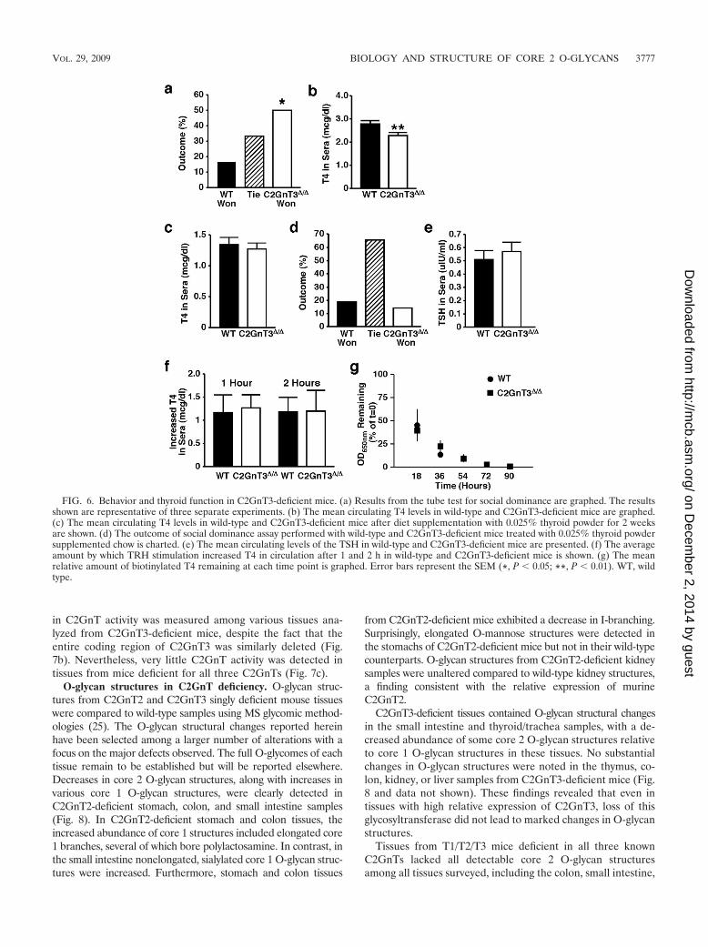
C2GnT3-deficient mice exhibit a behavioral abnormality linked toreduced thyroxine levels. Increased fighting was observed betweenmale mice in litters containing at least one C2GnT3-deficientanimal, but not among litters that included C2GnT2-deficientmice. The behavior of male mice lacking C2GnT3 was furtheranalyzed. C2GnT3-deficient male mice exhibited a significant in-crease in social dominance compared to wild-type male litter-mates (Fig. 6a). This assay is often used as a measure of aggres-sion (10). Hypothyroidism is one cause of aggression in mammals,including dogs and horses (4, 12, 15, 62). Further analyses re-
vealed that C2GnT3-deficient mice had a slight but significantdecrease in circulating thyroxine (T4) levels (Fig. 6b). To deter-mine whether the altered behavior observed in C2GnT3 defi-ciency was a result of insufficient T4 abundance, we fed C2GnT3-deficient mice and wild-type littermates chow supplemented with0.025% porcine thyroid powder (34). Wild-type and C2GnT3-deficient mice fed thyroid-powder supplemented chow achievedsimilar circulating T4 levels (Fig. 6c). When these mice wereretested in the social dominance assay, no difference betweenC2GnT3-deficient and wild-type mice was observed (Fig. 6d).This finding indicates that the altered behavior observed inC2GnT3-deficient mice is likely due to reduced T4 levels in cir-culation.
The thyroid is stimulated to release T4 in response to thesecretion of TSH from the pituitary. Thus, we investigated theabundance of TSH in circulation in C2GnT3-deficient mice.No difference was observed in circulating TSH abundance(Fig. 6e). Reduced levels of circulating T4, in the presence ofnormal levels of TSH, suggests secondary hypothyroidism be-cause thyroid hormone regulation involves a negative-feedbackloop in which T4 feeds back to the pituitary to reduce TSHsecretion (11, 16). Secondary hypothyroidism can be tested bystimulation with TRH, the hormone secreted from the hypo-thalamus that stimulates the pituitary to release TSH (41, 67).No differences were present in the levels of T4 secreted inresponse to TRH stimulation among wild-type and C2GnT3-deficient mice (Fig. 6f). This suggests that the slight reductionin T4 levels may not be sufficient to increase TSH levels via thenegative-feedback loop.
The mild hypothyroidism in C2GnT3-deficient mice may notbe a result of secondary hypothyroidism despite normal levelsof TSH. To determine whether T4 levels are reduced due todecreased half-life in circulation, we compared the in vivohalf-life of T4 in wild-type and C2GnT3-deficient mice. Nodifference in T4 half-life in wild-type and C2GnT3-deficientmice was detected (Fig. 6g).
Mice deficient for all three C2GnTs are viable. The extent towhich the three known glycosyltransferases with C2GnT activ-ity can biologically compensate for each other is unknown;thus, we chose to generate mice deficient for multiple C2GnTs.Since these three Gcnt genes reside on different chromosomes,we accomplished this by crossing the singly deficient states toeach other. All three possible C2GnT doubly deficient combi-nations (T1/T2, T1/T3, and T2/T3) were generated, and theseanimals were born without overt abnormalities and appearednormal. From these parental sources, offspring should theoret-ically include triple-null littermates (T1/T2/T3). Remarkably,mice deficient in all three C2GnTs were born viable and ap-peared to develop normally to adults. In addition, both maleand female T1/T2/T3 mice were fertile (data not shown).
C2GnT and C4GnT activity in C2GnT-deficient mice.C2GnT2 and C2GnT4 enzymatic activity levels were deter-mined among tissue lysates from animals bearing single andmultiple C2GnT deficiencies. C2GnT2-deficient mouse colonsand mesenteric lymph node samples contained significantlyreduced C2GnT activity compared to the level of activity inwild-type lysates (Fig. 7a). Furthermore, no significant C4GnTactivity was detected in any tissue tested from C2GnT2-defi-cient mice, implying that C4GnT activity is produced exclu-sively by C2GnT2 (50, 69). In contrast, no significant decrease
FIG. 5. Circulating and mucosal immunoglobulins in wild-type andC2GnT2-deficient mice. (a) The circulating levels of IgM, IgG1,IgG2a, IgG2b, IgG3, and IgA isotypes in wild-type and C2GnT2-deficient mice are graphed. The data shown are pooled from twoseparate experiments, each of at least five mice of each genotype peran experiment. (b) The mean mucosal IgA levels in wild-type andC2GnT2-deficient mice fecal samples are graphed. All values aremeans � the SEM (*, P � 0.05). WT, wild type.
3776 STONE ET AL. MOL. CELL. BIOL.
on Decem
ber 2, 2014 by guesthttp://m
cb.asm.org/
Dow
nloaded from
in C2GnT activity was measured among various tissues ana-lyzed from C2GnT3-deficient mice, despite the fact that theentire coding region of C2GnT3 was similarly deleted (Fig.7b). Nevertheless, very little C2GnT activity was detected intissues from mice deficient for all three C2GnTs (Fig. 7c).
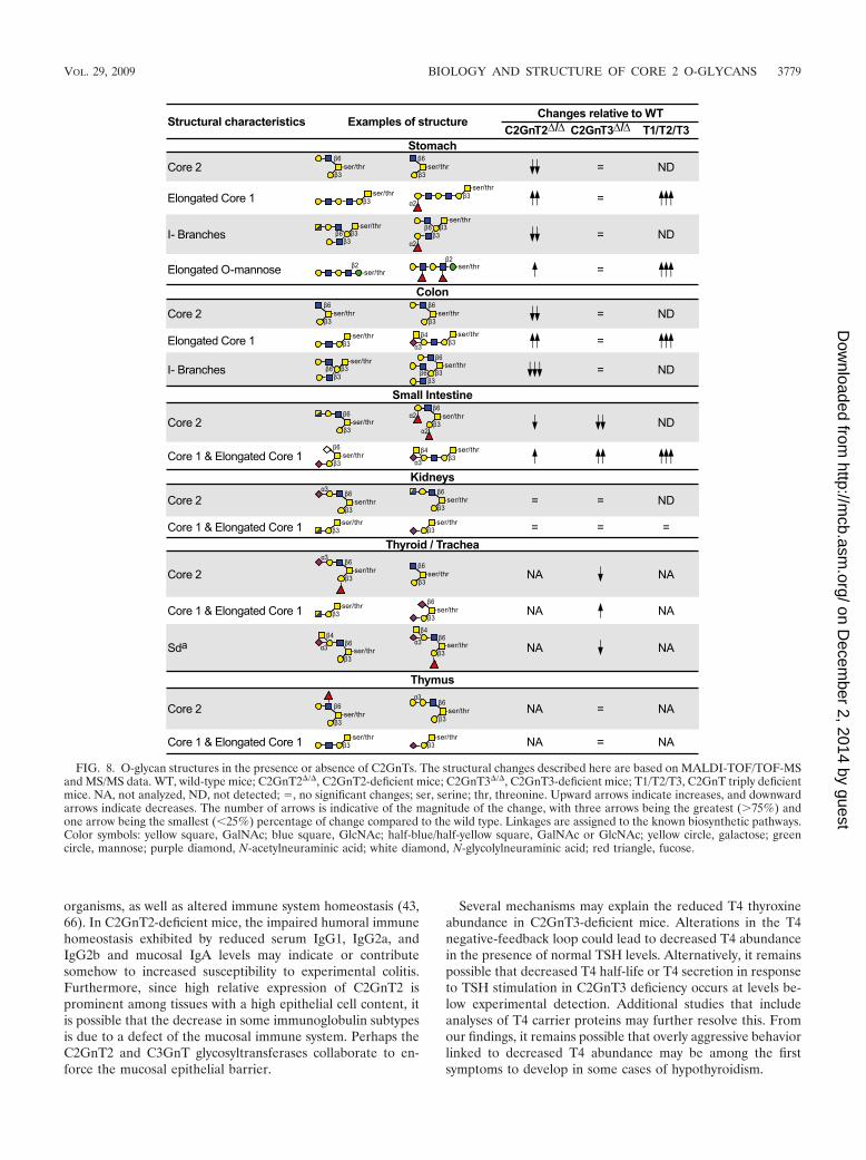
O-glycan structures in C2GnT deficiency. O-glycan struc-tures from C2GnT2 and C2GnT3 singly deficient mouse tissueswere compared to wild-type samples using MS glycomic method-ologies (25). The O-glycan structural changes reported hereinhave been selected among a larger number of alterations with afocus on the major defects observed. The full O-glycomes of eachtissue remain to be established but will be reported elsewhere.Decreases in core 2 O-glycan structures, along with increases invarious core 1 O-glycan structures, were clearly detected inC2GnT2-deficient stomach, colon, and small intestine samples(Fig. 8). In C2GnT2-deficient stomach and colon tissues, theincreased abundance of core 1 structures included elongated core1 branches, several of which bore polylactosamine. In contrast, inthe small intestine nonelongated, sialylated core 1 O-glycan struc-tures were increased. Furthermore, stomach and colon tissues
from C2GnT2-deficient mice exhibited a decrease in I-branching.Surprisingly, elongated O-mannose structures were detected inthe stomachs of C2GnT2-deficient mice but not in their wild-typecounterparts. O-glycan structures from C2GnT2-deficient kidneysamples were unaltered compared to wild-type kidney structures,a finding consistent with the relative expression of murineC2GnT2.
C2GnT3-deficient tissues contained O-glycan structural changesin the small intestine and thyroid/trachea samples, with a de-creased abundance of some core 2 O-glycan structures relativeto core 1 O-glycan structures in these tissues. No substantialchanges in O-glycan structures were noted in the thymus, co-lon, kidney, or liver samples from C2GnT3-deficient mice (Fig.8 and data not shown). These findings revealed that even intissues with high relative expression of C2GnT3, loss of thisglycosyltransferase did not lead to marked changes in O-glycanstructures.
Tissues from T1/T2/T3 mice deficient in all three knownC2GnTs lacked all detectable core 2 O-glycan structuresamong all tissues surveyed, including the colon, small intestine,
FIG. 6. Behavior and thyroid function in C2GnT3-deficient mice. (a) Results from the tube test for social dominance are graphed. The resultsshown are representative of three separate experiments. (b) The mean circulating T4 levels in wild-type and C2GnT3-deficient mice are graphed.(c) The mean circulating T4 levels in wild-type and C2GnT3-deficient mice after diet supplementation with 0.025% thyroid powder for 2 weeksare shown. (d) The outcome of social dominance assay performed with wild-type and C2GnT3-deficient mice treated with 0.025% thyroid powdersupplemented chow is charted. (e) The mean circulating levels of the TSH in wild-type and C2GnT3-deficient mice are presented. (f) The averageamount by which TRH stimulation increased T4 in circulation after 1 and 2 h in wild-type and C2GnT3-deficient mice is shown. (g) The meanrelative amount of biotinylated T4 remaining at each time point is graphed. Error bars represent the SEM (*, P � 0.05; **, P � 0.01). WT, wildtype.
VOL. 29, 2009 BIOLOGY AND STRUCTURE OF CORE 2 O-GLYCANS 3777
on Decem
ber 2, 2014 by guesthttp://m
cb.asm.org/
Dow
nloaded from

stomach, and kidney (Fig. 8). These results indicate that noother glycosyltransferases appear capable of synthesizing core2 O-glycans in vivo. Furthermore, small intestine, stomach, andcolon samples from T1/T2/T3 mice had a further increase inelongated Core 1 O-glycans compared to tissues lacking asingle C2GnT. Surprisingly, no O-glycan structures containingI-branches were detected in stomach and colon samples fromT1/T2/T3 mice. There was also an unexpected increase in elon-gated O-mannose structures in the stomach of T1/T2/T3 mice,even in comparison to C2GnT2-deficient stomach samples.
DISCUSSION
The presence of three conserved genes encoding glycosyl-transferases that can initiate the biosynthesis of Core 2 O-glycans suggests that each C2GnT may provide significantcompensation for one another as afforded by this close thoughnot complete overlap in enzymatic function. However, the dif-ferent expression profiles of distinct C2GnTs among tissuesand cell types further suggests that these closely related glyco-syltransferases may have unique biological roles. By generatingmice singly and multiply deficient in C2GnT activities, we havedeveloped mammalian animal models for studying the struc-ture and function of O glycosylation as controlled by C2GnTactivity. Core 2 and perhaps core 4 O-glycans produced byC2GnT2 operate in establishing the epithelial mucosal barrierand reducing disease pathology after chemical induction ofcolitis. Alterations in humoral and mucosal immune systemhomeostasis were also detected and may be related to thesefindings. In contrast, C2GnT3 does not provide a role in theformation or maintenance of the mucosal barrier but insteadalters behavioral phenotypes linked with T4 thyroxine abun-dance in circulation. Neither C2GnT2 nor C2GnT3 appear tofunction in the biosynthesis of selectin ligands, unlike findingsin mice lacking C2GnT1 (14). Each C2GnT glycosyltransferasetherefore has different biological roles among various physio-logical systems. Both expected and unexpected changes oc-curred in the repertoire of O-glycans detected among multipletissues from mice deficient in C2GnT activity. These structuralalterations of the glycome may aid in further resolving the
mechanistic features of O-glycan biosynthesis and the etiologyof these phenotypes.
Distinct and potential overlapping functions of C2GnTs.Neutrophilia in C2GnT3-deficient mice in the absence of ameasured defect in selectin ligand, as determined by cytom-etry, is reminiscent of mice deficient for (1,3)fucosyltrans-ferase-IV (FucT-IV). In contrast, no hematological or selectinligand biosynthetic defect was observed in C2GnT2 deficiency.Mice deficient for FucT-IV also exhibited a slight increase(20%) in neutrophils in circulation without measurable de-creases in binding of selectin chimeras to neutrophils (23).Nevertheless, FucT-IV was shown to contribute to selectinligand function in vivo, since FucT-IV deficiency increasedselectin-dependent leukocyte rolling velocity in microvessels(65). It remains possible that C2GnT3 contributes to selectinligand formation detectable by additional studies and ap-proaches. Alternatively, C2GnT3 may modulate neutrophilabundance in circulation by altering myelopoiesis. Several celladhesion molecules, including selectins and sialomucins, havebeen found to influence hematopoiesis, and altered glycosyla-tion has been shown to increase neutrophil production (23, 53,63). This may reflect altered binding between lectins and gly-coproteins in the context of stem cell turnover and differenti-ation. Nevertheless, the degree of increase observed amongcirculating neutrophils per se in C2GnT3-deficient mice is un-likely to significantly alter physiology.
Decreased mucosal barrier function is associated with in-flammatory bowel diseases (33, 55), including colitis. It is pos-sible that the increased mucosal permeability and disease signsthat occur with DSS treatment are related in C2GnT2-defi-cient mice. Moreover, the single C3GnT glycosyltransferasethat generates core 3 O-glycans is also essential for mucosalbarrier function and similarly decreases susceptibility to DSS-induced colitis (2). In C3GnT-deficient mice, these findingswere attributed to a reduced abundance of Muc2, a mucinknown to be necessary for protection from colitis (2, 60). Incontrast, C2GnT2-deficient mice did not appear to have de-creased expression of Muc2. Susceptibility to inflammatorybowel diseases is associated with factors other than alteredmucin levels, including changes in commensal and pathogenic
FIG. 7. Enzyme activity in tissue lysates from mice deficient for C2GnTs. (a) The relative C2GnT and C4GnT activities in tissue lysates fromC2GnT2-deficient mice are graphed. (b and c) The C2GnT activity in tissue lysates from C2GnT3-deficient (b) and triply deficient (c) mice relativeto the activity in wild-type control tissues is shown. Measurements shown represent means � the SEM (*, P � 0.05; ***, P � 0.001).
3778 STONE ET AL. MOL. CELL. BIOL.
on Decem
ber 2, 2014 by guesthttp://m
cb.asm.org/
Dow
nloaded from
organisms, as well as altered immune system homeostasis (43,66). In C2GnT2-deficient mice, the impaired humoral immunehomeostasis exhibited by reduced serum IgG1, IgG2a, andIgG2b and mucosal IgA levels may indicate or contributesomehow to increased susceptibility to experimental colitis.Furthermore, since high relative expression of C2GnT2 isprominent among tissues with a high epithelial cell content, itis possible that the decrease in some immunoglobulin subtypesis due to a defect of the mucosal immune system. Perhaps theC2GnT2 and C3GnT glycosyltransferases collaborate to en-force the mucosal epithelial barrier.
Several mechanisms may explain the reduced T4 thyroxineabundance in C2GnT3-deficient mice. Alterations in the T4negative-feedback loop could lead to decreased T4 abundancein the presence of normal TSH levels. Alternatively, it remainspossible that decreased T4 half-life or T4 secretion in responseto TSH stimulation in C2GnT3 deficiency occurs at levels be-low experimental detection. Additional studies that includeanalyses of T4 carrier proteins may further resolve this. Fromour findings, it remains possible that overly aggressive behaviorlinked to decreased T4 abundance may be among the firstsymptoms to develop in some cases of hypothyroidism.
FIG. 8. O-glycan structures in the presence or absence of C2GnTs. The structural changes described here are based on MALDI-TOF/TOF-MSand MS/MS data. WT, wild-type mice; C2GnT2�/�, C2GnT2-deficient mice; C2GnT3�/�, C2GnT3-deficient mice; T1/T2/T3, C2GnT triply deficientmice. NA, not analyzed, ND, not detected; , no significant changes; ser, serine; thr, threonine. Upward arrows indicate increases, and downwardarrows indicate decreases. The number of arrows is indicative of the magnitude of the change, with three arrows being the greatest (�75%) andone arrow being the smallest (�25%) percentage of change compared to the wild type. Linkages are assigned to the known biosynthetic pathways.Color symbols: yellow square, GalNAc; blue square, GlcNAc; half-blue/half-yellow square, GalNAc or GlcNAc; yellow circle, galactose; greencircle, mannose; purple diamond, N-acetylneuraminic acid; white diamond, N-glycolylneuraminic acid; red triangle, fucose.
VOL. 29, 2009 BIOLOGY AND STRUCTURE OF CORE 2 O-GLYCANS 3779
on Decem
ber 2, 2014 by guesthttp://m
cb.asm.org/
Dow
nloaded from

Although mice singly deficient for C2GnT1, C2GnT2, orC2GnT3 exhibited distinct phenotypes, the presence of novelphenotypes in mice deficient for multiple C2GnTs, but notpresent in any of the singly deficient models, would revealcollaboration and compensation among these glycosyltrans-ferases in physiologic processes. Preliminary data indicate thatT1/T2/T3 mice are unique among the other lesser deficiencystates with elevated levels of the liver enzyme alanine transam-inase, implying changes in liver function apparent only in theabsence of all three C2GnTs (unpublished observation). De-fining the biological mechanisms of the phenotypes evoked bythe loss of C2GnT activity will require additional resolutionthat is afforded by linking glycan structures to normal andpathological contexts.
Structural basis of O-glycan biosynthesis determined byC2GnT1, C2GnT2, and C2GnT3. The absence of any singleC2GnT glycosyltransferase results in a reduction of core 2O-glycan structures in one or more tissues in vivo. By compar-ison, the decrease in core 2 O-glycan structures in C2GnT3-deficient tissue samples was modest, and the C2GnT activitywas not measurably decreased in these tissues. This suggeststhat C2GnT3 may have a relatively minor contribution to core2 O-glycan biosynthesis in these tissue types in vivo. Core 2O-glycan structures were totally absent from tissues lacking allthree C2GnTs. Interestingly, tissue lysates from these triplydeficient animals harbored a small amount of C2GnT enzy-matic activity. This suggests the possibility that another un-identified glycosyltransferase may have a low level of C2GnTactivity when assayed in vitro. Nevertheless, the absence ofcore 2 O-glycan structures in T1/T2/T3 mice indicates that noadditional glycosyltransferases exist that can generate core 2O-glycans in vivo as detected by our analyses.
C2GnT deficiency further altered the repertoire and abun-dance of O-glycan structures not directly generated by C2GnTsand produced by different biosynthetic pathways. The largedecrease in the abundance of I-branching on O-glycan struc-tures from stomach and colon tissues of mice lacking C2GnT2suggests that in some tissues C2GnT2, and not IGnT, is thedominant I-branching glycosyltransferase in protein O glyco-sylation. The further loss of I-branching in stomach and colonsamples from mice lacking all three C2GnTs implies that eitherC2GnT1 or C2GnT3 may also have I-branching activity in vivo.In this regard, C2GnT3 has been characterized with a low levelof I-branching activity in vitro (51). The repertoire of core 1O-glycans and O-mannosylated glycans were also alteredamong various tissues. The increase of core 1 O-glycan struc-tures and their elongation in the absence of one or moreC2GnTs was linked with the formation of polylactosaminesthat may alter lectin-dependent binding and physiology. Thiselaboration of core 1 O-glycans is probably not due to reducedcompetition for acceptor substrates in the Golgi apparatus,since the formation of the core 2 O-glycan branch does notdecrease the efficiency of core 1 extension enzyme-�1,3-N-acetylglucosaminyltransferase (�3GnT3) (68). Alternate expla-nations include the possible elevation of �3GnT3 activity orperhaps an increase in the availability of the common UDP-GlcNAc donor substrate, which is also used in elaboratingglycans that arise from protein O mannosylation (70, 72). Core4 O-glycan structures, if present, exist below the level of de-tection in wild-type and C2GnT-deficient tissues; thus, it is not
currently known whether the loss of C4GnT activity inC2GnT2 deficiency correlates with a reduction or absence ofcore 4 O-glycans in vivo.
Core 2 O-glycans have been associated with interactionsamong commensal and pathogenic bacteria and their hosts.Loss of C2GnT2 may alter the potential or frequency of gas-trointestinal colonization by commensal or pathogenic bacte-ria. Recently, it has been shown that Helicobacter pylori andClostridium perfringens express proteins that bind to core 2O-glycans (5, 27), and preliminary data suggest that C2GnT3may be required for T-cell responses to an intestinal challenge(H. Ziltener, unpublished data). These findings may relate tothe phenotypes described here and suggest that challengingthese animals with additional stimuli may provide mechanisticinsights and perhaps further reveal novel biological roles forthe core 2 O-glycans. The relatively mild impact of core 2O-glycan deficiency in vivo may reflect compensation providedby the resulting induction of extended core 1 and O-mannosy-lated O-glycan structures. This has been observed in the for-mation of selectin ligands on extended Core 1 O-glycans inC2GnT1 deficiency (68). However, each of the three glycosyl-transferases that contribute to C2GnT or C4GnT activity has adistinct function, and this begins to explain the conservation ofthree separate genes encoding glycosyltransferases that initiatethe formation of core 2 O-glycans.
ACKNOWLEDGMENTS
We thank David Ditto for technical expertise with the hematologicalassays and Margo Streets and Trang Tran for technical expertise withthe behavior assays. Jeffery Long assisted with the statistical analysis ofthe behavioral data.
This study was funded in part by a Mizutani Foundation for Glyco-science award (J.D.M.), a Veterans Affairs Merit Review Grant(S.B.H.), the University of California San Diego Digestive DiseaseResearch Development Center (S.B.H), and National Institute ofHealth grants HL057345 (J.D.M.), HL78784 (J.D.M.), GMG2116(J.D.M.), CA33000 (M.F.), and P01CA71932 (M.F. and J.D.M.).J.D.M. is supported by the Howard Hughes Medical Institute. Thisstudy was also funded in part by Biotechnology and Biological SciencesResearch Council grant BBF0083091 (A.D.) and Public Health Servicegrant DK080506 (S.B.H.). M.N.I. is supported by University ScienceMalaysia, Ministry of Higher Education.
We declare that we have no competing financial interests.
REFERENCES
1. Altschuler, Y., C. L. Kinlough, P. A. Poland, J. B. Bruns, G. Apodaca, O. A.Weisz, and R. P. Hughey. 2000. Clathrin-mediated endocytosis of MUC1 ismodulated by its glycosylation state. Mol. Biol. Cell 11:819–831.
2. An, G., B. Wei, B. Xia, J. M. McDaniel, T. Ju, R. D. Cummings, J. Braun, andL. Xia. 2007. Increased susceptibility to colitis and colorectal tumors in micelacking core 3-derived O-glycans. J. Exp. Med. 204:1417–1429.
3. Arkan, M. C., A. L. Hevener, F. R. Greten, S. Maeda, Z. W. Li, J. M. Long,A. Wynshaw-Boris, G. Poli, J. Olefsky, and M. Karin. 2005. IKK0beta linksinflammation to obesity-induced insulin resistance. Nat. Med. 11:191–198.
4. Aronson, L. 1998. Animal behavior case of the month. Aggression directedtoward other horses. J. Am. Vet. Med. Assoc. 213:358–359.
5. Ashida, H., R. Maki, Y. H. Ozawa, Tani, M. Kiyohara, M. Fujita, A.Imamura, H. Ishida, M. Kiso, and K. Yamamoto. 2008. Characterization oftwo different endo--N-acetylgalactosaminidases from probiotic and patho-genic enterobacteria, Bifidobacterium longum and Clostridium perfringens.Glycobiology 18:727–734.
6. Baum, L. G. 2002. Developing a taste for sweets. Immunity 16:5–8.7. Bierhuizen, M. F., and M. Fukuda. 1992. Expression cloning of a cDNA en-
coding UDP-GlcNAc:Gal �1-3-GalNAc-R (GlcNAc to GalNAc) �1-6GlcNActransferase by gene transfer into CHO cells expressing polyoma large tumorantigen. Proc. Natl. Acad. Sci. USA 89:9326–9330.
8. Carlow, D. A., B. Ardman, and H. J. Ziltener. 1999. A novel CD8 T cell-restricted CD45RB epitope shared by CD43 is differentially affected byglycosylation. J. Immunol. 163:1441–1448.
3780 STONE ET AL. MOL. CELL. BIOL.
on Decem
ber 2, 2014 by guesthttp://m
cb.asm.org/
Dow
nloaded from

9. Cho, J. H. 2008. The genetics and immunopathogenesis of inflammatorybowel disease. Nat. Rev. Immunol. 8:458–466.
10. Crawley, J. N. 2007. What’s wrong with my mouse? Behavioral phenotypingof transgenic and knockout mice. Wiley Interscience, New York, NY.
11. Dahl, G. E., N. P. Evans, L. A. Thrun, and F. J. Karsch. 1994. A centralnegative feedback action of thyroid hormones on thyrotropin-releasing hor-mone secretion. Endocrinology 135:2392–2397.
12. Dodman, N. H., P. A. Mertens, and L. P. Aronson. 1995. Animal behaviorcase of the month: dogs were evaluated because of aggression. J. Am. Vet.Med. Assoc. 207:1168–1171.
13. deVos, T., and T. A. Dick. 1991. A rapid method to determine the isotype andspecificity of coproantibodies in mice infected with Trichinella or fed choleratoxin. J. Immunol. Methods 141:285–288.
14. Ellies, L. G., S. Tsuboi, B. Petryniak, J. B. Lowe, M. Fukuda, and J. D.Marth. 1998. Core 2 oligosaccharide biosynthesis distinguishes between se-lectin ligands essential for leukocyte homing and inflammation. Immunity9:881–890.
15. Fatjo, J., M. Amat, and X. Manteca. 2003. Animal behavior case of themonth: aggression in dogs. J. Am. Vet. Med. Assoc. 223:623–626.
16. Fliers, E., U. A. Unmehopa, and A. Alkemade. 2006. Functional neuroanat-omy of thyroid hormone feedback in the human hypothalamus and pituitarygland. Mol. Cell Endocrinol. 251:1–8.
17. Furuta, G. T., J. R. Turner, C. T. Taylor, R. M. Hershberg, K. Comerford, S.Narravula, D. K. Podolsky, and S. P. Colgan. 2001. Hypoxia-inducible factor1-dependent induction of intestinal trefoil factor protests barrier functionduring hypoxia. J. Exp. Med. 193:1027–1034.
18. Gauguet, J. M., S. D. Rosen, J. D. Marth, and U. H. von Andrian. 2004. Core2 branching �1,6-N-acetylglucosaminyltransferase and high endothelial cellN-acetylglucosamine-6-sulfotransferase exert differential control over B- andT-lymphocyte homing to peripheral lymph nodes. Blood 104:4104–4112.
19. Grewal, P. K., S. Uchiyama, D. Ditto, N. Varki, D. T. Le, V. Nizet, and J. D.Marth. 2008. The Ashwell receptor mitigates the lethal coagulopathy ofsepsis. Nat. Med. 14:648–655.
20. Hassan, H., C. A. Reis, E. P. Bennett, E. Mirgorodskaya, P. Roepstorff, M. A.Hollingsworth, J. Burchell, J. Taylor-Papadimitriou, and H. Clausen. 2000.The lectin domain of UDP-N-acetyl-D-galactosamine: polypeptide N-acetyl-galactosaminyltransferase-T4 directs its glycopeptide specificities. J. Biol.Chem. 275:38197–38205.
21. Hiraoka, N., H. Kawashima, B. Petryniak, J. Nakayama, J. Mitoma, J. D.Marth, J. B. Lowe, and M. Fukuda. 2004. Core 2 branching �1,6-N-acetyl-glucosaminyltransferase and high endothelial venule-restricted sulfotrans-ferase collaboratively control lymphocyte homing. J. Biol. Chem. 279:3058–3067.
22. Ho, S. B., L. A. Dvorak, R. E. Moor, A. C. Jacobson, M. R. Frey, J. Corredor,D. B. Polk, and L. L. Shekels. 2006. Cysteine-rich domains of Muc3 intestinalmucin promote cell migration, inhibit apoptosis, and accelerate wound heal-ing. Gastroenterology 131:1501–1517.
23. Homeister, J. W., A. D. Thall, B. Petryniak, P. Maly, C. E. Rogers, P. L.Smith, R. J. Kelly, K. M. Gersten, S. W. Askari, G. Cheng, G. Smithson,R. M. Marks, A. K. Misra, O. Hindsgaul, U. H. von Andrian, and J. B. Lowe.2001. The (1,3)fucosyltransferases FucT-IV and FucT-VII exert collabora-tive control over selectin-dependent leukocyte recruitment and lymphocytehoming. Immunity 15:115–126.
24. Iwai, T., N. Inaba, A. Naundorf, Y. Zhang, M. Gotoh, H. Iwasaki, T. Kudo,A. Togayachi, Y. Ishizuka, H. Nakanishi, and H. Narimatsu. 2002. Molecularcloning and characterization of a novel UDP-GlcNAc:GalNAc-peptide �1,3-N-acetylglucosaminyltransferase (�3Gn-T6), an enzyme synthesizing thecore 3 structure of O-glycans. J. Biol. Chem. 277:12802–12809.
25. Jang-Lee, J., S. J. North, M. Sutton-Smith, D. Goldberg, M. Panico, H.Morris, S. Haslam, and A. Dell. 2006. Glycomic profiling of cells and tissuesby mass spectrometry: fingerprinting and sequencing methodologies. Meth-ods Enzymol. 415:59–86.
26. Ju, T., K. Brewer, A. D’Souza, R. D. Cummings, and W. M. Canfield. 2002.Cloning and expression of human core 1 �1,3-galactosyltransferase. J. Biol.Chem. 277:178–186.
27. Kawakubo, Y. M., Ito, Y. Okimura, M. Kobayashi, K. Sakura, S. Kasama,M. N. Fukuda, M. Fukuda, T. Katsuyama, and J. Nakayama. 2004. Naturalantibiotic function of a human gastric mucin against Helicobacter pyloriinfection. Science 305:1003–1006.
28. Long, J. M., P. LaPorte, S. Merscher, B. Funke, B. Saint-Jore, A. Puech, R.Kucherlapati, B. E. Morrow, A. I. Skoultchi, and A. Wynshaw-Boris. 2006.Behavior of mice with mutations in the conserved region deleted in velocar-diofacial/DiGeorge syndrome. Neurogenetics 7:247–257.
29. Long, J. M., P. LaPorte, R. Paylor, and A. Wynshaw-Boris. 2004. Expandedcharacterization of the social interaction abnormalities in mice lacking Dvl1.Genes Brain Behav. 3:51–62.
30. Lowe, J. B. 2001. Glycosylation, immunity, and autoimmunity. Cell 104:809–812.
31. Lowe, J. B., and J. D. Marth. 2003. A genetic approach to mammalian glycanfunction. Annu. Rev. Biochem. 72:643–691.
32. Magnet, A. D., and M. Fukuda. 1997. Expression of the large I antigen
forming �-1,6-N-acetylglucosaminyltransferase in various tissues of adultmice. Glycobiology 7:285–295.
33. Mankertz, J., and J. D. Schulzke. 2007. Altered permeability in inflamma-tory bowel disease: pathophysiology and clinical implications. Curr. Opin.Gasteroenterol. 23:379–383.
34. Marians, R. C., L. Ng, H. C. Blair, P. Unger, P. N. Graves, and T. F. Davies.2002. Defining thyrotropin-dependent and -independent steps of thyroidhormone synthesis by using thyrotropin receptor-null mice. Proc. Natl. Acad.Sci. USA 99:15776–15781.
35. Marth., J. D. and P. K. Grewal. 2008. Mammalian glycosylation in immunity.Nat. Rev. Immunol. 8:874–887.
36. Merzaban, J. S., J. Zuccolo, S. Y. Corbel, M. J. Williams, and H. J. Ziltener.2005. An alternate Core 2�1,6-N-acetylglucosaminyltransferase selectivelycontributes to P-selectin ligand formation in activated CD8 T cells. J. Im-munol. 174:4051–4059.
37. Murthy, A. K., C. N. Dubose, J. A. Banas, J. J. Coalson, and B. P. Arulanan-dam. 2006. Contribution of polymeric immunoglobulin receptor to regula-tion of intestinal inflammation in dextran sulfate sodium-induced colitis.Gastroenterology 26:1372–1380.
38. Murthy, S. N., H. S. Cooper, H. Shim, R. S. Shah, S. A. Ibrahim, and D. J.Sedergran. 1993. Treatment of dextran sulfate sodium-induced murine co-litis by intracolonic cyclosporine. Dig. Dis. Sci. 38:1722–1734.
39. Nagy, A., J. Rossant, R. Nagy, W. Abramow-Newerly, and J. C. Roder. 1993.Derivation of completely cell culture-derived mice from early-passage em-bryonic stem cells. Proc. Natl. Acad. Sci. USA 90:8424–8428.
40. Okayasu, I., S. Hatakeyama, M. Yamada, T. Ohkusa, Y. Inagaki, and R.Nakaya. 1990. A novel method in the induction of reliable experimentalacute and chronic ulcerative colitis in mice. Gastroenterology 98:694–702.
41. Oliveira, K. J., T. M. Ortiga-Carvalho, A. Cabanelas, M. A. Veiga, K. Aoki,H. Ohki-Hamazaki, K. Wada, E. Wada, and C. C. Pazos-Moura. 2006.Disruption of neuromedin B receptor gene results in dysregulation of thepituitary-thyroid axis. J. Mol. Endocrinol. 36:73–80.
42. Perez-Vilar, J., and R. L. Hill. 1999. The structure and assembly of secretedmucins. J. Biol. Chem. 274:31751–31754.
43. Peterson, D. A., D. N. Frank, N. R. Pace, and J. I. Gordon. 2008. Meta-genomic approaches for defining the pathogenesis of inflammatory boweldisease. Cell Host Microbe 3:417–427.
44. Raman, J., T. A. Fritz, T. A. Gerken, O. Jamison, D. Live, M. Liu, and L. A.Tabak. 2008. The catalytic and lectin domains of UDP-GalNAc:polypeptide-N-acetylgalactosaminyltransferasae function in concert to direct glycosy-lation site selection. J. Biol. Chem. 283:22942–22951.
45. Rosen, S. D. 2004. Ligands for L-selectin: homing, inflammation, and be-yond. Annu. Rev. Immunol. 22:129–156.
46. Rossi, F. M. V., S. Y. Corbel, J. S. Merzaban, D. A. Carlow, K. Gossens, J.Duenas, L. So, L. Yi, and H. J. Ziltener. 2005. Recruitment of adult thymicprogenitors is regulated by P-selectin and its ligand PSGL-1. Nat. Immunol.6:626–634.
47. Satsangi, J., M. Parkes, E. Louis, L. Hashimoto, N. Kato, K. Welsh, J. D.Terwilliger, G. M. Lathrop, J. I. Bell, and D. P. Jewell. 1996. Two stagegenome-wide search in inflammatory bowel disease provides evidence forsusceptibility loci on chromosomes 3, 7, and 12. Nat. Genet. 14:199–202.
48. Schachter, H. 2000. The joys of HexNAc: the synthesis and function of N-and O-glycan branches. Glycoconj. J. 17:465–483.
49. Schachter, H., and I. Brockhausen. 1989. The biosynthesis of branchedO-glycans. Symp. Soc. Exp. Biol. 43:1–26.
50. Schwientek, T., M. Nomoto, S. B. Levery, G. Merkx, A. G. van Kessel, E. P.Bennett, M. A. Hollingsworth, and H. Clausen. 1999. Control of O-glycanbranch formation: molecular cloning of human cDNA encoding a novel�1,6-N-acetylglucosaminyltransferase forming core 2 and core 4. J. Biol.Chem. 274:4504–4512.
51. Schwientek, T., J. C. Yeh, S. B. Levery, B. Keck, G. Merkx, A. G. van Kessel,M. Fukuda, and H. Clausen. 2000. Control of O-glycan branch formation:molecular cloning and characterization of a novel thymus-associated Core 2�1,6-N-acteylglucosaminyltransferase. J. Biol. Chem. 275:11106–11113.
52. Shafi, R., S. P. Iyer, L. G. Ellies, N. O’Donnell, K. W. Marek, D. Chui, G. W.Hart, and J. D. Marth. 2000. The O-GlcNAc transferase gene resides on theX chromosome and is essential for embryonic stem cell viability and mouseontogeny. Proc. Natl. Acad. Sci. USA 97:5735–5739.
53. Simmons, P. J., J. Levesque, and D. N. Haylock. 2001. Mucin-like moleculesas modulators of the survival and proliferation of primitive hematopoieticcells. Ann. N. Y. Acad. Sci. 938:196–207.
54. Stanley, P. 2007. Regulation of Notch signaling by glycosylation. Curr. Opin.Struct. Biol. 17:530–535.
55. Stein, J., J. Ries, and K. B. Barrett. 1998. Disruption of intestinal barrierfunction associated with experimental colitis: possible role of mast cells.Am. J. Physiol. 274:G203–G209.
56. Sutton-Smith, M., H. R. Morris, and A. Dell. 2000. A rapid mass spectro-metric strategy suitable for the investigation of glycan alterations in knockoutmice. Tetrahedron Asymmetry 11:363–369.
57. Tarp, M. A., and H. Clausen. 2008. Mucin-type O glycosylation and itspotential use in drug and vaccine development. Biochim. Biophys. Acta1780:546–563.
VOL. 29, 2009 BIOLOGY AND STRUCTURE OF CORE 2 O-GLYCANS 3781
on Decem
ber 2, 2014 by guesthttp://m
cb.asm.org/
Dow
nloaded from

58. Ten Hagen, K. G., T. A. Fritz, and L. A. Tabak. 2003. All in the family: theUDP-GalNAc:polypeptide N-acetylgalactosaminyltransferases. Glycobiol-ogy 13:1R–16R.
59. Tenno, M., K. Ohtsubo, F. K. Hagen, D. Ditto, A. Zarbock, P. Schaerli, U. H.von Andrian, K. Ley, D. Le, L. A. Tabak, and J. D. Marth. 2007. Initiation ofprotein O glycosylation by polypeptide GalNAcT-1 in vascular biology andhumoral immunity. Mol. Cell. Biol. 27:8783–8796.
60. van der Sluis, M., B. A. E. de Koning, A. C. J. M. de Bruijn, A. Velcich,J. P. P. Meijerink, J. B. van Goudoever, H. A. Buller, and A. W. C. Einer-hand. 2006. Muc2-deficient mice spontaneously develop colitis, indicatingthat Muc2 is critical for colonic protection. Gastroenterology 131:117–129.
61. Varki, A. 2006. Nothing in glycobiology makes sense, except in the light ofevolution. Cell 126:841–845.
62. Venero, C., A. Guadano-Ferraz, A. I. Herrero, K. Nordstrom, J. Manzano,G. M. de Escobar, J. Bernal, and B. Vennstrom. 2005. Anxiety, memoryimpairment, and locomotor dysfunction caused by a mutant thyroid hormonereceptor 1 can be ameliorated by T3 treatment. Genes Dev. 19:2152–2163.
63. Verfaillie, C. M. 1998. Adhesion receptors as regulators of the hematopoieticprocess. Blood 92:2609–2612.
64. Wang, H., R. Tang, W. Zhang, K. Amirikian, Z. Geng, J. Geng, R. P. Hebbel,L. Xia, J. D. Marth, M. Fukuda, S. Katoh, and Y. Huo. 2009 Core 21-6-N-glucosaminyltransferase-I is crucial for the formation of atheroscle-rotic lesions in apolipoprotein E-deficient mice. Arterioscler. Thromb. Vasc.Biol. 29:180–187.
65. Weninger, W., L. H. Ulfman, G. Cheng, N. Souchkova, E. J. Quackenbush,J. B. Lowe, and U. H. von Andrian. 2000. Specialized contributions by
(1,3)-fucosyltransferase-IV and FucT-VII during leukocyte rolling in der-mal microvessels. Immunity 12:665–676.
66. Xavier, R. J., and D. K. Podolsky. 2007. Unraveling the pathogenesis ofinflammatory bowel disease. Nature 448:427–434.
67. Yamada, M., Y. Saga, N. Shibusawa, J. Hirato, M. Murakami, T. Iwasaki, K.Hashimoto, T. Satoh, K. Wakabayashi, M. M. Taketo, and M. Mori. 1997.Tertiary hypothyroidism and hyperglycemia in mice with targeted disruptionof the thyrotropin-releasing hormone gene. Proc. Natl. Acad. Sci. USA94:10862–10867.
68. Yeh, J. C., N. Hiraoka, B. Petryniak, J. Nakayama, L. G. Ellies, D. Rabuka,O. Hindsgaul, J. D. Marth, J. B. Lowe, and M. Fukuda. 2001. Novel sulfatedlymphocyte homing receptors and their control by a Core1 extension �1,3,-N-acetylglucosaminyltransferase. Cell 105:957–969.
69. Yeh, J. C., E. Ong, and M. Fukuda. 1999. Molecular cloning and expressionof a novel �-1,6-N-acetylglucosaminyltransferase that forms Core 2, Core 4,and I branches. J. Biol. Chem. 274:3215–3221.
70. Yoshida, A., K. Kobayashi, H. Manya, K. Taniguchi, H. Kano, M. Mizuno,T. Inazu, H. Mitsuhashi, S. Takahashi, M. Takeuchi, R. Herrmann, V.Straub, B. Talim, T. Voit, H. Topaloglu, T. Toda, and T. Endo. 2001. Mus-cular dystrophy and neuronal migration disorder caused by mutations in aglycosyltransferase, POMGnT1. Dev. Cell 1:717–724.
71. Zachara, N. E., and G. W. Hart. 2006. Cell signaling, the essential role ofO-GlcNAc. Biochim. Biophys. Acta 1761:599–617.
72. Zhang, W., D. Betel, and H. Schachter. 2002. Cloning and expression of anovel UDP-GlcNAc:-3-D-mannoside �1,2-N-acetylglucosaminyltransferaseI. Biochem. J. 361:153–162.
3782 STONE ET AL. MOL. CELL. BIOL.
on Decem
ber 2, 2014 by guesthttp://m
cb.asm.org/
Dow
nloaded from