Estimation of protein function using template-based alignment of enzyme active sites
11
Estimation of protein function using template-based alignment of enzyme active sites Hanson et al. Hanson et al. BMC Bioinformatics 2014, 15:87 http://www.biomedcentral.com/1471-2105/15/87
Transcript of Estimation of protein function using template-based alignment of enzyme active sites

Estimation of protein function using template-basedalignment of enzyme active sitesHanson et al.
Hanson et al. BMC Bioinformatics 2014, 15:87http://www.biomedcentral.com/1471-2105/15/87
Hanson et al. BMC Bioinformatics 2014, 15:87http://www.biomedcentral.com/1471-2105/15/87
SOFTWARE Open Access
Estimation of protein function using template-basedalignment of enzyme active sitesBrett Hanson1, Charles Westin2, Mario Rosa3, Alexander Grier3, Mikhail Osipovitch3, Madolyn L MacDonald3,Greg Dodge3, Paule M Boli3, Cyprian W Corwin3, Haeja Kessler3, Talia McKay3, Herbert J Bernstein4
and Paul A Craig5*
Abstract
Background: The accumulation of protein structural data occurs more rapidly than it can be characterized bytraditional laboratory means. This has motivated widespread efforts to predict enzyme function computationally.The most useful/accurate strategies employed to date are based on the detection of motifs in novel structures thatcorrespond to a specific function. Functional residues are critical components of predictively useful motifs. We haveimplemented a novel method, to complement current approaches, which detects motifs solely on the basis ofdistance restraints between catalytic residues.
Results: ProMOL is a plugin for the PyMOL molecular graphics environment that can be used to create active site motifsfor enzymes. A library of 181 active site motifs has been created with ProMOL, based on definitions published inthe Catalytic Site Atlas (CSA). Searches with ProMOL produce better than 50% useful Enzyme Commission (EC)class suggestions for level 1 searches in EC classes 1, 4 and 5, and produce some useful results for other classes. 261additional motifs automatically translated from Jonathan Barker’s JESS motif set [Bioinformatics 19:1644–1649, 2003] anda set of NMR motifs is under development. Alignments are evaluated by visual superposition, Levenshtein distance androot-mean-square deviation (RMSD) and are reasonably consistent with related search methods.
Conclusion: The ProMOL plugin for PyMOL provides ready access to template-based local alignments. Recentimprovements to ProMOL, including the expanded motif library, RMSD calculations and output selection formatting,have greatly increased the program’s usability and speed, and have improved the way that the results are presented.
Keywords: Motif, Enzyme, Catalytic site, Structural homology, Function prediction, Protein data bank
BackgroundStructural motifs corresponding to enzyme active sites areoften highly conserved in functionally related proteins as aresult of common ancestry or convergent evolutionary pro-cesses [1]. These features can be the basis for inferringfunction, and computational structural analysis using thisapproach is now an active area of research [2]. Efforts tothis end are motivated by a disparity between the capacityof structural genomics initiatives to generate structuresof macromolecules and the ability to characterize theseproteins using traditional laboratory methods [3]. Thepresent research is concerned with predicting the
* Correspondence: [email protected] Institute of Technology, School of Chemistry & MaterialsScience, 1 Lomb Memorial Drive, Rochester, NY 14623, USAFull list of author information is available at the end of the article
© 2014 Hanson et al.; licensee BioMed CentraCommons Attribution License (http://creativecreproduction in any medium, provided the or
function of proteins with known structure and un-known function and with contributing to a better un-derstanding of the structural basis of enzyme activity,primarily by in silico methods [4,5]. Numerous algorithmsand software applications have been developed for thesepurposes [6]. There are three major similarity-basedapproaches to in silico function assignment: sequencealignment, backbone alignment and template align-ment. Gapped BLAST and Position-Specific IteratedBLAST (PSI-BLAST) [7] and Pattern-Hit InitiatedBLAST (PHI-BLAST) [8] are examples of current prac-tice in sequence alignment. Backbone alignment andtemplate alignment are structurally based and allow forthe possibility that the same function may be achievedwith very different sequences. See [6] for the currentstate of protein structural alignment including DALI/
l Ltd. This is an Open Access article distributed under the terms of the Creativeommons.org/licenses/by/2.0), which permits unrestricted use, distribution, andiginal work is properly credited.

Hanson et al. BMC Bioinformatics 2014, 15:87 Page 2 of 10http://www.biomedcentral.com/1471-2105/15/87
FSSP [9] and ModBase [10]. More local structuralalignment has been done with JESS templates used bythe Catalytic Site Atlas (CSA) (http://www.ebi.ac.uk/thornton-srv/databases/CSA/) [1]. The failure of com-parison of sequences is not sufficient to preclude simi-larity of active sites [11], and in some cases, functionis determined locally, rather than globally. However,strong similarity of the local three-dimensional struc-ture of active sites may not be sufficient to determineactivity in situ or even in vitro, due, for example, tosteric constraints. See, for example, [12]. Thus there isvalue in use of active-site structural templates as acomplementary approach to sequence-based methodsand more global methods, but none of the similarity-based methods is sufficient to provide a rigorous deter-mination of activity.ProMOL is a molecular visualization and analysis tool
that uses a template-based approach to alignment. Theapproach followed in ProMOL is similar to that used inJESS or in FLORA [13] in that it ignores the physico-chemical properties of residues and instead assessescatalytic site structural conservation. It has been avail-able online since 2006 [14] and has been developed as aplugin for the widely used PyMOL molecular graphicsenvironment [15]. ProMOL is being developed collab-oratively and distributed freely as open source software(http://sourceforge.net/projects/sbevsl/).ProMOL uses a library of motif templates derived
from active site definitions in the CSA [16] and ana-lyzes spatial relationships and residue identities withina given structure to determine the presence of struc-tural features known to be associated with specificcatalytic functions. Features of ProMOL include theabilities
� To make and store motif templates in a growinglibrary of hundreds of existing motif templates,
� To request alignment with subsets of the motifs bytemplate source,
� To view the optimal alignment between a motiftemplate and a query structure, and
� To characterize a structure from the PDB or astructure provided by the user.
ProMOL, in combination with PyMOL, is a standaloneapplication that functions best with an Internet connec-tion (for access to the PDB). Its design as an open source(http://sourceforge.net/projects/sbevsl/files/ProMOL/)plugin to PyMOL is conducive to customization and fur-ther development by independent users.
ImplementationProMOL was developed for the PyMOL molecular graphicsenvironment in Windows, Mac OS X (including Lion), and
Linux. The program is written in Python, and requiresPython 2.6, or 2.7. When creating a template for the library,ProMOL uses PyMOL’s API commands to generate a motiftemplate of the active site for an enzyme for which thefunction is known. When testing a template or usingthe motif library, ProMOL can search for the motif in aquery structure, using the distance selection commandsin PyMOL and a set of constraints built-in to ProMOL'sMotif Finder module. Additionally, ProMOL contains amodule called EZ-Viz that allows the user to interactwith the GUI, rather than having to work directly withPyMOL's command line [17].ProMOL can contain multiple sets of motif templates.
The ProMOL release contains a set of motif templates(the P or ProMOL set) from the developers. Once Pro-MOL has been installed, users can generate their ownmotif templates (the U or user set). Additional sets areunder development.The P set of motifs was created within ProMOL using
catalytic site entries published by the CSA. The PDB ID,EC number, residue name (must be an amino acid), resi-due number, and chain for each of the catalytic residueswere entered in the Motif Maker tab. The process ofmotif creation is described below. Once the motif wascreated in ProMOL, it was tested against the templatestructure to ensure that it worked properly. If there wasan exact residue-by-residue match between the motifand the template structure, the motif was saved andtested against known homologs (to search for true positiveand false negatives) and random structures (to search fortrue negatives and false positives). Please note that thesenumbers provide a limited window on the reliability ofstructural alignments. In using ProMOL/PyMOL it is alsopossible to view the quality of the alignment directly, pro-viding a much more reliable method of validation. Themotifs were then saved to the P motif set within Pro-MOL's file structure.The U set of motifs can be created by any ProMOL user.
These motifs are stored in a separate directory on the user’scomputer. The location depends on the operating system.See the ProMOL User Guide (http://www.promol.org/home/download/download-now) for specific details.Initially, matches were ranked based on their Levensh-
tein distance - a measure of the difference between twosequences [18]. If the two sequences are identical, theLevenshtein distance between them is zero. If there isone difference between the sequences, the Levenshteindistance is one and so on. For example, if a motif con-tains histidine, serine and glutamate, and the matchingregion of the query protein contains histidine, cysteineand glutamate, the Levenshtein distance is one.The Levenshtein distance was incorporated in ProMOL
to provide a first level of screening and comparisonbetween the query structures and the motif templates.
Hanson et al. BMC Bioinformatics 2014, 15:87 Page 3 of 10http://www.biomedcentral.com/1471-2105/15/87
However, the Levenshtein distance is a coarse measure ofstructural similarity. To allow more exact quantitative com-parison of structural alignments, RMSD calculations wereadded to ProMOL. RMSD measures the L2 norm three-dimensional distance between the atoms in a match; thelower the RMSD between a query and a motif, the betterthe match. ProMOL can compute RMSD considering alphacarbons only, alpha and beta carbons, or all atoms within amatch.ProMOL works in Windows (including 8), Mac OS X
(including Lion) and Linux. The installation differs by oper-ating system; details can be found in the ProMOL UserGuide (http://www.promol.org/home/download/download-now). The current release of ProMOL should only be usedwith PyMOL release 1.3 or higher on all three operatingsystems.At present, ProMOL works well with PyMOL in sys-
tems that have Python versions 2.5, 2.6 or 2.7 installed.This combination is not functional in systems using Py-thon 3.0 or higher, because PyMOL is normally releasedagainst lower versions of Python.ProMOL can be accessed from the plugin dropdown
menu in PyMOL once it is installed properly. The usermust download one of the three compressed file formatsavailable: tarball, zip and exe (Windows installer). Ineach case the contents need to be expanded in thestartup folder for PyMOL. The location of the startupfolder varies by operating system and PyMOL installa-tion. Examples of the location of the startup folder andthe latest details can be found on the web site (www.pro-mol.org) and in the ProMOL User Guide.The motif library for ProMOL is located in the /pmg_tk/
startup/ProMOL/Motifs folder. As mentioned earlier, thisfolder contains the P motif templates (generated byProMOL). The N set of motif templates (based onNMR structures) and the J set of motif templates (gen-erated by algorithmic conversion of the CSA Jess mo-tifs) is under construction - check the web site foravailability. Each motif is a segment of Python codewhich should not be modified. A motif consists of aseries of calls to PyMOL to select residues by type andrelative distance. For example, for a serine protease,ProMOL searches for the serine-histidine-aspartatecatalytic triad. To start, it sends a call for the specifiedatoms in the sidechains of all the serines in a protein;as an option, backbone atoms can be added to theatom lists for the desired residues. The next segmentof code sends a call for all the histidine atoms that arewithin a specified distance of a serine residue. The onlyselections that proceed are those that contain bothserine and histidine within a specified distance of eachother. At this point, ProMOL sends a call to PyMOL tofind aspartate atoms that are within a specified distance ofserine atoms and histidine atoms. At the conclusion of
this search, ProMOL reports out the atoms that pass theselection process.The organization of the ProMOL code is shown in
Figure 1. The ProMol.py file controls the execution ofthe different functions that are located in the ProMOLtabs and buttons. The functions assigned to each tab aredescribed in detail in the ProMOL User Guide. The Pro-MOL code was created by the authors, with the notableexception of the treewidgets [19], which is required forthe RMSD calculation display. Treewidgets is a libraryfor Python and Tkinter for the display of trees contain-ing information based on a wide variety of data struc-tures. ProMOL also accesses the PDB Loader applicationthat ships with PyMOL to load PDB structures over theInternet [20].The GUI for ProMOL (Figure 2) includes four buttons
at the base of the window (Open, Fetch PDB, RandomPDB, and Clear) and five tabs (Welcome, EZ-Viz, MotifFinder, Motif Maker and View Options). These featuresare explained in much greater depth in the ProMOLUser Guide.
Motif creationA library of active site motif templates has been createdin ProMOL based on structures that were determinedby x-ray diffraction. A motif is a selected set of residuesand atoms identifying an active site. In general the rele-vant active sites have been taken from the Catalytic SiteAtlas. The orientation-independent geometry of such asite provides a means of identification of the presence ofsuch a site in another molecule. The motif creationmechanism within ProMOL is based on relative dis-tances among all the active site residues from the tem-plate molecules. A full description of motif creation willbe provided in a subsequent paper (Osipovitch, in prep-aration). The templates for the motifs were selectedfrom structures in the Catalytic Site Atlas that were usedto create the original JESS motif set [1], plus an expan-sion to include more complete coverage of the EC clas-ses. The motifs are lists of residue names, atom namesand their relative distances that must be matched in analignment. A tolerance value is included as an additionto the distances to broaden the range of acceptablematches. The default tolerance value for motif creationis 2.00 Angstroms and it can be adjusted as a motif istested. In addition to the tolerance that becomes part ofthe actual code of the motif, there is a precision factorthat can be used when searching for motifs with theMotif Finder. The precision factor is a multiplier of thedistances in the motif, providing an additional way torelax or tighten the constraints for a match. The use ofthe Motif Finder is described in detail in the ProMOLUser Guide, which can be obtained at http://www.pro-mol.org/home/download/download-now.
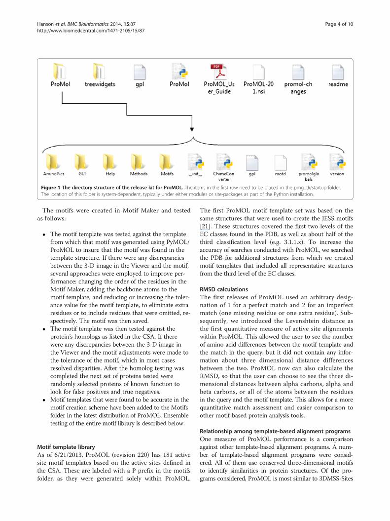
Figure 1 The directory structure of the release kit for ProMOL. The items in the first row need to be placed in the pmg_tk/startup folder.The location of this folder is system-dependent, typically under either modules or site-packages as part of the Python installation.
Hanson et al. BMC Bioinformatics 2014, 15:87 Page 4 of 10http://www.biomedcentral.com/1471-2105/15/87
The motifs were created in Motif Maker and testedas follows:
� The motif template was tested against the templatefrom which that motif was generated using PyMOL/ProMOL to insure that the motif was found in thetemplate structure. If there were any discrepanciesbetween the 3-D image in the Viewer and the motif,several approaches were employed to improve per-formance: changing the order of the residues in theMotif Maker, adding the backbone atoms to themotif template, and reducing or increasing the toler-ance value for the motif template, to eliminate extraresidues or to include residues that were omitted, re-spectively. The motif was then saved.
� The motif template was then tested against theprotein’s homologs as listed in the CSA. If therewere any discrepancies between the 3-D image inthe Viewer and the motif adjustments were made tothe tolerance of the motif, which in most casesresolved disparities. After the homolog testing wascompleted the next set of proteins tested wererandomly selected proteins of known function tolook for false positives and true negatives.
� Motif templates that were found to be accurate in themotif creation scheme have been added to the Motifsfolder in the latest distribution of ProMOL. Ensembletesting of the entire motif library is described below.
Motif template libraryAs of 6/21/2013, ProMOL (revision 220) has 181 activesite motif templates based on the active sites defined inthe CSA. These are labeled with a P prefix in the motifsfolder, as they were generated solely within ProMOL.
The first ProMOL motif template set was based on thesame structures that were used to create the JESS motifs[21]. These structures covered the first two levels of theEC classes found in the PDB, as well as about half of thethird classification level (e.g. 3.1.1.x). To increase theaccuracy of searches conducted with ProMOL, we searchedthe PDB for additional structures from which we createdmotif templates that included all representative structuresfrom the third level of the EC classes.
RMSD calculationsThe first releases of ProMOL used an arbitrary desig-nation of 1 for a perfect match and 2 for an imperfectmatch (one missing residue or one extra residue). Sub-sequently, we introduced the Levenshtein distance asthe first quantitative measure of active site alignmentswithin ProMOL. This allowed the user to see the numberof amino acid differences between the motif template andthe match in the query, but it did not contain any infor-mation about three dimensional distance differencesbetween the two. ProMOL now can also calculate theRMSD, so that the user can choose to see the three di-mensional distances between alpha carbons, alpha andbeta carbons, or all of the atoms between the residuesin the query and the motif template. This allows for a morequantitative match assessment and easier comparison toother motif-based protein analysis tools.
Relationship among template-based alignment programsOne measure of ProMOL performance is a comparisonagainst other template-based alignment programs. A num-ber of template-based alignment programs were consid-ered. All of them use conserved three-dimensional motifsto identify similarities in protein structures. Of the pro-grams considered, ProMOL is most similar to 3DMSS-Sites
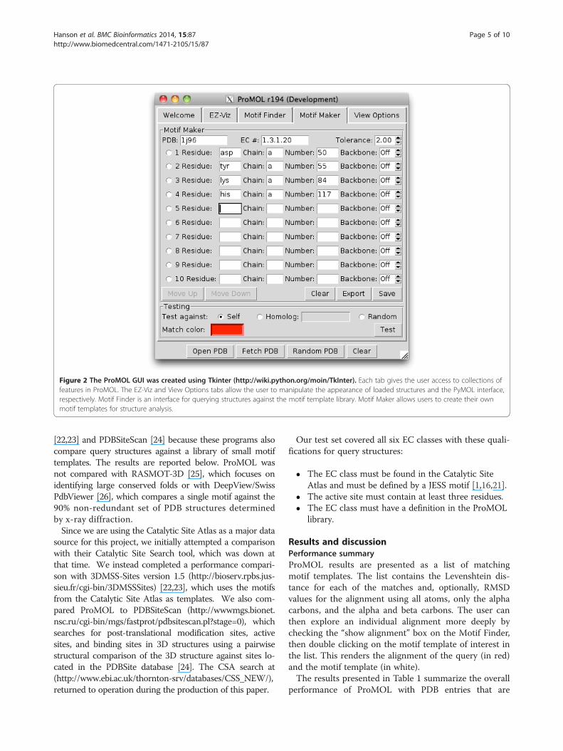
Figure 2 The ProMOL GUI was created using Tkinter (http://wiki.python.org/moin/TkInter). Each tab gives the user access to collections offeatures in ProMOL. The EZ-Viz and View Options tabs allow the user to manipulate the appearance of loaded structures and the PyMOL interface,respectively. Motif Finder is an interface for querying structures against the motif template library. Motif Maker allows users to create their ownmotif templates for structure analysis.
Hanson et al. BMC Bioinformatics 2014, 15:87 Page 5 of 10http://www.biomedcentral.com/1471-2105/15/87
[22,23] and PDBSiteScan [24] because these programs alsocompare query structures against a library of small motiftemplates. The results are reported below. ProMOL wasnot compared with RASMOT-3D [25], which focuses onidentifying large conserved folds or with DeepView/SwissPdbViewer [26], which compares a single motif against the90% non-redundant set of PDB structures determinedby x-ray diffraction.Since we are using the Catalytic Site Atlas as a major data
source for this project, we initially attempted a comparisonwith their Catalytic Site Search tool, which was down atthat time. We instead completed a performance compari-son with 3DMSS-Sites version 1.5 (http://bioserv.rpbs.jus-sieu.fr/cgi-bin/3DMSSSites) [22,23], which uses the motifsfrom the Catalytic Site Atlas as templates. We also com-pared ProMOL to PDBSiteScan (http://wwwmgs.bionet.nsc.ru/cgi-bin/mgs/fastprot/pdbsitescan.pl?stage=0), whichsearches for post-translational modification sites, activesites, and binding sites in 3D structures using a pairwisestructural comparison of the 3D structure against sites lo-cated in the PDBSite database [24]. The CSA search at(http://www.ebi.ac.uk/thornton-srv/databases/CSS_NEW/),returned to operation during the production of this paper.
Our test set covered all six EC classes with these quali-fications for query structures:
� The EC class must be found in the Catalytic SiteAtlas and must be defined by a JESS motif [1,16,21].
� The active site must contain at least three residues.� The EC class must have a definition in the ProMOL
library.
Results and discussionPerformance summaryProMOL results are presented as a list of matchingmotif templates. The list contains the Levenshtein dis-tance for each of the matches and, optionally, RMSDvalues for the alignment using all atoms, only the alphacarbons, and the alpha and beta carbons. The user canthen explore an individual alignment more deeply bychecking the “show alignment” box on the Motif Finder,then double clicking on the motif template of interest inthe list. This renders the alignment of the query (in red)and the motif template (in white).The results presented in Table 1 summarize the overall
performance of ProMOL with PDB entries that are
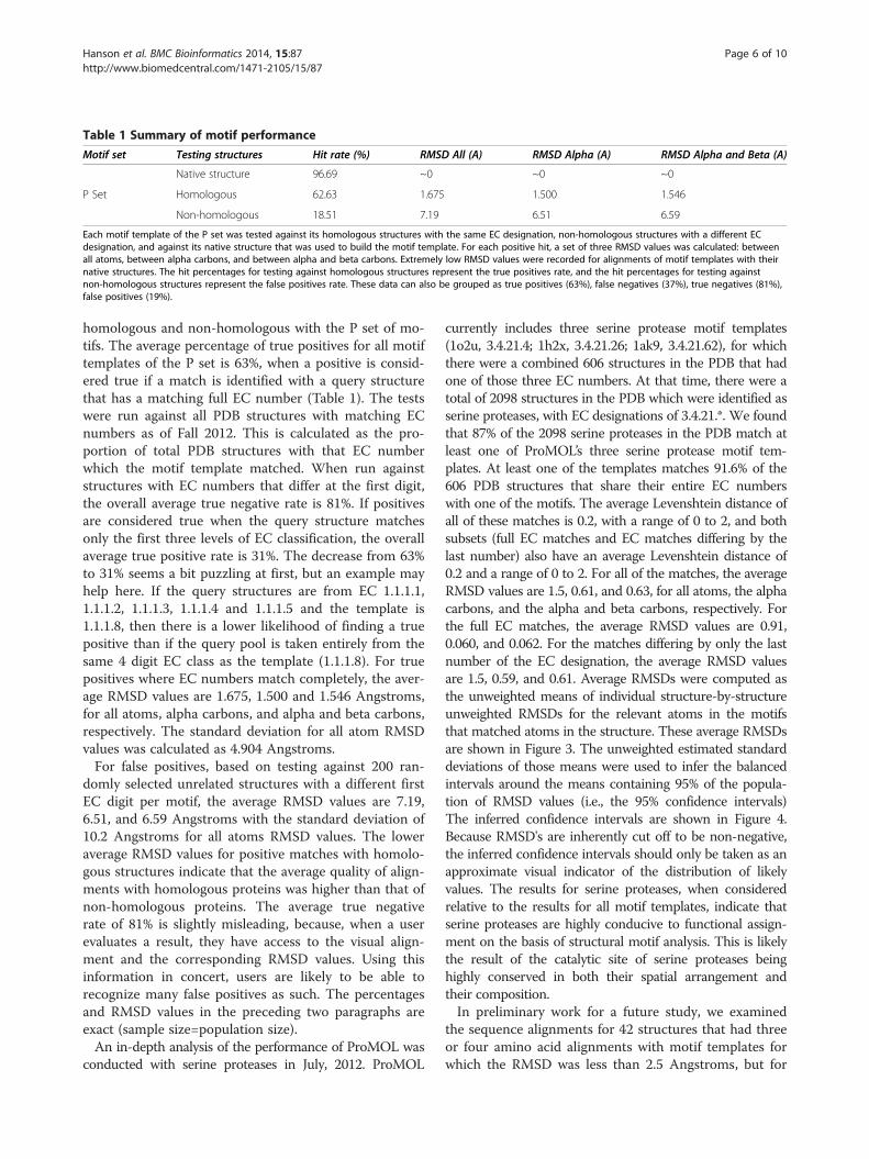
Table 1 Summary of motif performance
Motif set Testing structures Hit rate (%) RMSD All (A) RMSD Alpha (A) RMSD Alpha and Beta (A)
Native structure 96.69 ~0 ~0 ~0
P Set Homologous 62.63 1.675 1.500 1.546
Non-homologous 18.51 7.19 6.51 6.59
Each motif template of the P set was tested against its homologous structures with the same EC designation, non-homologous structures with a different ECdesignation, and against its native structure that was used to build the motif template. For each positive hit, a set of three RMSD values was calculated: betweenall atoms, between alpha carbons, and between alpha and beta carbons. Extremely low RMSD values were recorded for alignments of motif templates with theirnative structures. The hit percentages for testing against homologous structures represent the true positives rate, and the hit percentages for testing againstnon-homologous structures represent the false positives rate. These data can also be grouped as true positives (63%), false negatives (37%), true negatives (81%),false positives (19%).
Hanson et al. BMC Bioinformatics 2014, 15:87 Page 6 of 10http://www.biomedcentral.com/1471-2105/15/87
homologous and non-homologous with the P set of mo-tifs. The average percentage of true positives for all motiftemplates of the P set is 63%, when a positive is consid-ered true if a match is identified with a query structurethat has a matching full EC number (Table 1). The testswere run against all PDB structures with matching ECnumbers as of Fall 2012. This is calculated as the pro-portion of total PDB structures with that EC numberwhich the motif template matched. When run againststructures with EC numbers that differ at the first digit,the overall average true negative rate is 81%. If positivesare considered true when the query structure matchesonly the first three levels of EC classification, the overallaverage true positive rate is 31%. The decrease from 63%to 31% seems a bit puzzling at first, but an example mayhelp here. If the query structures are from EC 1.1.1.1,1.1.1.2, 1.1.1.3, 1.1.1.4 and 1.1.1.5 and the template is1.1.1.8, then there is a lower likelihood of finding a truepositive than if the query pool is taken entirely from thesame 4 digit EC class as the template (1.1.1.8). For truepositives where EC numbers match completely, the aver-age RMSD values are 1.675, 1.500 and 1.546 Angstroms,for all atoms, alpha carbons, and alpha and beta carbons,respectively. The standard deviation for all atom RMSDvalues was calculated as 4.904 Angstroms.For false positives, based on testing against 200 ran-
domly selected unrelated structures with a different firstEC digit per motif, the average RMSD values are 7.19,6.51, and 6.59 Angstroms with the standard deviation of10.2 Angstroms for all atoms RMSD values. The loweraverage RMSD values for positive matches with homolo-gous structures indicate that the average quality of align-ments with homologous proteins was higher than that ofnon-homologous proteins. The average true negativerate of 81% is slightly misleading, because, when a userevaluates a result, they have access to the visual align-ment and the corresponding RMSD values. Using thisinformation in concert, users are likely to be able torecognize many false positives as such. The percentagesand RMSD values in the preceding two paragraphs areexact (sample size=population size).An in-depth analysis of the performance of ProMOL was
conducted with serine proteases in July, 2012. ProMOL
currently includes three serine protease motif templates(1o2u, 3.4.21.4; 1h2x, 3.4.21.26; 1ak9, 3.4.21.62), for whichthere were a combined 606 structures in the PDB that hadone of those three EC numbers. At that time, there were atotal of 2098 structures in the PDB which were identified asserine proteases, with EC designations of 3.4.21.*. We foundthat 87% of the 2098 serine proteases in the PDB match atleast one of ProMOL’s three serine protease motif tem-plates. At least one of the templates matches 91.6% of the606 PDB structures that share their entire EC numberswith one of the motifs. The average Levenshtein distance ofall of these matches is 0.2, with a range of 0 to 2, and bothsubsets (full EC matches and EC matches differing by thelast number) also have an average Levenshtein distance of0.2 and a range of 0 to 2. For all of the matches, the averageRMSD values are 1.5, 0.61, and 0.63, for all atoms, the alphacarbons, and the alpha and beta carbons, respectively. Forthe full EC matches, the average RMSD values are 0.91,0.060, and 0.062. For the matches differing by only the lastnumber of the EC designation, the average RMSD valuesare 1.5, 0.59, and 0.61. Average RMSDs were computed asthe unweighted means of individual structure-by-structureunweighted RMSDs for the relevant atoms in the motifsthat matched atoms in the structure. These average RMSDsare shown in Figure 3. The unweighted estimated standarddeviations of those means were used to infer the balancedintervals around the means containing 95% of the popula-tion of RMSD values (i.e., the 95% confidence intervals)The inferred confidence intervals are shown in Figure 4.Because RMSD's are inherently cut off to be non-negative,the inferred confidence intervals should only be taken as anapproximate visual indicator of the distribution of likelyvalues. The results for serine proteases, when consideredrelative to the results for all motif templates, indicate thatserine proteases are highly conducive to functional assign-ment on the basis of structural motif analysis. This is likelythe result of the catalytic site of serine proteases beinghighly conserved in both their spatial arrangement andtheir composition.In preliminary work for a future study, we examined
the sequence alignments for 42 structures that had threeor four amino acid alignments with motif templates forwhich the RMSD was less than 2.5 Angstroms, but for
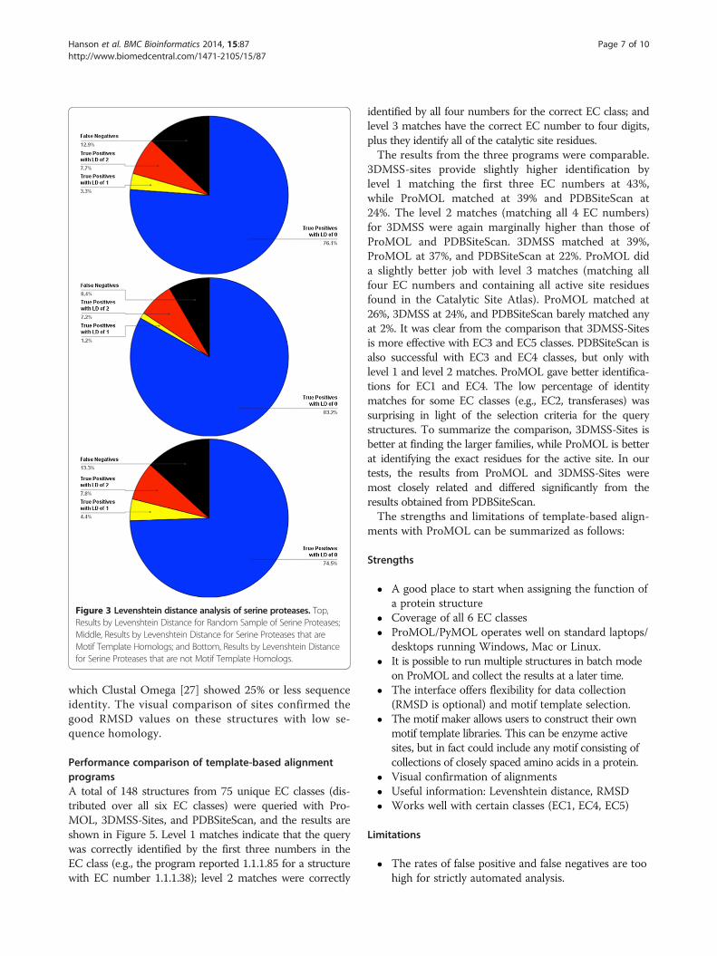
Figure 3 Levenshtein distance analysis of serine proteases. Top,Results by Levenshtein Distance for Random Sample of Serine Proteases;Middle, Results by Levenshtein Distance for Serine Proteases that areMotif Template Homologs; and Bottom, Results by Levenshtein Distancefor Serine Proteases that are not Motif Template Homologs.
Hanson et al. BMC Bioinformatics 2014, 15:87 Page 7 of 10http://www.biomedcentral.com/1471-2105/15/87
which Clustal Omega [27] showed 25% or less sequenceidentity. The visual comparison of sites confirmed thegood RMSD values on these structures with low se-quence homology.
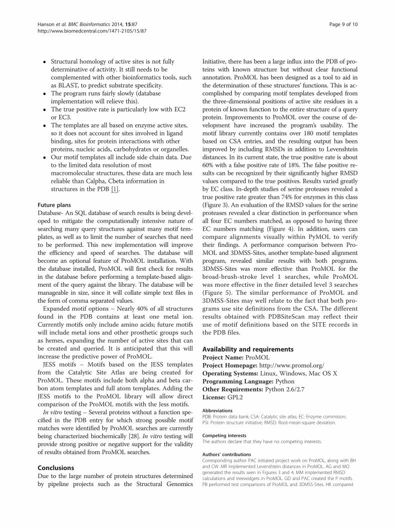
Performance comparison of template-based alignmentprogramsA total of 148 structures from 75 unique EC classes (dis-tributed over all six EC classes) were queried with Pro-MOL, 3DMSS-Sites, and PDBSiteScan, and the results areshown in Figure 5. Level 1 matches indicate that the querywas correctly identified by the first three numbers in theEC class (e.g., the program reported 1.1.1.85 for a structurewith EC number 1.1.1.38); level 2 matches were correctly
identified by all four numbers for the correct EC class; andlevel 3 matches have the correct EC number to four digits,plus they identify all of the catalytic site residues.The results from the three programs were comparable.
3DMSS-sites provide slightly higher identification bylevel 1 matching the first three EC numbers at 43%,while ProMOL matched at 39% and PDBSiteScan at24%. The level 2 matches (matching all 4 EC numbers)for 3DMSS were again marginally higher than those ofProMOL and PDBSiteScan. 3DMSS matched at 39%,ProMOL at 37%, and PDBSiteScan at 22%. ProMOL dida slightly better job with level 3 matches (matching allfour EC numbers and containing all active site residuesfound in the Catalytic Site Atlas). ProMOL matched at26%, 3DMSS at 24%, and PDBSiteScan barely matched anyat 2%. It was clear from the comparison that 3DMSS-Sitesis more effective with EC3 and EC5 classes. PDBSiteScan isalso successful with EC3 and EC4 classes, but only withlevel 1 and level 2 matches. ProMOL gave better identifica-tions for EC1 and EC4. The low percentage of identitymatches for some EC classes (e.g., EC2, transferases) wassurprising in light of the selection criteria for the querystructures. To summarize the comparison, 3DMSS-Sites isbetter at finding the larger families, while ProMOL is betterat identifying the exact residues for the active site. In ourtests, the results from ProMOL and 3DMSS-Sites weremost closely related and differed significantly from theresults obtained from PDBSiteScan.The strengths and limitations of template-based align-
ments with ProMOL can be summarized as follows:
Strengths
� A good place to start when assigning the function ofa protein structure
� Coverage of all 6 EC classes� ProMOL/PyMOL operates well on standard laptops/
desktops running Windows, Mac or Linux.� It is possible to run multiple structures in batch mode
on ProMOL and collect the results at a later time.� The interface offers flexibility for data collection
(RMSD is optional) and motif template selection.� The motif maker allows users to construct their own
motif template libraries. This can be enzyme activesites, but in fact could include any motif consisting ofcollections of closely spaced amino acids in a protein.
� Visual confirmation of alignments� Useful information: Levenshtein distance, RMSD� Works well with certain classes (EC1, EC4, EC5)
Limitations
� The rates of false positive and false negatives are toohigh for strictly automated analysis.
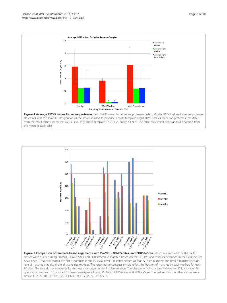
Figure 4 Average RMSD values for serine proteases. Left: RMSD values for all serine proteases tested; Middle: RMSD values for serine proteasestructures with the same EC designation as the structure used to produce a motif template; Right: RMSD values for serine proteases that differfrom the motif templates by the last EC level (e.g., motif Template 3.4.21.4 vs. query 3.4.21.5). The error bars reflect one standard deviation fromthe mean in each case.
Figure 5 Comparison of template-based alignments with ProMOL, 3DMSS-Sites, and PDBSiteScan. Structures from each of the six ECclasses were queried using ProMOL, 3DMSS-Sites, and PDBSiteScan. A match is based on the EC Class and residues described in the Catalytic SiteAtlas. Level 1 matches shared the first 3 numbers in the EC class; level 2 matches shared all four EC class numbers and level 3 matches includelevel 2 matches that also share all active site residues. The reported percentages simply reflect the fraction of matches by each method for eachEC class. The selection of structures for this test is described under Implementation. The distribution of structures follows: for EC1, a total of 26query structures from 16 unique EC classes were queried using ProMOL, 3DMSS-Sites and PDBSiteScan. The test sets for the other classes weresimilar: EC2 (29, 18), EC3 (20, 12), EC4 (23, 13), EC5 (25, 8), EC6 (25, 7).
Hanson et al. BMC Bioinformatics 2014, 15:87 Page 8 of 10http://www.biomedcentral.com/1471-2105/15/87
Hanson et al. BMC Bioinformatics 2014, 15:87 Page 9 of 10http://www.biomedcentral.com/1471-2105/15/87
� Structural homology of active sites is not fullydeterminative of activity. It still needs to becomplemented with other bioinformatics tools, suchas BLAST, to predict substrate specificity.
� The program runs fairly slowly (databaseimplementation will relieve this).
� The true positive rate is particularly low with EC2or EC3.
� The templates are all based on enzyme active sites,so it does not account for sites involved in ligandbinding, sites for protein interactions with otherproteins, nucleic acids, carbohydrates or organelles.
� Our motif templates all include side chain data. Dueto the limited data resolution of mostmacromolecular structures, these data are much lessreliable than Calpha, Cbeta information instructures in the PDB [1].
Future plansDatabase- An SQL database of search results is being devel-oped to mitigate the computationally intensive nature ofsearching many query structures against many motif tem-plates, as well as to limit the number of searches that needto be performed. This new implementation will improvethe efficiency and speed of searches. The database willbecome an optional feature of ProMOL installation. Withthe database installed, ProMOL will first check for resultsin the database before performing a template-based align-ment of the query against the library. The database will bemanageable in size, since it will collate simple text files inthe form of comma separated values.Expanded motif options – Nearly 40% of all structures
found in the PDB contains at least one metal ion.Currently motifs only include amino acids; future motifswill include metal ions and other prosthetic groups suchas hemes, expanding the number of active sites that canbe created and queried. It is anticipated that this willincrease the predictive power of ProMOL.JESS motifs – Motifs based on the JESS templates
from the Catalytic Site Atlas are being created forProMOL. These motifs include both alpha and beta car-bon atom templates and full atom templates. Adding theJESS motifs to the ProMOL library will allow directcomparison of the ProMOL motifs with the Jess motifs.In vitro testing – Several proteins without a function spe-
cified in the PDB entry for which strong possible motifmatches were identified by ProMOL searches are currentlybeing characterized biochemically [28]. In vitro testing willprovide strong positive or negative support for the validityof results obtained from ProMOL searches.
ConclusionsDue to the large number of protein structures determinedby pipeline projects such as the Structural Genomics
Initiative, there has been a large influx into the PDB of pro-teins with known structure but without clear functionalannotation. ProMOL has been designed as a tool to aid inthe determination of these structures’ functions. This is ac-complished by comparing motif templates developed fromthe three-dimensional positions of active site residues in aprotein of known function to the entire structure of a queryprotein. Improvements to ProMOL over the course of de-velopment have increased the program’s usability. Themotif library currently contains over 180 motif templatesbased on CSA entries, and the resulting output has beenimproved by including RMSDs in addition to Levenshteindistances. In its current state, the true positive rate is about60% with a false positive rate of 18%. The false positive re-sults can be recognized by their significantly higher RMSDvalues compared to the true positives. Results varied greatlyby EC class. In-depth studies of serine proteases revealed atrue positive rate greater than 74% for enzymes in this class(Figure 3). An evaluation of the RMSD values for the serineproteases revealed a clear distinction in performance whenall four EC numbers matched, as opposed to having threeEC numbers matching (Figure 4). In addition, users cancompare alignments visually within PyMOL to verifytheir findings. A performance comparison between Pro-MOL and 3DMSS-Sites, another template-based alignmentprogram, revealed similar results with both programs.3DMSS-Sites was more effective than ProMOL for thebroad-brush-stroke level 1 searches, while ProMOLwas more effective in the finer detailed level 3 searches(Figure 5). The similar performance of ProMOL and3DMSS-Sites may well relate to the fact that both pro-grams use site definitions from the CSA. The differentresults obtained with PDBSiteScan may reflect theiruse of motif definitions based on the SITE records inthe PDB files.
Availability and requirementsProject Name: ProMOLProject Homepage: http://www.promol.org/Operating Systems: Linux, Windows, Mac OS XProgramming Language: PythonOther Requirements: Python 2.6/2.7License: GPL2
AbbreviationsPDB: Protein data bank; CSA: Catalytic site atlas; EC: Enzyme commision;PSI: Protein structure initiative; RMSD: Root-mean-square deviation.
Competing interestsThe authors declare that they have no competing interests.
Authors’ contributionsCorresponding author PAC initiated project work on ProMOL, along with BHand CW. MR implemented Levenshtein distances in ProMOL. AG and MOgenerated the results seen in Figures 3 and 4. MM implemented RMSDcalculations and treewidgets in ProMOL. GD and PAC created the P motifs.PB performed test comparisons of ProMOL and 3DMSS-Sites. HK compared
Hanson et al. BMC Bioinformatics 2014, 15:87 Page 10 of 10http://www.biomedcentral.com/1471-2105/15/87
ProMOL with PDBSiteScan. TM compared ProMOL and BLAST results. CCcontributed to software design and debugging. HJB contributed to thesoftware design. All authors read and approved the final manuscript.
Authors’ informationHJB: Dept. of Math and Computer Science, Dowling College, Shirley, NY,11967, USA PAC, GD: School of Chemistry & Materials Science, RochesterInstitute of Technology, Rochester, NY, 14624, USA. MO, AG, MM, MR, BH,CW, HK: Thomas H. Gosnell School of Life Sciences, Rochester Institute ofTechnology, Rochester, NY, 14624, USA. PB, TM: College of Health Sciencesand Technology, Rochester Institute of Technology, Rochester, NY, 14624,USA. CC: Department of Computer Science, Rochester Institute ofTechnology, Rochester, NY, 14624, USA.
AcknowledgementsNSF DUE 0402408, NIGMS 1R15GM078077-01, NIGMS 3R15GM078077-01S,NIGMS 2R15GM078077-02, NIGMS 3R15GM078077-02S1, Dowling College,Rochester Institute of Technology. The creation of ProMOL distribution kitswas originated by Nikolay Darakev, who created the PyMOL MINGW win-dows build process in 2008 (see http://sourceforge.net/apps/mediawiki/sbevsl/index.php?title=PyMOL_MINGW_Build_Instructions). From the Roches-ter Institute of Technology: Eno Akpovwa, Lacey Andrews, Nicole Arroyo,Abdul Bangura, Daniel Bobo, Sean Bourne, Luticha Doucette, ChanelleFrancis, Katrina Henry, MaryEd Kenney, Desiree Matthews, Scott Mottarella.From Dowling College: Mogjan Asadi, Isaac Awuah Asiamah, Kethi Bardhi,Kostandina Bardhi, Darina Boycheva, Clarice Chigbo, Ricky Chachra, GeorgiDarakev, Nikolay Darakev, Damian Glinojecki, Jonathan Ihm, John Jemilawon,Kedian Jimenez, Nan Jia, Mia Jurjivec, Petko Kamburov, Barry LaPierre, MingLi, Stavros Louris, Gregory McQuillan, Kostadin Mitev, Daniel O’Brien, StojanRegodic, Limone Rosa, Matt Rousseau, Paul Sussman, Rohit Tripathi, GeorgiTodorov, Peter Zhivkov, Elena Zlateva.
Author details1University of California, Irvine, CA, USA. 2University of Louisville, Louisville,KY, USA. 3Rochester Institute of Technology, Rochester, NY, USA. 4DowlingCollege, Shirley, NY, USA. 5Rochester Institute of Technology, School ofChemistry & Materials Science, 1 Lomb Memorial Drive, Rochester,NY 14623, USA.
Received: 6 March 2013 Accepted: 24 January 2014Published: 27 March 2014
References1. Torrance JW, Bartlett GJ, Porter CT, Thornton JM: Using a library of
structural templates to recognise catalytic sites and explore theirevolution in homologous families. J Mol Biol 2005, 347:565–581.
2. Jones S, Thornton JM: Searching for functional sites in protein structures.Curr Opin Chem Biol 2004, 8:3–7.
3. Friedberg I: Automated protein function prediction—the genomicchallenge. Brief Bioinform 2006, 7:225–242.
4. Skolnick J, Fetrow JS: From genes to protein structure and function: novelapplications of computational approaches in the genomic era. TrendsBiotechnol 2000, 18:34–39.
5. Gerlt JA, Allen KN, Almo SC, Armstrong RN, Babbitt PC, Cronan JE,Dunaway-Mariano D, Imker HJ, Jacobson MP, Minor W, Poulter CD, Raushel FM,Sali A, Shoichet BK, Sweedler JV: The enzyme function initiative. Biochemistry2011, 50:9950–9962.
6. Hasegawa H, Holm L: Advances and pitfalls of protein structuralalignment. Curr Opin Struct Biol 2009, 19:341–348.
7. Altschul SF, Madden TL, Schäffer AA, Zhang J, Zhang Z, Miller W, Lipman DJ:Gapped BLAST and PSI-BLAST: a new generation of protein databasesearch programs. Nucleic Acids Res 1997, 25:3389–3402.
8. Zhang Z, Miller W, Schäffer AA, Madden TL, Lipman DJ, Koonin EV, AltschulSF: Protein sequence similarity searches using patterns as seeds. NucleicAcids Res 1998, 26:3986–3990.
9. Holm L, Sander C: Alignment of three-dimensional protein structures: networkserver for database searching. Methods Enzymol 1996, 266:653–662.
10. Pieper U, Webb BM, Barkan DT, Schneidman-Duhovny D, Schlessinger A,Braberg H, Yang Z, Meng EC, Pettersen EF, Huang CC, Datta RS, SampathkumarP, Madhusudhan MS, Sjölander K, Ferrin TE, Burley SK, Sali A: ModBase, a
database of annotated comparative protein structure models, andassociated resources. Nucleic Acids Res 2011, 39:D465–D474.
11. Schulz GE, Schirmer RH: Principles of Protein Structure. New York, NY, USA:Springer-Verlag; 1979.
12. Petty HR: Molecular Biology of Membranes: Structure and Function. New York,NY, USA: Plenum; 1993.
13. Redfern OC, Dessailly BH, Dallman TJ, Sillitoe I, Orengo CA: FLORA: a novelmethod to predict protein function from structure in diversesuperfamilies. PLoS Comput Biol 2009, 5:e1000485.
14. Hanson BR, Westin C, Craig PA: Using PyMOL’s selection algebra forenzyme catalytic site prediction. FASEB J 2007, 21:A296.
15. Delano WL: The PyMOL Molecular Graphics System. San Carlos, CA, USA:Schrodinger, LLC; 2002.
16. Porter CT: The catalytic site atlas: a resource of catalytic sites andresidues identified in enzymes using structural data. Nucleic Acids Res2004, 32:D129–D133.
17. Grell L, Parkin C, Slatest L, Craig PA: EZ-Viz, a tool for simplifying molecularviewing in PyMOL. Biochem Mol Biol Educ 2006, 34:402–407.
18. Levenshtein VI: Binary codes capable of correcting deletions, insertions,and reversals. Sov Phys-Dokl 1966, 10:707–710.
19. Gushee M: TreeWidgets. 2002 [http://matt.gushee.net/software/treewidgets/]20. Moad C: Remote PDB Loader. 2004 [http://www.pymolwiki.org/index.php/
Plugins_Tutorial]21. Barker JA, Thornton JM: An algorithm for constraint-based structural tem-
plate matching: application to 3D templates with statistical analysis.Bioinformatics 2003, 19:1644–1649.
22. Escalier V, Pothier J, Soldano H, Viari A: Pairwise and multiple identificationof three-dimensional common substructures in proteins. J Comput Biol1998, 5:41–56.
23. Petitjean M: Interactive maximal common 3D substructure searching withthe combined SDM/RMS Algorithm. Comp Chem 1998, 22:463–465.
24. Ivanisenko VA, Pintus SS, Grigorovich DA, Kolchanov NA: PDBSiteScan:a program for searching for active, binding and posttranslationalmodification sites in the 3D structures of proteins. Nucleic Acids Res 2004,32:W549–W554.
25. Debret G, Martel A, Cuniasse P: RASMOT-3D PRO: a 3D motif searchwebserver. Nucleic Acids Res 2009, 37:W459–W464.
26. Johansson MU, Zoete V, Michielin O, Guex N: Defining and searching forstructural motifs using DeepView/Swiss-PdbViewer. BMC Bioinforma 2012,13:173.
27. Sievers F, Wilm A, Dineen D, Gibson TJ, Karplus K, Li W, Lopez R, McWilliamH, Remmert M, Söding J, Thompson JD, Higgins DG: Fast, scalablegeneration of high-quality protein multiple sequence alignments usingclustal omega. Mol Syst Biol 2011, 7:539.
28. Dodge G, Arroyo EN, Bernstein HJ, Craig PA: Development and testing of asystematic approach for computational enzyme function determination.FASEB J 2013, 27:811.1.
doi:10.1186/1471-2105-15-87Cite this article as: Hanson et al.: Estimation of protein function usingtemplate-based alignment of enzyme active sites. BMC Bioinformatics2014 15:87.
Submit your next manuscript to BioMed Centraland take full advantage of:
• Convenient online submission
• Thorough peer review
• No space constraints or color figure charges
• Immediate publication on acceptance
• Inclusion in PubMed, CAS, Scopus and Google Scholar
• Research which is freely available for redistribution
Submit your manuscript at www.biomedcentral.com/submit