Multiple 40-kDa Heat-Shock Protein Chaperones Function in Tom70-dependent Mitochondrial Import
15
Molecular Biology of the Cell Vol. 18, 3414 –3428, September 2007 Multiple 40-kDa Heat-Shock Protein Chaperones Function in Tom70-dependent Mitochondrial Import Melanie K. Bhangoo, Stefan Tzankov, Anna C.Y. Fan, Kurt Dejgaard, David Y. Thomas, and Jason C. Young Department of Biochemistry, McGill University, Montreal, QC, H3G 1Y6, Canada Submitted January 31, 2007; Accepted June 19, 2007 Monitoring Editor: Jeffrey Brodsky Mitochondrial preproteins that are imported via the translocase of the mitochondrial outer membrane (Tom)70 receptor are complexed with cytosolic chaperones before targeting to the mitochondrial outer membrane. The adenine nucleotide transporter (ANT) follows this pathway, and its purified mature form is identical to the preprotein. Purified ANT was reconstituted with chaperones in reticulocyte lysate, and bound proteins were identified by mass spectrometry. In addition to 70-kDa heat-shock cognate protein (Hsc70) and 90-kDa heat-shock protein (Hsp90), a specific subset of cochaperones were found, but no mitochondria-specific targeting factors were found. Interestingly, three different Hsp40-related J-domain proteins were identified: DJA1, DJA2, and DJA4. The DJAs bound preproteins to different extents through their C-terminal regions. DJA dominant-negative mutants lacking the N-terminal J-domains impaired mitochon- drial import. The mutants blocked the binding of Hsc70 to preprotein, but with varying efficiency. The DJAs also showed significant differences in activation of the Hsc70 ATPase and Hsc70-dependent protein refolding. In HeLa cells, the DJAs increased new protein folding and mitochondrial import, although to different extents. No single DJA was superior to the others in all aspects, but each had a profile of partial specialization. The Hsp90 cochaperones p23 and Aha1 also regulated Hsp90 –preprotein interactions. We suggest that multiple cochaperones with similar yet partially specialized properties cooperate in optimal chaperone–preprotein complexes. INTRODUCTION The majority of mitochondrial proteins are encoded in the nucleus, translated on cytosolic ribosomes, and subse- quently imported into mitochondria (Reichert and Neupert, 2004). The translocase of the mitochondrial outer membrane (TOM) is responsible for polypeptide translocation across the outer membrane; and inside the organelle, various mech- anisms sort these polypeptides to their appropriate compart- ment—matrix, inner membrane or intermembrane space. The TOM complex contains integral membrane import re- ceptors, Tom20 and Tom70, which mediate targeting of mi- tochondrial preproteins, and a general import pore that provides an aqueous channel for translocation. Tom20 is thought to be responsible for the import of preproteins bearing classical matrix-directed N-terminal signal se- quences, or leader peptides. Tom70, the alternate receptor, is thought to mediate the import of preproteins that contain targeting information within their mature sequences (Koehler, 2004; Rehling et al., 2004). Although much of our knowledge of import derives from work in Saccharomyces cerevisiae and Neurospora crassa, the biological functions of the human Tom70 and Tom20 receptors seem to be largely conserved (Iwahashi et al., 1997; Schleiff et al., 1997; Yano et al., 1997; Suzuki et al., 2002). The Tom70 receptor seems to be most critical for the import of particularly hydrophobic preproteins, such as members of the inner membrane metabolite carrier family (Sollner et al., 1990; de Marcos-Lousa et al., 2006). Before import, these preproteins typically depend on cytosolic chaperones to maintain their solubility. The ATP-dependent chaperones 70-kDa heat-shock cognate protein (Hsc70) and 90-kDa heat-shock protein (Hsp90) have key roles in this mechanism (Young et al., 2004). In addition to maintaining the solubility of the bound preprotein, Hsc70 and Hsp90 can both dock onto to a specific binding site in Tom70, an essential first step in preprotein targeting. The chaperone docking site lies in the central region of Tom70 next to the single N-terminal transmembrane domain (Young et al., 2003). At this stage, preprotein is thought to contact Tom70 directly in a C-terminal region separate from the chaperone docking site (Brix et al., 2000; Wu and Sha, 2006). ATP- dependent cycling by Hsp90 and most probably Hsc70 then assists the translocation of preprotein via the outer mem- brane TOM machinery (Fan et al., 2006). In their other cellular functions, Hsc70 and Hsp90 interact with a wide range of cochaperone proteins, which connect additional specific chaperoning functions or biochemical ac- tivities to chaperone complexes. In some cases, the type of substrate polypeptide bound by a chaperone complex deter- mines which specific cochaperone is recruited to Hsp90. For example, the immunophilin FKBP52 usually associates with, and assists in the maturation of, steroid hormone receptors, whereas Cdc37 has a comparable role for kinases (Pratt and Toft, 2003). Such specificity is not absolute: both Cdc37 and This article was published online ahead of print in MBC in Press (http://www.molbiolcell.org/cgi/doi/10.1091/mbc.E07– 01– 0088) on June 27, 2007. Address correspondence to: Jason C. Young ([email protected]). Abbreviations used: ANT, adenine nucleotide transporter; GA, geldanamycin; Hsc70, 70-kDa heat-shock cognate protein; Hsp90, 90-kDA heat shock protein; ISP, Rieske iron-sulfur protein; PiC, phosphate carrier; PK, proteinase K; RL, reticulocyte lysate; TOM, translocase of the mitochondrial outer membrane; TPR, tetratri- copeptide repeat. 3414 © 2007 by The American Society for Cell Biology
-
Upload
independent -
Category
Documents
-
view
3 -
download
0
Transcript of Multiple 40-kDa Heat-Shock Protein Chaperones Function in Tom70-dependent Mitochondrial Import

Molecular Biology of the CellVol. 18, 3414–3428, September 2007
Multiple 40-kDa Heat-Shock Protein Chaperones Functionin Tom70-dependent Mitochondrial ImportMelanie K. Bhangoo, Stefan Tzankov, Anna C.Y. Fan, Kurt Dejgaard,David Y. Thomas, and Jason C. Young
Department of Biochemistry, McGill University, Montreal, QC, H3G 1Y6, Canada
Submitted January 31, 2007; Accepted June 19, 2007Monitoring Editor: Jeffrey Brodsky
Mitochondrial preproteins that are imported via the translocase of the mitochondrial outer membrane (Tom)70 receptorare complexed with cytosolic chaperones before targeting to the mitochondrial outer membrane. The adenine nucleotidetransporter (ANT) follows this pathway, and its purified mature form is identical to the preprotein. Purified ANT wasreconstituted with chaperones in reticulocyte lysate, and bound proteins were identified by mass spectrometry. Inaddition to 70-kDa heat-shock cognate protein (Hsc70) and 90-kDa heat-shock protein (Hsp90), a specific subset ofcochaperones were found, but no mitochondria-specific targeting factors were found. Interestingly, three differentHsp40-related J-domain proteins were identified: DJA1, DJA2, and DJA4. The DJAs bound preproteins to different extentsthrough their C-terminal regions. DJA dominant-negative mutants lacking the N-terminal J-domains impaired mitochon-drial import. The mutants blocked the binding of Hsc70 to preprotein, but with varying efficiency. The DJAs also showedsignificant differences in activation of the Hsc70 ATPase and Hsc70-dependent protein refolding. In HeLa cells, the DJAsincreased new protein folding and mitochondrial import, although to different extents. No single DJA was superior to theothers in all aspects, but each had a profile of partial specialization. The Hsp90 cochaperones p23 and Aha1 also regulatedHsp90–preprotein interactions. We suggest that multiple cochaperones with similar yet partially specialized propertiescooperate in optimal chaperone–preprotein complexes.
INTRODUCTION
The majority of mitochondrial proteins are encoded in thenucleus, translated on cytosolic ribosomes, and subse-quently imported into mitochondria (Reichert and Neupert,2004). The translocase of the mitochondrial outer membrane(TOM) is responsible for polypeptide translocation acrossthe outer membrane; and inside the organelle, various mech-anisms sort these polypeptides to their appropriate compart-ment—matrix, inner membrane or intermembrane space.The TOM complex contains integral membrane import re-ceptors, Tom20 and Tom70, which mediate targeting of mi-tochondrial preproteins, and a general import pore thatprovides an aqueous channel for translocation. Tom20 isthought to be responsible for the import of preproteinsbearing classical matrix-directed N-terminal signal se-quences, or leader peptides. Tom70, the alternate receptor, isthought to mediate the import of preproteins that containtargeting information within their mature sequences(Koehler, 2004; Rehling et al., 2004). Although much of ourknowledge of import derives from work in Saccharomycescerevisiae and Neurospora crassa, the biological functions of
the human Tom70 and Tom20 receptors seem to be largelyconserved (Iwahashi et al., 1997; Schleiff et al., 1997; Yano etal., 1997; Suzuki et al., 2002).
The Tom70 receptor seems to be most critical for theimport of particularly hydrophobic preproteins, such asmembers of the inner membrane metabolite carrier family(Sollner et al., 1990; de Marcos-Lousa et al., 2006). Beforeimport, these preproteins typically depend on cytosolicchaperones to maintain their solubility. The ATP-dependentchaperones 70-kDa heat-shock cognate protein (Hsc70) and90-kDa heat-shock protein (Hsp90) have key roles in thismechanism (Young et al., 2004). In addition to maintainingthe solubility of the bound preprotein, Hsc70 and Hsp90 canboth dock onto to a specific binding site in Tom70, anessential first step in preprotein targeting. The chaperonedocking site lies in the central region of Tom70 next to thesingle N-terminal transmembrane domain (Young et al.,2003). At this stage, preprotein is thought to contact Tom70directly in a C-terminal region separate from the chaperonedocking site (Brix et al., 2000; Wu and Sha, 2006). ATP-dependent cycling by Hsp90 and most probably Hsc70 thenassists the translocation of preprotein via the outer mem-brane TOM machinery (Fan et al., 2006).
In their other cellular functions, Hsc70 and Hsp90 interactwith a wide range of cochaperone proteins, which connectadditional specific chaperoning functions or biochemical ac-tivities to chaperone complexes. In some cases, the type ofsubstrate polypeptide bound by a chaperone complex deter-mines which specific cochaperone is recruited to Hsp90. Forexample, the immunophilin FKBP52 usually associates with,and assists in the maturation of, steroid hormone receptors,whereas Cdc37 has a comparable role for kinases (Pratt andToft, 2003). Such specificity is not absolute: both Cdc37 and
This article was published online ahead of print in MBC in Press(http://www.molbiolcell.org/cgi/doi/10.1091/mbc.E07–01–0088)on June 27, 2007.
Address correspondence to: Jason C. Young ([email protected]).
Abbreviations used: ANT, adenine nucleotide transporter; GA,geldanamycin; Hsc70, 70-kDa heat-shock cognate protein; Hsp90,90-kDA heat shock protein; ISP, Rieske iron-sulfur protein; PiC,phosphate carrier; PK, proteinase K; RL, reticulocyte lysate; TOM,translocase of the mitochondrial outer membrane; TPR, tetratri-copeptide repeat.
3414 © 2007 by The American Society for Cell Biology
the immunophilin FKBP8 have been found in chaperonecomplexes with a folding-deficient mutant of the cystic fi-brosis transmembrane conductance regulator (CFTR) chlo-ride channel (Wang et al., 2006). Many cochaperones containfunctionally conserved chaperone–interaction domains.Some interactions with Hsp90 and Hsc70 are mediated bystructurally conserved tetratricopeptide repeat (TPR) clampdomains that recognize the C-terminal motifs of the chaper-ones. The TPR-domains in FKBP52 and the related cyclophi-lin Cyp40 connect to Hsp90. Those in the cochaperone Hoprecognize Hsc70 and Hsp90 separately, allowing Hsc70–substrate complexes to recruit Hsp90 (Pratt and Toft, 2003).Also, a conserved TPR clamp domain in Tom70 forms itschaperone docking site (Young et al., 2003). J-domains,found in various cochaperones including the Hsp40-relatedproteins, stimulate ATP hydrolysis and substrate binding byHsc70. J-domains do not form stable contacts with Hsc70,but they are essential for all known functions of Hsc70(Walsh et al., 2004; Qiu et al., 2006). Several nucleotide ex-change factors including Bag-1 transiently promote ATPrebinding and Hsc70 dissociation from substrate (Mayer andBukau, 2005). Cochaperones that do not belong to thesestructural families include Cdc37, p23, and Aha1. p23 stabi-lizes the ATP- and substrate-binding state of Hsp90,whereas the recently discovered Aha1 stimulates the Hsp90ATPase (Panaretou et al., 2002; Sullivan et al., 2002; Lotz et al.,2003; Morishima et al., 2003; Pearl and Prodromou, 2006).
It is not known what proteins in addition to Hsc70 andHsp90 are complexed with Tom70-dependent preproteinsbefore import, or what proteins are involved in complexformation. It seems certain that cochaperone proteins par-ticipate in these complexes. The preproteins are not relatedto structural families of proteins known to prefer specificcochaperones, and the involvement of many cochaperonescannot be predicted. The preproteins seem to be in high-molecular-weight complexes of �600 kDa, similar to otherHsp90-bound complexes, but with some heterogeneity(Young et al., 2003; Fan et al., 2006). The identity and functionof bound cochaperones would lead to significant insight.First, are the cochaperones present specifically bound? Sec-ond, are there components unique to chaperone–preproteincomplexes, and what role might they have? Third, do thecochaperones bound have implications for the function ofHsc70 and Hsp90?
To address these questions, we reconstituted chaperonecomplexes in reticulocyte lysate (RL) as a model cytosol.This method was used to originally identify most of thecochaperones involved in steroid receptor and kinase mat-uration (Pratt and Toft, 2003). RL is uniquely suited for suchexperiments. Although it contains essentially no organellarcontamination, it is the basis for all experiments reconstitut-ing cell-free mitochondrial import; therefore, it contains allnecessary factors. A mitochondrial preprotein, the adeninenucleotide transporter (ANT) is uniquely purifiable in bio-chemical amounts (Pebay-Peyroula et al., 2003), and it wasused as the starting point for the reconstitution of com-plexes.
MATERIALS AND METHODS
Chemicals and ReagentsUnless stated otherwise, all chemical reagents were from Sigma DiagnosticsCanada or BioShop Canada (Mississauga, ON, Canada). Restriction enzymesand other recombinant DNA reagents were from New England Biolabs (Ip-swich, MA), Invitrogen (Carlsbad, CA), and Stratagene (San Diego, CA).Geldanamycin (GA) was from LC Laboratories (Boston, MA). Untreatedrabbit RL was from Green Hectares (Oregon, WI). Antibodies against Hsp90,Hsc70, Hop, FKBP52, and Hsp40 were from Assay Designs (Ann Arbor, MI);
those against Cdc37, Cyp40, and p23 were from Affinity BioReagents (Golden,CO); that against DJA1 was from Labvision/NeoMarkers (Fremont, CA); andthat against luciferase was from Sigma Diagnostics Canada. The antibodyagainst Tpr2 was a gift from Dr. W.M.J. Obermann (Houston, TX); thoseagainst DJA2 and DJA4 were gifts from Dr. M. Mori (Kumamoto, Japan), andthat against Hsp60 was a gift from Dr. F. U. Hartl (Martinsried, Germany).Additional antibodies specific for DJA1, DJA2, and DJA4 were raised inrabbits against the synthetic peptides LVDFDPNQER, PEVPNIIGET, andPNEQNWRQHR, respectively, and they were confirmed with immunoblotsagainst the purified DJA proteins, and HeLa cells were transfected with eachantibody. Antibodies against the myc (9E10) and hemagglutinin (HA.11)epitope tags were from Covance/BAbCO (Richmond, CA).
PlasmidsThe sequences encoding bovine phosphate carrier (PiC) and N. crassa Rieskeiron-sulfur protein (ISP) were in pGEM-3 (Promega, Madison, WI) (Sollner etal., 1989; Zara et al., 1992). The sequence encoding murine ANT2 (NM_007451)was amplified by polymerase chain reaction (PCR) from a cDNA library andinserted into pGEM-11Z (Promega) (Fan et al., 2006). The sequences encodingnontagged human Hsp90� and rat Hsc70 were in pET15b and pET11a(Novagen, San Diego, CA), respectively, and the sequences encoding residues566-732 of human Hsp90� (C-90) and residues 151-263 of human Bag-1(C-Bag) were in pProExHTa (Clontech, Mountain View, CA) (Young andHartl, 2000; Sondermann et al., 2001; Young et al., 2003). Recombinant non-tagged human p23 was in pET28 (Novagen) (Young and Hartl, 2000), and thesequence encoding human DJA1 (NM_001539) also in pET28a was a gift fromC.H.I. Ramos (Laboratorio Nacional de Luz Sıncrotron, Campinas SP, Brazil)(Borges et al., 2005). The sequences encoding human Hsc70 (NM_006597),DJA2 (NM_005880), and DJA4 (NM_018602) as well as the sequences encod-ing amino acids 98-397 of human DJA1 (C-A1), amino acids 100-412 of humanDJA2 (C-A2), and amino acids 99-397 of human DJA4 (C-A4) were amplifiedfrom a cDNA library and inserted into pProExHTa (Clontech). Sequences ofDJA1, DJA2, and DJA4 were inserted into pGEM-11Z (Promega) without tags,and into pcDNA3.1 myc-His C (Invitrogen). Human Aha1 in pProExHTa wasa gift from W.M.J. Obermann (Houston, TX) (Lotz et al., 2003). The sequenceencoding firefly luciferase was amplified by PCR from pGL3 (Promega) andinserted into pcDNA3.1 (Invitrogen) without a tag; the pSV40-�-galactosidasevector was from Promega. The vectors pCAGGS-PiC-3HA, pGR-�LBD, andpMTV-luciferase (glucocorticoid responsive element [GRE]-luciferase) wereas reported previously (Hollenberg et al., 1987; Hollenberg and Evans, 1988;Brychzy et al., 2003; Young et al., 2003).
Protein PurificationThe expression vectors for full-length His-tagged DJA1, DJA2, and DJA4 weregrown in Rosetta 2 Escherichia coli cells (Novagen), and protein expression wasinduced with 1 mM isopropyl-�-d-thiogalactopyranoside at 30°C for 1 h, at37°C for 30 min, and at 30°C for 3 h, respectively. The cells were harvestedand resuspended in buffer containing 750 mM NaCl, 60 mM imidazole and 20mM KH2PO4, pH 7.5, with Complete protease inhibitors (Roche Diagnostics,Indianapolis, IN). Cells were lysed by cavitation in a French press, and the celldebris was removed by centrifugation. The supernatant was loaded onto a5-ml nickel-Sepharose high-performance column (GE Healthcare, Little Chal-font, Buckinghamshire, United Kingdom), and proteins were eluted withbuffer containing 1 M imidazole, 500 mM NaCl, and 20 mM KH2PO4, pH 7.5.Peak fractions were loaded on a Superdex 200 Hi-Load 16/60 column (GEHealthcare) and eluted with buffer HS (500 mM NaCl, 20 mM HEPES-KOH,pH 7.5, and 5 mM MgOAc2). Nonaggregated peak fractions were collected,and the yield was determined by absorbance at 280 nm. The DJA proteinslacking the N-terminal J-domain, C-A1, C-A2, and C-A4, were purified iden-tically to the full-length proteins. Hsc70 was expressed for 4 h at 30°C andpurified on nickel-Sepharose high-performance equlibrated in buffer contain-ing 500 mM NaCl, 20 mM imidazole, and 20 mM KH2PO4, pH 7.5, and elutedin buffer containing 300 mM imidazole and 20 mM KH2PO4, pH 7.5. Hsc70was further purified by ion exchange on a Mono Q 5/50 GL column and gelfiltration on a Superdex 200 Hi-Load 16/60 column (GE Healthcare) equili-brated in buffer CG (100 mM KOAc, 20 mM HEPES-KOH, pH 7.5, and 5 mMMgOAc2). Bovine ANT was purified as described previously (Klingenberg etal., 1979; Pebay-Peyroula et al., 2003), with the following modifications. Mi-tochondria were prepared from bovine heart and treated with atractyloside,and then they were solubilized in buffer AB (500 mM NaCl, 10 mM HEPES-KOH, pH 7.5, and 1 mM EDTA) containing 2% Triton X-100. Insolublematerial was removed by ultracentrifugation at 140,000 � g for 30 min, andthe supernatant was loaded onto a hydroxyapatite HTP-gel column (Bio-Rad,Hercules, CA) equilibrated in buffer AB containing 0.1% Triton X-100, andpeak flow-through fractions containing ANT were collected. RecombinantC-90, C-Bag, Aha1, p23, and firefly luciferase were purified as describedpreviously (Young and Hartl, 2000; Brychzy et al., 2003; Lotz et al., 2003;Young et al., 2003).
Chaperone–ANT ComplexesRL was desalted into buffer CG on NAP-10 columns or a Hi-Prep 26/10 fastdesalting column (GE Healthcare). Purified ANT was chemically biotinylated
Hsp40 Chaperones in Mitochondrial Import
Vol. 18, September 2007 3415
on cysteine side chains with maleimide PEO2-biotin (Pierce Chemical, Rock-ford, IL) for 3 h at room temperature, and excess biotin was removed withNAP-10 columns equilibrated in buffer MT (100 mM NaCl, 10 mM HEPES-KOH, 1 mM EDTA and 0.1% Triton X-100). The biotinylated ANT (ANT-B)was bound to immobilized streptavidin-agarose (Pierce Chemical) for 1 h. TheANT-B was washed twice with buffer CG to remove stabilizing detergent, andit was reconstituted by incubation with 50% RL in buffer CG, supplementedwith 2 mM ATP. After 15 min at room temperature, the reactions wereterminated with 0.1 U/�l apyrase, grade VII. The reconstituted ANT-B com-plexes were reisolated, and the protein components were eluted with Lae-mmli loading buffer. After separation by Laemmli SDS-polyacrylamide gelelectrophoresis (PAGE) and total protein staining with Coomassie Blue, bandswere excised and subjected to in-gel tryptic digestion as described previously(Shevchenko et al., 1996). Peptides were analyzed by C18 reverse phaseseparation followed by ion trap liquid chromatography-tandem mass spec-trometry on a Daltonics Esquire HCT� instrument (Bruker, Newark, DE).Spectra were formatted and searched against mammalian sequences in theNational Center for Biotechnology Information database by using the Mascotsearch engine (Matrix Science, Boston, MA). Data were validated manually.
For gel filtration analysis, ANT-B was bound to monomeric avidin agarose(Pierce Chemical) for 1 h, washed with buffer CG, and eluted with 1% SDSand 8 M urea. The eluted ANT-B was diluted 1:400 into 50% RL in buffer CG,supplemented with 2 mM ATP. After 15 min at room temperature, thereaction was terminated with apyrase as described above and loaded onto aSuperose 6 10/300 GL column (GE Healthcare) equilibrated in buffer CG.Fractions were collected and separated by SDS-PAGE, transferred onto nitro-cellulose, detected with horseradish peroxidase-conjugated streptavidin(Pierce Chemical), and visualized on chemiluminescent-sensitive film. Incontrol reactions, cell-free translation reactions of ANT were performed usingthe TNT coupled RL system with SP6 polymerase (Promega), and they werelabeled with [35S]methionine (GE Healthcare). Reactions were diluted 1:4 intobuffer CG and analyzed by gel filtration as described above. Fractions wereanalyzed by SDS-PAGE and autoradiography. Phosphorimager quantitationwas performed with a FujiFilm BAS-1800II analyzer (FujiFilm, Stamford, CT)and ImageGauge software (Fuji, Tokyo, Japan).
Mitochondrial Import AssaysRat liver mitochondria were isolated and import reactions were performed asdescribed previously (Lingelbach et al., 1986; Young et al., 2003; Fan et al.,2006). For cleaner mitochondrial preparations by gradient centrifugation (Vi-jayasarathy et al., 1989), mitochondria were layered over 12-ml steps of 42, 45,and 60% sucrose in 10 mM HEPES-KOH, pH 7.5, and 1 mM EDTA, in SW28tubes (Beckman Coulter, Fullerton, CA). After centrifugation at 100,000 � gfor 1 h, mitochondria were harvested from the 45–60% interface, diluted inthe same buffer without sucrose, collected by centrifugation, and resuspendedat 10 mg/ml in buffer MC (250 mM sucrose, 80 mM KOAc, 20 mM HEPES-KOH, pH 7.5, and 5 mM MgOAc2) supplemented with 10 mM Na-succinate,1 mM dithiothreitol, 2 mM ATP, and 0.4 mM ADP. Cell-free translations ofthe preproteins were performed with the TNT-coupled RL system with SP6 orT7 polymerase supplemented with [35S]methionine, and then reactions wereterminated with 1 mM methionine and adjusted to 250 mM sucrose. Typicalimport reactions contained 25% reticulocyte lysate and 5 mg/ml mitochon-dria at 30°C for 30 min, and negative control reactions were inhibited with 1�M valinomycin. Mitochondria were reisolated and digested with 250 �g/mlproteinase K (PK) at 4°C for 10 min, followed by 1 mM phenylmethylsulfonylfluoride to stop the digestion. Undigested and digested samples were ana-lyzed by SDS-PAGE and autoradiography or phosphorimager analysis.
Inhibition of import by C-90, C-Bag, and geldanamycin (GA) was assayedas reported previously (Young et al., 2003; Fan et al., 2006). Purified C-90 andC-Bag were added to reactions at a final concentration of 20 and 5 �M,respectively. Then, 18 �M GA or the equivalent volume of vehicle controldimethyl sulfoxide (1%) was added to the translation reactions, and excessGA was removed before import reactions by using MicroBioSpin 6 columns(Bio-Rad) pre-equilibrated in buffer MC containing 0.4 mM EGTA, centri-fuged at 16°C with 2 mM ATP added to the collection tube. Inhibition ofimport by C-A1, C-A2, and C-A4 was assayed as for C-90, with each mutantadded at 20 �M, and reactions adjusted to a final concentration of 90 mMNaCl in buffer MC to maintain solubility of the mutants. Control reactionsadjusted to 90 mM NaCl with no additional protein showed that the addedionic strength had no significant effect on import.
Purified ANT was radiolabeled with Na-[125I] (GE Healthcare) by usingIodo-Beads (Pierce Chemical) according to the manufacturer’s instructions.125I-ANT was desalted using a NAP-10 column in buffer MT to remove excessradiolabel, concentrated by trichloroacetic acid precipitation, and resus-pended in 0.1% SDS. Import was initiated by 1:100 dilution into reactionscontaining 4 mg/ml mitochondria and 40% RL in buffer MC, supplementedwith 2 mM succinate, 0.2 mM dithiothreitol, 2 mM ATP, and 0.4 mM EGTA.To assay import of ANT-B, the biotinylated protein was recovered withmonomeric avidin agarose as described for gel filtration analysis. The elutedANT-B was diluted 1:10 into distilled water and then a further 1:100 dilutioninto reactions containing RL and mitochondria as described for 125I-ANT.Control reactions using cell-free translated ANT showed that the final con-
centration of 0.001% SDS did not interfere with mitochondrial import in anyway.
Coprecipitation ExperimentsTo assay binding of preproteins to the DJA proteins, an assay established forTom70 (Young et al., 2003; Fan et al., 2006) was adapted. DJA1, DJA2, andDJA4 or the truncation mutants C-A1, C-A2, and C-A4 were prebound onnickel-Sepharose in buffer HS for 30 min at 4°C. Cell-free translations of ANTand PiC were performed as described above with SP6 polymerase, diluted1:20 into buffer CG containing 20 mM imidazole, 0.1% Triton X-100, and 2mg/ml ovalbumin, and added to the immobilized DJA proteins. The finalreactions contained 5 �M wild-type DJA protein or 10 �M truncation mutant,and 5% translation mixture. After 5 min at room temperature, the bindingreactions were terminated by the addition of 0.1 U/�l apyrase. The proteincomplexes were recovered at 4°C for 30 min, and then they were washed withbuffer CG containing 20 mM imidazole and 0.1% Triton X-100. Protein com-plexes were eluted with Laemmli loading buffer, and they were analyzed bySDS-PAGE and autoradiography or phosphorimager quantitation. To formDJA heterocomplexes, ANT-B was diluted into 50% RL as described for thegel filtration analysis, but binding reactions were supplemented 1:20 withcell-free translations of each nontagged DJA. The reactions were then copre-cipitated with different His-tagged DJAs as described above.
To assay formation and dissociation of Hsc70, Hsp90, and DJA complexeswith ANT, methods established for the ligand binding domain of the glu-cocorticoid receptor (Young and Hartl, 2000; Sondermann et al., 2001; Brychzyet al., 2003) were adapted. To assay binding, ANT-B was prebound to strepta-vidin-agarose and washed with buffer CG as described above. Cell-free trans-lation reactions of Hsc70, Hsp90, or each DJA were performed as describedabove with T7 or SP6 polymerase, diluted 1:10 or 1:14 into 40% RL in bufferCG containing 2 mM ATP, and added to the immobilized ANT-B. Aliquotswere taken at the indicated time points, and complexes were reisolated andwashed with buffer CG containing 0.1% Triton X-100. Protein complexes wereeluted and analyzed as described above. Where indicated, binding reactionswere performed in the presence of 20 �M C-A1, C-A2, C-A4 or C90, or 4 �MAha1. To assay dissociation of complexes, cell-free translation reactions ofHsc70 and Hsp90 were complexed with ANT-B as described above for 15 minat room temperature, and complexes were reisolated and washed with bufferCG. Dissociation reactions consisting of buffer CG with or without 2 mM ATP,4 �M Aha1, 5 �M C-Bag, 5 �M p23, or 20 �M C-90 were incubated with thecomplexes for 10 min at room temperature. Released and remaining immo-bilized fractions were separated and analyzed by SDS-PAGE and autoradiog-raphy or phosphorimager quantitation.
Hsc70 ActivityATPase activities were measured as described previously (Young and Hartl,2000; Brychzy et al., 2003). Briefly, reactions contained 4 �M Hsc70, 20 �MC-Bag, and the indicated concentration of each DJA protein, in buffer CGsupplemented with 78 mM NaCl to maintain solubility of the DJA proteins, 4mM ATP, and 5 �Ci/ml �-[32P]ATP. Reactions were assembled on ice andinitiated by incubation at 30°C. At various times, aliquots were removed andterminated by adjusting to 25 mM EDTA. Samples were separated by thinlayer chromatography (TLC) on polyethylene-imine cellulose (MallinckrodtBaker, St. Louis, MO) developed in 0.5 M LiCl and 0.5 M formic acid. ADPproduced was quantified by phosphorimager analysis and linear rates calcu-lated.
To assay refolding of firefly luciferase, the protein was denatured in bufferCG containing 6 M guanidinium-HCl and 1 mM dithiothreitol for 15 min atroom temperature. Refolding reactions were preassembled on ice, containing4 �M Hsc70, 4 �M DJA protein, and 0.5 �M C-Bag, either with 1 mM ATP ortreated with 0.1 U/�l apyrase, in buffer CG supplemented with 39 mM NaCl,and warmed to 30°C immediately before use. Luciferase was diluted 1:100into reactions to a final concentration of 5.4 nM and incubated at 30°C.Control reactions contained 50% RL in buffer CG supplemented with 39 mMNaCl and 1 mM ATP. At various times, aliquots were diluted 1:45 intoluciferase assay reagent (Promega), and luciferase activity was measured in aLumat LB 9507 luminometer (Berthold Technologies, Bad Wildbad, Ger-many).
Cell CultureHeLa cells were maintained in DMEM containing 4.5 g/l glucose, 36 mg/lpyruvate, 2 mM glutamine, and 10% fetal bovine serum (Invitrogen). Cellswere transfected using Lipofectamine and PLUS Reagent (Invitrogen) insix-well plates with 4 �g of myc-His-tagged DJA plasmid or vector alone,0.5–1.5 �g of pSV40-�-galactosidase, and 0.5 �g of either pcDNA3.1-lucif-erase, or pCAGGS-PiC-3HA, or pMTV-luciferase with 0.5 �g of pGR-�LBD.Two days after transfection, cells were harvested and lysed by freeze-thaw in�-galactosidase Reporter lysis buffer (Promega). Lysates were cleared bycentrifugation at 20,000 � g for 10 min and assayed for �-galactosidase andluciferase activities with the appropriate kits (Promega). Equal amounts oflysate were analyzed by immunoblots. The immunoblots were quantifiedwith a FujiFilm LAS-1000 CH luminescent image analyzer and the Image-Gauge software.
M. K. Bhangoo et al.
Molecular Biology of the Cell3416

RESULTS
Mitochondrial Import of ANTMammalian ANT is an inner membrane metabolite carrierprotein, and it was expected to follow the chaperone–Tom70mitochondrial import pathway (de Marcos-Lousa et al.,2006), as has been established for PiC. Like many suchcarriers, ANT lacks a signal sequence, and it contains tar-geting information within its mature sequence, consistentwith Tom70 dependence. ANT is composed almost entirelyof six transmembrane helices, and its hydrophobicity sug-gested reliance on chaperones. Our previous work alsoshowed that the Hsp90 inhibitor novobiocin blocked importof ANT, consistent with strong chaperone involvement (Fanet al., 2006). However, the relative importance of Hsc70 andHsp90 to ANT import remained unknown, and this wasaddressed using previously established assays.
Both Hsc70 and Hsp90 dock onto a juxtamembrane TPRclamp domain of Tom70, a key step in the targeting ofchaperone-bound preproteins. The C-terminal 16-kDa frag-ment of Hsp90 (C-90), containing the TPR clamp domainrecognition motif, competes with Hsc70 and Hsp90 for bind-ing to Tom70, thereby blocking Tom70-dependent import(Young et al., 2003). The import of ANT was therefore testedin the presence of excess C-90 competitor. RadiolabeledANT was generated by cell-free translation in RL, and it wasimported into isolated rat liver mitochondria. ANT is notproteolytically processed upon import, so the extent of im-port was assessed by resistance to externally added PK.Imported ANT was resistant to digestion (Figure 1A, lanes 2and 6), but when import was abolished by valinomycin
disruption of the inner membrane potential, PK resistancewas reduced to a low background level (Figure 1A, lanes 3and 7). Addition of the C-90 competitor also reduced theamount of imported ANT, although not to the valinomycinbackground, whereas the addition of the control proteinserum albumin had no effect (Figure 1A, lanes 8 and 9).Quantitation revealed that C-90 impaired ANT import tobelow 40% of control reactions (Figure 1A). This effect wassimilar to that observed for Tom70-dependent PiC (Young etal., 2003), whereas import of the Tom20-dependent RieskeISP was only marginally affected. ANT thus seems to followthe Tom70 import route.
The individual contributions of Hsc70 and Hsp90 to ANTimport were tested. Hsc70 can be dysregulated by an excessof its nucleotide exchange factor, the C-terminal domain ofBag-1 (C-Bag), which promotes the release of polypeptidesubstrate from Hsc70 (Sondermann et al., 2001; Mayer andBukau, 2005). C-Bag inhibited the import of ANT to �50% ofcontrol reactions (Figure 1B), and as established previously(Young et al., 2003), PiC import was inhibited to �60%. Thespecific Hsp90 inhibitor GA (Pearl and Prodromou, 2006)was used to block Hsp90 function during import, and itpartially diminished ANT import to �72% of the control(Figure 1B). As reported previously (Young et al., 2003), GAinhibition of PiC import was also partial, to �60% of thecontrol (Figure 1B). Most importantly, combined inhibitionof Hsc70 and Hsp90 by C-Bag and GA further reduced ANTimport to �40% of the control, similar to the establishedlevel of �30% observed for PiC. In contrast, combined C-Bagand GA treatment had no inhibitory effect on ISP import(Figure 1B). In PiC, the combined effect of Hsc70 and Hsp90inhibition indicated that the chaperones substitute for eachother when one chaperone is inhibited (Young et al., 2003).For ANT, combined chaperone inhibition had an effectcloser to that of Hsc70 inhibition alone, suggesting that ANTis more dependent on Hsc70 relative to Hsp90 for its import.Combined chaperone inhibition blocked the import of ANTto the same level as C-90 competition for Tom70 (Figure 1, Aand B), as expected from the chaperone docking mechanism.
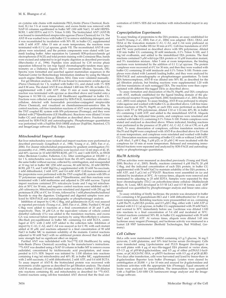
Reconstitution of Chaperone–ANT ComplexesANT can be purified in biochemical amounts from bovineheart (Pebay-Peyroula et al., 2003). Mass spectrometry anal-ysis determined that the purified protein was identical inmass to that predicted by the cDNA sequence (data notshown), indicating that mature ANT should be reverted toits preprotein state by denaturation. Dilution of denaturedANT into RL should then reconstitute the chaperone–pre-protein complexes involved in targeting. We confirmed thatpurified ANT could indeed be reimported into isolated mi-tochondria. First, purified ANT was radiolabeled by tyrosineiodination (Figure 2A, lane 6) and diluted into import reac-tions containing RL and mitochondria. After PK digestion,PK-resistant ANT was observed significantly above that inreactions treated with valinomycin (Figure 2A, lanes 7 and8). The PK-resistant material in the valinomycin controlreactions was similar to that normally observed in reactionsusing cell-free translated ANT (Figure 2A, lanes 1–5). In asecond approach, purified ANT was chemically biotinylatedon its cysteine side chains (Figure 2C, lanes 2–4, Coomassiestain). Import of ANT-B after dilution into reactions contain-ing RL and mitochondria was detected by streptavidin blots(Figure 2A, lanes 9–11). A number of high-molecular-weightbackground bands were observed due to endogenous bio-tinylated mitochondrial proteins (data not shown). Never-theless, imported ANT-B was detected above the valinomy-cin background after PK digestion (Figure 2A, lanes 9 and
Figure 1. Mitochondrial import of ANT. (A) ANT (input, lane 1)was radiolabeled by cell-free translation and imported into isolatedrat liver mitochondria (� mito, lanes 2–9). As a negative control,import was abolished with valinomycin (val, lanes 3 and 7). Thetrue extent of import was determined by resistance to added PK(lanes 6–9). Import was assayed in the presence of 20 �M C-90fragment, a competitor of chaperone-Tom70 interactions (C-90,lanes 4 and 8), or the same mass concentration of bovine serumalbumin as a negative control (BSA, lanes 5 and 9). Reactions wereanalyzed by SDS-PAGE and autoradiography. Right, phosphorim-ager quantitation of import of ANT, PiC, and ISP are shown relativeto control reactions. Standard deviations here, and in all figures,were determined from at least three independent experiments. (B)Import of ANT, PiC, and ISP was assayed upon inhibition with 5�M C-Bag, treatment with 18 �M GA, or combined inhibition withboth reagents, and the quantitation relative to control reactions isshown.
Hsp40 Chaperones in Mitochondrial Import
Vol. 18, September 2007 3417
10), and mock reactions lacking ANT-B showed no corre-sponding band (Figure 2A, lane 11). Thus, the purified ANTcan be reimported into mitochondria, even after chemicalmodification.
Chaperone complexes with Tom70-dependent prepro-teins in RL have a characteristic profile on gel filtrationchromatography. As reported previously (Young et al., 2003;Fan et al., 2006), cell-free translated ANT eluted in a broadpeak at �13-ml elution volume on a 24-ml Superose 6 col-umn, with an apparent molecular size of �600 kDa (Figure2B). Some ANT was also observed in the void volume of thecolumn (8-ml elution volume), most likely representing ag-gregated material (exclusion limit �5 MDa). In parallel,ANT-B was diluted into chaperone binding reactions con-taining RL and ATP for 15 min at room temperature (Youngand Hartl, 2000; Brychzy et al., 2003), and then it was ana-lyzed on the same gel filtration column. Streptavidin blotsdetected a similar peak eluting around 13 ml (Figure 2B), inaddition to void volume material. The reconstituted ANT-Bcomplexes, therefore, reproduce those normally studied bycell-free translation as preimport intermediates, in molecularsize as well as import competence.
To identify the components of chaperone–ANT com-plexes, ANT-B (Figure 2C, lane 4) was incubated with RL(Figure 2C, lane 5) as described above but in larger reactions.ANT-B complexes were then isolated with streptavidin-aga-rose and analyzed by total protein staining (Figure 2C, lane7). Several bands were visible in the 40- to 100-kDa range.Very little ANT-B was observed, because the streptavidin–biotin interaction was mostly resistant to the SDS-containingbuffer used to elute the beads. The protein pattern wasclearly distinct from that of the RL (Figure 2C, lane 5). Mockreactions lacking ANT-B returned far fewer bands (Figure2C, lane 8), and no bands were detected with streptavidin-agarose alone (Figure 2C, lane 6). Visible bands were sub-
jected to tryptic digestion followed by mass spectrometryidentification of peptides. Relevant proteins identified aresummarized in Table 1. Although the exact rabbit sequencesfor many of the proteins are not known, the very highdegree of conservation between chaperones and cochaper-ones within mammals (�97% identity) allowed unambigu-ous identification based on human, rat, mouse or pig se-quences. Accession numbers are for human sequences,unless otherwise indicated.
The band at �90 kDa (Figure 2C, lane 7, labeled a) wasidentified as a mixture of Hsp90� and Hsp90�, the twohighly similar cytosolic forms of the chaperone. The heavy
Figure 2. Reconstitution of chaperone–ANTcomplexes. (A) Left, a representative reactionof cell-free translated ANT (lane 1) importedinto mitochondria (� mito, lanes 2–5) as inFigure 1 is shown. Middle, purified ANT wasradioiodinated (ANT-I125, lane 6) and dilutedinto reactions containing RL, ATP, and mito-chondria (lanes 7 and 8). Right, purified ANTwas biotinylated (ANT-B) and diluted intoreactions containing RL, ATP, and mitochon-dria (lanes 9 and 10), whereas mock reactionscontained no ANT-B (lane 11). Negative con-trol reactions were treated with valinomycin(val) to abolish import (lanes 3, 5, 8, and 10).The extent of import was assessed by resis-tance to PK (lanes 4, 5, 7, 8, and 9–11). (B) Top,ANT was radiolabeled by cell-free translationand resolved on a Superose 6 10/300 GL col-umn. The amount of ANT in each fractionwas determined by SDS-PAGE and phospho-rimager quantitation, and arbitrary phospho-rimager units are represented. The columnvoid volume (V0) and elution volumes of mo-lecular size standards (indicated in kilodal-tons) are marked. Bottom, ANT-B was dilutedinto reactions containing RL and ATP andresolved on the same gel filtration column.
The ANT-B in each fraction was detected by SDS-PAGE and streptavidin blots. The elution volume corresponding to the fractions is marked(indicated in milliliters). (C) Molecular weight markers (indicated in kilodaltons) are shown (lane 1) and purified ANT (lane 2), ANT afterthe biotinylation reaction (ANT-B, lane 3) and removal of excess biotin by desalting (lane 4). ANT-B bound to streptavidin-agarose wasincubated in reactions containing RL (lane 5) and ATP. Complexes were recovered and eluted with Laemmli loading buffer (lane 7). Negativecontrol reactions contained no ANT-B or RL (lane 6) or no ANT-B (lane 8). Discrete bands labeled a, b, c, and the range between d and e, wereanalyzed by mass spectrometry. Bands in the negative control reactions labeled f, g, h, and i also were analyzed.
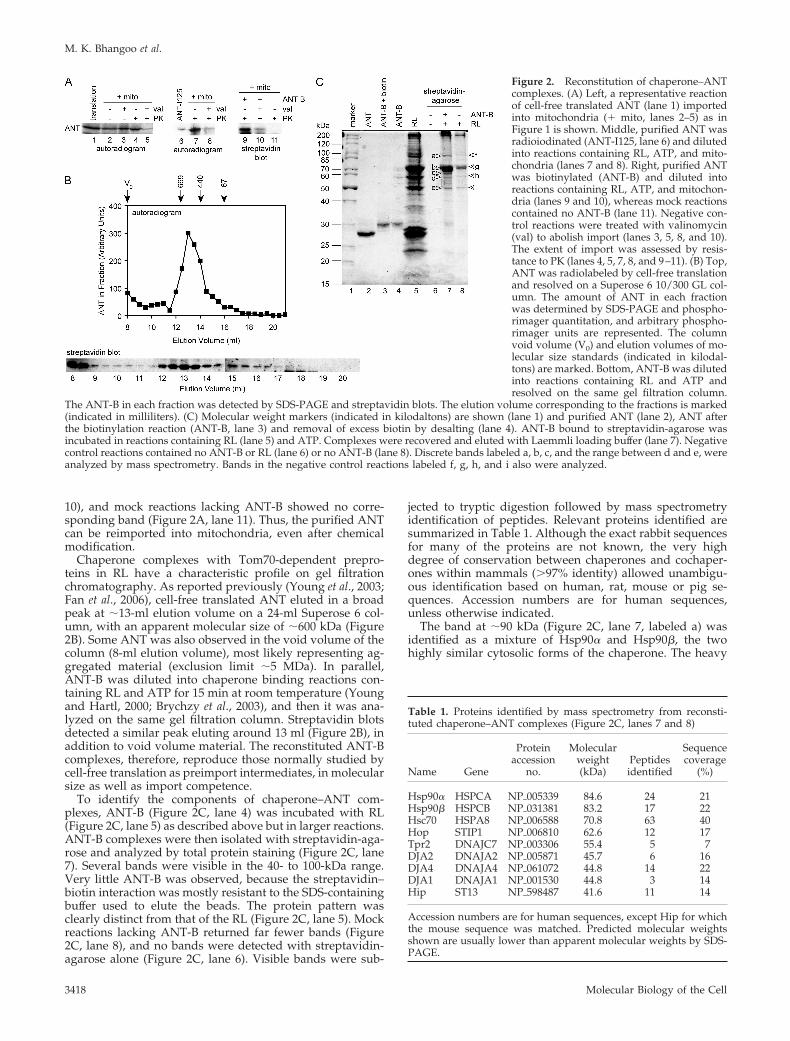
Table 1. Proteins identified by mass spectrometry from reconsti-tuted chaperone–ANT complexes (Figure 2C, lanes 7 and 8)
Name Gene
Proteinaccession
no.
Molecularweight(kDa)
Peptidesidentified
Sequencecoverage
(%)
Hsp90� HSPCA NP�005339 84.6 24 21Hsp90� HSPCB NP�031381 83.2 17 22Hsc70 HSPA8 NP�006588 70.8 63 40Hop STIP1 NP�006810 62.6 12 17Tpr2 DNAJC7 NP�003306 55.4 5 7DJA2 DNAJA2 NP�005871 45.7 6 16DJA4 DNAJA4 NP�061072 44.8 14 22DJA1 DNAJA1 NP�001530 44.8 3 14Hip ST13 NP�598487 41.6 11 14
Accession numbers are for human sequences, except Hip for whichthe mouse sequence was matched. Predicted molecular weightsshown are usually lower than apparent molecular weights by SDS-PAGE.
M. K. Bhangoo et al.
Molecular Biology of the Cell3418
band �70 kDa (labeled b) was found to be Hsc70. Thegreater amount of Hsc70 compared with Hsp90 is in agree-ment with higher dependence on Hsc70 for import (Figure1B). The bands below 70 kDa (labeled c, and a range betweend and e) contained a number of cochaperones of Hsc70 andHsp90. Samples from regions of the gels below the 40-kDamolecular weight did not return reliably identifiable pep-tides except for ANT itself. The major visible bands in neg-ative control reactions (Figure 2C, lane 8, labeled f, g, h, andi) were also analyzed, and they were found to be exclusivelykeratins and hemoglobin multimers (keratin 1, NP_006112;keratin 10, NP_000412; keratin 9, NP_000217; and hemoglo-bin �, NP_000509). These were unavoidable backgroundproteins and ruled out as chaperone complex components.Importantly, no peptides from the chaperone-related com-ponents were identified in the negative controls.
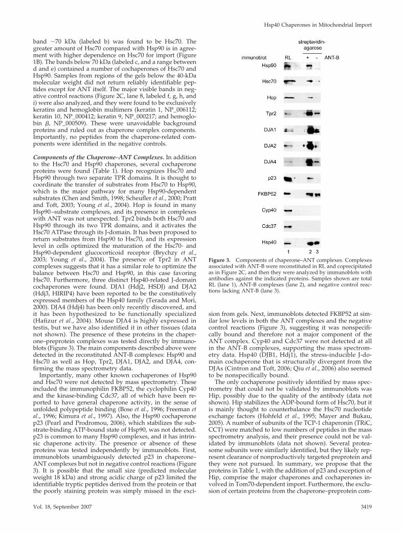
Components of the Chaperone–ANT Complexes. In additionto the Hsc70 and Hsp90 chaperones, several cochaperoneproteins were found (Table 1). Hop recognizes Hsc70 andHsp90 through two separate TPR domains. It is thought tocoordinate the transfer of substrates from Hsc70 to Hsp90,which is the major pathway for many Hsp90-dependentsubstrates (Chen and Smith, 1998; Scheufler et al., 2000; Prattand Toft, 2003; Young et al., 2004). Hop is found in manyHsp90–substrate complexes, and its presence in complexeswith ANT was not unexpected. Tpr2 binds both Hsc70 andHsp90 through its two TPR domains, and it activates theHsc70 ATPase through its J-domain. It has been proposed toreturn substrates from Hsp90 to Hsc70, and its expressionlevel in cells optimized the maturation of the Hsc70- andHsp90-dependent glucocorticoid receptor (Brychzy et al.,2003; Young et al., 2004). The presence of Tpr2 in ANTcomplexes suggests that it has a similar role to optimize thebalance between Hsc70 and Hsp90, in this case favoringHsc70. Furthermore, three distinct Hsp40-related J-domaincochaperones were found. DJA1 (Hdj2, HSDJ) and DJA2(Hdj3, HIRIP4) have been reported to be the constitutivelyexpressed members of the Hsp40 family (Terada and Mori,2000). DJA4 (Hdj4) has been only recently discovered, andit has been hypothesized to be functionally specialized(Hafizur et al., 2004). Mouse DJA4 is highly expressed intestis, but we have also identified it in other tissues (datanot shown). The presence of these proteins in the chaper-one–preprotein complexes was tested directly by immuno-blots (Figure 3). The main components described above weredetected in the reconstituted ANT-B complexes: Hsp90 andHsc70 as well as Hop, Tpr2, DJA1, DJA2, and DJA4, con-firming the mass spectrometry data.
Importantly, many other known cochaperones of Hsp90and Hsc70 were not detected by mass spectrometry. Theseincluded the immunophilin FKBP52, the cyclophilin Cyp40and the kinase-binding Cdc37, all of which have been re-ported to have general chaperone activity, in the sense ofunfolded polypeptide binding (Bose et al., 1996; Freeman etal., 1996; Kimura et al., 1997). Also, the Hsp90 cochaperonep23 (Pearl and Prodromou, 2006), which stabilizes the sub-strate-binding ATP-bound state of Hsp90, was not detected.p23 is common to many Hsp90 complexes, and it has intrin-sic chaperone activity. The presence or absence of theseproteins was tested independently by immunoblots. First,immunoblots unambiguously detected p23 in chaperone–ANT complexes but not in negative control reactions (Figure3). It is possible that the small size (predicted molecularweight 18 kDa) and strong acidic charge of p23 limited theidentifiable tryptic peptides derived from the protein or thatthe poorly staining protein was simply missed in the exci-
sion from gels. Next, immunoblots detected FKBP52 at sim-ilar low levels in both the ANT complexes and the negativecontrol reactions (Figure 3), suggesting it was nonspecifi-cally bound and therefore not a major component of theANT complex. Cyp40 and Cdc37 were not detected at allin the ANT-B complexes, supporting the mass spectrom-etry data. Hsp40 (DJB1, Hdj1), the stress-inducible J-do-main cochaperone that is structurally divergent from theDJAs (Cintron and Toft, 2006; Qiu et al., 2006) also seemedto be nonspecifically bound.
The only cochaperone positively identified by mass spec-trometry that could not be validated by immunoblots wasHip, possibly due to the quality of the antibody (data notshown). Hip stabilizes the ADP-bound form of Hsc70, but itis mainly thought to counterbalance the Hsc70 nucleotideexchange factors (Hohfeld et al., 1995; Mayer and Bukau,2005). A number of subunits of the TCP-1 chaperonin (TRiC,CCT) were matched to low numbers of peptides in the massspectrometry analysis, and their presence could not be val-idated by immunoblots (data not shown). Several protea-some subunits were similarly identified, but they likely rep-resent clearance of nonproductively targeted preprotein andthey were not pursued. In summary, we propose that theproteins in Table 1, with the addition of p23 and exception ofHip, comprise the major chaperones and cochaperones in-volved in Tom70-dependent import. Furthermore, the exclu-sion of certain proteins from the chaperone–preprotein com-
Figure 3. Components of chaperone–ANT complexes. Complexesassociated with ANT-B were reconstituted in RL and coprecipitatedas in Figure 2C, and then they were analyzed by immunoblots withantibodies against the indicated proteins. Samples shown are totalRL (lane 1), ANT-B complexes (lane 2), and negative control reac-tions lacking ANT-B (lane 3).
Hsp40 Chaperones in Mitochondrial Import
Vol. 18, September 2007 3419

plexes suggests a substantial degree of specificity in complexformation.
DJA Interaction with PreproteinWe were struck by the presence of three related DJA pro-teins in the chaperone–ANT complexes. Although all threewere present, it was possible that one in particular might bemore effective than the others. For example, the higher num-ber of peptides derived from DJA4 might suggest a greaterinvolvement of that cochaperone with preprotein com-plexes. Alternatively, there might be subtler differences be-tween them, such that they function together in import, andperhaps other biological processes. In the yeast S. cerevisiae,the two cytosolic Hsp40-related proteins Ydj1 and Sis1 havedistinct biological roles (Johnson and Craig, 2001; Fan et al.,2004); however, Ydj1 is orthologous to the three human DJAproteins, whereas Sis1 is related to the divergent Hsp40/DJB1. The homology between the human DJAs (70–85%similarity) suggests they have a closer functional relation-ship. The DJA proteins contain the J-domain, zinc fingerdomain, and C-terminal dimerization domain (Figure 4C,diagram) conserved from the eponymous DnaJ of E. coli(Terada et al., 1997; Terada and Mori, 2000; Borges et al.,2005; Qiu et al., 2006). As expected, their N-terminal J-do-mains were found to activate ATP hydrolysis by Hsc70(Terada and Mori, 2000; Hafizur et al., 2004). Based on ex-periments with the E. coli and S. cerevisiae orthologues, thebinding site for unfolded polypeptides should reside in thecentral- to C-terminal regions of the DJA proteins (Walsh etal., 2004; Mayer and Bukau, 2005; Cintron and Toft, 2006;Qiu et al., 2006). Like most J-domains, those in the DJAproteins are well conserved, and the greatest divergence in
sequence is within the expected substrate binding region.This suggested that one difference between the DJAs mightbe the range of substrates bound, or the strength of binding.However, sequences outside the J-domains of some proteinshave been implicated in Hsc70 interactions, so there mightalso be differences between the DJAs in the activation ofHsc70. We therefore addressed these possibilities systemat-ically.
The binding of the DJAs to the polypeptide substrate ANTwas tested first. The polypeptide binding specificities of thehuman DJAs are so far unknown. For ease and accuracy ofquantitation, we adapted a coprecipitation assay used tostudy preprotein interactions with Tom70 (Young et al.,2003; Fan et al., 2006). DJA1, DJA2, and DJA4 were purifiedas N-terminally His-tagged proteins. Radiolabeled ANTproduced by cell-free translation was incubated with iden-tical amounts (5 �M) of each DJA protein. The His-taggedproteins were reisolated with nickel-Sepharose, and the as-sociated ANT was quantified. All three DJAs bound ANT atlevels above the low background binding, but DJA2 boundnoticeably less preprotein than DJA1 or DJA4 (Figure 4A,lanes 2–5). Quantitation showed that DJA2 binding of ANTwas �40% of DJA1 binding, whereas DJA4 binding was notsignificantly different from that of DJA1 (Figure 4B). Forcomparison with another Tom70-dependent preprotein, theassay was applied to PiC (Figure 4, A and B). Again, DJA1bound PiC well, and DJA2 seemed to have the poorestbinding, at �40% of DJA1. However, DJA4 bound PiC rel-atively poorly, at �60% of DJA1 binding. These data sug-gested that the DJAs differ in their binding of polypeptides.
The full-length DJA proteins might bind polypeptides in adynamic equilibrium, because their J-domains promote the
Figure 4. DJA interaction with preprotein. A, ANT,and PiC were radiolabeled by cell-free translation (lane1) and coprecipitated with nickel-Sepharose alone(beads, lane 2), or with nickel-Sepharose and purifiedHis-tagged DJA1, DJA2, or DJA4 at a final concentration5 �M (lanes 3–5). Recovered material was analyzed bySDS-PAGE and autoradiography. (B) Phosphorimagerquantitation is shown of the coprecipitation of ANT andPiC with DJA1, DJA2 and DJA4, as in described in A,relative to that with DJA1 set to 1. (C) Diagram of DJA1,DJA2, and DJA4 architecture. J-domain (J, black), zincfinger (Zn, light gray), and C-terminal domains (C, darkgray) are indicated. The start sites of C-A1, C-A2, andC-A4 deletion mutants are marked (dashed line, aminoacid numbers indicated). (D) ANT and PiC were copre-cipitated with purified His-tagged C-A1, C-A2, andC-A4 as described in A. Phosphorimager quantitation ofthe coprecipitation is shown, relative to that with C-A1set to 1. (E) Cell-free translated ANT and PiC (lane 1)were imported into mitochondria (� mito, lanes 2–13)as described in Figure 1. As a negative control, importwas abolished with valinomycin (val, lanes 3 and 9).Reactions were supplemented with 90 mM NaCl alone(S, lanes 4 and 10) or with 20 �M C-A1, C-A2, or C-A4with 90 mM NaCl (lanes 5–7 and 11–13). The extent ofimport was assessed by resistance to PK (lanes 8–13).The mature noncleaved form of ANT (m) is visible, asare the precursor (p) and proteolytically cleaved mature(m) forms of PiC. (F) Phosphorimager quantitation isshown of the import of ANT, PiC, and ISP in the pres-ence of 20 �M C-A1, C-A2, or C-A4, as described in E,relative to control reactions with added NaCl.
M. K. Bhangoo et al.
Molecular Biology of the Cell3420

transfer of polypeptides to Hsc70. The association of ANTand PiC with the DJAs might then depend on interactionswith Hsc70 as well as the actual affinity of binding to theDJAs. To directly address the level of polypeptide binding,truncation mutants of the DJAs lacking the N-terminal J-domain were purified. The deletion mutants, C-A1 (residues98-397 of DJA1), C-A2 (residues 100-412 of DJA2), and C-A4(residues 99-397 of DJA4), were also N-terminally His-tagged, and they contained the complete zinc finger andC-terminal regions of the DJAs (Figure 4C). The binding ofcell-free translated ANT and PiC to the mutants was testedas described above. C-A1 was found to bind strongly to bothANT and PiC, and C-A2 binding to both polypeptides was�50% of C-A1 binding (Figure 4D). C-A4 bound slightly lessANT than C-A1 and significantly less PiC, at �60% relativeto C-A1. Overall, the interactions of the truncation mutantswith polypeptides were similar to those of the full-lengthDJAs, suggesting that Hsc70 interactions did not greatlychange the substrate binding profiles of full-length versustruncated DJAs. These results confirmed that the DJAs di-verge in polypeptide binding, with DJA1 apparently bind-ing the highest amount of preprotein. Also, as observed forDJA4, different polypeptides can be bound to varying de-grees.
We wanted to evaluate the importance of DJA binding topreprotein for mitochondrial import. Immunodepletion ofthe DJAs from RL could not be achieved with the availableantibodies (data not shown). So, we reasoned that the trun-cated DJAs should act as dominant-negative inhibitors ofwild-type DJA function, by competing for the same bindingsites in preprotein but disallowing transfer to Hsc70. Inhi-bition of Hsc70 binding should then impair the Tom70 dock-ing step required for import. Alternatively, Hsp40 can acti-vate substrate binding by Hsc70 without strongly interactingwith substrate (Minami et al., 1996; Cintron and Toft, 2006).If Hsp40 or some mechanism other than the DJAs wassufficient for Hsc70 binding, the truncated DJAs should haveless effect. Therefore, C-A1, C-A2, and C-A4 were added at20 �M to mitochondrial import reactions of ANT and PiC.Note that PiC is proteolytically processed upon import toproduce a faster migrating band on SDS-PAGE (Zara et al.,1992), but the extent of true import was quantified as de-scribed for ANT by resistance to externally added PK (Fig-ure 4E). Indeed, all three mutants clearly inhibited import ofANT and PiC to between 40 and 60% of control reactions(Figure 4F). The partial but significant inhibition of importapproached the level observed with the C-90 competitor orcombined Hsc70 and Hsp90 inhibition in Figure 1. As ex-pected, the mutant DJAs had little effect on the Tom20-dependent import of ISP (Figure 4F). For ANT and PiC, itseems that the effect of each dominant DJA mutant cannot beovercome by the other DJA proteins or J-domain cochaper-ones present in the lysate. These data suggest that each ofthe DJAs function in the Tom70 import pathway.
Hsc70–Preprotein InteractionsTo further test the importance of the DJAs for Hsc70 bindingto preprotein, the binding of Hsc70 to purified ANT wasmonitored over time. As established in previous work onHsc70 and Hsp90 interactions with the glucocorticoid recep-tor (Young and Hartl, 2000; Sondermann et al., 2001; Brychzyet al., 2003), radiolabeled Hsc70 generated by cell-free trans-lation was used for ease and accuracy of quantitation. Al-though unlabeled Hsc70 is in excess in the binding reactions,the radiolabeled protein acts as a tracer and matches thebehavior of unlabeled protein. At different times after addi-tion to ANT-B, the preprotein was recovered with strepta-
vidin-agarose, and bound Hsc70 was quantified. Hsc70bound with a fast initial increase and reached a maximum at�10 min (Figure 5A). When C-A1, C-A2, and C-A4 wereadded to the reactions at 20 �M, binding of Hsc70 wasclearly diminished (Figure 5A). Moreover, C-A2 was themost inhibitory, reducing Hsc70 binding as early as 1 mininto the reaction, with a final level of �30% of the controlreaction. C-A1 and C-A4 were slightly more moderate intheir effect, with inhibition visible by 2–4 min and a finalHsc70 level of �60% of the control. Thus, the three trunca-tion mutants directly inhibit Hsc70 interactions with prepro-tein, albeit to different degrees.
The dominant mutants seem to compete with wild-typeDJAs for polypeptide, preventing the wild-type cochaper-ones from initiating Hsc70 binding (Cintron and Toft, 2006).
Figure 5. Hsc70-preprotein interactions. (A) Top, Hsc70 was ra-diolabeled by cell-free translation, incubated with ANT-B immobi-lized on streptavidin-agarose, and coprecipitated after the indicatedtimes. Reactions contained either no addition, or 20 �M purifiedC-A1, C-A2, or C-A4. Recovered material was analyzed by SDS-PAGE and autoradiography. Bottom, quantitation of time courses ofHsc70 binding is shown. (B) Top, cell-free translated Hsc70 wascoprecipitated with ANT-B as described in A but after 15 min ofbinding. Samples were resuspended in dissociation reactions withor without ATP, containing buffer alone, or 5 �M purified p23 orC-Bag. After 10 min, released and remaining bound fractions wereseparated and analyzed. Bottom, quantitation of dissociation reac-tions.
Hsp40 Chaperones in Mitochondrial Import
Vol. 18, September 2007 3421
The partial effects of the mutants suggested that competitionwith the wild-type DJAs was not complete. This in turnimplied that the DJAs have distinct sets of binding sites thatonly partly overlap with each other. Interestingly, C-A2 wasthe strongest inhibitor of Hsc70 binding (Figure 5A), but itseemed to bind less overall preprotein than C-A1 or C-A4(Figure 4D). We suggest that C-A2 might bind fewer siteswithin the polypeptides, but these sites are relatively moreimportant for Hsc70 binding. In contrast, C-A1 and C-A4might bind more, but less distinctly important, sites. Theoverall result is that the three mutants inhibit preproteinimport to a similar degree (Figure 4F).
To confirm that the Hsc70–preprotein complexes ob-served in these experiments were formed normally, theirnucleotide dependence was assayed. In previous experi-ments with the glucocorticoid receptor, the C-Bag nucleotideexchange factor promoted Hsc70 dissociation from isolatedcomplexes in an ATP-dependent manner (Sondermann et al.,2001). Following a similar approach, Hsc70–ANT complexeswere isolated after 15 min of binding, and then they wereincubated under varying conditions in the absence or pres-ence of ATP. Dissociated and bead-bound fractions wereanalyzed separately. As observed previously, ATP alone didnot promote complex dissociation greatly over buffer with-out ATP (Figure 5B). As another negative control, the puri-fied Hsp90-specific cochaperone p23 also did not stronglydissociate Hsc70. However, C-Bag markedly promotedHsc70 dissociation but only in the presence of ATP (Figure5B). Thus, the Hsc70–preprotein complexes inhibited by theDJA mutants seem to be normally functional complexes.
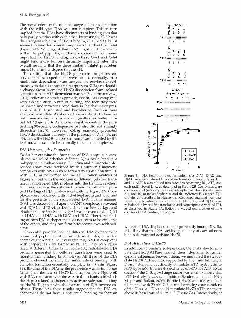
DJA Heterocomplex FormationTo further examine the formation of DJA–preprotein com-plexes, we asked whether different DJAs could bind to apolypeptide simultaneously. Experimental approaches de-scribed above were modified for this purpose. Chaperonecomplexes with ANT-B were formed by its dilution into RLwith ATP, as performed for the gel filtration analysis ofFigure 2B, but with the addition of different cell-free trans-lated, radiolabeled DJA proteins into the binding reaction.Each reaction was then allowed to bind to a different puri-fied His-tagged DJA protein identically to Figure 4A. Com-plexes were reisolated with nickel-Sepharose and analyzedfor the presence of the radiolabeled DJA. In this manner,DJA1 was detected in chaperone–ANT complexes recoveredwith DJA2 and DJA4, at levels above the low backgroundbinding (Figure 6A). Similar, DJA2 was recovered with DJA1and DJA4, and DJA4 with DJA1 and DJA2. Therefore, bind-ing of each DJA cochaperone does not seem to be exclusiveof the others, and they can form heterocomplexes with sub-strate.
It was also possible that the different DJA cochaperonesbound polypeptide substrate in a defined order, or with acharacteristic kinetic. To investigate this, ANT-B complexeswith chaperones were formed in RL, and they were reiso-lated at different times as in Figure 5A; radiolabeled DJAproteins generated by cell-free translation were used tomonitor their binding to complexes. All three of the DJAproteins showed the same fast initial rate of binding, withcomplex formation essentially complete in �5 min (Figure6B). Binding of the DJAs to the preprotein was as fast, if notfaster than, the rate of Hsc70 binding (compare Figure 6Bwith 5A), consistent with the expected mechanism in whichthe Hsp40-related cochaperones activate substrate bindingby Hsc70. Together with the formation of DJA heterocom-plexes (Figure 6A), these results suggest that the DJA co-chaperones do not have a sequential binding mechanism
where one DJA displaces another previously bound DJA. So,it is likely that the DJAs act independently of each other tobind substrate and activate Hsc70.
DJA Activation of Hsc70In addition to binding polypeptides, the DJAs should acti-vate the Hsc70 ATPase through their J domains. To furtherexplore differences between them, we measured the steady-state Hsc70 ATPase rates supported by the three full-lengthDJAs. J-domains specifically stimulate ATP hydrolysis toADP by Hsc70, but not the exchange of ADP for ATP, so anexcess of the C-Bag exchange factor was used to ensure thatATP hydrolysis was rate limiting (Sondermann et al., 2001;Mayer and Bukau, 2005). Purified Hsc70 at 4 �M was sup-plemented with 20 �M C-Bag and increasing concentrationsof the DJAs. All DJAs could stimulate Hsc70 ATPase activityabove its basal rate of �1 min�1 (Figure 7A). Interestingly, at
Figure 6. DJA heterocomplex formation. (A) DJA1, DJA2, andDJA4 were radiolabeled by cell-free translation (input, lanes 1, 5,and 9). ANT-B was diluted into reactions containing RL, ATP, andeach radiolabeled DJA, as described in Figure 2B. Complexes werecoprecipitated (recovery) with nickel-Sepharose alone (beads, lanes2, 6, and 10) or nickel-Sepharose and the indicated His-tagged DJAprotein, as described in Figure 4A. Recovered material was ana-lyzed by autoradiography. (B) Top, DJA1, DJA2, and DJA4 wereradiolabeled by cell-free translation and coprecipitated with ANT-Bas described in Figure 5A. Bottom, averaged quantitation of timecourses of DJA binding are shown.
M. K. Bhangoo et al.
Molecular Biology of the Cell3422

all concentrations above 1 �M, DJA1 was clearly more effi-cient at ATPase stimulation than DJA2 and DJA4, whichwere similar to each other (Figure 7A). At 8 �M DJA1, Hsc70activity reached around 7 min�1, in line with previouslyreported rates, whereas DJA2 and DJA4 at the same concen-trations supported rates �5 min�1. Our results are in thesame range as reported previously (Terada and Mori, 2000;Hafizur et al., 2004), with the difference that in our hands,DJA4 is somewhat more active than was thought. Althoughthe difference between DJA1 and the other two may notseem great, in a finely balanced biological system, the func-tional consequences of such variation may be amplified.Also, differences between DJA activation of Hsc70 seem tobe greatest (at least 2-fold) in the concentration range of 2–4�M DJAs, which lies closest to the estimated cellular con-centration of the DJAs, and the estimated ratio to Hsc70 incells.
Another property of the DJAs was their expected ability tosupport polypeptide refolding by Hsc70. A polypeptide sub-strate commonly used to study Hsc70 function is fireflyluciferase, which has a sensitive and reproducible enzymaticassay. Purified Hsc70 has been shown to assist luciferaserefolding in numerous studies (Hohfeld et al., 1995; Freemanand Morimoto, 1996; Minami et al., 1996; Hohfeld andJentsch, 1997; Terada et al., 1997; Luders et al., 2000; Teradaand Mori, 2000). However, in most cases the stress-induciblecochaperone Hsp40 was used, and a direct comparison be-tween all three DJAs with Hsc70 has not yet been reported.Thus, we tested the refolding of luciferase after dilution outof 6 M guanidine into reactions containing 4 �M Hsc70 and4 �M of each DJA in turn, as well as 0.5 �M C-Bag, whichwas found to be optimal for refolding at this concentration(data not shown). Refolding reactions contained 1 mM ATP,
whereas negative control reactions contained no ATP, andthey were treated with apyrase to destroy any endogenousATP. 50% RL with ATP served as a positive control. Sur-prisingly, DJA1 seemed to be the least effective in assistingrefolding (Figure 7B), despite providing the strongest stim-ulation of the Hsc70 ATPase activity (Figure 7A). Refoldingwith DJA1 reached slightly above 20% of the RL control after60 min (Figure 7B). In marked contrast, DJA2 was veryefficient at promoting luciferase refolding, supporting a re-folding level essentially the same as the RL control. DJA4, incontrast, was also relatively weak in promoting refolding,although the final activity reached was �40% of the RLcontrol and somewhat higher than with DJA1. In all cases,the negative control reactions lacking ATP produced nosignificant refolding (Figure 7B). Overall, the divergent re-folding function of the DJAs may result from a more com-plex combination of differences in polypeptide binding,Hsc70 ATPase stimulation, and perhaps other factors. In anearlier report, DJA1 and DJA2 promoted luciferase refoldingto similar degrees (Terada and Mori, 2000), and the diver-gence from our results seems due to apparently small dif-ferences in experimental conditions. It might be that DJA2 ismore robust and able to support refolding under a broaderrange of conditions. The variability of the results may also betaken as further evidence that minor differences between theDJAs might be amplified depending on the biological con-text.
DJA Function in CellsThe refolding of a guanidine-denatured protein can be dis-tinct from the cellular folding of the same protein during itssynthesis on ribosomes. Experiments in mammalian and S.
Figure 7. DJA activation of Hsc70. (A)Steady-state ATPase rates of purified Hsc70were measured in reactions containing 1 mMATP, 4 �M Hsc70, 20 �M C-Bag, and theindicated concentrations of DJA1, DJA2, orDJA4. Right inset, representative example ofthe data from which linear rates were calcu-lated. Reactions contained �-[32P]ATP and theamount of ADP produced at each time pointwas determined by separation on TLC andphosphorimager quantitation. (B) Refoldingof guanidine-denatured luciferase was moni-tored in reactions containing 50% RL and 2mM ATP, or 4 �M Hsc70, 0.5 �M C-Bag, and4 �M DJA1, DJA2, or DJA4 as indicated, withor without 2 mM ATP. The activity of lucif-erase refolded in RL reactions at 60 min wasset to 100%.
Hsp40 Chaperones in Mitochondrial Import
Vol. 18, September 2007 3423
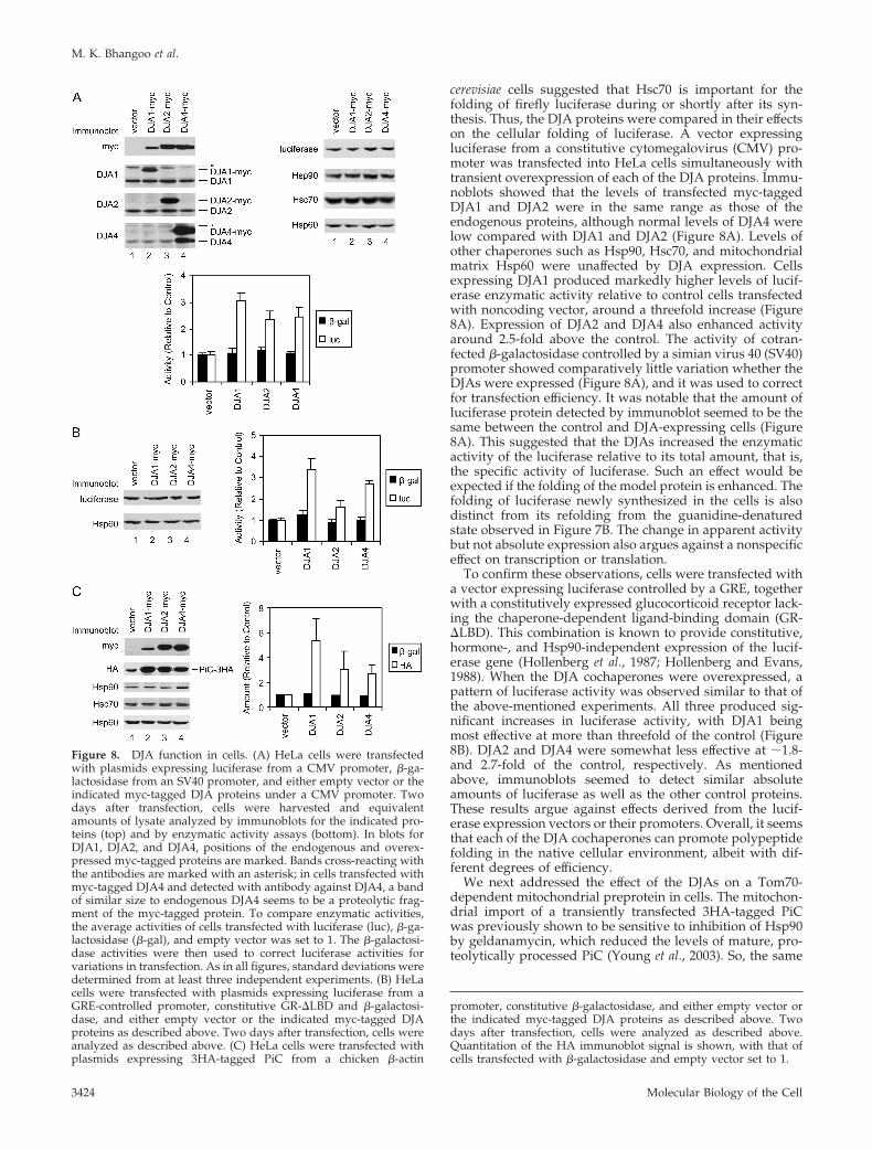
cerevisiae cells suggested that Hsc70 is important for thefolding of firefly luciferase during or shortly after its syn-thesis. Thus, the DJA proteins were compared in their effectson the cellular folding of luciferase. A vector expressingluciferase from a constitutive cytomegalovirus (CMV) pro-moter was transfected into HeLa cells simultaneously withtransient overexpression of each of the DJA proteins. Immu-noblots showed that the levels of transfected myc-taggedDJA1 and DJA2 were in the same range as those of theendogenous proteins, although normal levels of DJA4 werelow compared with DJA1 and DJA2 (Figure 8A). Levels ofother chaperones such as Hsp90, Hsc70, and mitochondrialmatrix Hsp60 were unaffected by DJA expression. Cellsexpressing DJA1 produced markedly higher levels of lucif-erase enzymatic activity relative to control cells transfectedwith noncoding vector, around a threefold increase (Figure8A). Expression of DJA2 and DJA4 also enhanced activityaround 2.5-fold above the control. The activity of cotran-fected �-galactosidase controlled by a simian virus 40 (SV40)promoter showed comparatively little variation whether theDJAs were expressed (Figure 8A), and it was used to correctfor transfection efficiency. It was notable that the amount ofluciferase protein detected by immunoblot seemed to be thesame between the control and DJA-expressing cells (Figure8A). This suggested that the DJAs increased the enzymaticactivity of the luciferase relative to its total amount, that is,the specific activity of luciferase. Such an effect would beexpected if the folding of the model protein is enhanced. Thefolding of luciferase newly synthesized in the cells is alsodistinct from its refolding from the guanidine-denaturedstate observed in Figure 7B. The change in apparent activitybut not absolute expression also argues against a nonspecificeffect on transcription or translation.
To confirm these observations, cells were transfected witha vector expressing luciferase controlled by a GRE, togetherwith a constitutively expressed glucocorticoid receptor lack-ing the chaperone-dependent ligand-binding domain (GR-�LBD). This combination is known to provide constitutive,hormone-, and Hsp90-independent expression of the lucif-erase gene (Hollenberg et al., 1987; Hollenberg and Evans,1988). When the DJA cochaperones were overexpressed, apattern of luciferase activity was observed similar to that ofthe above-mentioned experiments. All three produced sig-nificant increases in luciferase activity, with DJA1 beingmost effective at more than threefold of the control (Figure8B). DJA2 and DJA4 were somewhat less effective at �1.8-and 2.7-fold of the control, respectively. As mentionedabove, immunoblots seemed to detect similar absoluteamounts of luciferase as well as the other control proteins.These results argue against effects derived from the lucif-erase expression vectors or their promoters. Overall, it seemsthat each of the DJA cochaperones can promote polypeptidefolding in the native cellular environment, albeit with dif-ferent degrees of efficiency.
We next addressed the effect of the DJAs on a Tom70-dependent mitochondrial preprotein in cells. The mitochon-drial import of a transiently transfected 3HA-tagged PiCwas previously shown to be sensitive to inhibition of Hsp90by geldanamycin, which reduced the levels of mature, pro-teolytically processed PiC (Young et al., 2003). So, the same
Figure 8. DJA function in cells. (A) HeLa cells were transfectedwith plasmids expressing luciferase from a CMV promoter, �-ga-lactosidase from an SV40 promoter, and either empty vector or theindicated myc-tagged DJA proteins under a CMV promoter. Twodays after transfection, cells were harvested and equivalentamounts of lysate analyzed by immunoblots for the indicated pro-teins (top) and by enzymatic activity assays (bottom). In blots forDJA1, DJA2, and DJA4, positions of the endogenous and overex-pressed myc-tagged proteins are marked. Bands cross-reacting withthe antibodies are marked with an asterisk; in cells transfected withmyc-tagged DJA4 and detected with antibody against DJA4, a bandof similar size to endogenous DJA4 seems to be a proteolytic frag-ment of the myc-tagged protein. To compare enzymatic activities,the average activities of cells transfected with luciferase (luc), �-ga-lactosidase (�-gal), and empty vector was set to 1. The �-galactosi-dase activities were then used to correct luciferase activities forvariations in transfection. As in all figures, standard deviations weredetermined from at least three independent experiments. (B) HeLacells were transfected with plasmids expressing luciferase from aGRE-controlled promoter, constitutive GR-�LBD and �-galactosi-dase, and either empty vector or the indicated myc-tagged DJAproteins as described above. Two days after transfection, cells wereanalyzed as described above. (C) HeLa cells were transfected withplasmids expressing 3HA-tagged PiC from a chicken �-actin
promoter, constitutive �-galactosidase, and either empty vector orthe indicated myc-tagged DJA proteins as described above. Twodays after transfection, cells were analyzed as described above.Quantitation of the HA immunoblot signal is shown, with that ofcells transfected with �-galactosidase and empty vector set to 1.
M. K. Bhangoo et al.
Molecular Biology of the Cell3424

protein was constitutively expressed along with transientoverexpression of each DJA cochaperone in HeLa cells.When levels of mature PiC were tested by immunoblot,noticeably more protein was detected upon expression ofDJA1 relative to control cells (Figure 8C). More PiC was alsoobserved with expression of DJA2 and DJA4. Again, the�-galactosidase transfection control showed relatively littlevariation (Figure 8C). The increased accumulation of PiCwas most likely due to increased efficiency of import. ThePiC detected by immunoblots corresponded in size to themature form of the protein that is proteolytically processedafter import, suggesting that in the vector control, excess PiCthat could not be imported was removed by degradation,possibly by proteosomes. Furthermore, levels of mitochon-drial Hsp60 remained unchanged upon DJA expression (Fig-ure 8C), arguing against an effect on the Tom20-dependentimport pathway used by Hsp60, or a general expansion ofthe mitochondria. These data are consistent with a functionof the DJAs in promoting the import of the transfected PiC.
In the live cell experiments, transient overexpression ofDJA1 was typically less than that of DJA2 and DJA4, asjudged by detection of the myc-tags (Figure 8, A and C).DJA1 still showed the greatest effect on luciferase activityand the accumulation of PiC, within the limits of immuno-blot quantitation. Overall, our in vitro and live cell resultssuggest that the DJA proteins differ in separate aspects oftheir function—polypeptide binding, Hsc70 ATPase stimu-lation, assistance of protein folding or refolding, and mito-chondrial import—and that no single DJA is superior in allaspects.
Hsp90–Preprotein InteractionsIn the analysis of chaperone–preprotein complexes, Hsp90and its cochaperone p23 were positively identified. Hsp90seems to have a lesser, although still significant role in theimport of ANT, and we next addressed the characteristics ofits complex formation. Performed as described above forHsc70 (Figure 5A), the binding of Hsp90� to ANT-B wasmonitored over time by using cell-free translated radiola-beled Hsp90� as a tracer. Like Hsc70, Hsp90 bound with afast initial phase and reached its maximum level by 10 min(Figure 9A). In models of Hsp90 function based on steroidreceptor maturation, a complex mediated by Hop acts toload substrate polypeptide from Hsc70 onto Hsp90 (Chenand Smith, 1998; Pratt and Toft, 2003). However, in othercases such as with some kinases, Hop mediation seemsdispensable for Hsp90 entry into complexes (Lee et al., 2002;Lee et al., 2004). Because Hop was positively identified as acomponent of chaperone–ANT complexes (Figure 3 and Ta-ble 1), the importance of Hsp90 interactions with Hop forcomplex formation was assayed. The C-90 fragment com-petes with full-length Hsp90 for the binding site on Hop(Scheufler et al., 2000), and when added at 20 �M to Hsp90binding reactions with ANT-B, it markedly reduced theamount of Hsp90 bound to �30% of the control (Figure 9A).This suggests that a large fraction of Hsp90 requires theinteraction with Hop for recruitment to preprotein com-plexes. The three DJA truncation mutants lacking J domainsalso partially reduced Hsp90 binding (data not shown), infurther agreement with Hop-mediated recruitment of Hsp90to Hsc70-bound preprotein. Nevertheless, there is somelevel of Hop-independent binding of preprotein by Hsp90(Figure 9A), most likely by direct recognition of polypeptide.
The Hsp90 cochaperone Aha1 was recently found to stim-ulate the Hsp90 ATPase activity by binding to the centralregion of the chaperone (Panaretou et al., 2002; Lotz et al.,2003). Aha1 has been hypothesized to promote dissociation
of Hsp90 from polypeptide substrate, but no direct experi-mental demonstration of this has yet been reported. Al-though Aha1 was not found to be a component of ourchaperone–ANT complexes (Table 1; data not shown), it waspossible that a dissociation factor would not be stably bounddue to its function. We therefore asked what effect, if any,Aha1 would have on Hsp90 binding reactions with ANT.Intriguingly, 4 �M Aha1 caused a transient increase in theamount of Hsp90 bound, followed by a progressive decline(Figure 9A). The peak of Hsp90 binding in the presence ofadded Aha1 varied between 20 and 60% above the final levelof control reactions, and it was reached by 4–6 min; becausethe reason for the increase is unclear, a conservative repre-sentative time course experiment is shown. Final levels ofHsp90 binding with Aha1 continued to drop below 70% ofthe control after 10 min. Addition of p23 had no effect onHsp90 binding (data not shown). These data agree in partwith a function of Aha1 in dissociating Hsp90 complexes;
Figure 9. Hsp90–preprotein interactions. (A) Top, Hsp90 was ra-diolabeled by cell-free translation and coprecipitated with ANT-B asdescribed in Figure 5A. Reactions contained either no addition, or20 �M purified C-90 or 4 �M Aha1. Bottom, quantitation of timecourses of Hsp90 binding is shown. (B) Top, cell-free translatedHsp90 was coprecipitated with ANT-B, and the dissociation ofcomplexes monitored as described in Figure 5B. Dissociation reac-tions, with or without ATP, contained buffer alone, or 5 �M purifiedp23 or C-Bag, or 4 �M Aha1, or 5 �M p23 and 4 �M Aha1. Bottom,quantitation of dissociation reactions.
Hsp40 Chaperones in Mitochondrial Import
Vol. 18, September 2007 3425

however, the two-phase Hsp90 binding curve suggests thatthe dynamic equilibrium of the complexes may be changedover time by Aha1.
Next, the dissociation of Hsp90–preprotein complexeswas assayed. Hsp90 complexes with ANT-B were formedfor 15 min as described above and isolated, and then theywere incubated in the presence of various purified proteinsas described for Hsc70. The absence or presence of ATP hadno effect on Hsp90 dissociation when no protein was added(Figure 9B). As a negative control, the Hsc70-specific co-chaperone C-Bag also had no effect, independent of nucleo-tide presence. The cochaperone p23 at 5 �M favored somedissociation of Hsp90 in the presence of ATP (Figure 9B).This effect was less pronounced than previously observedwith glucocorticoid receptor complexes (Young and Hartl,2000; Brychzy et al., 2003), under somewhat different exper-imental conditions. Aha1 at 4 �M had a slightly strongerability to promote Hsp90 dissociation, again requiring ATP(Figure 9B). Combined treatment with p23 and Aha1 seemedto reduce dissociation to the level of the buffer control (Fig-ure 9B), suggesting there was no synergy of function be-tween the cochaperones and that they might counteract eachother. This would be consistent with their overlapping bind-ing sites on Hsp90 (Ali et al., 2006). The C-90 fragment hadno dissociation effect on Hsp90–ANT complexes (data notshown), indicating that once Hsp90 bound preprotein itsbinding became independent of Hsc70 and Hop. Our resultssuggest that Aha1 may indeed act as a substrate dissociationfactor for Hsp90. p23 may also aid dissociation in addition tostabilizing Hsp90 in chaperone–preprotein complexes.
DISCUSSION
Our results identify a basic set of proteins complexed withTom70-dependent mitochondrial preproteins in the cytosolbefore import: Hsp90 and Hsc70, Hop, Tpr2, DJA1, DJA2,DJA4, and p23. These components seem to be specificallybound, because a number of other chaperones and cochap-erones are excluded from such complexes. The cochaperonesbound outline what is thought to be a core Hsc70–Hsp90system or pathway (Young et al., 2004), starting with theHsp40-related DJAs that initiate Hsc70 binding, progressingvia Hop to Hsp90 binding, which is stabilized by p23. Thecore system components were also found in a recent pro-teomic analysis of chaperone-associated misfolded CFTRchloride channels, with several additional cochaperones in-cluding Cdc37 and FKBP8 (Wang et al., 2006). In our chap-erone–preprotein complexes, the presence of multiple DJAproteins may be seen as elaborations to the core system (onlyDJA1 was associated with the misfolded CFTR). The DJAproteins bind polypeptides themselves, and probably theycontribute to the solubility of preproteins as well as initiateHsc70 binding. We propose that the advantage of havingthree DJAs probably comes from the partial specialization ofeach, as discussed in more detail below. Tpr2 is a furtheraddition to the core system, and it is usually not observed asa major component of Hsp90 complexes. It is likely that Tpr2helps maintain the amount of Hsc70 bound to preproteins,in addition to Hsp90.
Interestingly, many cochaperones that are excluded fromthe chaperone–preprotein complexes are thought to associ-ate specifically with certain classes of Hsp90-bound sub-strates at later stages of their chaperone pathway. For exam-ple, FKBP52 is commonly associated with Hsp90-boundsteroid hormone receptors, Cdc37 with folding intermedi-ates of kinases, and UNC-45 (which is found in mammals aswell as the original Caenorhabditis elegans source) with myo-
sins (Pratt and Toft, 2003; Young et al., 2004). The exclusionof such cochaperones from preprotein complexes may re-flect biological parsimony. Preproteins do not need to beassisted to a specific native state, so the specialized chaper-oning functions of the substrate-specific cochaperoneswould not be required. Rather, preproteins must be main-tained in a soluble form capable of being imported intomitochondria. This requirement may be fulfilled largely bythe Hsc70 and Hsp90 chaperones themselves. Targetingcommences with the docking of Hsc70 and Hsp90 to theTom70 import receptor, and ATPase cycling by Hsp90 andmost likely Hsc70 assist preprotein translocation across theouter membrane. Thus, the cytosolic cochaperones com-plexed with preproteins would act mainly to support thebinding of Hsc70 and Hsp90 in a balance optimal for import.
A conceptual function not found in the chaperone–pre-protein complexes is a mitochondrial-specific sorting factor.This is perhaps not a great surprise: a mitochondrial equiv-alent of the secretory Signal Recognition Particle has provenelusive over many years. Most earlier approaches to findingnew cytosolic factors focused on Tom20-dependent prepro-teins with classical leader peptides. However, the majorimport receptor Tom20 seems designed to recognize thestructural features of amphipathic �-helical leader peptidesthemselves (Schleiff et al., 1997; Abe et al., 2000; Yano et al.,2004). The situation is less clear for Tom70-dependent pre-proteins, as the targeting information read out is still largelyundefined. There is evidence that Tom70 contacts prepro-teins directly, and a recent crystal structure of S. cerevisiaeTom70 revealed a potential preprotein-binding groove (Brixet al., 2000; Wu and Sha, 2006). Our results support the ideathat sorting of Tom70-dependent preproteins from otherpolypeptides is accomplished solely by Tom70 within theouter membrane translocase.
The discovery of the three cytosolic DJA cochaperones inpreprotein complexes suggested either that one DJA wasparticularly suited to aiding preprotein import and the oth-ers were less important or that each DJA might have char-acteristics favorable to import of the preproteins. Our resultsare most consistent with the latter idea. The truncated DJAslacking J domains seem to act as dominant inhibitors and toimpair mitochondrial import in vitro to the same level (Fig-ure 4, E and F). When different biochemical properties of theDJAs are compared, as summarized in Table 2, variationsbetween them become apparent, but without one standingout consistently above the others. DJA1 is the strongestactivator of the Hsc70 ATPase, and it binds the greatestamounts of the preproteins. DJA2 is most efficient at pro-moting luciferase refolding, and its truncation mutant blocks
Table 2. The results of Figures 4, 5, 7, and 8 are represented sche-matically
DJA1 DJA2 DJA4
Mutant inhibition of import � � �ANT binding �� � ��PiC binding �� � �Mutant blocking of Hsc70 binding � �� �Hsc70 ATPase activation �� � �Hsc70-mediated luciferase refolding � ��� ��New luciferase activity in cells �� � �PiC accumulation in cells � � �
Plus signs are assigned according to the rank order of each DJA forquantitative activity measured.
M. K. Bhangoo et al.
Molecular Biology of the Cell3426

Hsc70–preprotein binding most severely. DJA4 discrimi-nates between ANT and PiC in the levels of preproteinbound, and moderately promotes luciferase refolding. Inlive cells, DJA1 is more effective in promoting new luciferaseactivity and mitochondrial import of PiC. The distribution ofproperties forms a unique profile for each of the DJA pro-teins. DJA1 might be more important, based on new lucif-erase folding in live cells; in contrast, DJA2 might be con-sidered the most biochemically active, if the luciferaserefolding function reflects general effectiveness. However, itseems more likely that different polypeptides will be opti-mally handled by different DJAs, or combinations of DJAs.
We propose that this partial specialization between theDJA cochaperones provides an advantage above a singleHsp40-related factor. For each polypeptide substrate,whether it needs to be folded or transported, an optimalcombination of DJA properties might be postulated: on- andoff-rates of polypeptide binding, sites of binding, rates oftransfer to Hsc70. Unlike with a single DJA, the three DJAswill permit a larger, more complex range of polypeptides, asfound in the cytosol of higher animals, to be productivelychaperoned. Binding of each DJA to a polypeptide seems tobe independent of other DJAs, suggesting that they can actin any combination. Some polypeptides may be relativelyunrestricted as to which DJA can assist their folding. Otherpolypeptides may have a stringent requirement for oneapart from the others. Still other polypeptides may requiretwo or all three of the DJAs. Further insight might eventu-ally be derived from more comprehensive analyses of therange of polypeptides bound by each DJA.
The fourth J domain protein in the chaperone–preproteincomplexes was Tpr2. Unlike the DJAs, Tpr2 does not have asubstrate-binding domain or intrinsic chaperone activity.Tpr2 is thought to trigger Hsc70 binding to substrate whilepromoting dissociation of Hsp90–substrate interactions, re-sulting in the retrograde transfer of substrate between thechaperones (Brychzy et al., 2003). The cochaperone Hopseems to be important for recruiting Hsp90 to Hsc70–boundpreprotein but not for maintaining Hsp90 binding (Figure 9),similar to the expected chaperone pathway. Hop and Tpr2thus form an opposing pair. They bind both Hsp90 andHsc70 through their TPR clamp domains, and they likelyfunction to maintain the optimal balance between the chap-erones in preprotein complexes. Docking of the chaperonesonto the TPR clamp domain of Tom70 would displace bothHop and Tpr2 from preprotein complexes. Replacement ofthe cytosolic TPR cochaperones with the membrane-inte-grated Tom70 may help switch the function of the chaper-ones from preprotein targeting to preprotein translocationacross the mitochondrial outer membrane.
The Hsp90 cochaperones p23 and Aha1 may form anotheropposing pair. Both seem to favor the closed ATP-boundform of Hsp90, but p23 slows the ATPase cycle, whereasAha1 accelerates it (Pearl and Prodromou, 2006). The func-tional consequences of these regulatory cochaperones areless clear. p23 enhances Hsp90-dependent folding, and it isthought to stabilize substrate-binding by Hsp90, yet afterATP hydrolysis, p23 promotes Hsp90 dissociation fromsubstrate (Young and Hartl, 2000; Sullivan et al., 2002;Morishima et al., 2003). Aha1 also enhances Hsp90 functionin vivo, but it has been proposed to also dissociate Hsp90from substrate (Panaretou et al., 2002; Lotz et al., 2003). Ourresults now suggest that Aha1 is more efficient than p23 attriggering the ATP-dependent dissociation of Hsp90–pre-protein complexes. However, the unexplained effect of Aha1on the kinetics of Hsp90 binding to preprotein suggests ithas an additional effect on the nature of Hsp90 complexes
over time. Regardless, both p23 and Aha1 seem to regulateHsp90 interactions with polypeptides, in our case with pre-proteins. In parallel to the DJAs, they may provide anotherexample of how minor differences in an apparently similarfunction are combined for optimal biological efficiency.
ACKNOWLEDGMENTS
We thank Christoph Kloeckner for preliminary experiments, Megan Kooistrafor assistance with protein purifications, and Francois Bouthillier for technicalsupport. This work was supported by a Canadian Institutes of Health Re-search operating grant, the Canadian Foundation for Innovation, and theMinistry of Education of Quebec. A.C.Y.F. is supported by the CanadianInstitutes of Health Research Training Program in Chemical Biology. J.C.Y. isa Canada Research Chair in Molecular Chaperones.
REFERENCES
Abe, Y., Shodai, T., Muto, T., Mihara, K., Torii, H., Nishikawa, S., Endo, T.,and Kohda, D. (2000). Structural basis of presequence recognition by themitochondrial protein import receptor Tom20. Cell 100, 551–560.
Ali, M. M., Roe, S. M., Vaughan, C. K., Meyer, P., Panaretou, B., Piper, P. W.,Prodromou, C., and Pearl, L. H. (2006). Crystal structure of an Hsp90-nucle-otide-p23/Sba1 closed chaperone complex. Nature 440, 1013–1017.
Borges, J. C., Fischer, H., Craievich, A. F., and Ramos, C. H. (2005). Lowresolution structural study of two human HSP40 chaperones in solution.DJA1 from subfamily A and DJB4 from subfamily B have different quaternarystructures. J. Biol. Chem. 280, 13671–13681.
Bose, S., Weikl, T., Bugl, H., and Buchner, J. (1996). Chaperone function ofHsp90-associated proteins. Science 274, 1715–1717.
Brix, J., Ziegler, G. A., Dietmeier, K., Schneider-Mergener, J., Schulz, G. E., andPfanner, N. (2000). The mitochondrial import receptor Tom 70, identificationof a 25 kDa core domain with a specific binding site for preproteins. J. Mol.Biol. 303, 479–488.
Brychzy, A., Rein, T., Winklhofer, K. F., Hartl, F. U., Young, J. C., andObermann, W. M. (2003). Cofactor Tpr2 combines two TPR domains and a Jdomain to regulate the Hsp70/Hsp90 chaperone system. EMBO J. 22, 3613–3623.
Chen, S., and Smith, D. F. (1998). Hop as an adaptor in the heat shock protein70 (Hsp70) and hsp90 chaperone machinery. J. Biol. Chem. 273, 35194–35200.
Cintron, N. S., and Toft, D. (2006). Defining the requirements for Hsp40 andHsp70 in the Hsp90 chaperone pathway. J. Biol. Chem. 281, 26235–26244.
de Marcos-Lousa, C., Sideris, D. P., and Tokatlidis, K. (2006). Translocation ofmitochondrial inner-membrane proteins: conformation matters. Trends Bio-chem. Sci. 31, 259–267.
Fan, A. C., Bhangoo, M. K., and Young, J. C. (2006). Hsp90 functions in thetargeting and outer membrane translocation steps of Tom70-mediated mito-chondrial import. J. Biol. Chem. 281, 33313–33324.
Fan, C. Y., Lee, S., Ren, H. Y., and Cyr, D. M. (2004). Exchangeable chaperonemodules contribute to specification of type I and type II Hsp40 cellularfunction. Mol. Biol. Cell 15, 761–773.
Freeman, B. C., and Morimoto, R. I. (1996). The human cytosolic molecularchaperones hsp90, hsp70 (hsc70) and hdj-1 have distinct roles in recognitionof a non-native protein and protein refolding. EMBO J. 15, 2969–2979.
Freeman, B. C., Toft, D. O., and Morimoto, R. I. (1996). Molecular chaperonemachines: chaperone activities of the cyclophilin Cyp-40 and the steroidaporeceptor-associated protein p23. Science 274, 1718–1720.
Hafizur, R. M., Yano, M., Gotoh, T., Mori, M., and Terada, K. (2004). Modu-lation of chaperone activities of Hsp70 and Hsp70–2 by a mammalian DnaJ/Hsp40 homolog, DjA4. J. Biochem. 135, 193–200.
Hohfeld, J., and Jentsch, S. (1997). GrpE-like regulation of the hsc70 chaperoneby the anti-apoptotic protein BAG-1. EMBO J. 16, 6209–6216.
Hohfeld, J., Minami, Y., and Hartl, F. U. (1995). Hip, a novel cochaperoneinvolved in the eukaryotic Hsc70/Hsp40 reaction cycle. Cell 83, 589–598.
Hollenberg, S. M., and Evans, R. M. (1988). Multiple and cooperative trans-activation domains of the human glucocorticoid receptor. Cell 55, 899–906.
Hollenberg, S. M., Giguere, V., Segui, P., and Evans, R. M. (1987). Colocal-ization of DNA-binding and transcriptional activation functions in the humanglucocorticoid receptor. Cell 49, 39–46.
Hsp40 Chaperones in Mitochondrial Import
Vol. 18, September 2007 3427
Iwahashi, J., Yamazaki, S., Komiya, T., Nomura, N., Nishikawa, S., Endo, T.,and Mihara, K. (1997). Analysis of the functional domain of the rat livermitochondrial import receptor Tom20. J. Biol. Chem. 272, 18467–18472.
Johnson, J. L., and Craig, E. A. (2001). An essential role for the substrate-binding region of Hsp40s in Saccharomyces cerevisiae. J. Cell Biol. 152, 851–856.
Kimura, Y., Rutherford, S. L., Miyata, Y., Yahara, I., Freeman, B. C., Yue, L.,Morimoto, R. I., and Lindquist, S. (1997). Cdc37 is a molecular chaperone withspecific functions in signal transduction. Genes Dev. 11, 1775–1785.
Klingenberg, M., Aquila, H., and Riccio, P. (1979). Isolation of functionalmembrane proteins related to or identical with the ADP, ATP carrier ofmitochondria. Methods Enzymol. 56, 407–414.
Koehler, C. M. (2004). New developments in mitochondrial assembly. Annu.Rev. Cell Dev. Biol. 20, 309–335.
Lee, P., Rao, J., Fliss, A., Yang, E., Garrett, S., and Caplan, A. J. (2002). TheCdc37 protein kinase-binding domain is sufficient for protein kinase activityand cell viability. J. Cell Biol. 159, 1051–1059.
Lee, P., Shabbir, A., Cardozo, C., and Caplan, A. J. (2004). Sti1 and Cdc37 canstabilize Hsp90 in chaperone complexes with a protein kinase. Mol. Biol. Cell15, 1785–1792.
Lingelbach, K. R., Graf, L. J., Dunn, A. R., and Hoogenraad, N. J. (1986). Effectof deletions within the leader peptide of pre-ornithine transcarbamylase onmitochondrial import. Eur. J. Biochem. 161, 19–23.
Lotz, G. P., Lin, H., Harst, A., and Obermann, W. M. (2003). Aha1 binds to themiddle domain of Hsp90, contributes to client protein activation, and stimu-lates the ATPase activity of the molecular chaperone. J. Biol. Chem. 278,17228–17235.
Luders, J., Demand, J., Papp, O., and Hohfeld, J. (2000). Distinct isoforms ofthe cofactor BAG-1 differentially affect Hsc70 chaperone function. J. Biol.Chem. 275, 14817–14823.
Mayer, M. P., and Bukau, B. (2005). Hsp70 chaperones: cellular functions andmolecular mechanism. Cell Mol. Life Sci. 62, 670–684.
Minami, Y., Hohfeld, J., Ohtsuka, K., and Hartl, F. U. (1996). Regulation of theheat-shock protein 70 reaction cycle by the mammalian DnaJ homolog, Hsp40.J. Biol. Chem. 271, 19617–19624.
Morishima, Y., Kanelakis, K. C., Murphy, P. J., Lowe, E. R., Jenkins, G. J.,Osawa, Y., Sunahara, R. K., and Pratt, W. B. (2003). The hsp90 cochaperonep23 is the limiting component of the multiprotein hsp90/hsp70-based chap-erone system in vivo where it acts to stabilize the client protein: hsp90complex. J. Biol. Chem. 278, 48754–48763.
Panaretou, B. et al. (2002). Activation of the ATPase activity of hsp90 by thestress-regulated cochaperone aha1. Mol. Cell 10, 1307–1318.
Pearl, L. H., and Prodromou, C. (2006). Structure and mechanism of the hsp90molecular chaperone machinery. Annu. Rev. Biochem. 75, 271–294.
Pebay-Peyroula, E., Dahout-Gonzalez, C., Kahn, R., Trezeguet, V., Lauquin,G. J., and Brandolin, G. (2003). Structure of mitochondrial ADP/ATP carrierin complex with carboxyatractyloside. Nature 426, 39–44.
Pratt, W. B., and Toft, D. O. (2003). Regulation of signaling protein functionand trafficking by the hsp90/hsp70-based chaperone machinery. Exp. Biol.Med. 228, 111–133.
Qiu, X. B., Shao, Y. M., Miao, S., and Wang, L. (2006). The diversity of theDnaJ/Hsp40 family, the crucial partners for Hsp70 chaperones. Cell Mol. LifeSci. 63, 2560–2570.
Rehling, P., Brandner, K., and Pfanner, N. (2004). Mitochondrial import andthe twin-pore translocase. Nat. Rev. Mol. Cell Biol. 5, 519–530.
Reichert, A. S., and Neupert, W. (2004). Mitochondriomics or what makes usbreathe. Trends Genet 20, 555–562.
Scheufler, C., Brinker, A., Bourenkov, G., Pegoraro, S., Moroder, L., Bartunik,H., Hartl, F. U., and Moarefi, I. (2000). Structure of TPR domain-peptide
complexes: critical elements in the assembly of the Hsp70-Hsp90 multichap-erone machine. Cell 101, 199–210.
Schleiff, E., Shore, G. C., and Goping, I. S. (1997). Interactions of the humanmitochondrial protein import receptor, hTom20, with precursor proteins invitro reveal pleiotropic specificities and different receptor domain require-ments. J. Biol. Chem. 272, 17784–17789.
Shevchenko, A., Wilm, M., Vorm, O., and Mann, M. (1996). Mass spectromet-ric sequencing of proteins silver-stained polyacrylamide gels. Anal. Chem. 68,850–858.
Sollner, T., Griffiths, G., Pfaller, R., Pfanner, N., and Neupert, W. (1989).MOM19, an import receptor for mitochondrial precursor proteins. Cell 59,1061–1070.
Sollner, T., Pfaller, R., Griffiths, G., Pfanner, N., and Neupert, W. (1990). Amitochondrial import receptor for the ADP/ATP carrier. Cell 62, 107–115.
Sondermann, H., Scheufler, C., Schneider, C., Hohfeld, J., Hartl, F. U., andMoarefi, I. (2001). Structure of a Bag/Hsc70 complex: convergent functionalevolution of Hsp70 nucleotide exchange factors. Science 291, 1553–1557.
Sullivan, W. P., Owen, B. A., and Toft, D. O. (2002). The influence of ATP andp23 on the conformation of hsp90. J. Biol. Chem. 277, 45942–45948.
Suzuki, H., Maeda, M., and Mihara, K. (2002). Characterization of rat TOM70as a receptor of the preprotein translocase of the mitochondrial outer mem-brane. J. Cell Sci. 115, 1895–1905.
Terada, K., Kanazawa, M., Bukau, B., and Mori, M. (1997). The human DnaJhomologue dj2 facilitates mitochondrial protein import and luciferase refold-ing. J. Cell Biol. 139, 1089–1095.
Terada, K., and Mori, M. (2000). Human DnaJ homologs dj2 and dj3, andbag-1 are positive cochaperones of hsc70. J. Biol. Chem. 275, 24728–24734.
Vijayasarathy, C., Bhat, N. R., and Avadhani, N. G. (1989). Intramitochondrialfatty acylation of a cytoplasmic imported protein in animal cells. J. Biol.Chem. 264, 7772–7775.
Walsh, P., Bursac, D., Law, Y. C., Cyr, D., and Lithgow, T. (2004). The J-proteinfamily: modulating protein assembly, disassembly and translocation. EMBORep. 5, 567–571.
Wang, X. et al. (2006). Hsp90 cochaperone Aha1 downregulation rescuesmisfolding of CFTR in cystic fibrosis. Cell 127, 803–815.
Wu, Y., and Sha, B. (2006). Crystal structure of yeast mitochondrial outermembrane translocon member Tom70p. Nat. Struct. Mol. Biol. 13, 589–593.
Yano, M., Kanazawa, M., Terada, K., Namchai, C., Yamaizumi, M., Hanson,B., Hoogenraad, N., and Mori, M. (1997). Visualization of mitochondrialprotein import in cultured mammalian cells with green fluorescent proteinand effects of overexpression of the human import receptor Tom20. J. Biol.Chem. 272, 8459–8465.
Yano, M., Terada, K., and Mori, M. (2004). Mitochondrial import receptorsTom20 and Tom22 have chaperone-like activity. J. Biol. Chem. 279, 10808–10813.
Young, J. C., Agashe, V. R., Siegers, K., and Hartl, F. U. (2004). Pathways ofchaperone-mediated protein folding in the cytosol. Nat. Rev. Mol. Cell Biol. 5,781–791.
Young, J. C., and Hartl, F. U. (2000). Polypeptide release by Hsp90 involvesATP hydrolysis and is enhanced by the co-chaperone p23. EMBO J. 19,5930–5940.
Young, J. C., Hoogenraad, N. J., and Hartl, F. U. (2003). Molecular chaperonesHsp90 and Hsp70 deliver preproteins to the mitochondrial import receptorTom70. Cell 112, 41–50.
Zara, V., Palmieri, F., Mahlke, K., and Pfanner, N. (1992). The cleavablepresequence is not essential for import and assembly of the phosphate carrierof mammalian mitochondria but enhances the specificity and efficiency ofimport. J. Biol. Chem. 267, 12077–12081.
M. K. Bhangoo et al.
Molecular Biology of the Cell3428