Structural and functional characterization of glycosylation in an immunoglobulin G1 to Cryptococcus...
12
Molecular Immunology 43 (2006) 987–998 Structural and functional characterization of glycosylation in an immunoglobulin G1 to Cryptococcus neoformans glucuronoxylomannan Fang Wang a,b , Antonio Nakouzi c , Mauricio Alvarez c , Oscar Zaragoza c , Ruth Hogue Angeletti a,d , Arturo Casadevall c,∗ a Laboratory for Macromolecular Analysis and Proteomics, Albert Einstein College of Medicine, 1300 Morris Park Avenue, Bronx, NY 10461, USA b Department of Pathology, Albert Einstein College of Medicine, 1300 Morris Park Avenue, Bronx, NY 10461, USA c Department of Medicine (Division of Infectious Diseases) and Microbiology and Immunology, Albert Einstein College of Medicine, 1300 Morris Park Avenue, Bronx, NY 10461, USA d Developmental and Molecular Biology, Albert Einstein College of Medicine, 1300 Morris Park Avenue, Bronx, NY 10461, USA Received 3 April 2005 Available online 19 July 2005 Abstract Analysis of the N-linked oligosaccharides of the murine IgG1 monoclonal antibody (mAb) to Cryptococcus neoformans by LC/MS revealed five different core fucosylated, biantennary complex-type oligosaccharides at Asn-293, with the major species being a mono-galactosylated oligosaccharide with the glycosyl composition of Hex 4 HexNAc 4 Fuc (39% of the total glycan pool). The primary sequence predicted from nucleic acid sequencing differed from that measured by mass spectrometry at position 33 (ASN to ASP), a finding that may represent post- translational modification caused by spontaneous ASP deamination. Analysis of mAb 18B7 from three hybridoma clones revealed the same heterogenous N-glycan pattern, indicating that diversity in oligosaccharide structures originated from individual cells. The binding of native and de-glycosylated mAb 18B7 to cryptococcal Ag was comparable but the de-glycosylated 18B7 had shorter serum half-life and did not activate complement (C). De-glycosylated mAb 18B7 was opsonic for C. neoformans with murine macrophages through a mechanism that involved C-independent ingestion through the C receptor. Passive administration of de-glycosylated mAb 18B7 mediated comparable protective efficacy to the native mAb in mice with lethal infection. The results imply that the contribution of N-glycan structure to immunoglobulin function varies depending on the Ag–Ab system. © 2005 Published by Elsevier Ltd. Keywords: Antibodies; Antigens; Fungal; Phagocytosis; Rodent 1. Introduction Immunoglobulin G (IgG) is the predominant Ab class in serum and its presence in convalescent sera is associated with immunity for many infectious diseases. The majority of immunoglobulins in therapeutic use are IgG. Each IgG molecule is composed of two heavy and light chains that ∗ Corresponding author. Tel.: +1 718 430 2215; fax: +1 718 430 8968. E-mail address: [email protected] (A. Casadevall). are linked by disulfide bridges. IgG has a single glycosy- lation site at CH 2 domain of Fc region. IgG glycosylation is important for the interaction of immunoglobulin with the Fc receptor (Radaev and Sun, 2001), immunoglobulin sta- bility (Ghirlando et al., 1999), metabolism (Wright et al., 2000), half-life (Wawrzynczak et al., 1992), and comple- ment (C) activation (Wright and Morrison, 1994, 1998). The IgG is a dimer with two glycan molecules that occupy the space between the two Fc chains and their presence results in an open conformation (Krapp et al., 2003). The interaction between the IgG Fc domain and the Fc receptor is dependent 0161-5890/$ – see front matter © 2005 Published by Elsevier Ltd. doi:10.1016/j.molimm.2005.05.013
-
Upload
independent -
Category
Documents
-
view
0 -
download
0
Transcript of Structural and functional characterization of glycosylation in an immunoglobulin G1 to Cryptococcus...
Molecular Immunology 43 (2006) 987–998
Structural and functional characterization of glycosylationin an immunoglobulin G1 toCryptococcus neoformans
glucuronoxylomannan
Fang Wanga,b, Antonio Nakouzic, Mauricio Alvarezc, Oscar Zaragozac,Ruth Hogue Angelettia,d, Arturo Casadevallc,∗
a Laboratory for Macromolecular Analysis and Proteomics, Albert Einstein College of Medicine, 1300 Morris Park Avenue, Bronx, NY 10461, USAb Department of Pathology, Albert Einstein College of Medicine, 1300 Morris Park Avenue, Bronx, NY 10461, USA
c Department of Medicine (Division of Infectious Diseases) and Microbiology and Immunology, Albert Einstein College of Medicine,1300 Morris Park Avenue, Bronx, NY 10461, USA
d Developmental and Molecular Biology, Albert Einstein College of Medicine, 1300 Morris Park Avenue, Bronx, NY 10461, USA
Received 3 April 2005Available online 19 July 2005
A
fi actosylatedo fromn esent post-t d the sameh tive andd ot activatec lvedC tive efficacyt onv©
K
1
swom
sy-nhe-
-
theults in
ndent
0d
bstract
Analysis of the N-linked oligosaccharides of the murine IgG1 monoclonal antibody (mAb) toCryptococcus neoformans by LC/MS revealedve different core fucosylated, biantennary complex-type oligosaccharides at Asn-293, with the major species being a mono-galligosaccharide with the glycosyl composition of Hex4HexNAc4Fuc (39% of the total glycan pool). The primary sequence predicteducleic acid sequencing differed from that measured by mass spectrometry at position 33 (ASN to ASP), a finding that may repr
ranslational modification caused by spontaneous ASP deamination. Analysis of mAb 18B7 from three hybridoma clones revealeeterogenousN-glycan pattern, indicating that diversity in oligosaccharide structures originated from individual cells. The binding of nae-glycosylated mAb 18B7 to cryptococcal Ag was comparable but the de-glycosylated 18B7 had shorter serum half-life and did nomplement (C). De-glycosylated mAb 18B7 was opsonic forC. neoformans with murine macrophages through a mechanism that invo-independent ingestion through the C receptor. Passive administration of de-glycosylated mAb 18B7 mediated comparable protec
o the native mAb in mice with lethal infection. The results imply that the contribution ofN-glycan structure to immunoglobulin functiaries depending on the Ag–Ab system.2005 Published by Elsevier Ltd.
eywords: Antibodies; Antigens; Fungal; Phagocytosis; Rodent
. Introduction
Immunoglobulin G (IgG) is the predominant Ab class inerum and its presence in convalescent sera is associatedith immunity for many infectious diseases. The majorityf immunoglobulins in therapeutic use are IgG. Each IgGolecule is composed of two heavy and light chains that
∗ Corresponding author. Tel.: +1 718 430 2215; fax: +1 718 430 8968.E-mail address: [email protected] (A. Casadevall).
are linked by disulfide bridges. IgG has a single glycolation site at CH2 domain of Fc region. IgG glycosylatiois important for the interaction of immunoglobulin with tFc receptor (Radaev and Sun, 2001), immunoglobulin stability (Ghirlando et al., 1999), metabolism (Wright et al.,2000), half-life (Wawrzynczak et al., 1992), and complement (C) activation (Wright and Morrison, 1994, 1998). TheIgG is a dimer with two glycan molecules that occupyspace between the two Fc chains and their presence resan open conformation (Krapp et al., 2003). The interactionbetween the IgG Fc domain and the Fc receptor is depe
161-5890/$ – see front matter © 2005 Published by Elsevier Ltd.oi:10.1016/j.molimm.2005.05.013
988 F. Wang et al. / Molecular Immunology 43 (2006) 987–998
on IgG glycosylation, even though crystallographic analy-sis has shown that the carbohydrate structures are outside theinterface between the immunoglobulin and its receptor. Ther-modynamic studies suggest that carbohydrate chains functionto stabilize the immunoglobulin Fc structure and the interac-tion between the Ab molecule and its receptor (Mimura etal., 2001; Radaev and Sun, 2001).
The IgG1 murine mAb 18B7 binds glucuronoxylomannan(GXM) and is protective againstCryptococcus neoformans(Casadevall et al., 1998). MAb 18B7 is currently in clinicaltesting for the treatment of human cryptococcosis (Larsenet al., 2005). Since IgG glycosylation depends on the cellline that the immunoglobulin is expressed, and differences inglycosylation can translate into differences in Ab-dependentcellular cytotoxicity (Lifely et al., 1995), we have carried outa detailed study of mAb 18B7 glycan structure and function.The results indicate a complexN-glycan pattern wherebyfive core fucosylated, biantennary complex-type oligosac-charides are attached to Asn-293. In contrast to other IgGswhere glycans are essential for proper interaction of the Igmolecule with the Fc receptor, de-glycosylation of mAb 18B7did not abolish its opsonic properties because Ab-mediatedphagocytosis ofC. neoformans can occur through the Creceptor (CR). Our result show that the properties conferredby N-glycans to immunoglobulin function can vary depend-ing on the biological system.
2
2
inedf ol,i fromS psinw ing-gg kB ean)w
2
edi t inS ) at3 col-l dc , thece wth,w lutedw of1o
2.3. Preparation of mAb 18B7
MAb 18B7 was prepared at the Massachusetts PublicHealth Biologic Laboratories (Jamaica Plain, MA) and isfrom a lot of purified bulk material generated while synthe-sizing material for the clinical trial. Briefly, hybridoma 18B7cells were adapted to and grown in HyQ-CCM-1 (HycloneLaboratories) in a hollow fiber bioreactor and mAb 18B7protein was purified by protein A affinity chromatography,passed over a Q-sepharose column and equilibrated in PBS,pH 7.4. For studies ofN-glycan heterogeneity within the18B7 hybridoma cell line, clones were recovered by cloningin soft agar, the cells were expanded, supernatant was col-lected, concentrated and mAb 18B7 was purified by proteinG affinity chromatography.
2.4. Preparation of de-N-glycosylated mAb 18B7
A volume of 200�l of 400 mU PNGase F in 20 mMTris–HCl buffer, pH 7.5, containing 50 mM NaCl, and 50%glycerol was added to 350�l of 1.94 mg/ml mAb 18B7 andthe resulting solution was incubated for 24 h at 37◦C. Thepositive control was prepared without PNGaseF and the neg-ative control without mAb 18B7. For the circular dichroismstudy, de-glycosylated 18B7 was prepared using 400 mUGlyko N-glycanase in 20 mM Tris–HCl buffer, pH 7.5, con-t UP medb
21
iousdh LC.T dVacC iges-t eda stsw etry( toae ed tod at thefl n-t entB etica for3 fol-l -m wasd oiso-t h theQ eter( d to
. Materials and methods
.1. Chemicals and reagents
Sequanal grade guanidine hydrochloride was obtarom Pierce (Rockford, IL). Analytical grade dithiothreitodoacetamide, and 2-mercaptoethanol were obtainedigma (St. Louis, MO). Sequencing grade-modified tryas obtained from Promega (Madison, WI). Sequencrade peptide-N-glycosidase F (PNGaseF), GlykoN-lycanase, sialidase A,�-galactosidase (�-GALase, Jacean), and�-N-acetylhexosaminidase (Hexase, Jack Bere obtained from Prozyme (San Leandro, CA).
.2. Yeast strains and culture conditions
C. neoformans (CN) strains 24067 and H99 were usn our experiments. The cells were grown overnighabouraud’s dextrose broth (Difco, Sparks, MD, USA0◦C with agitation (150–180 rpm). The cells were then
ected at 3000 rpm at 4◦C, washed with PBS buffer anounted with a hemacytometer. To induce capsule sizeells were grown in Sabouraud medium at 30◦C with mod-rate shaking, collected in the logarithmic phase of groashed with PBS, and placed in Sabouraud (10%) diith MOPS buffer (50 mM, pH 7.3) at a cell density07 ml−1 and incubated with moderate shaking at 30◦Cvernight (Zaragoza et al., 2003).
aining 50 mM NaCl and 1 mM EDTA instead of 400 mNGase F. The de-glycosylated mAb 18B7 was confiry trypsin digestion and LC/MS/MS analysis.
.5. Characterizing N-linked oligosaccharides of mAb8B7 by LC/MS and LC/MS/MS
MAb 18B7 was reduced and alkylated using prevescribed method for IgM (Wang et al., 2003). The 18B7eavy chain was separated from the light chain by HPhe fractions were collected and centrifuged by Speeoncentrator to evaporate organic solvents. Trypsin d
ion of the heavy chain and light chain were performt 37◦C for overnight, respectively. The resulting digeere analyzed by HPLC coupled with mass spectrom
LC/MS). A volume of 20�l of the digest was loaded on1 mm× 250 mm Vydac C18 column. A HP 1100 HPLC
quipped with a degasser and a binary pump was usegas the solvents and generate acetonitrile gradientsow rate of 30�l/min. Solvent A was deionized water coaining 0.1% trifluoroacetic acid or formic acid and solv
was 80% (v/v) acetonitrile containing 0.1% trifluoroaccid or formic acid. The sample was desalted at 1%B0 min and separated by a 5 min 1%B–18%B gradient
owed by a 0.3%B min−1 gradient. After 20 min of comencing the acetonitrile gradient, the column effluentelivered directly to a mass spectrometer. The mon
opic masses of the tryptic peptides were measured witSTAR triple quadrupole time of flight mass spectrom
Applied Biosystems, Foster City, CA), which was tune
F. Wang et al. / Molecular Immunology 43 (2006) 987–998 989
provide a mass resolving power of ca. 8000 (full width halfmaximum, FWHM). MS/MS analysis of tryptic peptides wasperformed with the LCQ quadrupole ion trap mass spectrom-eter (Thermo, San Jose, CA), which was operated in the datadependent mode in concert with dynamic exclusion, detect-ing the intensity of the ions in them/z range of 400–2000and switching to the collision-induced dissociation (CID)mode to acquire a MS/MS spectrum when certain criteriawere met. The mass isolation window for CID mode was setas 3 mass units and dynamic exclusion time for each ion wasset as 1 min after 3 scans were acquired. The relative col-lision energy was set arbitrarily at 30%. The glycopeptideswere located during HPLC run by in-source CID with the iontrap mass spectrometer as described previously (Wang et al.,2003). The mass spectrometer was set to cycle between twodifferent scan functions. The mass spectra were recorded inthem/z range of 600–2000 in the first scan and in the secondscan, the mass spectrometer detected the daughter ions pro-duced during ion source CID at the collision energy of 25 Vin them/z range of 150–450.
2.6. Exoglycosidase digestion of glycopepitdes
The heavy chain tryptic digest was treated with sev-eral exoglycosidase combinations. Exoglycosidases (3�leach) were added to each sample at 0 and 12 h ofi liasew duessa� c-t inal� er 1, noe ,G erei eret
2
g aJ copicC /ml,u ctrab nnings th of1 liptic-i [w -s ec r)o ataw -t 2 nms
2.8. Immunofluorescence and ELISA binding studies
MAb 18B7 (10�g/ml) was added to a suspension of 106 C.neoformans cells and incubated at 37◦C for 30 min. The cellswere then washed three times with blocking solution (1%BSA, 0.5% horse serum), incubated with 10�g/ml of fluores-cein isothiocyanate-labeled goat anti-mouse IgG1 (SouthernBiotechnology, Birmingham, AL) for 30 min at 37◦C. Afterthree washes, cells were suspended in mounting medium(0.1 M of n-propyl gallate). The slides were viewed in anAX70 microscope (Olympus, Melville, NY). MAb bindingto GXM was studied by ELISA. Briefly, polysterene plateswere coated with 1�g/ml of GXM and blocked with 1% BSA.MAb binding was detected by using alkaline phosphatase-conjugated goat anti-mouse IgG1 followed by addition ofp-nitrophenyl phosphatase substrate.
2.9. Complement binding studies
A suspension of 2× 10⁶ CN cells with induced capsulewas used for each sample. Fresh mouse serum samples (50�l)were prepared containing 100�g/ml of the native and degly-cosylated 18B7. Negative controls consisting of cells withoutAb to the capsule were carried out in parallel to observe Clocalization in untreated cells. When indicated, EGTA 10 mMwas also included as an inhibitor of the classical C pathway.T 5, 5,a byd on( telyc , thec tc eatedw at3 al-i encew t Abt gGo ate( ng-h ce,c llsw hb ithb 50%g rev Y)a gitalc ging,B shop7
2
,1 cells
ncubation time. The exoglycosidases used were: siahich releases non-reducing terminal sialic acid resiuch as N-glycolylneuraminic acid (NeuGc) andN-cetylneuraminic acid,�-GALase which cleaves�(1–4),(1–6) and �(1–3)-linked, non-reducing terminal gala
ose, and HEXase which releases all non-reducing term-linked N-acetylglucosamine andN-acetylgalactosamin
esidues. Exoglycosidase combination were: reactionnzyme; reaction 2, sialiase A; reaction 3, sialiase�-ALase; 4, sialiase,�-GALase, HEXase. The samples w
ncubated at 37◦C for 24 h. The exoglycodidase digests when subjected to LC/MS/MS analysis.
.7. Circular dichroism spectroscopy
Circular dichroism (CD) spectra were recorded usinasco Model J-720 spectropolarimeter (Japan Spectroso., Tokyo, Japan), at mAb 18B7 concentration of 0.4 mgsing quartz cells with a 0.5 mm path length. CD speetween 200 and 250 nm were obtained using a scapeed of 50 nm/min, a time response of 2 s, a bandwidnm, and an average of five scans. The measured el
ty data were converted into mean residue ellipticity,θ],hich is defined as [θ] = θ/10nCl, where θ is the meaured ellipticity in millidegrees,l the path length of thell in centimeters,C the concentration (moles per litef the Ab, andn the number of its residues. The CD dere plotted as mean residue ellipticity [θ] (in degrees cen
imeter squared per decimole) versus wavelength, in 0.teps.
he cells were incubated for several time intervals (1.nd 15 min) at 37◦C, and C and Ab binding was stoppediluting the serum solution with 1 ml of blocking soluti0.5% fetal bovine serum, 1% BSA in PBS) and immediaollecting the cells by centrifugation. After three washesells were suspended in 100�l of blocking solution. To detecapsule by immunofluorescence in the samples not trith Ab, mAb 18B7 (10�g/ml) was added for 30 min7◦C. After the cells were washed, mAb to GXM and C loc
zed in the capsule were detected by immunofluorescith a fluorescein-isothiocynate (FITC) conjugated goa
o mouse C (5�g/ml, Cappel) and goat anti-mouse Ir IgM conjugated to tetramethyl-rhodamine-isothiocynTRITC) (Southern Biotechnology Associates, Inc, Birmiam, AL). To detect the cell wall by immunofluorescenalcofluor white (50�g/ml) was added in this step. The ceere then incubated for 30 min at 37◦C and washed witlocking solution. The cells were finally washed again wlocking solution, and suspended in mounting medium (lycerol and 50 mMN-propyl gallate in PBS). Cells weiewed in an AX70 microscope (Olympus, Melville, Nnd pictures were taken with a QImaging Retiga 1300 diamera using the QCapture Suite V2.46 software (QImaurnaby BC, Canada), and processed with Adobe Photo.0 for windows (San Jose, CA).
.10. Murine macrophage-like cells
Murine macrophage-like cell line J774.16 (Ralph et al.975) was used in the phagocytosis assays. J774.16
990 F. Wang et al. / Molecular Immunology 43 (2006) 987–998
express Fc and CR3 receptors and behave like primary murineperitoneal macrophages with regards to their phagocytic effi-cacy (Taborda and Casadevall, 2002). These macrophageswere grown at 37◦C, in an atmosphere containing 10%CO2 in feeding medium containing DMEM, 10% NCTC-109medium (Gibco), 10% heat-inactivated (56◦C for 30 min)FCS (Gemini Bio-products, Woodland, CA, USA), and 1%non-essential amino acids (Mediatech Cellgro, Washington,DC, USA).
2.11. Phagocytosis assays
Phagocytosis assays were performed as described previ-ously (Zaragoza et al., 2003). Briefly, 5× 10⁴ J774.16 cellswere plated and incubated overnight in each well of 96-welltissue culture plates,C. neoformans strain 24067 was addedat a ratio of 1:1, the cells were then incubated for 2 h, fixed,stained and the phagocytosis index was determined for eachwell. Phagocytosis via the C receptors (CR) was blocked byadding 10�g/ml Ab to CD18, CD11b, and CD11c (Pharmin-gen, San Diego, CA) for 1 h at 4◦C. The phagocytosis indexwas defined as the number of macrophages with ingestedCN out of 100 counted macrophages in total. Counting wascarried out in triplicate.
2.12. Pharmacokinetic studies
Ab1i am-p nt ofm rings 7s rumh ware(
2
tedm A/Jm sda,M oreio wasa
3. Results
3.1. Glycosylation of mAb 18B7
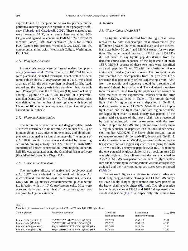
The tryptic peptides derived from the light chain wererecovered by both monoisotopic mass measurement (thedifference between the experimental mass and the theoret-ical mass below 50 ppm) and MS/MS except for two pep-tides. The experimental masses of 2929.2 and 2557.8 Dadid not match to any tryptic peptides derived from thededuced amino acid sequence of the light chain of mAb18B7. MS/MS spectra of these two ions were identifiedas tryptic peptides T1 and T2 with the substitution of oneamino acid residue (data not shown). The proteomic anal-ysis revealed two discrepancies from the predicted DNAsequence that presumably reflect sequencing errors. Ala7from the nucleic acid sequence should be threonine andthe Asn33 should be aspartic acid. The calculated monoiso-topic masses of these two tryptic peptides after correctionwere matched to the experimental masses with the errorbelow 0.12 Da, as listed inTable 1. The protein-derivedlight chain V region sequence is deposited in GenBankunder accession number AJ309277. MAb 18B7 has a kappalight chain and the light chain constant region sequencefor kappa light chain is used. Ninety two percent of theamino acid sequence of the heavy chain were recoveredb errorw ainV cces-s gions Banku renceh mAb1 ningt 3w d toA tidei ouslya posed(
r ver-i aly-s nt inti afters o
TM 18B7 li
T
PPP QSPKP PK
The serum half-life of native and de-glycosylated m8B7 was determined in Balb/c mice. An amount of 50�g of
mmunoglobulin was injected intravenously and blood sles were obtained at various time intervals. The amouAb 18B7 protein in serum was calculated by measu
erum Ab binding activity for GXM relative to mAb 18Btandards of known concentration. Immunoglobulin sealf-life was calculated using the GraphPad Prism softGraphPad Software, San Diego, CA).
.13. Mouse protection studies
The protective efficacy of native and de-glycosylaAb 18B7 was evaluated in 6–8 week old femaleice obtained from the National Cancer Institute (BetheD). Ab (100�g/mouse) was administered i.p. 24 h bef
.v. infection with 1× 10⁵ C. neoformans cells. Mice werebserved daily and the survival of the various groupsnalyzed by log–rank statistic.
able 1onoisotopic mass obtained for tryptic peptides T1 and T2 from IgG
ryptic peptide Amino acid sequence
eptide 1–24 (predicted) DVVMTQAPLALPVSLGDQASISCReptide 1–24 (MS/MS) DVVMTQTPLALPVSLGDQASISCReptide 25–50 (predicted) SSQSLAHSNGNTYLHWYLQKPGeptide 25–50 (MS/MS) SSQSLAHSDGNTYLHWYLQKPGQS
y both monoisotopic mass measurement with theithin 50 ppm and MS/MS. The protein-derived heavy chregion sequence is deposited in GenBank under a
ion number AJ309276. The heavy chain constant reequence of mouse hybridoma Ab 4F8, deposited in Gennder accession number M60432, was used as the refeeavy chain constant region sequence for analyzing the8B7 MS results. The tryptic peptide E288-R297 contai
he one potentialN-glycosylation site at position Asn-29as glycosylated. Five oligosaccharides were attachesn-293. MS/MS was performed on each of glycopep
ons and the carbohydrate compositions were unambigussigned and their corresponding structures were proTable 2).
The proposed oligosaccharide structures were furthefied using exoglycosidase cleavage and LC/MS/MS anis. Five doubly charged glycopeptide ions were presehe heavy chain tryptic digest (Fig. 1A). Two glycopeptideons withm/z values at 1536.9 and 1618.0 disappearedialidase digestion (Fig. 1B), respectively. Thus, these tw
ght chain
Calculatedmonoisotopic mass
Experimentalmonoisotopic mass
�mass(Da)
2527.277 2557.187 29.912557.288 2557.187 −0.10
2927.431 2928.294 0.862928.416 2928.294 −0.12

F.Wang
etal./Molecular
Imm
unology43
(2006)987–998
991
Table 2Oligosaccharides attached to Asn-293 of glycopeptides (E289–R297)
Monoisotopic mass ofglycopeptide (Da)a
Theoretical monoisotopicmass of glycopeptide (Da)
Mass difference(Da)
Carbohydrate compositionb Proposed oligosaccharidestructurec
Oligosaccharidetype
Relativeabundance (%)d
3232.102 3232.245 −0.14 Hex5HexNAc4FucNeuGc Hybrid 7
3070.051 3070.192 −0.14 Hex4HexNAc4FucNeuGc Complex 7
2925.010 2925.155 −0.15 Hex5HexNAc4Fuc Hybrid 14
2762.973 2763.102 −0.13 Hex4HexNAc4Fuc Complex 44
2600.916 2601.049 −0.13 Hex3HexNAc4Fuc Complex 29
a Monoisotopic masses of glycopeptides were obtained by LC coupled with Qstar QqTOF mass spectrometer.b Carbohydrate composition was assigned on the masses of glycans and MS/MS.c The structures ofN-glycans were proposed based on the fragment ions of glycopeptides obtained by MS/MS. () N-acetylglucosamine, () core mannose, (©) mannose, (�) fucose, (�) galactose and (♦)
NeuGC.d The relative abundance were calculated based on the relative intensities of glycopeptide ions obtained by LC/MS.

992 F. Wang et al. / Molecular Immunology 43 (2006) 987–998
Fig. 1. The mass spectra of glycopeptide ions after exoglycosidase treatment. (A) no reaction; (B) sialidase treatment; (C) sialidase and�-GALase treatment;(D) sialidase,�-GALase and HEXase treatment. (), N-acetylglucosamine; (©), mannose; (�), fucose; (�), galactose; (♦), N-glycolylneuraminic acid.
peptides contained terminal sialic acid. After treating the gly-copeptides with sialidase and�-GALase, only one glycopep-tide ion appeared atm/z 1302.0 (Fig. 1C), due to the release ofboth NeuGc and galactose. TwoN-acetylglucosamines werereleased after digestion with sialidase,�-GALase and HEX-ase, and a glycopepitde ion withm/z value at 1098.8 wasobserved (Fig. 1D).
Earlier studies had shown that the relative intensities inthe electrospray ionization mass spectrometry data corre-lated closely with the relative quantities of each glycoformfor glycopeptides with the same peptide sequence (Settineriand Burlingame, 1996). The mono-galactosylated, core fuco-sylated, biantennary complex-type oligosaccharide with theglycosyl composition of Hex4HexNAc4Fuc was the majorglycan species, which comprised approximately 39% of thetotal glycan pool (Table 2). Two oligosaccharides had termi-nalN-glycolylneuraminic acid (NeuGc) residues, and each ofwhich comprised approximately 8% of glycan pool. Analysisof the glycosylation pattern of three 18B7 subclones recov-ered from the parent 18B7 hybridoma by soft agar cloningin our laboratory revealed that each had the same glycosylal-
tion pattern as the original mAb 18B7 prepared for clinicaluse.
3.2. Circular dichroism spectra of native 18B7 andde-glycosylated 18B7
Circular dichroism spectroscopy is a sensitive methodfor probing the conformational changes in the secondarystructure of proteins. The CD spectra of native 18B7 andde-glycosylated 18B7 were essentially similar (Fig. 2).
3.3. Binding of native and de-glycosylated mAb 18B7 toGXM and C. neoformans
Native and de-glycosylated mAb 18B7 demonstrated sim-ilar binding to GXM by ELISA (Fig. 3). Similarly, the indirectimmunofluorescence pattern of native and de-glycosylatedbinding to encapsulatedC. neoformans cells was indistin-guishable. Hence, removal of glycans did not have a signifi-cant effect on the binding properties of mAb 18B7 for GXMby ELISA or on yeast cells.

F. Wang et al. / Molecular Immunology 43 (2006) 987–998 993
Fig. 2. Circular dichroism spectra of native (solid line) and de-glycosylated-18B7 (dashed line). The spectra were recorded at room temperature(∼21◦C).
3.4. Pharmacokinetics of native and de-glycosylatedmAb 18B7
De-glycosylated mAb 18B7 was cleared from serum fasterthan native mAb 18B7 with a half-life of 110± 4.4 and382± 9.0 h, respectively.
3.5. Activation of the C pathway by the native andde-glycosylated 18B7
Ab–Ag complexes can activate C through the classi-cal pathway.C. neoformans is unusual among encapsulatedmicrobes in that its capsule is a potent activator of the alter-native C pathway although this phenomenon is much slowerthan Ab-mediated classical pathway activation (Kozel, 1996;
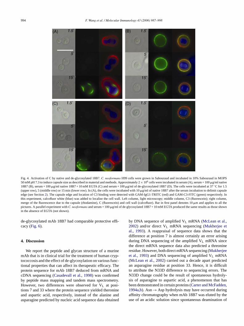
Kozel et al., 1998). Since Ab glycosylation is important forC1q binding and classical pathway activation we performeda kinetic study to infer the function of the native and de-glycosylated 18B7 in C activation in this system. As shownin Fig. 4, addition of native mAb 18B7 toC. neoformanscells produced fast deposition of C in the cryptococcal cap-sule with C3 being detected by immunofluorescence after1.5 min of incubation. This C activation occurred throughthe classical pathway, since it could be inhibited by EGTA(Fine et al., 1972). In the same conditions, de-glycosylated18B7 coated cells did not have any C bound, consistent witha requirement for glycosylation in classical C activation path-way. Longer incubations (15 min) resulted in C deposition inthe cryptococcal capsule in all the cases, even in EGTA- orde-glycosylated 18B7-treated cells, indicating that C3 depo-sition in the cryptococcal capsule was a result of alternativepathway activation.
3.6. Opsonic and protective efficacy of native andde-glycosylated 18B7
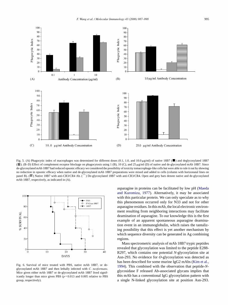
Removal of mAb 18B7N-glycans with PNGase F reduced,but did not abrogate, the opsonic efficacy of the IgG1 forC.neoformans with J774.16 macrophage-like cells (Fig. 5). Theability of de-glycosylated 18B7 to promote phagocytosis wasunexpected given that Fc glycans are essential for IgG inter-ac niza-t thata d thee 18.I educ-t reaso Ab1 e and
F Inset: t ycosyl1
ig. 3. Binding of native and de-glycosylated 18B7 to GXM by ELISA.8B7 (b) on binding to encapsulatedC. neoformans cells.
ction with the Fc receptor. Since antibodies toC. neoformansapsular polysaccharide are capable of promoting opsoion by inducing changes in the polysaccharide capsulellow C-independent ingestion by the CR we investigateffect of blocking CR with antibodies to CD11b and CD
n the presence of antibodies to CR there was a marked rion in the opsonic efficacy of de-glycosylated 18B7 whenly a modest reduction was observed for the native m8B7. Passive transfer experiments revealed that nativ
he indirect immunofluorescence patterns of native 18B7 (a) and de-glated
994 F. Wang et al. / Molecular Immunology 43 (2006) 987–998
Fig. 4. Activation of C by native and de-glycosylated 18B7.C. neoformans H99 cells were grown in Sabouraud and incubated in 10% Sabouraud in MOPS50 mM pH 7.3 to induce capsule size as described in material and methods. Approximately 2× 10⁶ cells were incubated in serum (A), serum + 100�g/ml native18B7 (B), serum + 100�g/ml native 18B7 + 10 mM EGTA (C) and serum + 100�g/ml of de-glycosylated 18B7 (D). The cells were incubated at 37◦C for 1.5(upper row), 5 (middle row) or 15 min (lower row). In (A), the cells were incubated with 10�g/ml of native 18B7 after the serum incubation to delimit capsuleedge (see Section2). The capsule edge and location of C3 binding were detected with GAM-IgG1-TRITC (red) and GAM-C3-FITC (green) respectively. Inthis experiment, calcofluor white (blue) was added to localize the cell wall. Left column, light microscopy; middle column, C3 (fluorescein); right column,merge of the fluorescence due to the capsule (rhodamine), C (fluorescein) and cell wall (calcofluor). Bar in first panel denotes 10�m and applies to all thepictures. A parallel experiment withC. neoformans and serum + 100�g/ml of de-glycosylated 18B7 + 10 mM EGTA produced the same results as those shownin the absence of EGTA (not shown).
de-glycosylated mAb 18B7 had comparable protective effi-cacy (Fig. 6).
4. Discussion
We report the peptide and glycan structure of a murinemAb that is in clinical trial for the treatment of human cryp-tococcosis and the effect of de-glycosylation on various func-tional properties that can affect its therapeutic efficacy. Theprotein sequence for mAb 18B7 deduced from mRNA andcDNA sequencing (Casadevall et al., 1998) was confirmedby peptide mass mapping and tandem mass spectrometry.However, two differences were observed for VL at posi-tions 7 and 33 where the protein sequence yielded theronineand aspartic acid, respectively, instead of the alanine andasparagine predicted by nucleic acid sequence data obtained
by DNA sequence of amplified VL mRNA (McLean et al.,2002) and/or direct VL mRNA sequencing (Mukherjee etal., 1993). A reappraisal of sequence data shows that thedifference at position 7 is almost certainly an error arisingduring DNA sequencing of the amplified VL mRNA sincethe direct mRNA sequence data also predicted a threonineresidue. However, both direct mRNA sequencing (Mukherjeeet al., 1993) and DNA sequencing of amplified VL mRNA(McLean et al., 2002) carried out a decade apart predictedan asparagine residue at position 33. Hence, it is difficultto attribute the N33D difference to sequencing errors. TheN33D change could be the result of spontaneous hydroly-sis of asparagine to aspartic acid, a phenomenon that hasbeen demonstrated in certain proteins (Carter and McFadden,1994a,b). Asn→ Asp hydrolysis may have occurred duringaffinity chromatography when mAb 18B7 was eluted by theuse of an acidic solution since spontaneous deamination of
F. Wang et al. / Molecular Immunology 43 (2006) 987–998 995
Fig. 5. (A) Phagocytic index of macrophages was determined for different doses (0.1, 1.0, and 10.0�g/ml) of native 18B7 ( ) and deglycosylated 18B7( ). (B–D) Effect of complement receptor blockage on phagocytosis using 1 (B), 10 (C), and 25�g/ml (D) of native and de-glycosylated mAb 18B7. Sincede-glycosylated mAb 18B7 had reduced opsonic efficacy we considered the possibility of toxicity tomacrophage-like cells but were able to rule it out by showingno reduction in opsonic efficacy when native and de-glycosylated mAb 18B7 preparations were mixed and added to cells (column with horizonatal lines onpanel B). ( ) Native 18B7 with anti-CR3/CR4 Ab. ( ) De-glycosylated 18B7 with anti-CR3/CR4. Open and grey bars denote native and de-glycosylatedmAb 18B7, respectively, as indicated in (A).
Fig. 6. Survival of mice treated with PBS, native mAb 18B7, or de-glycosylated mAb 18B7 and then lethally infected withC. neoformans.Mice given either mAb 18B7 or de-glycosylated mAb 18B7 lived signif-icantly longer than mice given PBS (p = 0.013 and 0.005 relative to PBSgroup, respectively).
asparagine in proteins can be facilitated by low pH (Maedaand Kuromizu, 1977). Alternatively, it may be associatedwith this particular protein. We can only speculate as to whythis phenomenon occurred only for N33 and not for otherasparagine residues. In this mAb, the local electronic environ-ment resulting from neighboring interactions may facilitatedeamination of asparagine. To our knowledge this is the firstexample of an apparent spontaneous asparagine deamina-tion event in an immunoglobulin, which raises the tantaliz-ing possibility that this effect is yet another mechanism bywhich sequence diversity can be generated in Ag combiningregions.
Mass spectrometric analysis of mAb 18B7 trypic peptidesrevealed that glycosylation was limited to the peptide E288-R297, which contains one potentialN-glycosylation site atAsn-293. No evidence forO-glycosylation was detected ashas been described for some murine IgG2 mAbs (Kim et al.,1994). This combined with the observation that peptide-N-glycosidase F released Ab-associated glycans implies thatthis mAb has a conventional IgG glycosylation pattern witha single N-linked glycosylation site at position Asn-293.

996 F. Wang et al. / Molecular Immunology 43 (2006) 987–998
The mAb 18B7 glycans manifested considerably less diversestructural diversity than reported for other murine IgG mAbs(Saba et al., 2002). For example, a murine IgG1K mAb wasreported to have 10 N-linked oligosaccharides, of which themajority are core-fucosyl, asialyl biantennary chains withvarying galactosylation and a minority are afusoyl, bisected,and mono-sialyl oligosacchrides (Saba et al., 2002). In con-trast, mAb 18B7 had five core fucosylated, biantennarycomplex-type oligosaccharides, of which four were galac-tosylated, comprising 71% of the total oligosaccharide pooland two were monosialylated byN-glycolyneuraminic acid,comprising 16% of the total oligosaccharide pool.
CD spectral analyses of native and de-glycosylated mAb18B7 were essentially identical, indicating no major changein secondary structure as a result ofN-glycan removal. Sincethe Ag recognized by mAb 18B7 is a large polysaccharide thatis presumably multivalent, Fc glycosylation could conceiv-ably affect the binding properties by affecting Fc–Fc interac-tions that influence avidity and/or the quartenary structure ofAg–Ab complexes. Comparison of native and deglycosylatedmAb 18B7 by ELISA revealed comparable binding curves.Similarly, no differences in immunofluorescence pattern wereobserved for the binding of native and deglycosylated mAb18B7 to theC. neoformans capsule. Remarkably, we foundthat deglycosylated mAb 18B7 was opsonic forC. neofor-mans with only a small reduction in opsonic efficacy relativet tra-t theiw dentp r cer-t 2T e ofAs teractdi ntsC notn otesc rther-m atedc dies.H ocy-t cro-b ssityf
thes vedh mea-s CHN 7.4t m inet dies,q por-t man
chimeric IgG1 mAbs expressed in cells unable to processhigh-mannose intermediates through the terminal glycosyla-tion products had significantly shorter serum half-life in mice(Wright and Morrison, 1994). The serum half-life of deglyco-sylated mAb 18B7 was significantly shorter than native mAb18B7 in mice. This result is consistent with studies indicatinga role for glycosylation in IgG serum half-life and that theserum clearance of glycoproteins can reflect interactions withmannose and/or asialoglycoprotein receptor in liver cells andthat this effect has been implicated in serum half-life differ-ences TNFR–IgG (Raju et al., 2001).
IgG Fc region glycosylation is essential for C activation.The IgG N-linked glycosylation site in CH2 is located nearthe binding site for C1q, the first component of the classicalC pathway.C. neoformans has the unusual characteristic thatits capsule is a potent activator of the alternative C pathway(Kozel, 1996). Consequently, incubation ofC. neoformanscells in serum results in C deposition in the capsule withoutthe need for specific Ab although this process is slower thanAb-mediated classical C activation (Kozel, 1996). Sincede-glycosylated mAb could conceivably alter C3 depositioneven if could not promote classical C activation by changingthe capsule properties and/or blocking C activation sites,we compared C3 deposition onC. neoformans cells coatedwith native and de-glycosylated mAb 18B7. Incubation ofC. neoformans cells coated with native mAb 18B7 in serumr ule,c tiono b1 sules andc thisr romp e Cp itioni ylatedI theC werw
tyh overf aredfc wasi ner-a thatt isesf ro-t ang oningt
turesa dingc rma-c de-g the
o native mAb 18B7 being evident at higher Ab concenions. Since IgG Fc region glycosylation is essential fornteraction with the Fc receptor (Wright and Morrison, 1997),e hypothesized that this effect was due to C-indepenhagocytosis through the CR, as has been described fo
ain mAbs toC. neoformans (Taborda and Casadevall, 200).his mechanism is postulated to involve the occurrencb-mediated structural change in theC. neoformans cap-ule such that the capsular polysaccharide can then inirectly with the CR (Taborda and Casadevall, 2002). The
nhibition of opsonic efficacy by Ab to the CR componeD11b and CD18c for deglycosylated mAb 18B7, butative 18B7, indicates that the deglycosylated IgG1 promomplement-independent phagocytosis through CR. Fuore, native and de-glycosylated mAb 18B7 demonstr
omparable protective efficacy in passive transfer stuence, for microbes where Ab binding can trigger phag
osis through an alternative receptor or by modifying miial surfaces, glycosylation may not be an absolute nece
or IgG opsonic efficacy.Fc N-linked glycosylation has been implicated in
erum half-life of immunoglobulins but the effect obseras varied with the IgG studied and the host used toure clearance. For a murine IgG2b, deletion of the2-linked glycosylation site reduced serum half-life from
o 4.8 days, an effect attributed to increased catabolisxtravascular tissues (Wawrzynczak et al., 1992). In addi-ion to the quantitative defects described in these stuualitative differences in glycosylation structure are im
ant for Ab pharmacokinetics. In this regard, mouse–hu
esulted in rapid binding of C3 to the rim of the capsonsistent with classical C activation. In contrast, incubaf C. neoformans cells coated with de-glycosylated mA8B7 in serum led to slow deposition of C3 on the capurface. Since de-glycosylated IgG does not bind C1qonsequently cannot activate the classical C pathwayesult implies C3 deposition in the capsule resulting folysaccharide-mediated activation of the alternativathway. Hence, the eventual outcome of C3 depos
n the presence and absence of native and de-glycosgG1 is qualitatively similar in that C3 is deposited in. neoformans surface, although the process is much sloith de-glycosylated IgG.To investigate the mechanism ofN-glycan heterogenei
ybridoma 18B7 cells were cloned in soft agar to recresh clones originating from single cells, mAb was preprom three different clones and protein analyzed forN-glycanontent. The glycan pattern in each of the three clonesdentical and corresponded to that in the mAb 18B7 geted from the parent cell line. This observation indicates
he variation in the glycosylation pattern of mAb 18B7 arrom individual cells, each of which produces hybridoma pein in multiple glycoforms. Hence, it is unlikely that one cenerate a more homogenous Ab preparation by subcl
his hybridoma cell line.In summary, we have characterized the glycan struc
ssociated with mAb 18B7 and compared the Ag binharacteristics, opsonizing properties, serum phaokinetics, and C-activating activities of the native andlycosylated forms of this therapeutic Ab. With regards to

F. Wang et al. / Molecular Immunology 43 (2006) 987–998 997
functional studies, Ag binding was not significantly affectedby removal of N-glycans. Consistent with prior studies,N-linked glycosylation in mAb 18B7 was essential for Fc�R-mediated phagocytosis and classical C activation. However,de-glycosylated mAb 18B7 remained opsonic by promotingC-independent phagocytosis through CR3 and serum incuba-tion of C. neoformans coated with de-glycosated mAb 18B7resulted in capsular deposition of C3 through activation of thealternative C pathway. These results illustrate how the effectof IgG glycosylation on immunoglobulin function can varydepending on the biological characteristics of the systembeing studied. In this system mAb 18B7 de-glycosylationdid not abrogate phagocytosis or C deposition on the capsulebecause certain characteristics of theC. neoformans capsuleallow the polysaccharide to interact directly with cellularreceptors when Ab binding alters capsule structure and toactivate the alternative C system. These results suggest thatIgG glycosylation is dispensable for some biological effectsof Ab function againstC. neoformans and imply that it maybe possible to use de-glycosylated mAbs in therapy. Forexample, de-glycosylated mAbs may have advantages indelivery of microbicidal radiation (Dadachova et al., 2003),since they would not trigger Fc�R-mediated phagocytosisand thereby spare certain host cells radiation damage. Hence,N-linked glycosylation function should be considered inthe context of the system being studied rather than beinga s toi anb turea rovet
A
althG ec-t
R
C rized. 13,
C aged.
C Gold-L.,ong,
an1446.
D izingngal
F R.M.,A. J.
Ghirlando, R., Lund, J., Goodall, M., Jefferis, R., 1999. Glycosylation ofhuman IgG-Fc: influences on structure revealed by differential scan-ning micro-calorimetry. Immunol. Lett. 68, 47–52.
Kim, H., Yamaguchi, Y., Masuda, K., Matsunaga, C., Yamamoto, K.,Irimura, T., Takahashi, N., Kato, K., Arata, Y., 1994.O-Glycosylationin hinge region of mouse immunoglobulin G2b. J. Biol. Chem. 269,12345–12350.
Kozel, T.R., 1996. Activation of the complement system by pathogenicfungi. Clin. Microbiol. Rev. 9, 34–46.
Kozel, T.R., MacGill, R.S., Wall, K.K., 1998. Bivalency is required foranticapsular monoclonal antibodies to optimally suppress activation ofthe alternative complement pathway by theCryptococcus neoformanscapsule. Infect. Immun. 66, 1547–1553.
Krapp, S., Mimura, Y., Jefferis, R., HUber, R., Sondermann, P., 2003.Structural analysis of human IgG-Fc glycoforms reveals a correla-tion between glycosylation and structural integrity. J. Exp. Med. 325,979–989.
Larsen, R.A., Pappas, P.G., Perfect, J., Aberg, J.A., Casadevall, A., Cloud,G.A., James, R., Filler, S., Dismukes, W.E., 2005. Phase I evaluationof the safety and pharmacokinetics of murine-derived anticryptococ-cal antibody 18B7 in subjects with treated cryptococcal meningitis.Antimicrob. Agents Chemother. 99, 952–958.
Lifely, M.R., Hale, C., Boyce, S., Keen, M.J., Phillips, J., 1995. Glycosy-lation and biological activity of CAMPATH-1H expressed in differentcell lines and grown under different culture conditions. Glycobiology5, 813–822.
Maeda, H., Kuromizu, K., 1977. Spontaneous deamidation of a proteinantibiotic, neocarzinostatin, at weakly acidic pH. Conversion to ahomologous inactive preneocarzinostatin due to change of asparagine83 to aspartic acid 83 accompanied by conformational and biologicalalterations. J. Biochem. (Tokyo) 81, 25–35.
M l, A.,r a
M S.P.,dues76,
M cteri-
n. J.
R eptor.s. J.
R 001.tion
e
R effec-nol.
S r, M.,gly-and
Anal.
S n ofpec-278.
T CR4ody-
W ite-f aog-
global attribute conferring singular effector propertiemmunoglobulin molecules. In fact, IgG glycosylation ce viewed as another facet of immunoglobulin architecmenable to engineering that can be modified to imp
he therapeutic usefulness of certain mAbs.
cknowledgments
This work was supported by National Institutes of Herants AI033142 and AI033774. ABI QSATR mass sp
rometer was acquired under NCRR Grant.
eferences
arter, D.A., McFadden, P.N., 1994a. Determination of beta-isomeaspartic acid as the corresponding alcohol. J. Protein Chem97–106.
arter, D.A., McFadden, P.N., 1994b. Trapping succinimides inpolypeptides by chemical reduction. J. Protein Chem. 13, 89–96
asadevall, A., Cleare, W., Feldmesser, M., Glatman-Freedman, A.,man, D.L., Kozel, T.R., Lendvai, N., Mukherjee, J., Pirofski,Rivera, J., Rosas, A.L., Scharff, M.D., Valadon, P., Westin, K., ZhZ., 1998. Characterization of a murine monoclonal antibody toCryp-tocococcus neoformans polysaccharide that is a candidate for humtherapeutic studies. Antimicrob. Agents Chemotherap. 42, 1437–
adachova, E., Nakouzi, A., Bryan, R.A., Casadevall, A., 2003. Ionradiation delivered by specific antibody is therapeutic against a fuinfection. Proc. Natl. Acad. Sci. U.S.A. 100, 10942–10947.
ine, D.P., Marney Jr., S.R., Colley, D.G., Sergent, J.S., Des Prez,1972. C3 shunt activation in human serum chelated with EGTImmunol. 109, 807–809.
cLean, G.R., Torres, M., Elguezabal, N., Nakouzi, A., Casadeval2002. Isotype can affect the fine specificity of an antibody fopolysaccharide antigen. J. Immunol. 169, 1379–1386.
imura, Y., Sondermann, P., Ghirlando, R., Lund, J., Young,Goodall, M., Jefferis, R., 2001. Role of oligosaccharide resiof IgG1-Fc in Fc gamma RIIb binding. J. Biol. Chem. 245539–45547.
ukherjee, J., Casadevall, A., Scharff, M.D., 1993. Molecular charazation of the antibody responses toCryptococcus neoformans infectionand glucuronoxylomannan-tetanus toxoid conjugate immunizatioExp. Med. 177, 1105–1106.
adaev, S., Sun, P.D., 2001. Recognition of IgG by Fcgamma recThe role of Fc glycosylation and the binding of peptide inhibitorBiol. Chem. 276, 16478–16483.
aju, T.S., Briggs, J.B., Chamow, S.M., Winkler, M.E., Jones, A.J., 2Glycoengineering of therapeutic glycoproteins: in vitro galactosylaand sialylation of glycoproteins with terminalN-acetylglucosaminand galactose residues. Biochemistry 40, 8868–8876.
alph, P., Prichard, J., Cohn, M., 1975. Reticulum cell sarcoma: antor cell in antibody-dependent cell-mediated immunity. J. Immu114, 898–905.
aba, J.A., Kunkel, J.P., Jan, D.C., Ens, W.E., Standing, K.G., ButleJamieson, J.C., Perreault, H., 2002. A study of immunoglobulin Gcosylation in monoclonal and polyclonal species by electrospraymatrix-assisted laser desorption/ionization mass spectrometry.Biochem. 305, 16–31.
ettineri, C.A., Burlingame, A.L., 1996. Structural characterizatioprotein glycosylation using HPLC/electrospray ionization mass strometry and glycosidase digestion. Methods Mol. Biol. 61, 255–
aborda, C.P., Casadevall, A., 2002. CR3 (CD11b/CD18) and(CD11c/CD18) are involved in complement-independent antibmediated phagocytosis ofCryptococcus neoformans. Immunity 16,791–802.
ang, F., Nakouzi, A., Angeletti, R.H., Casadevall, A., 2003. Sspecific characterization of the N-linked oligosaccharides omurine immunoglobulin M by high-performance liquid chromat

998 F. Wang et al. / Molecular Immunology 43 (2006) 987–998
raphy/electrospray mass spectroscopy. Anal. Biochem. 314, 266–280.
Wawrzynczak, E.J., Cumber, A.J., Parnell, G.D., Jones, P.T., Winter, G.,1992. Blood clearance in the rat of a recombinant mouse monoclonalantibody lacking the N-linked oligosaccharide side chains of the CH2
domains. Mol. Immunol. 29, 213–220.Wright, A., Morrison, S.L., 1994. Effect of altered CH2-associated car-
bohydrate structure on the functional properties and in vivo fateof chimeric mouse–human immunoglobulin G1. J. Exp. Med. 180,1087–1096.
Wright, A., Morrison, S.L., 1997. Effect of glycosylation on antibodyfunction: implications for genetic engineering. Trends Biotechnol. 15,26–32.
Wright, A., Morrison, S.L., 1998. Effect of C2-associated carbohydratestructure on Ig effector function: studies with chimeric mouse–humanIgG1 antibodies in glycosylation mutants of Chinese hamster ovarycells. J. Immunol. 160, 3393–3402.
Wright, A., Sato, Y., Okada, T., Chang, K., Endo, T., Morrison, S.,2000. In vivo trafficking and catabolism of IgG1 antibodies withFc associated carbohydrates of differing structure. Glycobiology 10,1347–1355.
Zaragoza, O., Fries, B.C., Casadevall, A., 2003. Induction of capsulegrowth inCryptococcus neoformans by mammalian serum and CO(2).Infect. Immun. 71, 6155–6164.
Glossary
Ag: antigen.C: complement.CD: cicular dichroism.CID: collision-induced dissociation.CN: Cryptococcus neoformans.CR: complement receptor.Fuc: fucose.GlcNAc: N-acetylglucosamine.GXM: glucuronoxylomannan.Hexase: �-N-acetylhexosaminidase.HexNAc: N-acetylhexosamine.Hex: hexose.IgG: immunoglobulin G.LC/MS: HPLC coupled with mass spectrometry.LC/MS/MS: HPLC coupled with tandem mass spectrometry.mAb: monoclonal antibody.MS: mass spectrometry.MS/MS: tandem mass spectrometry.NeuGc: N-glycolylneuraminic acid.PNGase F: peptide-N-glycosidase F.�-GALase: �-galactosidase.