
Microevolution of Serial Clinical Isolates of Cryptococcus ...
19
Microevolution of Serial Clinical Isolates of The MIT Faculty has made this article openly available. Please share how this access benefits you. Your story matters. Citation Chen, Yuan; Farrer, Rhys A.; Giamberardino, Charles; Sakthikumar, Sharadha; Jones, Alexander; Yang, Timothy; Tenor, Jennifer L. et al. “ Microevolution of Serial Clinical Isolates of Cryptococcus Neoformans Var. Grubii and C. Gattii.” Edited by Françoise Dromer. mBio 8, no. 2 (March 2017): e00166–17 © 2017 Chen et al As Published http://dx.doi.org/10.1128/mBio.00166-17 Publisher American Society for Microbiology Version Final published version Citable link http://hdl.handle.net/1721.1/109920 Terms of Use Creative Commons Attribution 4.0 International License Detailed Terms http://creativecommons.org/licenses/by/4.0/
-
Upload
khangminh22 -
Category
Documents
-
view
0 -
download
0
Transcript of Microevolution of Serial Clinical Isolates of Cryptococcus ...
Microevolution of Serial Clinical Isolates of
The MIT Faculty has made this article openly available. Please share how this access benefits you. Your story matters.
Citation Chen, Yuan; Farrer, Rhys A.; Giamberardino, Charles; Sakthikumar,Sharadha; Jones, Alexander; Yang, Timothy; Tenor, Jennifer L.et al. “ Microevolution of Serial Clinical Isolates of CryptococcusNeoformans Var. Grubii and C. Gattii.” Edited by Françoise Dromer.mBio 8, no. 2 (March 2017): e00166–17 © 2017 Chen et al
As Published http://dx.doi.org/10.1128/mBio.00166-17
Publisher American Society for Microbiology
Version Final published version
Citable link http://hdl.handle.net/1721.1/109920
Terms of Use Creative Commons Attribution 4.0 International License
Detailed Terms http://creativecommons.org/licenses/by/4.0/
Microevolution of Serial Clinical Isolatesof Cryptococcus neoformans var. grubiiand C. gattii
Yuan Chen,a,b* Rhys A. Farrer,c Charles Giamberardino,a Sharadha Sakthikumar,c
Alexander Jones,a Timothy Yang,a Jennifer L. Tenor,a Omar Wagih,d
Marelize Van Wyk,e Nelesh P. Govender,e Thomas G. Mitchell,b
Anastasia P. Litvintseva,f Christina A. Cuomo,c John R. Perfecta
Division of Infectious Diseases, Department of Medicine, Duke University Medical Center, Durham, NorthCarolina, USAa; Department of Molecular Genetics and Microbiology, Duke University Medical Center, Durham,North Carolina, USAb; Broad Institute of Massachusetts Institute of Technology and Harvard University,Cambridge, Massachusetts, USAc; European Molecular Biology Laboratory (EMBL), European BioinformaticsInstitute, Wellcome Genome Campus, Hinxton, Cambridge, United Kingdomd; National Institute forCommunicable Diseases (Centre for Opportunistic, Tropical, and Hospital Infections), A Division of the NationalHealth Laboratory Service, Johannesburg, South Africae; Mycotic Diseases Branch, Centers for Disease Controland Prevention, Atlanta, Georgia, USAf
ABSTRACT The pathogenic species of Cryptococcus are a major cause of mortalityowing to severe infections in immunocompromised as well as immunocompetent in-dividuals. Although antifungal treatment is usually effective, many patients relapseafter treatment, and in such cases, comparative analyses of the genomes of incidentand relapse isolates may reveal evidence of determinative, microevolutionary changeswithin the host. Here, we analyzed serial isolates cultured from cerebrospinal fluidspecimens of 18 South African patients with recurrent cryptococcal meningitis. Thetime between collection of the incident isolates and collection of the relapse isolatesranged from 124 days to 290 days, and the analyses revealed that, during this pe-riod within the patients, the isolates underwent several genetic and phenotypicchanges. Considering the vast genetic diversity of cryptococcal isolates in sub-Saharan Africa, it was not surprising to find that the relapse isolates had acquireddifferent genetic and correlative phenotypic changes. They exhibited various mecha-nisms for enhancing virulence, such as growth at 39°C, adaptation to stress, andcapsule production; a remarkable amplification of ERG11 at the native and unlinkedlocus may provide stable resistance to fluconazole. Our data provide a deeper un-derstanding of the microevolution of Cryptococcus species under pressure from anti-fungal chemotherapy and host immune responses. This investigation clearly sug-gests a promising strategy to identify novel targets for improved diagnosis, therapy,and prognosis.
IMPORTANCE Opportunistic infections caused by species of the pathogenic yeastCryptococcus lead to chronic meningoencephalitis and continue to ravage thousandsof patients with HIV/AIDS. Despite receiving antifungal treatment, over 10% of pa-tients develop recurrent disease. In this study, we collected isolates of Cryptococcusfrom cerebrospinal fluid specimens of 18 patients at the time of their diagnosis andwhen they relapsed several months later. We then sequenced and compared thegenomic DNAs of each pair of initial and relapse isolates. We also tested the isolatesfor several key properties related to cryptococcal virulence as well as for their sus-ceptibility to the antifungal drug fluconazole. These analyses revealed that the re-lapsing isolates manifested multiple genetic and chromosomal changes that affecteda variety of genes implicated in the pathogenicity of Cryptococcus or resistance tofluconazole. This application of comparative genomics to serial clinical isolates pro-
Received 2 February 2017 Accepted 6February 2017 Published 7 March 2017
Citation Chen Y, Farrer RA, Giamberardino C,Sakthikumar S, Jones A, Yang T, Tenor JL, WagihO, Van Wyk M, Govender NP, Mitchell TG,Litvintseva AP, Cuomo CA, Perfect JR. 2017.Microevolution of serial clinical isolates ofCryptococcus neoformans var. grubii andC. gattii. mBio 8:e00166-17. https://doi.org/10.1128/mBio.00166-17.
Editor Françoise Dromer, Institut Pasteur
Copyright © 2017 Chen et al. This is an open-access article distributed under the terms ofthe Creative Commons Attribution 4.0International license.
Address correspondence to Christina A.Cuomo, [email protected], or John R.Perfect, [email protected].
* Present address: Yuan Chen, Aperiomics, Inc.,Ashburn, Virginia, USA.
This article is a direct contribution from aFellow of the American Academy ofMicrobiology. External solicited reviewers:James Kronstad, University of British Columbia;Peter Williamson, National Institutes of Health.
RESEARCH ARTICLE
crossm
March/April 2017 Volume 8 Issue 2 e00166-17 ® mbio.asm.org 1
m
bio.asm.org
on May 19, 2017 - P
ublished by m
bio.asm.org
Dow
nloaded from
vides a blueprint for identifying the mechanisms whereby pathogenic microbesadapt within patients to prolong disease.
Pathogenic species of Cryptococcus are encapsulated yeasts that normally reside inthe environment, where they readily become airborne and can infect humans
through inhalation. These yeasts are neurotropic; after causing pulmonary infection,they can spread to the central nervous system (CNS) and cause life-threateningmeningoencephalitis. Cryptococcal species have evolved manifold attributes that en-able them to progress from latent infection to disease in immunocompetent individualsas well as in patients with suppressed cellular immunity, especially those with HIV/AIDS(1–4). Many of the human risks for increased susceptibility to cryptococcal disease havebeen determined. With regard to the pathogen, basic research has shown that theCryptococcus genome is dynamic in its plasticity (5), but little is known about thegenetic and phenotypic adaptations that the yeast cells undergo to invade, survive, andproliferate in the CNS of patients. To directly examine the properties of cryptococcalcells during this critical process, our previous study profiled the expression of Crypto-coccus genes at the site of human meningitis. In that study, genes related to catalyticactivity and transporters, such as ENA1 and CFO1, were significantly upregulated incerebrospinal fluid (CSF) during infection, suggesting the importance of such functionsfor survival of the yeasts in the harsh human environment, specifically within thesubarachnoid space (6, 7). Although these studies identified expression signaturesduring active infection, they provided only an initial glimpse of how this yeast is ableto adapt rapidly and continuously to assault by host immune responses and treatmentwith antifungal drugs.
An independent approach to look at adaptation is to examine mutations that occurover the course of an infection and determine if any confer a selective advantage in thehost. Between 10% and 20% of HIV/AIDS patients in South Africa develop recurrentcryptococcal meningitis (8). Consequently, genetic and phenotypic dissection of clinicalrelapse isolates may elucidate how the initial infecting isolate adapted to the CNS of thehost. Microevolution of isolates of Cryptococcus has been documented in several casesof recurrent infection as well as by in vitro and in vivo experiments (9–11). In a recentreport of a case of recurrent cryptococcal meningoencephalitis (CM), the genomicsequences of incident and relapse isolates were compared after 77 days in the patient’sCNS (12). Several mutations were identified between these serial isolates, and aframeshift mutation in the AT-rich interaction domain-containing (ARID) transcriptionalregulator was implicated in several phenotypic differences, including reduced capsulesize and altered carbon source preference. In addition, strains with higher rates ofspontaneous mutation may display accelerated microevolution within the host (11),and the serial passage of cryptococcal strains in the same host can change theirvirulence potential (13). These studies clearly indicated the importance of microevolu-tion in the adaptation of Cryptococcus to the human CNS and ability to cause disease.
In an earlier investigation, we examined the incident and relapse isolates of 81HIV/AIDS patients from South Africa with recurrent cryptococcal meningoencephalitis(14). Each isolate was genotyped by multilocus sequence typing (MLST). As expectedfrom previous studies, the vast majority of cases were caused by isolates of C. neofor-mans var. grubii, which consists of three distinct molecular types, VNI, VNII, and VNB (15,16). Infections with C. gattii are less common in South Africa and are caused bymolecular type VGI or VGIV (17). After initial diagnosis and treatment with fluconazole(FLZ) and/or amphotericin B (AMB), in most cases, recurrent disease can ensue becauseof reinfection with a different isolate or persistent infection by the initial isolate andrelapse, perhaps due to inadequate antifungal treatment or selection of the morevirulent clones within the patient. In this cohort, 89% of relapse episodes were causedby isolates with the same genotype(s) as the incident isolates (14).
To investigate the microevolution of C. neoformans var. grubii and C. gattii in the CNSunder selective pressure from the host immune responses and antifungal treatment, we
Chen et al. ®
March/April 2017 Volume 8 Issue 2 e00166-17 mbio.asm.org 2
m
bio.asm.org
on May 19, 2017 - P
ublished by m
bio.asm.org
Dow
nloaded from
analyzed the incident and relapse isolates from 18 patients. These serial isolatesappeared clonal based on MLST analysis (14). Using whole-genome sequencing, wetraced the mutational events of each isolate during its passage in the patient andcompared these mutations to key phenotypic changes in the same isolates. Correlativechanges in genotype and phenotype were detected during in vivo microevolution, andmultiple genes were implicated in shifts toward resistance to FLZ or adaptation to thehost.
RESULTSWhole-genome sequencing of serial isolates. This investigation included 32
isolates of C. neoformans var. grubii and six C. gattii isolates (see Table S1 in thesupplemental material). Each incident isolate was cultured from a specimen of CSF atthe diagnosis of cryptococcal meningoencephalitis but prior to treatment. Relapseisolates were obtained from subsequent CSF specimens more than 120 days followingcollection of the incident isolate.
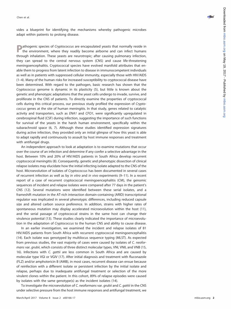
Genome-wide variants were identified for each isolate and compared to examinechanges during passage. Genomic DNA of each isolate was extracted from a singlecolony and analyzed by aligning Illumina reads to their most closely related referenceassemblies, allowing the identification of single nucleotide polymorphisms (SNPs) andindels (insertion or deletion events; see Materials and Methods). High-depth (268� onaverage) read alignments covered over 96% of the reference genome nucleotides formost isolates (Fig. 1; see also Fig. S1 and Table S2 in the supplemental material).Stringent filters were applied to SNP calls to reduce false positives, such as near-alignment errors due to adjacent indels, and the final calls were manually validated. Toverify the data and check for clonality of serial isolates, a phylogenetic tree of C. neo-
1 2 3 4 5 6 7 8 9 10 11 12 13 14
FIG 1 Aneuploid regions of all C. neoformans var. grubii isolates. For each sequenced isolate, the normalized read depth is shown in 5-kb windows along eachchromosome relative to the H99 reference, and average normalized depth on the y axis is relative to ploidy levels.
Microevolution of Cryptococcus Serial Clinical Isolates ®
March/April 2017 Volume 8 Issue 2 e00166-17 mbio.asm.org 3
m
bio.asm.org
on May 19, 2017 - P
ublished by m
bio.asm.org
Dow
nloaded from
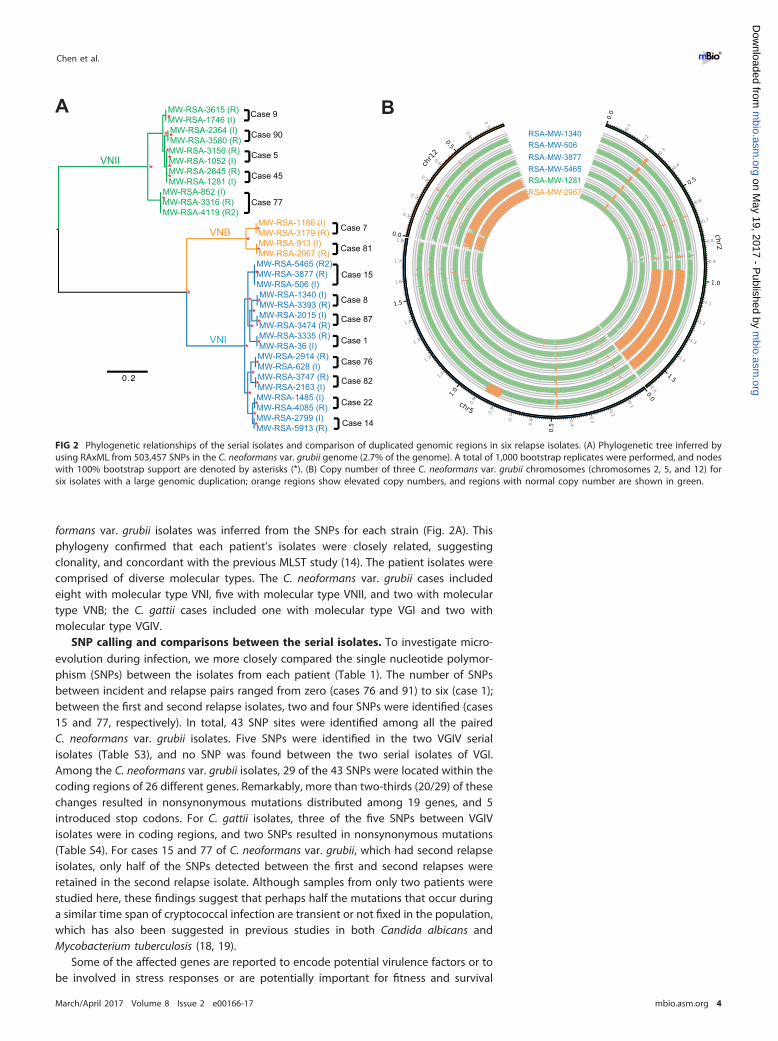
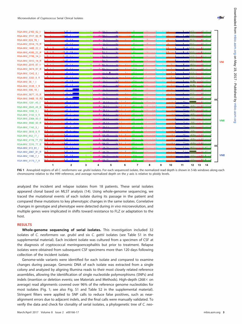
formans var. grubii isolates was inferred from the SNPs for each strain (Fig. 2A). Thisphylogeny confirmed that each patient’s isolates were closely related, suggestingclonality, and concordant with the previous MLST study (14). The patient isolates werecomprised of diverse molecular types. The C. neoformans var. grubii cases includedeight with molecular type VNI, five with molecular type VNII, and two with moleculartype VNB; the C. gattii cases included one with molecular type VGI and two withmolecular type VGIV.
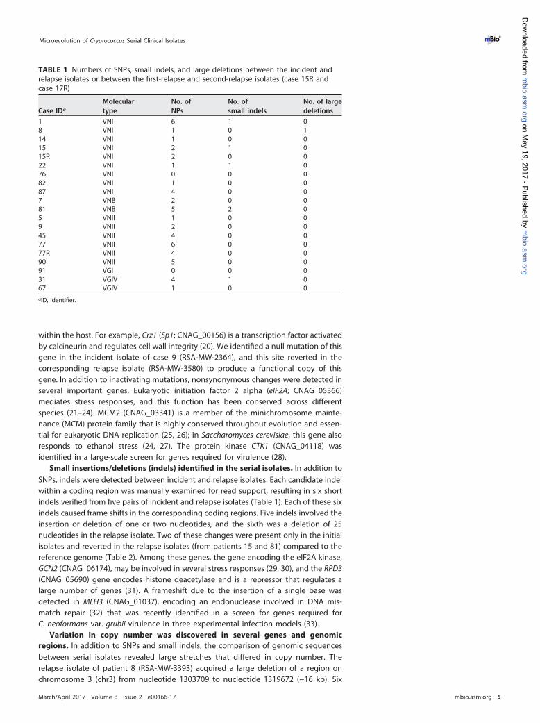
SNP calling and comparisons between the serial isolates. To investigate micro-evolution during infection, we more closely compared the single nucleotide polymor-phism (SNPs) between the isolates from each patient (Table 1). The number of SNPsbetween incident and relapse pairs ranged from zero (cases 76 and 91) to six (case 1);between the first and second relapse isolates, two and four SNPs were identified (cases15 and 77, respectively). In total, 43 SNP sites were identified among all the pairedC. neoformans var. grubii isolates. Five SNPs were identified in the two VGIV serialisolates (Table S3), and no SNP was found between the two serial isolates of VGI.Among the C. neoformans var. grubii isolates, 29 of the 43 SNPs were located within thecoding regions of 26 different genes. Remarkably, more than two-thirds (20/29) of thesechanges resulted in nonsynonymous mutations distributed among 19 genes, and 5introduced stop codons. For C. gattii isolates, three of the five SNPs between VGIVisolates were in coding regions, and two SNPs resulted in nonsynonymous mutations(Table S4). For cases 15 and 77 of C. neoformans var. grubii, which had second relapseisolates, only half of the SNPs detected between the first and second relapses wereretained in the second relapse isolate. Although samples from only two patients werestudied here, these findings suggest that perhaps half the mutations that occur duringa similar time span of cryptococcal infection are transient or not fixed in the population,which has also been suggested in previous studies in both Candida albicans andMycobacterium tuberculosis (18, 19).
Some of the affected genes are reported to encode potential virulence factors or tobe involved in stress responses or are potentially important for fitness and survival
0.2
MW-RSA-2015 (I)
MW-RSA-1186 (I)
MW-RSA-36 (I)
MW-RSA-913 (I)
MW-RSA-2914 (R)
MW-RSA-5465 (R2)
MW-RSA-3393 (R)
MW-RSA-3474 (R)
MW-RSA-1485 (I)
MW-RSA-3179 (R)
MW-RSA-4085 (R)
MW-RSA-1340 (I)
MW-RSA-2799 (I)
MW-RSA-3877 (R)
MW-RSA-3747 (R)
MW-RSA-506 (I)
MW-RSA-2967 (R)
MW-RSA-3335 (R)
MW-RSA-628 (I)
MW-RSA-4119 (R2)
MW-RSA-2645 (R)
MW-RSA-1746 (I)
MW-RSA-3316 (R)
MW-RSA-1281 (I)
MW-RSA-3615 (R)
MW-RSA-852 (I)
MW-RSA-3580 (R)
MW-RSA-1052 (I)
MW-RSA-2364 (I)
MW-RSA-3156 (R)
MW-RSA-2163 (I)
MW-RSA-5913 (R)
*
*
**
* *
*
*
*
*
*
*
*
**
*
*
*
*
*
*
*
*
*
*
*
*
VNII
VNB
VNI
Case 9
Case 90
Case 5
Case 45
Case 77
Case 7
Case 81
Case 15
Case 8
Case 87
Case 1
Case 76
Case 82
Case 22
Case 14
A
RSA-MW-2967
RSA-MW-1281
RSA-MW-5465
RSA-MW-3877
RSA-MW-506
RSA-MW-1340
B
*
FIG 2 Phylogenetic relationships of the serial isolates and comparison of duplicated genomic regions in six relapse isolates. (A) Phylogenetic tree inferred byusing RAxML from 503,457 SNPs in the C. neoformans var. grubii genome (2.7% of the genome). A total of 1,000 bootstrap replicates were performed, and nodeswith 100% bootstrap support are denoted by asterisks (*). (B) Copy number of three C. neoformans var. grubii chromosomes (chromosomes 2, 5, and 12) forsix isolates with a large genomic duplication; orange regions show elevated copy numbers, and regions with normal copy number are shown in green.
Chen et al. ®
March/April 2017 Volume 8 Issue 2 e00166-17 mbio.asm.org 4
m
bio.asm.org
on May 19, 2017 - P
ublished by m
bio.asm.org
Dow
nloaded from
within the host. For example, Crz1 (Sp1; CNAG_00156) is a transcription factor activatedby calcineurin and regulates cell wall integrity (20). We identified a null mutation of thisgene in the incident isolate of case 9 (RSA-MW-2364), and this site reverted in thecorresponding relapse isolate (RSA-MW-3580) to produce a functional copy of thisgene. In addition to inactivating mutations, nonsynonymous changes were detected inseveral important genes. Eukaryotic initiation factor 2 alpha (eIF2A; CNAG_05366)mediates stress responses, and this function has been conserved across differentspecies (21–24). MCM2 (CNAG_03341) is a member of the minichromosome mainte-nance (MCM) protein family that is highly conserved throughout evolution and essen-tial for eukaryotic DNA replication (25, 26); in Saccharomyces cerevisiae, this gene alsoresponds to ethanol stress (24, 27). The protein kinase CTK1 (CNAG_04118) wasidentified in a large-scale screen for genes required for virulence (28).
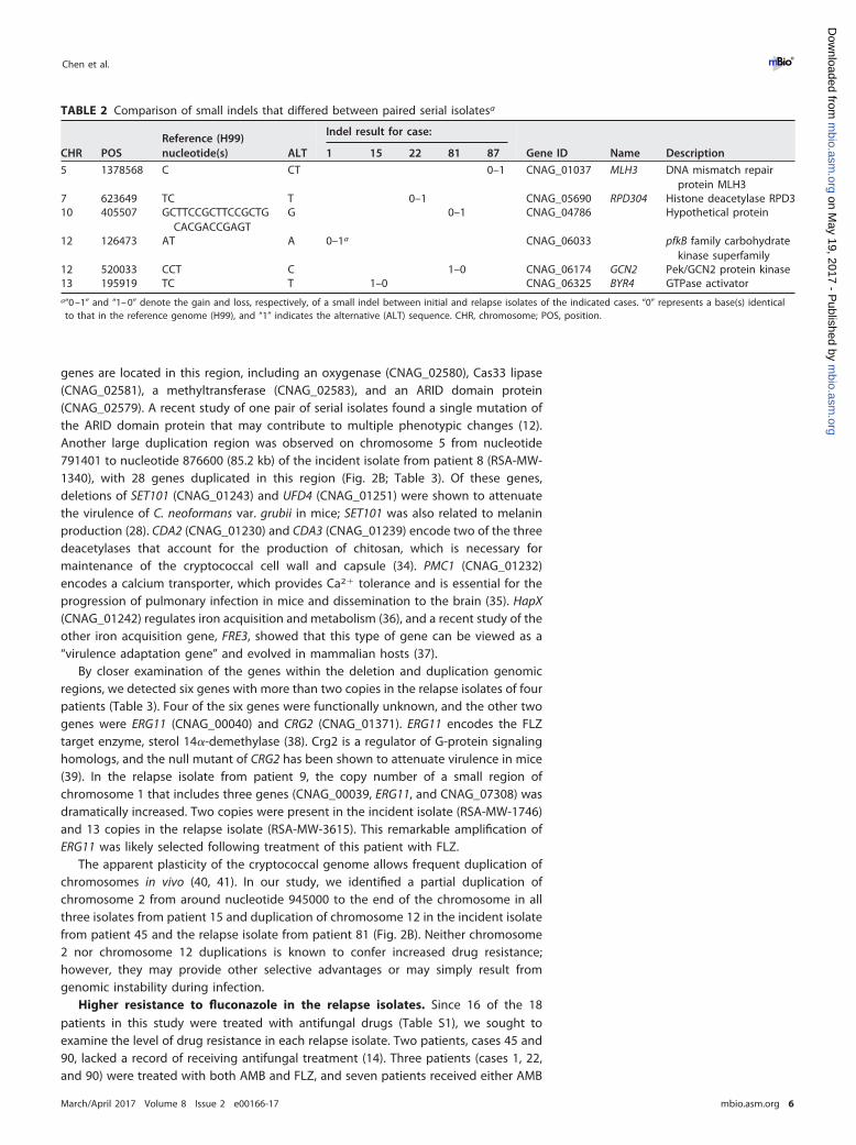
Small insertions/deletions (indels) identified in the serial isolates. In addition toSNPs, indels were detected between incident and relapse isolates. Each candidate indelwithin a coding region was manually examined for read support, resulting in six shortindels verified from five pairs of incident and relapse isolates (Table 1). Each of these sixindels caused frame shifts in the corresponding coding regions. Five indels involved theinsertion or deletion of one or two nucleotides, and the sixth was a deletion of 25nucleotides in the relapse isolate. Two of these changes were present only in the initialisolates and reverted in the relapse isolates (from patients 15 and 81) compared to thereference genome (Table 2). Among these genes, the gene encoding the eIF2A kinase,GCN2 (CNAG_06174), may be involved in several stress responses (29, 30), and the RPD3(CNAG_05690) gene encodes histone deacetylase and is a repressor that regulates alarge number of genes (31). A frameshift due to the insertion of a single base wasdetected in MLH3 (CNAG_01037), encoding an endonuclease involved in DNA mis-match repair (32) that was recently identified in a screen for genes required forC. neoformans var. grubii virulence in three experimental infection models (33).
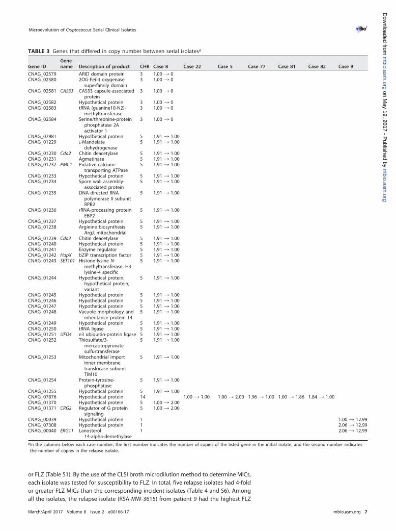
Variation in copy number was discovered in several genes and genomicregions. In addition to SNPs and small indels, the comparison of genomic sequencesbetween serial isolates revealed large stretches that differed in copy number. Therelapse isolate of patient 8 (RSA-MW-3393) acquired a large deletion of a region onchromosome 3 (chr3) from nucleotide 1303709 to nucleotide 1319672 (~16 kb). Six
TABLE 1 Numbers of SNPs, small indels, and large deletions between the incident andrelapse isolates or between the first-relapse and second-relapse isolates (case 15R andcase 17R)
Case IDa
Moleculartype
No. ofNPs
No. ofsmall indels
No. of largedeletions
1 VNI 6 1 08 VNI 1 0 114 VNI 1 0 015 VNI 2 1 015R VNI 2 0 022 VNI 1 1 076 VNI 0 0 082 VNI 1 0 087 VNI 4 0 07 VNB 2 0 081 VNB 5 2 05 VNII 1 0 09 VNII 2 0 045 VNII 4 0 077 VNII 6 0 077R VNII 4 0 090 VNII 5 0 091 VGI 0 0 031 VGIV 4 1 067 VGIV 1 0 0aID, identifier.
Microevolution of Cryptococcus Serial Clinical Isolates ®
March/April 2017 Volume 8 Issue 2 e00166-17 mbio.asm.org 5
m
bio.asm.org
on May 19, 2017 - P
ublished by m
bio.asm.org
Dow
nloaded from
genes are located in this region, including an oxygenase (CNAG_02580), Cas33 lipase(CNAG_02581), a methyltransferase (CNAG_02583), and an ARID domain protein(CNAG_02579). A recent study of one pair of serial isolates found a single mutation ofthe ARID domain protein that may contribute to multiple phenotypic changes (12).Another large duplication region was observed on chromosome 5 from nucleotide791401 to nucleotide 876600 (85.2 kb) of the incident isolate from patient 8 (RSA-MW-1340), with 28 genes duplicated in this region (Fig. 2B; Table 3). Of these genes,deletions of SET101 (CNAG_01243) and UFD4 (CNAG_01251) were shown to attenuatethe virulence of C. neoformans var. grubii in mice; SET101 was also related to melaninproduction (28). CDA2 (CNAG_01230) and CDA3 (CNAG_01239) encode two of the threedeacetylases that account for the production of chitosan, which is necessary formaintenance of the cryptococcal cell wall and capsule (34). PMC1 (CNAG_01232)encodes a calcium transporter, which provides Ca2� tolerance and is essential for theprogression of pulmonary infection in mice and dissemination to the brain (35). HapX(CNAG_01242) regulates iron acquisition and metabolism (36), and a recent study of theother iron acquisition gene, FRE3, showed that this type of gene can be viewed as a“virulence adaptation gene” and evolved in mammalian hosts (37).
By closer examination of the genes within the deletion and duplication genomicregions, we detected six genes with more than two copies in the relapse isolates of fourpatients (Table 3). Four of the six genes were functionally unknown, and the other twogenes were ERG11 (CNAG_00040) and CRG2 (CNAG_01371). ERG11 encodes the FLZtarget enzyme, sterol 14�-demethylase (38). Crg2 is a regulator of G-protein signalinghomologs, and the null mutant of CRG2 has been shown to attenuate virulence in mice(39). In the relapse isolate from patient 9, the copy number of a small region ofchromosome 1 that includes three genes (CNAG_00039, ERG11, and CNAG_07308) wasdramatically increased. Two copies were present in the incident isolate (RSA-MW-1746)and 13 copies in the relapse isolate (RSA-MW-3615). This remarkable amplification ofERG11 was likely selected following treatment of this patient with FLZ.
The apparent plasticity of the cryptococcal genome allows frequent duplication ofchromosomes in vivo (40, 41). In our study, we identified a partial duplication ofchromosome 2 from around nucleotide 945000 to the end of the chromosome in allthree isolates from patient 15 and duplication of chromosome 12 in the incident isolatefrom patient 45 and the relapse isolate from patient 81 (Fig. 2B). Neither chromosome2 nor chromosome 12 duplications is known to confer increased drug resistance;however, they may provide other selective advantages or may simply result fromgenomic instability during infection.
Higher resistance to fluconazole in the relapse isolates. Since 16 of the 18patients in this study were treated with antifungal drugs (Table S1), we sought toexamine the level of drug resistance in each relapse isolate. Two patients, cases 45 and90, lacked a record of receiving antifungal treatment (14). Three patients (cases 1, 22,and 90) were treated with both AMB and FLZ, and seven patients received either AMB
TABLE 2 Comparison of small indels that differed between paired serial isolatesa
CHR POSReference (H99)nucleotide(s) ALT
Indel result for case:
Gene ID Name Description1 15 22 81 87
5 1378568 C CT 0–1 CNAG_01037 MLH3 DNA mismatch repairprotein MLH3
7 623649 TC T 0–1 CNAG_05690 RPD304 Histone deacetylase RPD310 405507 GCTTCCGCTTCCGCTG
CACGACCGAGTG 0–1 CNAG_04786 Hypothetical protein
12 126473 AT A 0–1a CNAG_06033 pfkB family carbohydratekinase superfamily
12 520033 CCT C 1–0 CNAG_06174 GCN2 Pek/GCN2 protein kinase13 195919 TC T 1–0 CNAG_06325 BYR4 GTPase activatora“0 –1” and “1– 0” denote the gain and loss, respectively, of a small indel between initial and relapse isolates of the indicated cases. “0” represents a base(s) identicalto that in the reference genome (H99), and “1” indicates the alternative (ALT) sequence. CHR, chromosome; POS, position.
Chen et al. ®
March/April 2017 Volume 8 Issue 2 e00166-17 mbio.asm.org 6
m
bio.asm.org
on May 19, 2017 - P
ublished by m
bio.asm.org
Dow
nloaded from
or FLZ (Table S1). By the use of the CLSI broth microdilution method to determine MICs,each isolate was tested for susceptibility to FLZ. In total, five relapse isolates had 4-foldor greater FLZ MICs than the corresponding incident isolates (Table 4 and S6). Amongall the isolates, the relapse isolate (RSA-MW-3615) from patient 9 had the highest FLZ
TABLE 3 Genes that differed in copy number between serial isolatesa
Gene IDGenename Description of product CHR Case 8 Case 22 Case 5 Case 77 Case 81 Case 82 Case 9
CNAG_02579 ARID domain protein 3 1.00 ¡ 0CNAG_02580 2OG-Fe(II) oxygenase
superfamily domain3 1.00 ¡ 0
CNAG_02581 CAS33 CAS33 capsule-associatedprotein
3 1.00 ¡ 0
CNAG_02582 Hypothetical protein 3 1.00 ¡ 0CNAG_02583 tRNA (guanine10-N2)-
methyltransferase3 1.00 ¡ 0
CNAG_02584 Serine/threonine-proteinphosphatase 2Aactivator 1
3 1.00 ¡ 0
CNAG_07981 Hypothetical protein 5 1.91 ¡ 1.00CNAG_01229 L-Mandelate
dehydrogenase5 1.91 ¡ 1.00
CNAG_01230 Cda2 Chitin deacetylase 5 1.91 ¡ 1.00CNAG_01231 Agmatinase 5 1.91 ¡ 1.00CNAG_01232 PMC1 Putative calcium-
transporting ATPase5 1.91 ¡ 1.00
CNAG_01233 Hypothetical protein 5 1.91 ¡ 1.00CNAG_01234 Spore wall assembly-
associated protein5 1.91 ¡ 1.00
CNAG_01235 DNA-directed RNApolymerase II subunitRPB2
5 1.91 ¡ 1.00
CNAG_01236 rRNA-processing proteinEBP2
5 1.91 ¡ 1.00
CNAG_01237 Hypothetical protein 5 1.91 ¡ 1.00CNAG_01238 Arginine biosynthesis
ArgJ, mitochondrial5 1.91 ¡ 1.00
CNAG_01239 Cda3 Chitin deacetylase 5 1.91 ¡ 1.00CNAG_01240 Hypothetical protein 5 1.91 ¡ 1.00CNAG_01241 Enzyme regulator 5 1.91 ¡ 1.00CNAG_01242 HapX bZIP transcription factor 5 1.91 ¡ 1.00CNAG_01243 SET101 Histone-lysine N-
methyltransferase, H3lysine-4 specific
5 1.91 ¡ 1.00
CNAG_01244 Hypothetical protein,hypothetical protein,variant
5 1.91 ¡ 1.00
CNAG_01245 Hypothetical protein 5 1.91 ¡ 1.00CNAG_01246 Hypothetical protein 5 1.91 ¡ 1.00CNAG_01247 Hypothetical protein 5 1.91 ¡ 1.00CNAG_01248 Vacuole morphology and
inheritance protein 145 1.91 ¡ 1.00
CNAG_01249 Hypothetical protein 5 1.91 ¡ 1.00CNAG_01250 tRNA ligase 5 1.91 ¡ 1.00CNAG_01251 UFD4 e3 ubiquitin-protein ligase 5 1.91 ¡ 1.00CNAG_01252 Thiosulfate/3-
mercaptopyruvatesulfurtransferase
5 1.91 ¡ 1.00
CNAG_01253 Mitochondrial importinner membranetranslocase subunitTIM10
5 1.91 ¡ 1.00
CNAG_01254 Protein-tyrosine-phosphatase
5 1.91 ¡ 1.00
CNAG_01255 Hypothetical protein 5 1.91 ¡ 1.00CNAG_07876 Hypothetical protein 14 1.00 ¡ 1.90 1.00 ¡ 2.00 1.96 ¡ 1.00 1.00 ¡ 1.86 1.84 ¡ 1.00CNAG_01370 Hypothetical protein 5 1.00 ¡ 2.00CNAG_01371 CRG2 Regulator of G protein
signaling5 1.00 ¡ 2.00
CNAG_00039 Hypothetical protein 1 1.00 ¡ 12.99CNAG_07308 Hypothetical protein 1 2.06 ¡ 12.99CNAG_00040 ERG11 Lanosterol
14-alpha-demethylase1 2.06 ¡ 12.99
aIn the columns below each case number, the first number indicates the number of copies of the listed gene in the initial isolate, and the second number indicatesthe number of copies in the relapse isolate.
Microevolution of Cryptococcus Serial Clinical Isolates ®
March/April 2017 Volume 8 Issue 2 e00166-17 mbio.asm.org 7
m
bio.asm.org
on May 19, 2017 - P
ublished by m
bio.asm.org
Dow
nloaded from
MIC (128 �g/ml), 16-fold greater than the MIC seen with the incident isolate (RSA-MW-1746; MIC, 8 �g/ml). Two genes, BYR4 (CNAG_06325) and ERG11, were associated withincreased resistance to FLZ in case 15 and case 9, respectively. A single base deletionwas identified in BYR4 for the incident isolate from patient 15, but this change revertedin the relapse isolate (Table 2). Byr4 is a dosage-dependent regulator of cytokinesis (42),and it has been reported to be essential for resistance of C. neoformans var. grubii to FLZ(43). The deletion of the nucleotide caused a frameshift affecting two-thirds of theprotein length, likely to produce a loss of Bry4 function. Therefore, the changes in FLZresistance in case 15 serial isolates might be directly associated with this mutation.
The duplication of genes and genomic regions is another well-documented mech-anism for developing resistance to FLZ. In fact, heteroresistance in Cryptococcus hasbeen described and shown to be due to aneuploidy, with duplication of chromosome1 containing the gene (AFR1) encoding an efflux pump and the target (ERG11) of FLZ(44). This duplication can be observed in vivo when the yeast is under stress, but theextra copy is lost under nonstress growth conditions (40). Therefore, our sequencing ofa single colony after many passages in vitro may have missed some changes inchromosomal aneuploidy. Furthermore, we observed no changes or duplication in theAFR1 gene between any paired isolates. In case 9, an alternative yeast strategy ofresistance was used. The increased MIC of the relapse isolate can be attributed to theextensive local duplication of ERG11 and elevated production of Erg11, the target ofFLZ. Indeed, the copy number of this small genomic region increased from 2 in theincident isolate to 13 in the relapse isolate (Table 3). By more finely mapping the readcoverage boundaries of the duplicated regions in the two genomes, we found that theduplicated region on chromosome 1 of the incident isolate (from nucleotide 123657 tonucleotide 128150) was smaller than in the relapse isolate (from nucleotide 121438 tonucleotide 129598), suggesting that multiple independent amplifications can contrib-ute to resistance (Fig. 3A). Previous studies reported that increasing the copy numberof ERG11 or the related region can produce resistance to FLZ in C. neoformans var. grubii(40). However, to the best of our knowledge, this is the first observation that C. neo-formans var. grubii can so markedly amplify ERG11.
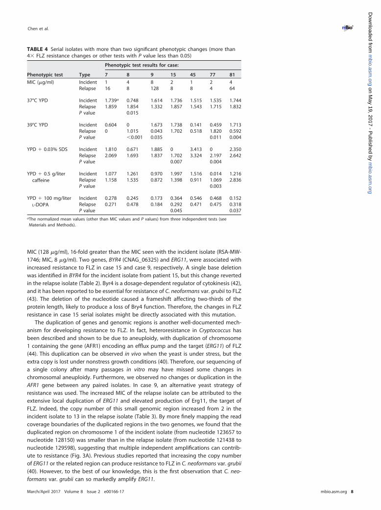
TABLE 4 Serial isolates with more than two significant phenotypic changes (more than4� FLZ resistance changes or other tests with P value less than 0.05)
Phenotypic test Type
Phenotypic test results for case:
7 8 9 15 45 77 81
MIC (�g/ml) Incident 1 4 8 2 1 2 4Relapse 16 8 128 8 8 4 64
37°C YPD Incident 1.739a 0.748 1.614 1.736 1.515 1.535 1.744Relapse 1.859 1.854 1.332 1.857 1.543 1.715 1.832P value 0.015
39°C YPD Incident 0.604 0 1.673 1.738 0.141 0.459 1.713Relapse 0 1.015 0.043 1.702 0.518 1.820 0.592P value �0.001 0.035 0.011 0.004
YPD � 0.03% SDS Incident 1.810 0.671 1.885 0 3.413 0 2.350Relapse 2.069 1.693 1.837 1.702 3.324 2.197 2.642P value 0.007 0.004
YPD � 0.5 g/litercaffeine
Incident 1.077 1.261 0.970 1.997 1.516 0.014 1.216Relapse 1.158 1.535 0.872 1.398 0.911 1.069 2.836P value 0.003
YPD � 100 mg/literL-DOPA
Incident 0.278 0.245 0.173 0.364 0.546 0.468 0.152Relapse 0.271 0.478 0.184 0.292 0.471 0.475 0.318P value 0.045 0.037
aThe normalized mean values (other than MIC values and P values) from three independent tests (seeMaterials and Methods).
Chen et al. ®
March/April 2017 Volume 8 Issue 2 e00166-17 mbio.asm.org 8
m
bio.asm.org
on May 19, 2017 - P
ublished by m
bio.asm.org
Dow
nloaded from
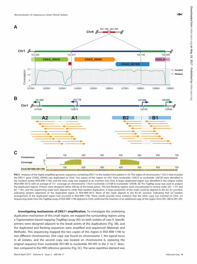
Investigating mechanisms of ERG11 amplification. To investigate the underlyingduplication mechanism of this small region, we mapped the surrounding regions usinga Tagmentation-based mapping (TagMap) assay (45) on both isolates of case 9. Specificprimers were designed adjacent to the break points of the duplications (Fig. 3B), andthe duplicated and flanking sequences were amplified and sequenced (Materials andMethods). This sequencing mapped the two copies of this region in RSA-MW-1746 totwo different chromosomes. One copy was found on chromosome 1, the typical locusin all isolates, and the second copy was located on chromosome 6, replacing theoriginal sequence from nucleotide 991188 to nucleotide 991395 in the 3=-to-5= direc-tion compared to the H99 reference genome (Fig. 3C). The same repetitive element was
0
5
10
15
121001 122001 123001 124001 125001 126001 127001 128001 129001
Incident
Consensus
Relapse
Cov
erag
e(x)
130001
121,438 123,675 128,150 129,598
CNAG_00039 CNAG_00040
CNAG_007308
CNAG_00043
121438 123675 128150 129598
121,602 127,988123,770 129,485
Chr1 A1 A2B1 B2
A2 A1 B2 B1
1 100 200 300 400 500 600 700
1 100 200 300 400 500 600 700
Chr1
991,188 991,395Chr6
RE
0043
A
C
B
Consensus
Coverage
Chr6:991000-991700
FIG 3 Analyses of the highly amplified genomic sequences containing ERG11 in the isolates from patient 9. (A) The region of chromosome 1 (Chr1) that includesthe ERG11 gene (CNAG_00040) was duplicated on Chr6. Two copies of this region on Chr1 from nucleotide 123675 to nucleotide 128150 were identified inthe incident isolate (RSA-MW-1746), and the extra copy was mapped as an insertion into Chr6. A larger duplicated region was identified in the relapse isolate(RSA-MW-3615) with an average of 13� coverage on chromosome 1 from nucleotide 121438 to nucleotide 129598. (B) The TagMap assay was used to analyzethe duplicated regions. Primers were designed within 200 bp of the break points. The two flanking regions were concatenated in reverse order (A2 ¡ A1 andB2 ¡ B1), and the sequencing reads were aligned to verify their tandem duplication. A large proportion of the reads could be aligned to the A2–A1 junction,indicating tandem alignment of the duplicated region in RSA-MW-3615. None of the reads aligned to the B2–B1 junction, indicating that no tandemarrangement of the duplicated region had occurred in RSA-MW-1746. These results provide more evidence that the other copy was inserted on Chr6. (C)Sequencing reads from the TagMap assay of RSA-MW-1746 aligned to Chr6 confirmed the insertion of an additional copy of this region from 991,188 to 991,395.
Microevolution of Cryptococcus Serial Clinical Isolates ®
March/April 2017 Volume 8 Issue 2 e00166-17 mbio.asm.org 9
m
bio.asm.org
on May 19, 2017 - P
ublished by m
bio.asm.org
Dow
nloaded from
located near the 5= end in both copies, suggesting that the duplication of this regionin RSA-MW-1746 could have been mediated by this element. In relapse isolate RSA-MW-3615, the extra copy on chromosome six was retained as in the incident isolate, buta tandem duplication of a larger region was observed on chromosome 1 which createdanother 11 extra copies of ERG11 and the two other genes. As noted, amplification ofthis small region correlated with the significant increase of FLZ resistance, and sequenc-ing revealed the precision and efficiency of this change.
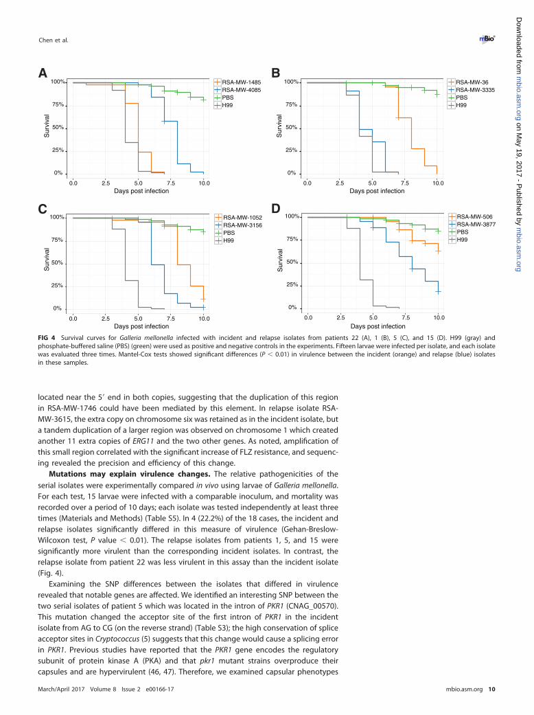
Mutations may explain virulence changes. The relative pathogenicities of theserial isolates were experimentally compared in vivo using larvae of Galleria mellonella.For each test, 15 larvae were infected with a comparable inoculum, and mortality wasrecorded over a period of 10 days; each isolate was tested independently at least threetimes (Materials and Methods) (Table S5). In 4 (22.2%) of the 18 cases, the incident andrelapse isolates significantly differed in this measure of virulence (Gehan-Breslow-Wilcoxon test, P value � 0.01). The relapse isolates from patients 1, 5, and 15 weresignificantly more virulent than the corresponding incident isolates. In contrast, therelapse isolate from patient 22 was less virulent in this assay than the incident isolate(Fig. 4).
Examining the SNP differences between the isolates that differed in virulencerevealed that notable genes are affected. We identified an interesting SNP between thetwo serial isolates of patient 5 which was located in the intron of PKR1 (CNAG_00570).This mutation changed the acceptor site of the first intron of PKR1 in the incidentisolate from AG to CG (on the reverse strand) (Table S3); the high conservation of spliceacceptor sites in Cryptococcus (5) suggests that this change would cause a splicing errorin PKR1. Previous studies have reported that the PKR1 gene encodes the regulatorysubunit of protein kinase A (PKA) and that pkr1 mutant strains overproduce theircapsules and are hypervirulent (46, 47). Therefore, we examined capsular phenotypes
0%
25%
50%
75%
100%
0.0 2.5 5.0 7.5 10.0Days post infection
Sur
viva
l
RSA-MW-36RSA-MW-3335PBSH99
A
0%
25%
50%
75%
100%
0.0 2.5 5.0 7.5 10.0
Sur
viva
l
RSA-MW-1485RSA-MW-4085PBSH99
Days post infection
B
0%
25%
50%
75%
100%
0.0 2.5 5.0 7.5 10.0
RSA-MW-506RSA-MW-3877PBSH99
Sur
viva
l
Days post infection
C
0%
25%
50%
75%
100%
0.0 2.5 5.0 7.5 10.0
Sur
viva
l
RSA-MW-1052RSA-MW-3156PBSH99
Days post infection
D
FIG 4 Survival curves for Galleria mellonella infected with incident and relapse isolates from patients 22 (A), 1 (B), 5 (C), and 15 (D). H99 (gray) andphosphate-buffered saline (PBS) (green) were used as positive and negative controls in the experiments. Fifteen larvae were infected per isolate, and each isolatewas evaluated three times. Mantel-Cox tests showed significant differences (P � 0.01) in virulence between the incident (orange) and relapse (blue) isolatesin these samples.
Chen et al. ®
March/April 2017 Volume 8 Issue 2 e00166-17 mbio.asm.org 10
m
bio.asm.org
on May 19, 2017 - P
ublished by m
bio.asm.org
Dow
nloaded from
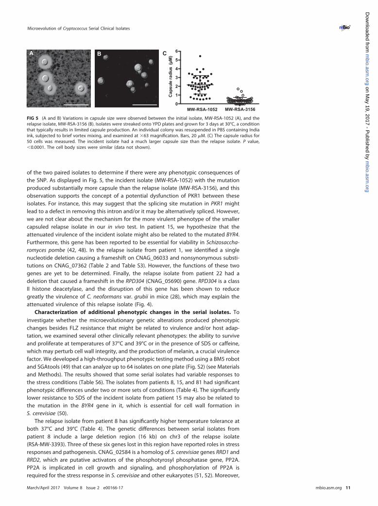
of the two paired isolates to determine if there were any phenotypic consequences ofthe SNP. As displayed in Fig. 5, the incident isolate (MW-RSA-1052) with the mutationproduced substantially more capsule than the relapse isolate (MW-RSA-3156), and thisobservation supports the concept of a potential dysfunction of PKR1 between theseisolates. For instance, this may suggest that the splicing site mutation in PKR1 mightlead to a defect in removing this intron and/or it may be alternatively spliced. However,we are not clear about the mechanism for the more virulent phenotype of the smallercapsuled relapse isolate in our in vivo test. In patient 15, we hypothesize that theattenuated virulence of the incident isolate might also be related to the mutated BYR4.Furthermore, this gene has been reported to be essential for viability in Schizosaccha-romyces pombe (42, 48). In the relapse isolate from patient 1, we identified a singlenucleotide deletion causing a frameshift on CNAG_06033 and nonsynonymous substi-tutions on CNAG_07362 (Table 2 and Table S3). However, the functions of these twogenes are yet to be determined. Finally, the relapse isolate from patient 22 had adeletion that caused a frameshift in the RPD304 (CNAG_05690) gene. RPD304 is a classII histone deacetylase, and the disruption of this gene has been shown to reducegreatly the virulence of C. neoformans var. grubii in mice (28), which may explain theattenuated virulence of this relapse isolate (Fig. 4).
Characterization of additional phenotypic changes in the serial isolates. Toinvestigate whether the microevolutionary genetic alterations produced phenotypicchanges besides FLZ resistance that might be related to virulence and/or host adap-tation, we examined several other clinically relevant phenotypes: the ability to surviveand proliferate at temperatures of 37°C and 39°C or in the presence of SDS or caffeine,which may perturb cell wall integrity, and the production of melanin, a crucial virulencefactor. We developed a high-throughput phenotypic testing method using a BM5 robotand SGAtools (49) that can analyze up to 64 isolates on one plate (Fig. S2) (see Materialsand Methods). The results showed that some serial isolates had variable responses tothe stress conditions (Table S6). The isolates from patients 8, 15, and 81 had significantphenotypic differences under two or more sets of conditions (Table 4). The significantlylower resistance to SDS of the incident isolate from patient 15 may also be related tothe mutation in the BYR4 gene in it, which is essential for cell wall formation inS. cerevisiae (50).
The relapse isolate from patient 8 has significantly higher temperature tolerance atboth 37°C and 39°C (Table 4). The genetic differences between serial isolates frompatient 8 include a large deletion region (16 kb) on chr3 of the relapse isolate(RSA-MW-3393). Three of these six genes lost in this region have reported roles in stressresponses and pathogenesis. CNAG_02584 is a homolog of S. cerevisiae genes RRD1 andRRD2, which are putative activators of the phosphotyrosyl phosphatase gene, PP2A.PP2A is implicated in cell growth and signaling, and phosphorylation of PP2A isrequired for the stress response in S. cerevisiae and other eukaryotes (51, 52). Moreover,
MW-RSA-3156MW-RSA-10520
1
2
3
4
5
6
Cap
sule
radi
us (
M)A B C
FIG 5 (A and B) Variations in capsule size were observed between the initial isolate, MW-RSA-1052 (A), and therelapse isolate, MW-RSA-3156 (B). Isolates were streaked onto YPD plates and grown for 3 days at 30°C, a conditionthat typically results in limited capsule production. An individual colony was resuspended in PBS containing Indiaink, subjected to brief vortex mixing, and examined at �63 magnification. Bars, 20 �M. (C) The capsule radius for50 cells was measured. The incident isolate had a much larger capsule size than the relapse isolate. P value,�0.0001. The cell body sizes were similar (data not shown).
Microevolution of Cryptococcus Serial Clinical Isolates ®
March/April 2017 Volume 8 Issue 2 e00166-17 mbio.asm.org 11
m
bio.asm.org
on May 19, 2017 - P
ublished by m
bio.asm.org
Dow
nloaded from
a previous study showed that deletion of RRD1 and RRD2 can enhance the resistanceof S. cerevisiae to caffeine (53). Cas33 (CNAG_02581) is involved in formation of thecryptococcal capsule (54), the major virulence factor of pathogenic Cryptococcus spe-cies (55). CNAG_02583 encodes a tRNA (guanine 10-N2) methyltransferase; methylationof tRNA has been linked to protein synthesis, infections, temperature, and immuneresponses in several systems (56, 57). The ARID domain protein (CNAG_02579), also inthis deleted region, was previously shown to be prematurely truncated by a frameshiftmutation in a relapse isolate (12).
In case 81, the relapse isolate had reduced growth at 39°C in comparison to itscorresponding incident isolate but produced more melanin (Table 4). Comparing thegenetic differences between these two isolates, a small indel in GCN2 (CNAG_06174)was identified in the incident isolate (RSA-MW-913) but was reverted in the relapseisolate (Table 2). While GCN2 is involved in responses to amino acid starvation,intercellular acid stress, and oxidative stress response in S. cerevisiae (58–60), a melaninsynthesis-related gene, Dct, is also regulated by GCN2 (30). Thus, the difference inmelanin production may be explained by the presence of the indel in GCN2.
DISCUSSION
Microbes have developed fascinating genetic plasticity to adapt to a variety of stressconditions within a very short time through mechanisms of microevolution. Under-standing these processes and the magnitude of their influence on host adaptability anddrug resistance may highlight targets for new strategies and/or drugs to treat infec-tions. However, most of the previous analyses on microevolution have been performedwith in vitro systems and animal models (5, 61). Here, the complete genome compar-isons of clonal serial isolates from relapsing patients provided a specific window toinvestigate the microevolution of Cryptococcus within the human subarachnoid space,primarily under conditions of drug treatment. In these longitudinal samples, theshortest time period between the collection of the incident isolate and collection of therelapse isolate was 124 days for our paired cases, and most isolates had a much longerexposure to the human CNS. We estimate that the mutation rate in the members of thiscohort averaged approximately one polymorphism every 58 days. This rate could havebeen higher in other isolates such as those with mutations in mismatch repair genes(SGF29 and MSH2), which have been identified in clinical isolates (62). In our study, weidentified a frameshift mutation in MLH3, which is involved in a particular class ofmismatch repair of frameshift mutations in S. cerevisiae (63), but this change did notappear to increase the observed mutation rate.
To study the microevolution of these isolates, we compared genetic and phenotypicchanges for all the paired isolates. High-coverage whole-genome sequencing data wereobtained for each isolate, and SNP and indel calling pipelines were optimized to reducefalse positives. In parallel, we established highly reproducible assays to evaluateclinically significant phenotypes in a large number of isolates. The combination of thesetwo approaches provided an efficient and reliable strategy to suggest associatedphenotypic and genetic changes.
The analyses of serial isolates from 18 patients revealed that only a few geneticchanges between incident and relapse isolates were associated with prolongeddisease. The low number of mutations is consistent with a previous report of asingle case of C. neoformans var. grubii serial isolates (64). Indeed, more than 40%of the SNPs between serial isolates of these treated patients produced nonsynony-mous mutations, which suggests the functional selection of genetic changes thatpromoted survival and proliferation of the cryptococcal cells. This supposition wassupported by the finding that several genetic alterations in the relapse isolates ledto phenotypic changes likely to enhance host adaptation or resistance to treatment.For example, the relapse isolate of patient 9 was found to have multiple duplica-tions of a small genomic region carrying ERG11, the target gene of FLZ, and thisincrease in gene copy number was associated with the acquisition of a dramaticincrease in resistance to FLZ. The acquisition of tandem duplications of ERG11 may
Chen et al. ®
March/April 2017 Volume 8 Issue 2 e00166-17 mbio.asm.org 12
m
bio.asm.org
on May 19, 2017 - P
ublished by m
bio.asm.org
Dow
nloaded from
provide drug resistance that is more stable than that of isolates that acquirechromosome 1 aneuploidies, known to contribute to heteroresistance. A differentdeletion, in patient 15, caused a frameshift mutation of the BYR4 gene in theincident isolate, and this genetic change provides an explanation for the increasedresistance to FLZ in the relapse isolate. Among the patients in this investigation, 10were known to have received FLZ, albeit with different regimens, and in 5 cases, therelapse isolates had 4-fold or greater increases in FLZ MICs (Table 4).
Apart from resistance to FLZ, five other clinically relevant phenotypes were evalu-ated and compared among the isolates. Among the other phenotypes that we com-pared, a minority of patient isolates differed in growth at higher temperature, responseto chemical stress, or melanin production. Virulence was tested with the standardmodel of infecting and quantifying the survival of wax moth larvae. The paired isolatesfrom four patients differed significantly in virulence; in three cases, the relapse isolatewas more lethal than the incident isolate, and in the fourth case, it was less virulentthan the incident isolate (Fig. 4). Few of the genetic alterations in these isolates werelinked to the genes that could account for these differences. However, the results arenot unexpected in that numerous studies have confirmed that cryptococcal pathoge-nicity is polygenic and that the genes required for growth in the mammalian CNS maynot be essential for larval lethality. In the subarachnoid space, cryptococcal cells areunder constant stress. Even in the absence of mutations or genomic rearrangements,the expression of critical genes may be regulated as needed. Nevertheless, as detailedin Results, the paired isolates from all four cases had indels or SNPs that altered genesreported to affect cryptococcal virulence, such as PKR1 and RPD304.
In this study, we tried to link phenotypic changes to genetic changes to reveal thecontribution of microevolution in building pathogenicity of a microorganism within thehuman body. Clearly, we successfully identified some mutations that were very likelyselected for enhancement of antifungal drug resistance or other stress conditions, butwe also found that strong phenotypic changes were observed between the pairedisolates in several samples but that no or very limited genetic changes were identified.We speculate that there are three major possible reasons for the results. First, thegenome sequencing method that we used was not able to detect the epigeneticmodifications between the serial isolates, which have been shown to affect host-pathogen interactions in C. albicans and other pathogens (65–67). The modulation ofgene expression through histone deacetylation or nucleotide methylation has alsobeen implicated in affecting the fitness and pathogenicity of C. neoformans var. grubii(68, 69). A recent comparison of closely related strains of C. gattii with differentphenotypes and genetic changes found mutations in two histone deacetylases (70).The second reason that is the general method used in SNP calling from the next-generation sequencing (NGS) data usually has around a 1% error rate (71, 72); conse-quently, we applied very rigorous settings in variant calling and followed that by amanual check to ensure the accuracy of the result. Thus, an increase in the false-negative rate may be inevitable. Third, reference genome-based analysis may missgenes unique to isolates from the other lineages. For example, our C. neoformans var.grubii reference genome that we used was H99, which is a representative of the VNIlineage, and there are two other lineages, VNII and VNB, in C. neoformans var. grubii(73).
In all, approximately a quarter of strains changed their stress and virulence pheno-types within 3 to 4 months of human subarachnoid space exposure. While geneticchanges of Cryptococcus occur on a real-time basis during human CNS infections,additional work will be needed to study the impact of specific changes to more directlylink them to a phenotype. The adaptation of Cryptococcus to the host response andtreatment may further highlight genes that are critical for infection and help tounderstand the dynamics of infection that are important to consider in developing newinterventions.
Microevolution of Cryptococcus Serial Clinical Isolates ®
March/April 2017 Volume 8 Issue 2 e00166-17 mbio.asm.org 13
m
bio.asm.org
on May 19, 2017 - P
ublished by m
bio.asm.org
Dow
nloaded from
MATERIALS AND METHODSDNA isolation and sequencing. From a previous investigation of HIV/AIDS patients who relapsed
with CM (14), we selected serial isolates from 15 cases of CM caused by C. neoformans var. grubii (8 ofmolecular type VNI, 2 VNB, and 5 VNI) and 3 cases due to C. gattii (1 VGI and 2 VGIV) (Table 1). From thesecases, we analyzed all 38 serial isolates (17 of molecular type VNI, 4 VNB, 11 VNII, 2 VGI, and 4 VGIV). Eachisolate was recovered from a freezer stock, thawed, streak-plated on yeast extract-peptone-dextrose(YPD) agar, and grown at 37°C for 36 to 48 h to produce isolated colonies. A single colony was streakedto a fresh YPD plate and grown for 24 h, a portion of the yeast cells (~100 �l) was used for DNA isolationwith a MasterPure yeast DNA purification kit (Epicenter, Madison, WI) from sheared genomic DNA of eachisolate as directed by the manufacturer’s instructions, and a small insertion library was constructed andsequenced on an Illumina HiSeq 2000 sequencer, generating between 14 and 150 million 101-bppaired-end reads per isolate. This resulted in a 56-fold to 603-fold average depth of aligned basescompared to C. neoformans var. grubii H99 genome or the known sequenced C. gattii genomes.
Read alignment and variant identification. Illumina reads were aligned to one of the referencegenome assemblies using Burrows-Wheeler Aligner (BWA) v0.7.4-r385 mem (76) and converted to sortedBAM format using SAMtools v0.1.9 (r783) (77). The Genome Analysis Toolkit (GATK) v2.7-4-g6f46d11 (78)was used to call both variant and reference nucleotides from the alignments. Briefly, the Picard toolsAddOrReplaceReadGroups, MarkDuplicates, CreateSequenceDictionary, and ReorderSam were used to pre-process the alignments (http://broadinstitute.github.io/picard/). We then used GATK RealignerTarget-Creator and IndelRealigner to resolve misaligned reads close to indels on serial isolate pairs to avoiddiscrepancies between isolates. Next, GATK Unified Genotyper (with the haploid Genotyper ploidysetting) was run with both SNP and indel genotype likelihood models (GLM). We also ran BaseRecalibrator and PrintReads for base quality score recalibration on those initial sites for GLM SNP andthen recalled variants with Unified Genotyper with the parameter “— output_mode EMIT_ALL_SITES.” Wemerged and sorted all of the calls and then ran Variant Filtration with the parameters “QD � 2.0, FS �60.0, MQ � 40.0.” Next, we removed any base that had less than a minimum genotype quality of 50, aminimum percent alternate allele (AD) of 80%, or a minimum depth of 10. Finally, we removed anypositions that were called by both GLMs (i.e., incompatible indels and SNPs), any marked as “LowQual”by GATK, any nested indels, and any sites that did not include a PASS flag. The final base calls covered�95% of the genome for any given isolate. We then categorized every single base between the serialisolates and annotated those changes using the General Feature Files (GFF).
Variant Call format (VCF) files generated by GATK were compared among isolates from same patient.All the identified SNPs between two adjacent time points in serial isolates were validated by manualinspection of read alignments using igvtools (79). All indels located in the coding regions were alsoverified manually using igvtools.
Phylogenetic analysis. In total, 503,457 positions (2.7% of the genome) were called a reference oran SNP (i.e., nonambiguous) position in every VCF and included a variant in at least one isolate. A FASTAfile of these positions was created and converted into phylip format, and a phylogenetic tree wasgenerated using RAxML v7.7.8 (80) with 1,000 bootstrap replications. RAxML was run with the general-ized time-reversible (GTR) and category (CAT) rate approximation with final evaluation of the tree usingGTR plus gamma-distributed rates.
Large genomic deletion and duplication region identification. To discover large deletions in thegenomes, we applied multiple deletion detection methods, including DELLY (version 0.0.9) (81), Break-Dancer (version 1.1) (82), Pindel (version 0.2.4t) (83), and CNVnator (0.2.7) (84). All candidate duplicationregions were identified using CNVnator. The read realignment files generated by GATK were followed byduplicate read removal using Picard tools and then processed using all the aforementioned softwareaccording to the following settings. A bin size of 50 bp was used for CNVnator. All the raw calls werefiltered for the size (�100 bp), call region around centromeres (� 200 bp), and repetitive sequences (50%overlapping). The average read depth of each copy number variation (CNV) call was calculated andcompared with the average read depth of the relevant chromosome. We retained only the deletionregions with �20% read depth and duplication regions with �180% read depth for the chromosome.Default settings were used for DELLY, BreakDancer, and Pindel, and the following filters were set forDELLY and BreakDancer. All raw deletion calls with quality scores of �20 or supporting reads of �5 wereremoved from DELLY, and deletion calls with quality scores of �90 and supporting reads of �5 wereremoved from the results of BreakDancer.
All the deletion calls from each of the four software methods were merged, and de novo assemblyof these regions was performed to verify the deletion calls. Briefly, Illumina reads falling into the intervals[(START_INNER �500 bp, START_INNER �100 bp) and (END_INNER �100 bp, END_INNER �500 bp)] fordeletion calls were obtained from the bam files using SAMtools and assembled using Velvet (version1.2.07) according to the insertion length of sequencing libraries. Next, the generated contigs werealigned against the putative deletion regions of the corresponding genomes with 200-bp flankingsequences using cross match (version 0.990319) (http://www.phrap.org). If pairwise alignment indicatedthe existence of the deletion and if the length of the aligned contig differed by �10% from the expectedlength, the deletion call was accepted. Identified deletion regions were compared between the isolatesfrom the same patient, and variant deletions were manually checked using igvtools.
Experimental investigation of the duplication regions. Genomic DNA of RSA-MW-1746 andRSA-MW-3615 was extracted as described above. Two primers were designed within 100 bp of theduplication breakpoints. The primer sequences of the RSA-MW-1746 duplication region were 5=-CCATCGTCCGTCGGAATCAGTC (left) and 5=-CTTCGAGAACACCTTGAGGACC (right), and the RSA-MW-3615primer sequences were 5=-GAAGAAATTGGTGGATGCAAGC (left) and 5=-GCCACTCATCTCATTGTTCATC
Chen et al. ®
March/April 2017 Volume 8 Issue 2 e00166-17 mbio.asm.org 14
m
bio.asm.org
on May 19, 2017 - P
ublished by m
bio.asm.org
Dow
nloaded from
(right). The TagMap assay was performed as previously described (45). After removal of the adaptorand primer sequences, all the sequence reads within 20 bp of the duplication breakpoints wereselected and aligned to the C. neoformans var. grubii H99 genome. The putative insertion region ofthe extra copies needed to have both ends perfectly aligned by the reads from the two duplicationends. To identify tandem duplication of the duplicated regions, artificial sequences were createdusing the 100-bp sequences from both duplication ends and concatenated from the 3= endsequence in 5=-to-3= order and from the 5= end sequences in 5=-to-3= order. Since the sequence readscould be aligned to the middle of the artificial sequence, formation of the tandem duplication in theduplication region was confirmed (Fig. 3B).
Galleria mellonella infections. The virulence of the isolates was evaluated by monitoring thesurvival of infected Galleria mellonella larvae (Vanderhorst, Inc., St. Marys, OH), as previously described(85). Healthy larvae weighing between 200 mg and 400 mg were used for each experiment. For eachisolate, an inoculum of ~50,000 cells (10 �l [5 � 106 CFU/ml]) was injected into the larvae, and 15 to 20larvae were used in each test. Infected larvae were placed in petri dishes and incubated at 37°C in thedark. The number of surviving larvae was recorded daily for 10 days postinoculation. Each experimentwas repeated three times. The survival data were analyzed using the Kaplan-Meier method withGraphPad Prism software.
Analyses of phenotypes. The recovered freezer stock of each isolate was streak-plated on YPD agarand incubated at 37°C for 36 to 48 h. A single colony was selected and transferred to three wells acrosstwo 96-well plates containing 200 to 300 �l of YPD broth (BD Difco) and incubated for 24 to 48 h at 30C.The colonies were then pin replicated into 384-well plates containing 80 �l of YPD broth (BD Difco) andincubated for 24 to 48 h at 30°C. The liquid culture was then transferred to rectangular agar plates(Greiner Bio-One, catalog no. 781186) at a density of 1,536 colonies per plate using a 384-pin replicatingtool with a BM5 robot (S&P Robotics Inc., Ontario, Canada). To minimize any effect of the location of thecolonies on the plate (e.g., edge versus center or proximity of colonies), each colony from the 384-wellmicroplate was replicated 24 times (on separate plates [6 sets of 4 blocks]) (see Fig. S2A in thesupplemental material). Moreover, for each test, we used two technical replicates, at least threeindependent tests were performed for each set of conditions (Fig. S2B), and each batch experimentincluded a control testing of growth at 30°C on YPD. Images of each plate were collected using the sameconditions such as aperture and exposure time once a day over 3 days.
The images of colony growth for all testing plates were analyzed using SGATools (49, 86) (Fig. S2C)(see below). Except for the production of melanin by the isolates on L-3,4-dihydroxyphenylalanine(L-DOPA) plates, which was measured by colony color, the growth of each isolate under variousconditions was quantified by colony size (see Table S6 in the supplemental material). To minimize systemerror caused by variations in colony size or color, we calculated averages of the values within the 25%to 75% range for 24 replicates, and then Pearson product-moment correlation coefficients (known as r)between two technical replicates within all the middle quadrants were calculated. Tests with an r scoreof less than 0.95 were removed from further analysis, and each set of conditions was required to havethree valid independent tests (Fig. S3 and S4).
To eliminate the impact of the different growth rates of these isolates, the colony size of each isolateafter incubation at 30°C on YPD agar was deemed to represent an isolate’s basal or control rate ofgrowth. For example, the colony sizes of isolates under conditions of incubation at 37°C or 39°C, and inmedia with SDS or caffeine, were normalized by dividing them by the corresponding colony sizes at 30°Con YPD to obtain a relative growth rate under each stress condition. However, for melanin production,the average colony pigment of the replicates for each isolate was normalized to the colony pigment ofthe isolate at 30°C on YPD in the same experimental batch. A t test was performed to compare the resultsfrom incident and relapse isolates from the same patient with the normalized average values from threetests. All the data analyses and statistical tests were performed in R (87).
Cell wall integrity was assessed on 2% agar YPD medium containing 0.5 g/liter caffeine and 0.03%SDS. Melanin was induced by the use of medium composed of 20 g/liter agar, 1 g/liter L-asparagine,1 g/liter glucose, 3 g/liter KH2PO4, 0.25 g/liter MgSO4 · 7H2O, 1 mg/liter thiamine HCl, 5 �g/liter biotin, and100 mg/liter L-DOPA.
Data Availability. The sequencing data of 38 isolates were submitted to SRA under umbrella project(PRJNA371609), and the project accession numbers are PRJNA227958 (RSA-MW-36), PRJNA227967(RSA-MW-3335), PRJNA227966 (RSA-MW-1340), PRJNA227944 (RSA-MW-3393), PRJNA227950 (RSA-MW-2799), PRJNA227941 (RSA-MW-5913), PRJNA227936 (RSA-MW-506), PRJNA227937 (RSA-MW-3877),PRJNA227935 (RSA-MW-5465), PRJNA227943 (RSA-MW-1485), PRJNA227946 (RSA-MW-4085), PRJNA227948(RSA-MW-628), PRJNA227955 (RSA-MW-2914), PRJNA227960 (RSA-MW-2163), PRJNA227959 (RSA-MW-3747),PRJNA227949 (RSA-MW-2015), PRJNA227942 (RSA-MW-3474), PRJNA227951 (RSA-MW-1186), PRJNA227953(RSA-MW-3179), PRJNA227965 (RSA-MW-913), PRJNA227940 (RSA-MW-2967), PRJNA227957 (RSA-MW-1052),PRJNA227968 (RSA-MW-3156), PRJNA227952 (RSA-MW-1746), PRJNA227956 (RSA-MW-3615), PRJNA227934(RSA-MW-1281), PRJNA227938 (RSA-MW-2645), PRJNA227969 (RSA-MW-852), PRJNA227954 (RSA-MW-3316),PRJNA227970 (RSA-MW-4119), PRJNA227963 (RSA-MW-2364), PRJNA227945 (RSA-MW-3580), PRJNA227975(RSA-MW-2399), PRJNA227972 (RSA-MW-4243), PRJNA227973 (RSA-MW-500), PRJNA227976 (RSA-MW-2343),PRJNA227974 (RSA-MW-3980), and PRJNA227971 (RSA-MW-6610). Sequences of the nuclear genome andGeneral Feature Files (GFF) for C. gattii isolates VGI WM276 (74), VGIV IND107 (75), and C. neoformans var.grubii isolate VNI H99 (5) are available at NCBI (GenBank project accession numbers GCA_000185945.1,GCA_000835755.1, and GCA_000149245.3, respectively).
Microevolution of Cryptococcus Serial Clinical Isolates ®
March/April 2017 Volume 8 Issue 2 e00166-17 mbio.asm.org 15
m
bio.asm.org
on May 19, 2017 - P
ublished by m
bio.asm.org
Dow
nloaded from
SUPPLEMENTAL MATERIALSupplemental material for this article may be found at https://doi.org/10.1128/
mBio.00166-17.FIG S1, TIF file, 0.5 MB.FIG S2, EPS file, 9.1 MB.FIG S3, EPS file, 1.5 MB.FIG S4, EPS file, 1.5 MB.TABLE S1, PDF file, 0.03 MB.TABLE S2, PDF file, 0.1 MB.TABLE S3, PDF file, 0.04 MB.TABLE S4, PDF file, 0.02 MB.TABLE S5, PDF file, 0.1 MB.TABLE S6, PDF file, 0.04 MB.
ACKNOWLEDGMENTSWe thank the Broad Institute Sequencing Platform for generating the Illumina
sequences. We thank Chen-Hsin Yu for helping on the data processing of the pheno-typic tests. We acknowledge the South African National Institute for CommunicableDiseases’ GERMS-SA surveillance network through which these isolates were originallycollected.
This project has been funded in whole or in part by the following U.S. Health andHuman Services grants from the National Institute of Allergy and Infectious Diseases:U19 AI110818 (Broad Institute), R01 AI93257 (J.R.P.), R01 AI73896 (J.R.P.), and R01AI025783 (T.G.M.). R.A.F. was supported by the Wellcome Trust. The funders had no rolein study design, data collection and analysis, decision to publish, or preparation of themanuscript. The content is solely our responsibility and does not necessarily representthe official views of the funders. The use of product names in this manuscript does notimply their endorsement by the U.S. Department of Health and Human Services. Thefindings and conclusions in this article are those of the authors and do not necessarilyrepresent the views of the CDC.
REFERENCES1. Kidd SE, Hagen F, Tscharke RL, Huynh M, Bartlett KH, Fyfe M, Macdougall
L, Boekhout T, Kwon-Chung KJ, Meyer W. 2004. A rare genotype ofCryptococcus gattii caused the cryptococcosis outbreak on VancouverIsland (British Columbia, Canada). Proc Natl Acad Sci U S A 101:17258 –17263. https://doi.org/10.1073/pnas.0402981101.
2. MacDougall L, Kidd SE, Galanis E, Mak S, Leslie MJ, Cieslak PR, KronstadJW, Morshed MG, Bartlett KH. 2007. Spread of Cryptococcus gattii inBritish Columbia, Canada, and detection in the Pacific Northwest, USA.Emerg Infect Dis 13:42–50. https://doi.org/10.3201/eid1301.060827.
3. Datta K, Bartlett KH, Baer R, Byrnes E, Galanis E, Heitman J, Hoang L,Leslie MJ, MacDougall L, Magill SS, Morshed MG, Marr KA; Cryptococcusgattii Working Group of the Pacific Northwest. 2009. Spread of Crypto-coccus gattii into Pacific Northwest region of the United States. EmergInfect Dis 15:1185–1191. https://doi.org/10.3201/eid1508.081384.
4. Park BJ, Wannemuehler KA, Marston BJ, Govender N, Pappas PG, ChillerTM. 2009. Estimation of the current global burden of cryptococcalmeningitis among persons living with HIV/AIDS. AIDS 23:525–530.https://doi.org/10.1097/QAD.0b013e328322ffac.
5. Janbon G, Ormerod KL, Paulet D, Byrnes EJ, Yadav V, Chatterjee G,Mullapudi N, Hon CC, Billmyre RB, Brunel F, Bahn YS, Chen W, Chen Y,Chow EWL, Coppée JY, Floyd-Averette A, Gaillardin C, Gerik KJ, GoldbergJ, Gonzalez-Hilarion S, Gujja S, Hamlin JL, Hsueh YP, Ianiri G, Jones S,Kodira CD, Kozubowski L, Lam W, Marra M, Mesner LD, Mieczkowski PA,Moyrand F, Nielsen K, Proux C, Rossignol T, Schein JE, Sun S, Wollsch-laeger C, Wood IA, Zeng Q, Neuvéglise C, Newlon CS, Perfect JR, LodgeJK, Idnurm A, Stajich JE, Kronstad JW, Sanyal K, Heitman J, Fraser JA, etal. 2014. Analysis of the genome and transcriptome of Cryptococcusneoformans var. grubii reveals complex RNA expression and microevo-lution leading to virulence attenuation. PLoS Genet 10:e1004261.https://doi.org/10.1371/journal.pgen.1004261.
6. Chen Y, Toffaletti DL, Tenor JL, Litvintseva AP, Fang C, Mitchell TG,McDonald TR, Nielsen K, Boulware DR, Bicanic T, Perfect JR. 2014. TheCryptococcus neoformans transcriptome at the site of human meningitis.mBio 5:e01087-13. https://doi.org/10.1128/mBio.01087-13.
7. Lee A, Toffaletti DL, Tenor J, Soderblom EJ, Thompson JW, Moseley MA,Price M, Perfect JR. 2010. Survival defects of Cryptococcus neoformansmutants exposed to human cerebrospinal fluid result in attenuatedvirulence in an experimental model of meningitis. Infect Immun 78:4213– 4225. https://doi.org/10.1128/IAI.00551-10.
8. Chang CC, Dorasamy AA, Gosnell BI, Elliott JH, Spelman T, Omarjee S,Naranbhai V, Coovadia Y, Ndung’u T, Moosa MY, Lewin SR, French MA.2013. Clinical and mycological predictors of cryptococcosis-associatedimmune reconstitution inflammatory syndrome. AIDS 27:2089 –2099.https://doi.org/10.1097/QAD.0b013e3283614a8d.
9. Blasi E, Brozzetti A, Francisci D, Neglia R, Cardinali G, Bistoni F, Vidotto V,Baldelli F. 2001. Evidence of microevolution in a clinical case of recurrentCryptococcus neoformans meningoencephalitis. Eur J Clin Microbiol In-fect Dis 20:535–543. https://doi.org/10.1007/s100960100549.
10. Sullivan D, Haynes K, Moran G, Shanley D, Coleman D. 1996. Persistence,replacement, and microevolution of Cryptococcus neoformans strains inrecurrent meningitis in AIDS patients. J Clin Microbiol 34:1739 –1744.
11. Magditch DA, Liu TB, Xue C, Idnurm A. 2012. DNA mutations mediatemicroevolution between host-adapted forms of the pathogenic fungusCryptococcus neoformans. PLoS Pathog 8:e1002936. https://doi.org/10.1371/journal.ppat.1002936.
12. Ormerod KL, Morrow CA, Chow EWL, Lee IR, Arras SDM, Schirra HJ, CoxGM, Fries BC, Fraser JA. 2013. Comparative genomics of serial isolates ofCryptococcus neoformans reveals gene associated with carbon utilizationand virulence. G3 (Bethesda) 3:675– 686 https://doi.org/10.1534/g3.113.005660.
Chen et al. ®
March/April 2017 Volume 8 Issue 2 e00166-17 mbio.asm.org 16
m
bio.asm.org
on May 19, 2017 - P
ublished by m
bio.asm.org
Dow
nloaded from

13. Fries BC, Casadevall A. 1998. Serial isolates of Cryptococcus neoformansfrom patients with AIDS differ in virulence for mice. J Infect Dis 178:1761–1766.
14. Van Wyk M, Govender NP, Mitchell TG, Litvintseva AP; GERMS-SA. 2014.Multilocus sequence typing of serially collected isolates of Cryptococcusfrom HIV-infected patients in South Africa. J Clin Microbiol 52:1921–1931. https://doi.org/10.1128/JCM.03177-13.
15. Litvintseva AP, Kestenbaum L, Vilgalys R, Mitchell TG. 2005. Comparativeanalysis of environmental and clinical populations of Cryptococcus neo-formans. J Clin Microbiol 43:556 –564. https://doi.org/10.1128/JCM.43.2.556-564.2005.
16. Chen Y, Litvintseva AP, Frazzitta AE, Haverkamp MR, Wang L, Fang C,Muthoga C, Mitchell TG, Perfect JR. 2015. Comparative analyses ofclinical and environmental populations of Cryptococcus neoformans inBotswana. Mol Ecol 24:3559 –3571. https://doi.org/10.1111/mec.13260.
17. Sloan DJ, Parris V. 2014. Cryptococcal meningitis: epidemiology andtherapeutic options. Clin Epidemiol 6:169 –182. https://doi.org/10.2147/CLEP.S38850.
18. Ford CB, Funt JM, Abbey D, Issi L, Guiducci C, Martinez DA, Delorey T, LiBY, White TC, Cuomo C, Rao RP, Berman J, Thompson DA, Regev A. 2015.The evolution of drug resistance in clinical isolates of Candida albicans.Elife 4:e00662. https://doi.org/10.7554/eLife.00662.
19. Meumann EM, Globan M, Fyfe JAM, Leslie D, Porter JL, Seemann T,Denholm J, Stinear TP. 26 November 2015. Genome sequence compar-isons of serial multi-drug-resistant Mycobacterium tuberculosis isolatesover 21 years of infection in a single patient. Microb Genom https://doi.org/10.1099/mgen.0.000037.
20. Lev S, Desmarini D, Chayakulkeeree M, Sorrell TC, Djordjevic JT. 2012.The Crz1/Sp1 transcription factor of Cryptococcus neoformans is acti-vated by calcineurin and regulates cell wall integrity. PLoS One7:e51403. https://doi.org/10.1371/journal.pone.0051403.
21. Zhan K, Narasimhan J, Wek RC. 2004. Differential activation of eIF2kinases in response to cellular stresses in Schizosaccharomyces pombe.Genetics 168:1867–1875. https://doi.org/10.1534/genetics.104.031443.
22. Lu PD, Harding HP, Ron D. 2004. Translation reinitiation at alternativeopen reading frames regulates gene expression in an integrated stressresponse. J Cell Biol 167:27–33. https://doi.org/10.1083/jcb.200408003.
23. Sullivan WJ, Narasimhan J, Bhatti MM, Wek RC. 2004. Parasite-specificeIF2 (eukaryotic initiation factor-2) kinase required for stress-inducedtranslation control. Biochem J 380:523–531. https://doi.org/10.1042/BJ20040262.
24. Reineke LC, Cao Y, Baus D, Hossain NM, Merrick WC. 2011. Insights intothe role of yeast eIF2A in IRES-mediated translation. PLoS One 6:e24492.https://doi.org/10.1371/journal.pone.0024492.
25. Sherman DA, Pasion SG, Forsburg SL. 1998. Multiple domains of fissionyeast Cdc19p (Mcm2) are required for its association with the core MCMcomplex. Mol Biol Cell 9:1833–1845. https://doi.org/10.1091/mbc.9.7.1833.
26. Yan H, Merchant AM, Tye BK. 1993. Cell cycle-regulated nuclear local-ization of MCM2 and MCM3, which are required for the initiation of DNAsynthesis at chromosomal replication origins in yeast. Genes Dev7:2149 –2160. https://doi.org/10.1101/gad.7.11.2149.
27. Ge XQ, Jackson DA, Blow JJ. 2007. Dormant origins licensed by excessMcm2-7 are required for human cells to survive replicative stress. GenesDev 21:3331–3341. https://doi.org/10.1101/gad.457807.
28. Liu OW, Chun CD, Chow ED, Chen C, Madhani HD, Noble SM. 2008.Systematic genetic analysis of virulence in the human fungal pathogenCryptococcus neoformans. Cell 135:174 –188. https://doi.org/10.1016/j.cell.2008.07.046.
29. Jiang HY, Wek SA, McGrath BC, Lu D, Hai T, Harding HP, Wang X, Ron D,Cavener DR, Wek RC. 2004. Activating transcription factor 3 is integral tothe eukaryotic initiation factor 2 kinase stress response. Mol Cell Biol24:1365–1377. https://doi.org/10.1128/MCB.24.3.1365-1377.2004.
30. Dang Do AN, Kimball SR, Cavener DR, Jefferson LS. 2009. EIF2alphakinases GCN2 and PERK modulate transcription and translation of dis-tinct sets of mRNAs in mouse liver. Physiol Genomics 38:328 –341.https://doi.org/10.1152/physiolgenomics.90396.2008.
31. Kurdistani SK, Robyr D, Tavazoie S, Grunstein M. 2002. Genome-widebinding map of the histone deacetylase Rpd3 in yeast. Nat Genet31:248 –254. https://doi.org/10.1038/ng907.
32. Nishant KT, Plys AJ, Alani E. 2008. A mutation in the putative MLH3endonuclease domain confers a defect in both mismatch repair andmeiosis in Saccharomyces cerevisiae. Genetics 179:747–755. https://doi.org/10.1534/genetics.108.086645.
33. Desalermos A, Tan X, Rajamuthiah R, Arvanitis M, Wang Y, Li D, Kourk-oumpetis TK, Fuchs BB, Mylonakis E. 2015. A multi-host approach for thesystematic analysis of virulence factors in Cryptococcus neoformans. JInfect Dis 211:298 –305. https://doi.org/10.1093/infdis/jiu441.
34. Baker LG, Specht CA, Donlin MJ, Lodge JK. 2007. Chitosan, the deacety-lated form of chitin, is necessary for cell wall integrity in Cryptococcusneoformans. Eukaryot Cell 6:855– 867. https://doi.org/10.1128/EC.00399-06.
35. Kmetzsch L, Staats CC, Cupertino JB, Fonseca FL, Rodrigues ML, SchrankA, Vainstein MH. 2013. The calcium transporter Pmc1 providesCa2�tolerance and influences the progression of murine cryptococcalinfection. FEBS J 280:4853– 4864. https://doi.org/10.1111/febs.12458.
36. Jung WH, Saikia S, Hu G, Wang J, Fung CK-y, D’Souza C, White R,Kronstad JW. 2010. HapX positively and negatively regulates the tran-scriptional response to iron deprivation in Cryptococcus neoformans.PLoS Pathog 6:e1001209. https://doi.org/10.1371/journal.ppat.1001209.
37. Hu G, Chen SH, Qiu J, Bennett JE, Myers TG, Williamson PR. 2014.Microevolution during serial mouse passage demonstrates FRE3 as avirulence adaptation gene in Cryptococcus neoformans. mBio 5:e00941-14. https://doi.org/10.1128/mBio.00941-14.
38. Selmecki A, Forche A, Berman J. 2006. Aneuploidy and Isochromosomeformation in drug-resistant Candida albicans. Science 313:367–370.https://doi.org/10.1126/science.1128242.
39. Shen G, Wang YL, Whittington A, Li L, Wang P. 2008. The RGS proteinCrg2 regulates pheromone and cyclic AMP signaling in Cryptococcusneoformans. Eukaryot Cell 7:1540 –1548. https://doi.org/10.1128/EC.00154-08.
40. Sionov E, Lee H, Chang YC, Kwon-Chung KJ. 2010. Cryptococcus neofor-mans overcomes stress of azole drugs by formation of disomy in specificmultiple chromosomes. PLoS Pathog 6:e1000848. https://doi.org/10.1371/journal.ppat.1000848.
41. Kwon-Chung KJ, Chang YC. 2012. Aneuploidy and drug resistance inpathogenic fungi. PLoS Pathog 8:e1003022. https://doi.org/10.1371/journal.ppat.1003022.
42. Jwa M, Song K. 1998. Byr4, a dosage-dependent regulator of cytokinesisin S. pombe, interacts with a possible small GTPase pathway includingSpg1 and Cdc16. Mol Cells 8:240 –245.
43. Ianiri G, Idnurm A. 2015. Essential gene discovery in the basidiomyceteCryptococcus neoformans for antifungal drug target prioritization. mBio6:e02334-14. https://doi.org/10.1128/mBio.02334-14.
44. Sionov E, Chang YC, Kwon-Chung KJ. 2013. Azole heteroresistance inCryptococcus neoformans: emergence of resistant clones with chromo-somal disomy in the mouse brain during fluconazole treatment. Anti-microb Agents Chemother 57:5127–5130. https://doi.org/10.1128/AAC.00694-13.
45. Stern DL. 2016. Tagmentation-based mapping (TagMap) of mobile DNAgenomic insertion sites. bioRxiv https://doi.org/10.1101/037762.
46. D’Souza CA, Alspaugh JA, Yue C, Harashima T, Cox GM, Perfect JR,Heitman J. 2001. Cyclic AMP-dependent protein kinase controls viru-lence of the fungal pathogen Cryptococcus neoformans. Mol Cell Biol21:3179 –3191. https://doi.org/10.1128/MCB.21.9.3179-3191.2001.
47. Choi J, Vogl AW, Kronstad JW. 2012. Regulated expression of cyclicAMP-dependent protein kinase A reveals an influence on cell size andthe secretion of virulence factors in Cryptococcus neoformans. Mol Mi-crobiol 85:700 –715. https://doi.org/10.1111/j.1365-2958.2012.08134.x.
48. Song K, Mach KE, Chen CY, Reynolds T, Albright CF. 1996. A novelsuppressor of ras1 in fission yeast, byr4, is a dosage-dependent inhibitorof cytokinesis. J Cell Biol 133:1307–1319. https://doi.org/10.1083/jcb.133.6.1307.
49. Wagih O, Usaj M, Baryshnikova A, VanderSluis B, Kuzmin E, Costanzo M,Myers CL, Andrews BJ, Boone CM, Parts L. 2013. SGAtools: one-stopanalysis and visualization of array-based genetic interaction screens.Nucleic Acids Res 41:W591–W596. https://doi.org/10.1093/nar/gkt400.
50. Rajagopalan S, Wachtler V, Balasubramanian M. 2003. Cytokinesis infission yeast: a story of rings, rafts and walls. Trends Genet 19:403– 408.https://doi.org/10.1016/S0168-9525(03)00149-5.
51. Santhanam A, Hartley A, Düvel K, Broach JR, Garrett S. 2004. PP2Aphosphatase activity is required for stress and Tor kinase regulation ofyeast stress response factor Msn2p. Eukaryot Cell 3:1261–1271. https://doi.org/10.1128/EC.3.5.1261-1271.2004.
52. Janssens V, Goris J. 2001. Protein phosphatase 2A: a highly regulatedfamily of serine/threonine phosphatases implicated in cell growth andsignalling. Biochem J 353:417– 439. https://doi.org/10.1042/bj3530417.
53. Rempola B, Kaniak A, Migdalski A, Rytka J, Slonimski PP, di Rago JP. 2000.
Microevolution of Cryptococcus Serial Clinical Isolates ®
March/April 2017 Volume 8 Issue 2 e00166-17 mbio.asm.org 17
m
bio.asm.org
on May 19, 2017 - P
ublished by m
bio.asm.org
Dow
nloaded from
Functional analysis of RRD1 (YIL153w) and RRD2 (YPL152w), whichencode two putative activators of the phosphotyrosyl phosphataseactivity of PP2A in Saccharomyces cerevisiae. Mol Gen Genet 262:1081–1092. https://doi.org/10.1007/PL00008651.
54. Moyrand F, Chang YC, Himmelreich U, Kwon-Chung KJ, Janbon G. 2004.Cas3p belongs to a seven-member family of capsule structure designerproteins. Eukaryot Cell 3:1513–1524. https://doi.org/10.1128/EC.3.6.1513-1524.2004.
55. O’Meara TR, Alspaugh JA. 2012. The Cryptococcus neoformans capsule: asword and a shield. Clin Microbiol Rev 25:387– 408. https://doi.org/10.1128/CMR.00001-12.
56. Hori H. 2014. Methylated nucleosides in tRNA and tRNA methyltrans-ferases. Front Genet 5:144. https://doi.org/10.3389/fgene.2014.00144.
57. Tomikawa C, Yokogawa T, Kanai T, Hori H. 2010. N7-Methylguanine atposition 46 (m7G46) in tRNA from Thermus thermophilus is required forcell viability at high temperatures through a tRNA modification network.Nucleic Acids Res 38:942–957. https://doi.org/10.1093/nar/gkp1059.
58. Hueso G, Aparicio-Sanchis R, Montesinos C, Lorenz S, Murguía JR, Ser-rano R. 2012. A novel role for protein kinase Gcn2 in yeast tolerance tointracellular acid stress. Biochem J 441:255–264. https://doi.org/10.1042/BJ20111264.
59. Chaveroux C, Lambert-Langlais S, Parry L, Carraro V, Jousse C, Maurin AC,Bruhat A, Marceau G, Sapin V, Averous J, Fafournoux P. 2011. Identifi-cation of GCN2 as new redox regulator for oxidative stress prevention invivo. Biochem Biophys Res Commun 415:120 –124. https://doi.org/10.1016/j.bbrc.2011.10.027.
60. Murguía JR, Serrano R. 2012. New functions of protein kinase Gcn2 inyeast and mammals. IUBMB Life 64:971–974 https://doi.org/10.1002/iub.1090.
61. McClelland EE, Perrine WT, Potts WK, Casadevall A. 2005. Relationship ofvirulence factor expression to evolved virulence in mouse-passagedCryptococcus neoformans lines. Infect Immun 73:7047–7050. https://doi.org/10.1128/IAI.73.10.7047-7050.2005.
62. Fraser JA. 2015. Parallel evolution of the Cryptococcus genome duringinfection of humans, abstr. 918. Abstr 19th Congress of the InternationalSociety for Human and Animal Mycology.
63. Flores-Rozas H, Kolodner RD. 1998. The Saccharomyces cerevisiae MLH3gene functions in MSH3-dependent suppression of frameshift muta-tions. Proc Natl Acad Sci U S A 95:12404 –12409. https://doi.org/10.1073/pnas.95.21.12404.
64. Ormerod KL, Fraser JA. 2013. Balancing stability and flexibility within thegenome of the pathogen Cryptococcus neoformans. PLoS Pathog9:e1003764. https://doi.org/10.1371/journal.ppat.1003764.
65. Soyer JL, El Ghalid M, Glaser N, Ollivier B, Linglin J, Grandaubert J,Balesdent MH, Connolly LR, Freitag M, Rouxel T, Fudal I. 2014. Epigeneticcontrol of effector gene expression in the plant pathogenic fungusLeptosphaeria maculans. PLoS Genet 10:e1004227. https://doi.org/10.1371/journal.pgen.1004227.
66. Gómez-Díaz E, Jordà M, Peinado MA, Rivero A. 2012. Epigenetics ofhost-pathogen interactions: the road ahead and the road behind. PLoSPathog 8:e1003007. https://doi.org/10.1371/journal.ppat.1003007.
67. Zordan RE, Galgoczy DJ, Johnson AD. 2006. Epigenetic properties ofwhite-opaque switching in Candida albicans are based on a self-sustaining transcriptional feedback loop. Proc Natl Acad Sci U S A103:12807–12812. https://doi.org/10.1073/pnas.0605138103.
68. Bouklas T, Pechuan X, Goldman DL, Edelman B, Bergman A, Fries BC.2013. Old Cryptococcus neoformans Cells contribute to virulence inchronic cryptococcosis. mBio 4:e00455-13. https://doi.org/10.1128/mBio.00455-13.
69. Brandão FA, Derengowski LS, Albuquerque P, Nicola AM, Silva-Pereira I,Poças-Fonseca MJ. 2015. Histone deacetylases inhibitors effects on Cryp-tococcus neoformans major virulence phenotypes. Virulence 6:618 – 630.https://doi.org/10.1080/21505594.2015.1038014.
70. Farrer RA, Voelz K, Henk DA, Johnston SA, Fisher MC, May RC, Cuomo CA.2016. Microevolutionary traits and comparative population genomics ofthe emerging pathogenic fungus Cryptococcus gattii. Philos Trans R SocLond B Biol Sci 371:20160021. https://doi.org/10.1098/rstb.2016.0021.
71. Nielsen R, Paul JS, Albrechtsen A, Song YS. 2011. Genotype and SNP
calling from next-generation sequencing data. Nat Rev Genet 12:443– 451. https://doi.org/10.1038/nrg2986.
72. Pirooznia M, Kramer M, Parla J, Goes FS, Potash JB, McCombie WR, ZandiPP. 2014. Validation and assessment of variant calling pipelines fornext-generation sequencing. Hum Genomics 8:14. https://doi.org/10.1186/1479-7364-8-14.
73. Litvintseva AP, Thakur R, Vilgalys R, Mitchell TG. 2006. Multilocus se-quence typing reveals three genetic subpopulations of Cryptococcusneoformans var. grubii (serotype A), including a unique population inBotswana. Genetics 172:2223–2238. https://doi.org/10.1534/genetics.105.046672.
74. D’Souza CA, Kronstad JW, Taylor G, Warren R, Yuen M, Hu G, Jung WH,Sham A, Kidd SE, Tangen K, Lee N, Zeilmaker T, Sawkins J, McVicker G,Shah S, Gnerre S, Griggs A, Zeng Q, Bartlett K, Li W, Wang X, Heitman J,Stajich JE, Fraser JA, Meyer W, Carter D, Schein J, Krzywinski M, Kwon-Chung KJ, Varma A, Wang J, Brunham R, Fyfe M, Ouellette BFF, SiddiquiA, Marra M, Jones S, Holt R, Birren BW, Galagan JE, Cuomo CA. 2011.Genome variation in Cryptococcus gattii, an emerging pathogen ofimmunocompetent hosts. mBio 2:e00342-10. https://doi.org/10.1128/mBio.00342-10.
75. Farrer RA, Desjardins CA, Sakthikumar S, Gujja S, Saif S, Zeng Q, Chen Y,Voelz K, Heitman J, May RC, Fisher MC, Cuomo CA. 2015. Genomeevolution and innovation across the four major lineages of Cryptococcusgattii. mBio 6:e00868-15. https://doi.org/10.1128/mBio.00868-15.
76. Li H, Durbin R. 2010. Fast and accurate long-read alignment withBurrows-Wheeler transform. Bioinformatics 26:589 –595. https://doi.org/10.1093/bioinformatics/btp698.
77. Li H, Handsaker B, Wysoker A, Fennell T, Ruan J, Homer N, Marth G,Abecasis G, Durbin R; 1000 Genome Project Data Processing Subgroup.2009. The sequence alignment/map format and SAMtools. Bioinformat-ics 25:2078 –2079. https://doi.org/10.1093/bioinformatics/btp352.
78. McKenna A, Hanna M, Banks E, Sivachenko A, Cibulskis K, Kernytsky A,Garimella K, Altshuler D, Gabriel S, Daly M, DePristo MA. 2010. Thegenome analysis toolkit: a MapReduce framework for analyzing next-generation DNA sequencing data. Genome Res 20:1297–1303. https://doi.org/10.1101/gr.107524.110.
79. Robinson JT, Thorvaldsdóttir H, Winckler W, Guttman M, Lander ES, GetzG, Mesirov JP. 2011. Integrative genomics viewer. Nat Biotechnol 29:24 –26. https://doi.org/10.1038/nbt.1754.
80. Stamatakis A. 2014. RAxML version 8: a tool for phylogenetic analysisand post-analysis of large phylogenies. Bioinformatics 30:1312–1313.https://doi.org/10.1093/bioinformatics/btu033.
81. Rausch TT, Zichner TT, Schlattl AA, Stütz AMA, Benes VV, Korbel JOJ.2012. DELLY: structural variant discovery by integrated paired-end andsplit-read analysis. Bioinformatics 28:i333–i339. https://doi.org/10.1093/bioinformatics/bts378.
82. Chen K, Wallis JW, McLellan MD, Larson DE, Kalicki JM, Pohl CS, McGrathSD, Wendl MC, Zhang Q, Locke DP, Shi X, Fulton RS, Ley TJ, Wilson RK,Ding L, Mardis ER. 2009. BreakDancer: an algorithm for high-resolutionmapping of genomic structural variation. Nat Methods 6:677– 681.https://doi.org/10.1038/nmeth.1363.
83. Ye K, Schulz MH, Long Q, Apweiler R, Ning Z. 2009. Pindel: a patterngrowth approach to detect break points of large deletions and mediumsized insertions from paired-end short reads. Bioinformatics 25:2865–2871. https://doi.org/10.1093/bioinformatics/btp394.
84. Abyzov A, Urban AE, Snyder M, Gerstein M. 2011. CNVnator: an approachto discover, genotype, and characterize typical and atypical CNVs fromfamily and population genome sequencing. Genome Res 21:974 –984.https://doi.org/10.1101/gr.114876.110.
85. Mylonakis E, Moreno R, El Khoury JB, Idnurm A, Heitman J, CalderwoodSB, Ausubel FM, Diener A. 2005. Galleria mellonella as a model system tostudy Cryptococcus neoformans pathogenesis. Infect Immun 73:3842–3850. https://doi.org/10.1128/IAI.73.7.3842-3850.2005.
86. Wagih O, Parts L. 2014. Gitter: A robust and accurate method forquantification of Colony sizes from Plate images. G3 (Bethesda)4:547–552. https://doi.org/10.1534/g3.113.009431.
87. R Development Core Team. 2008. R: a language and environment forstatistical computing. R Foundation for Statistical Computing, Vienna,Austria. http://www.R-project.org.
Chen et al. ®
March/April 2017 Volume 8 Issue 2 e00166-17 mbio.asm.org 18
m
bio.asm.org
on May 19, 2017 - P
ublished by m
bio.asm.org
Dow
nloaded from


![North Carolina register [serial] - NC OAH](https://static.fdokumen.com/doc/165x107/633bb631a24651471c0221d8/north-carolina-register-serial-nc-oah.jpg)


![The Chanticleer [serial]](https://static.fdokumen.com/doc/165x107/632863a3051fac18490eb46f/the-chanticleer-serial.jpg)


![The Rhododendron [serial] - Internet Archive](https://static.fdokumen.com/doc/165x107/63237b81117b4414ec0c57ee/the-rhododendron-serial-internet-archive.jpg)
![Yackety yack [serial] - Internet Archive](https://static.fdokumen.com/doc/165x107/63276564e491bcb36c0b431c/yackety-yack-serial-internet-archive.jpg)


