Microevolution of Serial Clinical Isolates of Cryptococcus ...
Upload
independentCategory
view
1download
0
f u n g a l b i o l o g y 1 1 4 ( 2 0 1 0 ) 4 3 8 – 4 4 5
journa l homepage : www. e l sev ier . com/ loca te / funb io
Molecular characterization and evaluation of virulence factorsof Cryptococcus laurentii and Cryptococcus neoformans strainsisolated from external hospital areas
Leonardo ANDRADE-SILVAa, Kennio FERREIRA-PAIMb, Mario Leon SILVA-VERGARAb,Andre Luiz PEDROSAa,*aDisciplina de Biologia Molecular, Departamento de Ciencias Biologicas, Universidade Federal do Triangulo Mineiro, Avenida Frei Paulino,
30, CEP 38025-180, Uberaba, Minas Gerais, BrazilbDisciplina de Doencas Infecciosas e Parasitarias, Departamento de Clınica Medica, Universidade Federal do Triangulo Mineiro,
Avenida Frei Paulino, 30, CEP 38025-180, Uberaba, Minas Gerais, Brazil
a r t i c l e i n f o
Article history:
Received 10 August 2009
Received in revised form
1 March 2010
Accepted 4 March 2010
Available online 10 March 2010
Corresponding Editor: Michael Lorenz
Keywords:
Cryptococcus spp.
Epidemiology
Genotyping
Virulence factors
* Corresponding author. Tel.: þ55 34 3318 562E-mail address: [email protected]
1878-6146/$ – see front matter ª 2010 The Bdoi:10.1016/j.funbio.2010.03.005
a b s t r a c t
Cryptococcosis is a common opportunistic fungal infection that is mainly caused by the
species Cryptococcus neoformans and Cryptococcus gattii, but there have recently been several
reports of infection by non-neoformans Cryptococcus species. The aims of this study were to
genetically characterize Cryptococcus spp. isolated from external hospital areas in Minas
Gerais State, Brazil, and to evaluate their pathogenic potential, analyzing their phospholi-
pase and melanin production and the capacity for capsule enlargement. Seventy-three
different samples were collected: 62 from bird droppings and 11 from tree detritus.
C. neoformans alone was isolated from 43.8 % of the samples, Cryptococcus laurentii alone
from 23.3 % and both fungi were found together in 10.9 %. C. laurentii was exclusively
isolated from 45 % (5/11) of the tree samples (Anacardium occidentale, Guazuma ulmifolia,
Mangifera indica and Ficus benjamina). Among the 51 C. neoformans isolates, 47 were classified
as type VNI and four as type VNII. All of the C. neoformans isolates were of MATa type.
Among the 21 isolates of C. laurentii genotyped using the URA5-RFLP technique, 16 ampli-
fied a 1.6 kb amplicon which produced a specific restriction profile in 15 isolates. In
C. neoformans, 76.4 % of the isolates were capable of capsule enlargement in the induction
medium and 92.1 % were phospholipase producers. In C. laurentii, 7.4 % of the isolates were
capable of capsule enlargement and 85.1 % were phospholipase producers. Characteriza-
tion of the genotypes and the pathogenic potential of the Cryptococcus spp. isolates studied
may contribute towards better understanding of the epidemiology of cryptococcosis and
the ecology of agents causing this disease in our region.
ª 2010 The British Mycological Society. Published by Elsevier Ltd. All rights reserved.
Introduction and AD), Cryptococcus gattii (serotypes B and C) and interspe-
Cryptococcosis is mainly caused by two pathogenic species in
the genus Cryptococcus: Cryptococcus neoformans (serotypes A, D
8; fax: þ55 34 3312 1487.m.edu.brritish Mycological Society
cies hybrids between both species (Springer & Chaturvedi
2010). C. neoformans has a worldwide distribution, is mainly
associated with bird droppings and especially affects patients
. Published by Elsevier Ltd. All rights reserved.
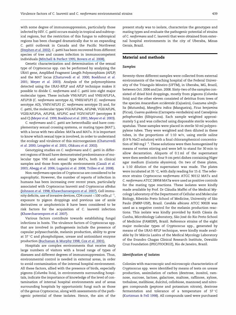
Virulence factors of C. laurentii and C. neoformans environmental strains 439
with some degree of immunosuppression, particularly those
infected by HIV. C. gattii occurs mainly in tropical and subtrop-
ical regions, but the restriction of this fungus to subtropical
regions has been changed dramatically due to an expanding
C. gattii outbreak in Canada and the Pacific Northwest
(Stephen et al. 2002). C. gattii has been recovered from different
species of tree and causes infection in immunocompetent
individuals (Mitchell & Perfect 1995; Bovers et al. 2008).
Genetic characterization and determination of the sexual
type of Cryptococcus spp. can be performed by analyzing the
URA5 gene, Amplified Fragment Length Polymorphism (AFLP)
and the MAT locus (Chaturvedi et al. 2000; Boekhout et al.
2001; Meyer et al. 2003). Analysis of the polymorphisms
detected using the URA5-RFLP and AFLP technique makes it
possible to divide C. neoformans and C. gattii into eight major
molecular types. These include VNI/AFLP1 and VNII/AFLP1A,
AFLP1B (C. neoformans serotype A), VNIII/AFLP3 (C. neoformans
serotype AD), VNIV/AFLP2 (C. neoformans serotype D) and, for
C. gattii, the molecular types VGI/AFLP4A, AFLP4B, VGII/AFLP6,
VGIII/AFLP5A, AFLP5B, AFLP5C and VGIV/AFLP7 (serotypes B
and C) (Meyer et al. 1999; Boekhout et al. 2001; Meyer et al. 2003).
C. neoformans and C. gattii are heterothallic and have com-
plementary sexual crossing systems, or mating types (MAT ),
with a locus with two alleles: MATa and MATa. It is important
to know which sexual type is involved, in order to understand
the ecology and virulence of this microorganism (Chaturvedi
et al. 2000; Lengeler et al. 2001; Ohkusu et al. 2002).
Genotyping studies on C. neoformans and C. gattii in differ-
ent regions of Brazil have demonstrated predominance of mo-
lecular type VNI and sexual type MATa, both in clinical
samples and those from specific environments (Casali et al.
2003; Abegg et al. 2006; Lugarini et al. 2008; Trilles et al. 2008).
Non-neoformans species of Cryptococcus are considered to be
saprophytic. However, the number of reports of infection in
humans has been increasing over recent years, particularly
associated with Cryptococcus laurentii and Cryptococcus albidus
(Johnson et al. 1998; Khawcharoenporn et al. 2007). Cell immu-
nity deficits, use of invasive devices, CD4 count<100 cells ml�1,
exposure to pigeon droppings and previous use of azole
derivatives or amphotericin B have been considered to be
risk factors for the acquisition of C. laurentii infection
(Khawcharoenporn et al. 2007).
Various factors contribute towards establishing fungal
infections in hosts. The virulence factors of Cryptococcus spp.
that are involved in pathogenesis include the presence of
capsular polysaccharide, melanin production, ability to grow
at 37 �C and phospholipase, urease and antioxidant enzyme
production (Buchanan & Murphy 1998; Cox et al. 2001).
Hospitals are complex environments that receive daily
large numbers of visitors with a broad range of types of
diseases and different degrees of immunosuppression. Thus,
environmental control is needed in external areas, in order
to avoid contamination of the internal hospital environment.
All these factors, allied with the presence of birds, especially
pigeons (Columba livia), in environments surrounding hospi-
tals, indicate the importance of knowledge of the level of con-
tamination of internal hospital environments and of areas
surrounding hospitals by opportunistic fungi such as those
of the genus Cryptococcus, along with assessments of the path-
ogenic potential of these isolates. Hence, the aim of the
present study was to isolate, characterize the genotypes and
mating types and evaluate the pathogenic potential of strains
of C. neoformans and C. laurentii that were obtained from exter-
nal hospital environments in the city of Uberaba, Minas
Gerais, Brazil.
Material and methods
Samples
Seventy-three different samples were collected from external
environments of the teaching hospital of the Federal Univer-
sity of the Triangulo Mineiro (UFTM), in Uberaba, MG, Brazil,
between Oct. 2006 and Jan. 2008. Sixty-two of the samples con-
sisted of dried bird droppings, mostly from pigeons (Columba
livia) and the other eleven consisted of detritus from trees of
the species Anacardium occidentale (Cajueiro), Guazuma ulmifo-
lia (Mutumba), Mangifera indica (Mangueira), Ficus benjamina
(Ficus), Guarea guidonia (Carrapeta-verdadeira) and Caesalpinia
peltophoroides (Sibipiruna). Each sample weighted approxi-
mately 5 g and was collected using disposable sterile wooden
spatulas. These samples were placed in sterile 50 ml polypro-
pylene tubes. They were weighted and then diluted in these
tubes, in the proportions of 1:10 w/v, using sterile saline
(0.9 % NaCl solution) with a final chloramphenicol concentra-
tion of 360 mg l�1. These solutions were then homogenized by
means of vortex stirring and were left to stand for 30 min to
allow decantation. Aliquots of 0.1 ml of the supernatant
were then seeded onto four 9 cm petri dishes containing Niger
agar medium (Guizotia abyssinica). On two of these plates,
a 1:10 dilution of the supernatant was made. The plates
were incubated at 35 �C, with daily reading for 15 d. The refer-
ence strains Cryptococcus neoformans ATCC 90112 MATa and
C. neoformans ATCC 28958 MATa were used as positive controls
for the mating type reactions. These isolates were kindly
made available by Prof. Dr Claudia Maffei of the Medical My-
cology Laboratory of the Department of Cellular and Molecular
Biology, Ribeirao Preto School of Medicine, University of Sao
Paulo (FMRP-USP), Brazil. Candida albicans ATCC 90028 was
used as a negative control for the melanin production reac-
tions. This isolate was kindly provided by Keith Cassia da
Cunha, Microbiology Laboratory, Sao Jose do Rio Preto School
of Medicine (FAMERP), Brazil. Reference strains of the eight
major molecular types of Cryptococcus spp., generated by
means of the URA5-RFLP technique, were kindly made avail-
able by Dr Marcia Lazera of the Medical Mycology Laboratory
of the Evandro Chagas Clinical Research Institute, Oswaldo
Cruz Foundation (IPEC/FIOCRUZ), Rio de Janeiro, Brazil.
Identification of isolates
Colonies with macroscopic and microscopic characteristics of
Cryptococcus spp. were identified by means of tests on urease
production, assimilation of carbon (dextrose, inositol, ram-
nose, sucrose, lactose, galactose, maltose, raffinose, xylose,
trehalose, melibiose, dulcitol, cellobiose, mannose) and nitro-
gen compounds (peptone and potassium nitrate), dextrose
fermentation and tolerance of a temperature of 37 �C
(Kurtzman & Fell 1998). All compounds used were purchased

440 L. Andrade-Silva et al.
from VETEC (Vetec, Brazil). To differentiate between Cryptococ-
cus neoformans and Cryptococcus gattii, the canavanine-glycine-
bromothymol blue (CGB) medium was used (Kwon-Chung
et al. 1982). Different colonies from the same sample were
identified by a number followed by a letter. After identifica-
tion, the strains were kept in water at a temperature of
approximately 4 �C and then frozen at �20 �C in YPD medium
containing 30 % (v/v) of glycerol.
Extraction of fungal DNA
Genomic DNA was extracted in accordance with a previously
described technique (Meyer et al. 2003). The extract was
diluted in 500 ml of TE (10 mM Tris–HCl and 0.5 M EDTA)
containing 50 mg ml�1 of RNAse A (Invitrogen, USA) and was
stored at 4 �C. Quantification and determination of the purity
of the extracted DNA were performed by spectrophotometry
(Eppendorf, Biophotometer 22331, USA).
Determination of mating types
The reaction was performed in accordance with a previously
described technique (Chaturvedi et al. 2000) in volumes of
25 ml, containing 25 ng of DNA, PCR 1� buffer (10 mM Tris–
HCl at pH 8.3, 50 mM KCl and 1.5 mM MgCl2), 0.2 mM of the
dNTPs (dATP, dCTP, dGTP and dTTP) (Invitrogen, USA), 3 mM
of MgCl2, 1.5 U of Taq DNA polymerase (Invitrogen, Brazil)
1.68 pmol of the MFa (MATa1) forward primer (50-CTTCACTGC
CATCTTCACCA-30) and 1.71 pmol of the MFa (MATa2) reverse
primer (50-GACACAAAGGGTCATGCCA-30). The MATa primer
pair was (MATa1: 50-CGCCTTCACTGCTACCTTCT-30) and
(MATa2: 50-AACGCAAGAGTAAGTCGGGC-30). The amplifica-
tion reactions were performed in a thermocycler (MJ Research
PTC-100, Germany) that was programmed for an initial dena-
turing at 95 �C (4 min) and 34 cycles at 94 �C (1 min), with
annealing at 63 �C (1 min) for MATa and 60 �C for MATa and
extension at 72 �C (1 min), followed by an extension cycle of
10 min at 72 �C. All of the amplification products obtained
from the PCR reactions and from the URA5-RFLP reaction
were separated by means of electrophoresis on agarose gel
at a concentration of 1.5 % for the PCR reactions and 3 % for
the URA5-RFLP reactions, at 90 V. This was followed by stain-
ing with 0.5 mg ml�1 of ethidium bromide, and the product
was viewed in a transilluminator under ultraviolet light and
photographed using a digital camera.
URA5-RFLP
The PCR reaction on the URA5 gene for isolates of Cryptococcus
neoformans was performed in accordance with a previously
described technique (Meyer et al. 1999, 2003) in a final volume
of 25 ml, containing 25 ng of DNA, PCR 1� buffer (10 mM Tris–
HCl at pH 8.3, 50 mM KCl and 1.5 mM MgCl2), 0.2 mM of the
dNTPs (dATP, dCTP, dGTP and dTTP) (Invitrogen, Brazil),
3 mM of MgCl2, 1.5 U of Taq DNA polymerase (Invitrogen, Bra-
zil) and 3.32 pmol of the URA5 primer (50-ATGTCCTCC
CAAGCCCTCGACTCCG-30) and 3.44 pmol of the SJO1 primer
(50-TTAAGACCTCTGAACACCGTACTC-30). The thermocycler
was programmed for an initial denaturing at 95 �C for 4 min
and 34 cycles of denaturing at 94 �C for 45 s, with association
at 57 �C for 1 min and extension at 72 �C for 1 min, followed
by an extension cycle for 10 min at 72 �C. The PCR reaction
with primers URA5 and SJO1 to Cryptococcus laurentii was per-
formed with the same concentration of primers, but with
a higher stringency. The thermocycler was programmed for
an initial denaturing at 95 �C for 4 min and 34 cycles of dena-
turing at 94 �C for 45 s, with association at 61 �C for 45 s and
extension at 72 �C for 1 min and 30 s, followed by an extension
cycle for 10 min at 72 �C. Aliquots of 25 ml of the PCR product of
both species of Cryptococcus were subjected to double diges-
tion using Sau96I (10 U ml�1) and HhaI (20 U ml�1) (New England
Biolabs, USA). The molecular types were determined by com-
parison with the profiles obtained from the standard strains of
the eight principal genotypes (VNI–VNIV) and (VGI–VGIV) of
C. neoformans and Cryptococcus gattii (Meyer et al. 2003).
Measurement of capsule enlargement and yeast diameter
The capsule synthesis capacity was evaluated as the differ-
ence between the capsule thickness in the control medium
and its thickness after cultivation in the medium for inducing
capsule enlargement (Sabouraud broth diluted 1:10 with
50 mM 3-(N-morpholino)propanesulfonic acid, MOPS, pH 7.3)
(Zaragoza & Casadevall 2004). Aliquots of 5� 105 cfu deter-
mined by counting in a Neubauer counting chamber, were in-
oculated in 10 ml tubes containing 2 ml of Sabouraud broth
(control) and Sabouraud broth diluted 1:10 in MOPS (induc-
tion). These were maintained under incubation at 35 �C for
48 h, with stirring at 100 rpm. The capsule thickness and yeast
diameter were measured by negative staining of the cells with
China ink (Trident, Sao Paulo, Brazil), under an optical micro-
scope (Ken-A-Vision, USA) at a magnification of 1000�. The
readings were made with the aid of a micrometric eyepiece
(Shimadzu-Kalnew, Japan), calibrated with the micrometric
slide (Spencer Lenz Company, USA). Twenty different cells
from each isolate were measured. Capsule induction was
evaluated quantitatively. We considered that an isolate was
able to promote capsule enlargement when its mean capsule
thickness in the induction medium (MOPS) was greater than
the mean thickness plus two standard deviations of isolates
in the control medium (Sabouraud).
Phospholipase production
The samples were, in duplicate, transferred to (5 ml inoculum
of the order of 106 cfu ml�1, determined by counting in a Neu-
bauer counting chamber) petri dishes containing phospholi-
pase agar and were incubated at 35 �C for 5 d. Briefly,
phospholipase agar containing Sabouraud Dextrose Agar
(Oxoid, England) 1.0 M sodium chloride, 5.0 mM calcium chlo-
ride and 8 % sterile egg yolk (Oxoid, England). At the end of this
period, the diameters of the colony and precipitation halo
were measured, and the Pz for each strain was determined
(Samaranayake et al. 1984). The results were presented as
classes (Price et al. 1982).
Melanin production and growth ability at 37 �C
Melanin production was determined in 9 cm petri dishes con-
taining Niger agar (Guizotia abyssinica) incubated for 5 d at

Virulence factors of C. laurentii and C. neoformans environmental strains 441
35 �C. Phenol-oxidase production was observed by colony
colors. The Candida albicans (ATCC 90028) was used as negative
control for melanin production. Melanin production was qual-
itatively assayed, by visually determining the color of colo-
nies. Growth ability at 37 �C was evaluated qualitatively by
viewing the growth of the isolates after 5 d of incubation in
Sabouraud dextrose agar.
Statistical analysis
The data were analyzed with the Bioestat� 4.0 (www.mamir-
aua.org.br) and MS Excel� (Microsoft Corporation) softwares.
The Kolmogorov–Smirnov test was used to evaluate whether
the data presented normal distribution. Variables with normal
distribution were tested regarding the differences in mean
capsule thickness, yeast diameter and phospholipase produc-
tion between the control and induction media, using
Student’s t-test and the Z test. Variables without normal
distribution were tested regarding the difference between
the means, using the Mann–Whitney test. The relationship
between the Pz value and the capsule thickness in the control
and induction media was tested using Spearman’s correlation
coefficient. For all tests, the significance level was set at 5 %
(P< 0.05).
Results
C. neoformans and C. laurentii phenotype characterization
Cryptococcus laurentii was differentiated from Cryptococcus neo-
formans by lactose assimilation, a carbohydrate not assimi-
lated by C. neoformans (Kurtzman & Fell 1998) (data not
shown). All strains of C. laurentii and C. neoformans were posi-
tive for melanin production in Niger agar and growth ability at
37 �C in Sabouraud dextrose agar. C. laurentii colonies had
a brown lighter color than the colonies of C. neoformans (data
not shown). Out of the 73 samples collected, 41 (56.1 %) were
positive for C. neoformans or C. laurentii. C. neoformans alone
was isolated from 24 plates (58.5 %), and C. laurentii alone
was isolated from nine plates (21.9 %). C. laurentii and C. neofor-
mans were isolated concomitantly in eight (19.5 %) of the
41 samples. Differentiation between C. neoformans and
Table 1 – Cryptococcus spp. isolated from peri- and intra-hospi
Places Samples
Bird droppings Debris of trees To
Air conditioning 02 00 0
External corridors 21 00 2
Roofs, eaves
and windows nets
23 00 2
Outpatient wards 09 00 0
Building facade 07 03 1
Trees 00 08 0
Total 62 11 7
Cl – C. laurentii; Cn – C. neoformans.
a Isolated from bird droppings.
Cryptococcus gattii was done using the CGB medium, and all
the isolates were found to be C. neoformans. More than one
strain of the same species was isolated from 33 % (8/24) of
the samples that were positive for C. neoformans alone; from
33 % (3/9) of the samples that were positive for C. laurentii
alone; and from 50 % (4/8) of the samples that were positive
for these two fungi simultaneously. Thus, a total of 51 isolates
of C. neoformans and 27 of C. laurentii were obtained. Five
(45.5 %) of the samples obtained from tree detritus were posi-
tive for C. laurentii. The tree species that were positive for
C. laurentii were Anacardium occidentale (Cajueiro), Guazuma
ulmifolia (Mutumba), Mangifera indica (Mangueira) and Ficus
benjamina (Ficus) (Table 1).
C. neoformans and C. laurentii molecular characterization
All the isolates of Cryptococcus neoformans amplified a fragment
of approximately 750 bp of the URA5 gene, using the URA5 and
SJO1 primers (data not shown). Digestion of the specific frag-
ment using the endonucleases Sau96I and HhaI showed that,
out of the 51 isolates of C. neoformans, 47 (92.1 %) presented
the molecular type VNI and four (7.9 %) presented VNII
(Fig 1C). All of the isolates were classified as MATa. In one of
the positive samples, the molecular types VNI and VNII were
found concomitantly (isolates 93A and 93B) (Fig 1C).
Using the URA5 and SJO1 primers for C. neoformans and
Cryptococcus gattii, we observed amplification of a fragment
of approximately 1.6 kb in 59.3 % (16/27) of the Cryptococcus
laurentii isolates (data not shown). Among the 16 C. laurentii
isolates that amplified a fragment of the URA5 gene, fifteen
presented similar restriction profiles, while the isolate 42B
demonstrated a different profile (Fig 1A and B).
Induction of capsule enlargement in C. neoformans andC. laurentii
The 51 isolates of Cryptococcus neoformans presented a mean
capsule thickness of 0.91� 0.32 mm in Sabouraud medium
(control) and 2.58� 1.46 mm in the induction medium
(P< 0.001) (Fig 2A). The isolates of C. neoformans presented
a positive capacity of 76.4 % (39/51) for capsule enlargement
(Fig 2A). There was no difference in mean size of the yeasts
in the two media (results not shown). No significant
tal sampling.
Positive samples
tal C. neoformans C. laurentii Cnþ Cl Total
2 01 00 00 01
1 09 00 03 12
3 09 02 03 14
9 01 02 02 05
0 04a 00 00 04
8 00 05 00 05
3 24 09 08 41

Fig 1 – URA5-RFLP analysis of Cryptococcus neoformans and Cryptococcus laurentii isolates. Representative agarose gels
showing the URA5 amplicons of C. neoformans (C) and C. laurentii (A and B) isolates digested with Sau96I and HhaI. MM – 50 bp
DNA ladder (Invitrogen, USA), Cn – C. neoformans ATCC 90112.
442 L. Andrade-Silva et al.
differences were found from comparison between the molec-
ular types VNI and VNII of the isolates in relation to the mean
yeast size and capsule thickness in the culture media used.
The 27 isolates of Cryptococcus laurentii had a mean capsule
thickness of 0.78� 0.26 mm in the Sabouraud medium (control)
and 1.0� 0.51 mm in the induction medium (P< 0.003) (Fig 2B).
Two (7.4 %) of the 27 isolates of C. laurentii were capable of
capsule enlargement (Fig 2B). This species also did not present
any significant differences in yeast size between the two
media (results not shown).
The C. neoformans isolates presented a greater mean cap-
sule thickness than shown by C. laurentii, both in the induction
and in the control medium, with mean values of 2.58� 1.46
versus 1.00� 0.5 (P< 0.001) and 0.91� 0.32 versus 0.78� 0.26
(P< 0.001), respectively. No difference was found between
Fig 2 – The mean capsule thickness of the isolates of C. neoforman
of the capsule after 48 h of cultivation in control medium (Sabo
the two species regarding yeast size in the two media (results
not shown).
C. neoformans and C. laurentii phospholipase production
The Cryptococcus neoformans isolates presented a positive rate
of 92.1 % (47/51) for phospholipase production, with a mean
Pz of 0.63� 0.16. The classes of intermediate producers and
strong producers accounted for 39.2 % (20/51) and 52.9 %
(27/51), respectively. No correlation was found between the
capsule thicknesses in the control and induction media for
molecular types VNI and VNII of C. neoformans and the phos-
pholipase production.
The Cryptococcus laurentii isolates presented a positive rate
of 85.1 % (23/27), with a mean Pz of 0.69� 0.18, and the classes
s (A) and C. laurentii (B) tested for their ability to enlargement
uraud) and inducing medium (MOPS).

Virulence factors of C. laurentii and C. neoformans environmental strains 443
of intermediate and strong producers accounted for 44.4 %
(12/27) and 40.7 % (11/27), respectively. There was no signifi-
cant difference in phospholipase production between the
two species. No correlation was found between the capsule
thicknesses in the control and induction media for C. laurentii
isolates and the phospholipase production.
Discussion
The positive findings of Cryptococcus laurentii and Cryptococcus
neoformans in the samples of the present study are in agree-
ment with the results obtained by other authors, who also
demonstrated that C. neoformans predominates in isolates
from different environments. This can be explained by the
great frequency of Columbiformes in the environment from
which our samples were obtained (Casali et al. 2003; Abegg
et al. 2006).
The genotype VNI predominated in the majority of the
C. neoformans isolates (92 %), as demonstrated by other
authors (Casali et al. 2003; Meyer et al. 2003; Trilles et al.
2008). However, findings of different genotypes even in the
same sample (isolates 93A and 93B) suggest that there is
genetic variability among C. neoformans isolates from this en-
vironment. This may be translated into phenotypic variations
such as differences in virulence or in susceptibility to thera-
peutic agents (Casali et al. 2001; Jain et al. 2005) The determina-
tion of mating types showed that MATa presented 100 %
prevalence in the C. neoformans isolates, which is in agreement
with descriptions from several other authors (Casali et al. 2003;
Abegg et al. 2006; Lugarini et al. 2008).
The positive findings of C. laurentii in bird droppings in the
present study is concordant with the results from other au-
thors such as Pedroso et al. (2009), Bernardo et al. (2001) and
Tay et al. (2008), who reported positive findings of 12.9 %,
40.0 % and 4.6 %, respectively. C. laurentii was isolated con-
comitantly with C. neoformans in 10.9 % of the samples, which
exceeds the results described recently (2.3 %) in another
region of Brazil (Pedroso et al. 2009). This may indicate that
C. laurentii and C. neoformans share the same habitat in the
region studied.
Data from other studies have demonstrated positive find-
ings of C. laurentii in tree detritus at rates ranging from 12.7 %
to 77.0 % (Montenegro & Paula 2000; Bernardo et al. 2001; Tay
et al. 2008). The results from the present study resemble those
obtained by Montenegro & Paula (2000), who reported a positive
rate of 58.3 %, for samples from detritus of trees of the genus
Eucalyptus, in public parks in the city of Sao Paulo. Isolation
of C. laurentii from the tree species Ficus benjamina, Guazuma
ulmifolia, Anacardium occidentale and Mangifera indica had not
previously been described in the literature and it suggests
that these species may also be a habitat for C. laurentii, in the
same way as previously described for C. neoformans regarding
the genus Fıcus (Lazera et al. 2000). In the present study, C. neo-
formans was not recovered from any of the tree samples. One
possible explanation for this might be the better adaptation
of C. laurentii to this environment, as demonstrated by the
greater frequency of reports of this species in trees, compared
with C. neoformans (Lazera et al. 2000; Montenegro & Paula 2000;
Bernardo et al. 2001; Tay et al. 2008).
We used the URA5 and SJO1 primers for C. neoformans and
Cryptococcus gattii genotyping with the aim of investigating
their capacity for molecular typing with regard to C. laurentii.
Sixteen isolates (59.3 %) of the latter species amplified a DNA
fragment of approximately 1.6 kb, thus showing that among
the fungus species studied, this site presents conserved re-
gions. Furthermore, the amplified fragments were polymor-
phic, since 15 isolates presented the same restriction profile
and one isolate had a distinct profile. On the other hand, the
low frequency of amplification may have occurred because
of the large intraspecies genetic variability that has already
been described for this species (Sugita et al. 2000; Takashima
et al. 2003).
The expression of virulence factors and increased pathoge-
nicity of infections caused by C. neoformans has been evaluated
by several authors (Buchanan & Murphy 1998; Vidotto et al.
1998; Zaragoza & Casadevall 2004; Zaragoza et al. 2008). Iso-
lates with the potential to induce capsules were found in
C. neoformans (76.4 %) and in C. laurentii (7.1 %), this result in
combination with the data melanin, growth ability at 37 �C
and phospholipases production, suggests that both species
are potentially virulent. Variability in capsule induction has
been described previously and is strain-dependent (Zaragoza
& Casadevall 2004; Zaragoza et al. 2008), as also observed in
the present study. C. neoformans presented a high capacity
for induction of capsule enlargement in the different isolates.
In reviewing the literature, we did not find any data related
to the formation of capsule by C. laurentii, but some authors
have described similarities in the production and constituents
of the capsule in this species, in relation to C. neoformans. This
characteristic, strengthened through descriptions of cases of
human infection by C. laurentii (Khawcharoenporn et al.
2007), might also be related to greater virulence in some iso-
lates of C. laurentii. Proof or disproof for this hypothesis may
only come through virulence tests on animal models or mac-
rophage experiments (Ma et al. 2009).
The greater virulence of C. neoformans isolates that produce
phospholipases has already been demonstrated in animal
virulence experiments that compared isolates with high
production and absence of production, and from comparisons
between wild strains and strains known to have the gene
deletions responsible for producing these enzymes (Chen
et al. 1997; Cox et al. 2001). In our study, the positive rate for
phospholipase production in C. neoformans isolates was 92 %,
and the majority of the isolates were classified as strong pro-
ducers (53 %). These results exceed those already described
from C. neoformans isolates from different environments,
which ranged from 26 % to 73 % (Vidotto et al. 1998; Casali
et al. 2003; Abegg et al. 2006).
The positive rate of phospholipase production for the C.
laurentii isolates was 85.18 %. Data describing phospholipase
production in C. laurentii are scarce in the literature. The exist-
ing data report that phospholipase producing isolates have
pathogenic potential, since C. laurentii isolated from the cen-
tral nervous system of dogs showed this phenotype (Campos
2006). Likewise, this has been reported in isolates from the clo-
aca of birds, coming from different environments (Cafarchia
et al. 2008). These data, together with other results such as
the growth of all C. laurentii isolates at 37 �C and the response
to capsule enlargement induction tests by some isolates,

444 L. Andrade-Silva et al.
suggest that some of the isolates of this species are potentially
pathogenic.
The genotyping and mating type results add information
regarding the distribution of molecular types from Cryptococ-
cus spp. isolates from different environments. Furthermore,
they demonstrate that this method, which has already been
used to characterize C. neoformans and C. gattii, has the capac-
ity to demonstrate genetic variability in C. laurentii. The results
also indicate that C. laurentii and C. neoformans may share the
same environment and that some of these isolates have path-
ogenic potential, thereby contributing towards the informa-
tion on the ecology and epidemiology of these two species.
Acknowledgements
We wish to thank Dr Eliane Lages-Silva, Dr Luiz E. Ramırez and
Dr Virmondes Rodrigues Junior (UFTM, Brazil) for permitting
the use of the DNA analysis equipments, Delio J.M. Amador Ju-
nior (UFTM, Brazil) for providing the PCR protocols used in this
work, Dr Claudia Maffei (FMRP-USP, Brazil) and Dr Marcia Laz-
era (FIOCRUZ, Brazil) for providing Cryptococcus spp. reference
strains. This study was supported by Fundacao de Amparo A
Pesquisa do Estado de Minas Gerais (FAPEMIG, Grant CDS-
1881/06) and Conselho Nacional de Desenvolvimento Cientı-
fico e Tecnologico (CNPq). All experiments were conducted
according to Brazilian regulations.
r e f e r e n c e s
Abegg MA, Cella FL, Faganello J, Valente P, Schrank A,Vainstein MH, 2006. Cryptococcus neoformans and Cryptococcusgattii isolated from the excreta of psittaciformes in a southernBrazilian zoological garden. Mycopathologia 161: 83–91.
Bernardo FM, Martins HM, Martins ML, 2001. Urban Sources ofCryptococcus spp – Lisbon (Portugal). Revista Portuguesa deCiencias Veterinarias 96: 157–160.
Boekhout T, Theelen B, Diaz M, Fell JW, Hop WC, Abeln EC,Dromer F, Meyer W, 2001. Hybrid genotypes in the pathogenicyeast Cryptococcus neoformans. Microbiology 147: 891–907.
Bovers M, Hagen F, Boekhout T, 2008. Diversity of the Cryptococcusneoformans–Cryptococcus gattii species complex. Revista Iberoa-mericana de Micologıa 25: S4–S12.
Buchanan KL, Murphy JW, 1998. What makes Cryptococcusneoformans a pathogen? Emerging Infectious Diseases 4: 71–83.
Cafarchia C, Romito D, Coccioli C, Camarda A, Otranto D, 2008.Phospholipase activity of yeasts from wild birds and possibleimplications for human disease. Medical Mycology 46: 429–434.
Campos FL, 2006. Biotipagem, sorogrupagem e producao de pro-tease e fosfolipase por Cryptococcus neoformans isolados de caese gatos nos municıpios de Rio de Janeiro-RJ e Sao Paulo-SP.Master Thesis.1: 1–62. Seropedica, Rio de Janeiro, Brasil. In-stituto de Veterinaria, Curso de Pos-Graduacao em Microbio-logia Veterinaria. Universidade Federal Rural do Rio de Janeiro.
Casali AK, Goulart L, Rosa e Silva LK, Ribeiro AM, Amaral AA,Alves SH, Schrank A, Meyer W, Vainstein MH, 2003. Moleculartyping of clinical and environmental Cryptococcus neoformansisolates in the Brazilian state Rio Grande do Sul. FEMS YeastResearch 3: 405–415.
Casali AK, Schrank A, Vainstein MH, 2001. Cryptococcusneoformans – Aspectos moleculares e epidemiologicos.Biotecnologia Ciencia & Desenvolvimento 20: 34–37.
Chaturvedi S, Rodeghier B, Fan J, McClelland CM, Wickes BL,Chaturvedi V, 2000. Direct PCR of Cryptococcus neoformansMATalpha and MATa pheromones to determine mating type,ploidy, and variety: a tool for epidemiological and molecularpathogenesis studies. Journal of Clinical Microbiology 38:2007–2009.
Chen SC, Muller M, Zhou JZ, Wright LC, Sorrell TC, 1997. Phos-pholipase activity in Cryptococcus neoformans: a new virulencefactor? Journal of Infectious Diseases 175: 414–420.
Cox GM, McDade HC, Chen SC, Tucker SC, Gottfredsson M,Wright LC, Sorrell TC, Leidich SD, Casadevall A,Ghannoum MA, Perfect JR, 2001. Extracellular phospholipaseactivity is a virulence factor for Cryptococcus neoformans.Molecular Microbiology 39: 166–175.
Jain N, Wickes BL, Keller SM, Fu J, Casadevall A, Jain P, Ragan MA,Banerjee U, Fries BC, 2005. Molecular epidemiology of clinicalCryptococcus neoformans strains from India. Journal of ClinicalMicrobiology 43: 5733–5742.
Johnson LB, Bradley SF, Kauffman CA, 1998. Fungaemia due toCryptococcus laurentii and a review of non-neoformanscryptococcaemia. Mycoses 41: 277–280.
Khawcharoenporn T, Apisarnthanarak A, Mundy LM, 2007.Non-neoformans cryptococcal infections: a systematic review.Infection 35: 51–58.
Kurtzman CP, Fell JW, 1998. The Yeasts: a taxonomic study, 4th edn.Elsevier Science, Amsterdam.
Kwon-Chung KJ, Polacheck I, Bennett JE, 1982. Improved diag-nostic medium for separation of Cryptococcus neoformans var.neoformans (serotypes A and D) and Cryptococcus neoformansvar. gattii (serotypes B and C). Journal of Clinical Microbiology 15:535–537.
Lazera MS, Salmito Cavalcanti MA, Londero AT, Trilles L,Nishikawa MM, Wanke B, 2000. Possible primary ecologicalniche of Cryptococcus neoformans. Medical Mycology 38: 379–383.
Lengeler KB, Cox GM, Heitman J, 2001. Serotype AD strains ofCryptococcus neoformans are diploid or aneuploid and areheterozygous at the mating-type locus. Infection and Immunity69: 115–122.
Lugarini C, Goebel CS, Condas LA, Muro MD, de Farias MR,Ferreira FM, Vainstein MH, 2008. Cryptococcus neoformansisolated from Passerine and Psittacine bird excreta in the stateof Parana, Brazil. Mycopathologia 166: 61–69.
Ma H, Hagen F, Stekel DJ, Johnston SA, Sionov E, Falk R,Polacheck I, Boekhout T, May RC, 2009. The fatal fungaloutbreak on Vancouver Island is characterized by enhancedintracellular parasitism driven by mitochondrial regulation.Proceedings of the National Academy of Sciences U S A 106:12980–12985.
Meyer W, Castaneda A, Jackson S, Huynh M, Castaneda E, 2003.Molecular typing of IberoAmerican Cryptococcus neoformansisolates. Emerging Infectious Diseases 9: 189–195.
Meyer W, Marszewska K, Amirmostofian M, Igreja RP, Hardtke C,Methling K, Viviani MA, Chindamporn A, Sukroongreung S,John MA, Ellis DH, Sorrell TC, 1999. Molecular typing of globalisolates of Cryptococcus neoformans var. neoformans by poly-merase chain reaction fingerprinting and randomly amplifiedpolymorphic DNAda pilot study to standardize techniques onwhich to base a detailed epidemiological survey. Electrophore-sis 20: 1790–1799.
Mitchell TG, Perfect JR, 1995. Cryptococcosis in the era ofAIDSd100 years after the discovery of Cryptococcus neoformans.Clinical Microbiology Reviews 8: 515–548.
Montenegro H, Paula CR, 2000. Environmental isolation of Cryp-tococcus neoformans var. gattii and C. neoformans var. neoformansin the city of Sao Paulo, Brazil. Medical Mycology 38: 385–390.
Ohkusu M, Tangonan N, Takeo K, Kishida E, Ohkubo M, Aoki S,Nakamura K, Fujii T, Siqueira IC, Maciel EA, Sakabe S,Almeida GM, Heins-Vaccari EM, Lacaz CS, 2002. Serotype,

Virulence factors of C. laurentii and C. neoformans environmental strains 445
mating type and ploidy of Cryptococcus neoformans strainsisolated from patients in Brazil. Revista do Instituto de MedicinaTropical de Sao Paulo 44: 299–302.
Pedroso RS, Ferreira JC, Candido RC, 2009. The isolation andcharacterization of virulence factors of Cryptococcus spp. fromsaprophytic sources in the city of Ribeirao Preto, Sao Paulo,Brazil. Microbiological Research 164: 221–227.
Price MF, Wilkinson ID, Gentry LO, 1982. Plate method for detection ofphospholipase activity in Candida albicans. Sabouraudia 20: 7–14.
Samaranayake LP, Raeside JM, MacFarlane TW, 1984. Factorsaffecting the phospholipase activity of Candida species in vitro.Sabouraudia 22: 201–207.
Springer DJ, Chaturvedi V, 2010. Projecting global occurrence ofCryptococcus gattii. Emerging Infectious Diseases 16: 14–20.
Stephen C, Lester S, Black W, Fyfe M, Raverty S, 2002. Multispeciesoutbreak of cryptococcosis on southern Vancouver Island,British Columbia. The Canadian Veterinary Journal 43: 792–794.
Sugita T, Takashima M, Ikeda R, Nakase T, Shinoda T, 2000.Intraspecies diversity of Cryptococcus laurentii as revealed bysequences of internal transcribed spacer regions and 28SrRNA gene and taxonomic position of C. laurentii clinicalisolates. Journal of Clinical Microbiology 38: 1468–1471.
Takashima M, Sugita T, Shinoda T, Nakase T, 2003. Three newcombinations from the Cryptococcus laurentii complex:
Cryptococcus aureus, Cryptococcus carnescens and Cryptococcuspeneaus. International Journal of Systematic and Evolutionary Mi-crobiology 53: 1187–1194.
Tay ST, Na SL, Tajuddin TH, 2008. Natural occurrence and growthreaction on canavanine-glycine-bromothymol blue agar ofnon-neoformans Cryptococcus sppin Malaysia. Mycoses 51:515–519.
Trilles L, Lazera Mdos S, Wanke B, Oliveira RV, Barbosa GG,Nishikawa MM, Morales BP, Meyer W, 2008. Regional patternof the molecular types of Cryptococcus neoformans and Crypto-coccus gattii in Brazil. Memorias do Instituto Oswaldo Cruz 103:455–462.
Vidotto V, Leone R, Sinicco A, Ito-kuwa S, Criseo G, 1998. Com-parison of phospholipase production in Cryptococcus neofor-mans isolates from AIDS patients and bird droppings.Mycopathologia 142: 71–76.
Zaragoza O, Casadevall A, 2004. Experimental modulation ofcapsule size in Cryptococcus neoformans. Biological ProceduresOnline 6: 10–15.
Zaragoza O, Chrisman CJ, Castelli MV, Frases S, Cuenca-Estrella M, Rodriguez-Tudela JL, Casadevall A, 2008. Capsuleenlargement in Cryptococcus neoformans confers resistance tooxidative stress suggesting a mechanism for intracellularsurvival. Cellular Microbiology 10: 2043–2057.
Copyright © 2022 FDOKUMEN