Regional pattern of the molecular types of Cryptococcus neoformans and Cryptococcus gattii in Brazil
Upload
independentCategory
view
2download
0
ORIGINAL ARTICLE
Endophytic fungal compounds active againstCryptococcus neoformans and C. gattiiCristiane B Pereira1, Djalma M de Oliveira2, Alice FS Hughes1, Markus Kohlhoff3, Mariana LA Vieira1,Aline B Martins Vaz1, Mariana C Ferreira1, Camila R Carvalho1, Luiz H Rosa1, Carlos A Rosa1,Tânia MA Alves3, Carlos L Zani3, Susana Johann1 and Betania B Cota3
Infections with Cryptococcus are invasive mycoses associated with significant morbidity and mortality, mainly in
immunosuppressed patients. Several drugs have been introduced to combat these opportunistic infections. However, resistance
of this organism to antifungal drugs has increased, causing difficulties in the treatment. The goal of this work was to evaluate
the antifungal activity of ethanol extracts from endophytic fungi isolated from plants collected from different Brazilian
ecosystems and to perform the fractionation of the most promising extract. Four-hundred fungal extracts were investigated by
microdilution broth assays against Cryptococcus neoformans and Cryptococcus gattii at a concentration of 500 μgml−1. Among
them, the extract of Mycosphaerella sp. UFMGCB 2032, an endophytic fungus isolated from the plant Eugenia bimarginata DC.
(Myrtaceae) exhibited outstanding antifungal activity against C. neoformans and C. gattii, with MIC values of 31.2 μg ml−1 and
7.8 μg ml−1, respectively. The fractionation of this extract using liquid–liquid partitioning and semi-preparative HPLC afforded
two eicosanoic acids with antifungal activity, compound 1, (2 S,3 R,4 R)-(E)-2-amino-3,4-dihydroxy-2-(hydroxymethyl)-14-
oxoeicos-6,12-dienoic acid with MIC values ranging from 1.3–2.50 μgml−1, and compound 2, known as myriocin, with MIC
values of 0.5 μgml−1 against C. neoformans and C. gattii. These compounds are reported for the first time in the
Mycosphaerella genus.
The Journal of Antibiotics (2015) 00, 1–9. doi:10.1038/ja.2015.11
INTRODUCTION
Cryptococcosis is a systemic invasive mycosis associated withsignificant morbidity and mortality with worldwide distribution ofsome species.1,2 Cryptococcus neoformans and Cryptococcus gattiiare the most important pathogenic species.3 Cryptococcosis is anopportunistic infection that affects immunosuppressed patients,whereas C. gattii yeasts may also affect immunocompetentindividuals.1,4 Currently, the antifungal drugs used for cryptococcosistreatment are amphotericin B, 5-flucytosine and fluconazole. How-ever, the pharmacological management of the disease is difficultbecause of the complexity of the infections and treatments and thereports of resistance and/or tolerance to the antifungals and their toxiceffects.5 The clinical syndromes and the treatment of infections causedby C. neoformans and C. gattii are very similar, but in some cases,a longer induction therapy may be required for a C. gattiiinfection. Moreover, this yeast has a propensity to promote massiveinflammation. Mixed infections and drug resistance can occur,contributing to disease persistence and therapeutic failure.6,7 It istherefore expected that new classes of antifungal agents can helpovercome these problems.
Endophytic microorganisms can live in the intra-and/or intercel-lular environments of plant tissues without causing any apparentnegative effect to the plants.8 They are considered rich sources ofbioactive natural products,9,10 which have been considered promisingalternatives for a variety of human health concerns.10 Endophyticfungi associated with plants belonging to unique ecosystems,owing to environmental conditions, may produce specific secondarymetabolites.9 In this view, Brazil has a rich diversity of plant speciesthat may be associated with endophytic fungi communities and whichremains poorly explored for antifungal activity.11 Ecosystems withunique characteristics such as microbial community, temperature,salinity and pH may contain many novel microorganisms species,including those able to produce compounds with undiscoveredapplications.9,10
The aim of the present study was to investigate the antifungalactivity of crude ethanol extracts obtained from endophytic fungiassociated with plants from different Brazilian ecosystems against thepathogenic yeasts C. neoformans and C. gattii, using a microbrothdilution susceptibility test and to perform the fractionation of the mostpromising extract. The present communication is concerned with the
1Departamento de Microbiologia, Instituto de Ciências Biológicas, Universidade Federal de Minas Gerais, Belo Horizonte, MG, Brazil; 2Departamento de Química e Exatas,Universidade Estadual do Sudoeste da Bahia, Campus Universitário de Jequié, Jequié, Bahia, Brazil and 3Laboratório de Química de Produtos Naturais, Centro de Pesquisa RenéRachou, Fundação OswaldoQ2 Cruz, Belo Horizonte, MG, BrazilCorrespondence: BB Cota, Laboratório de Química de Produtos Naturais, Centro de Pesquisa René Rachou, Fundação Oswaldo CruzQ3 , Av Augusto de Lima 1715, Belo Horizonte,30190-002 MG, Brazil.E-mail: [email protected] 15 October 2014; revised 16 December 2014; accepted 19 January 2015
The Journal of Antibiotics (2015), 1–9& 2015 Japan Antibiotics Research Association All rights reserved 0021-8820/15www.nature.com/ja

isolation and structural characterization of two eicosanoic-acid deri-vatives. These are antifungal compounds isolated and identified fromthe endophytic fungus Mycosphaerella sp., obtained from Eugeniabimarginata DC., a plant from the Myrtaceae family present in theBrazilian savannah.
MATERIALS AND METHODS
Sources of endophytic fungiAll plant samples were obtained from adult specimens collected in Brazil(savannah, Atlantic forest and Amazon rainforest). The plant identity, plantpart collected, geographic coordinates and total number of endophytic fungiextracts evaluated are summarized in Table 1. Voucher specimens weredeposited at the ‘Instituto de Ciências Biológicas’ herbarium (BHCB), ‘Uni-versidade Federal de Minas Gerais (ICB, UFMG)’ and at the herbarium of‘Universidade Federal de Roraima’, Brazil.
Isolation of the endophytic fungiAll samples of plant material were processed and the fungi was isolated asdescribed previously.12 In brief, the tissue samples were cut into pieces, placedin sterilized individual plastic bags and stored for o24 h at 10 °C prior to theisolation of endophytic fungi. Before surface sterilization, the leaves were cutinto ~ 5-mm-long fragments using a flame-sterilized scalpel in a laminar flowhood to avoid air contamination of the tissues, and all samples were processedseparately. The fragments were surface-sterilized by immersion in 2% Extran(1min), 70% EtOH (1min) and 2% sodium hypochlorite (3min), which wasfollowed by a wash with sterile distilled water (2min). Afterwards, thefragments were plated on Petri plates containing potato dextrose agar (PDA,
Q4 Himedia, India) supplemented with chloramphenicol (200 μgml− 1) (Sigma, StLouis, MO, USA). The plates were incubated for up to 60 days at 25 °C, and thehyphal tip of each fungus growing out from the plant tissue was excised andtransferred to a new PDA plate. After incubation at 25 °C for 10 days, culturepurity was assessed using colony morphology. To test the effectiveness of thesurface sterilization, 100 μl of the last water rinse was plated on PDA andincubated at 25 °C. Long-term samples of the filamentous fungal colonies werestored in cryotubes with 15% sterile glycerol at − 80 °C and in sterile distilledwater at room temperature. All endophytic fungal isolates were deposited in theCulture Collection of Microorganisms and Cells at the ‘Universidade Federal deMinas Gerais’ under the code UFMGCB.
Fungi cultivation and extract preparationTwo-millimeter-diameter plugs of each endophytic fungus culture wereinoculated into the centers of Petri dishes (90mm diameter) containing20ml of PDA and incubated at 25± 2 °C for 15 days. The cultures weretransferred to 50-ml vial tubes containing 40ml of EtOH and macerated for48 h at ambient temperature. The organic phase was decanted, and the solventwas removed in a vacuum centrifuge at 35 °C.13
BioassayFungal isolates and inoculum. The antifungal activity of the extracts wasevaluated using C. neoformans ATCC 24067 and C. gattii ATCC 24065. Theseyeast strains were stored frozen at − 80 °C, and the inocula were preparedaccording to document 7.1.14 The starting inocula of Cryptococcus isolates were
cultured onto Sabouraud dextrose agar twice for 48 h at 30 °C to ensure purityand viability. The inoculum was prepared by picking five distinct colonies thatwere suspended in 5ml of sterile saline (0.85% saline), and the resultingsuspension transmittance was adjusted with a spectrophotometer to reach the1.0 McFarland turbidity standard at 530 nm wavelength. This procedure yieldeda yeast stock suspension of 1–5× 10⁶ CFUml− 1, from which was prepared aworking suspension (1–5× 10⁵ CFUml− 1) in sterile distilled water.
Screening for antifungal activityAll crude extracts were diluted to a final concentration of 500 μgml− 1 in RPMI1640 (InLab, São Paulo, Brazil) supplemented with 2% glucose (Vetec, Rio deJaneiro, Brazil) and MOPS (Sigma). Amphotericin B (Sigma) at 2.0 μgml− 1
was used as a positive control. The wells (except the blank control) wereinoculated with the yeasts to obtain a final inoculum of 0.5–2.5× 10⁵
CFUml− 1. The microplates were incubated at 30± 2 °C for 48 h, after which10 μl of a 5mgml− 1 of MTT (3-(4,5-dimetylthiazol-2-yl)-2,5-diphenyltetrazo-lium bromide) (Amresco, USA Q5) solution was added to each well. The plateswere then incubated for 4 h and the absorbance measured at 570 nm using amicrotitre plate spectrophotometer (VERSAmax, Molecular Devices, Sunnyvale,CA, USA).13,14 The data were collected and analyzed using the instrumentsoftware (Softmax, Molecular Devices). The percentages of inhibition (%inhib.) of the growth of the yeasts were calculated according to the followingequation:
%Inhib: ¼ OD of negative control well� OD of sample testedð ÞOD of negative control well
´ 100
Extracts with 75% inhibition of fungal growth against at least one Cryptococcusspecies were considered active. All susceptibility tests were performed induplicate in three independent experiments.
Determination of MICThe microdilution assays used to determinate the MIC were performedaccording to document 7.1 with modifications.14 The concentrations of thesamples ranged from 500 to 0.97 μg ml− 1. Fluconazole (Sigma) and amphoter-icin B (Sigma) were used as positive controls at concentrations of 0.125 to64 μgml− 1 and 0.008–1 μgml− 1, respectively. MIC end points were defined asthe lowest concentration of samples that inhibited yeast growth (yellow well)with inhibition equal or superior to 90% of the growth in the control well.14 AllMIC values were obtained in duplicate in three independent experiments.
Molecular identification of the fungal isolate UFMGCB 2032The internal transcribed spacer (ITS) region was amplified with the universalprimers ITS1 and ITS4.15 The amplification of the ITS region was performed asdescribed previously.16 Successfully amplified PCR products were purified usingan ethanol/ethylenediaminetetraacetic acid precipitation protocol (AppliedBiosystems Q6) and the sequencing reactions were performed at Myleus Biotech-nology (www.myleus.com, Belo Horizonte, Brazil) on an ABI3130 automatedsequencer (Applied Biosystems, Life Technologies Q7, CA, USA) using POP7polymer and BigDye v3.1 with the same primers that were used in theamplification reactions. Quality control of individual sequences was performedby PHRED (http://asparagin.cenargen.embrapa.br/phph), and only thesequences with at least 400 nucleotides and a PHRED quality score above 20were considered. The consensus sequence was then generated by the CAP3
Table 1 Host plant, data and number of extracts of endophytic fungi (EFE) assessed against Cryptococcus species
Host plant Collected part Habitat and GPS location Total of EFE
Solanum cernuum Leaves and stems Atlantic forest (19°52′S, 43°58′W) 24
Eugenia bimarginata Leaves Savannah (19°52′S, 43°58′W) 7
Myrciaria floribunda Leaves Savannah (09°20′S, 49°58′W) 7
Alchornea castaneifolia Leaves Savannah (09°20′S, 49°58′W) 21
Stryphnodendron adstrigens Leaves and stems Savannah (21°05′S, 44°12′W) and (19°30′S, 43°54′W) 151
Carapa guianensis Leaves Amazon rainforest (0°57′02″N, 59°54′41″W) 194
Activity of endophytic fungal compoundQ1
sCB Pereira et al
2
The Journal of Antibiotics

program (http://asparagin.cenargen.embrapa.br/cgi bin/phph/cap3.pl).17 Theconsensus sequence (PHRED420) was subjected to phylogenetic inferences,which were estimated using MEGAVersion 5.0.18 The alignments wereprepared including sequences of all relevant type strains or reference strainsfrom culture collections deposited in GenBank to ensure accurate identification.The sequences were aligned using the MUSCLE method,19 with manualadjustments for visual improvement where necessary. The Akaike informationcriterion was used to identify the most appropriate model of evolution for thedata set. For ITS sequences, the data were estimated using the maximumlikelihood method based on the general time reversible model, and a discretegamma distribution was used to model evolutionary rate differences amongsites (five categories (+G, parameter= 0.5758). The rate variation modelallowed some sites to be evolutionarily invariable ((+I), 0.00% sites). Therobustness of the trees was estimated by a bootstrap analysis20 with 1000replicates. The consensus sequence was submitted to GenBank and assigned anaccession number from KF681521.
General experimental proceduresThin layer chromatography analyses were conducted on pre-coated silica gelG-60/F254 plates (0.25mm, Merck, Darmstadt, Germany). Thin layer chroma-tography plates were eluted in a pre-saturated chamber using mixtures withdifferent proportions of CHCl3:MeOH:NH4OH. The spots were visualizedunder visible and UV light (254 nm and 366 nm), and after heating the platewas sprayed with vanillin-H2SO4 reagent. Analytical reversed phase-HPLCanalyses were run in a HPLC system (Shimadzu, Kyoto, Japan) equipped with aLC6AD pump and a SPD M-10 A VP diode-array detector. Analytical analyseswere carried out on a Shim-pack C18 column (5 μm, 4.6 mm×250mm, i.d.Q8 )eluted with a gradient of CH2CN:H2O at a flow rate of 1mlmin− 1. Semi-preparative purifications were done on a Shim-pack C18 column (5 μm,20×250mm, i.d.) using mixtures of MeOH:H2O or CH2CN:H2O as eluents.The flow rate was set at 8 mlmin− 1 and the effluent absorption measured at220 nm on a Shimadzu SPD-10 A-UV detector (Shimadzu). Solvents wereremoved using a vacuum centrifuge (Thermo-Savant SPD SC250 Express,Holbrook, NY, USA). UV spectrum was recorded in MeOH on a Beckman DUSeries 600 spectrophotometer and optical rotations were determined on aModular Circular Polarimeter MCP 300 (Anton Paar, Ashland, VA, USA).
Large-scale cultivation and extraction proceduresFive 2-mm-diameter plugs of the culture of Mycosphaerella sp. were inoculatedinto each of 600 Petri dishes (90-mm diameter) containing 20ml of PDA. Theplates were incubated at 25± 2 °C for 15 days. The mycelium was separatedfrom the medium with a spatula and both the medium and mycelium wereextracted four times at 48 h intervals using ethyl acetate. The organic phaseswere grouped and concentrated on a rotary evaporator. The residual solventwas removed on a vacuum centrifuge at 40 °C to yield the crude extracts. Theseextracts were diluted in a MeOH:H2O mixture (1:3) and successively parti-tioned between hexane, dichloromethane and ethyl acetate (EtOAc). Thisprocedure afforded the organic fractions denominated as hexane, dichloro-methane and EtOAc, and Q9Aq.
Isolation of antifungal compounds. An 800-mg portion of the EtOAc fractionresidue was dissolved in MeOH:H2O (1:1) and then centrifuged to removesuspended and insolubilized material. Nine aliquots containing ~ 100mg ofextract were injected into the semi-prep column. The column was eluted with alinear gradient of MeOH in H2O starting at 10% and going to 50% in 30min,from 50 to 95% in 20min, from 95 to 100% in 1min, and held at 100% for17min. Twenty-eight sub-fractions were obtained and tested in an antifungalassay. Active sub-fractions 13 (41mg) and 14 (59mg) were further fractionatedby semi-preparative reversed phase-HPLC. The elution started with CH2CN:H2O 30% during 10min, and the CH2CN proportion increased to 50% in14min, 75% in 26min, and then to 100% in 10min and kept at 100% for30min. Sub-fraction 13 yielded 12mg of compound 1, whereas sub-fraction 14afforded 21.5mg of compound 2.
Spectral data of the isolated compoundsOne- and two-dimensional NMR experiments were run on a Bruker Avance400MHz spectrometer with standard pulse sequences using deuterated solventscontaining 0.1% TMS Q10.LC-DAD-MS analyses were performed using a Thermo Surveyor Plus
chromatograph (Thermo Fisher Scientific, Waltham, MA, USA) equipped witha Finnigan Surveyor PDA Plus diode-array detector. The analysis was carriedout on a reverse phase column (Atlantis C18, Waters, Q11USA, 3 μm particlediameter, 150mm×2.1mm i.d.) using a linear gradient of CH2CN:H2O from
Mycosphaerella scytalidii CPC 10998 [DQ303016]T
Mycosphaerella scytalidii CPC 10988 [DQ303015]R
Mycosphaerella scytalidii CBS 516.93 [KF901616]R
Uncultured endophytic fungus [KC978081]U
Mycosphaerella sp. UFMGCB 2032 [KF681521]
Mycosphaerella sp. CBS 110991 [DQ303035]R
Mycosphaerella sp. CPC 10986 [DQ303037]R
Mycosphaerella sp. CPC 11002 [DQ303038]R
Mycosphaerella endophytica CBS 114662 [DQ302953]T
Mycosphaerella endophytica CBS 111519 [KF901712]R
Mycosphaerella gregaria [AY509757] P
Mycosphaerella gregaria [AY509755] P
Mycosphaerella stramenti CPC 11545 [DQ303042]T
Passalora zambiae CBS 112971 [KF901810]R
Passalora zambiae CBS 112970 [KF901811]T
Botryosphaeria obtusa CBS 119049 [DQ458889]R
Botryosphaeria parva [AY343467] P100
100
89
45
97
29
64
27
20
19
86
14
13
2
Figure 1 A consensus tree representing the phylogenetic analysis of the ITS region sequences of mycelia fungus (name and (accession number) in bold)isolates from Eugenia bimarginata DC. (Myrtaceae), in comparison with T (type strain), R (reference strains), P (from publications) or U (Unpublished) of theclosest species (Mycosphaerella sp. (accession no.)), following BLAST analysis, deposited in the GenBank database. The tree was rooted with Passalorazambiae, Botryosphaeria obtusa and Botryosphaeria parva as the outgroup. Numbers at the branches are the bootstrap values of 1000 replications. Therewere a total of 726 positions in the final data set. The tree was constructed using MEGA software with the maximum composite likelihood method.
Activity of endophytic fungal compoundQ1
sCB Pereira et al
3
The Journal of Antibiotics
1 to 100% in 12.5min. The effluent was entirely directed to the Bruker ETD-maXis quadrupole TOF (Bruker Daltonics, Bremen, Germany) for ESI in thepositive and negative ion modes. The mass detector was set to the m/z range50–1500Da. The instrument was operated under the following conditions: endplate offset, − 500 V; capillary voltage, 4500 V; nebulizer pressure, 0.4 bar; drygas (nitrogen) flow rate, 4.0 l min− 1; dry temperature, 180 °C; ISCIDQ12 energy,25 eV; collision energy, 7 eV; ion cooler RF, 25 Vpp and transfer time, 45–49 μs.
RESULTS ANDQ13 DISCUSSION
Screening for antifungal activity toward Cryptococcus speciesAlthough endophytic fungi have been studied as producers ofantifungal natural products, screenings against Cryptococcus as themain target using microdilution broth assays have beenunderexplored.21,22 In our continued search for new compounds withantifungal activity from endophytic fungi,11,13,23–25 attention wasfocused on the ethanol extracts of the endophytic fungi isolated fromsix plants growing in different Brazilian ecosystems (Table 1). Theyyielded 404 extracts that were tested at 500 μgml− 1 againstC. neoformans and C. gattii. Four extracts (1.2% of isolates) showed475% inhibition of fungal growth against at least one Cryptococcusspecies.The MIC value of the extract from UFMGCB 2032 (7.8 μgml− 1)
was close to that observed for fluconazole (4.0 μg ml− 1) in the assay
with C. gattii. Fluconazole is a major antifungal agent used in thetreatment of cryptococcosis because of its high efficacy, low toxicityand ability to be administered orally and parentally.26 Therefore, theisolate UFMGCB 2032 was chosen to be submitted to chromato-graphic fractionation with the aim of identifying its antifungalcompounds.
Identification of fungal isolate UFMGCB 2032The identification of the fungal isolate UFMGCB 2032 was based onthe analysis of the ITS region because it was not possible to obtainsporulating cultures with the specific conditions and culture mediaused to perform the morphological identification technique. Thenucleotide sequence of UFMGCB 2032 did not show 100% alignmentand 100% identity with any species deposited in GenBank. Othernuclear genomic loci such as β-tubulin and translation elongationfactor 1-alpha were used to identify the isolate, but the amplificationsuccess rate failed to amplify these loci. The sequence of the fungusUFMGCB 2032 showed identity and coverage ranging from 99 to98%, with sequences of uncultured endophytic and different species ofMycosphaerella; for this reason the fungal isolate was identified only atthe genus level. The related sequences from the ITS region of theribosomal RNA were retrieved from GenBank and were used for thephylogenetic analysis with MEGA 5 using the neighbor-joiningmethod with 1000 bootstrap replicates. The phylogenetic analysisconfirmed the distinct clustering of the isolate obtained in the studywith Mycosphaerella species as the closest genotypes to the fungalisolate (Figure 1).
Chromatographic fractionation of Mycosphaerella sp. extractThe endophytic fungus Mycosphaerella sp. (UFMGCB 2032) wasisolated from E. bimarginata, a plant from the Brazilian savannah.A literature search revealed that species of the Eugenia genus havesome interesting properties such as antioxidant, anti-inflammatory,cytotoxic, anti-Trypanosoma cruzi, hypoglycemic and hypo-cholesterolemic activities.27–30 In addition, antifungal activities havealso been reported for a few species of the genus Eugenia. Cloveoleoresin from Eugenia caryophyllata has shown fungicidal activityagainst Candida albicans, Penicillium citrinum, Aspergillus niger andTrichophyton mentagrophytes.31 Essential oil from Eugenia uniflora L.was active toward several species of opportunistic pathogens belonging
Table 2 MIC values of samples from Mycosphaerella sp. UFMGCB
2032 against Cryptococcus neoformans ATCC 24067 and
Cryptococcus gattii ATCC 24065 using microdilution broth assays
C. neoformans C. gatti
Sample MIC (μgml−1) MIC (μgml−1)
EtOAc crude extract 31.2 7.8
Hex fraction 500 62.5
DCM fraction 4500 4500
EtOAc fraction 31.2 7.8
Aq fraction 4500 4500
Compound 1 1.3 2.5
Compound 2 0.5 0.5
Amphotericin B 0.5 0.3
Fluconazole 4.0 4.0
2
2´
OH
1
O
OH
3
4 6
7
12
OH
OH
NH2
13 15
20
O
SR
R
OH
O
OH
OH
OH
NH2O
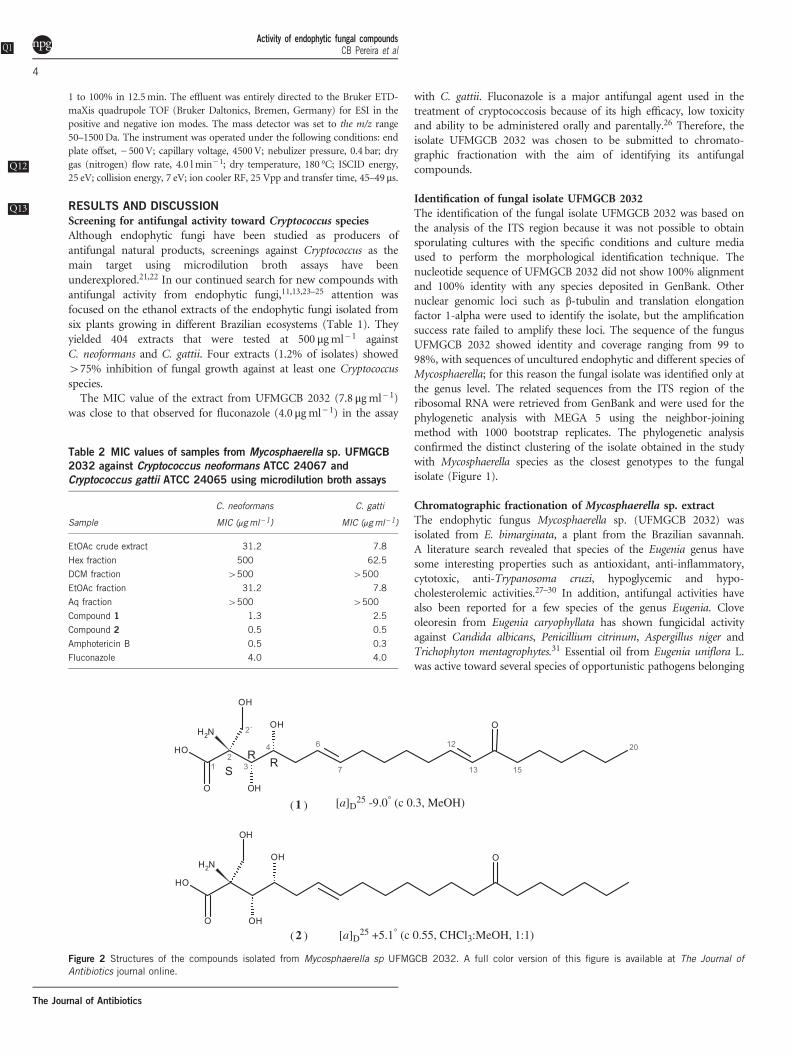
(1 ) [a]D25 -9.0° (c 0.3, MeOH)
[a]D25 +5.1° (c 0.55, CHCl3:MeOH, 1:1)(2 )
Figure 2 Structures of the compounds isolated from Mycosphaerella sp UFMGCB 2032. A full color version of this figure is available at The Journal ofAntibiotics journal online.
Activity of endophytic fungal compoundQ1
sCB Pereira et al
4
The Journal of Antibiotics

to the genera Candida (MIC 37.5–2.3 μgml− 1) and Cryptococcus,including C. gattii (MIC 18.0–22.0 μgml− 1) and C. neoformans (MIC11.0 μg ml− 1). In this study, the major constituents of Eugenia unifloraL. essential oil, identified by analysis by GC-MS, are the sesquiterpenesatractylone (26.78%) and curzerene (17.96%).27
The ethyl acetate extract of Mycosphaerella sp. mycelium grown inPDA medium exhibited MIC values of 31.2 μgml− 1 againstC. neoformans and 7.8 μg ml− 1 against C. gattii (Table 2), respectively.The extract was subjected to a liquid–liquid partition sequence, andthis procedure afforded four fractions: hexane, dichloromethane,EtOAc and Aq. They were tested against the two Cryptococcus species
Table 3 Physicochemical properties of compound 1
Properties Compound 1
Appearance White powder
Molecular formula C21H37NO6
HRMS (ESI-QTOF) (m/z)Found 398.2533 [M−H]−
Calcd. for C21H36NO6 398. 2543 for [M−H]−
UV λMeOHmax nm (ε) 227 (2.55)
[α]D25 (c 0.3 MeOH) –9.0
Soluble DMSO, methanol
-H -
-
C3H6NO3
C20H30NO3
C21H36NO6
C3H4NO2C8H15O
Figure 3 The ESI-(-)-LC-MS/MS spectra of compound 1 and proposal of the key fragments. A full color version of this figure is available at The Journal ofAntibiotics journal online.
Activity of endophytic fungal compoundQ1
sCB Pereira et al
5
The Journal of Antibiotics

and the results showed that the EtOAc fraction exhibited MIC valuescomparable to the crude extract (Table 2). In order to identify itsactive compounds, the EtOAc fraction was chromatographed twice bysemi-preparative reverse phase HPLC to afford two active compounds(1 and 2) (Figure 2).
Structural elucidation of the active compounds isolated fromMycosphaerella sp.The chemical structures of compounds 1 and 2 were established byanalysis of NMR and mass spectrometric data and their comparisonwith the literature data. Compound 1 was isolated as an amorphouswhite solid with a specific rotation [α]D25 − 9.0° (c 0.3 in MeOH)(Table 3). It gives a reddish-brown spot (Rf= 0.45; CHCl3:MeOH:NH4OH/10:6.25:0.25) after spraying the plates with vanillin-H2SO4.The negativeQ14 HRMS (ESI-QTOF) spectrum showed a quasi-molecularion peak [M−H]− at m/z 398.2533 (calcd. 398.2543 for C21H36NO6),consistent with the molecular formula C21H37NO6. The mass spec-trum obtained using tandem MS of compound 1 gave a base peak atm/z 104.0351(C3H6NO3) attributed to the fragment [M−H− 294]−,and major fragments at m/z 332.2226 (C20H30NO3), m/z 127.1124(C8H15O) and m/z 86.0246 (C3H4NO2). The proposal of the keyfragments is shown in Figure 3. The UV spectrum of 1 showed λmax at227 nm. NMR spectra (one and two dimensional) showed 1H signalscharacteristic of a hydrocarbon chain at δ 2.54–1.21, corresponding tomethylene carbons at δ 39.0–21.9 in the 13C spectrum. Furthermore,the 13C NMR spectrum of compound 1 showed peaks owing to four
olefinic carbons resonating at δ 147.1 and 130.2, 131.6 and 127.0.These peaks were correlated to four olefinic protons at δ 6.83, 6.07,5.43 and 5.35 in the 1H NMR spectrum (Table 4). The geometryof the C6/C7 and C12/C13 alkene bonds were trans (E), as evidencedby the large vicinal coupling constants (J6–7= 15.5 Hz andJ12–13= 16.0 Hz). The 13C NMR (DEPT) spectrum of 1 showed signalsat δ 200.0 and at δ 169.9 of carbonyl carbons; the former wasattributed to a ketone group and the latter to a carboxylic acid group.Correlation peaks between the H-13 (δ 6.07) and H-15 (δ 2.54) signalsand the carbonyl at δ 200.0 in HMBC were observed and confirmedthe location of the ketone group. HMBC correlations of the carboxylicacid carbonyl group at δ 169.9 confirmed its position at C-1 owing tocross peaks with H-2′ (δ 3.56 and δ 3.69). The doublets at δ 3.69 (1H,d, J= 10.5, H-2′a) and δ 3.56 (1H, d, J= 10.5, H-2’b) were attributedto the geminal hydrogens of an oxygenated primary carbon. Inaddition, the 1H NMR spectrum showed signals for two protonsbound to the oxygenated tertiary carbons at δ 3.64 (1H, td, J 7.1, 1.5,H-4) and δ 3.61 (1H, br s, H-3). The values of the coupling constantsof the signals at δ 3.64 and at δ 3.61 are similar to the values publishedfor natural mycestericin A32 and synthetic myriocin.33,34 The 1H–1HCOSY cross-peak between H-4 (δ 3.64) and H-5 (δ 2.09) suggests theirvicinal location. The 1H NMR and 13C NMR spectral data ofcompound 1 exhibited the characteristic signals of aliphatic chains(δ 1.21–2.54/ δ 21.9–39.0) and signals due to a terminal methyl groupwere observed at δ 0.85 (1H, t, J= 6.75Hz, H-20) and at δ 13.9(C-20). The similarity between shifts and coupling constants values
Table 4 NMR data for compounds 1 and 2 (DMSO-d6) at 400/100 MHz, δ in ppm and J in Hz
Compound 1 Compound 2
Position δ 13C (DEPT) δ 1H (J) 1H-1H COSY HMBC (H→ C) 13C (DEPT) δ 1H (J) 1H-1H COSY HMBC (H→ C)
1 169.9 (C) H-2′a, H-2′b 169.7 (C) H-2′a, H-2′b2 67.6 (C) H-2′, H-3 67.8 (C)
3 69.3 (CH) 3.61 (br s) 69.3 (CH) 3.61 (br s)3-OH 5.02 (br s)a 5.02 (br s)a H-3
4 70.7 (CH) 3.64 (td, J 7.0, 1.5) H-5 70.7 (CH) 3.64 (td, J 7.0, 1.5) H-5
5 37.5 (CH2) a: 2.09 (ddd, J 7.0) H-4, H-6, H-7 37.5 (CH2) a: 2.09 (ddd, J 7.0) H-5b, H-6 H-4, H-6, H-7
b: 2.16 (m) b: 2.15 (ddd, J 7.0) H-5a
6 131.6 (CH) 5.43 (dt, J 15.5, 6.5) H-5, H-7 H-8 131.9 (CH) 5.42 (dt, J 15.3, 6.5) H-7, H-8 H-8
7 127.0 (CH) 5.35 (dt, J 15.5, 6.5) H-6, H-8 H-8 126.7 (CH) 5.34 (dt, J 15.4, 6.5) H-5, H-6, H-8 H-8
8 31.8 (CH2) 1.96 (br q, J 6.5) H-7 H-6, H-7 32.1 (CH2) 1.93 (q, J 6.7) H-6, H-10 H-6, H-7
9 28.4 (CH2) 1.34 (br q, J 7.5) H-8 H-8, H-10 28.4 (CH2)b 1.31–1.29 (m)a H-8
10 27.0 (CH2) 1.42 (br q,7.5) H-11 H-8, H-9, H-11 28.8 (CH2) 1.28–1.27 (m)a H-8 H-8
11 31.6 (CH2) 2.19 (br q, J 7.0) H-10, H-12, H-13 H-12, H-13 28.3 (CH2)b 1.26–1.25 (m)a H-10 H-9, H-12
12 147.1 (CH) 6.83 (br dt, J 16.0,
7.0)
H-11, H-13 H-11 23.2 (CH2) 1.44 (qui, J 7.0) H-13 H-11, H-13
13 130.2 (CH) 6.07 (dt, J 16.0, 2.0) H-11, H-12 H-11 41.8 (CH2) 2.38 (t, J 7.2) H-12 H-12
14 200.0 (C) – H-12, H-13, H-15 210.58 (C) H-12, H-13, H-15,
H-16
15 39.0 (CH2) 2.54 (t, J 7.3) H-16 H-16 41.8 (CH2) 2.38 (t, J 7.2) H-16 H-16
16 23.7 (CH2) 1.47 (br quin, J 7.0) H-15, H-18 H-15 23.2 (CH2) 1.44 (qui, J 7.1) H-15, H-17 H-15
17 28.2 (CH2) 1.23–1.21 (m)a H-15, H-16 28.5 (CH2)b 1.22–1.20 (m)a H-16 H-15, H-16
18 31.0 (CH 2) 1.26–1.24 (m)a H-16, H-20 H-16, H-19, H-20 31.1 (CH2) 1.24–1,23 (m)a H-20 H-19, H-20
19 21.9 (CH2) 1.30–1.27 (m)a H-20 H-17, H-20 21.9 (CH2) 1.26–1.25 (m)a H-20 H-17, H-20
20 13.9 (CH3) 0.85 (t, J 7.0) H-18, H-19 H-18 13.9 (CH3) 0.85 (t, J 7.0) H-19 H-18
2′a 62.4 (CH2) a: 3.69 (d, J 10.5) H-2′b 62.35 (CH2) a: 3.70 (d, J 10.5) H-2′b2′b b: 3.56 (d, J 10.5) H-2′a b: 3.58 (d, J 10.5) H-2′a
a1H NMR signals assignment was rationalized by HSQC contour map and Perch NMR software calculations.bInterchangeable signals.
Activity of endophytic fungal compoundQ1
sCB Pereira et al
6
The Journal of Antibiotics

found in the 13C NMR and 1H spectra of both 1 and 2, at C-1 to C-5and H-3 to H-5 proton signals, respectively, suggested that these twostructural moieties are identical in 1 and 2. However, the comparisonbetween the [α]D values for 1 (−9.0°, c 0.3, MeOH) and myriocin(+5.1°, c 0.18, MeOH)35 shows that they are comparable in magnitudebut with opposite signs. This reversal effect on the [α] sign of 1 incomparison with 2 could result from conformational effects due thedouble bond at C-12.36 Fujita et al.37 also reported that the compound6,7-dihydromyriocin, obtained by the hydrogenation of myriocin, is alevorotatory compound ([α]D − 6.5°, c 0.108, MeOH). This sign
reversal effect was also found when comparing [α]D values formyriocin35 and 14-epi-mycestericin A.38 Thus, the relative stereoche-mistries at C-2, C-3 and C-4 of 1 were assigned as 2 S, 3 R and 4 R,respectively. On the basis of these observations, compound 1 wasidentified as (2 S,3 R,4 R)-(E)-2-amino-3,4-dihydroxy-2-(hydroxy-methyl)-14-oxoeicos-6,12-dienoic acid, as shown in Figure 2.Compound 2 was isolated as an amorphous white solid, and the UV
spectrum exhibited no absorption maximum above 200 nm. It showeda positive specific rotation ([α]D25 +5.1°, c 0.55 in CHCl3:MeOH 1:1).The negative HRMS (ESI-QTOF) spectrum of compound 2 showed a
--H
-H-
-
C3H6NO3
C3H4NO2
C18H31O3
C20H32NO3
C21H38NO6
Figure 4 The ESI-(-)-LC-MS/MS spectra of compound 2 and proposal of the key fragments. A full color version of this figure is available at The Journal ofAntibiotics journal online.
Activity of endophytic fungal compoundQ1
sCB Pereira et al
7
The Journal of Antibiotics

quasi-molecular ion peak [M−H]− at m/z 400.2707 (calcd. 400.2699for C21H38NO6), consistent with the molecular formula C21H39NO6.The mass spectrum obtained using tandem MS of compound 2showed an ion peak [M−H− 296]− at m/z 104.0356 (base peak,C3H6NO3), and the main fragments at m/z 334.2389 (C20H32NO3), m/z 295.2281 (C18H31O3) and m/z 86.0249 (C3H4NO2), respectively. Theproposal of the key fragments is shown in Figure 4. Analysis of theNMR and HRMS spectra of compound 2 showed that its maindifference from compound 1 is the absence of a double bond at C12–
C13. Furthermore, the respective 1H NMR spectrum signals for H-2' (aand b hydroxymethylene protons), as well as the [α]D value reportedfor the isomeric compound 2-epi-myriocin,39 differ significantly fromthose found for compound 2. On the basis of these observations, 2 wasidentified as (2 S,3 R,4 R)-(E)-2-amino-3,4-dihydroxy-2-(hydroxy-methyl)-14-oxoeicos-6-enoic acid, known as (+)-myriocin (2) andshown in Figure 2. All 1H and 13C NMR (one and two dimensional)data for 1 and 2 can be seen in Table 4.
Antifungal activity of isolated compoundsMycosphaerella spp. are important phytopathogenic species, causingdiseases in wheat, banana, Eucalyptus species and other crops.40,41
Bioactive compounds were isolated from other species of Myco-sphaerella. Asteromine, a 6,6′-binaphtho α-pyrone isolated fromM. asteroma, has antifungal activity against Ustilago maydis,Ophiostoma ulmi and Geotrichum candidum.42 Cercosporin, a peryle-nequinone metabolite of polyketide origin, was isolated fromMycosphaerella sp. nov. strain F2140 associated with the foliage ofthe plant Psychotria horizontalis (Rubiaceae). Cercosporin exhibits highcytotoxicity activity (MCF-7, IC50 4.68 μM) and also non-selectiveanti-protozoan activity against Plasmodium falciparum (IC50 1.03 μM),Leishmania donovani (IC50 0.46 μM) and Trypanosoma cruzi(IC50 1.08 μM).43In the present work we show that eicosanoic acids, the oxidized
derivatives of C20-fatty acids produced by Mycosphaerella sp.UFMGCB 2032, possess antifungal activity. Compound 1 exhibitedMIC values of 1.3 and 2.5 μgml− 1 against C. neoformans and C. gattii,respectively. Compound 2 showed a MIC value of 0.5 μgml− 1 to bothspecies of Cryptococcus tested. The antifungal activity of naturalproduct analogs from the eicosanoid family is still little explored,specifically against Cryptococcus pathogenic species. According to theliterature, there are no previous reports of the isolation of compound1, whereas compound 2 (myriocin) was first described by Kluepfelet al.44 and later isolated from Myriococcum albomyces, Melanconisflavovirens, Isaria sinclairii and Paecilomyces variotii ATCC 74097.45
They are reported herein for the first time as natural products fromthis endophytic species. Myriocin is a serine palmitoyltransferaseinhibitor46 and has immunosuppressant activity. This metabolite alsoexhibits antifungal properties against several pathogens (Alternariasolani, C. albicans, C. parapsilosis, C. pseudotropicalis, C. tropicalis,Ceratocytis ulmi¸ Cryptococcus laurentii, Fusarium oxysporum, Rhizo-mucor miehei and Streptomyces spp.44,47
These two eicosanoic-acid compounds exhibited higher antifungalactivities against Cryptococcus species than the crude original extractand fractions. Considering the antifungal activity of the compoundsdemonstrated in this work, their in vivo efficacy in a murine model ofCryptococcus infection should be assessed.
CONFLICT OF INTERESTThe authors declare no conflict of interest.
ACKNOWLEDGEMENTS
We thank the ‘Conselho Nacional de Desenvolvimento Científico e
Tecnológico’ (CNPq) for financial support and for a scholarship. This work was
also supported in part by a grant from ‘Fundação de Amparo à Pesquisa do
Estado de Minas Gerais’ (FAPEMIG). We thank the Program of Technological
Development in Tools for Health PDTIS-FIOCRUZ, RPT13A platform, and
especially Eliane G Carvalho for the use of its facilities.
1 Perfect, J. R. et al. Clinical practice guidelines for the management of cryptococcaldisease: update by the infectious diseases society of America. Clin. Infect. Dis. 50,291–322 (2010).
2 Kurtzman, C. P., Fell, J. W. & Boekhout, T. The Yeasts: a Taxonomic Study (Elsevier:New York, NY, USA, 2011).
3 Rosario, I., Acosta, B. & Colom, F. La paloma y otras aves como reservorio deCryptococcus spp. Rev. Iberoam. Micol. 25, S13–S18 (2008).
4 Nucci, M. & Perfect, J. R. When primary antifungal therapy fails. Clin. Infect. Dis. 46,1426–1433 (2008).
5 Vandeputte, P., Ferrari, S. & Coste, A. T. Antifungal resistance and new strategies tocontrol fungal infections. Int. J. Microbiol. 2012: ID 713687 1–26 (2012).
6 Desnos-Ollivier, M. et al. Mixed infections and in vivo evolution in the human fungalpathogen Cryptococcus neoformans. MBio 1, e00091–10 (2010).
7 Del Poeta, M. & Casadevall, A. Ten challenges on Cryptococcus and Cryptococcosis.Mycopathologia 173, 303–310 (2012).
8 Bacon, C. W. & White, J. F. Microbial Endophytes (Marcel Dekker: New York, NY, USA,2000).
9 Strobel, G. A. Endophytes as sources of bioactive products. Microbes Infect. 5,534–535 (2003).
10 Pimentel, M. R., Molina, G., Dionísio, A. P., Maróstica, M. R. J. & Pastore, G. M. Theuse of endophytes to obtain bioactive compounds and their application in biotransfor-mation process. Biotechnol. Res. Int. 2011: ID 576286 1–11 (2011).
11 Vieira, M. L. A et al. Diversity and antimicr Q15obial activities of the fungal endophytecommunity associated with the traditional Brazilian medicinal plant Solanum cernuumVell. (Solanaceae). Can. J. Microbiol. 58, 54–66 (2012).
12 Rosa, L. H. et al. Leishmanicidal, trypanocidal, and citotoxic activities of endophyticfungi associated with bioactive plants in Brazil. Braz. J. Microbiol. 41,420–430 (2010).
13 Carvalho, C. R. et al. The diversity, antimicrobial and anticancer activitiesof the endophytic fungi associated with the medicinal plant Stryphnodendronadstringens (Mart.) Coville (Fabaceae) from the Brazilian savannah. Symbiosis 57,1–13 (2012).
14 Eucast-Subcommittee of antifungal susceptibility testing of the European Committee onAntibiotic Susceptibility Testing (EUCAST). Method for determination of MinimalInhibitory Concentration (MIC) by broth dilution of fermentative yeasts (Document7.1. Taufkirchen: Germany, ECSMID, 2002).
15 White, T. J., Bruns, T., Lee, S. & Taylor, J. Amplification and direct sequencing offungal ribosomal RNA genes for phylogenetics. in PCR Protocols: a guide to methodsand applications (eds Innis M. A., Gelfand D. H., Sninsky J. J., White T. J.)315–322 (Academic Press: San Diego, USA, 1990).
16 Rosa, L. H., Vaz, A. B. M., Caligiorne, R. L. B., Campolina, S. A. & Rosa, C. A.Endophytic fungi associated with the Antarctic grass Deschampsia antarctica Desv.(Poaceae). Polar Biol. 32, 161–167 (2009).
17 Togawa, R. C. & Brigido, M. M. Web based tool for simple electropherogram qualityanalysis Q16. Abstracts of Papers of 1st International Conference on Bioinformatics andComputational Biology (Ribeirão Preto: Brazil, 2003).
18 Tamura, K. et al. MEGA5: molecular evolutionary genetics analysis using maximumlikelihood, evolutionary distance, and maximum parsimony methods. Mol. Biol. Evol.28, 2731–2739 (2011).
19 Edgar, R. C. MUSCLE: multiple sequence alignment with high accuracy and highthroughput. Nucleic Acids Res. 32, 1792–1797 (2004).
20 Felsenstein, J. Confidence limits on phylogenies: an approach using the bootstrap. Int.J. Evol. 39, 783–791 (1985).
21 Phongpaichit, S., Rungjindamai, N., Rukachaisirikul, V. & Sakayaroj, J. Antimicrobialactivity in cultures of endophytic fungi isolated from Garcinia species. FEMS Immunol.Med. Microbiol. 48, 367–372 (2006).
22 Sun, Z. L., Zhang, M, Zhang, J. F. & Feng, J. Antifungal and cytotoxic activities of thesecondary metabolites from endophytic fungus Massrison sp. Phytomedicine 18,859–862 (2011).
23 Wang, X. et al. Antifungal activity against plant pathogens of metabolites from theendophytic fungus Cladosporium cladosporioides. J. Agric. Food Chem. 61,4551–4555 (2013).
24 Vaz, A. B. M. et al. Diversity and antimicrobial activity of fungal endophyte communitiesassociated with plants of Brazilian savanna ecosystems. Afr. J. Microbiol. Res. 55,62–70 (2012).
25 Rosa, L. H. et al. Coniochaeta ligniaria: antifungal activity of the cryptic endophyticfungus associated with autotrophic tissue cultures of the medicinal plant Smallanthussonchifolius (Asteraceae). Symbiosis 60, 133–142 (2013).
26 Fica, A. C. Tratamiento de infecciones fúngicas sistêmicas primeira parte: fluconazol,itraconazol y voriconazol. Rev. Chil. Infectol. 21, 26–38 (2004).
Activity of endophytic fungal compoundQ1
sCB Pereira et al
8
The Journal of Antibiotics

27 Lago, J. H. et al. Chemical and biological evaluation of essential oils from two species ofMyrtaceae - Eugenia uniflora L. and Plinia trunciflora (O. berg) Kausel. Molecules 16,9827–9837 (2011).
28 Stefanello, M. É., Pascoal, A. C. & Salvador, M. J. Essential oils from neotropicalMyrtaceae: chemical diversity and biological properties. Chem. Biodivers. 8,73–94 (2011).
29 Santos, K. K. et al. Anti-Trypanosoma cruzi and cytotoxic activities of Eugeniauniflora L. Exp. Parasitol. 131, 130–132 (2012).
30 Sharma, S. B., Tanwar, R. S., Nasir, A. & Prabhu, K. M. Antihyperlipidemic effect ofactive principle isolated from seed of Eugenia jambolana on alloxan-induced diabeticrabbits. J. Med. Food. 14, 353–359 (2011).
31 Nunez, L., D'Aquino, M. & Chirife, J. Antifungal properties of clove oil (Eugeniacaryophylata) in sugar solution. Braz. J. Microbiol. 32, 123–126 (2001).
32 Sasaki, S. et al. Fungal metabolites. Part 14. Novel potent immunosuppressantmycestericins, produced by Mycelia sterilia. J. Antibiot. 47, 420−433 (1994).
33 Sano, S. et al. Asymmetric total synthesis of ISP-I (myriocin, thermozymocidin), apotent immunosuppressive principle in the Isaria sinclairii metabolite. Tetrahedron Lett.36, 2097–2100 (1995).
34 Jones, M. C. & Marsden, S. P. Total synthesis of the immunosuppressants myriocin and2-epi-myriocin. Org. Lett. 10, 4125–4128 (2008).
35 Fairhurst, N. W. G., Mahon, M. F., Munday, R. H. & Carbery, D. R. Remote stereocontrolin [3,3]-sigmatropic rearrangements: application to the total synthesis of the immuno-suppressant mycestericin G. Org. Lett. 14, 756–759 (2012).
36 Wiberg, K. B., Vaccaro, P. H. & Cheeseman, J. R. Conformational effects onoptical rotation. 3-substituted 1-butenes. J. Am. Chem. Soc. 125,1888–1896 (2003).
37 Fujita, T. et al. Fungal metabolites. Part 12. Potent immunosuppressant, 14-deoxo-myriocin, (2 S, 3 R, 4 R)-(E)-2-amino-3,4-dihydroxy-2-hydroxymethyleicos-6-enoic acid
and structure-activity relationships of myriocin derivatives. J. Antibiot. 47,216–224 (1994).
38 Yamanaka, H. et al. Total synthesis of mycestericin A and its 14-epimer. Tetrahedron65, 9188−9201 (2009).
39 Yoshikawa, M., Yokokawa, Y., Okuno, Y., Yagi, N. & Murakami, N. Syntheses,immunosuppressive activity, and structure-activity relationships of myriocin analogs,2-epi-myriocin, 14-deoxomyriocin, Z-14-deoxomyriocin, and nor-deoxomyriocins.Chem. Pharm. Bull. 43, 1647–1653 (1995).
40 Park, R. F., Keane, P. J., Wingfield, M. J. & Crous, P. W. Fungal diseases ofeucalypt foliage. in Diseases and Pathogens of Eucalypts (eds Keane P. J.,Kile G. A., Podger F. D., Brown B. N.) 153–239 (CSIRO Publishing: Collingwood, VIC,Australia, 2000).
41 Irish, B. M., Goenaga, R., Rios, C., Chavarria-Carvajal, J. & Ploetz, R. Evaluation ofbanana hybrids for tolerance to black leaf streak (Mycosphaerella fijiensis Morelet) inPuerto Rico. Crop Prot. 54, 229–238 (2013).
42 Arnone, A., Assante, G., Montorsi, M. & Nasini, G. Asteromine, a bioactive secondarymetabolite from a strain of Mycosphaerella asteroma. Phytochemistry 38,595–597 (1995).
43 Moreno, E. et al. Chemical constituents of the new endophytic fungus Mycosphaerellasp. nov. and their anti-parasitic activity. Nat. Prod. Commun. 6, 835–840 (2011).
44 Kluepfel, D. et al. Myriocin, a new antifungal antibiotic from Myriococcum albomyces.J. Antibiot. 25, 109–115 (1972).
45 Dictionary of Natural Products on DVD-ROM (Chapman & Hall: London, 2013).46 Castro, E. V. et al. Myriocin, a serine palmitoyltransferase inhibitor, blocks cytokinesis in
Leishmania (Viannia) braziliensis promastigotes. J. Eukaryot Microbiol. 60, 377–387(2013).
47 Horn, W. S. et al. (Merck & Co., Inc.). Antibiotic eicosenoic acids. US5,233,062A(1993).
Activity of endophytic fungal compoundQ1
sCB Pereira et al
9
The Journal of Antibiotics
Copyright © 2022 FDOKUMEN