Identification of manipulated variables for a glycosylation control strategy
Upload
independentCategory
view
3download
0
Biochemical and structural insights of the early glycosylationsteps in calicheamicin biosynthesis
Changsheng Zhang1, Eduard Bitto2, Randal D. Goff1, Shanteri Singh1, Craig A. Bingman2,Byron R. Griffith1, Christoph Albermann1, George N. Phillips Jr.2,*, and Jon S. Thorson1,*1Laboratory for Biosynthetic Chemistry, Pharmaceutical Sciences Division, School of Pharmacy,UW-National Cooperative Drug Discovery Group Program, University of Wisconsin-Madison, 777Highland Avenue, Madison, Wisconsin 53705-2222, USA2Department of Biochemistry, University of Wisconsin-Madison, 777 Highland Avenue, Madison,Wisconsin 53706-1544, USA
SummaryThe enediyne antibiotic calicheamicin (CLM) γ1
I is a prominent antitumor agent that is targeted toDNA by a novel aryltetrasaccharide comprised of an aromatic unit and four unusual carbohydrates.Herein we report the heterologous expression and the biochemical characterization of the two‘internal’ glycosyltransferases CalG3 and CalG2 and the first structural elucidation of an enediyneglycosyltransferase (CalG3). In conjunction with the previous characterization of the ‘external’ CLMGTs CalG1 and CalG4, this study completes the functional assignment of all four CLM GTs, extendsthe utility of enediyne GT-catalyzed reaction reversibility, and presents conclusive evidence of asequential glycosylation pathway in CLM biosynthesis. This work also reveals the common GT-Bstructural fold can now be extended to include enediyne GTs.
Keywordsenediyne; cancer; glycosyltransferase; carbohydrate; biosynthesis; crystallography
IntroductionCalicheamicin (CLM) γ1
I (Fig. 1, 1) from Micromonospora echinospora spp. calichensis is aprominent member of the enediyne family due to its unprecedented molecular architecture,remarkable mechanism of action, and clinical utility (Galm et al., 2005; Thorson et al., 2000;Van Lanen and Shen, 2008). Structurally, CLM is a member of the 10-membered enediyneswhich all share a signature bicyclo[7.3.1]enediyne core.Like all enediynes, CLM-inducedoxidative DNA strand scission is enabled by rapid enediyne cycloaromatization to form ahighly reactive diradical species (Zein et al., 1989; Zein et al., 1988). This reactive intermediateis exquisitely positioned by the CLM aryltetrasaccharide (Fig. 1), the critical DNA dockingelement of CLM (Kumar et al., 1997; Walker et al., 1993). The incredible potency of CLMhas also been harnessed for clinical use via conjugation to tumor-targeting antibodies, as
© 2008 Elsevier Ltd. All rights reserved.*To whom correspondence should be addressed: [email protected]; [email protected]'s Disclaimer: This is a PDF file of an unedited manuscript that has been accepted for publication. As a service to our customerswe are providing this early version of the manuscript. The manuscript will undergo copyediting, typesetting, and review of the resultingproof before it is published in its final citable form. Please note that during the production process errors may be discovered which couldaffect the content, and all legal disclaimers that apply to the journal pertain.
NIH Public AccessAuthor ManuscriptChem Biol. Author manuscript; available in PMC 2010 October 29.
Published in final edited form as:Chem Biol. 2008 August 25; 15(8): 842–853. doi:10.1016/j.chembiol.2008.06.011.
NIH
-PA Author Manuscript
NIH
-PA Author Manuscript
NIH
-PA Author Manuscript

exemplified by the γ-CD33 antibody conjugate (Mylotarg) approved by FDA in 2000 to treatacute myelogenous leukemia (AML) (Sievers and Linenberger, 2001). Similarly-appendedCLM-antibody conjugates to treat other cancers are steadily progressing through clinical trials(Boghaert et al., 2004; DiJoseph et al., 2005; Hamann et al., 2005).
Consistent with their many novel structural and pharmacological features, enediynebiosynthetic pathways are also rich with unique enzyme-catalyzed biotransformations. Earlymetabolic labeling studies suggested the 9- and 10-membered enediynes to derive from distinctbiosynthetic pathways (Hensens et al., 1989; Lam et al., 1993; Tokiwa et al., 1992). In contrast,the recent cloning and characterization of gene clusters encoding both 9-membered – includingC-1027 (Fig. 1, 4) (Liu et al., 2002), neocarzinostatin (Fig. 1, 5) (Liu et al., 2005), andmaduropeptin (Fig. 1, 6) (Van Lanen et al., 2007) - and 10-membered – including CLM (Ahlertet al., 2002), esperamicin (Fig. 1, 2) (Ahlert, Shepard and Thorson, unpublished, AY267372),and dynemicin (Fig. 1, 3) (Gao and Thorson, 2008; Zazopoulos et al., 2003) - enediynesrevealed a unified, divergent polyketide paradigm for enediyne core biosynthesis (Ahlert etal., 2002; Liu et al., 2002; Zhang et al., 2008). Some enediyne-producing organisms also relyupon a novel ‘self-sacrifice’ resistance protein (as exemplified by the CLM protein, CalC) forenediyne self-resistance (Biggins et al., 2003; Singh et al., 2006). Shen and coworkers werethe first to demonstrate the elegant application of pathway engineering to producechromoprotein enediyne analogs with drastically differing activities (Kennedy et al., 2007a;Kennedy et al., 2007b; Liu et al., 2002) while ‘sugar exchange’ and ‘aglycon exchange’reactions catalyzed by the CLM glycosyltransferases (GTs) CalG1 and CalG4 recently enabledthe production of more than 70 differentially-glycosylated CLM variants (Zhang et al.,2006b). While this latter study also provided in vitro biochemical characterization of CalG1and CalG4 as the CLM 3-O-methyl-rhamnosyltransferase and aminopentosyltransferase,respectively (Zhang et al., 2006b), the function of the remaining CLM glycosyltransferasesCalG2 and CalG3 remain unresolved. In addition, while the structures for a variety of naturalproduct-associated glycosyltransferase have emerged in recent years (Bolam et al., 2007;Mittler et al., 2007; Mulichak et al., 2003; Mulichak et al., 2001; Mulichak et al., 2004),enediyne GTs remain structurally uncharacterized (Van Lanen and Shen, 2008).
Herein we report the further study of the ‘internal’ stages of CLM glycosylation. Specifically,using a combination of GT reversibility and sugar nucleotide surrogates, CalG3 was verifiedas the requisite calicheamicinone 4,6-dideoxy-4-hydroxylamino-α-D-glucosyltransferase anddemonstrated to accept a set of 10 alternative sugar nucleotide donors. The structural studieshighlighted herein, also revealed, for the first time, that enediyne GTs such as CalG3 also adopta GT-B fold common to natural product GTs. This structural study also illuminated keycatalytic residues and snapshots of a dynamic loop anticipated to participate in NDP-binding.The application of a surrogate sugar nucleotide also enabled the confirmation of CalG2 as theremaining ‘internal’ GT – the 4-deoxy-thio-α-D-digitoxosyltransferase – and the firstcharacterized hydroxylamino glycosidic bond-forming GT. In conjunction with our previousreport (Zhang et al., 2006b), this study completes the functional assignment of all four CLMGTs, extends the concept of reversibility of enediyne GT-catalyzed reactions, and highlightsthe first crystal structure of an enediyne GT.
ResultsOverexpression and purification of CalG3 and CalG2
Analysis of the CLM biosynthetic gene cluster from M. echinospora revealed four putativeGT-encoded genes, calG1, calG2, calG3 and calG4 (Ahlert et al., 2002), implicating a distinctGT for each sugar of the CLM aryltetrasaccharide. Consistent with this, in vitro biochemicalcharacterization confirmed CalG1 and CalG4 as the CLM 3-O-methyl-rhamnosyltransferaseand aminopentosyltransferase, respectively (Zhang et al., 2006b). Analysis of CalG3 revealed
Zhang et al. Page 2
Chem Biol. Author manuscript; available in PMC 2010 October 29.
NIH
-PA Author Manuscript
NIH
-PA Author Manuscript
NIH
-PA Author Manuscript

highest homology to characterized GTs which operate upon aromatic acceptors, such as thenogalamycin SnogD (37% identity) (Torkkell et al., 1997) and elloramycin ElmGT (36%identity) (Blanco et al., 2001), while CalG2 more closely resembled GTs which act uponcarbohydrate acceptors including the CLM CalG4 (50% identity) (Zhang et al., 2006b) andavermectin AveBI (42% identity) (Wohlert et al., 2001; Zhang et al., 2006a). Based upon thissimple analysis, CalG3 was proposed as the putative calicheamicinone 4,6-dideoxy-4-hydroxylamino-α-D-glucosyltransferase and CalG2 postulated as the subsequent 4-deoxy-thio-α-D-digitoxosyltransferase (anticipated to form the signature CLM hydroxylaminoglycosidic bond). To complete the CLM GT annotation, recombinant N-His10-CalG2 and N-His10-CalG3 fusion proteins were overproduced in E. coli and subsequently purified by nickel-affinity chromatography to >95% homogeneity (Fig. S1) with overall yields of 10–15 mg perliter of culture.
Preparation of the putative acceptor CLM T0The availability of an appropriate acceptor was critical to the in vitro characterization of CalG2and CalG3. To address this issue, the truncated CLM analogue CLM T0 (Fig. 2A, 9) wasprepared via a slight modification of a literature procedure (Walker et al., 1992). Specifically,CLM α3
I (Fig. 2A, 7 and Fig. 2B, i) was refluxed in wet acetone with pyridine p-toluenesulfonate and the reaction monitored via HPLC. After refluxing for 19 h, a productclearly distinct from starting material, but with the characteristic CLM core UV signature, wasgenerated (Fig. 2B, ii). The product was purified (Fig. 2B, iii) and determined to have a massof 625.1 [M+H] by APCI-MS analysis - 40 daltons greater than expected product 9.Subsequent 1H NMR analysis (Fig. S2) revealed the new product to be 8 (Fig. 2A), anisopropylidene adduct formed by the dehydration of acetone in the presence of tosylate.Reasoning that the adduct could be easily removed under mild acidic conditions, 8 wasincubated with 0.2% TFA to provide 9 in 4 h (Fig. 2B, iv and v). The identity of 9 was verifiedby high resolution ESI-MS ([M+Na]+, m/z C24H28N2NaO9S3, calc. 607.0855, found607.0866) and 1H NMR (Fig. S3).
Reversibility of CalG3-catalyzed reactionFollowing recently established protocols for GT reversibility (Minami et al., 2005; Zhang etal., 2006a; Zhang et al., 2006b; Zhang et al., 2007), CalG3 and CalG2 were first investigatedfor their ability to catalyze the excision of 4,6-dideoxy-4-hydroxylamino-α-D-glucosyl moietyfrom CLM T0 (9) (Fig. 3A). No reaction was observed with 9 (50 µM) and CalG2 (7.5 µM)in the presence of 2 mM NDP (ADP, CDP, GDP, UDP or TDP) in Tris-HCl (10 mM, pH 7.6).In contrast, the incubation of 9 (50 µM) with CalG3 (7.5 µM) in the presence of various NDPs(2 mM) led to new product after 2 h at 30 °C (Fig. 3B, i–v). This transformation was determinedto be both CalG3- and NDP-dependent (Fig. 3B, vi) and the new product was subsequentlyidentified as the deglycosylated calicheamicinone (10, Fig. 3A) by LC-MS (calcd. 423, found446.0 [M+Na]+ and 422 [M-H]−) (Table S1). Unlike prior reports on the reversibility of GT-catalyzed reactions which indicated such transformations to be NDP-specific, CalG3reversibility was surprisingly observed with ADP (conversion rate of 40%; Fig. 3B, i), GDP(23%; Fig. 3B, ii), UDP (24%; Fig. 3B, iii), TDP (trace; Fig. 3B, iv) and even CDP (trace; Fig.3B, v). The apparent reversibility of this reaction was also enhanced at lower pH (Fig. S4A).Cumulatively, these studies highlight the clear reversibility of the CalG3-catalyzed reactionand are consistent with CalG3 as the requisite 4,6-dideoxy-4-hydroxylamino-α-D-glucosyltransferase involved in 1 biosynthesis.
CalG3-catalyzed ‘sugar exchange’Given the lack of calicheamicinone availability (10, Fig. 3A), the CalG3 sugar nucleotidespecificity was alternatively probed with 9 as an acceptor for putative GT-catalyzed ‘sugar-
Zhang et al. Page 3
Chem Biol. Author manuscript; available in PMC 2010 October 29.
NIH
-PA Author Manuscript
NIH
-PA Author Manuscript
NIH
-PA Author Manuscript

exchange’ reactions. In a GT-catalyzed sugar exchange reaction, first observed in the contextof CalG1 catalysis (Zhang et al., 2006b), the native sugar of a natural glycoside can besubstituted in situ by unnatural sugars supplied as NDP-sugar donors. Five commerciallyavailable NDP-glucoses, including ADP-, CDP-, GDP-, UDP- and TDP-Glc were examinedin the CalG3 ‘sugar exchange’ reaction with 9. Remarkably, all 5 NDP-glucoses wereestablished as CalG3 donor substrates, albeit with varying ‘sugar exchange’ efficiencies in theend point assay (50 µM 9, 2 mM NDP-Glc, 7.5 µM CalG3, 30 °C overnight). Interestingly,TDP-glucose exhibited the highest conversion rate of 9 to 9a (49.3%), followed by UDP-Glc(36.0%), CDP-Glc (25.3%), GDP-Glc (10.6%) and ADP-Glc (9.8%). In contrast to theinfluence of pH upon reaction reversibility, the CalG3-catalyzed sugar exchange reaction wasenhanced at higher pH (Fig. S4B and Fig. S4C).
The sugar exchange promiscuity of CalG3 was subsequently probed directly with 9 and a smalllibrary of 22 TDP-sugars (Fig. S5) comprised of 20 TDP-D-sugars (including commerciallyavailable TDP-α-D-glucose and unnatural sugar nucleotides generated via chemoenzymaticsynthesis with functionality variations such as deoxy, amino, and azido at the sugar C2, C3,C4, C5 or C6 positions) and two TDP-L-sugars (TDP-β-L-rhamnose and TDP-α-L-rhamnose).From this substrate specificity analysis, 10 sugar nucleotides were identified as CalG3substrates (Fig. 3A & 3C, i–x) to ultimately provide 10 novel CLM variants 9a–j (Fig. 3A,Table S1) with ‘sugar exchange’ conversions ranging from 15 to 70% (Fig. 3C). Cumulatively,these studies revealed CalG3 to be a relatively promiscuous GT and further highlighted GT-catalyzed ‘sugar exchange’ as an expeditious method for natural product diversification.
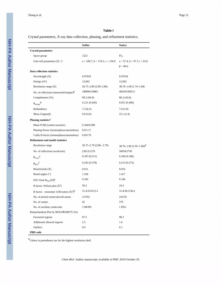
CalG3 structureThe structure of CalG3 was determined by single wavelength anomalous diffraction using a2.8 Å dataset collected from SeMet-labeled crystals (from I222 crystal form) and refinedagainst this dataset as well as a native dataset to a resolution of 1.9 Å (from P21 crystal form).Data collection, phasing and refinement statistics are summarized in Table I. Consistent withgel filtration, asymmetric units of both crystal forms revealed two molecules of CalG3 arrangedinto dimers with C2 symmetry (Fig. 4A). Analysis of the binding interface of these dimers bythe PISA server (Krissinel and Henrick, 2007) revealed the total buried surface area for thecomplex of 3300–3600 Å2. The dimers of both crystal forms align with an all-atom rmsd of0.76 Å, while all observed monomer conformers from both crystal forms align with rmsd ~0.48Å. These results confirm that CalG3 forms dimers with a closely similar quaternaryarrangement in both crystal forms and this form of protein thus may be relevant in vivo. Adetailed backbone comparison of the CalG3 molecules found in the two crystal forms revealedthat several secondary structure elements undergo slight shifts; these include surface exposedsegments containing residues 54–76, 205–212, and 219–226. In addition, a tetraglycine loopspanning residues 285–288 (Fig. 4B, pink) undergoes a conformational change.
The structure of the CalG3 monomer revealed that this protein consists of two closely opposedglobular domains, the N-terminal domain (residues 1–193, cyan in Fig. 4B) and the C–terminaldomain (residues 209–360, khaki in Fig. 4B) connected with a linker (residues 194–208, yellowin Fig. 4B) and stabilized by interaction of the C-terminal helix (residues 362–375, green inFig. 4B) and the N-terminal domain. Both the N- and C-terminal domain adopt a fold withRossmann topology and a 3-layer α-β-α sandwich architecture that shows homology toglycogen phoshorylase B. The structurally homologous cores of these domains can be alignedwith a rmsd of 3.1 Å for 103 residues. However, the domains show dramatic variations in sizeand conformation of several loop regions as well as topology of their C-termini. Full lengthCalG3 belongs to the GT-B clan of GTs and adopts a UDP-glycosyltransferase/glycogenphosphorylase fold found in many GTs. Based on the VAST server (Madej et al., 1995), theclosest overall structural homologs of CalG3 include Streptomyces fradiae dTDP-D-olivose-
Zhang et al. Page 4
Chem Biol. Author manuscript; available in PMC 2010 October 29.
NIH
-PA Author Manuscript
NIH
-PA Author Manuscript
NIH
-PA Author Manuscript

transferase UrdGT2 (which structurally aligns with CalG3 for 369 residues with 24% identityand rmsd of 3.1 Å, PDB ID 2p6p) (Mittler et al., 2007) and Amycolatopsis orientalis TDP-epi-vancosaminyltransferase GtfA (363 residues with 20% identity and rmsd of 4.0 Å, PDBID 1pnv, 1pn3, 1rrv) (Mulichak et al., 2003).
In vitro characterization of CalG2Given the lack of availability of the natural substrate NDP-4-deoxy-4-thio-α-D-digitoxose, thebiochemical function of CalG2 was examined with a small library of 22 surrogate TDP-sugarsubstrates (Fig. S5). Of this set, only one, TDP-4,6-dideoxy-α-D-glucose (12, Fig. 5) led to theCalG2-catalyzed transformation of 9 to a new product with a mass (calcd. 714, found 737 [M+Na]+) (Table S1) consistent with 13 (Fig. 5, i). Notably, this new product was clearly distinctfrom the CalG3-catalyzed ‘sugar exchange’ product 9e (Fig. 5, iii), derived from the same setof starting materials 9 and 12 (Fig. 5), and its production was enzyme-dependent (Fig. 5, ii).The incubation of 8 with 12 and CalG2 under identical conditions led to no change, consistentwith the 8 isopropylidene as masking the CalG2 acceptor nucleophile. Cumulatively, these invitro studies confirmed that CalG2 was capable of adding a sugar to 9, consistent with CalG2as the requisite CLM 4-deoxy-4-thio-α-D-digitoxosyltransferase.
DiscussionCLM GTs and CLM biosynthesis
Calicheamicin (1) has two distinct structural regions: the enediyne aglycon, or ‘warhead’,which consists of a highly functionalized bicyclo[7.3.1.]tridecadiyne core structure with anallylic trisulfide serving as the initial trigger for warhead cycloaromatization; thearyltetrasaccharide, which is composed of a set of unusual carbohydrate and aromatic unitsand docks the metabolite specifically into the minor groove of DNA (Kumar et al., 1997;Walker et al., 1994). Two possible scenarios for the final stages of CLM biosynthesis havebeen put forth – sequential GT-catalyzed glycosylation of the calicheamicinone core (ashighlighted in Fig. 6) or coupling an intact aryltetrasaccharide unit to the calicheamicinonecore (Rothstein and Love, 1991; Thorson et al., 1999). The first of these was based upon theconventional routes to secondary metabolite glycosylation while the second derived from thestructural similarities between the CLM aryltetrasaccharide and the orthosomycins avilamycinor evernimicin (Hosted et al., 2001; Weitnauer et al., 2001). Consistent with either putativeNDP-sugar or aryltetrasaccharide intermediates, random chemical mutagenesis of the CLMproducing strain M. echinospora led to undefined water soluble intermediates that could betransformed to CLM by other blocked mutant strains (Rothstein and Love, 1991).Characterization of the CLM biosynthetic gene cluster (Ahlert et al., 2002) revealed fourputative GT-encoding genes (calG1, calG2, calG3 and calG4) which enabled the initialbiochemical characterization of the CLM GTs CalG1 and CalG4 (Zhang et al., 2006b),providing the first support for the pathway highlighted in Fig. 6. Specifically, this prior workrevealed CalG1 and CalG4 as the 3-O-rhamonsyltransferase and aminopentosyltransferase,respectively, the order of which appeared indiscriminate. The current study extends this workby confirming CalG3 and CalG2 as the ‘internal’ sequential 4,6-dideoxy-4-hydroxylamino-α-D-glucosyltransferaseand 4-deoxy-thio-α-D-digitoxosyltransferase, respectively. Thus, thisstudy completes the functional assignment of the four CLM GTs and provides further supportfor a sequential glycosylation pathway in CLM biosynthesis.
CalG3 reaction reversibility and sugar exchangeThe reversibility of GT-catalyzed reactions has enabled GT biochemical characterization, thesyntheses of exotic sugar nucleotides, and the differential glycosylation of various complexnatural products (Zhang et al., 2006a; Zhang et al., 2006b; Zhang et al., 2007). As describedin the present study, a similar strategy facilitated the functional assignment of CalG3 as the
Zhang et al. Page 5
Chem Biol. Author manuscript; available in PMC 2010 October 29.
NIH
-PA Author Manuscript
NIH
-PA Author Manuscript
NIH
-PA Author Manuscript

requisite CLM 4,6-dideoxy-4-hydroxylamino-α-D-glucosyltransferase and allowed for thegeneration a small set of novel CLM analogs via CalG3-catalyzed ‘sugar exchange’. Whilethis study revealed CalG3 to be among the growing list of inherently promiscuous GTs (Salasand Mendez, 2007; Thibodeaux et al., 2007), a remarkable distinction of the CalG3 reactionfrom previously studied examples (Minami et al., 2005; Zhang et al., 2006a; Zhang et al.,2006b; Zhang et al., 2007) is the apparent reversibility with ‘non-native’ NDPs. Specifically,biochemical characterization of the CLM sugar nucleotide pathways reveals all fouraryltetrasaccharide sugars to derive from TDP/UDP-sugar precursors (Bililign et al., 2002;Johnson et al., unpublished) while the present study reveals purine-based nucleotides to beoptimal substrates in the reverse direction. Although this highlights the use of caution whenemploying reversibility as a means to determine GT substrate specificity, the nucleotidespecificity of CalG3 sugar exchange reactions with the NDP-Glc series is consistent with aTDP/UDP-sugar–dependent process. Simulation of GT-catalysis has revealed the equilibriumconstant (Keq) to be the single most critical factor governing reaction efficiency (Melancon etal., 2006). The observed modulation of the CalG3 equilibrium via nucleotides (Fig. 3 and Fig.3B) and/or pH (Fig. S4) is also consistent with a thermodynamically-controlled process.
CalG3 structureWhile the sequence homology of GalG3 and its closest structural homologs is low (<25%identity), the modular design and the structural similarity between GT-B glycosyltransferasesimplicate the C-terminal domain as involved in sugar nucleotide-binding. Specifically, thetetraglycine loop 285–288 (Fig 4B, inset), which adopted different conformations within thedistinct CalG3 crystal forms, most likely interacts with the sugar nucleotide pyrophosphatewhile an adjacent large cavity within the C-terminal domain of CalG3 is anticipated toaccommodate the corresponding nucleoside. The N-terminal domain also contains a largecavity, which in our high-resolution model binds an extended chemical entity (likely an orderedfragment of polyethylene glycol used during crystallization, Fig. 4B, brown). Based onstructural homology of CalG3 to other natural product GTs, this cavity likely bindscalicheamicinone.
Using this information as a guide, the products of the reaction, CLM T0 and dTDP, weremanually docked into the CalG3 model (Fig. 4C). Specifically, the dinucleotide positioningwas initially guided by the high structural conservation of C-terminal domains of CalG3 andoleandomycin glycosytransferase OleI in complex with UDP (PDB ID 2iya) (Bolam et al.,2007). Subsequent CLM T0 orientation in the corresponding N-terminal cavity was defined bythe surface of the cavity and the geometric constraints of a putative SN2 reaction between theacceptor hydroxyl group and the sugar nucleotide anomeric carbon. Based upon this model,the acceptor hydroxyl group is located near a putative catalytic diad (His11 and Glu115). Thefirst residue of the diad, His11, is highly conserved as His or Asp among GT homologs (basedupon a multiple sequence alignment of 1070 sequences from NR85S database by FFA03(Jaroszewski et al., 2005) and a structure-based multiple sequence alignment (Fig. S6).Consistent with this, mutation of the His11 equivalent in the oleadromycin GT OleI (His25 toAla) led to complete loss of catalytic activity (Bolam et al., 2007). Moreover, the CalG3 His11-containing loop (N1 loop, residues 5–12, Fig. 4B, inset, black arrow) has to undergoconformational changes during the substrate binding and catalysis. In the CalG3 model, thisdynamic loop resides between the calicheamicinone binding cavity and an internal cavity filledwith a cluster of well ordered water molecules, the removal of which allows for an expansionof the calicheamicinone binding cavity to accommodate the requisite hexose.
The second member of the CalG3 putative catalytic diad (Glu115), while less conserved ingeneral, is most often found to be Asp or Glu in GT sequence homologs (Fig. S6). The CalG3-substrate complex model also predicts the access to the calicheamicinone hydroxyl nucleophile
Zhang et al. Page 6
Chem Biol. Author manuscript; available in PMC 2010 October 29.
NIH
-PA Author Manuscript
NIH
-PA Author Manuscript
NIH
-PA Author Manuscript

to be sterically hindered, and thus, activation of this hydroxyl may require a water-mediatedprocess. Similar water-containing catalytic ‘triads’ have been described in variety of enzymes,including phospholipases (Scott et al., 1990). In some flavonoid GTs, a neighboring serinewithin the same loop has been implicated (along with the His/Asp-Asp/Glu diad) as part of acatalytic ‘triad’ (Offen et al. 2006). Interestingly, CalG3 also contains a Ser within the N1 loopbut in a different position, the function of which remains to be elucidated. In addition, consistentwith other GTs, the CalG3-substrate model is consistent with the conserved Gln311 – sugar,His284 – pyrophosphate and Trp268 – base stacking components of the sugar nucleotide (Fig.S6). In comparison to other UDP/dTDP-sugar nucleotide-utilizing GTs, CalG3 Leu271 istopologically equivalent to the TDP-sugar-utilizing GT GtfA Leu280, an amino acid noted asa potential selectivity filter for 2’-deoxy nucleotides. In contrast, the TDP-sugar-utilizing GtOleI has a Gln315 at the same position which hydrogen bonds the nucleotide 2’-OH. Finally,this model also implicates the cavity formed by the residues Leu14, Pro15, Gln137, and Arg135may accommodate the CLM trisulfide-SSSMe trigger.
SignificanceIn conjunction with our previous report (Zhang et al., 2006b), this study completes thefunctional assignment of all four CLM GTs, extends the concept of reversibility of enediyneGT-catalyzed reactions, and highlights the first crystal structure of an enediyne GT. From astructural biology perspective, this work reveals the common GT-B structural fold can now beextended to include enediyne GTs. Given the notable architectural distinctions of enediynesfrom other natural products for which glycosyltransferase structures have been elucidated(including glycopeptides, aromatic polyketides and macrolides), this work adds to the structuralblueprints for engineering and/or evolving novel glycosylation catalysts (Williams et al.,2007; Williams et al., 2008a; Williams et al. 2008b). From a biosynthetic perspective, thiswork also completes the functional annotation of the four calicheamicin GTs, highlights thefirst characterization of a hydroxylamino glycosidic bond-forming GT, and presents conclusiveevidence of a sequential glycosylation pathway in CLM biosynthesis. Furthermore, this workhighlights the utility of the reversibility of GT-catalyzed reactions and sugar nucleotidesurrogate substrates for both the elucidation of enzyme function and the diversification oftherapeutically important natural products.
Materials and methodsMaterials
E coli DH5α and BL21(DE3) competent cells were purchased from Invitrogen (Carlsbad, CA).The pET-16b E coli expression vector was purchased from Novagen (Madison, WI). Primerswere purchased from Intergrated DNA Technology (Coralville, IA). Pfu DNA polymerase waspurchased from Stratagene (La Jolla, CA). Restriction enzymes and T4 DNA ligase werepurchased from New England Biolabs (Ipswich, MA). All other chemicals were reagent gradeor better and purchased from Sigma (St. Louis, MO). Calicheamicinα3
I (7, Fig. 2) was providedby Wyeth Research (Pearl River, New York). Analytical HPLC was run on a Varian Prostar210/216 system connected to a Prostar 330 photodiode array detector (Varian, Walnut Creek,CA). Mass spectra (MS) were obtained by using electrospray ionization on an Agilent 1100HPLC-MSD SL quadrupole mass spectrometer (Agilent Technologies, Palo Alto, CA)connected with a UV/Vis diode array detector.
Chemoenzymatic synthesis of TDP-sugarsThe set of TDP-sugars employed were generated chemoenzymatically as previously described(Barton et al., 2002; Jiang et al., 2001; Jiang J. et al., 2000). Specifically, the RmlA (Ep,glucose-1-phosphate thymidylyltransferase) reaction was carried out in Tris-HCl buffer (50mM, pH 8.0) containing 5 mM MgCl2, 1 U inorganic pyrophosphatase, 10 µM of purified
Zhang et al. Page 7
Chem Biol. Author manuscript; available in PMC 2010 October 29.
NIH
-PA Author Manuscript
NIH
-PA Author Manuscript
NIH
-PA Author Manuscript

Ep, 8 mM sugar-1-phosphate and 6 mM TTP, and incubated at 37°C for 2 hrs. The formationof sugar nucleotides (5 – 24, Fig. 2) was analyzed by HPLC using a reverse phase column LunaC18, 5 µm, 250 × 4.6 mm with UV detection at 254 nm. A phosphate buffer A (30 mMpotassium phosphate pH 6.0, 5 mM tetrabutylammonium hydrogensulfate, 2 % acetonitrile)was used as mobile phase and the HPLC was run with a 0 – 50 % gradient of buffer B(acetonitrile) over 30 min.
Preparation of CLM T0 (9) from calicheamicin α3I (7)CLM α3
I (7, 20 mg, 0.017 mmol) was dissolved in wet acetone (20 mL) followed by the additionof pyridine p-toluenesulfonate (0.4 mg, 0.002 mmol). The reaction was heated to reflux (65oC) and monitored by TLC (9:1 CHCl3:MeOH) and reverse-phase HPLC (Phenomenex LunaC18, 4.6 × 250 mm column) (Fig. 2). After 19 h, full conversion of 7 to a product with a UVspectrum characteristic to the CLM core structure had occurred. Purification was performedby silica gel column chromatography (9:1 CHCl3:MeOH) to yield the isopropylidenylatedanalog 8 as a light brown oil (10 mg, 97%, TLC Rf = 0.35 9:1 CHCl3:MeOH), which wascharacterized by 1H NMR on a Varian UNITYInova 400 MHz instrument (Fig. S3) and by APCI-MS using an Agilent 1100 Series LC/MSD. Compound 8 (10 mg, 0.016 mmol) was hydrolyzedby dissolving in 1 mL of a 1:1 MeOH:H2O solution in the presence of TFA (12 µL) and agitatingfor 2 d, using TLC and reverse-phase HPLC to observe the reaction (Fig. 2). The solvent wasremoved in vacuo and the crude oil purified by preparatory reverse-phase HPLC (SupelcoDiscovery BIO 10 × 250 mm, 5 µm column) using a gradient of 10–100% acetonitrile in waterover 20 min. at a rate of 10 mL/min with UV detection at 280 nm. Lyophilization yielded 9 asa white powder (5.8 mg, 62%, TLC Rf = 0.32 9:1 CHCl3:MeOH). Characterization wasperformed on a Varian UNITYInova 500 MHz NMR with a capillary probe (Fig. S2) and byhigh-resolution MS using a Waters LCT time-of-flight mass spectrometer. Compund 8 -1HNMR (d6-acetone, 400 MHz)δ 7.86 (s, 1 H), 6.46 (dd, J= 10.3, 5.0 Hz, 1 H), 6.15 (d, J = 1.5Hz, 1 H), 6.04 (d, J = 9.4 Hz, 1 H), 5.98 (dd, J = 9.4, 1.5 Hz, 1 H), 4.75 (d, J = 7.9 Hz, 1 H),4.23 (t, J= 9.1 Hz, 1 H), 4.19-4.13 (m, 2 H), 4.03 (t, J = 9.2 Hz, 1 H), 3.94 (dd, J = 14.9, 5.0Hz, 1 H), 3.65 (s, 3 H), 3.48 (t, J= 8.4 Hz, 1 H), 3.05 (d, J= 17.2 Hz, 1 H), 2.73 (d, J= 17.2 Hz,1 H), 2.54 (s, 3 H), 2.28 (s, 3 H), 2.13 (s, 3 H), 1.18 (d, J= 6.2 Hz, 3 H); MS (APCI) m/zC27H33N2O9S3 ([M+H]+) 625.1, calc. 625.1. Compund 9 - 1H NMR (d6-acetone, 500 MHz)δ 6.46 (dd, J= 10.3, 4.9 Hz, 1 H), 6.14 (s, 1 H), 6.03 (d, J= 9.3 Hz, 1 H), 5.97 (d, J= 9.3 Hz, 1H), 4.72 (d, J= 7.7 Hz, 1 H), 4.23 (t, J= 9.2 Hz, 1 H), 4.18-4.11 (m, 2 H), 4.00 (t, J = 9.3 Hz,1 H), 3.93 (dd, J = 14.9, 4.9 Hz, 1 H), 3.65 (s, 3 H), 3.49 (dd, J= 9.2, 7.7 Hz, 1 H), 3.05 (d, J= 17.0 Hz, 1 H), 2.73 (d, J= 17.0 Hz, 1 H), 2.53 (s, 3 H), 1.17 (d, J = 6.0 Hz, 3 H); HRMS(ESI) m/z C24H28N2NaO9S3 ([M+Na]+) 607.0866, calc. 607.0855.
Cloning, expression and purification of GTsThe calG2 and calG3 genes from the calicheamicin producer,Micromonospora echinosporaLL6600, were amplified from genomic DNA by using primer pairs: 5’-cacggacggagtcgcatatggcccacctc - 3’ (forward, NdeI) and 5’- gccggtggatccgcggggcg−3’(reverse, BamHI) for calG2; 5’-gaagggctcccatatgcgcgtgctgttc-3’(forward,NdeI)and5’-gggcgacgagatctgctcaacccgagatg - 3’ (reverse, BglII) for calG3, using Pfu DNA polymerase.PCR products were digested with NdeI/BamHI (calG2) or NdeI/BglII (calG3) and ligated intothe pET16b expression vector (NdeI/BamHI - to generate the N-terminal MGHHHHHHHHHHfusion) to give plasmids pCAM3.2 (CalG2) and pCAM11.2 (CalG3), respectively.
For CalG3 expression, a single transformant of E. coli BL21(DE3)/pCAM11.2 was inoculatedinto 4 ml LB medium supplemented with 100 µg/ml of ampicilin and grown at 37 °C overnight.The starter cultures were inoculated into 1 L LB medium with 100 µg/ml of ampicilin and wasinitially grown at 28 °C to an OD600 value of 0.5 – 0.7. Expression was induced with theaddition of 0.4 mM of isopropyl-β-D-thiogalactopyranoside (IPTG) followed by continued
Zhang et al. Page 8
Chem Biol. Author manuscript; available in PMC 2010 October 29.
NIH
-PA Author Manuscript
NIH
-PA Author Manuscript
NIH
-PA Author Manuscript

growth with shaking for 16 hrs. The cells obtained from 1 L of culture were washed twice withbuffer A (20 mM NaH2PO4, pH 7.5, 500 mM NaCl, 10 mM imidazole) and resuspended in 30ml of buffer A supplemented with 1 mg/ml of lysozyme. After 10 min incubation on ice, thecells were lysed by 3 rounds of French-press (1,200 psi, Thermo IEC) and the insoluble materialwas removed by centrifugation at 30,000 g for 1 hr (4°C). The supernatant was loaded ontothe HisTrap HT column (1 ml, GE Healthcare) and the N-(His)10-tagged CalG3 was elutedwith a linear gradient of imidazole (10 – 500 mM) in buffer A using a FPLC-AKTA system(GE Healthcare). The purified protein was desalted through PD-10 column (GE Healthcare)and stored in the buffer containing 10 mM Tris-HCl (pH 8.0), 100 mM NaCl and 10 % glyceroluntil use. Protein concentration was determined by Bradford assay. N-(His)10-tagged CalG2was expressed and purified following the same protocol from E. coli overexpression strainBL21(DE3)/pCAM3.2.
CalG3 assaysGenerally, CalG3 assays were performed in a total volume of 100 µl containing 50 µM of CLMT0 (9) and 2 mM NDP (Fig. 3B) or 300 µM of TDP-sugars (Fig. S4) with incubation at 30°Cfor 2 h (reverse reaction) or overnight (sugar exchange) in the presence of 7.5 µM of CalG3,in Tris-HCl buffer (10 mM, pH 7.6) containing 1 mM of MgCl2. The assay mixtures lackingCalG3 served as controls. The reactions were subsequently quenched by the addition of 100µl methanol and were centrifuged to remove proteins. The reactions were monitored by HPLC(Phenomenex Luna C18, 5µm, 250 × 4,6 mm; 10% CH3CN to 100% CH3CN over 20 min, 1mL/min, 280 nm). The conversion rate was calculated by dividing the integrated area ofglycosylated product with the sum of integrated area of product and the remaining substrate.All newly-formed products were also analyzed by LC-MS (ESI) with both positive (+) andnegative (−) modes.
To assess the pH range for CalG3 catalysis, potassium phosphate buffers (50 mM, pH 6.0, pH7.0, pH 7.6 and pH 8.0) were used (9 was instable > pH 8.0). The reaction mixtures contained50 µM 9, 2 mM NDP (or 2 mM NDP-glucose) and 7.5 µM CalG3 and were incubated at 30 °C for 2 h (reverse reaction) or overnight (sugar exchange).
CalG2 assaysGenerally, CalG2 assays were performed in a total volume of 100 µl containing 50 µM of CLMT0 (9) and 2 mM of NDP-glucoses (Fig. 3B) or 300 µM of TDP-sugars (Fig. S4) with incubationat 30°C overnight in the presence of 7.5 µM CalG2, in Tris-HCl buffer (10 mM, pH 7.6)containing 1 mM of MgCl2. The assay mixtures without addition of CalG2 served as controls.The reactions were quenched by the addition of 100 µl methanol and were centrifuged toremove proteins. The formation of new CLM products was monitored by HPLC analysis asdescribed above for CalG3 assays.
CalG3 crystallizationCrystals of selenomethionine-labeled (SeMet) CalG3 were grown at 277 K by the hanging dropmethod from a 10 mg/ml protein solution in a protein buffer (50 mM NaCl, 10 mM TRIS pH7.5) mixed with an equal amount of the well solution (16% (w/v) polyethylene glycol 4000,200 mM triammonium citrate, 100mM MOPS pH 7.0). Rod-shaped crystals with dimensionsup to 400 µm × 20 µm × 20 µm grew fused in parallel clusters, only occasionally as usablesingle needles. Crystals were cryoprotected at 277 K by soaking in the well solution containing0%, 10%, and 20% (v/v) ethylene glycol and were flash frozen in a stream of cryogenic nitrogengas at 100 K. The native crystals of CalG3 that lead to a high resolution dataset were obtainedby sitting drop method from a 10 mg/ml protein solution in the protein buffer mixed with anequal amount of the well solution [25% (w/v) polyethylene glycol 1500; condition D1 ofHampton IndexHT screen]. Crystals were discovered 8 months after the initial set-up and were
Zhang et al. Page 9
Chem Biol. Author manuscript; available in PMC 2010 October 29.
NIH
-PA Author Manuscript
NIH
-PA Author Manuscript
NIH
-PA Author Manuscript

never again successfully reproduced. Crystals were cryoprotected at 277 K in FomblinMW2500 (Aldrich) and were flash frozen in a stream of cryogenic nitrogen gas at 100 K. Thediffraction quality of crystals was evaluated using a laboratory X-ray diffraction instrumentequipped with a Bruker AXS Proteum R CCD detector and a Microstar rotating anode generatorusing copper Ka radiation (Bruker, Madison, WI). All attempts to prepare crystals of complexesof CalG3 with its substrates, via co-crystallization with ligands or soaking crystals with ligands,were unsuccessful.
CalG3 structure determinationX-ray diffraction data for both SeMet-labeled and native CalG3 were collected at the GeneralMedicine and Cancer Institute Collaborative Access Team (GM/CA-CAT) 23-ID-D beamlineat the Advanced Photon Source at Argonne National Laboratory. Each of the 290 diffractionimages for the native crystal was collected at the crystal-to-detector distance of 220 mm andexposed for 2 s with 100-fold attenuation of the incident beam. The data was collected in asingle pass with 1.25 deg oscillation per frame. Each of the 180 diffraction images for theselenomethionine-labeled crystal was collected at wavelength of 0.97918 Å at the crystal-to-detector distance of 300 mm and exposed for 6 s with 100-fold attenuation of the incident beam.The data was collected in a single pass with 1 deg oscillation per frame. The diffraction imageswere integrated and scaled using HKL2000 (Otwinowski and Minor, 1997). The native crystalsbelong to the space group P21 with unit cell parameters a = 57.4 Å, b = 97.7 Å, c = 63.0 Å, β= 90.6°. The selenomethionine-labeled crystals belong to the space group I222 with unit cellparameters a = 106.7 Å, b = 119.3 Å, c = 155.9 Å.
The selenomethionine substructure of the SeMet-labeled crystals of CalG3 was determinedusing HySS (Adams et al., 2002; Uson and Sheldrick, 1999). These programs identified 13consensus anomalous sites. The structure was automatically phased using autoSHARP (de laFortelle and Bricogne, 1997) with the help of auxiliary programs from the CCP4 (CollaborativeComputational Project Number 4, 1994) suite. The initial phase information was significantlyimproved by two fold averaging during the density modification as implemented in CNS(Brunger et al., 1998). Resulting map at 2.8 Å resolution was of a high quality and allowed forpartial building of the model in ARP/wARP (Perrakis et al., 1999). This model was improvedby manual building in COOT (Emsley and Cowtan, 2004) and used as a search model inmolecular replacement trials in MOLREP (Vagin and Teplyakov, 1997) against the 1.9 Ånative diffraction data. The high resolution model of CalG3 was next built in ARP/wARP usingthe phase information derived from the successfully placed low resolution model. Theautomatically build model contained 614 residues of which 591 had side-chains assigned andhad R = 25.3 % (Rfree = 31.0%). The structure was completed in multiple cycles of manualbuilding in COOT and refinement in REFMAC5 (Murshudov et al., 1997). Final refinementprotocol included TLS refinement with 5 TLS-groups per monomer based on the TLSMDserver (Painter and Merritt, 2006) analysis. The final refined model has R = 16.0% (Rfree =21.2%). In addition to residues −2–375 of monomer A and residues −6–374 of monomer B,the final model contains 579 waters and a molecule of polyethylene glycol. Finally, we usedthe final high-resolution model of CalG3 (in P21) as a starting point for refinement of the lower-resolution model of the SeMet-labeled CalG3 (in I222).
PDB depositionsThe CalG3 1.9 Å native and 2.8 Å Se-Met structures have been deposited under the pdbaccession numbers 3DOR and 3DOQ, respectively.
Supplementary MaterialRefer to Web version on PubMed Central for supplementary material.
Zhang et al. Page 10
Chem Biol. Author manuscript; available in PMC 2010 October 29.
NIH
-PA Author Manuscript
NIH
-PA Author Manuscript
NIH
-PA Author Manuscript

AcknowledgmentsWe thank the University of Wisconsin–Madison School of Pharmacy Analytical Facility for analytical support, Dr.Philip R. Hamann (Wyeth Research) for providing CLM α3I and Dr. Gavin J. Williams for helpful discussion. B.R.G.is a postdoctoral fellow of the American Cancer Society (PF-05-016-01-CDD) and J.S.T is a UW HI Romnes Fellow.This research was supported in part by National Institutes of Health Grants CA84374 and National Cooperative DrugDiscovery Group Grant U19 CA113297 from the National Cancer Institute. GM/CA-CAT has been funded in wholeor in part with Federal funds from the National Cancer Institute (Y1-CO-1020) and the National Institute of GeneralMedical Science (Y1-GM-1104). Use of the Advanced Photon Source was supported by the U.S. Department ofEnergy, Basic Energy Sciences, Office of Science, under contract No. W-31-109-ENG-38.
ReferencesAdams PD, Grosse-Kunstleve RW, Hung LW, Ioerger TR, McCoy AJ, Moriarty NW, Read RJ,
Sacchettini JC, Sauter NK, Terwilliger TC. PHENIX: building new software for automatedcrystallographic structure determination. Acta Crystallogr D Biol Crystallogr 2002;58:1948–1954.[PubMed: 12393927]
Ahlert J, Shepard E, Lomovskaya N, Zazopoulos E, Staffa A, Bachmann BO, Huang K, Fonstein L,Czisny A, Whitwam RE, et al. The calicheamicin gene cluster and its iterative type I enediyne PKS.Science 2002;297:1173–1176. [PubMed: 12183629]
Barton WA, Biggins JB, Jiang J, Thorson JS, Nikolov DB. Expanding pyrimidine diphosphosugarlibraries via structure-based nucleotidylyltransferase engineering. Proc. Natl. Acad. Sci. USA2002;99:13397–13402. [PubMed: 12374866]
Biggins JB, Onwueme KC, Thorson JS. Resistance to enediyne antitumor antibiotics by CalC self-sacrifice. Science 2003;301:1537–1541. [PubMed: 12970566]
Bililign T, Shepard EM, Ahlert J, Thorson JS. On the origin of deoxypentoses: evidence to support aglucose progenitor in the biosynthesis of calicheamicin. ChemBioChem 2002;3:1143–1146. [PubMed:12404643]
Blanco G, Patallo EP, Brana AF, Trefzer A, Bechthold A, Rohr J, Mendez C, Salas JA. Identification ofa sugar flexible glycosyltransferase from Streptomyces olivaceus, the producer of the antitumorpolyketide elloramycin. Chem. Biol 2001;8:253–263. [PubMed: 11306350]
Boghaert ER, Sridharan L, Armellino DC, Khandke KM, DiJoseph JF, Kunz A, Dougher MM, Jiang F,Kalyandrug LB, Hamann PR, et al. Antibody-targeted chemotherapy with the calicheamicin conjugatehu3S193-N-Acetyl gamma calicheamicin dimethyl hydrazide targets lewisy and eliminates Lewis(y)-positive human carcinoma cells and xenografts. Clin. Cancer Res 2004;10:4538–4549. [PubMed:15240546]
Bolam DN, Roberts S, Proctor MR, Turkenburg JP, Dodson EJ, Martinez-Fleites C, Yang M, Davis BG,Davies GJ, Gilbert HJ. The crystal structure of two macrolide glycosyltransferases provides a blueprintfor host cell antibiotic immunity. Proc. Natl. Acad. Sci. USA 2007;104:5336–5341. [PubMed:17376874]
DiJoseph JF, Popplewell A, Tickle S, Ladyman H, Lawson A, Kunz A, Khandke K, Armellino DC,Boghaert ER, Hamann PR, et al. Antibody-targeted chemotherapy of B-cell lymphoma usingcalicheamicin conjugated to murine or humanized antibody against CD22. Cancer Immunol. Immun2005;54:11–24.
Brunger AT, Adams PD, Clore GM, DeLano WL, Gros P, Grosse-Kunstleve RW, Jiang JS, KuszewskiJ, Nilges M, Pannu NS, et al. Crystallography & NMR system: A new software suite formacromolecular structure determination. Acta Crystallogr D Biol Crystallogr 1998;54(Pt 5):905–921. [PubMed: 9757107]
Collaborative Computational Project Number 4. The CCP4 suite: programs for protein crystallography.Acta Crystallogr D Biol Crystallogr 1994;50:760–763. [PubMed: 15299374]
de la Fortelle E, Bricogne G. Maximum-likelihood heavy-atom parameter refinement for multipleisomorphous replacement and multiwavelength anomalous diffraction methods. Method Enzymol1997;276:472–494.
DiJoseph JF, Popplewell A, Tickle S, Ladyman H, Lawson A, Kunz A, Khandke K, Armellino DC,Boghaert ER, Hamann PR, et al. Antibody-targeted chemotherapy of B-cell lymphoma using
Zhang et al. Page 11
Chem Biol. Author manuscript; available in PMC 2010 October 29.
NIH
-PA Author Manuscript
NIH
-PA Author Manuscript
NIH
-PA Author Manuscript
calicheamicin conjugated to murine or humanized antibody against CD22. Cancer Immunol Immun2005;54:11–24.
Emsley P, Cowtan K. Coot: model-building tools for molecular graphics. Acta Crystallogr D BiolCrystallogr 2004;60:2126–2132. [PubMed: 15572765]
Galm U, Hager MH, Van Lanen SG, Ju J, Thorson JS, Shen B. Antitumor antibiotics: bleomycin,enediynes, and mitomycin. Chem. Rev 2005;105:739–758. [PubMed: 15700963]
Gao Q, Thorson JS. The biosynthetic genes encoding for the production of the dynemicin enediyne corein Micromonospora chersina ATCC53710. FEMS Microbiol Lett 2008;282:105–114. [PubMed:18328078]
Hamann PR, Hinman LM, Beyer CF, Lindh D, Upeslacis J, Shochat D, Mountain A. A calicheamicinconjugate with a fully humanized anti-MUC1 antibody shows potent antitumor effects in breast andovarian tumor xenografts. Bioconjugate Chem 2005;16:354–360.
Hensens OD, Giner JL, Goldberg IH. Biosynthesis of Ncs chrom-a, the chromophore of the antitumorantibiotic neocarzinostatin. Journal of the American Chemical Society 1989;111:3295–3299.
Hosted TJ, Wang TX, Alexander DC, Horan AC. Characterization of the biosynthetic gene cluster forthe oligosaccharide antibiotic, evernimicin, in Micromonospora carbonacea var. africana ATCC39149. J. Ind. Microbiol. Biotechnol 2001;27:386–392. [PubMed: 11774004]
Jaroszewski L, Rychlewski L, Li Z, Li W, Godzik A. FFAS03: a server for profile--profile sequencealignments. Nucleic Acids Res 2005;33:W284–W288. [PubMed: 15980471]
Jiang J, Biggins JB, Thorson JS. Expanding the pyrimidine diphosphosugar repertoire: Thechemoenzymatic synthesis of amino- and acetamidoglucopyranosyl derivatives. Angew. Chem. Int.Ed. Engl 2001;40:1502–1505. [PubMed: 11317315]
Jiang J, Biggins JB, T JS. A general enzymatic method for the synthesis of natural and "unnatural" UDP-and TDP-nucleotide sugars. J. Am. Chem. Soc 2000;122:6803–6804.
Kennedy DR, Gawron LS, Ju J, Liu W, Shen B, Beerman TA. Single chemical modifications of theC-1027 enediyne core, a radiomimetic antitumor drug, affect both drug potency and the role of ataxia-telangiectasia mutated in cellular responses to DNA double-strand breaks. Cancer Res 2007a;67:773–781. [PubMed: 17234789]
Kennedy DR, Ju J, Shen B, Beerman TA. Single modifications of an enediyne chromophore can conferan ability to induce DNA double strand breaks, interstrand crosslinks or both with concominantchanges in PIKK regulation of DNA damage response. Proc. Natl. Acad. Sci. USA 2007b;104:17632–17637. 104:17632–17637. [PubMed: 17978180]
Krissinel E, Henrick K. Inference of macromolecular assemblies from crystalline state. J Mol Biol2007;372:774–797. [PubMed: 17681537]
Kumar RA, Ikemoto N, Patel DJ. Solution structure of the calicheamicin gamma1I-DNA complex. J.
Mol. Biol 1997;265:187–201. [PubMed: 9020982]Lam KS, Veitch JA, Golik J, Krishnan B, Klohr SE, Volk KJ, Forenza S, Doyle TW. Biosynthesis of
esperamicin-a(1), an enediyne antitumor antibiotic. Journal of the American Chemical Society1993;115:12340–12345.
Liu W, Christenson SD, Standage S, Shen B. Biosynthesis of the enediyne antitumor antibiotic C-1027.Science 2002;297:1170–1173. [PubMed: 12183628]
Liu W, Nonaka K, Nie L, Zhang J, Christenson SD, Bae J, Van Lanen SG, Zazopoulos E, Farnet CM,Yang CF, Shen B. The neocarzinostatin biosynthetic gene cluster from Streptomycescarzinostaticus ATCC 15944 involving two iterative type I polyketide synthases. Chem. Biol2005;12:293–302. [PubMed: 15797213]
Madej T, Gibrat JF, Bryant SH. Threading a database of protein cores. Proteins 1995;23:356–369.[PubMed: 8710828]
Melancon CE, Thibodeaux CJ 3rd, Liu HW. Glyco-stripping and glyco-swapping. ACS Chem Biol2006;1:499–504. [PubMed: 17168536]
Minami A, Kakinuma K, Eguchi T. Aglycon switch approach toward unnatural glycosides from naturalglycoside with glycosyltransferase VinC. Tetrahedron Lett 2005;46:6187–6190.
Mittler M, Bechthold A, Schulz GE. Structure and action of the C-C bond-forming glycosyltransferaseUrdGT2 involved in the biosynthesis of the antibiotic urdamycin. J. Mol. Biol 2007;372:67–76.[PubMed: 17640665]
Zhang et al. Page 12
Chem Biol. Author manuscript; available in PMC 2010 October 29.
NIH
-PA Author Manuscript
NIH
-PA Author Manuscript
NIH
-PA Author Manuscript

Mulichak AM, Losey HC, Lu W, Wawrzak Z, Walsh CT, Garavito RM. Structure of the TDP-epi-vancosaminyltransferase GtfA from the chloroeremomycin biosynthetic pathway. Proc. Natl. Acad.Sci. USA 2003;100:9238–9243. [PubMed: 12874381]
Mulichak AM, Losey HC, Walsh CT, Garavito RM. Structure of the UDP-glucosyltransferase GtfB thatmodifies the heptapeptide aglycone in the biosynthesis of vancomycin group antibiotics. Structure2001;9:547–557. [PubMed: 11470430]
Mulichak AM, Lu W, Losey HC, Walsh CT, Garavito RM. Crystal structure of vancosaminyltransferaseGtfD from the vancomycin biosynthetic pathway: interactions with acceptor and nucleotide ligands.Biochemistry 2004;43:5170–5180. [PubMed: 15122882]
Murshudov GN, Vagin AA, Dodson EJ. Refinement of macromolecular structures by the maximum-likelihood method. Acta Crystallogr D Biol Crystallogr 1997;53:240–255. [PubMed: 15299926]
Offen W, Martinez-Fleites C, Yang M, Kiat-Lim E, Davis BG, Tarling CA, Ford CM, Bowles DJ, DaviesGJ. Structure of a flavonoid glucosyltransferase reveals the basis for plant natural productmodification. Embo J 2006;25:1396–1405. [PubMed: 16482224]
Otwinowski Z, Minor W. Processing of X-ray diffraction data collected in oscillation mode. MethodEnzymol 1997;276:307–326.
Painter J, Merritt EA. Optimal description of a protein structure in terms of multiple groups undergoingTLS motion. Acta Crystallogr D Biol Crystallogr 2006;62:439–450. [PubMed: 16552146]
Perrakis A, Morris R, Lamzin VS. Automated protein model building combined with iterative structurerefinement. Nat Struct Biol 1999;6:458–463. [PubMed: 10331874]
Rothstein DM, Love SF. Isolation of Mutants Blocked in Calicheamicin Biosynthesis. J. Bacteriol1991;173:7716–7718. [PubMed: 1938969]
Salas JA, Mendez C. Engineering the glycosylation of natural products in actinomycetes. TrendsMicrobiol 2007;15:219–232. [PubMed: 17412593]
Sievers EL, Linenberger M. Mylotarg: antibody-targeted chemotherapy comes of age. Curr. Opin. Oncol2001;13:522–527. [PubMed: 11673694]
Scott DL, White SP, Otwinowski Z, Yuan W, Gelb MH, Sigler PB. “Interfacial catalysis: the mechanismof phospholipase A2.”. Science 1990;250:1541–1546. [PubMed: 2274785]
Singh S, Hager MH, Zhang CS, Griffith BR, Lee MS, Hallenga K, Markley JL, Thorson JS. Structuralinsight into the self-sacrifice mechanism of enediyne resistance. ACS Chemical Biology 2006;1:451–460. [PubMed: 17168523]
Thibodeaux CJ, Melancon CE, Liu HW. Unusual sugar biosynthesis and natural productglycodiversification. Nature 2007;446:1008–1016. [PubMed: 17460661]
Thorson JS, Shen B, Whitwam RE, Liu W, Li Y, Ahlert J. Enediyne biosynthesis and self-resistance: Aprogress report. Bioorg. Chem 1999;27:172–188.
Thorson JS, Sievers EL, Ahlert J, Shepard E, Whitwam RE, Onwueme KC, Ruppen M. Understandingand exploiting nature's chemical arsenal: the past, present and future of calicheamicin research. Curr.Pharm. Des 2000;6:1841–1879. [PubMed: 11102565]
Tokiwa Y, Miyoshisaitoh M, Kobayashi H, Sunaga R, Konishi M, Oki T, Iwasaki S. Biosynthesis ofdynemicin-a, a 3-ene-1,5-diyne antitumor antibiotic. Journal of the American Chemical Society1992;114:4107–4110.
Torkkell S, Ylihonko K, Hakala J, Skurnik M, Mantsala P. Characterization of Streptomyces nogalatergenes encoding enzymes involved in glycosylation steps in nogalamycin biosynthesis. Mol. Gen.Genet 1997;256:203–209. [PubMed: 9349712]
Van Lanen SG, Oh T-J, Liu W, Wendt-Pienkowski E, Shen B. Characterization of the maduropeptinbiosynthetic gene cluster from Actinomadura madurae ATCC 39144 supporting a unifying paradigmfor enediyne biosynthesis. J. Am. Chem. Soc 2007;129:13082–13094. [PubMed: 17918933]
Uson I, Sheldrick GM. Advances in direct methods for protein crystallography. Curr Opin Struct Biol1999;9:643–648. [PubMed: 10508770]
Vagin A, Teplyakov A. MOLREP: an automated program for molecular replacement. Journal of AppliedCrystallography 1997;30:1022–1025.
Van Lanen SG, Shen B. Biosynthesis of enediyne antitumor antibiotics. Curr. Topics Med. Chem 2008;8in press.
Zhang et al. Page 13
Chem Biol. Author manuscript; available in PMC 2010 October 29.
NIH
-PA Author Manuscript
NIH
-PA Author Manuscript
NIH
-PA Author Manuscript

Walker S, Landovitz R, Ding WD, Ellestad GA, Kahne D. Cleavage behavior of calicheamicingamma1
I and calicheamicin T. Proc. Natl. Acad. Sci. USA 1992;89:4608–4612. [PubMed: 1584797]Walker S, Murnick J, Kahne D. Structural characterization of a calichemicin-DNA complex by NMR. J.
Am. Chem. Soc 1993;115:7954–7961.Walker SL, Andreotti AH, Kahne DE. NMR characterization of calicheamicin gamma1
I bound to DNA.Tetrahedron 1994;50:1351–1360.
Williams GJ, Zhang C, Thorson JS. Directed evolution of a natural product glycosyltransferase. Nat.Chem. Biol 2007;3:657–662. [PubMed: 17828251]
Williams GJ, Thorson JS. A high-throughput fluorescence-based glycosyltransferase screen and itsapplication in glycosyltransferase directed-evolution. Nat. Protocols 2008a;3:357–362.
Williams GJ, Goff RD, Zhang C, Thorson JS. Optimizing glycosyltransferase specificity via ‘hot spot’saturation mutagenesis presents a new catalyst for novobiocin glycorandomization. Chem. Biol.2008b in press.
Weitnauer G, Muhlenweg A, Trefzer A, Hoffmeister D, Sussmuth RD, Jung G, Welzel K, Vente A,Girreser U, Bechthold A. Biosynthesis of the orthosomycin antibiotic avilamycin A: Deductions fromthe molecular analysis of the avi biosynthetic gene cluster of Streptomyces viridochromogenes Tü57and production of new antibiotics. Chem. Biol 2001;8:569–581. [PubMed: 11410376]
Wohlert S, Lomovskaya N, Kulowski K, Fonstein L, Occi JL, Gewain KM, MacNeil DJ, HutchinsonCR. Insights about the biosynthesis of the avermectin deoxysugar L-oleandrose through heterologousexpression of Streptomyces avermitilis deoxysugar genes in Streptomyces lividans. Chem. Biol2001;8:681–700. [PubMed: 11451669]
Zazopoulos E, Huang K, Staffa A, Liu W, Bachmann BO, Nonaka K, Ahlert J, Thorson JS, Shen B,Farnet CM. A genomics-guided approach for discovering and expressing cryptic metabolic pathways.Nat Biotechnol 2003;21:187–190. [PubMed: 12536216]
Zein N, Poncin M, Nilakantan R, Ellestad GA. Calicheamicin gamma1I and DNA: Molecular recognition
process responsible for site-specificity. Science 1989;244:697–699. [PubMed: 2717946]Zein N, Sinha AM, McGahren WJ, Ellestad GA. Calicheamicin gamma1
I: An antitumor antibiotic thatcleaves double-stranded DNA site specifically. Science 1988;240:1198–1201. [PubMed: 3240341]
Zhang C, Albermann C, Fu X, Thorson JS. The in vitro characterization of the iterative avermectinglycosyltransferase AveBI reveals reaction reversibility and sugar nucleotide flexibility. J. Am.Chem. Soc 2006a;128:16420–16421. [PubMed: 17177349]
Zhang C, Fu Q, Albermann C, Li L, Thorson JS. The in vitro characterization of the erythronolidemycarosyltransferase EryBV and its utility in macrolide diversification. ChemBioChem 2007;8:385–390. [PubMed: 17262863]
Zhang C, Griffith BR, Fu Q, Albermann C, Fu X, Lee IK, Li L, Thorson JS. Exploiting the reversibilityof natural product glycosyltransferase-catalyzed reactions. Science 2006b;313:1291–1294.[PubMed: 16946071]
Zhang J, Van Lanen SG, Ju J, Liu W, Dorrestein PC, Li W, Kelleher NL, Shen B. Aphosphopantetheinylating polyketide synthase producing a linear polyene to initiate enediyneantitumor antibiotic biosynthesis. Proc. Natl. Acad. Sci. USA 2008;105:1460–1465. [PubMed:18223152]
Zhang et al. Page 14
Chem Biol. Author manuscript; available in PMC 2010 October 29.
NIH
-PA Author Manuscript
NIH
-PA Author Manuscript
NIH
-PA Author Manuscript

Fig. 1. Representative naturally occurring enediynesncluding the 10-membered enediynes calicheamcin γ1
I (1), esperamicin (2), dynemicin (3) and9-membered chromoprotein enediynes C-1027 (4), neocarzinostatin (5), and maduropeptin(6). The CLM aryltetrasaccharide is highlighted in blue.
Zhang et al. Page 15
Chem Biol. Author manuscript; available in PMC 2010 October 29.
NIH
-PA Author Manuscript
NIH
-PA Author Manuscript
NIH
-PA Author Manuscript

Fig. 2. Preparation of CLM T0 (9) from CLM α3I (7)
(A) Schematic of the strategy – (a) refluxing in acetone, 65°C, 20 h; (b) incubated in 0.2%TFA, RT, 4 h. (B) HPLC analyses of the preparation - (i) the starting material 7; (ii) refluxedfor 20 h; (iii) purified 8 from the reaction mixture of (ii); (iv) 8 incubated with 0.2% TFA atRT for 2 h; and (iv) 8 incubated with 0.2% TFA at RT for 4 h.
Zhang et al. Page 16
Chem Biol. Author manuscript; available in PMC 2010 October 29.
NIH
-PA Author Manuscript
NIH
-PA Author Manuscript
NIH
-PA Author Manuscript

Fig. 3. CalG3-catalyzed reverse reaction and ‘sugar exchange’ reaction(A) Schematic of the CalG3-catalyzed formation of 10 from 9 via reverse catalysis and theproduction of 10 novel CLM variants 9a–j via ‘sugar exchange’. (B) HPLC analyses of CalG3-catalyzed reverse reactions. In these reactions, 50 µM 9 was incubated with 7.5 µM CalG3 for2 h at 30 °C in the presence of (i) 2 mM ADP; (ii) 2 mM GDP; (iii) 2 mM UDP; (iv) 2 mMCDP; (v) 2 mM TDP and (vi) no NDP. Percent conversions were indicated in the parentheses.(C) The production of 9a–j via CalG3-catalyzed ‘sugar exchange’. 50 µM 9 was incubatedwith 7.5 µM CalG3 at 30°C overnight in the presence of various TDP-sugars (300 µM, Fig.S4). Percent conversions were indicated in the parentheses.
Zhang et al. Page 17
Chem Biol. Author manuscript; available in PMC 2010 October 29.
NIH
-PA Author Manuscript
NIH
-PA Author Manuscript
NIH
-PA Author Manuscript

Fig. 4. Structure of CalG3(A) A ribbon diagram of the CalG3 dimer with monomers color-coded in red and cyan. (B)The CalG3 monomer is formed by closely opposed N-terminal- (cyan) and the C-terminal-domains (khaki). These distinct domains are connected by a linker (yellow) and their interactionis stabilized by the C-terminal helix (green). The blue arrow indicates the putative catalyticloop, the magenta arrow points to a pyrophosphate-binding tetraglycine loop spanning residues285–288. An ordered portion of a polyethylene glycol molecule (brown) has been found in thecavity formed by the N-domain. The inset highlights Cot-trace of CalG3 (cyan, yellow, khaki)in the active site with putative catalytic diad residues H11 and E115 highlighted. The grayCα-trace is that of a docked model, which incorporates experimentally-observedconformational changes in the pyrophosphate binding loop (magenta) and modeled changesof the "catalytic loop" (blue, black arrow). (C) Manually docked model of the CalG3 with CLMT0 (carbon-cyan, oxygen-red, sulfur-yellow, nitrogen-blue) and a dinucleotide TDP (carbon-yellow, oxygen-red, nitrogen-blue, phosphorus-orange) in the active site.
Zhang et al. Page 18
Chem Biol. Author manuscript; available in PMC 2010 October 29.
NIH
-PA Author Manuscript
NIH
-PA Author Manuscript
NIH
-PA Author Manuscript
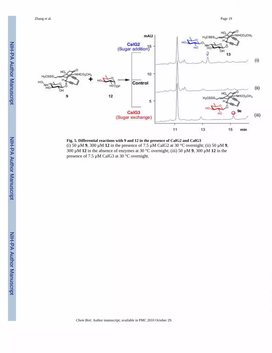
Fig. 5. Differential reactions with 9 and 12 in the presence of CalG2 and CalG3(i) 50 µM 9, 300 µM 12 in the presence of 7.5 µM CalG2 at 30 °C overnight; (ii) 50 µM 9,300 µM 12 in the absence of enzymes at 30 °C overnight; (iii) 50 µM 9, 300 µM 12 in thepresence of 7.5 µM CalG3 at 30 °C overnight.
Zhang et al. Page 19
Chem Biol. Author manuscript; available in PMC 2010 October 29.
NIH
-PA Author Manuscript
NIH
-PA Author Manuscript
NIH
-PA Author Manuscript

Fig. 6. The proposed CLM glycosylation pathway
Zhang et al. Page 20
Chem Biol. Author manuscript; available in PMC 2010 October 29.
NIH
-PA Author Manuscript
NIH
-PA Author Manuscript
NIH
-PA Author Manuscript
NIH
-PA Author Manuscript
NIH
-PA Author Manuscript
NIH
-PA Author Manuscript
Zhang et al. Page 21
Table I
Crystal parameters, X-ray data collection, phasing, and refinement statistics.
SeMet Native
Crystal parameters
Space group 1222 P21
Unit-cell parameters (Å, °) a = 106.7, b = 119.3, c = 156.0 a = 57.4, b = 97.7,c = 63.0
β = 90.6
Data collection statistics
Wavelength (Å) 0.97918 0.97918
Energy (eV) 12,662 12,662
Resolution range (Å) 26.75–2.80 (2.90–2.80) 38.76–1.68 (1.74–1.68)
No. of reflections (measured/unique)a 180890/24883 482455/68513
Completeness (%) 99.2 (94.9) 86.3 (43.6)
Rmergeb 0.121 (0.426) 0.051 (0.496)
Redundancy 7.3 (6.3) 7.0 (3.9)
Mean l/sigma(l) 9.8 (4.0) 25.1 (2.4)
Phasing statisticsc
Mean FOM (centric/acentric) 0.344/0.090
Phasing Power (isomorphous/anomalous) 0.0/1.17
Cullis R-factor (isomorphous/anomalous) 0.0/0.79
Refinement and model statistics
Resolution range 26.75–2.79 (2.86– 2.79) 38.76–1.90 (1.95–1.90)d
No. of reflections (work/test) 23613/1270 50954/2745
Rcryste 0.187 (0.311) 0.160 (0.186)
Rfreef 0.243 (0.378) 0.212 (0.275)
Rmsd bonds (Å) 0.011 0.014
Rmsd angles (°) 1.336 1.417
ESU from Rfree(Å)g 0.342 0.146
B factor–Wilson plot (Å2) 50.3 24.3
B factor – monomer A/B/waters (Å2)h 31.4/33.0/23.3 31.4/30.2/36.4
No. of protein molecules/all atoms 2/5783 2/6378
No. of waters 45 579
No. of auxiliary molecules 2 MOPS 1 PEG
Ramachandran Plot by MOLPROBITY (%)
Favoured regions 97.5 98.3
Additional allowed regions 2.5 1.6
Outliers 0.0 0.1
PBD code
aValues in parentheses are for the highest resolution shell.
Chem Biol. Author manuscript; available in PMC 2010 October 29.

NIH
-PA Author Manuscript
NIH
-PA Author Manuscript
NIH
-PA Author Manuscript
Zhang et al. Page 22
bRmerge = ΣhΣi | Ii(h) - <I(h)> |/ΣhΣiIi(h), where li(h) is the intensity of an individual measurement of the reflection and <I(h)> is the mean intensity
of the reflection.
cResolution range for phasing in SHARP was (26.66–3.2) Å.
dResolution range for refinement was cut (38.76–1.90) Å due to low completeness and signal in the remaining resolution shells.
eRcryst = Σh | | Fobs | - | Fcalc | |/Σh | Fobs |, where Fobs and Fcalc are the observed and calculated structure-factor amplitudes, respectively.
fRfree was calculated as Rcryst using ~5.0% of the randomly selected unique reflections that were omitted from structure refinement.
gEstimated standard uncertainty based on Rfree.
hB-factors from the model refined without TLS.
Chem Biol. Author manuscript; available in PMC 2010 October 29.
Copyright © 2022 FDOKUMEN