The diurnal cycle of shallow cumulus clouds over land: A single‐column model intercomparison study
Upload
khangminh22Category
view
1download
0
Glycosylation related actions of glycodelin: gamete, cumuluscell, immune cell and clinical associations
M.Seppala1,4, H.Koistinen1, R.Koistinen1,2, P.C.N.Chiu3 and W.S.B.Yeung3
1Department of Clinical Chemistry, University of Helsinki, Helsinki University Central Hospital, Biomedicum Helsinki, 4th floor,
Haartmaninkatu 8, 00290 Helsinki, Finland, 2Department of obstetrics and Gynaecology, University of Helsinki, Helsinki University
Central Hospital, Biomedicum Helsinki, 4th floor, Haartmaninkatu 8, 00290 Helsinki, Finland and 3Department of Obstetrics and
Gynaecology, University of Hong Kong, Queen Mary Hospital, Pokulfam Road, Hong Kong, People’s Republic of China
4Correspondence address. Pihlajatie 20 B 15, 00270 Helsinki, Finland. E-mail: [email protected]
Glycodelin is an example of a glycoprotein whose complex-type glycans mediate biological actions in human reproduc-tion and immune reactions. Being attached to an identical protein backbone, glycodelin oligosaccharides vary signifi-cantly from one reproductive tissue to another and have an effect on its own secretion and role in cell communication.For instance, uterine glycodelin-A inhibits sperm–oocyte interaction by binding on the sperm head. This is a glyco-sylation-dependent phenomenon, in which fucosyltransferase-5 plays a key role. Glycodelin-S from seminal plasmabinds evenly around the sperm head and maintains an uncapacitated state in the spermatozoa, until the isoform isdetached during sperm passage through the cervix. Glycodelin-F from follicular fluid and Fallopian tube binds tothe acrosomal region of the sperm head, thereby inhibiting both the sperm–oocyte binding and premature progester-one-induced acrosome reaction. The cumulus cells surrounding the oocyte can capture glycodelin-A and -F from thesurrounding environment and convert these isoforms to a cumulus cell isoform, glycodelin-C. It differs by glycosyla-tion from the other isoforms, and it too attaches on the sperm head, with the highest density in the equatorial region.Glycodelin-C is capable of detaching the sperm-bound inhibitory isoforms so that the sperm–oocyte binding isenhanced. Glycodelin-A also has immunosuppressive actions directed to cellular, humoral and innate immunity.Although these actions depend mainly on the protein backbone, glycosylation also plays a part. Glycosylated glyco-delin may be involved in the protection of spermatozoa against maternal immune reactions, and glycodelin alsohas apoptogenic activity. Some glycosylation patterns of glycodelin may mask its apoptogenic domain. This reviewupdates the recent research and clinical associations of glycodelin, highlighting the role of glycosylation.
Key words: cumulus cells/fertilization/immunosuppression
Immunological and structural background
Nomenclature, antibodies and immunodetection
The name ‘glycodelin’ was proposed by the Helsinki team to
their collaborators who had resolved the glycan structures and bio-
logical actions of purified glycoprotein (Riittinen et al., 1991), pre-
viously known as placental protein 14 or progesterone associated
endometrial protein (Dell et al., 1995; Kamarainen et al., 1996;
Koistinen et al., 1996; Morris et al., 1996). The name ‘glycodelin’
was subsequently agreed by the pioneers who had launched these
and a variety of other names for this protein into the literature
(reviewed in Seppala et al., 1998). The name ‘glycodelin’ high-
lights the importance of glycosylation for the biological activity
of the glycoprotein that is formed in various sites of the body.
Glycodelin is sugar-rich, comprising 17.5 wt.% carbohydrate
(Bohn et al., 1982). On the basis of the differences in glyco-
sylation, the isoforms so far characterized are designated as
glycodelin-A (amniotic fluid, endometrium/decidua and maternal
serum) (Dell et al., 1995; Koistinen et al., 2003), glycodelin-S
(seminal plasma and seminal vesicles) (Morris et al., 1996),
glycodelin-F (follicular fluid and the oviduct) (Tse et al., 2002)
and glycodelin-C (cumulus oophorus) (Chiu et al., 2006). Poly-
clonal and monoclonal anti-glycodelin antibodies generated
using purified glycodelin protein from amniotic fluid and
seminal plasma show similar immunoreactivity against these
four glycodelin isoforms. Although carbohydrates play an
active role in protein folding and participate in the formation of
discontinuous epitopes, the differentially glycosylated glycodelin
isoforms, such as glycodelin-A and glycodelin-S, share similar
thermodynamic parameters of reversible denaturation, suggesting
that their native folding is not influenced by different glycosyla-
tion patterns (Koistinen et al., 1999). This may explain why the
generation of antibodies specific for either glycodelin-A or
glycodelin-S has turned out to be difficult, and all the four
# The Author 2007. Published by Oxford University Press on behalf of the European Society of Human Reproduction and Embryology. All rights reserved. For
Permissions, please email: [email protected] 275
Human Reproduction Update, Vol.13, No.3 pp. 275–287, 2007 doi:10.1093/humupd/dmm004
Advance Access publication February 28, 2007
Dow
nloaded from https://academ
ic.oup.com/hum
upd/article/13/3/275/2458704 by guest on 10 July 2022
glycosylated isoforms react in the same way with anti-glycodelin
antibodies.
In addition to the conventional monoclonal and polyclonal anti-
bodies generated with the use of native glycosylated glycodelin
that has been purified from biological materials, polyclonal anti-
bodies have been produced against a synthetic linear femtopeptide
consisting of amino acids 69–83 of the glycodelin sequence –
NH2-Lys-Lys-Val-Leu-Gly-Glu-Lys-Thr-Glu-Asn-Pro-Lys-Lys-
Phe-Lys-COOH (Poddar et al., 1998). Not unexpectedly, the
results obtained with the use of this peptide antibody (Horowitz
et al., 2001; Song et al., 2001) are different from those obtained
with the anti-glycodelin antibodies (Julkunen et al., 1986a;
Waites et al., 1990; Mandelin et al., 2003). The difference is
seen both in normal tissues and in tumours, and cross-testing of
the two types of antibodies (R. Koistinen and M. Seppala, unpub-
lished observation) revealed that, while both react with glycodelin,
the femtopeptide antibody shows a wider spread of immunostain-
ing in tumours, normal tissues and, notably, in blood vessels
in which the glycodelin antibodies show no reactivity. Most
importantly, not all immunoreactivity of the femtopeptide anti-
body can be abolished by absorption with purified glycodelin-A,
demonstrating that its additional reactivity is glycodelin unrelated.
Given that about one half of the sequence of the linear femtopep-
tide used for immunization (Poddar et al., 1998) is similar to the
sequences present in many human proteins, such as SCP-1
peptide (100% identity with the first seven amino acids of the
femtopeptide), cutaneous T cell Iymphoma (CTCL) tumour
antigen se2-1, bullous pemphigoid antigen 1, dynein heavy
chain domain 3, KIAA1503 protein, dystonin isoform 1eA precur-
sor and a number of other proteins, any of these proteins/peptides
may potentially cross-react with the femtopeptide antibody.
Therefore, until the nature of the glycodelin unrelated additional
immunoreactivity of the peptide antibody has been specified, the
name ‘glycodelin’ is misleading in this context. Perhaps, a name
‘glycodelin-derived peptide’ should more accurately describe
this immunoreactivity to distinguish it from glycodelin.
Glycodelin gene
The Human Genome Organization (HUGO) has registered
progestagen-associated endometrial protein (PAEP) as the official
symbol of the glycodelin gene (Kamarainen et al., 1991). The gene
is 5.05 kb long and, like many other lipocalin genes, it is divided
into seven exons (Vaisse et al., 1990). The nucleotide sequence
encoding the retinol-binding motif of b-lactoglobulins is con-
served in the glycodelin gene that comprises four putative gluco-
corticoid/progesterone response elements (PRE) in the promoter
region (Vaisse et al., 1990). The presence of PREs is compatible
with the observations that progesterone is involved in the regu-
lation of glycodelin synthesis.
Primary structure
Glycodelin is a member of the lipocalin family of proteins.
Its primary sequence of 180 amino acid residues (Julkunen
et al., 1988) has significant similarity with b-lactoglobulins from
several species. The crystal structure of glycodelin is not known,
and information on its tertiary structure rests on the Swiss model
deduced from the crystal structure of bovine b-lactoglobulin
(Koistinen et al., 1999).
Carbohydrate moieties
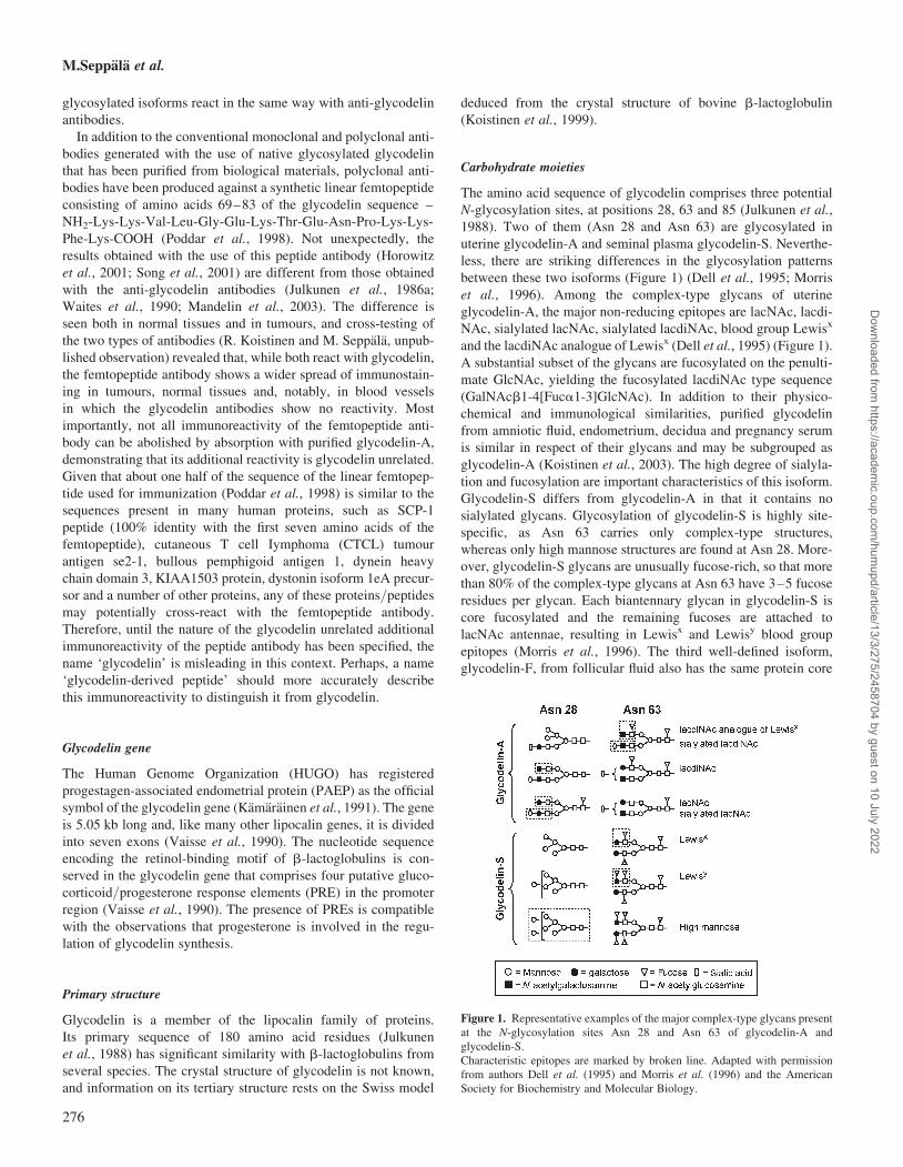
The amino acid sequence of glycodelin comprises three potential
N-glycosylation sites, at positions 28, 63 and 85 (Julkunen et al.,
1988). Two of them (Asn 28 and Asn 63) are glycosylated in
uterine glycodelin-A and seminal plasma glycodelin-S. Neverthe-
less, there are striking differences in the glycosylation patterns
between these two isoforms (Figure 1) (Dell et al., 1995; Morris
et al., 1996). Among the complex-type glycans of uterine
glycodelin-A, the major non-reducing epitopes are lacNAc, lacdi-
NAc, sialylated lacNAc, sialylated lacdiNAc, blood group Lewisx
and the lacdiNAc analogue of Lewisx (Dell et al., 1995) (Figure 1).
A substantial subset of the glycans are fucosylated on the penulti-
mate GlcNAc, yielding the fucosylated lacdiNAc type sequence
(GalNAcb1-4[Fuca1-3]GlcNAc). In addition to their physico-
chemical and immunological similarities, purified glycodelin
from amniotic fluid, endometrium, decidua and pregnancy serum
is similar in respect of their glycans and may be subgrouped as
glycodelin-A (Koistinen et al., 2003). The high degree of sialyla-
tion and fucosylation are important characteristics of this isoform.
Glycodelin-S differs from glycodelin-A in that it contains no
sialylated glycans. Glycosylation of glycodelin-S is highly site-
specific, as Asn 63 carries only complex-type structures,
whereas only high mannose structures are found at Asn 28. More-
over, glycodelin-S glycans are unusually fucose-rich, so that more
than 80% of the complex-type glycans at Asn 63 have 3–5 fucose
residues per glycan. Each biantennary glycan in glycodelin-S is
core fucosylated and the remaining fucoses are attached to
lacNAc antennae, resulting in Lewisx and Lewisy blood group
epitopes (Morris et al., 1996). The third well-defined isoform,
glycodelin-F, from follicular fluid also has the same protein core
Figure 1. Representative examples of the major complex-type glycans present
at the N-glycosylation sites Asn 28 and Asn 63 of glycodelin-A and
glycodelin-S.
Characteristic epitopes are marked by broken line. Adapted with permission
from authors Dell et al. (1995) and Morris et al. (1996) and the American
Society for Biochemistry and Molecular Biology.
M.Seppala et al.
276
Dow
nloaded from https://academ
ic.oup.com/hum
upd/article/13/3/275/2458704 by guest on 10 July 2022
as glycodelin-A and glycodelin-S, but it too has its own specific
glycosylation, as indicated by studies employing fluorophore-
assisted carbohydrate electrophoresis and lectin binding character-
istics (Chiu et al., 2003b). The fourth isoform, glycodelin-C,
is modified from glycodelin-A and -F by the cumulus cells. Com-
pared with its parent isoforms, its molecular size is smaller and the
isoelectric point is higher, and their lectin binding properties are
different. Glycodelin-C reacts strongly with concanavalin-A,
Wisteria floribunda agglutinin, Ricinus communis agglutinin,
Ulex europaeus agglutinin and Dolichos biflorus agglutinin but,
unlike its parent isoforms, not with Sambucus nigra bark agglutinin
that reacts with the a-NeuNAc(2-6)gal/galNAc residues (Table I).
Glycodelin produced by recombinant technologies may be
either non-glycosylated or glycosylated, depending on the cells
used for synthesis. For instance, recombinant glycodelin from
Chinese hamster ovary cells is glycosylated and it reacts immuno-
logically in the same way as glycodelin-A, yet their glycosylation
patterns are different. By contrast, glycodelin produced in the
human embryonic kidney 293 cells fulfils both immunological
and glycosylation based criteria of glycodelin-A (Van den
Nieuwenhof et al., 2000). Therefore, in the absence of specific
information on the type of glycosylation, term glycodelin
without any isoform designation is recommended.
Cellular origin and secretion in reproductive tissues
Glycodelin-A is synthesized in glandular and luminal surface
epithelium of progesterone- or progestagen-exposed endometrium
(Julkunen et al., 1986a; Mandelin et al., 1997, 2001) (Table I).
During the estrogen dominated fertile window of a normal ovula-
tory cycle, absence of glycodelin-A synthesis in the endometrium
is significant, because the glycoprotein has anti-fertilization
activity (discussed later). In a normal ovulatory cycle, glycodelin
expression peaks in the secretory endometrium 8–10 days after
ovulation, whereas in maternal serum and amniotic fluid, the
levels are highest at 10 and 16 weeks of pregnancy, respectively
(Julkunen et al., 1985). Glycodelin-A is secreted mainly into
endometrial/decidual gland lumen, from there to uterine fluid or
amniotic fluid, and less to serum. Results of a study employing
mutagenesis of the asparagines at the N-glycosylation sites (Asn
28 and Asn 63) suggest that glycosylation at Asn 28 plays a key
role in the extracellular secretion of glycodelin, as mutation of
Asn 28 brings about a significant decrease in the amount of
secreted protein. Loss of both glycosylation sites dramatically
reduces the secretion (Jayachandran et al., 2004). This study
shows that glycosylation is essential for glycodelin secretion.
Glycodelin-S is one of the major secretory proteins in the
seminal plasma, produced in seminal vesicle glands (Petrunin
et al., 1980; Bohn et al., 1982; Julkunen et al., 1984; Koistinen
et al., 1997). No clinically meaningful association has been
found between the glycodelin concentration in seminal plasma
and sperm pathology (Julkunen et al., 1984), or the success of
in vitro fertilization (IVF) (Koistinen et al., 2000).
Glycodelin-F is synthesized in luteinized granulosa cells of the
ovary and it is present in follicular fluid (Table I). Glycodelin has
also been found in the oviduct, synthesized by its epithelial cells
(Julkunen et al., 1986b, 1990; Laird et al., 1995). Part of oviductal
glycodelin is similar to glycodelin-F (Yeung et al., 2006).
Cumulus oophorus cells contain glycodelin immunoreactivity
but not glycodelin mRNA, indicating uptake rather than synthesis
(Tse et al., 2002). The cumulus cell isoform is immunologically
similar but it differs from the other three isoforms by glycosylation
and charge (Chiu et al., 2006).
Outside the pelvic organs, glycodelin has been found in haema-
topoietic cells of the bone marrow (Kamarainen et al., 1994;
Morrow et al., 1994), normal breast (Kamarainen et al., 1999),
glandular tissues in the lung and eccrine sweat glands (Kamarai-
nen et al., 1997). The type of glycosylation of glycodelin from
these tissues remains to be characterized.
Biological behaviour and actions in the reproductive system
The difference in glycosylation between glycodelin and
b-lactoglobulin is reflected in their different biological actions.
Unlike b-lactoglobulin and many other lipocalins, glycodelin-A
has not been found to bind retinol, retinoic acid or other lipocalin
ligands (Koistinen et al., 1999). Glycodelin inhibits E-selectin
mediated cell adhesion (Jeschke et al., 2003). This propensity is
compatible with an observation that oligosaccharides with fucosy-
lated lacdiNAc antennae present in glycodelin-A have been shown
to block selectin-mediated adhesions, and a biantennary N-linked
oligosaccharide bearing GalNAcb1-4(Fuca1-3)GlcNAc antennae
is an inhibitor of E-selectin-mediated adhesion (Grinnell et al.,
1994). Glycodelin has also been shown to take part in cell differ-
entiation, and it may be involved in tissue modelling (Kamarainen
et al., 1997; Koistinen et al., 2005; Uchida et al., 2005).
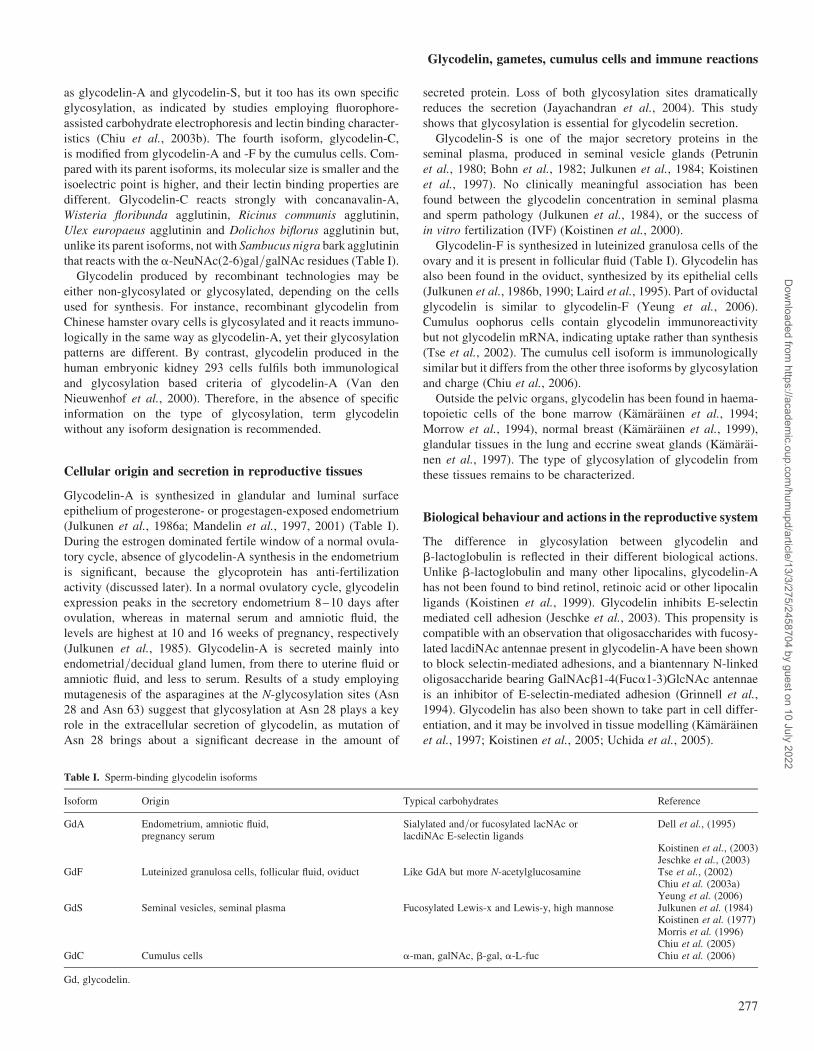
Table I. Sperm-binding glycodelin isoforms
Isoform Origin Typical carbohydrates Reference
GdA Endometrium, amniotic fluid,pregnancy serum
Sialylated and/or fucosylated lacNAc orlacdiNAc E-selectin ligands
Dell et al., (1995)
Koistinen et al., (2003)Jeschke et al., (2003)
GdF Luteinized granulosa cells, follicular fluid, oviduct Like GdA but more N-acetylglucosamine Tse et al., (2002)Chiu et al. (2003a)Yeung et al. (2006)
GdS Seminal vesicles, seminal plasma Fucosylated Lewis-x and Lewis-y, high mannose Julkunen et al. (1984)Koistinen et al. (1977)Morris et al. (1996)Chiu et al. (2005)
GdC Cumulus cells a-man, galNAc, b-gal, a-L-fuc Chiu et al. (2006)
Gd, glycodelin.
Glycodelin, gametes, cumulus cells and immune reactions
277
Dow
nloaded from https://academ
ic.oup.com/hum
upd/article/13/3/275/2458704 by guest on 10 July 2022
Binding of glycodelin isoforms on spermatozoa
All the four isoforms, glycodelin-A, -S, -F and -C, bind on the
human sperm head (Tables II and III). Glycodelin-F has two
binding sites. One of them is shared with glycodelin-A, and
glycodelin-A can displace maximally 70% of labelled glycodelin-F
bound on the spermatozoa (Chiu et al., 2003b). Immunocytochemi-
cal staining localizes glycodelin-F binding sites to the acrosomal
region of the human spermatozoa. Studies on neoglycoproteins
have shown that the binding of glycodelin-A to spermatozoa
involves mannose, fucose and possibly E-selectin ligands,
whereas that of glycodelin-F involves mannose, fucose and
N-acetylglucosamine, but not the selectin ligands (Chiu et al., 2004).
Fucosyltransferases (FUT) constitute a family of glycosyltrans-
ferases that incorporate fucosyl residues into glycolipid or glyco-
protein glycans, providing one of the possible termination steps of
glycoconjugate biosynthesis of the sialyl Lewisx or sialyl Lewisa
determinant that plays a role in cell–cell interaction (Borsig
et al., 1996). It is believed that the function of glycosyltransferases
on the cell surface is confined to their carbohydrate binding ability
rather than glycosyltransferase function because of a lack of sugar
nucleotide donors (Colley, 1997). Using chemical cross-linking and
anti-glycodelin antibody immunoprecipitation, the glycodelin-
receptor complex was isolated and analysed by mass spectrometry
and Western blot analysis using specific anti-FUT5 antibodies
(Chiu et al., 2007). The results suggested that FUT5 serves as
the receptor of glycodelin-A on human spermatozoa (Table IV).
Differential extraction of the surface labelled sperm proteins and
immunofluorescence staining suggested that sperm FUT5 is an
externally oriented integral membrane protein in the acrosomal
region of the human spermatozoa. Subsequently, Chiu et al.
(2007) succeeded in purification of biologically active FUT5
from human spermatozoa and, in co-immunoprecipitation exper-
iments, they confirmed that the interaction between glycodelin-A
and sperm FUT5 or recombinant FUT5 is highly specific. Accord-
ing to binding kinetic analyses, the KD of sperm FUT5 binding to
solubilized zona pellucida is 43 pmol ml21. The ability of sperm
FUT5 to bind to both glycodelin-A and -F, and to the zona
pellucida, suggests that human sperm FUT5 is a receptor of
glycodelin-A (and -F) and zona pellucida glycoproteins (Chiu
et al., 2007). The likely mechanism by which these glycodelin iso-
forms inhibit spermatozoa–zona pellucida binding is by blocking
the binding of sperm FUT5 to the zona pellucida.
Like glycodelin-A, glycodelin-S binds on the human sperm
head via two binding sites that are saturable and reversible
(Chiu et al., 2005). The binding sites of glycodelin-S are different
because glycodelin-A and -F cannot displace glycodelin-S from
its binding sites. Although the binding of glycodelin-S is specific,
its affinity is low. The low affinity binding sites are more abun-
dant than the high affinity binding sites. On the basis of indirect
immunofluorescence staining the sperm-bound glycodelin-S
covers the sperm head completely and this immunoreactivity is
removed when spermatozoa migrate through cervical mucus sur-
rogates. The low binding affinity of the carbohydrate-based inter-
actions is likely to allow detachment of glycodelin-S from
spermatozoa.
In follicular fluid, glycodelin-F is the main sperm–oocyte
binding inhibitory isoform (Chiu et al., 2003a), but follicular
fluid also contains small amounts of glycodelin-A (P.C.N. Chiu
and W.S.B. Yeung, unpublished observation). Glycodelin-C has
been converted from glycodelin-A and -F by the cumulus cells
(Chiu et al., 2006). Its protein core is identical with that of the
other glycodelin isoforms, but glycodelin-C has a smaller molecu-
lar size, a higher isoelectric point and different lectin binding prop-
erties compared with the other isoforms (Table I).
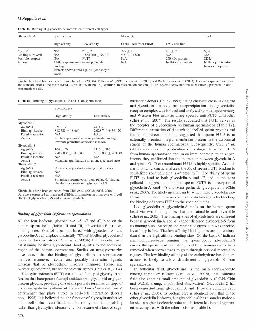
Table II. Binding of glycodelin-A isoforms on different cell types
Glycodelin-A Spermatozoa Monocyte T cell
High affinity Low affinity CD14þ cell from PBMC U937 cell line
KD (nM) N/A 21 + 2 6.7 + 1.1 48 + 21 N/ABinding sites /cell N/A 1 884 260 + 66 220 9 510–35 820 N/A N/APossible receptor N/A FUT5 N/A 250 kDa protein CD45Action Inhibits spermatozoa–zona pellucida
bindingProtects spermatozoa against lymphocyteattack
N/A Inhibits chemotaxis Inhibits proliferationInduces apoptosis
Kinetic data have been extracted from Chiu et al. (2003b), Miller et al. (1998), Vigne et al. (2001) and Rachmilewitz et al. (2003). Data are expressed as meanand standard error of the mean (SEM). N/A, not available; KD, equilibrium dissociation constant, FUT5, sperm fucosyltransferase-5; PBMC, peripheral bloodmononuclear cells.
Table III. Binding of glycodelin-F, -S and -C on spermatozoa
Spermatozoa
High affinity Low affinity
Glycodelin-FKD (nM) 3.9 + 0.1 25 + 2Binding sites/cell 818 720 + 18 060 2 028 740 + 36 120Possible receptor N/A FUT5Action Inhibits spermatozoa–zona pellucida binding
Prevents premature acrosome reactionGlycodelin-S
KD (nM) 104 + 20 1413 + 316Binding sites/cell 2 408 000 + 301 000 5 117 000 + 903 000Possible receptor N/A N/AAction Maintains spermatozoa in an uncapacitated state
Glycodelin-CKD (nM) Positive co-operativity among binding sitesBinding sites/cell N/APossible receptor N/AAction Stimulates spermatozoa–zona pellucida binding
Displaces sperm-bound glycodelin-A/F
Kinetic data have been extracted from Chiu et al. (2003b, 2005, 2006).Data were expressed as mean and (SEM). Information on monocyte or T celleffects of glycodelin-F, -S and -C is not available.
M.Seppala et al.
278
Dow
nloaded from https://academ
ic.oup.com/hum
upd/article/13/3/275/2458704 by guest on 10 July 2022
Capacitation
This is defined as a series of transformations that spermatozoa
normally undergo during their migration through the female
genital tract to reach and bind to the zona pellucida. During capaci-
tation, extensive changes take place in all sperm compartments. Ion
fluxes induce biochemical modifications, membrane lipids
and proteins are reorganized, and complex signal transduction
mechanisms are initiated (de Lamirande et al., 1997). At high phys-
iological concentrations (.900 pmol ml21) glycodelin-S signifi-
cantly suppresses bovine serum albumin-induced capacitation of
human spermatozoa, suggesting that glycodelin-S may contribute
to an uncapacitated state of human spermatozoa in seminal
plasma and prevent premature capacitation (Chiu et al., 2005).
Deglycosylated glycodelin-S has no similar inhibitory effect,
demonstrating the importance of glycosylation in this process. Inter-
estingly, another study employing recombinant glycodelin has
shown that whereas glycosylated glycodelin inhibits human sperm
capacitation, non-glycosylated glycodelin stimulates it (Dutta
et al., 2001). Compared with the binding kinetics of the other gly-
codelin isoforms, glycodelin-S binds to and detaches from sperma-
tozoa at a faster rate than the other two isoforms do. The fast
kinetics is obviously important for its biological action to take
place because spermatozoa will be in contact with seminal plasma
during a short time after ejaculation only. The weak binding affinity
of glycodelin-S may also explain why labelled glycodelin immunor-
eactivity can be demonstrated on spermatozoa only when the cells
are treated with high physiological concentration of glycodelin-S
(Chiu et al., 2003b, 2005). It also explains the readiness of
removal of bound glycodelin-S from spermatozoa during migration
through the cervical mucus. Interestingly, glycodelin immunoreac-
tivity has been found in the cervical mucus (Pockley et al., 1989),
but its origin remains to be determined.
There is cholesterol efflux from human spermatozoa during
bovine serum albumin and cyclodextrin induced capacitation of
human spermatozoa. Glycodelin-S significantly reduces this efflux
induced by either of the stimulators, and it exerts this effect
upstream of protein kinase activation in the adenylyl cyclase/protein kinase A/tyrosine kinase signalling pathway. Again, these
findings demonstrate the importance of carbohydrate moieties of
glycodelin-S for its biological action, particularly because the said
processes are activated upon removal of glycodelin-S from the sper-
matozoa. In vivo, the dissociation probably takes place during the
passage of spermatozoa through the cervix, as suggested by
in vitro experiments employing a cervical fluid surrogate (Chiu
et al., 2005). In view of these observations glycodelin-S appears
to play a role in maintaining an uncapacitated state in the human
spermatozoa before their passage through the cervix.
Inhibition of sperm binding to the zona pellucida
Glycodelin-A was the first endogenous human glycoprotein that
was found to potently and dose-dependently inhibit the binding
of spermatozoa to the zona pellucida (Oehninger et al., 1995).
This observation came from studies employing a hemizona
assay (HZA) that was originally developed to predict the fertiliz-
ing potential of spermatozoa in the human, without inadvertent
fertilization in vitro (Burkman et al., 1988). The HZA uses the
bisected matching halves of a human zona pellucida, providing
an internal control on zona to zona variability. One of the two
halves can be exposed to a test substance, whereas the other
serves as a control. In the case of glycodelin, the inhibition of
sperm–oocyte binding was observed in HZA after a short
pre-incubation time of glycodelin and the spermatozoa. This pro-
pensity of uterine glycodelin-A was glycosylation dependent,
because differently glycosylated glycodelin-S had no similar
activity (Morris et al., 1996). The result indicates that uterine
glycodelin-A has anti-fertilizing activity (Table II). This overtly
surprising finding does not contradict with the current knowledge
of the sequence of events in human reproduction, because glyco-
delin secretion is cyclically absent from endometrium during the
fertile midcycle when the spermatozoa migrate through the
uterine cavity to fertilize an oocyte in the fallopian tube.
Acrosome reaction
Initial sperm–oocyte binding in the mammals involves recognition
of glycosylated proteins of the zona pellucida by glycosylated
proteins on sperm surfaces (Benoff, 1997). Once the binding of
spermatozoa to the zona pellucida is engaged, acrosome reaction
is induced via a G protein-mediated event (Ward and Kopf,
1993). Glycodelin-F participates in the regulation of the acrosome
reaction in an interesting fashion. The isoform is secreted from
luteinized ovarian granulosa cells into pre-ovulatory follicular
fluid (Tse et al., 2002) and transferred with the cumulus oophorus
complex into the oviduct at ovulation. Glycodelin-F binds on the
acrosome region of the sperm head, thereby inhibiting progesterone
induced acrosome reaction. Glycodelin-A does not have this prop-
erty. Glycodelin-F has even stronger an inhibitory activity on
sperm-zona binding than glycodelin-A does (Chiu et al., 2003a),
and this capacity is lost upon deglycosylation (Chiu et al., 2003b).
Role of the cumulus oophorus
At ovulation the oocyte and its associated cumulus cell mass
containing follicular fluid are released and transported to the
oviduct. The cumulus cells that surround the oocyte secrete
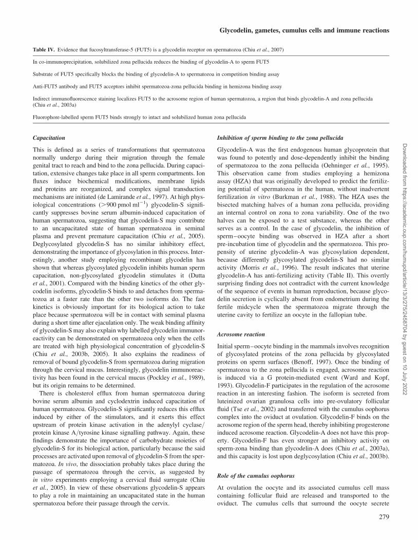
Table IV. Evidence that fucosyltransferase-5 (FUT5) is a glycodelin receptor on spermatozoa (Chiu et al., 2007)
In co-immunoprecipitation, solubilized zona pellucida reduces the binding of glycodelin-A to sperm FUT5
Substrate of FUT5 specifically blocks the binding of glycodelin-A to spermatozoa in competition binding assay
Anti-FUT5 antibody and FUT5 acceptors inhibit spermatozoa-zona pellucida binding in hemizona binding assay
Indirect immunofluorescence staining localizes FUT5 to the acrosome region of human spermatozoa, a region that binds glycodelin-A and zona pellucida(Chiu et al., 2003a)
Fluorophore-labelled sperm FUT5 binds strongly to intact and solubilized human zona pellucida
Glycodelin, gametes, cumulus cells and immune reactions
279
Dow
nloaded from https://academ
ic.oup.com/hum
upd/article/13/3/275/2458704 by guest on 10 July 2022
hormones (Shutt and Lopata, 1981), and they have been suggested
to be involved in nutrition, selecting spermatozoa with normal
morphology, induction of the acrosome reaction and support of
oocyte nuclear maturation, fertilization and the subsequent
embryo development (Carrell et al., 1993; Yanagimachi, 1994;
Wongsrikeao et al., 2005). Failure of cumulus oophorus formation
results in problems in fertilization. For fertilization of the oocyte,
the spermatozoa have to migrate through the cumulus cell mass.
An interesting set of observations led to the discovery of the role
of glycodelin-F in the fertilization process. Human follicular fluid
was found to inhibit human sperm–zona pellucida binding (Yao
et al., 1996), and the cumulus cells reduced this effect (Hong
et al., 2003). To clarify the factors behind these observations, col-
laborative research between Hong Kong and Helsinki identified
glycodelin as an important effector molecule in this phenomenon.
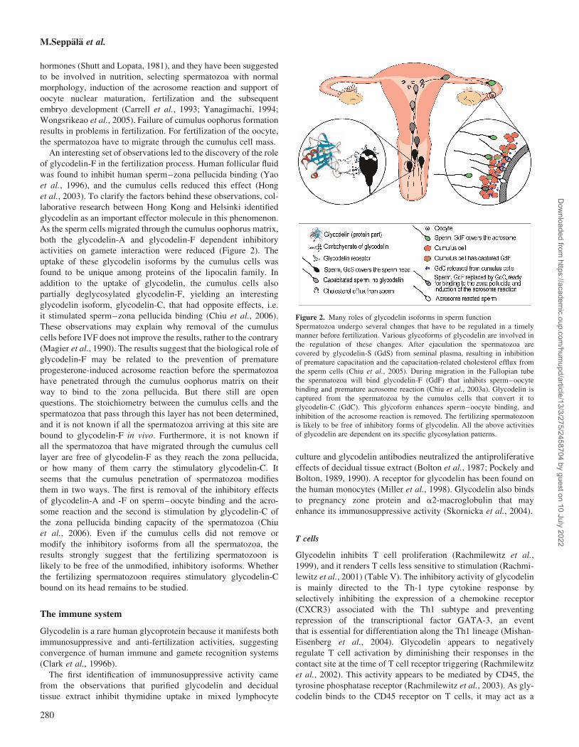
As the sperm cells migrated through the cumulus oophorus matrix,
both the glycodelin-A and glycodelin-F dependent inhibitory
activities on gamete interaction were reduced (Figure 2). The
uptake of these glycodelin isoforms by the cumulus cells was
found to be unique among proteins of the lipocalin family. In
addition to the uptake of glycodelin, the cumulus cells also
partially deglycosylated glycodelin-F, yielding an interesting
glycodelin isoform, glycodelin-C, that had opposite effects, i.e.
it stimulated sperm–zona pellucida binding (Chiu et al., 2006).
These observations may explain why removal of the cumulus
cells before IVF does not improve the results, rather to the contrary
(Magier et al., 1990). The results suggest that the biological role of
glycodelin-F may be related to the prevention of premature
progesterone-induced acrosome reaction before the spermatozoa
have penetrated through the cumulus oophorus matrix on their
way to bind to the zona pellucida. But there still are open
questions. The stoichiometry between the cumulus cells and the
spermatozoa that pass through this layer has not been determined,
and it is not known if all the spermatozoa arriving at this site are
bound to glycodelin-F in vivo. Furthermore, it is not known if
all the spermatozoa that have migrated through the cumulus cell
layer are free of glycodelin-F as they reach the zona pellucida,
or how many of them carry the stimulatory glycodelin-C. It
seems that the cumulus penetration of spermatozoa modifies
them in two ways. The first is removal of the inhibitory effects
of glycodelin-A and -F on sperm–oocyte binding and the acro-
some reaction and the second is stimulation by glycodelin-C of
the zona pellucida binding capacity of the spermatozoa (Chiu
et al., 2006). Even if the cumulus cells did not remove or
modify the inhibitory isoforms from all the spermatozoa, the
results strongly suggest that the fertilizing spermatozoon is
likely to be free of the unmodified, inhibitory isoforms. Whether
the fertilizing spermatozoon requires stimulatory glycodelin-C
bound on its head remains to be studied.
The immune system
Glycodelin is a rare human glycoprotein because it manifests both
immunosuppressive and anti-fertilization activities, suggesting
convergence of human immune and gamete recognition systems
(Clark et al., 1996b).
The first identification of immunosuppressive activity came
from the observations that purified glycodelin and decidual
tissue extract inhibit thymidine uptake in mixed lymphocyte
culture and glycodelin antibodies neutralized the antiproliferative
effects of decidual tissue extract (Bolton et al., 1987; Pockely and
Bolton, 1989, 1990). A receptor for glycodelin has been found on
the human monocytes (Miller et al., 1998). Glycodelin also binds
to pregnancy zone protein and a2-macroglobulin that may
enhance its immunosuppressive activity (Skornicka et al., 2004).
T cells
Glycodelin inhibits T cell proliferation (Rachmilewitz et al.,
1999), and it renders T cells less sensitive to stimulation (Rachmi-
lewitz et al., 2001) (Table V). The inhibitory activity of glycodelin
is mainly directed to the Th-1 type cytokine response by
selectively inhibiting the expression of a chemokine receptor
(CXCR3) associated with the Th1 subtype and preventing
repression of the transcriptional factor GATA-3, an event
that is essential for differentiation along the Th1 lineage (Mishan-
Eisenberg et al., 2004). Glycodelin appears to negatively
regulate T cell activation by diminishing their responses in the
contact site at the time of T cell receptor triggering (Rachmilewitz
et al., 2002). This activity appears to be mediated by CD45, the
tyrosine phosphatase receptor (Rachmilewitz et al., 2003). As gly-
codelin binds to the CD45 receptor on T cells, it may act as a
Figure 2. Many roles of glycodelin isoforms in sperm function
Spermatozoa undergo several changes that have to be regulated in a timely
manner before fertilization. Various glycoforms of glycodelin are involved in
the regulation of these changes. After ejaculation the spermatozoa are
covered by glycodelin-S (GdS) from seminal plasma, resulting in inhibition
of premature capacitation and the capacitation-related cholesterol efflux from
the sperm cells (Chiu et al., 2005). During migration in the Fallopian tube
the spermatozoa will bind glycodelin-F (GdF) that inhibits sperm–oocyte
binding and premature acrosome reaction (Chiu et al., 2003a). Glycodelin is
captured from the spermatozoa by the cumulus cells that convert it to
glycodelin-C (GdC). This glycoform enhances sperm–oocyte binding, and
inhibition of the acrosome reaction is removed. The fertilizing spermatozoon
is likely to be free of inhibitory forms of glycodelin. All the above activities
of glycodelin are dependent on its specific glycosylation patterns.
M.Seppala et al.
280
Dow
nloaded from https://academ
ic.oup.com/hum
upd/article/13/3/275/2458704 by guest on 10 July 2022

calcium-dependent lectin to bind other T cell surface glyco-
proteins to mediate its immunoregulatory activities (Ish-Shalom
et al., 2006). Importantly, glycodelin binding to T cells can be
competitively inhibited with oligosaccharides, showing that glyco-
delin binds to T cell surfaces in a carbohydrate-dependent manner.
Glycodelin has been suggested to act through its distinct receptors
that are decorated by carbohydrates and expressed in different
cells, wherein one of its surface molecular targets, CD45, mediates
its T cell inhibitory activity (Yaniv et al., 2003).
B cells
Glycodelin also has an effect on the B cells (Table V). A human B
cell inhibitory receptor, CD22, binds to the sialylated lacNAc
sequences (Powell and Varki, 1994). It has been suggested that the
oligosaccharides bearing sialylated lacNAc or lacdiNAc antennae
present in glycodelin-A may manifest immunosuppressive effects
by specifically blocking the adhesive and activation related events
mediated by CD22 (Dell et al., 1995). Preliminary evidence indicates
that glycodelin inhibits B cell receptor mediated activation of human
B cells (Yaniv et al., 2003). Here, glycodelin inhibits B cell prolifer-
ation and the up-regulation of IgM and major histocompatibility
complex (MHC), but not CD69 and CD86, regardless of the extent
of B cell receptor triggering. These findings suggest that glycodelin
affects some but not all B cell responses. The B cell inhibition by gly-
codelin is different from the T cell inhibition in that the extent of
glycodelin-mediated inhibition does not correlate to the level of B
cell receptor triggering, suggesting that glycodelin interferes with
late events of B cell receptor signalling (Yaniv et al., 2003). Interest-
ingly, CD22 binds to CD45, the leukocyte specific receptor linked
phosphotyrosine phosphatase involved in T cell activation (Stamen-
kovic et al., 1991). Taken together, these studies suggest that glyco-
delin is a soluble regulatory factor capable of interacting with both T
and B cells in a carbohydrate-dependent manner to affect both cellu-
lar and humoral immune responses (Yaniv et al., 2003).
Innate immunity
Besides its actions on humoral and cellular immunity, glycodelin
inhibits cytotoxicity of peripheral blood natural killer (NK) cells
(Okamoto et al., 1991) (Table V). These cells are of specific
interest because they need no prior exposure to react with
foreign antigens, such as bacteria, viruses or an embryo, a semi-
allograft. It is believed that uterine NK cells are derived from a
subset of the NK cells in peripheral blood (reviewed in Dosiou
and Giudice, 2005). There is selective, increased expression of
glycodelin in the NK cells isolated from pregnancy decidua
(Koopman et al., 2003). However, direct evidence that glycodelin
inhibits cytotoxicity of uterine NK cells is not available yet, and
the role of glycosylation in this process remains to be clarified.
Immunoprotection of spermatozoa
The expression of glycoconjugates by normal cells may protect
them from immune responses, especially in those cases in which
the MHC recognition is minimal or absent (Clark et al., 1997).
In spite of the frequent exposure to antigens in spermatozoa and
in seminal plasma, women are rarely immunized against sperma-
tozoa. Should it happen, the question remains whether the various
glycodelin glycoforms that bind on the human spermatozoa would
protect them against immune responses in the female body. The
immunosuppressive properties of the Lewisx/y epitopes present
in glycodelin-S may contribute to the low immunogenicity
of sperm in women (Morris et al., 1996). Recent findings
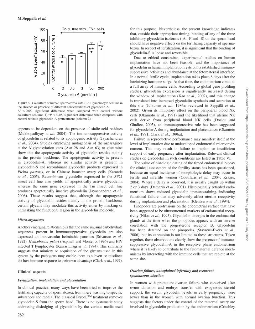
(R. Tsang and W.S.B. Yeung, unpublished results) indicate that
glycodelin-A treatment maintains viability of human spermatozoa
in co-culture with a human lymphocyte cell line (Figure 3), and
glycodelin bound spermatozoa are less likely to activate the lym-
phocytes. Obviously, glycosylation plays an important part here
because, in the absence of glycosylation, glycodelin does not
bind on the spermatozoa (Chiu et al., 2003b), and deglycosylated
glycodelin does not have the same effect. But, there are other
mechanisms, e.g. attenuation of immune responses against the
gametes or the blastocyst may be manifested by binding of selectin
or ‘selectin-like’ receptors on the spermatozoa to the oligosacchar-
ide ligands on immune effector cells (Clark et al., 1996b).
Other aspects
Oligosaccharides with at least one fucosylated lacdiNAc antenna,
such as present in glycodelin-A (Figure 1), have been shown to be
15–20-fold more potent inhibitors of selectin-mediated adhesions
than either sialyl- or sulpho-Lewisx/a type oligosaccharides
(Grinnell et al., 1994). The presence of such oligosaccharides in
glycodelin suggests indirectly that glycodelin may manifest
some of its immunosuppressive effects by blocking selectin-
dependent adhesions (Clark et al., 1996a). More recently, direct
evidence has been provided that glycodelin inhibits E-selectin-
mediated cell adhesion (Jeschke et al., 2003).
Apoptosis
Glycodelin-A but not glycodelin-S induces apoptosis in T cells,
but not in monocytes (Mukhopadhyay et al., 2001). This function
Table V. Immunosuppressive activity of glycodelin
Type of immune cell Effect by glycodelin Reference
T cells Inhibits T cell proliferation Rachmilewitz et al., (1999)Renders T cells less sensitive to stimulation Rachmilewitz et al., (2001)Diminishes T cell responses in the contact site at the time of T cell receptor triggering Rachmilewitz et al., (2002)Inhibition mediated by CD45, the tyrosine phosphatase receptor Rachmilewitz et al., (2003)Inhibits expression of a chemokine receptor CXCR3 Mishan-Eisenberg et al., (2004)
B cells Inhibits B cell proliferation, IgM secretion and the surface expression of MHC class II Yaniv et al., (2003)Inhibits B cell receptor mediated activation of human B cells Yaniv et al., (2003)
NK cells Inhibits cytotoxic activity of NK cells from peripheral blood Okamoto et al., (1991)Increased expression in uterine NK cells suggesting local activity Koopman et al., (2003)
Glycodelin, gametes, cumulus cells and immune reactions
281
Dow
nloaded from https://academ
ic.oup.com/hum
upd/article/13/3/275/2458704 by guest on 10 July 2022
appears to be dependent on the presence of sialic acid residues
(Mukhopadhyay et al., 2004). The immunosuppressive activity
of glycodelin is related to its apoptogenic activity (Jayachandran
et al., 2004). Studies employing mutagenesis of the asparagines
at the N-glycosylation sites (Asn 28 and Asn 63) to glutamine
show that the apoptogenic activity of glycodelin resides mainly
in the protein backbone. The apoptogenic activity is present
in glycodelin-A, whereas no similar activity is present in
glycodelin-S and recombinant glycodelin products expressed in
Pichia pastoris, or in Chinese hamster ovary cells (Karande
et al., 2005). Recombinant glycodelin expressed in the SF21
insect cell line also yields an apoptotically active glycodelin,
whereas the same gene expressed in the Tni insect cell line
produces apoptotically inactive glycodelin (Jayachandran et al.,
2006). These results suggest that, although the apoptogenic
activity of glycodelin resides mainly in the protein backbone,
certain glycans may modulate this activity either by masking or
unmasking the functional region in the glycodelin molecule.
Micro-organisms
Another emerging relationship is that the same unusual carbohydrate
sequences present in immunosuppressive glycodelin are also
expressed on intravascular helminthic parasites (Srivatsan et al.,
1992), Helicobacter pylori (Aspinall and Monteiro, 1996) and HIV
infected T lymphocytes (Kawashinagi et al., 1994). This similarity
suggests that mimicry or acquisition of the glycans used in this
system by the pathogens may enable them to subvert or misdirect
the host immune response to their own advantage (Clark et al., 1997).
Clinical aspects
Fertilization, implantation and placentation
In clinical practice, many ways have been tried to improve the
fertilizing capacity of spermatozoa, from mere washing to specific
substances and media. The classical PercollTM treatment removes
glycodelin-S from the sperm head. There is no systematic study
addressing dislodging of glycodelin by the various media used
for this purpose. Nevertheless, the present knowledge indicates
that, outside their appropriate timing, binding of any of the three
inhibitory glycodelin isoforms (-A, -F and -S) on the sperm head
should have negative effects on the fertilizing capacity of sperma-
tozoa. In respect of fertilization, it is significant that the binding of
glycodelin-S is loose and reversible.
Due to ethical constraints, experimental studies on human
implantation have not been feasible, and the importance of
glycodelin in human implantation rests on its established immuno-
suppressive activities and abundance at the fetomaternal interface.
In a normal fertile cycle, implantation takes place 8 days after the
luteinizing hormone surge. At that time, the endometrium contains
a full array of immune cells. According to global gene profiling
studies, glycodelin expression is significantly increased during
the window of implantation (Kao et al., 2002), and this change
is translated into increased glycodelin synthesis and secretion at
this site (Julkunen et al., 1986a; reviewed in Seppala et al.,
2002). Given its inhibitory effect on the peripheral blood NK
cells (Okamoto et al., 1991) and the likelihood that uterine NK
cells derive from peripheral blood NK cells (Dosiou and
Giudice, 2005), an immunoprotective role has been suggested
for glycodelin-A during implantation and placentation (Okamoto
et al., 1991; Clark et al., 1996a).
Failure in reproductive performance may manifest itself at the
level of implantation due to undeveloped endometrial microenvir-
onment. This may result in failure to implant or insufficient
support of early pregnancy after implantation. Relevant clinical
studies on glycodelin in such conditions are listed in Table VI.
The value of histologic dating of the timed endometrial biopsy
for clinical assessment of the fertility status has been questioned,
because an equal incidence of morphologic delay may occur in
fertile and infertile women (Coutifaris et al., 2004; Krazer,
2004). Where a delay is observed, it is usually caught up within
2 or 3 days (Damario et al., 2001). Histologically retarded endo-
metrium shows reduced glycodelin immunostaining, indicating
functional failure that may adversely affect uterine receptivity
during implantation and placentation (Klentzeris et al., 1994).
Pinopodes are protrusions on the endometrial surface that have
been suggested to be ultrastructural markers of endometrial recep-
tivity (Nikas et al., 1995). Glycodelin emerges in the endometrial
glands at the time when the pinopodes appear, with an inverse
correlation with the progesterone receptor B. Glycodelin
has been detected on the pinopodes (Stavreus-Evers et al.,
2006), but its expression is not limited to these structures. Taken
together, these observations clearly show the presence of immuno-
suppressive glycodelin-A in the receptive phase endometrium
where it is likely to contribute to the fetomaternal defence mech-
anisms by interacting with the immune cells that are replete at the
same site.
Ovarian failure, unexplained infertility and recurrent
spontaneous abortion
In women with premature ovarian failure who conceived after
ovum donation and embryo transfer with exogenous steroid
support, the serum glycodelin levels in early pregnancy were
lower than in the women with normal ovarian function. This
suggests that factors under the control of the maternal ovary are
involved in glycodelin production by the endometrium (Critchley
Figure 3. Co-culture of human spermatozoa with JE6.1 lymphocyte cell line in
the absence or presence of different concentrations of glycodelin-A.
*P , 0.05, significant difference when compared with control without
co-culture (column 1).*P , 0.05, significant difference when compared with
control without glycodelin-A pretreatment (column 2).
M.Seppala et al.
282
Dow
nloaded from https://academ
ic.oup.com/hum
upd/article/13/3/275/2458704 by guest on 10 July 2022
et al., 1992). Besides progesterone that was replaced, relaxin
appears to be another corpus luteum hormone that stimulates
glycodelin secretion (Stewart et al., 1997; Tseng et al., 1999). Sub-
normal secretion or absence of ovarian relaxin secretion may have
contributed to the observed difference in women with premature
ovarian failure.
Subnormal peri-implantation phase glycodelin levels have been
reported in the uterine fluid of non-pregnant patients with unex-
plained infertility (Mackenna et al., 1993) and also in women
with a history of recurrent spontaneous abortion (Dalton et al.,
1998; Salim et al., 2006). In the latter condition, also the circulat-
ing glycodelin concentration may be reduced at the mid-luteal
phase (Tulppala et al., 1995). Although the glycodelin concen-
tration of uterine secretions correlates well with endometrial
morphology during the menstrual cycle (Li et al., 1993b), particu-
larly during the peri-implantation period (Li et al., 1993a), the
question remains whether this measurement would be clinically
any more useful than immunohistochemical detection of glycode-
lin in endometrial biopsies, or the routine histologic dating.
An advantage of determining glycodelin secretion from uterine
flushings instead of endometrial biopsy is that uterine flushings
should give a more comprehensive picture of uterine glycodelin
secretion because, in biopsies, glycodelin expression varies from
one endometrial site to another (Li et al., 1991). Therefore, in
spite of its invasive nature, the measurement of glycodelin from
uterine flushings has clinical potential, should a standardized
methodology become widely applicable.
Polycystic ovary syndrome
This condition is frequently associated with increased secretion of
luteinizing hormone, androgen and insulin, resulting in problems
with ovulation, implantation and early pregnancy loss. In
ovulatory cycles of the women with polycystic ovary syndrome
(PCOS), glycodelin serum level increases during treatment with
metformin, an insulin-reducing agent (Jakubowicz et al., 2001).
During pregnancy, reduced glycodelin serum concentrations in
the first trimester are associated with early pregnancy loss,
suggesting failure in placentation (Jakubowicz et al., 2004).
Interestingly, insulin has no acute glycodelin reducing effect
(Seppala et al., 2005), whereas its long-term effects on glycodelin
secretion remain to be studied. Furthermore, it remains to be
proven if the low glycodelin serum level plays any part in the
pathogenesis of PCOS-related early pregnancy loss due to
reduced fetomaternal defence mechanisms at the placentation site.
In vitro fertilization
Given the steep rise of serum glycodelin level from implantation
onwards (Julkunen et al., 1985), prediction of the outcome of
IVF by serum glycodelin levels has been thoroughly explored.
Rather disappointedly, in many studies, the subnormal glycodelin
serum levels at the implantation phase have not predicted fertile or
infertile cycles in any consistent way (reviewed in Seppala et al.,
2002). More recent reports point to potential clinical utility under
specific circumstances. For instance, there is a significant corre-
lation between low serum glycodelin levels on day 21 of the
pretreatment cycle and a higher pregnancy rate following IVF/intracytoplasmic sperm injection in normogonadotrophic women
subjected to the long protocol of pituitary down-regulation
and gonadotrophin stimulation (Westergaard et al., 2004). In the
subsequent treatment cycle, the glycodelin serum levels were sig-
nificantly higher in conception than in non-conception cycles. The
authors suggest that measuring the mid-luteal serum glycodelin
level in the pretreatment cycle may offer a clinical test to decide
whether infertility treatment should be initiated in that cycle or
not. Perhaps, the low serum glycodelin level identified a specific
group of women whose infertility was related to endometrial
dysfunction, and this condition was corrected during ovarian
stimulation for IVF, contributing to the high success rate.
Another approach is also of interest (Liu et al., 2006). In indi-
vidual IVF cycles, comparison between serum glycodelin levels
taken on the day of oocyte retrieval and on the day of embryo
transfer gave information on success in terms of achieving a preg-
nancy. Although no overall difference in serum glycodelin was
found on the day of oocyte retrieval or embryo transfer between
the non-pregnant and pregnant groups, both the ratio and the
difference of serum glycodelin levels on the days of embryo trans-
fer and oocyte retrieval were higher in the pregnant group than in
the non-pregnant group. This approach is of interest because it
addresses intra-individual rather than inter-individual differences
and, therefore, is free of the problem of wide individual variation
in the glycodelin levels. The results are compatible with the
fetoembryonic defence system hypothesis (Clark et al., 1996a).
Both studies suggest clinical potential of the glycodelin test in
predicting a short-term IVF success under specific situations, but
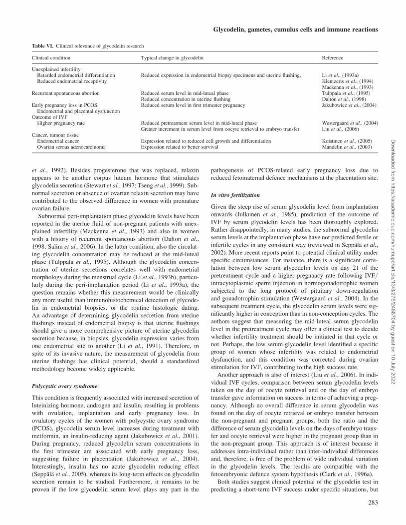
Table VI. Clinical relevance of glycodelin research
Clinical condition Typical change in glycodelin Reference
Unexplained infertilityRetarded endometrial differentiation Reduced expression in endometrial biopsy specimens and uterine flushing, Li et al., (1993a)Reduced endometrial receptivity Klentzeris et al., (1994)
Mackenna et al., (1993)Recurrent spontaneous abortion Reduced serum level in mid-luteal phase Tulppala et al., (1995)
Reduced concentration in uterine flushing Dalton et al., (1998)Early pregnancy loss in PCOS Reduced serum level in first trimester pregnancy Jakubowicz et al., (2004)
Endometrial and placental dysfunctionOutcome of IVF
Higher pregnancy rate Reduced pretreatment serum level in mid-luteal phase Westergaard et al., (2004)Greater increment in serum level from oocyte retrieval to embryo transfer Liu et al., (2006)
Cancer, tumour tissueEndometrial cancer Expression related to reduced cell growth and differentiation Koistinen et al., (2005)Ovarian serous adenocarcinoma Expression related to better survival Mandelin et al., (2003)
Glycodelin, gametes, cumulus cells and immune reactions
283
Dow
nloaded from https://academ
ic.oup.com/hum
upd/article/13/3/275/2458704 by guest on 10 July 2022
in different ways. The results are not mutually controversial
because the first study addressed low pretreatment levels and the
second study measured the difference from oocyte retrieval to
embryo transfer in the hormonally stimulated treatment cycles.
Another viewpoint in IVF comes from the observed apopto-
genic activity of glycodelin, i.e. whether the glycodelin isoforms
taken up (and possibly internalized) by the cumulus cells can
exert apoptogenic activity before fertilization. The question is of
interest because apoptosis has been used to estimate ovarian
reserve in women undergoing IVF (Seifer et al., 1996). Here,
the gametes and embryos derived from the cumulus complexes
with no or minor apoptosis were found to have an increased
chance of giving rise to optimum blastocysts (Corn et al., 2005).
Contraception
Contraceptive methods employing progestogens have an effect
on endometrial glycodelin secretion. This has been shown
in levonorgestrel-releasing intrauterine contraceptive system
(Mandelin et al., 1997), subdermal implants (Mandelin et al.,
2001) and levonorgestrel only containing pills taken for emer-
gency contraception before the LH surge (Durand et al., 2005).
In view of the anti-fertilizing effects of glycodelin-A, it would
seem that induction of glycodelin synthesis before fertilization
could contribute to the contraceptive effect of the above
methods. However, the significance of this effect remains to be
determined, because evidence of the magnitude of the effect
in vivo is not available and the oocyte/cumulus cell complex
can remove glycodelin from the spermatozoa and modify its
activity before fertilization (see Role of the cumulus oophorus).
Cancer
Endometrial cancer
When endometrial adenocarcinoma cells (Ishikawa cells) were
co-cultured with normal endometrial stromal cells in the presence
of progesterone, proliferation was reduced concomitantly with
induced glycodelin expression (Arnold et al., 2002). At the same
time, the adenocarcinoma cells underwent conversion to a
normal phenotype. These findings raised a question of whether
glycodelin was a cause or a consequence of the reduction of malig-
nant characteristics. Recent evidence indicates that glycodelin
may be primary to the observed change. Experiments employing
transfection of glycodelin cDNA into glycodelin-negative
endometrial adenocarcinoma cells brought about induction of gly-
codelin expression at the same time as they showed increased
differentiation and reduced tumour cell growth. A significant con-
comitant observation was reduced expression of the Bcl-XL gene,
an anti-apoptotic survival gene involved in tumour cell growth and
chemoresistance (Koistinen et al., 2005). The glycosylation profile
of glycodelin induced by transfection in endometrial adenocarci-
noma cells has not been determined, but the results are compatible
with glycodelin’s apoptogenic isoforms (see Apoptosis). Given
that progesterone and progesterone antagonists stimulate glycode-
lin gene expression, the results may encourage a reappraisal
of glycodelin synthesis-stimulating pathways in supporting
chemotherapy of malignant endometrial tumours, notably those
expressing the progesterone receptor. However, the results
obtained with the glycodelin femtopeptide antibody are
contradictory to the above results, as they show increased ‘-
glycodelin’ immunoreactivity in advanced malignant tumours
(Horowwitz et al., 2001). Obviously the glycodelin-unrelated
specificity of the polyclonal anti-peptide antibody may contribute
to the different results (see Nomenclature, antibodies and
immunodetection).
Ovarian serous carcinoma
Ovarian cancer consists of many subtypes, serous carcinoma being
the most common of them. Clinical stage and histological grade
are the gold standards in the selection of clinical management,
and many clinical and prognostic markers have been explored.
Studies employing anti-glycodelin antibodies show that glycode-
lin expression in tumour cells is more frequent in well differen-
tiated than in poorly differentiated carcinomas, and glycodelin
expression is more frequent in early stage compared with
advanced stage tumours (Mandelin et al., 2003). Importantly, in
grade I/stage III patients, the 5-year overall survival of the
patients with glycodelin expressing tumours is significantly
higher than in those patients whose tumours did not contain glyco-
delin. The study shows that glycodelin expression in ovarian
serous carcinoma is a favourable prognostic sign. These results
are at variance with those employing the glycodelin femtopeptide
antibody (Horowitz et al., 2001; Song et al., 2001), as the femto-
peptide antibody shows more intense staining in advanced
tumours compared with local tumours or normal tissues, and it
shows strong immunostaining in tumour blood vessels that are
negative with the use of the anti-glycodelin antibody (Mandelin
et al., 2003). Again, these differences are likely due to different
specificities of the antibodies used in these studies.
Concluding remarks
It is now firmly established that glycodelin interacts by its unique
carbohydrates with the cell surface of many cell types, particularly
the gametes and the immune cells. In the gametes, most biological
actions of glycodelin are inhibitory, such as the inhibition of sperm
capacitation (glycodelin-S), sperm–oocyte binding (glycodelin-A
and -F) and the acrosome reaction (glycodelin-F). All these activi-
ties involve binding of the specific glycodelin glycoform on
the sperm head. Recent studies have uncovered an important
role for the cumulus oophorus cells. These cells can take up
glycodelin-A and -F and modify their glycans in such a way that
the resulting glycodelin-C has stimulatory effects on the sperm–
zona pellucida binding. The structure and biological role of the
cumulus cell-modified glycodelin is now unfolding. Evidence
for the involvement of glycodelin oligosaccharides in the cell sig-
nalling processes of the cellular, humoral and innate immune
responses and apoptosis is also accumulating.
Finally, clinical research points to a number of areas in which
significant changes take place in glycodelin secretion, providing
functional information. As the removal of specific N-glycosylation
sites by mutagenesis clearly shows that glycosylation is requisite
for glycodelin secretion (Jayachandran et al., 2004), glycodelin
secretion may be affected by either inappropriate glycosylation
machinery or by another type of clinical dysfunction. So far, inap-
propriate glycosylation patterns of glycodelin have been addressed
in one clinical study only, i.e. in respect of the fertilization poten-
tial of spermatozoa in vitro (Koistinen et al., 2000). The paucity of
M.Seppala et al.
284
Dow
nloaded from https://academ
ic.oup.com/hum
upd/article/13/3/275/2458704 by guest on 10 July 2022
such studies is not surprising because aberrant glycosylation of
glycodelin cannot be identified by any available routine test.
Nevertheless, research on glycodelin glycosylation has led the
way to a better understanding of the biology and physiology of
reproduction, immunology and even cancer. Today, detection of
inappropriate glycoprotein glycosylation remains a challenge for
clinical research and practice alike, an glycodelin provides a well-
characterized example of this complex issue for future studies.
Abbreviations
Asn, asparagine; Gly, glycine; Glu, glutamic acid; Leu, leucine; Lys,
lysine; Phe, phenylalanine; Pro, proline; Thr, threonine; Val, valine.
Acknowledgements
The authors wish to thank Mrs Annikki Lofhjelm for skillful assist-ance. Original studies of this review have been supported by grantsfrom the University of Helsinki, the Academy of Finland, HelsinkiUniversity Central Hospital Research Funds, Finnish CancerFoundation and the Research Grant Council, Hong Kong. Originalstudies by the authors of this review have been approved by therespective Institutional Review Boards/Ethical Committees at theDepartment of Obstetrics and Gynaecology, Helsinki UniversityCentral Hospital, Helsinki, Finland and the Department of Obstetricsand Gynaecology, University of Hong Kong, Queen Mary Hospital,Hong Kong, China.
References
Arnold JT, Lessey BA, Seppala M et al. Effect of normal endometrial stroma ongrowth and differentiation in Ishikawa endometrial adenocarcinoma cells.Cancer Res 2002;62:79–88.
Aspinall GO, Monteiro MA. Lipopolysaccharides of the Helicobacter pyloristrains P466 and M019: structures of the O antigen and coreoligosaccharide regions. Biochemistry 1996;35:2498–504.
Benoff S. Carbohydrates and fertilization: an overview. Mol Hum Reprod1997;3:599–637.
Bohn H, Kraus W, Winckler W. New soluble placental tissue proteins: theirisolation, characterization, localization and quantification. In: Klopper A.(ed). Immunology of Human Placental Proteins. Praeger Publ Placenta1982;S4:67–81.
Bolton AE, Pockley AG, Clough KJ et al. Identification of placental protein 14as an immunosuppressive factor in human reproduction. Lancet 1987;1:593–5.
Borsig L, Kleene R, Dinter A et al. Immunodetection of alpha 1-3fucosyltransferase (FucT-V). Eur J Cell Biol, 1996;70:42–53.
Burkman LJ, Coddington CC, Franken DR et al. The hemizona assay (HZA):development of a diagnostic test for the binding of human spermatozoa tothe human hemizona pellucida to predict fertilization potential. FertilSteril 1988;49:688–97.
Carrell DT, Middleton RG, Peterson CM et al. Role of the cumulus in theselection of morphologically normal sperm and induction of the acrosomereaction during human in vitro fertilization. Arch Androl 1993;31:133–7.
Chiu PCN, Chung M-K, Koistinen R et al. Cumulus oophorus-associatedglycodelin-C displaces sperm bound glycodelin-A and -F and stimulatesspermatozoa-zona pellucida binding. J Biol Chem 2006; Dec 27 (Epubahead of print).
Chiu PCN, Chung M-K, Koistinen R et al. Glycodelin-A interacts withfucosyltransferase on human sperm plasma membrane to inhibitspermatozoa-zona pellucida binding. J Cell Sci 2007;120:33–44.
Chiu PCN, Koistinen R, Koistinen H et al. Zona binding inhibitory factor-1from human follicular fluid is an isoform of glycodelin. Biol Reprod2003a;69:365–72.
Chiu PCN, Koistinen R, Koistinen H et al. Binding of zona binding inhibitoryfactor-1 (ZIF-1) from human follicular fluid on spermatozoa. J Biol Chem2003b;278:13570–7.
Chiu PCN, Tsang HY, Chung MK et al. Glycodelin-S in human seminal plasmareduces cholesterol efflux and capacitation of spermatozoa. J Biol Chem2005;280:25580–9.
Chiu PCN, Tsang H-Y, Koistinen R et al. The contribution of D-mannose,L-fucose, N-acetylglucosamine and selectin residues on the bindingof glycodelin isoforms to human spermatozoa. Biol Reprod 2004;70:1710–9.
Clark GF, Dell A, Morris HR et al. Viewing AIDS from a glycobiologicalperspective: potential linkages to the human fetoembryonic systemhypothesis. Mol Hum Reprod 1997;3:5–13.
Clark GF, Oehninger S, Patankar MS et al. A role for glycoconjugates in humandevelopment: the human feto-embryonic defense system hypothesis. HumReprod 1996a;11:467–73.
Clark GF, Oehninger S, Seppala M. Role of glycoconjugates in cellularcommunication in the human reproductive system. Mol Hum Reprod1996b;2:513–7.
Colley KJ. Golgi localization of glycosyltransferases: more questions thananswers. Glycobiology 1997;7:1–13.
Corn CM, Hauser-Kronberger C, Moser M et al. Predictive value of cumuluscell apoptosis with regard to blastocyst development of correspondinggametes. Fertil Steril 2005;84:627–33.
Coutifaris C, Myers ER, Guzick DS et al. Histologic dating of timedendometrial biopsy tissue is not related to fertility status. Fertil Steril2004;82:1264–72.
Critchley HOD, Chard T, Olajide F et al. Role of the ovary in the synthesis ofplacental protein-14. J Clin Endocrinol Metab 1992;75:97–100.
Dalton CF, Laird SM, Estdale SE et al. Endometrial protein PP14 and CA-125in recurrent miscarriage patients: Correlation with pregnancy outcome.Hum Reprod 1998;13:3197–202.
Damario MA, Lesnick TG, Lessey BA et al. Endometrial markers ofuterine receptivity utilizing the donor oocyte model. Hum Reprod2001;16:1893–9.
de Lamirande E, Leclerc P, Gagnon C. Capacitation as a regulatory event thatprimes spermatozoa for the acrosome reaction and fertilization. Mol HumReprod 1997;3:175–94.
Dell A, Morris HR, Easton R et al. Structural analysis of the oligosaccharidesderived from glycodelin, a human glycoprotein with potentimmunosuppressive and contraceptive activities. J Biol Chem 1995;270:24116–26.
Dosiou C, Giudice LC. Natural killer cells in pregnancy and recurrentpregnancy loss: Endocrine and immunologic properties. Endocr Rev2006;26:44–62.
Durand M, Seppala M, del Carmen Cravioto M et al. Late follicular phaseadministration of levonorgestrel as an emergency contraceptive changesthe secretory pattern of glycodelin in serum and endometrium duringluteal phase of the menstrual cycle. Contraception 2005;71:451–7.
Dutta B, Ain R, Seshagiri PB et al. Differential influence of recombinantnon-glycosylated and glycosylated glycodelin on human sperm function:comparative studies with hamster spermatozoa. Reprod Fertil Dev2001;13:111–8.
Grinnell BW, Hermann RB, Yan BS. Human protein C inhibitsselectin-mediated cell adhesion: role of a unique fucosylatedoligosaccharide. Glycobiology 1994;4:221–5.
Hong SJ, Tse JY, Ho PC et al. Cumulus cells reduce the spermatozoa-zonabinding inhibitory activity of human follicular fluid. Fertil Steril 2003;79(suppl. 1):802–7.
Horowitz IR, Cho CH, Song MQ et al. Increased glycodelin levels ingynecological malignancies. Int J Gynecol Cancer 2001;11:173–9.
Ish-Shalom E, Gargir A, Andre S et al. Alpha2,6-Sialylation promotes bindingof placental protein 14 via its Ca2þ -dependent lectin activity: insightsinto differential effects on CD45RO and CD45RA T cells. Glycobiology2006;16:173–3.
Jakubowicz DJ, Essah PA, Seppala M et al. Reduced glycodelin andinsulin-like growth factor-binding protein 1 in women with polycysticovary syndrome during first trimester of pregnancy. J Clin EndocrinolMetab 2004;89:833–9.
Jakubowicz DJ, Seppala M, Jakubowicz S et al. Insulin reduction withmetformin increases luteal phase serum glycodelin and insulin-likegrowth factor binding protein 1 concentrations and enhances uterinevascularity and blood flow in the polycystic ovary syndrome. J ClinEndocrinol Metab 2001;86:1126–33.
Jayachandran R, Radcliffe CM, Royle L et al. Oligosaccharides modulateapoptotic activity of glycodelin. Glycobiology 2006;16:1052–63.
Jayachandran R, Shaila MS, Karande AA. Analysis of the role ofoligosaccharides in the apoptotic activity of glycodelin A. J Biol Chem2004;279:8585–91.
Glycodelin, gametes, cumulus cells and immune reactions
285
Dow
nloaded from https://academ
ic.oup.com/hum
upd/article/13/3/275/2458704 by guest on 10 July 2022
Jeschke U, Wang X, Briese V et al. Glycodelin and amniotic fluid transferrin asinhibitors of E-selectin-mediated cell adhesion. Histochem Cell Biol2003;119:345–54.
Julkunen M, Koistinen R, Sjoberg J et al. Secretory endometrium synthesizesplacental protein 14. Endocrinology 1986a;118:1782–6.
Julkunen M, Koistinen R, Suikkari AM et al. Identification by hybridizationhistochemistry of human endometrial cells expressing mRNAs encodinga uterine b-lactoglobulin homologue and insulin-like growthfactor-binding protein-1. Mol Endocrinol 1990;4:700–7.
Julkunen M, Rutanen EM, Koskimies AI et al. Distribution of placental protein14 in tissues and body fluids during pregnancy. Br J Obstet Gynaecol1985;442:571–2.
Julkunen M, Seppala M, Janne OA. Complete amino acid sequence ofhuman placental protein 14: A progestogerone-regulated uterine proteinhomologous to ß-lactoglobulins. Proc Natl Acad Sci USA 1988;85:8845–9.
Julkunen M, Wahlstrom T, Seppala M. Human fallopian tube containsplacental protein 14. Am J Obstet Gynecol 1986b;154:1076–9.
Julkunen M, Wahlstrom T, Seppala M. et al. Detection and localization ofplacental protein 14-like protein in human seminal plasma and in themale genital tract. Arch Androl 1984; (Suppl. 12):59–67.
Kamarainen M, Halttunen M, Koistinen R et al. Expression of glycodelin inhuman breast and breast cancer. Int J Cancer 1999;83:738–742.
Kamarainen M, Julkunen M, Seppala M. HinfI polymorphism in the humanprogesterone associated endometrial protein (PAEP). Nucleic Acids Res1991;19:5092.
Kamarainen M, Leivo I, Koistinen R et al. Normal human ovary and ovariantumors express glycodelin, a glycoprotein with immunosuppressive andcontraceptive properties. Evidence from immunohistochemical stainingand in situ hybridization. Am J Pathol 1996;148:1435–43.
Kamarainen M, Riittinen L, Seppala M et al. Progesterone-associatedendometrial protein – a constitutive marker of human erythroidprecursors. Blood 1994;84:467–73.
Kamarainen M, Seppala M, Virtanen I et al. Expression of glycodelin inMCF-7 breast cancer cells induces epithelial differentiation intoorganized acinar epithelium. Lab Invest 1997;77:565–73.
Kao LC, Tulac S, Lobo S et al. Global gene profiling in human endometriumduring the window of implantation. Endocrinology 2002;143:2119–38.
Karande AA, Mukhopadyay D, Jayachandran R et al. Mechanism of theimmunomodulatory activity of glycodelin. Indian J Physiol Pharmacol2005;49:271–83.
Kawashiwagi N, Gill MJ, Adachi M. Lymphocyte membrane modificationsinduced by HIV infection. Tohoku J Exp Med 1994;173:115–31.
Klentzeris LD, Bulmer JN, Seppala M et al. Placental protein 14 in cycleswith normal and retarded endometrial differentation. Hum Reprod1994;9:394–8.
Koistinen H, Easton RL, Koistinen R et al. Differences in glycosylation andsperm-egg binding inhibition of pregnancy-related glycodelin. BiolReprod 2003;69:1545–51.
Koistinen H, Koistinen R, Dell A et al. Glycodelin from seminal plasma is adifferentially glycosylated form of contraceptive glycodelin-A. MolHum Reprod 1996;2:759–65.
Koistinen H, Koistinen R, Hyden-Granskog C et al. Seminal plasma glycodelinand fertilization in vitro. J Androl 2000;21:636–40.
Koistinen H, Koistinen R, Kamarainen M et al. Multiple forms of messengerribonucleic acid encoding glycodelin in male genital tract. Lab Invest1997;76:683–90.
Koistinen H, Koistinen R, Seppala M et al. Glycodelin and ß-lactoglobulin,lipocalins with a high structural similarity, differ in ligand bindingproperties. FEBS Lett 1999;450:158–62.
Koistinen H, Seppala M, Nagy B et al. Glycodelin reduces carcinoma-associatedgene expression in endometrial adenocarcinoma cells. Am J Obstet Gynecol2005;193:1955–60.
Koopman LA, Kopcow HD, Rybalov B et al. Human decidual natural killercells are a unique NK cell subset with immunomodulatory potential.J Exp Med 2003;198:1201–12.
Krazer RR. Endometrial biopsy should be abandoned as a routine component inthe infertility evaluation. Fertil Steril 2004;82:1297–8.
Laird SM, Hill CJ, Warren MA et al. The production of placental protein14 by human uterine tubal epithelial cells in culture. Hum Reprod1995;10: 1346–51.
Li TC, Dalton C, Hunjan KS et al. The correlation of placental protein 14concentrations in uterine flushing and endometrial morphology in theperi-implantation period. Hum Reprod 1993a;8:1923–7.
Li TC, Ling E, Dalton C et al. Concentration of endometrial protein PP14 inuterine flushings throughout the menstrual cycle in normal fertilewomen. Br J Obstet Gynaecol 1993b;100:460–4.
Li TC, Pockley G, Bolton AE et al. The variation of endometrial protein PP14in different parts of the human endometrium. Int J Gynecol Obstet1991;34:257–60.
Liu HM, Xing FQ, Wu FL. Glycodelin in IVF-ET cycles: its association withendometrial receptivity and impact on the outcome of pregnancy. NanFang Yi De Da Xue Bao 2006;26:1227–9.
Mackenna A, Li T-C, Dalton C et al. Placental protein 14 levels in uterineflushing and plasma of women with unexplained infertility. Fertil Steril1993;59:577–82.
Magier S, van der Ven HH, Diedrich K et al. Significance of cumulus oophorusin in-vitro fertilization and oocyte viablity and fertility. Hum Reprod1990;5:847–52.
Mandelin E, Koistinen H, Koistinen R et al. Levonorgestrel-releasingIUD-wearing women express contraceptive glycodelin-A inendometrium during midcycle: Another contraceptive mechanism? HumReprod 1997;12:2671–5.
Mandelin E, Koistinen H, Koistinen R et al. Endometrial expression ofglycodelin in levonorgestrel-releasing subdermal implant wearingwomen. Fertil Steril 2001;76:474–8.
Mandelin E, Lassus H, Seppala M et al. Glycodelin in ovarian serouscarcinoma: association with differentiation and survival. Cancer Res2003;63:6258–64.
Miller RE, Fayen JD, Chakraborty S et al. A receptor for the lipocalin placentalprotein 14 on human monocytes. FEBS Lett 1998;436:455–60.
Mishan-Eisenberg G, Borovsky Z, Weber MC et al. Differential regulation ofTh1/Th2 cytokine responses by placental protein 14. J Immunol2004;173:5524–30.
Morris HR, Dell A, Easton RL et al. Gender specific glycosylation ofhuman glycodelin affects contraceptive activity. J Biol Chem 1996;271:32159–67.
Morrow DM, Xiong N, Getty RR et al. Hematopoietic placental protein 14. Animmunosuppressive factor in cells of the megakaryocytic lineage. Am JPathol 1994;145:1485–95.
Mukhopadhyay D, SundarRaj S, Alok A et al. Glycodelin-A, not glycodelin-S,is apoptotically active. Relevance of sialic acid modification. J Biol Chem2004;279:8577–84.
Mukhopadhyay D, Sundereshan S, Rao C et al. Placental protein 14 (PP14)induces apoptosis in T cells, but not in monocytes. J Biol Chem2001;276:28268–73.
Nikas G, Drakakis P, Loutradis D et al. Uterine pinopodes as markers of the‘nidation window’ in cycling women receiving exogenous oestradioland progesterone. Hum Reprod 1995;10:1208–13.
Oehninger S, Coddington CC, Hodgen GD et al. Factors affecting fertilization:endometrial placental protein 14 reduces the capacity of humanspermatozoa to bind to the human zona pellucida. Fertil Steril 1995;63:377–83.
Okamoto N, Uchida A, Takakura K et al. Suppression by human placentalprotein 14 of natural killer cell activity. Am J Reprod Immunol1991;26:137–42.
Petrunin DD, Kozliaeva GS, Mesniankina NV et al. Detection of chorionica2-microglobulin in the endometrium in the secretory phase of themenstrual cycle and in the male sperm. Akush Ginecol (Mosk) 1980;3:22–3.
Pockley AG, Bolton AE. Placental protein 14 (PP14) inhibits the synthesis ofinterleukin-2 and the release of soluble interleukin-2 receptors fromphytohaemagglutinin-stimulated lymphocytes. Clin Exp Immunol 1989;77:252–6.
Pockley AG, Bolton AE. The effect of human placental protein 14 (PP14) onthe production of interleukin-1 from mitogenically stimulatedmononuclear cell cultures. Immunology 1990;69:277–81.
Pockley AG, Barratt CL, Bolton AE. Placental protein 14 (pp14) content andimmunosupressive activity of human cervical mucus. Symp Soc ExpBiol 1989;43:317–23.
Poddar AS, Kim JG, Gill KP et al. Generation and characterization ofpolyclonal antipeptide antibody to human glycodelin. Fertil Steril1998;69:543–8.
Powell LD, Varki A. The oligosaccharide binding specificities of CD22b, asialic acid-specific lectin of B cells. J Biol Chem 1994;269:10628–36.
Rachmilewitz J, Borowsky Z, Mishan-Eisenberg G et al. Focal localization ofplacental protein 14 toward sites of TCR engagement. J Immunol2002;168:2745–50.
M.Seppala et al.
286
Dow
nloaded from https://academ
ic.oup.com/hum
upd/article/13/3/275/2458704 by guest on 10 July 2022
Rachmilewitz J, Borovsky Z, Riely GJ et al. Negative regulation of T cellactivation by placental protein 14 is mediated by the tyrosinephosphatase receptor CD45. J Biol Chem 2003;278:14059–65.
Rachmilewitz J, Riely GJ, Huang JH et al. A rheostatic mechanism for T-cellinhibition based on elevation of activation thresholds. Blood 2001;98:3727–32.
Rachmilewitz J, Riely GJ, Tykocinski ML. Placental protein 14 functions as adirect T-cell inhibitor. Cell Immunol 1999;191:26–33.
Riittinen L, Narvanen O, Virtanen I et al. Monoclonal antibodies againstendometrial protein PP14 and their use for purification andradioimmunoassay of PP14. J Immunol Methods 1991;136:85–90.
Salim R, Miel J, Savvas M et al. A comparative study of glycodelinconcentrations in uterine flushings in women with subseptate uteri,history of unexplained recurrent miscarriage and healthy controls. Eur JObstet Gynecol Reprod Biol 2006; Oct 16 (Epub ahead of print).
Seifer DB, Gardiner AC, Ferreira KA et al. Apoptosis as a function of ovarianreserve in women undergoing in vitro fertilization. Fertil Steril 1996;66:593–8.
Seppala M, Bohn H, Tatarinov Y. Glycodelins. Tumor Biol 1998;19:213–20.Seppala M, Mandelin E, Koistinen R et al. Glycodelin responses to
hyperinsulinaemic clamp vary according to basal glycodelin secretion.Clin Endocrinol 2005;62:611–5.
Seppala M, Taylor RN, Koistinen H et al. Glycodelin: A major lipocalin proteinof the reproductive axis with diverse actions in cell recognition anddifferentiation. Endocr Rev 2002;23:401–30.
Shutt DA, Lopata A. The secretion of hormones during the culture of humanpreimplantation embryos with corona cells. Fertil Steril 1981;35:413–6.
Skornicka EL, Kiyatkina N, Weber MC et al. Pregnancy zone protein is acarrier and modulator of placental protein-14 in T-cell growth andcytokine production. Cell Immunol 2004;232:144–56.
Song M, Ramaswamy S, Ramachandran S et al. Angiogenic role for glycodelinin tumorigenesis. Proc Natl Acad Sci USA 2001;98:9265–70.
Srivatsan J, Smith DF, Cummins RD. Schistosoma mansioni synthesizes novelbiantennary Asn-linked oligosaccharides containing terminal b-linkedN-acetylgalactosamine. Glycobiology 1992;2:445–52.
Stamenkovic I, Sgroi D, Aruffo A et al. The B lymphocyte adhesion moleculeCD22 interacts with leukocyte common antigen CD45RO on T cells anda2-6 sialyltransferase, CD75, on B cells. Cell 1991;66:1133–44.
Stavreus-Evers A, Mandelin E, Koistinen R et al. Glycodelin is present inpinopodes of receptive-phase human endometrium and is associatedwith down-regulation of progesterone receptor B. Fertil Steril 2006;85:1803–11.
Stewart DR, Erikson MS, Erikson ME et al. The role of relaxin in glycodelinsecretion. J Clin Endocrinol Metab 1997;82:839–46.
Tse JYM, Chiu PCN, Lee KF et al. The synthesis and fate of glycodelin inhuman ovary during folliculogenesis. Mol Hum Reprod 2002;8:142–8.
Tseng L, Zhu HH, Mazella J et al. Relaxin stimulates glycodelin mRNA andprotein concentrations in human endometrial glandular epithelial cells.Mol Hum Reprod 1999;5:372–5.
Tulppala M, Julkunen M, Tiitinen A et al. Habitual abortion is accompanied bylow serum levels of placental protein 14 in the luteal phase of the fertilecycle. Fertil Steril 1995;63:792–5.
Uchida H, Maruyama T, Nagashima T et al. Histone deacetylase inhibitorsinduce differentiation of human endometrial adenocarcinoma cellsthrough up-reguation of glycodelin. Endocrinology 2005;146:5365–73.
Vaisse C, Atger M, Potier B et al. Human placental protein 14 gene:sequence and characterization of a short duplication. DNA Cell Biol1990;9:401–3.
Van den Nieuwenhof IM, Koistinen H, Easton RL et al. Recombinantglycodelin carrying the same type of glycan structures as contraceptiveglycodelin-A can be produced in human kidney 293 cells but not inChinese hamster ovary cells. Eur J Biochem 2000;267:4753–62.
Vigne JL, Hornung D, Mueller MD et al. Purification and characterization of animmunomodulatory endometrial protein, glycodelin. J Biol Chem2001;276:17101–5.
Waites GT, Bell SC, Walker RA et al. Immunohistochemical distribution of thesecretory endometrial protein, ‘pregnancy-associated endometriala2-globulin’. A glycosylated b-lactoglobulin homologue, in the humanfetus and adult employing monoclonal antibodies. Hum Reprod1990;5:105–11.
Ward CR, Kopf GS. Molecular events mediating sperm activation. Dev Biol1993;158:9–34.
Westergaard LG, Yding Andersen C, Erb K et al. Placental protein 14concentrations in circulation related to hormonal parameters andreproductive outcome in women unergoing IVF/ICSI. Reprod BiomedOnline 2004;8:91–8.
Wongsrikeao P, Kaneshige Y, Ooki R et al. Effect of removal of cumulus cellson the nuclear maturation, fertilization and development of porcineoocytes. Reprod Domest Anim 2005;40:166–70.
Yanagimachi R. Fertility of mammalian spermatozoa: its development andrelativity. Zygote 1994;2:371–2.
Yaniv E, Borovsky Z, Mishan-Eisenberg G et al. Placental protein 14 regulatesselective B cell responses. Cell Immunol 2003;222:156–63.
Yao YQ, Yeung WS, Ho PC. Human follicular fluid inhibits the binding ofhuman spermatozoa to zona pellucida in vitro. Hum Reprod 1996;11:2674–80.
Yeung WSB, Lee K-F, Koistinen R et al. Roles of glycodelin in modulatingsperm function. Mol Cell Endocrinol 2006;250:149–56.
Submitted on July 11, 2006; resubmitted on October 22, 2006; resubmitted onDecember 21, 2006; accepted on January 8, 2007
Glycodelin, gametes, cumulus cells and immune reactions
287
Dow
nloaded from https://academ
ic.oup.com/hum
upd/article/13/3/275/2458704 by guest on 10 July 2022
Copyright © 2022 FDOKUMEN