Expression and analysis of the glycosylation properties of recombinant human erythropoietin...
7
Expression and analysis of the glycosylation properties of recombinant human erythropoietin expressed in Pichia pastoris Ser Huy Teh 1 , Mun Yik Fong 2 and Zulqarnain Mohamed 1,3 1 Unit of Genetics and Molecular Biology, Institute of Biological Sciences, Faculty of Science, University of Malaya, Kuala Lumpur, Malaysia. 2 Department of Parasitology, Faculty of Medicine, University of Malaya, Kuala Lumpur, Malaysia. 3 Center of Research in Biotechnology for Agriculture, University of Malaya, Kuala Lumpur, Malaysia Abstract The Pichia pastoris expression system was used to produce recombinant human erythropoietin, a protein synthe- sized by the adult kidney and responsible for the regulation of red blood cell production. The entire recombinant hu- man erythropoietin (rhEPO) gene was constructed using the Splicing by Overlap Extension by PCR (SOE-PCR) technique, cloned and expressed through the secretory pathway of the Pichia expression system. Recombinant erythropoietin was successfully expressed in P. pastoris. The estimated molecular mass of the expressed protein ranged from 32 kDa to 75 kDa, with the variation in size being attributed to the presence of rhEPO glycosylation analogs. A crude functional analysis of the soluble proteins showed that all of the forms were active in vivo. Key words: erythropoietin, glycosylation, Pichia pastoris, SOE-PCR. Received: June 6, 2010; Accepted: January 25, 2011. Introduction Erythropoietin (EPO) is a glycoprotein hormone re- sponsible for the regulation of red blood cell production. This hormone triggers the proliferation, differentiation and maturation of bone marrow erythroid precursors into func- tional erythrocytes when blood oxygen availability is de- creased, such as during hypoxia. EPO binds to and activates the receptor on erythroid progenitor cells. The treatment of anemic patients with EPO significantly reduces their de- pendence on blood transfusions and minimizes potential side effects such as iron overload, infections and adverse reactions to leukocyte antigens. In addition to its role in hematopoeisis, EPO is neuroprotective in the nervous sys- tem and can also protect other organs (Genc et al., 2004). Prior to the 1980s, human EPO for the treatment of kidney failure and other related blood disorders had to be extracted from donors. However, under normal conditions, the expression of EPO is generally low, meaning that many donors are necessary to obtain sufficient material for treat- ment. Successful cloning of the epo gene and subsequent expression in Chinese hamster ovary (CHO) cells led to the production of several types of commercially available re- combinant human EPO (rhEPO) for human use. Since then, the possibility of increasing the yield and production effi- ciency by using other cells, such as tobacco cell lines (Matsumoto et al., 1995), Schneider cell lines (derived from Drosophila) (Kim et al., 2005) and mammary gland cells (Zhang et al., 2000), has been explored. However, most of these systems presented serious problems and were not pursued further. Improvement in our understanding of the biological activities of EPO in vivo has created a need for technologies that can provide higher yields, prolong the half-life of recombinant EPO and increase the activity of this protein in vivo. In this report, we describe the cloning, expression and analysis of rhEPO produced in the methylotrophic yeast Pichia pastoris. Yeasts have long been a model organism for biochemical and genetic studies because of the advan- tages they offer compared to bacterial systems, including the ease with which they can cultured and maintained, and the fact that they share several important biological charac- teristics with eukaryotic cells, such as splicing and other processes involved in post-translational modifications. Se- veral yeast species have been used to generate recombinant proteins, including Saccharomyces cerevisiae, Hansenula polymorpha and Pichia pastoris (reviewed in Böer et al., 2007). While S. cerevisiae offers similar advantages to other yeasts, e.g., ease of DNA manipulation, shorter culti- vation time and the ability to perform post-translational events, the tendency for heterologous proteins to undergo hypermannosylation (with the addition of up to 150 Genetics and Molecular Biology, 34, 3, 464-470 (2011) Copyright © 2011, Sociedade Brasileira de Genética. Printed in Brazil www.sbg.org.br Send correspondence to Zulqarnain Mohamed. Unit of Genetics and Molecular Biology, Institute of Biological Sciences, Faculty of Science, University of Malaya, 50603 Kuala Lumpur, Malaysia. E-mail: [email protected]. Research Article
-
Upload
sanata-dharma -
Category
Documents
-
view
1 -
download
0
Transcript of Expression and analysis of the glycosylation properties of recombinant human erythropoietin...
Expression and analysis of the glycosylation properties of recombinanthuman erythropoietin expressed in Pichia pastoris
Ser Huy Teh1, Mun Yik Fong2 and Zulqarnain Mohamed1,3
1Unit of Genetics and Molecular Biology, Institute of Biological Sciences, Faculty of Science,
University of Malaya, Kuala Lumpur, Malaysia.2Department of Parasitology, Faculty of Medicine, University of Malaya, Kuala Lumpur, Malaysia.3Center of Research in Biotechnology for Agriculture, University of Malaya, Kuala Lumpur, Malaysia
Abstract
The Pichia pastoris expression system was used to produce recombinant human erythropoietin, a protein synthe-sized by the adult kidney and responsible for the regulation of red blood cell production. The entire recombinant hu-man erythropoietin (rhEPO) gene was constructed using the Splicing by Overlap Extension by PCR (SOE-PCR)technique, cloned and expressed through the secretory pathway of the Pichia expression system. Recombinanterythropoietin was successfully expressed in P. pastoris. The estimated molecular mass of the expressed proteinranged from 32 kDa to 75 kDa, with the variation in size being attributed to the presence of rhEPO glycosylationanalogs. A crude functional analysis of the soluble proteins showed that all of the forms were active in vivo.
Key words: erythropoietin, glycosylation, Pichia pastoris, SOE-PCR.
Received: June 6, 2010; Accepted: January 25, 2011.
Introduction
Erythropoietin (EPO) is a glycoprotein hormone re-
sponsible for the regulation of red blood cell production.
This hormone triggers the proliferation, differentiation and
maturation of bone marrow erythroid precursors into func-
tional erythrocytes when blood oxygen availability is de-
creased, such as during hypoxia. EPO binds to and activates
the receptor on erythroid progenitor cells. The treatment of
anemic patients with EPO significantly reduces their de-
pendence on blood transfusions and minimizes potential
side effects such as iron overload, infections and adverse
reactions to leukocyte antigens. In addition to its role in
hematopoeisis, EPO is neuroprotective in the nervous sys-
tem and can also protect other organs (Genc et al., 2004).
Prior to the 1980s, human EPO for the treatment of
kidney failure and other related blood disorders had to be
extracted from donors. However, under normal conditions,
the expression of EPO is generally low, meaning that many
donors are necessary to obtain sufficient material for treat-
ment. Successful cloning of the epo gene and subsequent
expression in Chinese hamster ovary (CHO) cells led to the
production of several types of commercially available re-
combinant human EPO (rhEPO) for human use. Since then,
the possibility of increasing the yield and production effi-
ciency by using other cells, such as tobacco cell lines
(Matsumoto et al., 1995), Schneider cell lines (derived
from Drosophila) (Kim et al., 2005) and mammary gland
cells (Zhang et al., 2000), has been explored. However,
most of these systems presented serious problems and were
not pursued further. Improvement in our understanding of
the biological activities of EPO in vivo has created a need
for technologies that can provide higher yields, prolong the
half-life of recombinant EPO and increase the activity of
this protein in vivo.
In this report, we describe the cloning, expression and
analysis of rhEPO produced in the methylotrophic yeast
Pichia pastoris. Yeasts have long been a model organism
for biochemical and genetic studies because of the advan-
tages they offer compared to bacterial systems, including
the ease with which they can cultured and maintained, and
the fact that they share several important biological charac-
teristics with eukaryotic cells, such as splicing and other
processes involved in post-translational modifications. Se-
veral yeast species have been used to generate recombinant
proteins, including Saccharomyces cerevisiae, Hansenula
polymorpha and Pichia pastoris (reviewed in Böer et al.,
2007). While S. cerevisiae offers similar advantages to
other yeasts, e.g., ease of DNA manipulation, shorter culti-
vation time and the ability to perform post-translational
events, the tendency for heterologous proteins to undergo
hypermannosylation (with the addition of up to 150
Genetics and Molecular Biology, 34, 3, 464-470 (2011)
Copyright © 2011, Sociedade Brasileira de Genética. Printed in Brazil
www.sbg.org.br
Send correspondence to Zulqarnain Mohamed. Unit of Geneticsand Molecular Biology, Institute of Biological Sciences, Faculty ofScience, University of Malaya, 50603 Kuala Lumpur, Malaysia.E-mail: [email protected].
Research Article
mannoses) to glycoproteins (Wu et al., 2002) raises the risk
of immunogenicity, particularly for therapeutic products.
Excessive glycosylation or mannosylation may also ham-
per efficient secretion of the recombinant protein as the
sheer size of the glycogen causes the protein to be retained
in the periplasmic membrane, despite the presence of a se-
cretion signal sequence to indicate that the protein should
be exported (Kang et al., 1998). For this reason,
methylotrophic yeasts such Pichia pastoris and Hansenula
polymorpha are preferred as the overall length of the
mannose outer chains is shorter than in S. cerevisiae (Kang
et al., 1998). Other species, such Kluyveromyces lactis and
Yarrowia lipolytica, are still being investigated for their
usefulness as cloning and expression systems.
Materials and Methods
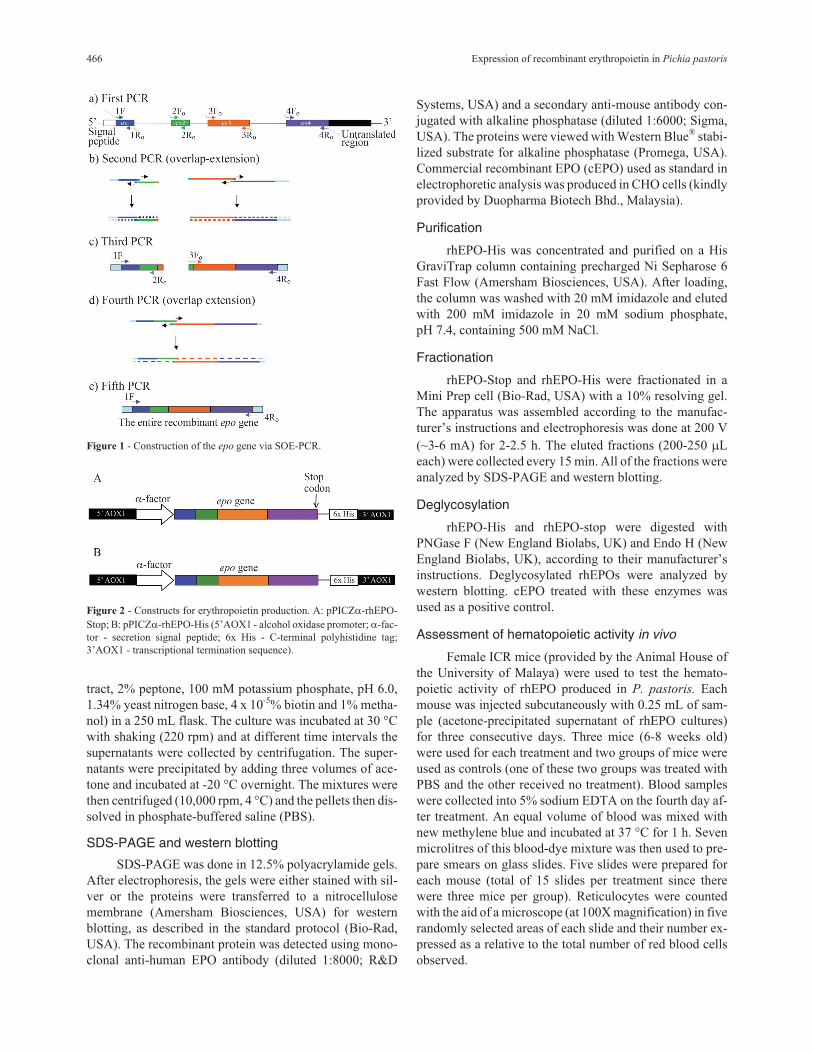
In vitro construction of the epo gene
The entire human erythropoietin gene was con-
structed using the Splicing by Overlap-Extension by PCR
(SOE-PCR) technique. Four sets of primers were designed
to amplify the four exons of the epo gene based on the
GenBank nucleotide sequence (GenBank accession num-
ber: X02158; Jacob et al., 1985). Primers covering the
exon-intron boundaries were designed to contain six nu-
cleotides that were complementary with the adjacent exons
in order to facilitate overlapping. An EcoRI site
(GAATTC) was included in primers at both ends of the tar-
get gene to generate sticky ends that would facilitate clon-
ing into the expression vector pPICZ�A. The primer sets
used are indicated in Table 1.
The four exons of the epo gene were initially ampli-
fied singly using human genomic DNA as the template. Af-
ter PCR, the products were purified by gel extraction using
a commercial kit (Qiagen, USA). Adjacent exons were as-
sembled in a second PCR reaction by allowing the exons to
form partial heteroduplexes in the overlapping regions fol-
lowed by selective amplification using terminal primers
(see Figure 1 for more details). The PCR was done using
1 U of Pfu polymerase (Fermentas, Lithuania), 1X of reac-
tion buffer, 1.5 mM Mg2+ and 200 �M of each dNTPs
(Fermentas, Lithuania). The PCR cycling conditions con-
sisted of 10 cycles of denaturation at 95 °C for 45 s, anneal-
ing at 60 °C for 45 s and elongation at 72 °C for 1 min. The
selective PCR cycling conditions included an initial dena-
turation at 95 °C for 3 min, followed by 32 cycles that in-
cluded denaturation at 95 °C for 45 s, annealing at 60 °C for
45 s and elongation at 72 °C for 1 min, with a final elonga-
tion at 72 °C for 5 min. These three steps of PCR were re-
peated until the entire epo gene was obtained.
Cloning of the recombinant epo gene
The entire epo gene construct was digested with
EcoRI (Promega, USA) and ligated into the corresponding
site in the Pichia plasmid expression vector pPICZ�A.
This vector has a highly-inducible promoter (AOX1) and
secretion signal (�-factor). The recombinant plasmids were
transformed into E. coli (TOP10F’ strain) for scale-up iso-
lation. The recombinant pPICZ�A was then linearized by
treatment with SalI (Fermentas, Lithuania) and trans-
formed chemically into P. pastoris (X-33 strain) by follow-
ing the protocol in the Pichia expression manual (Invi-
trogen, USA). The nucleotide sequence of the recombinant
epo gene construct was confirmed by DNA sequencing.
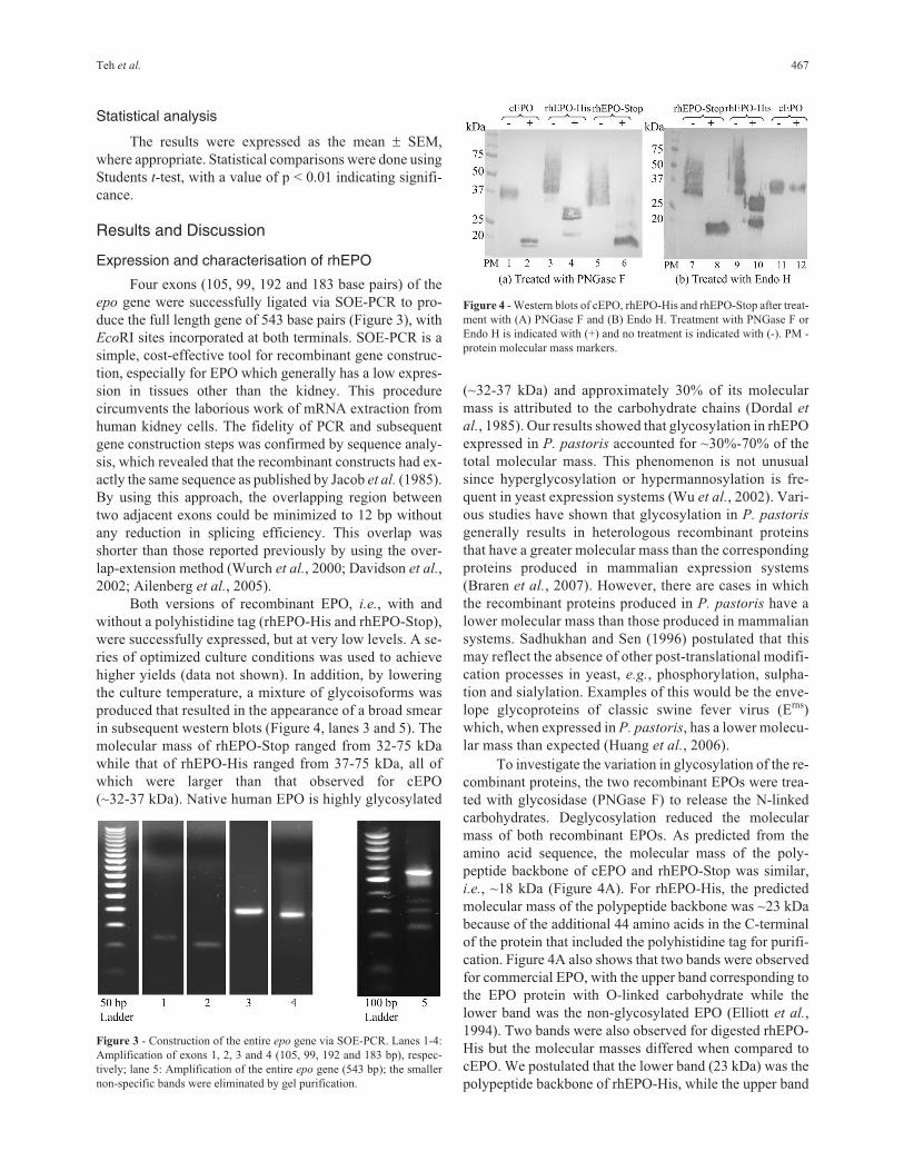
Two recombinant epo genes (pPICZ�-rhEPO-Stop
and pPICZ�-rhEPO-His) were constructed (Figure 2). A
stop codon (TGA) was introduced into pPICZ�-rhEPO-
Stop to produce a mature EPO of 165 amino acids. To ex-
press a fusion protein containing the polyhistidine tag
(pPICZ�-rhEPO-His) the rhEPO gene was cloned in frame
with the C-terminal peptide. Both vectors contained a na-
tive �-factor signal sequence that provides efficient secre-
tion of most proteins from P. pastoris.
Expression of the recombinant epo gene in P.pastoris
For small scale culture, a single colony or frozen
stock culture was grown overnight with shaking (220 rpm,
30 °C) in 10 mL of BMGY (1% yeast extract, 2% peptone,
100 mM potassium phosphate, pH 6.0, 1.34% yeast nitro-
gen base, 4 x 10⁻⁵ % biotin and 1% glycerol). The cells were
then pelleted by centrifugation (3000 g, 10 min, room tem-
perature) and transferred to 50 mL of BMMY (1% yeast ex-
Teh et al. 465
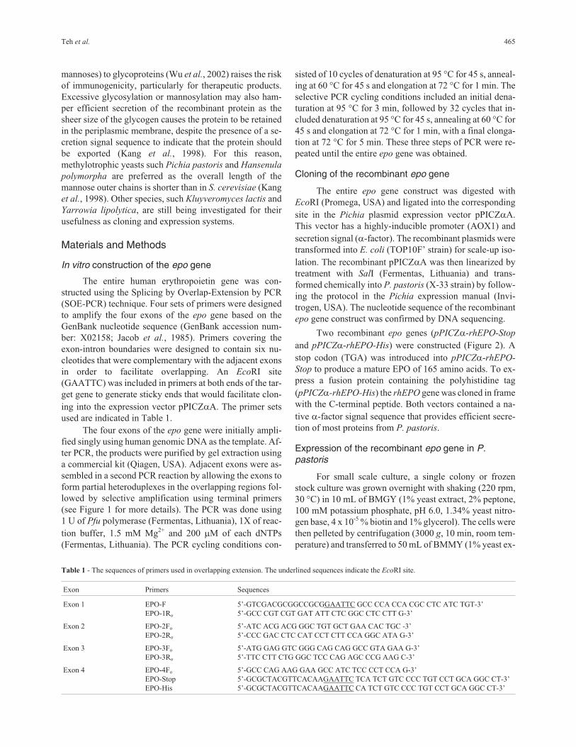
Table 1 - The sequences of primers used in overlapping extension. The underlined sequences indicate the EcoRI site.
Exon Primers Sequences
Exon 1 EPO-F
EPO-1Ro
5’-GTCGACGCGGCCGCGGAATTC GCC CCA CCA CGC CTC ATC TGT-3’
5’-GCC CGT CGT GAT ATT CTC GGC CTC CTT G-3’
Exon 2 EPO-2Fo
EPO-2Ro
5’-ATC ACG ACG GGC TGT GCT GAA CAC TGC -3’
5’-CCC GAC CTC CAT CCT CTT CCA GGC ATA G-3’
Exon 3 EPO-3Fo
EPO-3Ro
5’-ATG GAG GTC GGG CAG CAG GCC GTA GAA G-3’
5’-TTC CTT CTG GGC TCC CAG AGC CCG AAG C-3’
Exon 4 EPO-4Fo
EPO-Stop
EPO-His
5’-GCC CAG AAG GAA GCC ATC TCC CCT CCA G-3’
5’-GCGCTACGTTCACAAGAATTC TCA TCT GTC CCC TGT CCT GCA GGC CT-3’
5’-GCGCTACGTTCACAAGAATTC CA TCT GTC CCC TGT CCT GCA GGC CT-3’
tract, 2% peptone, 100 mM potassium phosphate, pH 6.0,
1.34% yeast nitrogen base, 4 x 10⁻⁵% biotin and 1% metha-
nol) in a 250 mL flask. The culture was incubated at 30 °C
with shaking (220 rpm) and at different time intervals the
supernatants were collected by centrifugation. The super-
natants were precipitated by adding three volumes of ace-
tone and incubated at -20 °C overnight. The mixtures were
then centrifuged (10,000 rpm, 4 °C) and the pellets then dis-
solved in phosphate-buffered saline (PBS).
SDS-PAGE and western blotting
SDS-PAGE was done in 12.5% polyacrylamide gels.
After electrophoresis, the gels were either stained with sil-
ver or the proteins were transferred to a nitrocellulose
membrane (Amersham Biosciences, USA) for western
blotting, as described in the standard protocol (Bio-Rad,
USA). The recombinant protein was detected using mono-
clonal anti-human EPO antibody (diluted 1:8000; R&D
Systems, USA) and a secondary anti-mouse antibody con-
jugated with alkaline phosphatase (diluted 1:6000; Sigma,
USA). The proteins were viewed with Western Blue® stabi-
lized substrate for alkaline phosphatase (Promega, USA).
Commercial recombinant EPO (cEPO) used as standard in
electrophoretic analysis was produced in CHO cells (kindly
provided by Duopharma Biotech Bhd., Malaysia).
Purification
rhEPO-His was concentrated and purified on a His
GraviTrap column containing precharged Ni Sepharose 6
Fast Flow (Amersham Biosciences, USA). After loading,
the column was washed with 20 mM imidazole and eluted
with 200 mM imidazole in 20 mM sodium phosphate,
pH 7.4, containing 500 mM NaCl.
Fractionation
rhEPO-Stop and rhEPO-His were fractionated in a
Mini Prep cell (Bio-Rad, USA) with a 10% resolving gel.
The apparatus was assembled according to the manufac-
turer’s instructions and electrophoresis was done at 200 V
(~3-6 mA) for 2-2.5 h. The eluted fractions (200-250 �L
each) were collected every 15 min. All of the fractions were
analyzed by SDS-PAGE and western blotting.
Deglycosylation
rhEPO-His and rhEPO-stop were digested with
PNGase F (New England Biolabs, UK) and Endo H (New
England Biolabs, UK), according to their manufacturer’s
instructions. Deglycosylated rhEPOs were analyzed by
western blotting. cEPO treated with these enzymes was
used as a positive control.
Assessment of hematopoietic activity in vivo
Female ICR mice (provided by the Animal House of
the University of Malaya) were used to test the hemato-
poietic activity of rhEPO produced in P. pastoris. Each
mouse was injected subcutaneously with 0.25 mL of sam-
ple (acetone-precipitated supernatant of rhEPO cultures)
for three consecutive days. Three mice (6-8 weeks old)
were used for each treatment and two groups of mice were
used as controls (one of these two groups was treated with
PBS and the other received no treatment). Blood samples
were collected into 5% sodium EDTA on the fourth day af-
ter treatment. An equal volume of blood was mixed with
new methylene blue and incubated at 37 °C for 1 h. Seven
microlitres of this blood-dye mixture was then used to pre-
pare smears on glass slides. Five slides were prepared for
each mouse (total of 15 slides per treatment since there
were three mice per group). Reticulocytes were counted
with the aid of a microscope (at 100X magnification) in five
randomly selected areas of each slide and their number ex-
pressed as a relative to the total number of red blood cells
observed.
466 Expression of recombinant erythropoietin in Pichia pastoris
Figure 1 - Construction of the epo gene via SOE-PCR.
Figure 2 - Constructs for erythropoietin production. A: pPICZ�-rhEPO-
Stop; B: pPICZ�-rhEPO-His (5’AOX1 - alcohol oxidase promoter; �-fac-
tor - secretion signal peptide; 6x His - C-terminal polyhistidine tag;
3’AOX1 - transcriptional termination sequence).
Statistical analysis
The results were expressed as the mean � SEM,
where appropriate. Statistical comparisons were done using
Students t-test, with a value of p < 0.01 indicating signifi-
cance.
Results and Discussion
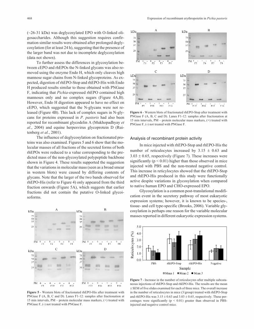
Expression and characterisation of rhEPO
Four exons (105, 99, 192 and 183 base pairs) of the
epo gene were successfully ligated via SOE-PCR to pro-
duce the full length gene of 543 base pairs (Figure 3), with
EcoRI sites incorporated at both terminals. SOE-PCR is a
simple, cost-effective tool for recombinant gene construc-
tion, especially for EPO which generally has a low expres-
sion in tissues other than the kidney. This procedure
circumvents the laborious work of mRNA extraction from
human kidney cells. The fidelity of PCR and subsequent
gene construction steps was confirmed by sequence analy-
sis, which revealed that the recombinant constructs had ex-
actly the same sequence as published by Jacob et al. (1985).
By using this approach, the overlapping region between
two adjacent exons could be minimized to 12 bp without
any reduction in splicing efficiency. This overlap was
shorter than those reported previously by using the over-
lap-extension method (Wurch et al., 2000; Davidson et al.,
2002; Ailenberg et al., 2005).
Both versions of recombinant EPO, i.e., with and
without a polyhistidine tag (rhEPO-His and rhEPO-Stop),
were successfully expressed, but at very low levels. A se-
ries of optimized culture conditions was used to achieve
higher yields (data not shown). In addition, by lowering
the culture temperature, a mixture of glycoisoforms was
produced that resulted in the appearance of a broad smear
in subsequent western blots (Figure 4, lanes 3 and 5). The
molecular mass of rhEPO-Stop ranged from 32-75 kDa
while that of rhEPO-His ranged from 37-75 kDa, all of
which were larger than that observed for cEPO
(~32-37 kDa). Native human EPO is highly glycosylated
(~32-37 kDa) and approximately 30% of its molecular
mass is attributed to the carbohydrate chains (Dordal et
al., 1985). Our results showed that glycosylation in rhEPO
expressed in P. pastoris accounted for ~30%-70% of the
total molecular mass. This phenomenon is not unusual
since hyperglycosylation or hypermannosylation is fre-
quent in yeast expression systems (Wu et al., 2002). Vari-
ous studies have shown that glycosylation in P. pastoris
generally results in heterologous recombinant proteins
that have a greater molecular mass than the corresponding
proteins produced in mammalian expression systems
(Braren et al., 2007). However, there are cases in which
the recombinant proteins produced in P. pastoris have a
lower molecular mass than those produced in mammalian
systems. Sadhukhan and Sen (1996) postulated that this
may reflect the absence of other post-translational modifi-
cation processes in yeast, e.g., phosphorylation, sulpha-
tion and sialylation. Examples of this would be the enve-
lope glycoproteins of classic swine fever virus (Erns)
which, when expressed in P. pastoris, has a lower molecu-
lar mass than expected (Huang et al., 2006).
To investigate the variation in glycosylation of the re-
combinant proteins, the two recombinant EPOs were trea-
ted with glycosidase (PNGase F) to release the N-linked
carbohydrates. Deglycosylation reduced the molecular
mass of both recombinant EPOs. As predicted from the
amino acid sequence, the molecular mass of the poly-
peptide backbone of cEPO and rhEPO-Stop was similar,
i.e., ~18 kDa (Figure 4A). For rhEPO-His, the predicted
molecular mass of the polypeptide backbone was ~23 kDa
because of the additional 44 amino acids in the C-terminal
of the protein that included the polyhistidine tag for purifi-
cation. Figure 4A also shows that two bands were observed
for commercial EPO, with the upper band corresponding to
the EPO protein with O-linked carbohydrate while the
lower band was the non-glycosylated EPO (Elliott et al.,
1994). Two bands were also observed for digested rhEPO-
His but the molecular masses differed when compared to
cEPO. We postulated that the lower band (23 kDa) was the
polypeptide backbone of rhEPO-His, while the upper band
Teh et al. 467
Figure 3 - Construction of the entire epo gene via SOE-PCR. Lanes 1-4:
Amplification of exons 1, 2, 3 and 4 (105, 99, 192 and 183 bp), respec-
tively; lane 5: Amplification of the entire epo gene (543 bp); the smaller
non-specific bands were eliminated by gel purification.
Figure 4 - Western blots of cEPO, rhEPO-His and rhEPO-Stop after treat-
ment with (A) PNGase F and (B) Endo H. Treatment with PNGase F or
Endo H is indicated with (+) and no treatment is indicated with (-). PM -
protein molecular mass markers.
(~26-31 kDa) was deglycosylated EPO with O-linked oli-
gosaccharides. Although this suggestion requires confir-
mation similar results were obtained after prolonged degly-
cosylation (for at least 24 h), suggesting that the presence of
the larger band was not due to incomplete deglycosylation
(data not shown).
To further assess the differences in glycosylation be-
tween cEPO and rhEPOs the N-linked glycans was also re-
moved using the enzyme Endo H, which only cleaves high
mannose sugar chains from N-linked glycoproteins. As ex-
pected, digestion of rhEPO-Stop and rhEPO-His with Endo
H produced results similar to those obtained with PNGase
F, indicating that Pichia-expressed rhEPO contained high
mannoses only and no complex sugars (Figure 4A,B).
However, Endo H digestion appeared to have no effect on
cEPO, which suggested that the N-glycans were not re-
leased (Figure 4B). This lack of complex sugars in N-gly-
cans for proteins expressed in P. pastoris had also been
reported for recombinant glycodelin A (Mukhopadhyay et
al., 2004) and equine herpesvirus glycoprotein D (Rui-
tenberg et al., 2001).
The influence of deglycosylation on fractionated pro-
teins was also examined. Figures 5 and 6 show that the mo-
lecular masses of all fractions of the secreted forms of both
rhEPOs were reduced to a value corresponding to the pre-
dicted mass of the non-glycosylated polypeptide backbone
shown in Figure 4. These results supported the suggestion
that the variations in molecular mass (seen as a broad smear
in western blots) were caused by differing contents of
glycans. Note that the larger of the two bands observed for
rhEPO-His (refer to Figure 4) only appeared from the third
fraction onwards (Figure 5A), which suggests that earlier
fractions did not contain the putative O-linked glycoi-
soforms.
Analysis of recombinant protein activity
In mice injected with rhEPO-Stop and rhEPO-His the
number of reticulocytes increased by 3.15 � 0.63 and
3.03 � 0.65, respectively (Figure 7). These increases were
significantly (p < 0.01) higher than those observed in mice
injected with PBS and the non-treated negative control.
This increase in reticylocytes showed that the rhEPO-Stop
and rhEPO-His produced in this study were functionally
active despite variations in glycosylation when compared
to native human EPO and CHO-expressed EPO.
Glycosylation is a common post-translational modifi-
cation event in the secretory pathway of most eukaryotic
expression systems; however, it is known to be species-,
tissue- and cell type-specific (Brooks, 2006). Variable gly-
cosylation is perhaps one reason for the variable molecular
masses reported in different eukaryotic expression systems.
468 Expression of recombinant erythropoietin in Pichia pastoris
Figure 5 - Western blots of fractionated rhEPO-His after treatment with
PNGase F (A, B, C and D). Lanes F1-12: samples after fractionation at
15 min intervals, PM – protein molecular mass markers, (+) treated with
PNGase F, (-) not treated with PNGase F.
Figure 6 - Western blots of fractionated rhEPO-Stop after treatment with
PNGase F (A, B, C and D). Lanes F1-12: samples after fractionation at
15 min intervals, PM – protein molecular mass markers, (+) treated with
PNGase F, (-) not treated with PNGase F.
Figure 7 - Increase in the number of reticulocytes after multiple subcuta-
neous injections of rhEPO-Stop and rhEPO-His. The results are the mean
� SEM of five slides examined for each of three mice. The overall increase
in the number of reticulocytes in mice (3/group) treated with rhEPO-Stop
and rhEPO-His was 3.15 � 0.63 and 3.03 � 0.65, respectively. These per-
centages were significantly (p < 0.01) greater than observed in PBS-
injected and negative control mice.

For example, recombinant EPO was ~35 kDa in CHO cells
(Lin et al., 1985), ~31 kDa in tobacco cells (Matsumoto et
al., 1995), ~25 kDa in Drosophila S2 cells (Kim et al.,
2005), > 29 kDa in S. cerevisiae (Elliott et al., 1989) and
~30 kDa in Physcomitrella patens (Weise et al., 2007),
while human urinary EPO (or native EPO) is reportedly
~34 kDa (Dordal et al., 1985). More recently, Celik et al.
(2007) reported that the molecular mass of rHuEPO was
30 kDa and that the major glycan attached to all three
N-linked glycosylation sites was Man17(GlcNAc)2. Curi-
ously, other than for recombinant EPO produced commer-
cially in CHO cells, none of the other studies mentioned
above commented on the possible correlation or associa-
tion between different rhEPO glycan profiles and the bioac-
tivity of the protein. This aspect deserves further investiga-
tion.
Perhaps the most important issue to be addressed is
whether proteins produced in yeast can be made to be
more like the corresponding proteins in humans. Both
EPO� and EPO�, which have different isoform composi-
tions, have been produced in mammalian cell systems
(Storring et al., 1998), and analyses of these EPOs have
shown that in this case differences in their glycosylation
patterns did not result in differences in immunogenicity
(Hermeling et al., 2003). Recombinant proteins produced
in P. pastoris tend to be hypermannosylated. Although
there are no detailed reports on the potential conse-
quence(s) of hypermannosylation in the immune response
of humans, hypermannosylation in Pichia-expressed re-
combinant proteins may be an important risk factor for in-
creased immunogenicity.
Hypermannosylation in P. pastoris is initiated by the
activity of �-1,6-mannosyltransferase (Och1p) (Dean,
1999). Inactivation of the OCH1 gene, which leads to the
elimination or minimization of N-linked glycosylation in P.
pastoris, has been considered an important step in the “hu-
manization” of these host cells (Bretthauer, 2003; Ver-
vecken et al., 2004). Other relevant “humanization” steps
include the introduction of a gene that confers �1,2-man-
nosidase activity (to remove �1,2-mannose residues) and
another gene (GlcNAc transferase I) that adds �1,2-linked
GlcNAc residues to the �1,3-mannose arm. Engineered P.
pastoris strains that can produce the so-called ‘hybrid-type’
N-glycan structures have been produced, with promising
outcomes and minimal effect on yeast viability (Bretthauer,
2003; Choi et al., 2003; Hamilton et al., 2003; Vervecken et
al., 2004). More recently, Hamilton et al. (2006) reported
extensive engineering of P. pastoris that also allowed the
production of complex terminally sialylated glycoproteins,
which is expected to increase the half-life and therapeutic
potency of most glycoproteins. The availability of such
yeast cell lines with `improved’ protein products will sig-
nificantly reduce the risk of problems associated with
immunogenicity. This property, together with a reduction
in the time and cost of production, may eliminate the need
to use mammalian cells in the future.
Acknowledgments
This study was sponsored by the University of Ma-
laya (Postgraduate Research Grant) and the Malaysia Toray
Science Foundation.
References
Ailenberg M, Goldenberg NM and Silverman M (2005) Descrip-
tion of a PCR-based technique for DNA splicing and muta-
genesis by producing 5’ overhangs with run through stop
DNA synthesis utilizing Ara-C. BMC Biotechnology 5:e23.
Böer E, Steinborn G, Kunze G and Gellissen G (2007) Yeast ex-
pression platforms. Appl Microbiol Biotechnol 77:513-523.
Braren I, Greunke K, Umland O, Deckers S, Bredehorst R and
Spillner E (2007) Comparative expression of different anti-
body formats in mammalian cells and Pichia pastoris.
Biotechnol Appl Biochem 47:205-214.
Bretthauer RK (2003) Genetic engineering of Pichia pastoris to
humanize N-glycosylation of proteins. Trends Biotechnol
21:459-462.
Brooks SA (2006) Protein glycosylation in diverse cell systems:
Implications for modification and analysis of recombinant
proteins. Expert Rev Proteomics 3:345-359.
Celik E, Calik P, Halloran SM and Oliver SG (2007) Production
of recombinant human erythropoietin from Pichia pastoris
and its structural analysis. J Appl Microbiol 103:2084-2094.
Choi BK, Bobrowicz P, Davidson RC, Hamilton SR, Kung DH,
Li H, Miele RG, Nett JH, Wildt S and Gerngross TU (2003)
Use of combinatorial genetic libraries to humanize N-linked
glycosylation in the yeast Pichia pastoris. Proc Natl Acad
Sci USA 100:5022-5027.
Davidson RC, Blankenship JR, Kraus PR, Jesus Berrios M, Hull
CM, D’Souza C, Wang P and Heitman J (2002) A PCR-
based strategy to generate integrative targeting alleles with
large regions of homology. Microbiology 148:2601-2615.
Dean N (1999) Asparagine-linked glycosylation in the yeast Gol-
gi. Biochim Biophys Acta 1426:309-322.
Dordal MS, Wang FF and Goldwasser E (1985) The role of carbo-
hydrate in erythropoietin action. Endocrinology 116:2293-
2299.
Elliott S, Giffin J, Suggs S, Lau EP and Banks AR (1989) Secre-
tion of glycosylated human erythropoietin from yeast di-
rected by the �-factor leader region. Gene 79:167-180.
Elliott S, Bartley T, Delorme E, Derby P, Hunt R, Lorenzini T,
Parker V, Rohde MF and Stoney K (1994) Structural re-
quirements for addition of O-linked carbohydrate to recom-
binant erythropoietin. Biochemistry 33:11237-11245.
Genc S, Koroglu TF and Genc K (2004) Erythropoietin and the
nervous system. Brain Res 1000:19-31.
Hamilton SR, Bobrowicz P, Bobrowicz B, Davidson RC, Li HJ,
Mitchell T, Nett JH, Rausch S, Stadheim TA, Wischnewski
H, et al. (2003) Production of complex human glycoproteins
in yeast. Science 301:1244-1246.
Hamilton SR, Davidson RC, Sethuraman N, Nett JH, Jiang Y,
Rios S, Bobrowicz P, Stadheim TA, Li H, Choi BK, et al.
(2006) Humanization of yeast to produce complex termi-
nally sialylated glycoproteins. Science 313:1441-1443.
Teh et al. 469
Hermeling S, Schellekens H, Crommelin DJA and Jiskoot W
(2003) Miscelle-associated protein in epoetin formulations:
A risk factor for immunogenicity. Pharm Res 20:1903-1907.
Huang C, Chien MS, Hu CM, Chen CW and Hsieh PC (2006) Se-
creted expression of the classical swine fever virus glyco-
protein Erns in yeast and application to a sandwich blocking
ELISA. J Virol Meth 132:40-47.
Jacob K, Shoemaker C, Rudersdorf R, Neill SD, Kaufman RJ,
Mufson A, Seehra J, Jones SS, Hewick R, Fitch EF, et al.
(1985) Isolation and characterization of genomic and cDNA
clones of human erythropoietin. Nature 313:806-810.
Kang HA, Sohn JH, Choi ES, Chung BH, Yu MH and Rhee SK
(1998) Glycosylation of human �1-antitrypsin in
Saccharomyces cerevisiae and methylotrophic yeasts. Yeast
14:371-381.
Kim YK, Shin HS, Tomiya N, Lee YC, Betenbaugh MJ and Cha
HJ (2005) Production and N-glycan analysis of secreted hu-
man erythropoietin glycoprotein in stably transfected
Drosophila S2 cells. Biotechnol Bioeng 92:452-461.
Lin FK, Suggs S, Lin CH, Browne JK, Smalling R, Egrie JC, Chen
KK, Fox GM, Martin F, Stabinsky Z, et al. (1985) Cloning
and expression of the human erythropoietin gene. Proc Natl
Acad Sci USA 82:7580-7584.
Matsumoto S, Ikura K, Ueda M and Sasaki R (1995) Characteriza-
tion of a human glycoprotein (erythropoietin) produced in
cultured tobacco cells. Plant Mol Biol 27:1163-1172.
Mukhopadhyay D, SundarRaj S, Alok A and Karande AA (2004)
Glycodelin A, not glycodelin S, is apoptotically active: Rel-
evance of sialic acid modification. J Biol Chem 279:8577-
8584.
Ruitenberg KM, Gilkerson JR, Wellington JE, Love DN and
Whalley JM (2001) Equine herpesvirus 1 glycoprotein D ex-
pressed in Pichia pastoris is hyperglycosylated and elicits a
protective immune response in the mouse model of EHV-1
disease. Virus Res 79:125-135.
Sadhukhan R and Sen I (1996) Different glycosylation require-
ments for the synthesis of enzymatically active angioten-
sin-converting enzyme in mammalian cells and yeast. J Biol
Chem 271:6429-6434.
Storring PL, Tiplady RJ, Gaines Das RE, Stenning BE, Lamikanra
A, Rafferty B and Lee J (1998) Epoetin alfa and beta differ
in their erythropoietin isoform compositions and biological
properties. Br J Haematol 100:79-89.
Vervecken W, Kaigorodov V, Callewaert N, Geysens S, Vusser
KD and Contreras R (2004) In vivo synthesis of mamma-
lian-like, hybrid-type N-glycans in Pichia pastoris. Appl
Environ Microbiol 70:2639-2646.
Weise A, Altmann F, Rodriguez-Franco M, Sjoberg ER, Baumer
W, Launhardt H, Kietzmann M and Gorr G (2007) High-
level expression of secreted complex glycosylated recombi-
nant human erythropoietin in the Physcomitrella �-fuc-t
�-xyl-t mutant. Plant Biotechnol J 5:389-401.
Wu JM, Lee CK and Hsu TA (2002) Asparagine-linked glyco-
sylational modifications in yeast. In: Al-Rubeai M (ed) Cell
Engineering: Glycosylation. Volume 3. Kluwer Academic
Publishers, New York, pp 215-232.
Wurch T, Lestienne F and Pauwels PJ (2000) Optimisation of
overlap extension PCR-based mutagenesis of a GC-rich
DNA template: Application to the human �2C-adrenoceptor
cDNA. Biotechnol Lett 22:1603-1609.
Zhang XC, Cheng GX, Chen JQ, Cheng Y, Wei YR, Yao WL, Xu
SF and Lao WD (2000) Expression of human erythropoietin
directed by mWAP promoter in mammary gland of trans-
genic mice. Chin Sci Bull 45:226-228.
Associate Editor: Carlos F.M. Menck
License information: This is an open-access article distributed under the terms of theCreative Commons Attribution License, which permits unrestricted use, distribution, andreproduction in any medium, provided the original work is properly cited.
470 Expression of recombinant erythropoietin in Pichia pastoris