Multiple Roles of Biosurfactants in Structural Biofilm Development by Pseudomonas aeruginosa
10
Published Ahead of Print 12 January 2007. 2007, 189(6):2531. DOI: 10.1128/JB.01515-06. J. Bacteriol. Sünje Johanna Pamp and Tim Tolker-Nielsen Pseudomonas aeruginosa Structural Biofilm Development by Multiple Roles of Biosurfactants in http://jb.asm.org/content/189/6/2531 Updated information and services can be found at: These include: REFERENCES http://jb.asm.org/content/189/6/2531#ref-list-1 at: This article cites 55 articles, 30 of which can be accessed free CONTENT ALERTS more» articles cite this article), Receive: RSS Feeds, eTOCs, free email alerts (when new http://journals.asm.org/site/misc/reprints.xhtml Information about commercial reprint orders: http://journals.asm.org/site/subscriptions/ To subscribe to to another ASM Journal go to: on February 21, 2014 by guest http://jb.asm.org/ Downloaded from on February 21, 2014 by guest http://jb.asm.org/ Downloaded from
Transcript of Multiple Roles of Biosurfactants in Structural Biofilm Development by Pseudomonas aeruginosa

Published Ahead of Print 12 January 2007. 2007, 189(6):2531. DOI: 10.1128/JB.01515-06. J. Bacteriol.
Sünje Johanna Pamp and Tim Tolker-Nielsen Pseudomonas aeruginosaStructural Biofilm Development by Multiple Roles of Biosurfactants in
http://jb.asm.org/content/189/6/2531Updated information and services can be found at:
These include:
REFERENCEShttp://jb.asm.org/content/189/6/2531#ref-list-1at:
This article cites 55 articles, 30 of which can be accessed free
CONTENT ALERTS more»articles cite this article),
Receive: RSS Feeds, eTOCs, free email alerts (when new
http://journals.asm.org/site/misc/reprints.xhtmlInformation about commercial reprint orders: http://journals.asm.org/site/subscriptions/To subscribe to to another ASM Journal go to:
on February 21, 2014 by guest
http://jb.asm.org/
Dow
nloaded from
on February 21, 2014 by guest
http://jb.asm.org/
Dow
nloaded from
JOURNAL OF BACTERIOLOGY, Mar. 2007, p. 2531–2539 Vol. 189, No. 60021-9193/07/$08.00�0 doi:10.1128/JB.01515-06Copyright © 2007, American Society for Microbiology. All Rights Reserved.
Multiple Roles of Biosurfactants in Structural BiofilmDevelopment by Pseudomonas aeruginosa�
Sunje Johanna Pamp and Tim Tolker-Nielsen*Centre for BioScience Technology, BioCentrum-DTU, The Technical University of Denmark, DK-2800 Lyngby, Denmark
Received 28 September 2006/Accepted 29 December 2006
Recent studies have indicated that biosurfactants produced by Pseudomonas aeruginosa play a role both inmaintaining channels between multicellular structures in biofilms and in dispersal of cells from biofilms.Through the use of flow cell technology and enhanced confocal laser scanning microscopy, we have obtainedresults which suggest that the biosurfactants produced by P. aeruginosa play additional roles in structuralbiofilm development. We present genetic evidence that during biofilm development by P. aeruginosa, biosur-factants promote microcolony formation in the initial phase and facilitate migration-dependent structuraldevelopment in the later phase. P. aeruginosa rhlA mutants, deficient in synthesis of biosurfactants, were notcapable of forming microcolonies in the initial phase of biofilm formation. Experiments involving two-color-coded mixed-strain biofilms showed that P. aeruginosa rhlA mutants were defective in migration-dependentdevelopment of mushroom-shaped multicellular structures in the later phase of biofilm formation. Experi-ments involving three-color-coded mixed-strain P. aeruginosa biofilms demonstrated that the wild-type andrhlA and pilA mutant strains formed distinct subpopulations on top of each other dependent on their abilityto migrate and produce biosurfactants.
Evidence is accumulating that many infections display ele-vated tolerance towards antimicrobial attack because the caus-ative bacteria reside in biofilms (8, 15, 51). The opportunistichuman pathogen Pseudomonas aeruginosa has become a modelorganism for studying biofilm development. When P. aerugi-nosa is grown as a biofilm in flow chambers, it often developsmushroom-shaped multicellular structures separated by liquid-filled channels (9, 26, 32). The cap-forming subpopulation andthe stalk-forming subpopulation of these mushroom-shapedstructures in many cases display differential tolerance to anti-microbial compounds. For example, the antibiotic tobramycinwas shown to kill preferentially bacteria in the cap portion ofthe mushroom-shaped structures, whereas the antibiotic colis-tin, the detergent sodium dodecyl sulfate, and the chelatorEDTA were shown to kill preferentially bacteria in the stalkportion of the mushroom-shaped structures (3, 5, 19, 21).Knowledge about subpopulation development in biofilms, andthe way the subpopulations interact and change during struc-tural biofilm development, may be useful for creating strategiesto control biofilm formation and eradicate persistent infec-tions.
Studies of liquid flow and molecular diffusion in flow cham-ber-grown biofilms have led to the proposal that the channelsand interstitial voids between the microcolonies may functionas a circulation system for efficient nutrient supply and wasteproduct removal (11, 52). Biosurfactants produced by P.aeruginosa via the RhlA gene product were shown to be im-portant for maintenance of the water channels between themushroom-shaped multicellular structures in biofilms (10). A
study which employed fluorescent reporter genes indicatedthat biosurfactant synthesis preferentially takes place in mi-crocolonies during early stages of biofilm development andin the stalk portion of the mushroom-shaped structures inmore mature P. aeruginosa biofilms (33). In addition, evi-dence has been presented that the biosurfactants producedby P. aeruginosa are involved in the dispersion of cells frombiofilms (5, 24, 47).
P. aeruginosa produces a number of biosurfactants, of whichthe three most abundant are 3-(3-hydroxyalkanoyloxy)alcanoicacid (HAA), L-rhamnosyl-3-hydroxydecanoyl-3-hydroxydecanoate(mono-rhamnolipid), and L-rhamnosyl-L-rhamnosyl-3-hydroxyde-canoyl-3-hydroxydecanoate (di-rhamnolipid) (13, 14, 44, 46).HAA is synthesized via the RhlA enzyme and is converted tomono-rhamnolipid by the RhlB enzyme (14, 39). Mono-rham-nolipid is converted to di-rhamnolipid by the RhlC enzyme(44). The rlhAB operon and rhlC gene are induced by acylhomoserine lactone-activated RhlR and are thus under quo-rum-sensing control (39–42, 44). In addition, phosphate limi-tation and the presence of nitrate have been shown to promotethe synthesis of rhamnolipids, while ammonium and highamounts of iron have been shown to repress the production ofrhamnolipids (18, 37, 38).
The formation of the mushroom-shaped multicellularstructures in P. aeruginosa biofilms under some conditionsoccurs in a sequential process which involves a nonmotilebacterial subpopulation that forms the mushroom stalks byclonal cell proliferation in certain foci on the substratumand a migrating bacterial subpopulation that forms themushroom caps via a process which requires type IV pili(26). Type IV pili have been implicated in two kinds ofsurface-associated motility in P. aeruginosa. One form ofsurface-associated translocation of P. aeruginosa which re-quires functional flagella and biosurfactant production, andunder some conditions type IV pili, has been termed swarm-
* Corresponding author. Mailing address: BioCentrum-DTU, Build-ing 301, The Technical University of Denmark, DK-2800 Lyngby,Denmark. Phone: 45 45 25 27 93. Fax: 45 45 88 73 28. E-mail: [email protected].
� Published ahead of print on 12 January 2007.
2531
on February 21, 2014 by guest
http://jb.asm.org/
Dow
nloaded from

ing motility (29, 45, 49). Another form of surface-associatedtranslocation of P. aeruginosa, which requires type IV pili,has been termed twitching motility (20, 34, 48). Presently, itis not known whether the type IV pili-driven motility thatplays a role in mushroom cap formation in P. aeruginosabiofilms should be regarded as twitching, swarming, or an-other kind of surface-associated motility.
Because surface-associated motility and biosurfactant pro-duction evidently both have roles in structural biofilm forma-tion by P. aeruginosa, we found it of interest to investigatewhether the biosurfactants produced by P. aeruginosa mightplay a role in structural biofilm development by facilitatingsurface-associated motility. We present evidence that migra-tion-dependent formation of the cap portion of the mushroom-shaped structures in P. aeruginosa biofilms is facilitated bybiosurfactant production. In addition, we present evidence thatbiosurfactant production is necessary for initial microcolonyformation in P. aeruginosa biofilms.
MATERIALS AND METHODS
Bacterial strains, plasmids, and growth conditions. The bacterial strains andplasmids used in this study are listed in Table 1. Escherichia coli strains werecultured in Luria broth (LB) medium at 37°C. P. aeruginosa strains were culturedin LB medium at 37°C during the procedure of genetic manipulation. In motilityplate assays, orcinol assays, and for cultivation of biofilms, AB minimal mediumsupplemented with glucose as indicated was used. AB medium consists of(NH4)2SO4 (15.1 mM), Na2HPO4 � 2H2O (33.7 mM), KH2PO4 (22.0 mM), NaCl(0.051 M), MgCl2 (1 mM), CaCl2 (0.1 mM), and trace metals (100 �l/liter). Thetrace metal solution contained CaSO4 � 2H2O (200 mg/liter), FeSO4 � 7H2O(200 mg/liter), MnSO4 � H2O (20 mg/liter), CuSO4 � 5H2O (20 mg/liter),ZnSO4 � 7H2O (20 mg/liter), CoSO4 � 7H2O (10 mg/liter), NaMoO4 � H2O, andH3BO3 (5 mg/liter). Antimicrobial agents were used where appropriate at thefollowing concentrations: for E. coli, ampicillin (Vepidan ApS, Denmark) at 100�g/ml; for P. aeruginosa, gentamicin sulfate (Biochrome AG, Germany) at 30�g/ml, streptomycin sulfate (Sigma) at 300 �g/ml, potassium tellurite (Sigma) at150 �g/ml, and carbenicillin (Sigma) at 200 �g/ml. The P. aeruginosa PAO1 (23)strain from John Matticks’ laboratory and an isogenic pilA::Telr derivative (27)were used in this study. The rhlA mutant strain was obtained by transferring themutated rhlA gene from P. aeruginosa PAO1 rhlA::Gmr (44) (kindly provided byG. A. O’Toole) into P. aeruginosa PAO1, using the transducing phage E79tv2
TABLE 1. Strains, plasmids, and primers used in this study
Strain, plasmid, or primer Relevant characteristics or sequence Source and/or reference
P. aeruginosa strainsPAO1 Wild type 23; obtained from J. S.
Mattick, University ofQueensland Brisbane
PAO1 Gfp PAO1 tagged with EGFP in a mini-Tn7 construct; Gmr 27PAO1 Yfp PAO1 tagged with EYFP in a mini-Tn7 construct; Gmr 27PAO1 rhlA rhlA::Gmr 44; kindly provided by
G. O’Toole, DartmouthMedical School
PAO1 rhlA Yfp rhlA inactivated by transferring rhlA::Gmr from strain PAO1 rhlA viatransduction into PAO1 wild type; tagged with EYFP in a mini-Tn7construct; Gmr Smr
This study
PAO1 rhlA Cfp rhlA inactivated by transferring rhlA::Gmr from strain PAO1 rhlA viatransduction into PAO1 wild type; tagged with ECFP in a mini-Tn7construct; Gmr Smr
This study
PAO1 pilA pilA inactivated by allelic displacement with a tellurite resistance cassetteusing pTTN80; Telr
27
PAO1 pilA Cfp PAO1 pilA tagged with CFP; Telr Smr This studyPAO1 pilArhlA Cfp pilA inactivated by allelic displacement in strain PAO1 rhlA CFP using
pTTN80; Telr Gmr SmrThis study
E. coli HB101 recA thi pro leu hsdRM; Smr; strain used for maintenance andproliferation of pTTN80 and the plasmids used for fluorescent tagging
25
PlasmidspTTN80 pCK318 with pilA::Telr cassette cloned in the NotI site; Apr Telr; used for
allelic displacement27
pUX-BF13 mob� ori-R6K; Apr; helper plasmid providing the Tn7 transpositionfunctions in trans
4
pBK-mini-Tn7(Km,Sm)PA1/04/03-eyfp-a
mob�; Apr Smr Kmr; delivery plasmid for mini-Tn7(Km, Sm)PA1/04/03-eyfp 31
pBK-mini-Tn7(Km,Sm)PA1/04/03-ecfp-a
mob�; Apr Smr Kmr; delivery plasmid for mini-Tn7(Km, Sm)PA1/04/03-ecfp 31
pEX1.8 pEX1 carrying ori (P. aeruginosa) as 1.8-kb PstI fragment from pRO1614in StyI site
41
pEX1.8-rhlAB pEX1.8 carrying rhlAB including the rhlA promoter 5
PrimersTn7-GlmS 5�-AATCTGGCCAAGTCGGTGAC-3�Tn7-R109 5�-CAGCATAACTGGACTGATTTCAG-3�rhlA-fw 5�-CCGCTGAGTTACTTGTCTGC-3�rhlA-rev 5�-TGCGTTATGCAACCGCAAAG-3�pilA-fw 5�-GAAGTACGCGGTCACCTG-3�pilA-rev 5�-CGGAGATGCCTACAAAGAGC-3�
2532 PAMP AND TOLKER-NIELSEN J. BACTERIOL.
on February 21, 2014 by guest
http://jb.asm.org/
Dow
nloaded from

(36). The P. aeruginosa PAO1 pilA rhlA double mutant strain was derived fromthe rhlA mutant by allelic displacement of pilA with pilA::Telr using the knockoutplasmid pTTN80. The strains were fluorescently tagged at an intergenic neutralchromosomal locus downstream of the glmS gene with ecfp, eyfp, or egfp inmini-Tn7 constructs as described previously (31). Plasmids were transformedinto P. aeruginosa strains using electroporation (25 �F, 200 �, �5 ms, 2.5 kV).
Rhamnolipid assay. The concentration of rhamnolipids in culture superna-tants was determined by the orcinol method as previously described (5, 28, 47),with modifications. Briefly, P. aeruginosa strains were grown at 30°C for 4 days inAB minimal medium supplemented with 10 mM glucose. A 0.5-ml aliquot ofculture supernatant was extracted twice with 2 volumes of diethyl ether (high-performance liquid chromatography grade; Sigma). The ether fractions werepooled, evaporated to dryness, and reconstituted in 0.25 ml distilled H2O. A100-�l aliquot of each sample was diluted 1:10 in orcinol reagent and heated at80°C for 30 min. Orcinol reagent was prepared immediately prior to use andconsisted of 7.5 volumes of 60% (vol/vol) sulfuric acid and 1 volume of 1.6%(wt/vol) orcinol in distilled water. After heating, the samples were allowed to coolat room temperature for 15 min, and absorbance (A421) was measured andcompared with rhamnose standards. The concentration of rhamnolipids wasdetermined by the relation that 1.0 mg of rhamnose corresponds to 2.5 mg ofrhamnolipid (41).
Twitching motility plate assay. Twitching motility was assayed on plates com-posed of AB minimal medium supplemented with 10 mM glucose and solidifiedwith 1.0% agar. The plates were dried overnight at room temperature and pointinoculated through the agar surface with 2.5-�l aliquots taken from overnightcultures of P. aeruginosa. Overnight cultures of P. aeruginosa were grown in ABminimal medium, supplemented with 30 mM glucose, at 30°C under vigorousshaking. All plates were incubated at 30°C for 48 h. Twitching motility wasvisualized by staining with Coomassie brilliant blue R250 (Sigma) as described bySemmler et al. (48). To complement the twitching motility-deficient phenotypeof the rhlA mutant, 0.0005% Tween 20 was added to the agar medium whereindicated. Twitching motility of P. aeruginosa rhlA containing either pEX1.8 orpEX1.8-rhlAB was examined in the presence of carbenicillin.
Cultivation of biofilms. Biofilms were cultivated in three-channel flow cellswith individual channel dimensions of 1 by 4 by 40 mm and covered with a glasscoverslip serving as substratum for biofilm formation. The flow system wasassembled and prepared as described elsewhere (50). AB minimal mediumsupplemented with 0.3 mM glucose as carbon source was used as growth me-dium. Individual flow chambers were inoculated with 300-�l aliquots taken fromovernight cultures of P. aeruginosa, adjusted to an optical density at 500 nm of0.005. Overnight cultures of P. aeruginosa strains were grown in AB minimalmedium supplemented with 30 mM glucose at 30°C under vigorous shaking. P.aeruginosa rhlA strains containing either pEX1.8 or pEX1.8-rhlAB were grown asovernight cultures in the presence of carbenicillin. After inoculation, flow cham-bers were left without flow for 1 h to allow bacterial attachment. The flow systemwas incubated at 30°C, and a laminar flow with a mean flow velocity of 0.2 mms�1 was achieved using a Watson Marlow 205S peristaltic pump. P. aeruginosarhlA strains harboring either pEX1.8 or pEX1.8-rhlAB were cultivated as biofilmswithout supplementation of carbenicillin. Loss of plasmid during 4 days ofgrowth in biofilms was not observed as examined by plating of cells derived frombiofilms on LB agar medium with and without carbenicillin.
Microscopy and image processing. Image acquisition was performed by theuse of a Zeiss LSM 510 confocal microscope (Carl Zeiss, Jena, Germany)equipped with an argon laser and with detectors and filter sets for simultaneousmonitoring of cyan fluorescent protein (CFP) and yellow fluorescent protein(YFP). Spectral imaging of mixed three-color-coded biofilms (CFP, green fluo-rescent protein [GFP], and YFP) was carried out using the Zeiss META module.Lambda stacks within a spectral range from 453.5 to 549.8 nm were recordedusing laser excitation at 458, 488, and 514 nm. Reference emission spectra fromsingle-color-coded biofilms were used for linear unmixing of the mixed color-coded biofilms. Images were obtained using a 40�/1.3 Plan-Neofluar oil objec-tive. Simulated three-dimensional images, shadow projections, and vertical cross-sections were generated using the Imaris software package (Bitplane AG).
RESULTS
In order to examine the effect of biosurfactant production onbiofilm development by P. aeruginosa, we constructed isogenicP. aeruginosa strains with and without the capability to producebiosurfactants. We inactivated the rhlA gene in our P. aerugi-nosa PAO1 strain by the use of transduction with phages that
had been propagated on a P. aeruginosa rhlA::Gmr strain. In-sertion of the rhlA::Gmr cassette in our PAO1 strain was con-firmed by PCR, and deficiency of biosurfactant production wasconfirmed by a drop-collapsing assay (data not shown). Inaddition, an orcinol assay showed that the wild-type PAO1produced 50.9 �g rhamnolipid/ml (n � 3; standard deviation[SD], 7.1) when it was grown in the medium used to cultivatebiofilms, compared to 0.58 �g rhamnolipid/ml (n � 3; SD, 1.1)measured in cultures of the rhlA mutant. As biosurfactantshave been described to facilitate swarming motility (25), weexamined the ability of the PAO1 rhlA mutant and wild type toswarm on semisolid glucose minimal medium and found thatthe rhlA mutant was deficient in swarming, as expected (datanot shown). Subsequently, we tested the ability of the rhlAmutant to perform twitching motility and found that the rate ofexpansion at the interstitial phase between a plastic surfaceand agar was significantly reduced for the rhlA mutant com-pared to the wild type (Fig. 1A and Table 2), suggesting thatbiosurfactants can also facilitate twitching motility. The P.aeruginosa pilA mutant (which does not produce type IV pili)was included as a nontwitching control (Fig. 1A). The twitch-ing defect of the rhlA mutant was complemented in P. aerugi-nosa PAO1 rhlA(pEX1.8-rhlAB), which contains a plasmid-
FIG. 1. Twitching motility plate assays. Glucose minimal mediumagar plates were point inoculated with P. aeruginosa PAO1 strains andincubated at 30°C for 48 h. The twitching zones were visualized bystaining with Coomassie brilliant blue after incubation. (A) Twitchingzones of P. aeruginosa PAO1 Yfp wild type, P. aeruginosa PAO1pilA::Telr (Cfp), and P. aeruginosa PAO1 rhlA::Gmr Yfp. (B) Twitchingzones of P. aeruginosa PAO1 rhlA::Gmr Yfp harboring either thevector control plasmid pEX1.8 or the plasmid pEX1.8-rhlAB express-ing rhlAB in trans. (C) Twitching zones of P. aeruginosa PAO1 Yfp wildtype and P. aeruginosa PAO1 rhlA::Gmr Yfp on medium containing0.0005% Tween 20.
VOL. 189, 2007 ROLES OF BIOSURFACTANTS IN P. AERUGINOSA BIOFILMS 2533
on February 21, 2014 by guest
http://jb.asm.org/
Dow
nloaded from

borne rhlAB operon (Fig. 1B and Table 2). To furthersubstantiate that surfactants can facilitate twitching motility,we investigated if the presence of the surfactant Tween 20could restore twitching motility of the rhlA mutant in the plateassay. In the presence of very low concentrations of Tween 20,the rhlA mutant was able to migrate by twitching motility at theplastic-agar interstitial surface to the same extent as the wildtype (with or without Tween 20) (Fig. 1C and Table 2).
We have previously found that when P. aeruginosa wasgrown in flow chambers with glucose as carbon source, thebacteria differentiated into a nonmotile and a motile subpopu-lation and formed mushroom-shaped multicellular structureswith the nonmotile subpopulation in the stalk portion and themotile subpopulation eventually settling in the cap portion
(26). When a biofilm was initiated with a mixture of P. aerugi-nosa wild type and P. aeruginosa pilA mutant, the nonmotilepilA mutant cells formed mushroom stalks only, whereas themotile wild-type bacteria were able to form mushroom caps ontop of the mushroom stalks formed by the pilA mutants (26).To address whether biosurfactants may facilitate cap formationby the motile subpopulation in P. aeruginosa biofilms, we in-oculated glucose minimal medium-perfused flow chamberswith 1:1 mixtures of CFP-tagged pilA mutant and either YFP-tagged wild type or YFP-tagged rhlA mutant and investigatedstructural biofilm development in the flow chambers by the useof confocal laser scanning microscopy (CLSM). After 4 days,the wild type had formed cap-shaped structures on top of thestalk-shaped structures formed by the pilA mutant (Fig. 2A), ashas been shown previously by Klausen et al. (26). By day 4 therhlA mutant also had formed cap-shaped structures on top ofthe stalk-forming pilA mutant, but the caps formed by the rhlAmutant were significantly smaller than those formed by thewild type (Fig. 2). The defect of the rhlA mutant in cap struc-ture formation could be complemented by introducing theplasmid pEX1.8-rhlAB, expressing the rhlAB operon in trans.The rhlA(pEX1.8-rhlAB) strain formed caps on top of thestalk-forming pilA mutant which were similar to those formedby the wild type, whereas the rhlA(pEX1.8) strain formed capssimilar to those formed by the rhlA mutant (Fig. 2). Analysis of50 randomly chosen mushroom-shaped structures in each of
TABLE 2. Quantification of twitching motility
P. aeruginosa strain and supplementationMediandiama
(cm) SD
P. aeruginosa PAO1 Yfp .........................................................3.25 0.12P. aeruginosa PAO1 rhlA Yfp.................................................1.60 0.11P. aeruginosa PAO1 rhlA Yfp pEX1.8 ..................................1.60 0.21P. aeruginosa PAO1 rhlA Yfp pEX1.8-rhlAB .......................2.85 0.16P. aeruginosa PAO1 Yfp � 0.0005% Tween 20 ..................3.30 0.19P. aeruginosa PAO1 rhlA Yfp � 0.0005% Tween 20..........3.15 0.21
a Based on 10 replicates for each strain/supplement combination.
FIG. 2. Confocal laser scanning micrographs of 4-day-old biofilms, initiated with a 1:1 mixture of either P. aeruginosa pilA::Telr Cfp and P.aeruginosa Yfp wild type (A), P. aeruginosa pilA::Telr Cfp and P. aeruginosa PAO1 rhlA::Gmr Yfp (B), P. aeruginosa pilA::Telr Cfp and P. aeruginosaPAO1 rhlA::Gmr Yfp(pEX1.8-rhlAB) (C), or P. aeruginosa pilA::Telr Cfp and P. aeruginosa PAO1 rhlA::Gmr Yfp(pEX1.8) (D). The images showtop-down shadow projections (230 �m by 230 �m) with two flanking images representing sections in the x-z and the y-z planes. Bar, 40 �m.
2534 PAMP AND TOLKER-NIELSEN J. BACTERIOL.
on February 21, 2014 by guest
http://jb.asm.org/
Dow
nloaded from
the 4-day-old mixed-strain biofilms showed that the wild typeformed caps with an average height of 65.34 �m (SD, 9.80),while the rhlA mutant formed caps with an average height of31.30 �m (SD, 5.99). The complemented strain rhlA(pEX1.8-rhlAB) formed caps with an average height of 65.71 �m (SD,10.37), whereas the vector control strain rhlA(pEX1.8) formedcaps with an average height of 27.02 �m (SD, 5.28). The experi-ment therefore suggested that biosurfactants produced by P.aeruginosa have a role in facilitating mushroom cap formation.
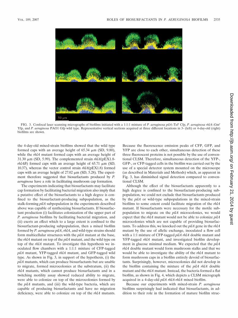
The experiments indicating that biosurfactants may facilitatecap formation by facilitating bacterial migration also imply thata putative effect of the biosurfactants to a high degree is con-fined to the biosurfactant-producing subpopulation, as thestalk-forming pilA subpopulation in the experiments describedabove was capable of synthesizing biosurfactants. If biosurfac-tant production (i) facilitates colonization of the upper part ofP. aeruginosa biofilms by facilitating bacterial migration, and(ii) exerts an effect which to a large extent is confined to thebiosurfactant-producing subpopulation, then a mixed biofilmformed by P. aeruginosa pilA, rhlA, and wild-type strains shouldform multicellular structures with the pilA mutant at the base,the rhlA mutant on top of the pilA mutant, and the wild type ontop of the rhlA mutant. To investigate this hypothesis we in-oculated flow chambers with a 1:1:1 mixture of CFP-taggedpilA mutant, YFP-tagged rhlA mutant, and GFP-tagged wildtype. As shown in Fig. 3, in support of the hypothesis, (i) thepilA mutants, which can produce biosurfactants but are unableto migrate, formed microcolonies at the substratum, (ii) therhlA mutants, which cannot produce biosurfactants and in atwitching motility assay showed reduced ability to migrate,were able to colonize on top of the microcolonies formed bythe pilA mutants, and (iii) the wild-type bacteria, which arecapable of producing biosurfactants and have no migrationdeficiency, were able to colonize on top of the rhlA mutants.
Because the fluorescence emission peaks of CFP, GFP, andYFP are close to each other, simultaneous detection of thesethree fluorescent proteins is not possible by the use of conven-tional CLSM. Therefore, simultaneous detection of the YFP-,GFP-, or CFP-tagged cells in the biofilm was carried out by theuse of a special detector system mounted on the microscope(as described in Materials and Methods) which, as apparent inFig. 3, has diminished signal detection compared to conven-tional CLSM.
Although the effect of the biosurfactants apparently to ahigh degree is confined to the biosurfactant-producing sub-population, we could not exclude that biosurfactants producedby the pilA or wild-type subpopulations in the mixed-strainbiofilms to some extent could facilitate migration of the rhlAmutant. If biosurfactants were necessary for the motile sub-population to migrate on the pilA microcolonies, we wouldexpect that the rhlA mutant would not be able to colonize pilAmicrocolonies which are not capable of providing biosurfac-tants. To address this, we knocked out the pilA gene in the rhlAmutant by the use of allelic exchange, inoculated a flow cellwith a 1:1 mixture of CFP-tagged pilA rhlA double mutant andYFP-tagged rhlA mutant, and investigated biofilm develop-ment in glucose minimal medium. We expected that the pilArhlA double mutant would form mushroom stalks and that wewould be able to investigate the ability of the rhlA mutant toform mushroom caps in a biofilm entirely devoid of biosurfac-tants. Surprisingly, however, microcolonies did not develop inthe biofilm containing the mixture of the pilA rhlA doublemutant and the rhlA mutant. Instead, the bacteria formed a flatbiofilm, as shown in Fig. 4, which depicts a CLSM micrographacquired in a 4-day-old pilA rhlA-rhlA mixed biofilm.
Because our experiments with mixed-strain P. aeruginosabiofilms surprisingly had indicated that biosurfactants, in ad-dition to their role in the formation of mature biofilm struc-
FIG. 3. Confocal laser scanning micrographs of biofilms initiated with a 1:1:1 mixture of P. aeruginosa pilA::Telr Cfp, P. aeruginosa rhlA::Gmr
Yfp, and P. aeruginosa PAO1 Gfp wild type. Representative vertical sections acquired at three different locations in 3- (left) or 4-day-old (right)biofilms are shown.
VOL. 189, 2007 ROLES OF BIOSURFACTANTS IN P. AERUGINOSA BIOFILMS 2535
on February 21, 2014 by guest
http://jb.asm.org/
Dow
nloaded from

tures, might also play a role in the formation of the initialmicrocolonies, we found it of interest to examine the role ofbiosurfactant production on biofilm development in more sim-ple model systems consisting of mono-strain P. aeruginosa bio-
films. While the wild type and the pilA mutant after 4 days ofdevelopment had formed a biofilm with mushroom-shapedstructures (Fig. 5A) and irregular protruding structures (Fig.5C), respectively, the rhlA mutant and the pilA rhlA doublemutant both formed flat biofilms (Fig. 5B and D), supportingthe hypothesis that biosurfactants are necessary for initialmicrocolony formation.
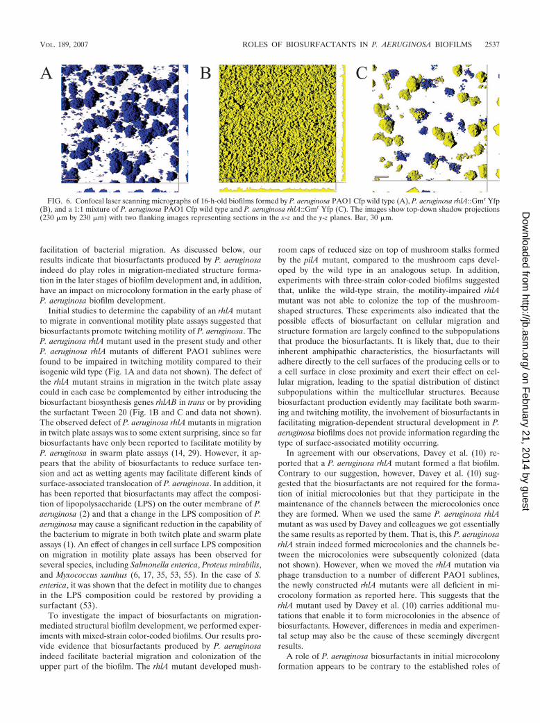
To examine further the role of biosurfactants in initial mi-crocolony formation, we examined young P. aeruginosa bio-films. After 16 h of development in glucose minimal medium-perfused flow chambers, the wild type had formed smallmicrocolonies (Fig. 6A), whereas the rhlA mutant had formeda flat thin biofilm (Fig. 6B). When the rhlA mutant and the wildtype were present in the flow chamber in a 1:1 mixture, the rhlAmutant was able to form initial microcolonies of the same sizeas those formed by the wild type (Fig. 6C), suggesting thatbiosurfactant produced by the wild type enabled microcolonyformation by the rhlA mutant.
DISCUSSION
Because previous studies had indicated that surface-associ-ated motility and biosurfactant production both play roles instructure formation in P. aeruginosa biofilms (10, 26), we foundit of interest to investigate if the role of biosurfactants instructural P. aeruginosa biofilm formation can be attributed to
FIG. 4. Confocal laser scanning micrograph of a 4-day-old biofilm,initiated with a 1:1 mixture of P. aeruginosa rhlA::Gmr Yfp and P.aeruginosa pilA::Telr rhlA::Gmr Cfp. The image shows a top-downshadow projection (230 �m by 230 �m) with two flanking imagesrepresenting sections in the x-z and the y-z planes. Bar, 20 �m.
FIG. 5. Confocal laser scanning micrographs of 4-day-old biofilms formed by P. aeruginosa PAO1 Yfp wild type (A), P. aeruginosa rhlA::Gmr
Yfp (B), P. aeruginosa pilA::Telr Cfp (C), and P. aeruginosa pilA::Telr rhlA::Gmr Cfp (D). The images show top-down shadow projections (230 �mby 230 �m) with two flanking images representing sections in the x-z and the y-z planes. Bar, 40 �m.
2536 PAMP AND TOLKER-NIELSEN J. BACTERIOL.
on February 21, 2014 by guest
http://jb.asm.org/
Dow
nloaded from
facilitation of bacterial migration. As discussed below, ourresults indicate that biosurfactants produced by P. aeruginosaindeed do play roles in migration-mediated structure forma-tion in the later stages of biofilm development and, in addition,have an impact on microcolony formation in the early phase ofP. aeruginosa biofilm development.
Initial studies to determine the capability of an rhlA mutantto migrate in conventional motility plate assays suggested thatbiosurfactants promote twitching motility of P. aeruginosa. TheP. aeruginosa rhlA mutant used in the present study and otherP. aeruginosa rhlA mutants of different PAO1 sublines werefound to be impaired in twitching motility compared to theirisogenic wild type (Fig. 1A and data not shown). The defect ofthe rhlA mutant strains in migration in the twitch plate assaycould in each case be complemented by either introducing thebiosurfactant biosynthesis genes rhlAB in trans or by providingthe surfactant Tween 20 (Fig. 1B and C and data not shown).The observed defect of P. aeruginosa rhlA mutants in migrationin twitch plate assays was to some extent surprising, since so farbiosurfactants have only been reported to facilitate motility byP. aeruginosa in swarm plate assays (14, 29). However, it ap-pears that the ability of biosurfactants to reduce surface ten-sion and act as wetting agents may facilitate different kinds ofsurface-associated translocation of P. aeruginosa. In addition, ithas been reported that biosurfactants may affect the composi-tion of lipopolysaccharide (LPS) on the outer membrane of P.aeruginosa (2) and that a change in the LPS composition of P.aeruginosa may cause a significant reduction in the capability ofthe bacterium to migrate in both twitch plate and swarm plateassays (1). An effect of changes in cell surface LPS compositionon migration in motility plate assays has been observed forseveral species, including Salmonella enterica, Proteus mirabilis,and Myxococcus xanthus (6, 17, 35, 53, 55). In the case of S.enterica, it was shown that the defect in motility due to changesin the LPS composition could be restored by providing asurfactant (53).
To investigate the impact of biosurfactants on migration-mediated structural biofilm development, we performed exper-iments with mixed-strain color-coded biofilms. Our results pro-vide evidence that biosurfactants produced by P. aeruginosaindeed facilitate bacterial migration and colonization of theupper part of the biofilm. The rhlA mutant developed mush-
room caps of reduced size on top of mushroom stalks formedby the pilA mutant, compared to the mushroom caps devel-oped by the wild type in an analogous setup. In addition,experiments with three-strain color-coded biofilms suggestedthat, unlike the wild-type strain, the motility-impaired rhlAmutant was not able to colonize the top of the mushroom-shaped structures. These experiments also indicated that thepossible effects of biosurfactant on cellular migration andstructure formation are largely confined to the subpopulationsthat produce the biosurfactants. It is likely that, due to theirinherent amphipathic characteristics, the biosurfactants willadhere directly to the cell surfaces of the producing cells or toa cell surface in close proximity and exert their effect on cel-lular migration, leading to the spatial distribution of distinctsubpopulations within the multicellular structures. Becausebiosurfactant production evidently may facilitate both swarm-ing and twitching motility, the involvement of biosurfactants infacilitating migration-dependent structural development in P.aeruginosa biofilms does not provide information regarding thetype of surface-associated motility occurring.
In agreement with our observations, Davey et al. (10) re-ported that a P. aeruginosa rhlA mutant formed a flat biofilm.Contrary to our suggestion, however, Davey et al. (10) sug-gested that the biosurfactants are not required for the forma-tion of initial microcolonies but that they participate in themaintenance of the channels between the microcolonies oncethey are formed. When we used the same P. aeruginosa rhlAmutant as was used by Davey and colleagues we got essentiallythe same results as reported by them. That is, this P. aeruginosarhlA strain indeed formed microcolonies and the channels be-tween the microcolonies were subsequently colonized (datanot shown). However, when we moved the rhlA mutation viaphage transduction to a number of different PAO1 sublines,the newly constructed rhlA mutants were all deficient in mi-crocolony formation as reported here. This suggests that therhlA mutant used by Davey et al. (10) carries additional mu-tations that enable it to form microcolonies in the absence ofbiosurfactants. However, differences in media and experimen-tal setup may also be the cause of these seemingly divergentresults.
A role of P. aeruginosa biosurfactants in initial microcolonyformation appears to be contrary to the established roles of
FIG. 6. Confocal laser scanning micrographs of 16-h-old biofilms formed by P. aeruginosa PAO1 Cfp wild type (A), P. aeruginosa rhlA::Gmr Yfp(B), and a 1:1 mixture of P. aeruginosa PAO1 Cfp wild type and P. aeruginosa rhlA::Gmr Yfp (C). The images show top-down shadow projections(230 �m by 230 �m) with two flanking images representing sections in the x-z and the y-z planes. Bar, 30 �m.
VOL. 189, 2007 ROLES OF BIOSURFACTANTS IN P. AERUGINOSA BIOFILMS 2537
on February 21, 2014 by guest
http://jb.asm.org/
Dow
nloaded from
biosurfactants in maintaining channels between microcolonies(10) and in dispersal of biofilm (5, 24, 47). However, in supportof the proposed role of P. aeruginosa biosurfactant in initialmicrocolony formation, it has been shown that low concentra-tions of rhamnolipid enhance the cell surface hydrophobicityof P. aeruginosa by causing a release of LPS from the cellsurface (2, 56). An increase in cell surface hydrophobicitycould increase the adhesiveness of the bacteria to a level whichis critical for initial microcolony formation in biofilms. In sup-port of this suggestion, Herman et al. (22) showed that addi-tion of low concentrations of rhamnolipid induced the forma-tion of multicellular aggregates in P. aeruginosa suspensions. Itis possible that the bacteria in wild-type P. aeruginosa biofilmsproduce low amounts of biosurfactants, sufficient to facilitatemicrocolony formation, in the early phase of biofilm develop-ment when the cell concentration is still not high enough toconstitute a quorum. When the cell concentration reacheshigher levels, the production of high concentrations of P.aeruginosa biosurfactants is expected to be induced via quorumsensing, and these high concentrations of P. aeruginosa biosur-factants might have the proposed effects on cellular migration,channel maintenance, and biofilm dispersal. At present wecannot explain why the effect of biosurfactants on structureformation in developed biofilms seemed to be confined largelyto biosurfactant-producing subpopulations, whereas biosurfac-tant production by the wild type in young wild-type–rhlA mixedbiofilms apparently could facilitate microcolony formation ofboth the wild type and the rhlA mutant. However, this might berelated to differences in the amount and composition of bio-surfactants required in the different processes. Caiazza andcoworkers recently described that the various biosurfactantcompounds produced by P. aeruginosa have different effects oncell surface hydrophobicity and cellular migration (7).
Due to the occurrence of nutrient and oxygen gradients inbiofilms, the top of the mushroom-shaped structures is be-lieved to be a favored location, because the cells present therewill receive more substrate from the bulk liquid than the cellssituated within the multicellular structures or close to the sub-stratum (12, 16, 30, 43, 54). It appears that only cells which arefully capable of type IV pilus-driven migration succeed inreaching the favorable top of the mushroom-shaped structures.Experiments to investigate a possible role of chemotaxis incoordinating the cellular migration involved in cap formationare under way in our laboratory.
In conclusion, the present study together with the studies ofothers suggest that P. aeruginosa biosurfactants have multipleroles in P. aeruginosa biofilm development: (i) they are neces-sary for initial microcolony formation, (ii) they facilitate sur-face-associated bacterial migration and thereby the formationof mushroom-shaped structures, (iii) they prevent colonizationof the channels between the mushroom-shaped structures (10),and (iv) they play a role in biofilm dispersion (5, 47).
ACKNOWLEDGMENTS
This work was supported by grants from the Danish ResearchAgency.
We thank G. A. O’Toole for providing a P. aeruginosa rhlA strainand for critical reading of the manuscript. We are grateful to P. K.Singh for providing the plasmids pEX1.8 and pEX1.8-rhlAB.
REFERENCES
1. Abeyrathne, P. D., C. Daniels, K. K. Poon, M. J. Matewish, and J. S. Lam.2005. Functional characterization of WaaL, a ligase associated with linkingO-antigen polysaccharide to the core of Pseudomonas aeruginosa lipopoly-saccharide. J. Bacteriol. 187:3002–3012.
2. Al-Tahhan, R. A., T. R. Sandrin, A. A. Bodour, and R. M. Maier. 2000.Rhamnolipid-induced removal of lipopolysaccharide from Pseudomonasaeruginosa: effect on cell surface properties and interaction with hydrophobicsubstrates. Appl. Environ. Microbiol. 66:3262–3268.
3. Banin, E., K. M. Brady, and E. P. Greenberg. 2006. Chelator-induced dis-persal and killing of Pseudomonas aeruginosa cells in a biofilm. Appl. Envi-ron. Microbiol. 72:2064–2069.
4. Bao, Y., D. P. Lies, H. Fu, and G. P. Roberts. 1991. An improved Tn7-basedsystem for the single-copy insertion of cloned genes into chromosomes ofgram-negative bacteria. Gene 109:167–168.
5. Boles, B. R., M. Thoendel, and P. K. Singh. 2005. Rhamnolipids mediatedetachment of Pseudomonas aeruginosa from biofilms. Mol. Microbiol. 57:1210–1223.
6. Bowden, M. G., and H. B. Kaplan. 1998. The Myxococcus xanthus lipopoly-saccharide O-antigen is required for social motility and multicellular devel-opment. Mol. Microbiol. 30:275–284.
7. Caiazza, N. C., R. M. Shanks, and G. A. O’Toole. 2005. Rhamnolipidsmodulate swarming motility patterns of Pseudomonas aeruginosa. J. Bacte-riol. 187:7351–7361.
8. Costerton, J. W., P. S. Stewart, and E. P. Greenberg. 1999. Bacterial biofilms:a common cause of persistent infections. Science 284:1318–1322.
9. Costerton, J. W., Z. Lewandowski, D. E. Caldwell, D. R. Korber, and H. M.Lappin-Scott. 1995. Microbial biofilms. Annu. Rev. Microbiol. 49:711–745.
10. Davey, M. E., N. C. Caiazza, and G. A. O’Toole. 2003. Rhamnolipid surfac-tant production affects biofilm architecture in Pseudomonas aeruginosaPAO1. J. Bacteriol. 185:1027–1036.
11. De Beer, D., R. Srinivasan, and P. S. Stewart. 1994. Direct measurement ofchlorine penetration into biofilms during disinfection. Appl. Environ. Mi-crobiol. 60:4339–4344.
12. De Beer, D., P. Stoodley, F. Roe, and Z. Lewandowski. 1994. Effects ofbiofilm structures on oxygen distribution and mass transport. Biotechnol.Bioeng. 43:1131–1138.
13. Deziel, E., F. Lepine, D. Dennie, D. Boismenu, O. A. Mamer, and R. Villemur.1999. Liquid chromatography/mass spectrometry analysis of mixtures of rham-nolipids produced by Pseudomonas aeruginosa strain 57RP grown on mannitolor naphthalene. Biochim. Biophys. Acta 1440:244–252.
14. Deziel, E., F. Lepine, S. Milot, and R. Villemur. 2003. rhlA is required for theproduction of a novel biosurfactant promoting swarming motility in Pseudo-monas aeruginosa: 3-(3-hydroxy-alkanoyloxy)alkanoic acids (HAAs), the pre-cursors of rhamnolipids. Microbiology 149:2005–2013.
15. Donlan, R. M., and J. W. Costerton. 2002. Biofilms: survival mechanisms ofclinically relevant microorganisms. Clin. Microbiol. Rev. 15:167–193.
16. Eberl, H. J., D. F. Parker, and M. C. M. van Loosdrecht. 2001. A newdeterministic spatio/temporal continuum model for biofilm development. J.Theor. Med. 3:161–175.
17. Gue, M., V. Dupont, A. Dufour, and O. Sire. 2001. Bacterial swarming: abiochemical time-resolved FTIR-ATR study of Proteus mirabilis swarm-celldifferentiation. Biochemistry 40:11938–11945.
18. Guerra-Santos, L., O. Kappeli, and A. Fiechter. 1984. Pseudomonas aerugi-nosa biosurfactant production in continuous culture with glucose as carbonsource. Appl. Environ. Microbiol. 48:301–305.
19. Haagensen, J. A., M. Klausen, R. K. Ernst, S. I. Miller, A. Folkesson, T.Tolker-Nielsen, and S. Molin. 2007. Differentiation and distribution of colis-tin- and sodium dodecyl sulfate-tolerant cells in Pseudomonas aeruginosabiofilms. J. Bacteriol. 189:28–37.
20. Henrichsen, J. 1972. Bacterial surface translocation: a survey and a classifi-cation. Bacteriol. Rev. 36:478–503.
21. Hentzer, M., H. Wu, J. B. Andersen, K. Riedel, T. B. Rasmussen, N. Bagge,N. Kumar, M. A. Schembri, Z. Song, P. Kristoffersen, M. Manefield, J. W.Costerton, S. Molin, L. Eberl, P. Steinberg, S. Kjelleberg, N. Hoiby, and M.Givskov. 2003. Attenuation of Pseudomonas aeruginosa virulence by quorumsensing inhibitors. EMBO J. 22:3803–3815.
22. Herman, D. C., Y. Zhang, and R. M. Miller. 1997. Rhamnolipid (biosurfac-tant) effects on cell aggregation and biodegradation of residual hexadecaneunder saturated flow conditions. Appl. Environ. Microbiol. 63:3622–3627.
23. Holloway, B. W., and A. F. Morgan. 1986. Genome organization in Pseudo-monas. Annu. Rev. Microbiol. 40:79–105.
24. Irie, Y., G. A. O’Toole, and M. H. Yuk. 2005. Pseudomonas aeruginosarhamnolipids disperse Bordetella bronchiseptica biofilms. FEMS Microbiol.Lett. 250:237–243.
25. Kessler, B., V. de Lorenzo, and K. N. Timmis. 1992. A general system tointegrate lacZ fusions into the chromosomes of gram-negative eubacteria:regulation of the Pm promoter of the TOL plasmid studied with all control-ling elements in monocopy. Mol. Gen. Genet. 233:293–301.
26. Klausen, M., A. Aaes-Jørgensen, S. Molin, and T. Tolker-Nielsen. 2003.Involvement of bacterial migration in the development of complex multicel-
2538 PAMP AND TOLKER-NIELSEN J. BACTERIOL.
on February 21, 2014 by guest
http://jb.asm.org/
Dow
nloaded from

lular structures in Pseudomonas aeruginosa biofilms. Mol. Microbiol. 50:61–68.
27. Klausen, M., A. Heydorn, P. Ragas, L. Lambertsen, A. Aaes-Jorgensen, S.Molin, and T. Tolker-Nielsen. 2003. Biofilm formation by Pseudomonasaeruginosa wild type, flagella and type IV pili mutants. Mol. Microbiol.48:1511–1524.
28. Koch, A. K., O. Kappeli, A Fiechter, and J Reiser. 1991. Hydrocarbonassimilation and biosurfactant production in Pseudomonas aeruginosa mu-tants. J. Bacteriol. 173:4212–4219.
29. Kohler, T., L. K. Curty, F. Barja, C. van Delden, and J. C. Pechere. 2000.Swarming of Pseudomonas aeruginosa is dependent on cell-to-cell signalingand requires flagella and pili. J. Bacteriol. 182:5990–5996.
30. Kreft, J. U., C. Picioreanu, J. W. Wimpenny, and M. C. van Loosdrecht.2001. Individual-based modelling of biofilms. Microbiology 147:2897–2912.
31. Lambertsen, L., C. Sternberg, and S. Molin. 2004. Mini-Tn7 transposons forsite-specific tagging of bacteria with fluorescent proteins. Environ. Micro-biol. 6:726–732.
32. Lawrence, J. R., D. R. Korber, B. D. Hoyle, J. W. Costerton, and D. E.Caldwell. 1991. Optical sectioning of microbial biofilms. J. Bacteriol. 173:6558–6567.
33. Lequette, Y., and E. P. Greenberg. 2005. Timing and localization of rham-nolipid synthesis gene expression in Pseudomonas aeruginosa biofilms. J.Bacteriol. 187:37–44.
34. Mattick, J. S. 2002. Type IV pili and twitching motility. Annu. Rev. Micro-biol. 56:289–314.
35. McCoy, A. J., H. Liu, T. J. Falla, and J. S. Gunn. 2001. Identification ofProteus mirabilis mutants with increased sensitivity to antimicrobial peptides.Antimicrob. Agents Chemother. 45:2030–2037.
36. Morgan, A. F. 1979. Transduction of Pseudomonas aeruginosa with a mutantof bacteriophage E79tv2. J. Bacteriol. 139:137–140.
37. Mulligan, C. N., and B. F. Gibbs. 1989. Correlation of nitrogen metabolismwith biosurfactant production by Pseudomonas aeruginosa. Appl. Environ.Microbiol. 55:3016–3019.
38. Mulligan, C. N., G. Mahmourides, and B. F. Gibbs. 1989. The influence ofphosphate metabolism on biosurfactant production by Pseudomonas aerugi-nosa. J. Biotechnol. 12:199–210.
39. Ochsner, U. A., A. Fiechter, and J. Reiser. 1994. Isolation, characterization,and expression in Escherichia coli of the Pseudomonas aeruginosa rhlABgenes encoding a rhamnosyltransferase involved in rhamnolipid biosurfac-tant synthesis. J. Biol. Chem. 269:19787–19795.
40. Ochsner, U. A., and J. Reiser. 1995. Autoinducer-mediated regulation ofrhamnolipid biosurfactant synthesis in Pseudomonas aeruginosa. Proc. Natl.Acad. Sci. USA 92:6424–6428.
41. Pearson, J. P., E. C. Pesci, and B. H. Iglewski. 1997. Roles of Pseudomonasaeruginosa las and rhl quorum-sensing systems in control of elastase andrhamnolipid biosynthesis genes. J. Bacteriol. 179:5756–5767.
42. Pesci, E. C., J. P. Pearson, P. C. Seed, and B. H. Iglewski. 1997. Regulationof las and rhl quorum sensing in Pseudomonas aeruginosa. J. Bacteriol.179:3127–3132.
43. Picioreanu, C., M. C. M. van Loosdrecht, and J. J. Heijnen. 1998. Mathe-matical modeling of biofilm structure with a hybrid differential-discrete cel-lular automaton approach. Biotechnol. Bioeng. 58:101–116.
44. Rahim, R., U. A. Ochsner, C. Olvera, M. Graninger, P. Messner, J. S. Lam,and G. Soberon-Chavez. 2001. Cloning and functional characterization of thePseudomonas aeruginosa rhlC gene that encodes rhamnosyltransferase 2, anenzyme responsible for di-rhamnolipid biosynthesis. Mol. Microbiol. 40:708–718.
45. Rashid, M. H., and A. Kornberg. 2000. Inorganic polyphosphate is neededfor swimming, swarming, and twitching motilities of Pseudomonas aerugi-nosa. Proc. Natl. Acad. Sci. USA 25:4885–4890.
46. Rendell, N. B., G. W. Taylor, M. Somerville, H. Todd, R. Wilson, and P. J.Cole. 1990. Characterisation of Pseudomonas rhamnolipids. Biochim. Bio-phys. Acta 16:189–193.
47. Schooling, S. R., U. K. Charaf, D. G. Allison, and P. Gilbert. 2004. A role forrhamnolipid in biofilm dispersion. Biofilms 1:91–99.
48. Semmler, A. B., C. B. Whitchurch, and J. S. Mattick. 1999. A re-examinationof twitching motility in Pseudomonas aeruginosa. Microbiology 145:2863–2873.
49. Sharma, M., and S. K. Anand. 2002. Swarming: a coordinated bacterialactivity. Curr. Sci. 83:707–715.
50. Sternberg, C., and T. Tolker-Nielsen. 2005. Growing and analyzing biofilmsin flow cells, p. 1B.2.1–1B.2.15. In R. Coico, T. Kowalik, J. Quarles, B.Stevenson, and R. Taylor (ed.), Current protocols in microbiology. JohnWiley & Sons, Inc., New York, N.Y.
51. Stewart, P. S., and J. W. Costerton. 2001. Antibiotic resistance of bacteria inbiofilms. Lancet 14:135–138.
52. Stoodley, P., D. De Beer, and Z. Lewandowski. 1994. Liquid flow in biofilmsystems. Appl. Environ. Microbiol. 60:2711–2716.
53. Toguchi, A., M. Siano, M. Burkart, and R. M. Harshey. 2000. Genetics ofswarming motility in Salmonella enterica serovar Typhimurium: critical rolefor lipopolysaccharide. J. Bacteriol. 182:6308–6321.
54. Wimpenny, J. W. T., and R. Colasanti. 1997. A unifying hypothesis for thestructure of microbial biofilms based on cellular automated models. FEMSMicrobiol. Ecol. 22:1–16.
55. Yang, Z., D. Guo, M. G. Bowden, H. Sun, L. Tong, Z. Li, A. E. Brown, H. B.Kaplan, and W. Shi. 2000. The Myxococcus xanthus wbgB gene encodes aglycosyltransferase homologue required for lipopolysaccharide O-antigenbiosynthesis. Arch. Microbiol. 174:399–405.
56. Zhang, Y., and R. M. Miller. 1994. Effect of a Pseudomonas rhamnolipidbiosurfactant on cell hydrophobicity and biodegradation of octadecane.Appl. Environ. Microbiol. 60:2101–2106.
VOL. 189, 2007 ROLES OF BIOSURFACTANTS IN P. AERUGINOSA BIOFILMS 2539
on February 21, 2014 by guest
http://jb.asm.org/
Dow
nloaded from