Multiple N Methylation of MT-II Backbone Amide Bonds Leads to Melanocortin Receptor Subtype hMC1R...
14
Multiple N-Methylation of MT-II Backbone Amide Bonds Leads to Melanocortin Receptor Subtype hMC1R Selectivity: Pharmacological and Conformational Studies Lucas Doedens, †,‡ Florian Opperer, †,‡ Minying Cai, ‡,§ Johannes G. Beck, †,‡ Matt Dedek, § Erin Palmer, § Victor J. Hruby,* ,§ and Horst Kessler* ,† Institute for AdVanced Study and Center for Integrated Protein Science at the Technische UniVersita ¨t Mu ¨nchen, Lichtenbergstrasse 4, 85747 Garching, Germany, and Department of Chemistry and Biochemistry, UniVersity of Arizona, Tucson, Arizona, 85721 Received February 18, 2010; E-mail: [email protected]; [email protected] Abstract: Multiple N-methylation is a novel technology to improve bioavailability of peptides and increase receptor subtype selectivity. This technique has been applied here to the superpotent but nonselective cyclic peptide MT-II. A library of all possible 31 backbone N-methylated derivatives has been synthesized and tested for binding and activation at melanocortin receptor subtypes 1, 3, 4, and 5. It turned out that selectivity is improved with every introduced N-methyl group, resulting in several N-methylated selective and potent agonists for the hMC1R. The most potent of these derivatives is N-methylated on four out of five amide bonds in the cyclic structure. Its solution structure indicates a strongly preferred backbone conformation that resembles other R-MSH analogs but possesses much less flexibility and in addition distinct differences in the spatial arrangement of individual amino acid side chains. Introduction N-Methylation of peptide bonds is long known 1-3 and has often been used to modify biological properties of bioactive peptides. 4 However, it has become evident only recently that multiple N-methylation is a novel technology to improve the pharmacological properties of peptides 5 and in extreme cases even achieve oral bioavailability 6 such as found for the antibiotic Cyclosporin. 7,8 Here we present a complete N-methylation library of the cyclic analogue of R-MSH, Ac-Nle-c[Asp-His- D-Phe-Arg-Trp-Lys]-NH 2 (MT-II), a biologically important and potent but nonselective agonist for the melanocortin receptor subtype family. 9,10 The goal of our present study was to obtain new peptidic compounds with melanocortin receptor activity and improved selectivity, possessing pharmacological properties superior to those of MT-II. Therefore, we exchanged in a systematic manner His, D-Phe, Arg, Trp and Lys by their N R - methylated analogs and studied all of the 31 (2 5 - 1 ) 31) possible MT-II-derivatives with one or more methylated back- bone nitrogens (Figure 1). The melanocortin receptors are members of the 7 transmem- brane (TM) spanning G-protein coupled receptor (GPCR) superfamily. Five melanocortin receptor subtypes (MCR1-5) † Institute for Advanced Study and Center for Integrated Protein Science at the Technische Universita ¨t Mu ¨nchen. ‡ These authors contributed equally to this manuscript. § University of Arizona. (1) Lindenberg, H. J. Prakt. Chem. 1875, 12, 244–259. (2) Fischer, E.; Bergmann, M. Liebigs Ann. Chem. 1913, 398, 96–124. (3) Fischer, E.; Lipschitz, W. Ber. Dtsch. Chem. Ges. 1915, 48, 360– 378. (4) Gilon, C.; Dechantsreiter, M. A.; Burkhart, F.; Frieder, A.; Kessler, H. In Houben-Weyl Methods of Organic Chemistry; Georg Theime Verlag: Stuttgart, 2003; Vol. E22c, pp 215-271. (5) Chatterjee, J.; Gilon, C.; Hoffman, A.; Kessler, H. Acc. Chem. Res. 2008, 41, 1331–1342. (6) Biron, E.; Chatterjee, J.; Ovadia, O.; Langenegger, D.; Brueggen, J.; Hoyer, D.; Schmid, H. A.; Jelinek, R.; Gilon, C.; Hoffman, A.; Kessler, H. Angew. Chem., Int. Ed. 2008, 47, 2595–2599. (7) Smith, J. M.; Hows, J. M.; Gordon-Smith, E. C. J. Clin. Pathol. 1983, 36, 41–43. (8) Sangalli, L.; Bortolotti, A.; Jiritano, L.; Bonati, M. Drug. Metab. Dispos. 1988, 16, 749–753. (9) Al-Obeidi, F.; Hadley, M. E.; Pettitt, B. M.; Hruby, V. J. J. Am. Chem. Soc. 1989, 111, 3413–3416. (10) Al-Obeidi, F.; Castrucci, A. M.; Hadley, M. E.; Hruby, V. J. J. Med. Chem. 1989, 32, 2555–2561. Figure 1. MT-II (Ac-Nle-cyclo(5f10)(Asp 5 -His 6 -D-Phe 7 -Arg 8 -Trp 9 - Lys 10 )-NH 2 ) with sites of N-methylation indicated by arrows. Published on Web 05/24/2010 10.1021/ja101428m 2010 American Chemical Society J. AM. CHEM. SOC. 2010, 132, 8115–8128 9 8115
-
Upload
independent -
Category
Documents
-
view
2 -
download
0
Transcript of Multiple N Methylation of MT-II Backbone Amide Bonds Leads to Melanocortin Receptor Subtype hMC1R...
Multiple N-Methylation of MT-II Backbone Amide Bonds Leadsto Melanocortin Receptor Subtype hMC1R Selectivity:
Pharmacological and Conformational Studies
Lucas Doedens,†,‡ Florian Opperer,†,‡ Minying Cai,‡,§ Johannes G. Beck,†,‡
Matt Dedek,§ Erin Palmer,§ Victor J. Hruby,*,§ and Horst Kessler*,†
Institute for AdVanced Study and Center for Integrated Protein Science at the TechnischeUniVersitat Munchen, Lichtenbergstrasse 4, 85747 Garching, Germany, and Department of
Chemistry and Biochemistry, UniVersity of Arizona, Tucson, Arizona, 85721
Received February 18, 2010; E-mail: [email protected]; [email protected]
Abstract: Multiple N-methylation is a novel technology to improve bioavailability of peptides and increasereceptor subtype selectivity. This technique has been applied here to the superpotent but nonselectivecyclic peptide MT-II. A library of all possible 31 backbone N-methylated derivatives has been synthesizedand tested for binding and activation at melanocortin receptor subtypes 1, 3, 4, and 5. It turned out thatselectivity is improved with every introduced N-methyl group, resulting in several N-methylated selectiveand potent agonists for the hMC1R. The most potent of these derivatives is N-methylated on four out offive amide bonds in the cyclic structure. Its solution structure indicates a strongly preferred backboneconformation that resembles other R-MSH analogs but possesses much less flexibility and in addition distinctdifferences in the spatial arrangement of individual amino acid side chains.
Introduction
N-Methylation of peptide bonds is long known1-3 and hasoften been used to modify biological properties of bioactivepeptides.4 However, it has become evident only recently thatmultiple N-methylation is a novel technology to improve thepharmacological properties of peptides5 and in extreme caseseven achieve oral bioavailability6 such as found for the antibioticCyclosporin.7,8 Here we present a complete N-methylationlibrary of the cyclic analogue of R-MSH, Ac-Nle-c[Asp-His-D-Phe-Arg-Trp-Lys]-NH2 (MT-II), a biologically important andpotent but nonselective agonist for the melanocortin receptorsubtype family.9,10 The goal of our present study was to obtainnew peptidic compounds with melanocortin receptor activity
and improved selectivity, possessing pharmacological propertiessuperior to those of MT-II. Therefore, we exchanged in asystematic manner His, D-Phe, Arg, Trp and Lys by their NR-methylated analogs and studied all of the 31 (25 - 1 ) 31)possible MT-II-derivatives with one or more methylated back-bone nitrogens (Figure 1).
The melanocortin receptors are members of the 7 transmem-brane (TM) spanning G-protein coupled receptor (GPCR)superfamily. Five melanocortin receptor subtypes (MCR1-5)
† Institute for Advanced Study and Center for Integrated Protein Scienceat the Technische Universitat Munchen.
‡ These authors contributed equally to this manuscript.§ University of Arizona.(1) Lindenberg, H. J. Prakt. Chem. 1875, 12, 244–259.(2) Fischer, E.; Bergmann, M. Liebigs Ann. Chem. 1913, 398, 96–124.(3) Fischer, E.; Lipschitz, W. Ber. Dtsch. Chem. Ges. 1915, 48, 360–
378.(4) Gilon, C.; Dechantsreiter, M. A.; Burkhart, F.; Frieder, A.; Kessler,
H. In Houben-Weyl Methods of Organic Chemistry; Georg TheimeVerlag: Stuttgart, 2003; Vol. E22c, pp 215-271.
(5) Chatterjee, J.; Gilon, C.; Hoffman, A.; Kessler, H. Acc. Chem. Res.2008, 41, 1331–1342.
(6) Biron, E.; Chatterjee, J.; Ovadia, O.; Langenegger, D.; Brueggen,J.; Hoyer, D.; Schmid, H. A.; Jelinek, R.; Gilon, C.; Hoffman, A.;Kessler, H. Angew. Chem., Int. Ed. 2008, 47, 2595–2599.
(7) Smith, J. M.; Hows, J. M.; Gordon-Smith, E. C. J. Clin. Pathol. 1983,36, 41–43.
(8) Sangalli, L.; Bortolotti, A.; Jiritano, L.; Bonati, M. Drug. Metab.Dispos. 1988, 16, 749–753.
(9) Al-Obeidi, F.; Hadley, M. E.; Pettitt, B. M.; Hruby, V. J. J. Am.Chem. Soc. 1989, 111, 3413–3416.
(10) Al-Obeidi, F.; Castrucci, A. M.; Hadley, M. E.; Hruby, V. J. J. Med.Chem. 1989, 32, 2555–2561.
Figure 1. MT-II (Ac-Nle-cyclo(5�f10ε)(Asp5-His6-D-Phe7-Arg8-Trp9-Lys10)-NH2) with sites of N-methylation indicated by arrows.
Published on Web 05/24/2010
10.1021/ja101428m 2010 American Chemical Society J. AM. CHEM. SOC. 2010, 132, 8115–8128 9 8115
have been discovered thus far.8,11-17 They all intracellularlymediate their effects by activating pathways that are cyclicadenosine monophosphate (cAMP) dependent. The diverseMCRs are distributed widely in mammalian tissues, and regulatenumerous functions in the body including skin and haircoloration,18-20 inflammation21 and immunomodulation,22 ste-roid production and release,23,24 cardiovascular functions,25,26
energy homeostasis,27 feeding behavior,28-30 penile erection andsexual behavior31-33 and many other functions. These verydiverse biological activities generally are associated with only1 or 2 of the melanocortin receptors that are expressed indifferent parts of the body. Thus, the search for highly selectiveand potent agonist and antagonist analogues is critical.
The natural endogenous agonistic ligands R-melanocytestimulating hormone (R-MSH, Ac-Ser1-Tyr2-Ser3-Met4-Glu5-His6-Phe7-Arg8-Trp9-Gly10-Lys11-Pro12-Val13-NH2), �-MSH,γ-MSH and adrenocorticotropin (ACTH) all are derived fromthe same precursor protein, pro-opiomelanocortin (POMC).Structure-function examinations of R-MSH and of R-MSHfragments have led to the identification of His-Phe-Arg-Trp asthe critical pharmacophore for pigmentary activity and otherfunctions of the melanotropin peptides.19,20,34-41 Truncation ofthe R-MSH sequence, keeping amino acids 4-10, exchange of
Met4 with Nle, Glu5 with Asp, Phe7 with D-Phe and Gly10 withLys, acetylation of the Nle4-R-nitrogen, amidation of theC-terminal carboxyl function, and lactam cyclization resultedin the lactam bridged compound MT-II, a super potent butnonselective agonist at 4 melanocortin receptors MC1R, MC3R,MC4R, MC5R9,10,41 (not at the MC2R which has ACTH as itsligand, and where R-MSH and its analogues are not active).
A major focus in synthesizing new melanotropins is gainingselectivity and improved pharmacological properties that aresuitable for medical applications of these compounds. Whencreating peptidic ligands, one possible way to obtain theseproperties is N-methylation of the amide bonds,4–6,42-44 sincesubstitution of amide protons with methyl groups can result inreceptor subtype selectivity,45-48 but also other pharmacologicalproperties can be improved, such as metabolic stability6,49-51
lipophilicity,49–51 potency,52-55 bioavailability6,56,57 and con-
(11) The Melanotropic Peptides. Ann. N.Y. Acad. Sci. 1993, 680, 1-687.
(12) Chhajlani, V.; Wikberg, J. E. FEBS Lett. 1992, 309, 417–420.(13) Chhajlani, V.; Muceniece, R.; Wikberg, J. E. Biochem. Biophys. Res.
Commun. 1993, 195, 866–873.(14) Gantz, I.; Konda, Y.; Tashiro, T.; Shimoto, Y.; Miwa, H.; Munzert,
G.; Watson, S. J.; DelValle, J.; Yamada, T. J. Biol. Chem. 1993,268, 8246–8250.
(15) Gantz, I.; Miwa, H.; Konda, Y.; Shimoto, Y.; Tashiro, T.; Watson,S. J.; DelValle, J.; Yamada, T. J. Biol. Chem. 1993, 268, 15174–15179.
(16) Gantz, I.; Shimoto, Y.; Konda, Y.; Miwa, H.; Dickinson, C. J.;Yamada, T. Biochem. Biophys. Res. Commun. 1994, 200, 1214–1220.
(17) Labbe, O.; Desarnaud, F.; Eggerickx, D.; Vassart, G.; Parmentier,M. Biochemistry 1994, 33, 4543–4549.
(18) The Melanocortin System. Ann. N.Y. Acad. Sci. 2003, 994, 1-367,provides the proceedings of a recent symposium discussing the latestdevelopments in this area.
(19) Hadley, M. E. in The Melanotropin Peptides; CRC Press: Boca Raton,FL, 1989.
(20) Eberle, A. N. The Melanotropins: Chemistry, Physiology andMechanisms of Action; Karger: Basel, 1988.
(21) Manna, S. K.; Aggarwal, B. B. J. Immunol. 1998, 161, 2873–2880.(22) Maaser, C.; Kannengiesser, K.; Specht, C.; Lugering, A.; Brzoska,
T.; Luger, T. A.; Domschke, W.; Kucharzik, T. Gut 2006, 55, 1415–1422.
(23) Xia, Y.; Wikberg, J. E. Cell Tissue Res. 1996, 286, 63–68.(24) Buckley, D. I.; Ramachandran, J. Proc. Natl. Acad. Sci. U.S.A. 1981,
78, 7431–7435.(25) Low, M. J. J. Endocrinol. InVest. 2004, 27, 95–100.(26) Li, S. J.; Varga, K.; Archer, P.; Hruby, V. J.; Sharma, S. D.;
Kesterson, R. A.; Cone, R. D.; Kunos, G. J. Neurosci. 1996, 16,5182–5188.
(27) Gantz, I.; Fong, T. M. Am. J. Physiol. Endocrinol. Metab. 2003, 284,E468–474.
(28) Fan, W.; Boston, B. A.; Kesterson, R. A.; Hruby, V. J.; Cone, R. D.Nature 1997, 385, 165–168.
(29) Farooqi, I. S.; Keogh, J. M.; Yeo, G. S.; Lank, E. J.; Cheetham, T.;O’Rahilly, S. N. Engl. J. Med. 2003, 348, 1085–1095.
(30) Branson, R.; Potoczna, N.; Kral, J. G.; Lentes, K. U.; Hoehe, M. R.;Horber, F. F. N. Engl. J. Med. 2003, 348, 1096–1103.
(31) Wessells, H.; Gralnek, D.; Dorr, R.; Hruby, V. J.; Hadley, M. E.;Levine, N. Urology 2000, 56, 641–646.
(32) Wessells, H.; Levine, N.; Hadley, M. E.; Dorr, R.; Hruby, V. Int. J.Impot. Res. 2000, 12 (4), S74–79.
(33) King, S. H.; Mayorov, A. V.; Balse-Srinivasan, P.; Hruby, V. J.;Vanderah, T. W.; Wessells, H. Curr. Top. Med. Chem. 2007, 7, 1098–1106.
(34) Eberle, A.; Hubscher, W. HelV. Chim. Acta 1979, 62, 2460–2483.(35) Eberle, A.; Schwyzer, R. HelV. Chim. Acta 1979, 62, 2452–2459.
(36) Medzihradszky, K. Recent DeVelopments in the Chemistry of NaturalCarbon Compounds;, Hungarian Academy of Science: Budapest,1976; pp 207-250.
(37) Schwyzer, R.; Eberle, A. Frontiers of Hormone Research; Karger:Basel, 1977; Vol. 4, pp 18-25.
(38) Hruby, V. J.; Wilkes, B. C.; Hadley, M. E.; Al-Obeidi, F.; Sawyer,T. K.; Staples, D. J.; de Vaux, A. E.; Dym, O.; Castrucci, A. M.;Hintz, M. F.; Riehm, J. P.; Rao, K. R. J. Med. Chem. 1987, 30, 2126–2130.
(39) Castrucci, A. M.; Hadley, M. E.; Sawyer, T. K.; Wilkes, B. C.; Al-Obeidi, F.; Staples, D. J.; de Vaux, A. E.; Dym, O.; Hintz, M. F.;Riehm, J. P.; Rao, K. R.; Hruby, V. J. Gen. Comp. Endrocrinol.1989, 73, 157–163.
(40) Hruby, V. J.; Cai, M.; Grieco, P.; Han, G.; Kavarana, M.; Trivedi,D. Ann. N.Y. Acad. Sci. 2003, 994, 12–20.
(41) Hruby, V. J.; Cai, M.; Cain, J. P.; Mayorov, A. V.; Dedek, M. M.;Trivedi, D. Curr. Top. Med. Chem. 2007, 7, 1107–1119.
(42) Rajeswaran, W. G.; Hocart, S. J.; Murphy, W. A.; Taylor, J. E.; Coy,D. H. J. Med. Chem. 2001, 44, 1416–1421.
(43) Laufer, R.; Gilon, C.; Chorev, M.; Selinger, Z. J. Biol. Chem. 1986,261, 10257–10263.
(44) Laufer, R.; Wormser, U.; Friedman, Z. Y.; Gilon, C.; Chorev, M.;Selinger, Z. Proc. Natl. Acad. Sci. U.S.A. 1985, 82, 7444–7448.
(45) Kawasaki, A. M.; Knapp, R.; Wire, W.; Kramer, T.; Yamamura, H. I.;Burks, T. F.; Hruby, V. J. Peptides: Chemistry, Structure and Biology,Proceedings of the EleVenth American Peptide Symposium 1990,337-338.
(46) Hruby, V. J.; Fang, S. A.; Knapp, R.; Kazmierski, W.; Lui, G. K.;Yamamura, H. I. Int. J. Pept. Protein Res. 1990, 35, 566–573.
(47) Knapp, R. J.; Vaughn, L. K.; Fang, S. N.; Bogert, C. L.; Yamamura,M. S.; Hruby, V. J.; Yamamura, H. I. J. Pharmacol. Exp. Ther. 1990,255, 1278–1286.
(48) Chatterjee, J.; Ovadia, O.; Zahn, G.; Marinelli, L.; Hoffman, A.;Gilon, C.; Kessler, H. J. Med. Chem. 2007, 50, 5878–5881.
(49) Cody, W. L.; He, J. X.; Reily, M. D.; Haleen, S. J.; Walker, D. M.;Reyner, E. L.; Stewart, B. H.; Doherty, A. M. J. Med. Chem. 1997,40, 2228–2240.
(50) Haviv, F.; Fitzpatrick, T. D.; Swenson, R. E.; Nichols, C. J.; Mort,N. A.; Bush, E. N.; Diaz, G.; Bammert, G.; Nguyen, A.; Rhutasel,N. S.; Nellans, H. N.; Hoffman, D. J.; Johnson, E. S.; Greer, J. J. Med.Chem. 1993, 36, 363–369.
(51) Fairlie, D. P.; Abbenante, G.; March, D. R. Curr. Med. Chem. 1995,2, 654–686.
(52) Tonelli, A. E. Biopolymers 1976, 15, 1615–1622.(53) Manavalan, P.; Momany, F. A. Biopolymers 1980, 19, 1943–1973.(54) Ron, D.; Gilon, C.; Hanani, M.; Vromen, A.; Selinger, Z.; Chorev,
M. J. Med. Chem. 1992, 35, 2806–2811.(55) Dechantsreiter, M. A.; Planker, E.; Matha, B.; Lohof, E.; Holzemann,
G.; Jonczyk, A.; Goodman, S. L.; Kessler, H. J. Med. Chem. 1999,42, 3033–3040.
(56) Ali, F. E.; Bennett, D. B.; Calvo, R. R.; Elliott, J. D.; Hwang, S. M.;Ku, T. W.; Lago, M. A.; Nichols, A. J.; Romoff, T. T.; Shah, D. H.;Vasko, J. A.; Wong, A. S.; Yellin, T. O.; Yuan, C.-K.; Samanen,J. M. J. Med. Chem. 1994, 37, 769–780.
(57) Mazur, R. H.; James, P. A.; Tyner, D. A.; Hallinan, E. A.; Sanner,J. H.; Schulze, R. J. Med. Chem. 1980, 23, 758–763.
8116 J. AM. CHEM. SOC. 9 VOL. 132, NO. 23, 2010
A R T I C L E S Doedens et al.
formational rigidity.48,51,58 It may also turn an agonist into anantagonist.57
Application of the N-alkyl scan concept59 to the highly activeand selective cyclic pentapeptide cyclo(-RGDfV-)60 has led toone of the most active (0.5 nM) and selective inhibitors for theRv�3 integrin,55,61 the analog cyclo(-Arg-Gly-Asp-D-Phe-(Me)-Val-], Cilengitide,55,61 which is now in clinical phase III fortreatment of glioblastoma. N-Alkylation is also present innaturally occurring peptides from plants, marine sources andvarious microorganisms. Several of these compounds exhibitbiological activity like antibiotic,62-64 antitumor65-67 andimmunosuppressor activity.68
Peptide Design. Ac-Nle4-c[Asp5, D-Phe7, Lys10]R-MSH(4-10)-NH2 (MT-II) was chosen as a template for the design of acombinatorial library to search for more selective melanotropinpeptides. Some N-methyl amino acids are commercially avail-able, but most are expensive. However, several methods forsynthesizing N-methylated amino acids in solution and on solidsupports have been developed.69 We decided to do the N-methylations on a solid support, which gives flexibility in librarydesign and facilitated coupling, because no coupling of N-methylated amino acids to an N-methylated peptide is neces-sary.6 We used the procedure originally described by Millerand Scanlan70 which has been optimized by Biron et al. andwhich is compatible with all commonly used amino acids.71
A major advantage of the o-NBS protecting group coupledto the free amine prior to N-methylation is that deprotectionwith mercaptoethanol is selective for N-methylated derivativesand does not occur when the protected amine is not alkylated.71
Hence, it provides an inherent purification step during synthesis.Couplings on NR-methylamino acids are known to be more
challenging than normal couplings.72 Hence, these couplingswere performed with HATU and HOAt instead of TBTU andHOBt using DIEA as base in NMP yielding in completecouplings after 3 h. Nevertheless, some peptides (3, 13, 25)could not be purified as sharp peaks by HPLC and were
observed as broad peaks or mixtures of two peaks with identicalmass, even after repeated synthesis. When the peaks wereseparated and reinjected, similar chromatograms were observed.This indicated the presence of conformational isomers ratherthan diastereomers. To study the influence of N-methylation onthe conformation of the pharmacophore of MT-II, a library ofMT-II analogues with single and multiple N-methylation in thecore sequence of MT-II has been designed and synthesized(Figure 1 and Table 1).
Results and Discussion
Biological Data. Competitive binding assays with [125I]-[Nle4,D-Phe7]-R-MSH (NDP-R-MSH), and adenylate cyclaseassays were performed on HEK293 cells stably expressing thehMC1R, hMC3R, hMC4R, and hMC5R (Table 1 and Table 2,respectively).
In the first group of peptides with the single N-methylationscreen (peptides 2-6), it is revealed that N-Me-D-Phe caused atotal loss of binding as well as adenylate cyclase activities(Tables 1 and 2) at the hMC1R, hMC3R, hMC4R and hMC5Rfor peptide 5. For the rest of the core sequence singleN-methylation does not cause this drastic loss of bindingactivities (Table 1). The cAMP assay data (Table 2) show thatthe N-methylated peptides 2, 3 and 6 still retain full agonist orpartial agonist activity toward all subtypes of melanocortinreceptors. This phenomenon parallels earlier studies that haveshown that D-Phe is the most critical amino acid for the bindingand cAMP activities to hMCRs.43,73,75 The N-Me-D-Phe7
substitution apparently led to a conformation which preventsinteraction of the aromatic ring with the third and the sixthtransmembrane binding domains aromatic groups.76 The N-Me-His6 analog reveals increased binding selectivity for the hMC1Rand hMC3R. This result parallels our earlier observation thatconstraining the His residue can lead to potent antagonistsespecially for the hMC3R.77
The second group of peptides are dimethylated derivativesin the core sequence of MT-II (peptides 7-16). In this group,some exciting results are observed. It is first demonstrated thatany site of N-methylation combined with N-Me-D-Phe7 will losethe binding as well as cAMP activities at all hMCRs (peptides9, 12, 14, 16) confirming the results from monomethylation.N-Methylation combined with N-Me-Lys reduces binding at thehMC4R and hMC5R (peptide 7, 8, 10). The cAMP functionalassays show that except 7 at the hMC5R these peptides retainpartial agonist activities. N-Methylation combined with N-Me-Trp increases the selective agonist activity at the hMC1R(peptides 11, 13), and selective antagonist activity at hMC3R(peptide 13) and lead to large decreases in binding and functionalactivity at the hMC4R and hMC5R resulting in increasedselectivity of peptide 12 for the hMC3R. The combination ofN-Me-His and N-Me-Arg (peptide 15) reduces the binding at
(58) Vitoux, B.; Aubry, A.; Cung, M. T.; Marraud, M. Int. J. Pept. ProteinRes. 1986, 27, 617–632.
(59) Sugano, H.; Higaki, K.; Miyoshi, M. Bull. Chem. Soc. Jpn. 1973,46, 226–230.
(60) Aumailley, M.; Gurrath, M.; Muller, G.; Calvete, J.; Timpl, R.;Kessler, H. FEBS Lett. 1991, 291, 50–54.
(61) Dechantsreiter, M. A.; Matha, B.; Jonczyk, A.; Goodman, S. L.;Kessler, H. Peptides 1996; Mayflower Scientific: Kingswinford, 1996;p 329.
(62) Shemyakin, M. M.; Ovchinnkov, Y. A.; Ivanov, V. T.; Kiryushkin,A. A. Tetrahedron 1963, 19, 581–591.
(63) Bevan, K.; Davies, J. S.; Hall, M. J.; Hassall, C. H.; Morton, R. B.;Phillips, D. A.; Ogihara, Y.; Thomas, W. A. Experientia 1970, 26,122–123.
(64) Corbaz, R.; Ettlinger, L.; Gaumann, E.; Keller-Schierlein, W.;Kradolfer, F.; Neipp, L.; Prelog, V. HelV. Chim. Acta 1957, 23, 199.
(65) Jolad, S. D.; Hoffmann, J. J.; Torrance, S. J.; Wiedhopf, R. M.; Cole,J. R.; Arora, S. K.; Bates, R. B.; Gargiulo, R. L.; Kriek, G. R. J. Am.Chem. Soc. 1977, 99, 8040–8044.
(66) Pettit, G. R.; Kamano, Y.; Dufresne, C.; Cerny, R. L.; Herald, C. L.;Schmidt, J. M. J. Org. Chem. 1989, 54, 6005.
(67) Pettit, G. R.; Kamano, Y.; Herald, C. L.; Tuinman, A. A.; Boettner,F. E.; Kizu, H.; Schmidt, J. M.; Baczynskyj, L.; Tomer, K. B.;Bontems, R. J. J. Am. Chem. Soc. 1987, 109, 6883–6885.
(68) Ruegger, A.; Kuhn, M.; Lichti, H.; Loosli, H.-R.; Huguenin, R.;Quiquerez, C.; von Wartburg, A. HelV. Chim. Acta 1976, 59, 1075.
(69) Biron, E.; Kessler, H. J. Org. Chem. 2005, 70, 5183–5189.(70) Miller, S. C.; Scanlan, T. S. J. Am. Chem. Soc. 1997, 119, 2301–
2302.(71) Biron, E.; Chatterjee, J.; Kessler, H. J. Pept. Sci. 2006, 12, 213–
219.(72) Teixido, M.; Albericio, F.; Giralt, E. J. Pept. Res. 2005, 65, 153–
166.
(73) Haskell-Luevano, C.; Miwa, H.; Dickinson, C.; Hruby, V. J.; Yamada,T.; Gantz, I. Biochem. Biophys. Res. Commun. 1994, 204, 1137–1142.
(74) Haskell-Luevano, C.; Sawyer, T. K.; Hendrata, S.; North, C.;Panahinia, L.; Stum, M.; Staples, D. J.; Castrucci, A. M.; Hadley,M. F.; Hruby, V. J. Peptides 1996, 17, 995–1002.
(75) Sawyer, T. K.; Sanfilippo, P. J.; Hruby, V. J.; Engel, E. H.; Heward,C. B.; Burnett, J. B.; Hadley, M. E. Proc. Natl. Acad. Sci. U.S.A.1980, 77, 5754–5758.
(76) Chen, M.; Cai, M.; Aprahamian, C. J.; Georgeson, K. E.; Hruby,V.; Harmon, C. M.; Yang, Y. J. Biol. Chem. 2007, 282, 21712–21719.
(77) Grieco, P.; Cai, M.; Han, G.; Trivedi, D.; Campiglia, P.; Novellino,E.; Hruby, V. J. Peptides 2007, 28, 1191–1196.
J. AM. CHEM. SOC. 9 VOL. 132, NO. 23, 2010 8117
Melanocortin Receptor Subtype hMC1R Selectivity A R T I C L E S

Tab
le1.
Bin
ding
Ass
ayof
N-M
ethy
late
dM
T-I
IA
nalo
gues
athM
CR
sa
aG
ray
high
light
edam
ino
acid
sar
eN
-met
hyla
ted.
IC50
)co
ncen
trat
ion
ofpe
ptid
eat
50%
spec
ific
bind
ing
(N)
4).
NB
)0%
of12
5 I-N
DP-R
-MSH
disp
lace
men
tob
serv
edat
10µM
.Pe
rcen
tB
indi
ngE
ffici
ency
)m
axim
al%
of12
5 I-N
DP-R
-MSH
disp
lace
men
tob
serv
edat
10µM
.
8118 J. AM. CHEM. SOC. 9 VOL. 132, NO. 23, 2010
A R T I C L E S Doedens et al.

Tab
le2.
cAM
PA
ssay
ofN
-Met
hyla
ted
MT
-II
Ana
logu
esat
hMC
Rsa
aG
ray
high
light
edam
ino
acid
sar
eN
-met
hyla
ted.
EC
50)
Eff
ectiv
eco
ncen
trat
ion
ofpe
ptid
eth
atw
asab
leto
gene
rate
50%
max
imal
intr
acel
lula
rcA
MP
accu
mul
atio
n(N
)4)
.Pe
rcen
tA
ctiv
ity)
%of
cAM
Ppr
oduc
edat
10µM
ligan
dco
ncen
trat
ion,
inre
latio
nto
MT
-II.
NA
)0%
cAM
Pac
cum
ulat
ion
obse
rved
at10
µM.
The
pept
ides
wer
ete
sted
ata
rang
eof
conc
entr
atio
nfr
om10
-10
to10
-5
M.
J. AM. CHEM. SOC. 9 VOL. 132, NO. 23, 2010 8119
Melanocortin Receptor Subtype hMC1R Selectivity A R T I C L E S
the hMC4R and the hMC5R, therefore increase the agonistselectivity for the hMC1R and hMC3R.
More interesting results are found in the third group ofpeptides (peptides 17-26), tri-N-methylated derivatives in thecore sequence of MT-II caused a loss in binding affinities(peptides 18, 20, 22, 23, 25, 26), especially, with any of thecombinations with N-Me-D-Phe. In this group, as long as D-Pheis not N-methylated, most ligands will retain binding activityat hMCRs (peptides 17, 19, 21, 24), but they all have reducedcAMP activity (Table 2) except at the hMC1R (partial agonists).Tri-N-methylations generally increase the agonist selectivity forthe hMC1R (peptides 17, 19, 21, 24). It can be seen from Table1 that 24 has totally lost activity for all receptors but the hMC1R,which still exhibits good binding affinity and >1000 foldselectivity vs the hMC3, hMC4 and hMC5 receptors. Thecompound has agonist activity as well (Table 2).
Selectivity, but also loss of activity, are further shown forthe five tetra-N-methylated MT-II derivatives (peptides 27-31).Tetra-N-methylation caused a loss in binding affinities andcAMP activities at all subtypes of hMCRs except for peptide28, which has potent binding affinity (14 nM) at the hMC1Rand has full agonist activity. Thus, a highly potent and selectiveagonist of hMC1R has been obtained. Peptides 29, 30 andespecially 32 are weak, but selective antagonists for the hMC5R.
In addition to 31 NR-methylated analogs, one ε-N-methylatedlysine analogue (peptide 33) was investigated. It is interestingto note that in the case of peptide 33, there was a loss in bindingto the hMC4R, while selective antagonist activity was seen forthe hMC5R in the nanomolar range (peptide 33 is still a fullagonist at the hMC1R and the hMC3R). Thus far there are fewcases of selective peptide antagonists at the hMC5R.
It has been demonstrated that the MC1R, which is activatedselectively by the 4-fold N-methylated peptide 28 is involvedin pain, in immune response, and in melanoma cancer.78 Thus,highly selective MC1R ligands have potential for treatment ofpigmentary disorders,18–20,79,80 treatment of pain,78 cancertherapy,81,82 and inflammatory disorders of the skin. The hMC1Ris just one subtype of hMCRs in the melanocortin system. It ismainly distributed in the peripheral system in mammals, andthe other subtypes of hMCRs are distributed in both of theperipheral and the central nervous system.18 Designing highlyconstrained, lipophilic hMC1R selective N-methylated MT-IIderivatives will have widest application for specific targetingof disease.
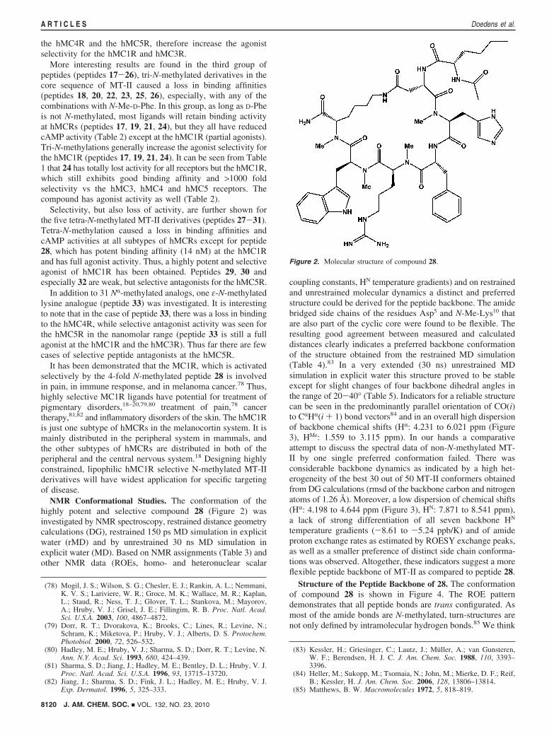
NMR Conformational Studies. The conformation of thehighly potent and selective compound 28 (Figure 2) wasinvestigated by NMR spectroscopy, restrained distance geometrycalculations (DG), restrained 150 ps MD simulation in explicitwater (rMD) and by unrestrained 30 ns MD simulation inexplicit water (MD). Based on NMR assignments (Table 3) andother NMR data (ROEs, homo- and heteronuclear scalar
coupling constants, HN temperature gradients) and on restrainedand unrestrained molecular dynamics a distinct and preferredstructure could be derived for the peptide backbone. The amidebridged side chains of the residues Asp5 and N-Me-Lys10 thatare also part of the cyclic core were found to be flexible. Theresulting good agreement between measured and calculateddistances clearly indicates a preferred backbone conformationof the structure obtained from the restrained MD simulation(Table 4).83 In a very extended (30 ns) unrestrained MDsimulation in explicit water this structure proved to be stableexcept for slight changes of four backbone dihedral angles inthe range of 20-40° (Table 5). Indicators for a reliable structurecan be seen in the predominantly parallel orientation of CO(i)to CRHR(i + 1) bond vectors84 and in an overall high dispersionof backbone chemical shifts (HR: 4.231 to 6.021 ppm (Figure3), HMe: 1.559 to 3.115 ppm). In our hands a comparativeattempt to discuss the spectral data of non-N-methylated MT-II by one single preferred conformation failed. There wasconsiderable backbone dynamics as indicated by a high het-erogeneity of the best 30 out of 50 MT-II conformers obtainedfrom DG calculations (rmsd of the backbone carbon and nitrogenatoms of 1.26 Å). Moreover, a low dispersion of chemical shifts(HR: 4.198 to 4.644 ppm (Figure 3), HN: 7.871 to 8.541 ppm),a lack of strong differentiation of all seven backbone HN
temperature gradients (-8.61 to -5.24 ppb/K) and of amideproton exchange rates as estimated by ROESY exchange peaks,as well as a smaller preference of distinct side chain conforma-tions was observed. Altogether, these indicators suggest a moreflexible peptide backbone of MT-II as compared to peptide 28.
Structure of the Peptide Backbone of 28. The conformationof compound 28 is shown in Figure 4. The ROE patterndemonstrates that all peptide bonds are trans configurated. Asmost of the amide bonds are N-methylated, turn-structures arenot only defined by intramolecular hydrogen bonds.85 We think
(78) Mogil, J. S.; Wilson, S. G.; Chesler, E. J.; Rankin, A. L.; Nemmani,K. V. S.; Lariviere, W. R.; Groce, M. K.; Wallace, M. R.; Kaplan,L.; Staud, R.; Ness, T. J.; Glover, T. L.; Stankova, M.; Mayorov,A.; Hruby, V. J.; Grisel, J. E.; Fillingim, R. B. Proc. Natl. Acad.Sci. U.S.A. 2003, 100, 4867–4872.
(79) Dorr, R. T.; Dvorakova, K.; Brooks, C.; Lines, R.; Levine, N.;Schram, K.; Miketova, P.; Hruby, V. J.; Alberts, D. S. Protochem.Photobiol. 2000, 72, 526–532.
(80) Hadley, M. E.; Hruby, V. J.; Sharma, S. D.; Dorr, R. T.; Levine, N.Ann. N.Y. Acad. Sci. 1993, 680, 424–439.
(81) Sharma, S. D.; Jiang, J.; Hadley, M. E.; Bentley, D. L.; Hruby, V. J.Proc. Natl. Acad. Sci. U.S.A. 1996, 93, 13715–13720.
(82) Jiang, J.; Sharma, S. D.; Fink, J. L.; Hadley, M. E.; Hruby, V. J.Exp. Dermatol. 1996, 5, 325–333.
(83) Kessler, H.; Griesinger, C.; Lautz, J.; Muller, A.; van Gunsteren,W. F.; Berendsen, H. J. C. J. Am. Chem. Soc. 1988, 110, 3393–3396.
(84) Heller, M.; Sukopp, M.; Tsomaia, N.; John, M.; Mierke, D. F.; Reif,B.; Kessler, H. J. Am. Chem. Soc. 2006, 128, 13806–13814.
(85) Matthews, B. W. Macromolecules 1972, 5, 818–819.
Figure 2. Molecular structure of compound 28.
8120 J. AM. CHEM. SOC. 9 VOL. 132, NO. 23, 2010
A R T I C L E S Doedens et al.
that steric effects and dipole orientation such as the above-mentioned parallel orientation of CO(i) to CRHR(i + 1) bondvectors84 contribute most strongly to the conformation of smallerN-methylated cyclic peptides.
According to the N-Me-His6 backbone dihedral angles (Φ )-98°, Ψ ) 78°) the structure obtained from restrained MDsimulation possesses an inverted γ-turn85 centered at N-Me-His6. A distance of 3.4 Å between the Asp5 carbonyl oxygenand the D-Phe7 amide nitrogen, a hydrogen bond angle of 141°and a moderately negative temperature gradient of D-Phe7 HN
(-5.92 ppb/K in the aqueous buffer, -5.55 ppb/K in DMSO)indicate that the hydrogen bond within this inverted γ-turn israther weak and protection form the solvent is incomplete.According to the minimal requirement for �-turns,86 whichconsists in a distance of less than 7 Å between CR
i and CRi+3,
the inverted γ-turn is located within a �-turn ranging from Asp5
to N-Me-Arg8 (CR-CR distance: 6.4 Å). A distance of 7.6 Åbetween the R-carbon atoms of N-Me-His6 and N-Me-Trp9
almost fulfills the criterion for a second overlapping �-turn,which is close to type II′ �-turn geometry, as D-Phe7 Φ, D-Phe7
Ψ, N-Me-Arg8 Φ, and N-Me-Arg8 Ψ possess dihedral anglesof 96°, -126°, -135° and 80°, respectively. The overlappingturns result in a virtually complete helical twist (R-turn) thatextends from residues Asp5 to N-Me-Trp9. Hydrophobic cluster-ing of the N-Me-Trp9 N-methyl group with the Asp5 H�, N-Me-Arg8 HR, N-Me-Lys10 Hγ atoms and the N-Me-His6 N-methylgroup (indicated by the presence of ROESY cross peaks betweenthe N-Me-Trp9 methyl protons and the Asp5 H�, N-Me-Arg8 HR,N-Me-Lys10 Hγ, N-Me-His6 N-methyl protons) seems to stabilizethis helical twist. Within the unrestrained 30 ns MD simulationstarting from the structure of the restrained MD, slight changesoccurred in a few backbone dihedral angles as compared to theaverage structure from the restrained MD simulation. Asp5 Ψ,N-Me-His6 Ψ, D-Phe7 Φ and N-Me-Trp9 Ψ were most affectedand changed from 144° to 113°, 78° to 117°, 96° to 75° and63° to 96°, respectively (Table 5). The rmsd between the atomsof the peptide backbone from Asp5 to N-Me-Lys10 of structuresobtained from the unrestrained and restrained MD simulationis 1.0 Å. According to the N-Me-His6 backbone dihedral angles(Φ ) -102°, Ψ ) 117°) the inverted γ-turn is less pronouncedin the structure obtained from unrestrained MD simulation.Upper bounds of some distance restraints within the cyclic corestructure (Asp5H�-D-Phe7HN; Asp5H�-N-Me-His6HMe; D-Phe7HN-N-Me-Arg8HR; N-Me-Arg8HR-N-Me-Lys10HMe; N-Me-Trp9HMe-
N-Me-Lys10HR) were violated during the unrestrained MDsimulation. This can be traced back directly to the aforemen-tioned changes in backbone dihedral angles. As illustrated inmore detail in the sections describing the side chain dynamicsand structure calculations, we think that the changes in thebackbone dihedral angles were caused by artificial strains inthe amide linked Asp5 and N-Me-Lys10 side chains. These wereintroduced within the DG calculation as our structure calculationprotocol did not take conformational averaging explicitly intoaccount. Accordingly, the conformer obtained from restrainedMD seems to be the best structural model for the peptidebackbone and we focused the analysis of side chain conforma-tion on this structure.
In consideration of the high binding affinity to hMC1R (IC50
) 14 nm) and the strong restriction that cyclization and 4-foldN-methylation pose on conformational changes within thebackbone of peptide 28, we think that its backbone conformationin aqueous solution is very close to the conformation presentin the receptor bound state.
The conformation of the peptide backbone also offers anexplanation for the strong interference of D-Phe7 N-methylationwith hMCR affinity, as N-methylation goes in hand with anincreased spatial requirement and increased hydrophobicity incomparison to the replaced amide proton. If D-Phe7 is N-methylated, these hydrophobic and steric effects would prohibitthe formation of the backbone conformation present in peptide28, as close proximity between the Asp5 carbonyl oxygen andthe D-Phe7 N-methyl group is disfavored. Hence, the N-Me-D-Phe7 substitution would not simply displace a hydrogen bonddonor, but also lead to an altered conformation of the peptidebackbone, which would affect the presentation of the pharma-cophore and prevent interaction of the aromatic ring with thethird and the sixth transmembrane binding domains aromaticgroups.76
Side Chain Structure and Dynamics. Investigation of sidechain conformation about the �1 angle requires a careful analysisof homo- and heteronuclear J-couplings as well as the consid-eration of NOE distances in stereospecifically assigned �protons.87 Extended MD simulations of a solution structure inexplicit solvent can further clarify which structural flips of sidechain dihedral angles are correlated.
The sums of the 3JHR-H� coupling constants of the individualamino acid side chains are in the order of 15 Hz, which excludes
(86) Venkatachalam, C. M. Biopolymers 1968, 6, 1425–1436.(87) Kessler, H.; Griesinger, C.; Wagner, K. J. Am. Chem. Soc. 1987,
109, 6927–6933.
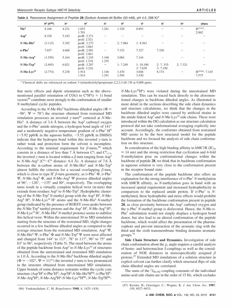
Table 3. Resonance Assignment of Peptide 28 (Sodium Acetate-d4 Buffer (50 mM), pH 4.5, 298 K)a
HN (HNMe) Hr H� Hγ Hδ Hε H� Hη others
Nle4 8.166 4.231 1.653 1.281 1.320 0.893 - - HAcetyl:1.701 2.062
Asp5 8.328 5.183 proR: 2.271 - - - - - -proS: 2.521
N-Me-His6 (3.115) 5.392 proR: 3.272 - 2: 7.084 1: 8.563 - - -proS: 3.063
D-Phe7 7.657 4.668 proR: 2.901 - 7.152 7.327 7.320 - -proS: 2.661
N-Me-Arg8 (1.559) 5.164 proR: 1.245 1.168 3.084 7.144 - - -proS: 1.518 1.255
N-Me-Trp9 (2.695) 6.021 proR: 3.287 - 1: 7.249 1: 10.180 2: 7. 535 2: 7.321 -proS: 3.216 3: 7.639 3: 7.150
N-Me-Lys10 (2.774) 5.238 1.914 1.146 1.470 3.046 8.174 - HAmide: 7.1451.914 1.291 1.594 3.530 7.575
a Chemical shifts are referenced on sodium 3-(trimethylsilyl)propionate-2,2,3,3-d4 (1H at 0.000 ppm).
J. AM. CHEM. SOC. 9 VOL. 132, NO. 23, 2010 8121
Melanocortin Receptor Subtype hMC1R Selectivity A R T I C L E S
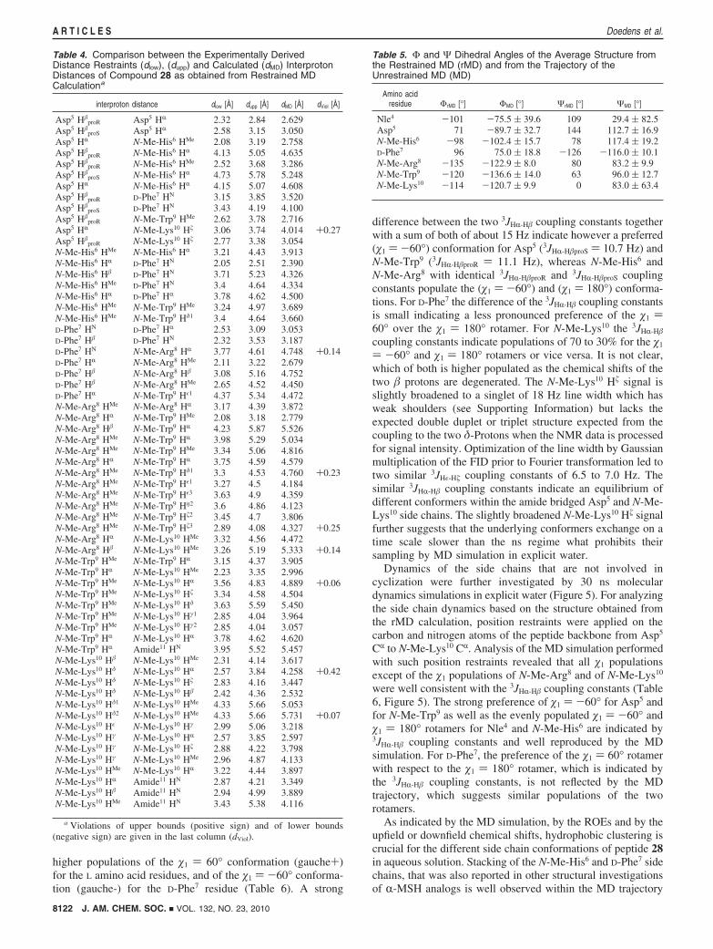
higher populations of the �1 ) 60° conformation (gauche+)for the L amino acid residues, and of the �1 ) -60° conforma-tion (gauche-) for the D-Phe7 residue (Table 6). A strong
difference between the two 3JHR-H� coupling constants togetherwith a sum of both of about 15 Hz indicate however a preferred(�1 ) -60°) conformation for Asp5 (3JHR-H�proS ) 10.7 Hz) andN-Me-Trp9 (3JHR-H�proR ) 11.1 Hz), whereas N-Me-His6 andN-Me-Arg8 with identical 3JHR-H�proR and 3JHR-H�proS couplingconstants populate the (�1 ) -60°) and (�1 ) 180°) conforma-tions. For D-Phe7 the difference of the 3JHR-H� coupling constantsis small indicating a less pronounced preference of the �1 )60° over the �1 ) 180° rotamer. For N-Me-Lys10 the 3JHR-H�
coupling constants indicate populations of 70 to 30% for the �1
) -60° and �1 ) 180° rotamers or vice versa. It is not clear,which of both is higher populated as the chemical shifts of thetwo � protons are degenerated. The N-Me-Lys10 H� signal isslightly broadened to a singlet of 18 Hz line width which hasweak shoulders (see Supporting Information) but lacks theexpected double duplet or triplet structure expected from thecoupling to the two δ-Protons when the NMR data is processedfor signal intensity. Optimization of the line width by Gaussianmultiplication of the FID prior to Fourier transformation led totwo similar 3JHε-H� coupling constants of 6.5 to 7.0 Hz. Thesimilar 3JHR-H� coupling constants indicate an equilibrium ofdifferent conformers within the amide bridged Asp5 and N-Me-Lys10 side chains. The slightly broadened N-Me-Lys10 H� signalfurther suggests that the underlying conformers exchange on atime scale slower than the ns regime what prohibits theirsampling by MD simulation in explicit water.
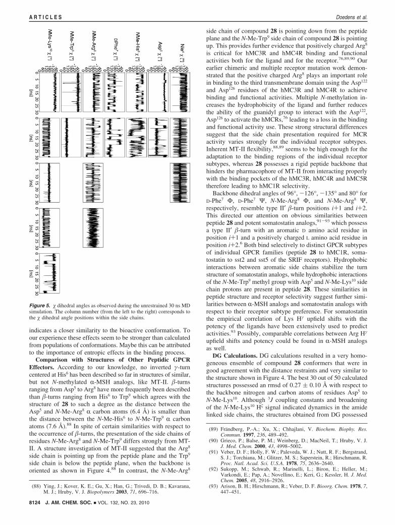
Dynamics of the side chains that are not involved incyclization were further investigated by 30 ns moleculardynamics simulations in explicit water (Figure 5). For analyzingthe side chain dynamics based on the structure obtained fromthe rMD calculation, position restraints were applied on thecarbon and nitrogen atoms of the peptide backbone from Asp5
CR to N-Me-Lys10 CR. Analysis of the MD simulation performedwith such position restraints revealed that all �1 populationsexcept of the �1 populations of N-Me-Arg8 and of N-Me-Lys10
were well consistent with the 3JHR-H� coupling constants (Table6, Figure 5). The strong preference of �1 ) -60° for Asp5 andfor N-Me-Trp9 as well as the evenly populated �1 ) -60° and�1 ) 180° rotamers for Nle4 and N-Me-His6 are indicated by3JHR-H� coupling constants and well reproduced by the MDsimulation. For D-Phe7, the preference of the �1 ) 60° rotamerwith respect to the �1 ) 180° rotamer, which is indicated bythe 3JHR-H� coupling constants, is not reflected by the MDtrajectory, which suggests similar populations of the tworotamers.
As indicated by the MD simulation, by the ROEs and by theupfield or downfield chemical shifts, hydrophobic clustering iscrucial for the different side chain conformations of peptide 28in aqueous solution. Stacking of the N-Me-His6 and D-Phe7 sidechains, that was also reported in other structural investigationsof R-MSH analogs is well observed within the MD trajectory
Table 4. Comparison between the Experimentally DerivedDistance Restraints (dlow), (dupp) and Calculated (dMD) InterprotonDistances of Compound 28 as obtained from Restrained MDCalculationa
interproton distance dlow [Å] dupp [Å] dMD [Å] dViol [Å]
Asp5 H�proR Asp5 HR 2.32 2.84 2.629
Asp5 H�proS Asp5 HR 2.58 3.15 3.050
Asp5 HR N-Me-His6 HMe 2.08 3.19 2.758Asp5 H�
proR N-Me-His6 HR 4.13 5.05 4.635Asp5 H�
proR N-Me-His6 HMe 2.52 3.68 3.286Asp5 H�
proS N-Me-His6 HR 4.73 5.78 5.248Asp5 HR N-Me-His6 HR 4.15 5.07 4.608Asp5 H�
proR D-Phe7 HN 3.15 3.85 3.520Asp5 H�
proS D-Phe7 HN 3.43 4.19 4.100Asp5 H�
proR N-Me-Trp9 HMe 2.62 3.78 2.716Asp5 HR N-Me-Lys10 H� 3.06 3.74 4.014 +0.27Asp5 H�
proR N-Me-Lys10 H� 2.77 3.38 3.054N-Me-His6 HMe N-Me-His6 HR 3.21 4.43 3.913N-Me-His6 HR D-Phe7 HN 2.05 2.51 2.390N-Me-His6 H� D-Phe7 HN 3.71 5.23 4.326N-Me-His6 HMe D-Phe7 HN 3.4 4.64 4.334N-Me-His6 HR D-Phe7 HR 3.78 4.62 4.500N-Me-His6 HMe N-Me-Trp9 HMe 3.24 4.97 3.689N-Me-His6 HMe N-Me-Trp9 Hδ1 3.4 4.64 3.660D-Phe7 HN D-Phe7 HR 2.53 3.09 3.053D-Phe7 H� D-Phe7 HN 2.32 3.53 3.187D-Phe7 HN N-Me-Arg8 HR 3.77 4.61 4.748 +0.14D-Phe7 HR N-Me-Arg8 HMe 2.11 3.22 2.679D-Phe7 H� N-Me-Arg8 H� 3.08 5.16 4.752D-Phe7 H� N-Me-Arg8 HMe 2.65 4.52 4.450D-Phe7 HR N-Me-Trp9 Hε1 4.37 5.34 4.472N-Me-Arg8 HMe N-Me-Arg8 HR 3.17 4.39 3.872N-Me-Arg8 HR N-Me-Trp9 HMe 2.08 3.18 2.779N-Me-Arg8 H� N-Me-Trp9 HR 4.23 5.87 5.526N-Me-Arg8 HMe N-Me-Trp9 HR 3.98 5.29 5.034N-Me-Arg8 HMe N-Me-Trp9 HMe 3.34 5.06 4.816N-Me-Arg8 HR N-Me-Trp9 HR 3.75 4.59 4.579N-Me-Arg8 HMe N-Me-Trp9 Hδ1 3.3 4.53 4.760 +0.23N-Me-Arg8 HMe N-Me-Trp9 Hε1 3.27 4.5 4.184N-Me-Arg8 HMe N-Me-Trp9 Hε3 3.63 4.9 4.359N-Me-Arg8 HMe N-Me-Trp9 Hη2 3.6 4.86 4.123N-Me-Arg8 HMe N-Me-Trp9 H�2 3.45 4.7 3.806N-Me-Arg8 HMe N-Me-Trp9 H�3 2.89 4.08 4.327 +0.25N-Me-Arg8 HR N-Me-Lys10 HMe 3.32 4.56 4.472N-Me-Arg8 H� N-Me-Lys10 HMe 3.26 5.19 5.333 +0.14N-Me-Trp9 HMe N-Me-Trp9 HR 3.15 4.37 3.905N-Me-Trp9 HR N-Me-Lys10 HMe 2.23 3.35 2.996N-Me-Trp9 HMe N-Me-Lys10 HR 3.56 4.83 4.889 +0.06N-Me-Trp9 HMe N-Me-Lys10 H� 3.34 4.58 4.504N-Me-Trp9 HMe N-Me-Lys10 Hδ 3.63 5.59 5.450N-Me-Trp9 HMe N-Me-Lys10 Hγ1 2.85 4.04 3.964N-Me-Trp9 HMe N-Me-Lys10 Hγ2 2.85 4.04 3.057N-Me-Trp9 HR N-Me-Lys10 HR 3.78 4.62 4.620N-Me-Trp9 HR Amide11 HN 3.95 5.52 5.457N-Me-Lys10 H� N-Me-Lys10 HMe 2.31 4.14 3.617N-Me-Lys10 Hδ N-Me-Lys10 HR 2.57 3.84 4.258 +0.42N-Me-Lys10 Hδ N-Me-Lys10 H� 2.83 4.16 3.447N-Me-Lys10 Hδ N-Me-Lys10 H� 2.42 4.36 2.532N-Me-Lys10 Hδ1 N-Me-Lys10 HMe 4.33 5.66 5.053N-Me-Lys10 Hδ2 N-Me-Lys10 HMe 4.33 5.66 5.731 +0.07N-Me-Lys10 Hε N-Me-Lys10 Hγ 2.99 5.06 3.218N-Me-Lys10 Hγ N-Me-Lys10 HR 2.57 3.85 2.597N-Me-Lys10 Hγ N-Me-Lys10 H� 2.88 4.22 3.798N-Me-Lys10 Hγ N-Me-Lys10 HMe 2.96 4.87 4.133N-Me-Lys10 HMe N-Me-Lys10 HR 3.22 4.44 3.897N-Me-Lys10 HR Amide11 HN 2.87 4.21 3.349N-Me-Lys10 H� Amide11 HN 2.94 4.99 3.889N-Me-Lys10 HMe Amide11 HN 3.43 5.38 4.116
a Violations of upper bounds (positive sign) and of lower bounds(negative sign) are given in the last column (dViol).
Table 5. Φ and Ψ Dihedral Angles of the Average Structure fromthe Restrained MD (rMD) and from the Trajectory of theUnrestrained MD (MD)
Amino acidresidue ΦrMD [°] ΦMD [°] ΨrMD [°] ΨMD [°]
Nle4 -101 -75.5 ( 39.6 109 29.4 ( 82.5Asp5 71 -89.7 ( 32.7 144 112.7 ( 16.9N-Me-His6 -98 -102.4 ( 15.7 78 117.4 ( 19.2D-Phe7 96 75.0 ( 18.8 -126 -116.0 ( 10.1N-Me-Arg8 -135 -122.9 ( 8.0 80 83.2 ( 9.9N-Me-Trp9 -120 -136.6 ( 14.0 63 96.0 ( 12.7N-Me-Lys10 -114 -120.7 ( 9.9 0 83.0 ( 63.4
8122 J. AM. CHEM. SOC. 9 VOL. 132, NO. 23, 2010
A R T I C L E S Doedens et al.

and is also present in the exemplary conformation shown inFigure 4A. Hydrophobic contacts between the D-Phe7 and N-Me-Arg8 side chains that agree with strong upfield shifts of theN-Me-Arg8 � and γ protons are also observed within the MDtrajectory and depicted in the conformation shown in Figure4B. Overall side chain dynamics as indicated by 3JHR-H� couplingconstants, is reflected by the rotamers shown for N-Me-His6,D-Phe7 and N-Me-Arg8 in Figure 4A and B, respectively. Theclustering of the N-Me-Trp9 indolyl ring with the N-methylgroup of N-Me-Arg8, which is observed in Figures 4A andB, isconfirmed by ROE contacts between methyl protons and all
indolyl protons as well as by a strongly upfield shifted HMe
resonance at 1.559 ppm. ROEs between the N-Me-His6 methylprotons and the N-Me-Trp9 δ1 and ε1 protons as well as betweenD-Phe7 HR and N-Me-Trp9 Hε1 are well consistent with theorientation of the indolyl ring given in Figure 4A and B. Anadditional ROE between the N-Me-His6 methyl protons and theN-Me-Trp9 ε3 proton indicates another orientation of the indolylgroup that was not sampled during 30 ns unrestrained MDsimulation. In this orientation the indolyl group also seems tostack on top of the N-Me-Arg8 N-methyl group as shown inFigure 4 but with the six membered ring of the indolyl grouppointing to the left and the five membered ring pointing to theright.
A dispersion of chemical shifts of peptide 28 in DMSO-d6(HR: 4.245 to 5.824 ppm (Figure 3), HMe: 1.893 to 3.058 ppm),which is similar to the dispersion in the aqueous buffer (HR:4.231 to 6.021 ppm (Figure 3), HMe: 1.559 to 3.115 ppm),indicates that hydrophobic interactions observed in aqueousbuffer are also present in the slightly more hydrophobic DMSO.This suggests that such hydrophobic interactions might also bepresent in hMC1R bound state.
In addition it is often found that stronger conformationalpreference which is accompanied by stronger biological activity
Figure 3. HR regions of 1H NMR spectra of peptide 28 (A and B) and MT-II (C and D). Spectra A and C were detected in 50 mM sodium acetate-d4 D2Obuffer (pH 4.5), B and D were detected in DMSO-d6. Numbers refer to HR atoms of the respective residues in the R-MSH sequence.
Figure 4. Stereoviews of the solution structure of peptide 28 as determinedby NMR spectroscopy and MD calculations. The conformers shown in Aand B possess the same cyclic core structure. The different side chainconformations of N-Me-His6, D-Phe7, and N-Me-Arg8 in A and B,respectively, reflect dynamics in � dihedral angles observed within the MD.
Table 6. 3JHR-H� Coupling Constants as Experimentally Determinedfrom E.COSYa
3JHR-H� [Hz]
HR-H� proR HR-H� proSp(�1 ) -60°)
[%]p(�1 ) 180°)
[%]p(�1 ) 60°)
[%]
Nle4 8.2; 6.2 84 16Asp5 3.8 10.7 74 11 15N-Me-His6 7.9 7.9 48 48 4D-Phe7 5.3 9.2 14 25 61N-Me-Arg8 7.0 7.0 40 40 20N-Me-Trp9 11.1 4.7 78 19 3N-Me-Lys10 9.5, 6 100 0
a The according �1 populations were derived by a linear combinationof 3JHR-H�(ap) ) 12 Hz, 3JHR-H�(ga) ) 3.5 Hz.
J. AM. CHEM. SOC. 9 VOL. 132, NO. 23, 2010 8123
Melanocortin Receptor Subtype hMC1R Selectivity A R T I C L E S
indicates a closer similarity to the bioactive conformation. Toour experience these effects seem to be stronger than calculatedfrom populations of conformations. Maybe this can be attributedto the importance of entropic effects in the binding process.
Comparison with Structures of Other Peptidic GPCREffectors. According to our knowledge, no inverted γ-turncentered at His6 has been described so far in structures of similar,but not N-methylated R-MSH analogs, like MT-II. �-turnsranging from Asp5 to Arg8 have more frequently been describedthan �-turns ranging from His6 to Trp9 which agrees with thestructure of 28 to such a degree as the distance between theAsp5 and N-Me-Arg8 R carbon atoms (6.4 Å) is smaller thanthe distance between the N-Me-His6 to N-Me-Trp9 R carbonatoms (7.6 Å).88 In spite of certain similarities with respect tothe occurrence of �-turns, the presentation of the side chains ofresidues N-Me-Arg8 and N-Me-Trp9 differs strongly from MT-II. A structure investigation of MT-II suggested that the Arg8
side chain is pointing up from the peptide plane and the Trp9
side chain is below the peptide plane, when the backbone isoriented as shown in Figure 4.88 In contrast, the N-Me-Arg8
side chain of compound 28 is pointing down from the peptideplane and the N-Me-Trp9 side chain of compound 28 is pointingup. This provides further evidence that positively charged Arg8
is critical for hMC3R and hMC4R binding and functionalactivities both for the ligand and for the receptor.76,89,90 Ourearlier chimeric and multiple receptor mutation work demon-strated that the positive charged Arg8 plays an important rolein binding to the third transmembrane domain using the Asp122
and Asp126 residues of the hMC3R and hMC4R to achievebinding and functional activities. Multiple N-methylation in-creases the hydrophobicity of the ligand and further reducesthe ability of the guanidyl group to interact with the Asp122,Asp126 to activate the hMCRs,76 leading to a loss in the bindingand functional activity use. These strong structural differencessuggest that the side chain presentation required for MCRactivity varies strongly for the individual receptor subtypes.Inherent MT-II flexibility,88,89 seems to be high enough for theadaptation to the binding regions of the individual receptorsubtypes, whereas 28 possesses a rigid peptide backbone thathinders the pharmacophore of MT-II from interacting properlywith the binding pockets of the hMC3R, hMC4R and hMC5Rtherefore leading to hMC1R selectivity.
Backbone dihedral angles of 96°, -126°, -135° and 80° forD-Phe7 Φ, D-Phe7 Ψ, N-Me-Arg8 Φ, and N-Me-Arg8 Ψ,respectively, resemble type II′ �-turn positions i+1 and i+2.This directed our attention on obvious similarities betweenpeptide 28 and potent somatostatin analogs,91-93 which possessa type II′ �-turn with an aromatic D amino acid residue inposition i+1 and a positively charged L amino acid residue inposition i+2.6 Both bind selectively to distinct GPCR subtypesof individual GPCR families (peptide 28 to hMC1R, soma-tostatin to sst2 and sst5 of the SRIF receptors). Hydrophobicinteractions between aromatic side chains stabilize the turnstructure of somatostatin analogs, while hydrophobic interactionsof the N-Me-Trp9 methyl group with Asp5 and N-Me-Lys10 sidechain protons are present in peptide 28. These similarities inpeptide structure and receptor selectivity suggest further simi-larities between R-MSH analogs and somatostatin analogs withrespect to their receptor subtype preference. For somatostatinthe empirical correlation of Lys Hγ upfield shifts with thepotency of the ligands have been extensively used to predictactivities.93 Possibly, comparable correlations between Arg Hγ
upfield shifts and potency could be found in R-MSH analogsas well.
DG Calculations. DG calculations resulted in a very homo-geneous ensemble of compound 28 conformers that were ingood agreement with the distance restraints and very similar tothe structure shown in Figure 4. The best 30 out of 50 calculatedstructures possessed an rmsd of 0.27 ( 0.10 Å with respect tothe backbone nitrogen and carbon atoms of residues Asp5 toN-Me-Lys10. Although 3J coupling constants and broadeningof the N-Me-Lys10 H� signal indicated dynamics in the amidelinked side chains, the structures obtained from DG possessed
(88) Ying, J.; Kover, K. E.; Gu, X.; Han, G.; Trivedi, D. B.; Kavarana,M. J.; Hruby, V. J. Biopolymers 2003, 71, 696–716.
(89) Frandberg, P.-A.; Xu, X.; Chhajlani, V. Biochem. Biophy. Res.Commun. 1997, 236, 489–492.
(90) Grieco, P.; Balse, P. M.; Weinberg, D.; MacNeil, T.; Hruby, V. J.J. Med. Chem. 2000, 43, 4998–5002.
(91) Veber, D. F.; Holly, F. W.; Paleveda, W. J.; Nutt, R. F.; Bergstrand,S. J.; Torchiana, M.; Glitzer, M. S.; Saperstein, R.; Hirschmann, R.Proc. Natl. Acad. Sci. U.S.A. 1978, 75, 2636–2640.
(92) Sukopp, M.; Schwab, R.; Marinelli, L.; Biron, E.; Heller, M.;Varkondi, E.; Pap, A.; Novellino, E.; Keri, G.; Kessler, H. J. Med.Chem. 2005, 48, 2916–2926.
(93) Arison, B. H.; Hirschmann, R.; Veber, D. F. Bioorg. Chem. 1978, 7,447–451.
Figure 5. � dihedral angles as observed during the unrestrained 30 ns MDsimulation. The column number (from the left to the right) corresponds tothe � dihedral angle positions within the side chains.
8124 J. AM. CHEM. SOC. 9 VOL. 132, NO. 23, 2010
A R T I C L E S Doedens et al.
very similar homogeneous N-Me-Lys10 �1 and �2 dihedral angles(�1 ) -55.2° +/- 2.01 Å, �2 ) -109° +/- 3.73°). Theobservation of only one preferred �1 dihedral angle did notreflect the 3JHR-H� coupling constants (Table 6) and the onlypopulated eclipsed �2 rotamer was thermodynamically unfavor-able. Apparently, conformation averaging in the N-Me-Lys10
side chain led to a number of conformation averaged distancerestraints that forced the lactam bridged N-Me-Lys10 side chaininto conformations during DG calculations that could not behighly populated at equilibrium.
Restrained Molecular Dynamics Calculations. The best DGstructure in respect of violations of experimental distancerestraints was subjected to refinement by restrained moleculardynamics (rMD) calculation in explicit water. The averagestructure of the rMD calculation was very similar to the DGstarting structure (rmsd ) 0.69 Å with respect to backboneheavy atoms from Asp5 to N-Me-Lys10). Within the cyclic corestructure significant changes occurred only in the N-Me-Lys10
side chain, that was strained in the starting conformer as sidechain dihedral angles were eclipsed (�2 ) -111° and �3 )-137°). As expected, the force field applied during restrainedMD calculation rotated the dihedral angles within the N-Me-Lys10 side chain to thermodynamically feasible values (�2 )-169° and �3 ) 163°).
Unrestrained Molecular Dynamics Calculations. The averagestructure obtained from the rMD calculation was further refinedby extended (30 ns) unrestrained MD simulation in explicitwater. This lead to significant changes of some backbonedihedral angles (Table 5) that were accompanied by furtherconformational changes in the lactam bridged N-Me-Lys10 sidechain. The changes of backbone dihedral angles led to significantviolations of distance restraints and most 3JHR-H� couplingconstants were not consistent with the �1 populations extractedfrom the MD trajectory. Consistency with 3JHR-H� couplingconstants and with distance restraints was much higher in atrajectory of an MD simulation in which the conformation ofthe peptide backbone obtained from rMD was conserved byapplying position restraints on all backbone carbon and nitrogenatoms from Asp5 CR to N-Me-Lys10 CR.
We believe that the artificial strains in the lactam bridgedside chains, that were described above, induced the conforma-tional changes that were observed in the peptide backboneduring 30 ns non position restrained MD simulation. The slightvariations in Φ and Ψ dihedral angles seemed to be energeticallyless demanding than flips between individual staggered con-formations within the N-Me-Lys10 side chain. Apparently, thebackbone compensated for the strains within the lactam bridgedside chains during the unrestrained MD simulation and positionrestraining of the backbone during the unrestrained MD simula-tion seems to be a legitimate way to avoid faulty changes ofbackbone dihedral angles.
Conclusions
Multiple N-methylation in MT-II led to several derivativesin which the affinity and activity for hMC1R could be retained,but simultaneously the binding affinity to the other 3 melano-cortin receptors was completely lost. Hence, selectivity andretained activity was achieved in this way. A careful analysisof the conformation of the most active (13 nM) compoundreveals a strong preference of one backbone conformation,which indicates a strongly reduced conformational space incomparison to the nonmethylated MT-II. Hence, this work againshows that restriction of peptide backbone conformation resulted
in strong receptor subtype selectivity with retained or evenimproved activity for an individual receptor subtype. Thisapproach, discussed by us already a quarter of a century ago94,95
is not achievable only by cyclization but can be further increasedby multiple N-methylation.48 With an increasing extent ofintroduction of N-methyl groups into its backbone, MT-IIderivatives lose their ability to bind to particular hMCRs outof the total family of melanocortin receptors. This leads in twocases (peptides 24 and 28) to highly selective and potent agonistsfor the hMC1R, but also to antagonists and compounds withboth, agonist and antagonist activities. These compounds possessmultiple amide bonds that are N-methylated which was recentlyreported to be a possible key toward bioavailability5 and maybealso enhanced oral bioavailability6 of peptides. Whereas theeffect of N-methylation on receptor subtype selectivity isstrongly supported by our results, the effect on pharmacologicalproperties that have been improved in other examples4–6 has tobe demonstrated for the N-methylated MT-II derivatives. Theseinvestigations are in progress.
Experimental Section
Materials. NR-Fmoc-amino acids, peptide coupling reagents,Rink amide MBHA resin and solvents were reagent grade and usedwithout further purification unless otherwise noted. These chemicalswere obtained from Aldrich, Novabiochem, Iris Biotech GmbH,Merck, NeoMPS, ORPEGEN Pharma and GLS. The followingamino acids were used: Fmoc-Lys(Alloc)-OH, Fmoc-Trp(Boc)-OH,Fmoc-Arg(Pbf)-OH, Fmoc-D-Phe-OH, Fmoc-His(Trt)-OH, Fmoc-Asp(OAllyl)-OH and Fmoc-Nle-OH. The polypropylene reactionvessels (syringes with frits) were purchased from B. BraunMelsungen AG. The purity of the peptides was checked byanalytical reverse-phase HPLC using an Amersham PharmaciaBiotech Akta Basic 10F with a ODS-A C18 (120 Å, 5 µm, 250mm ×4.6 mm) column (Omnicrom YMC Europe GmbH) moni-tored at 220 and 254 nm and by high resolution mass spectralanalysis.
Synthesis. The MT-II analogues were synthesized manually bythe Fmoc-SPPS using methods described in refs 96 and 97.
Rink amide MBHA resin (0.3 g, 4-(2′,4′-dimethoxyphenyl-Fmoc-aminomethyl)-phenoxyacetamido-norleucyl-MBHA resin) in a 10mL syringe with a frit on the bottom was swollen in NMP (3 mL)for 10 min. The Fmoc protecting group on the Rink linker wasremoved by 20% piperidine in NMP (1 × 8 min, 1 × 12 min).The resin was washed with NMP (5 × 3 mL). The first NR-Fmoc-amino acid was coupled using preactivated ester (3 equiv NR-Fmoc-amino acid, 3 equiv TBTU, 3 equiv HOBt and 8 equiv DIEA) inNMP. The coupling mixture was transferred into the syringe withthe resin and shaken for 3 h. Afterward the resin was washed withNMP (5 × 3 mL). Complete coupling was confirmed by HPLCmonitoring. Therefore the resin was washed with DCM (2 × 3mL). A small amount of resin was taken out of the syringe andthree drops of a mixture of trifluoracetic acid/triethylsilane/water(95:2.5:2.5, v/v/v) was added. After 10 min, acetonitrile and waterwere added, and after filtration, HPLC and ESI-MS showedcomplete coupling. The peptide sequences were continued byconsecutively coupling of the particular amino acids. N-Methylationwas carried out after coupling of the corresponding amino acid.This procedure includes three steps. First, the N-terminal NR-Fmoc-deprotected amino acid was protected as an NR-o-nitrobenzenesulfonamide by reaction with 5 equiv of o-nitrobenzenesulfonylchloride (o-NBS) and 10 equiv of collidine in NMP (15 min). The
(94) Kessler, H. Angew. Chem., Int. Ed. 1982, 21, 512–523.(95) Hruby, V. J. Life Sci. 1982, 31, 189–199.(96) Mayorov, A. V.; Han, S. Y.; Cai, M.; Hammer, M. R.; Trivedi, D.;
Hruby, V. J. Chem. Biol. Drug. Des. 2006, 67, 329–335.(97) Heckmann, D.; Kessler, H. Methods Enzymol. 2007, 426, 463–503.
J. AM. CHEM. SOC. 9 VOL. 132, NO. 23, 2010 8125
Melanocortin Receptor Subtype hMC1R Selectivity A R T I C L E S
methylation was done under Mitsunobu conditions98 with 5 equivof triphenylphosphine, 10 equiv of methanol and 5 equiv ofdiisopropyl azodicarboxylate (DIAD) in THF (10 min). Then theo-NBS group was removed with 10 equiv of 2-mercaptoethanoland 5 equiv of DBU in NMP (5 min). This deproctection procedurewas repeated once. For the coupling of the next amino acid, 3 equivHATU, 3 equiv HOAt and 8 equiv DIEA were used. After couplingof the last amino acid Fmoc-Nle-OH and Fmoc deprotection asdescribed above, acetylation was achieved by treatment of the DCMwashed resin (3 × 3 mL) with acetic anhydride/DCM/DIEA (25:75:2.5, v/v/v, 3 mL) for 30 min and washing of the resin with DCM(5 × 3 mL). The next step was the removal of the Nε-Alloc groupof Lys and the �-allyl group of Asp.96,97,99 For this, a solution ofPhSiH3 (24 equiv) in 1 mL DCM was added to the peptide resins.Then a solution of Pd(PPh3)4 (0.25 equiv) in 2 mL DCM was addedand the reaction allowed to proceed for 30 min. The peptide resinswere washed with DCM (3 × 3 mL), NMP (2 × 3 mL), afterwardtwice with 0.5% DIEA in DMF (v/v) and 0.5% sodium dieth-yldithiocarbamate trihydrate (DEDTC) in DMF (w/w) to removeany remaining traces of the Pd catalyst,100 followed by NMP (5 ×3 mL). The macrocyclic lactam ring formation was then mediatedby addition of TBTU (3 equiv), HOBt (3 equiv) and DIEA (8 equiv)for 3 h. The cyclized peptides were cleaved off solid support using3 mL trifluoracetic acid/triethylsilane/water (95:2.5:2.5, v/v/v, 1 ×3 h, 1 × 15 min), and the crude peptides were precipitated bydropping the solution in chilled diethyl ether to give white to lightyellow precipitates. The resulting peptide suspensions were cen-trifuged for 5 min at 4000 rpm, and the liquid was decanted. Thecrude peptides were washed with chilled diethyl ether (2 × 30 mL),and after the final centrifugation, dried under vacuum overnight.Purification was performed by HPLC using a ODS-A C18 (120Å´, 5 µm, 250 mm ×20 mm) column (Omnicrom YMC EuropeGmbH) eluting with a linear gradient of acetonitrile and watercontaining 0.1% TFA. The products were obtained by lyophilizationof the appropriate fractions after removal of the acetonitrile by rotaryevaporation. Analysis by analytical HPLC and HPLC-MS (Sup-porting Information) showed the peptides to be pure (>95%). Thepurified peptides were isolated in 2-14% overall yields.
Biological Activity Assays
Receptor Binding Assay. Competition binding experimentswere carried out using whole HEK293 cells stably expressinghuman MC1, MC3, MC4, and MC5 receptors as describedbefore.101-103 HEK293 cells transfected with hMCRs101–103
were seeded on 96-well plates 48 h before assay (50,000 cells/well). For the assay, the cell culture medium was aspirated andthe cells were washed once with a freshly prepared MEM buffercontaining 100% minimum essential medium with Earle’s salt(MEM, GIBCO), and 25 mM sodium bicarbonate. Next, thecells were incubated for 40 min at 37 °C with differentconcentrations of unlabeled peptide and labeled [125I]-[Nle4,D-Phe7]-R-MSH (Perkin-Elmer Life Science, 20 000 cpm/well,33.06 pM) diluted in a 125 µL of freshly prepared binding buffercontaining 100% MEM, 25 mM HEPES (pH 7.4), 0.2% bovineserum albumin, 1 mM 1,10-phenanthrolone, 0.5 mg/L leupeptin,
200 mg/L bacitracin. The assay medium was subsequentlyremoved, the cells were washed once with basic medium, andthen lysed by the addition of 100 µL of 0.1 M NaOH and 100µL of 1% Triton X-100. The lysed cells were transferred to 12× 75 mm borosilicate glass tubes, and the radioactivity wasmeasured by a Wallac 1470 WIZARD Gamma Counter. Theresults are shown in Table 1.
Adenylate Cyclase Assay. HEK 293 cells transfected withhuman melanocortin receptors103 were grown to confluence inMEM medium (GIBCO) containing 10% fetal bovine serum,100 units/mL penicillin and streptomycin, and 1 mM sodiumpyruvate. The cells were seeded on 96-well plates 48 h beforeassay (50 000 cells/well). For the assay, the cell culture mediumwas removed and the cells were rinsed with 100 µL of MEMbuffer (GIBCO). An aliquot (100 µL) of the Earle’s balancedsalt solution with 5 nM isobutylmethylxanthine (IBMX) wasplaced in each well along for 1 min at 37 °C. Next, aliquots(25 µL) of melanotropin peptides of varying concentration wereadded, and the cells were incubated for 3 min at 37 °C. Thereaction was stopped by aspirating the assay buffer and adding60 µL ice-cold Tris/EDTA buffer to each well, then placingthe plates in a boiling water bath for 7 min. The cell lysateswere then centrifuged for 10 min at 2,300g. A 50 µL aliquot ofthe supernatant was transferred to another 96-well plate andplaced with 50 µL [3H] cAMP and 100 µL protein kinase A(PKA) buffer in an ice bath for 2-3 h. The PKA bufferconsisted of Tris/EDTA buffer with 60 µg/mL PKA and 0.1%bovine serum albumin by weight. The incubation mixture wasfiltered through 1.0 µm glass fiber filters in MultiScreen-FB 96-well plates (Millipore, Billerica, MA). The total [3H] cAMPwas measured by a Wallac MicroBeta TriLux 1450 LSC andLuminescence Counter (PerkinElmer Life Science, Boston,MA). The cAMP accumulation data for each peptide analoguewas determined with the help of a cAMP standard curvegenerated by the same method as described above. IC50 andEC50 values represent the mean of two experiments performedin triplicate. IC50 and EC50 estimates and their associatedstandard errors were determined by fitting the data using anonlinear least-squares analysis, with the help of GraphPadPrism 4 (GraphPad Software, San Diego, CA). The maximalcAMP produced at 10 µM concentration of each ligand wascompared to the amount of cAMP produced at 10 µMconcentration of the standard agonist MT-II, and is expressedin percent (as % max effect) in the Table 2. The antagonistproperties of the lead compounds were evaluated by their abilityto competitively displace the MT-II agonist in a dose-dependentmanner, at up to 10 µM.
Data Analysis. IC50 and EC50 values represent the mean oftwo experiments performed in triplicate. IC50 and EC50 estimatesand their associated standard errors were determined by fittingthe data using a nonlinear least-squares analysis, with the helpof GraphPad Prism 4 (GraphPad Software, San Diego, CA).
NMR Spectroscopy. DQF-COSY, E.COSY, TOCSY, ROE-SY, 13C-HMBC and 13C-HSQC spectra were recorded at 298K on a Bruker DMX spectrometer operating at 600 MHz. ACOLOC spectrum104 was recorded at 298 K on a Bruker AvanceIII spectrometer operating at 600 MHz. A phase sensitiveHMBC and a reference HSQC spectrum with offset and rf-amplitude-compensated BEBOP105,106 and BIBOP pulses107,108
(98) Mitsunobu, O. Synthesis 1981, 1, 1–28.(99) Thieriet, N.; Alsina, J.; Giralt, E.; Guibe, F.; Albericio, F. Tetrahedron
Lett. 1997, 38, 7275–7278.(100) Mayorov, A. V.; Cai, M.; Chandler, K. B.; Petrov, R. R.; VanScoy,
A. R.; Yu, Z.; Tanaka, D. K.; Trivedi, D.; Hruby, V. J. J. Med. Chem.2006, 49, 1946–1952.
(101) Cai, M.; Mayorov, A. V.; Ying, J.; Stankova, M.; Trivedi, D.; Cabello,C.; Hruby, V. J. Peptides 2005, 26, 1481–1485.
(102) Cai, M.; Mayorov, A. V.; Cabello, C.; Stankova, M.; Trivedi, D.;Hruby, V. J. J. Med. Chem. 2005, 48, 1839–1848.
(103) Cai, M.; Cai, C.; Mayorov, A. V.; Xiong, C.; Cabello, C. M.;Soloshonok, V. A.; Swift, J. R.; Trivedi, D.; Hruby, V. J. J. Pept.Res. 2004, 63, 116–131.
(104) Kessler, H.; Griesinger, C.; Zarbock, J.; Loosli, H.-R. J. Magn. Reson.1984, 57, 331–336.
(105) Skinner, T. E.; Reiss, T. O.; Luy, B.; Khaneja, N.; Glaser, S. J. J.Magn. Reson. 2003, 163, 8–15.
8126 J. AM. CHEM. SOC. 9 VOL. 132, NO. 23, 2010
A R T I C L E S Doedens et al.

were detected at 298K on a Bruker Avance III spectrometeroperating at 750 MHz.109,110 Samples were prepared in 50 mMSodium acetate-d4 buffer (pH 4.5, 10% D2O, 0.05% NaN3) atconcentrations of 12-38 mM. This low pH was chosen to keephydrogen exchange induced line broadening small and toenhance comparability with numerous earlier structural studieson MCR effectors that were performed under the same condi-tions. Sodium 3-(trimethylsilyl)propionate-2,2,3,3-d4 (1H at0.000 ppm) was used as internal standard. Data were processedwith Topspin 1.3 software from Bruker. The homo- andheteronuclear experiments DQF-COSY, E.COSY, TOCSY,ROESY, and magnitude mode 13C-HMBC were performed witha spectral width of 11 ppm for 1H and 180 ppm for 13C.Individual HSQC spectra covering aliphatic (13C offset ) 35ppm, spectral width ) 50 ppm) and aromatic 13C resonances(13C offset ) 120 ppm, spectral width ) 30 ppm) were detected.The phase sensitive HMBC spectrum and the reference HSQCspectrum were detected with a spectral width of 9 ppm for 1Hand 190 ppm for 13C. A COLOC spectrum was detected withspectral widths of 9.5 ppm for 1H and 190 ppm for 13C. Theincrements in t1 and t2 were adjusted to the informationextracted from the individual experiments, ranging from 384to 2048 increments in t1 and from 4096 to 16 384 complex datapoints in t2. Depending on the sample concentration and theindividual experiments, 16 to 48 transients were averaged foreach t1 value. A mixing time of 80 ms was used for TOCSY(spin lock field: 6 kHz; mixing sequence MLEV-17). Watersignal suppression was achieved by WATERGATE tech-niques.111 The sequential assignment was obtained from het-eronuclear J correlations that were extracted from HSQC andHMBC spectra. A compensated ROESY experiment, which wasused for the extraction of inter proton distances, was performedwith 150 ms mixing time and a spin lock field of 4000 kHz.112
The volume integrals of the individually assigned cross-peakswere compensated for offset effects and converted into distanceconstraints using the isolated spin pair approximation.113 Tocompensate for watergate solvent signal suppression artifacts,of any two cross peaks representing an internuclear distance,the peak with the higher water resonance offset in the directdimension was preferably considered for distance calculations.The ROESY cross-peak volumes were calibrated against thedistance (1.78 Å) between the prochiral N-Me-Lys10 Hε protons.Upper and lower distance limits were set to plus and minus10% of the calculated distances, respectively. For distancerestraints referring to nonstereospecifically assigned methyleneprotons, another 0.7 Å was added on upper bounds as pseudoa-toms and multiplicity corrections. For distance restraints refer-ring to methyl protons, subtraction of 10% for the lower boundswas omitted and another 0.9 Å was added on upper bounds aspseudoatoms and multiplicity corrections. 18 intraresidual, 36sequential interresidual and 9 nonsequential interresidual ROE
derived distance restraints were used for structure calculations,with restraints between the lactam bridged residues Asp5 andN-Me-Lys10 counted as sequential.
3JHN-HR coupling constants were determined from 1D 1H NMRspectra, 3JHR-H� coupling constants from E.COSY and hetero-nuclear 3JC-H coupling constants from HMBC and referenceHSQC spectra.109,110,114,115
Structure Calculations for 28. The structural NMR refinementprotocol included distance geometry (DG) calculations, energyminimizations, and molecular dynamics (MD) simulations. TheDG program DISGEO was used to generate structures consistentwith the 63 distance restraints derived from the ROEs.116,117
The DG procedure started with the embedding of 50 structuresusing random metrization. The structures obtained from DGwere evaluated according to the lowest total restraint violationsand additionally counterchecked for close contacts of protonsthat did not show corresponding ROESY crosspeaks. Among30 structures that were in best accordance with experimentallyderived restraints, all had identical peptide backbone conforma-tions.
Restrained MD calculations (rMD) were carried out employ-ing the module DISCOVER of the INSIGHT II 2001 program(Biosym/MSI Inc.) with the CVFF force field. The ROErestraints were included with a force constant of 10 kcal mol-1
Å-2. The calculations were done with the explicit-image modelof periodic boundary conditions. The best structure resultingfrom the DG calculation was placed in a cubic box of length30 Å and soaked with water. After energy minimization usingsteepest descent and conjugate gradient, the system was heatedgradually starting from 10 K and increasing to 50, 100, 150,200, 250 and 300 K in 1 ps steps, each by direct scaling ofvelocities. The system was equilibrated for 50 ps with temper-ature bath coupling. Coordinates were saved every 100 fs foranother 150 ps. The average structure of the 150 ps rMDcalculation was subjected to energy minimization and furtherinvestigated by unrestrained MD simulations.
The GROMACS 4.0 software package (www.gromacs.org)118-120 was used to perform unrestrained MD calculations.Visualization of the simulation trajectories was performed usingthe software packages VMD121 and SYBYL 8.0.122 The scriptsg_cluster, g_dist and g_angle, that were used for analysis ofthe MD trajectory, were all packaged with GROMACS. The53a6 united atom (CH, CH2 and CH3 groups represented as asingle atom) forcefield, one of the GROMOS96 force fields,123
was used for the molecular dynamic simulations. Rigid SPCwater which was constrained using SETTLE124 served as watermodel. Solute bonds were constrained by the SHAKE algo-
(106) Skinner, T. E.; Reiss, T. O.; Luy, B.; Khaneja, N.; Glaser, S. J. J.Magn. Reson. 2004, 167, 68–74.
(107) Kobzar, K.; Skinner, T. E.; Khaneja, N.; Glaser, S. J.; Luy, B. J.Magn. Reson. 2004, 170, 236–243.
(108) Luy, B.; Kobzar, K.; Skinner, T. E.; Khaneja, N.; Glaser, S. J. J.Magn. Reson. 2005, 176, 179–186.
(109) Verdier, L.; Sakhaii, P.; Zweckstetter, M.; Griesinger, C. J. Magn.Reson. 2003, 163, 353–359.
(110) Kobzar, K.; Luy, B. J. Magn. Reson. 2007, 186, 131–141.(111) Piotto, M.; Saudek, V.; Sklenar, V. J. Biomol. NMR 1992, 2, 661–
665.(112) Griesinger, C.; Ernst, R. J. Magn. Reson. 1987, 75, 261–271.(113) Kumar, A.; Wagner, G.; Ernst, R.; Wuethrich, K. J. Am. Chem. Soc.
1981, 103, 3654–3658.
(114) Eberstadt, M.; Gemmecker, G.; Mierke, D. F.; Kessler, H. Angew.Chem., Int. Ed. 1995, 34, 1671–1695.
(115) Richardson, J. M.; Titman, J. J.; Keeler, J.; Neuhaus, D. J. Magn.Reson. 1991, 93, 533–553.
(116) Mierke, D. F.; Kessler, H. Biopolymers 1993, 33, 1003–1017.(117) Havel, T. F. Prog. Biophys. Mol. Biol. 1991, 56, 43–78.(118) Lindahl, E.; Hess, B.; van der Spoel, D. J. Mol. Model. 2001, 7,
306–317.(119) van der Spoel, D.; Lindahl, E.; Hess, B.; Groenhof, G.; Mark, A. E.;
Berendsen, H. J. C. J. Comput. Chem. 2005, 26, 1701–1718.(120) van der Spoel, D.; Lindahl, E.; Hess, B.; van Buuren, A. R.; Apol,
P. J.; Meulenhoff, P. J.; Tieleman, D. P.; Sijbers, A. L. T. M.;Feenstra, K. A.; van Drunen, R.; Berendsen, H. J. C. Gromacs UserManual Version 4.0, 2005; http://www.gromacs.org.
(121) Humphrey, W.; Dalke, A.; Schulten, K. J. Mol. Graph. 1996, 14(33-38), 27–38.
(122) SYBYL 7.3; Tripos International: St. Louis, MO, 2006.
J. AM. CHEM. SOC. 9 VOL. 132, NO. 23, 2010 8127
Melanocortin Receptor Subtype hMC1R Selectivity A R T I C L E S
rithm125 and temperature and pressure control was executed byBerendsen coupling.126 Periodic boundary conditions wereemployed on a octahedral simulation box, which was built witha distance of 1.4 nm for the solute. Cut off distances of 1.4 nmfor electrostatic and Lennard-Jones nonbonding interactions wereapplied. Simulation time steps were set to 2 fs. Upon additionof two acetate counterions and soaking of the box with water,the system was equilibrated by an initial minimization andsubsequent 50 ps MD simulations at 50, 100, 150, 200, 250,and 298 K using position restraints. Within the individual MDsteps, the temperature was gradually increased, while the forceconstants of the position restraints were decreased exponentiallyfrom 250 000 KJmol-1nm-2 at 50 K to 25 KJmol-1nm-2 at 250K. At 298 K no position restraints were applied. For adjacentpressure equilibration a 100 ps MD simulation was performed
at 298 K. The final 30 ns MD simulation was carried out at298 K. Coordinates were saved every 10 ps. For the MDsimulation with strict conservation of the peptide backboneobtained from the restrained MD simulation, position restraintsof 250 000 KJmol-1nm-2 were applied on all backbone carbonand nitrogen atoms between Asp5 CR and N-Me-Lys10 CR.
Acknowledgment. These studies were supported in parts bygrants from the U.S. Public Health Service, National Institutes ofHealth, DK017420 and DA06284. We also thank the HumboldtFoundation for the support via the Max-Planck Award, theDeutschen Forschungsgemeinschaft (DFG) for financial support andProf. Eberle in Basel for initial measurements of activities. J.G.B.thanks Oliver Demmer for help with GROMACS and Dr. BurkhardLuy for help with HMBC and reference HSQC spectra.
Supporting Information Available: Table listing sequencesand physicochemical properties of the N-methylated MT-IIanalogs, HPLC chromatograms, 1H NMR spectra, and f2 sliceof the ROESY crosspeak between N-Me-Lys10 H� and Asp5
H�proS. This material is available free of charge via the Internet
at http://pubs.acs.org.
JA101428M
(123) van Gunsteren, W. F.; Billeter, S. R.; Eising, A. A.; Hunenberger,P. H.; Krueger, P.; Mark, A. E.; Scott, W. R. P.; Tironi, I. G. inBiomolecular simulation: the GROMOS96 manual and user guide,1st ed., Hochschulverlag AG an der ETH Zurich, Zurich, 1996.
(124) Miyamoto, S.; Kollman, P. A. J. Comput. Chem. 1992, 13, 952–962.
(125) Ryckaert, J. P.; Ciccotti, G.; Berendsen, H. J. C. J. Comput. Phys.1977, 23, 327–341.
(126) Berendsen, H. J. C.; Postma, J. P. M.; van Gunsteren, W. F.; Dinola,A.; Haak, J. R. J. Chem. Phys. 1984, 81, 3684–3690.
8128 J. AM. CHEM. SOC. 9 VOL. 132, NO. 23, 2010
A R T I C L E S Doedens et al.