
Molecular Imaging Probes Derived from Natural Peptides
26
Molecular Imaging Probes Derived from Natural Peptides Journal: Natural Product Reports Manuscript ID NP-REV-07-2015-000083.R2 Article Type: Review Article Date Submitted by the Author: 29-Jan-2016 Complete List of Authors: Charron, Carlie; University of Western Ontario, Chemistry Hickey, Jennifer; University of Western Ontario, Chemistry Nsiama, Tienabe; London Health Sciences Centre, London Regional Cancer Program; Medical Imaging Cruickshank, Dana; University of Western Ontario, Chemistry Turnbull, William; University of Western Ontario, Chemistry Luyt, Leonard; University of Western Ontario, London Regional Cancer Program Natural Product Reports
-
Upload
khangminh22 -
Category
Documents
-
view
0 -
download
0
Transcript of Molecular Imaging Probes Derived from Natural Peptides
Molecular Imaging Probes Derived from Natural Peptides
Journal: Natural Product Reports
Manuscript ID NP-REV-07-2015-000083.R2
Article Type: Review Article
Date Submitted by the Author: 29-Jan-2016
Complete List of Authors: Charron, Carlie; University of Western Ontario, Chemistry Hickey, Jennifer; University of Western Ontario, Chemistry Nsiama, Tienabe; London Health Sciences Centre, London Regional Cancer Program; Medical Imaging Cruickshank, Dana; University of Western Ontario, Chemistry Turnbull, William; University of Western Ontario, Chemistry Luyt, Leonard; University of Western Ontario, London Regional Cancer Program
Natural Product Reports
Natural Product Reports RSCPublishing
ARTICLE
This journal is © The Royal Society of Chemistry 2013 J. Name., 2013, 00, 1-3 | 1
Cite this: DOI: 10.1039/x0xx00000x
Received 00th January 2012,
Accepted 00th January 2012
DOI: 10.1039/x0xx00000x
www.rsc.org/
Molecular Imaging Probes Derived from Natural
Peptides
C.L. Charron, J.L. Hickey, T.K. Nsiama, D.R. Cruickshank, W.L. Turnbull and L.G Luyt
Peptides are naturally occurring compounds that play an important role in all living systems
and are responsible for a range of essential functions. Peptide receptors have been implicated
in disease states such as oncology, metabolic disorders and cardiovascular disease. Therefore,
natural peptides have been exploited as diagnostic and therapeutic agents due to the unique
target specificity for their endogenous receptors. This review discusses a variety of natural
peptides highlighting their discovery, endogenous receptors, as well as their derivatization to
create molecular imaging agents, with an emphasis on the design of radiolabelled peptides.
This review also highlights methods for discovering new and novel peptides when knowledge
of specific targets and endogenous ligands are not available.
Introduction
The complexity of all living systems is a daunting task to understand. Yet, when broken down into fundamental building blocks, the complexity of these systems appears to be an eloquent work of art. One fundamental building block of all complex living systems is peptides. Found in all living systems, peptides are diverse naturally occurring compounds responsible for many biological functions. They are capable of acting as neurotransmitters, growth factors or antimicrobials, as well as facilitating cell-to-cell communication and ion-channel regulation, to name only a few. Peptides fit a specialized niche between the two molecular weight extremes of small molecules and proteins. They are able to combine the benefits of small molecules, such as low cost, membrane permeability and metabolic stability, with target specificity and high potency seen in proteins and antibodies. More importantly, peptides and their endogenous receptors have been implicated in disease states such as oncology, metabolic disorders and cardiovascular disease. Natural peptides, composed entirely of natural components, are known to have a relatively short in vivo half-life and are readily metabolized by endo- and exo- peptidases. Fortunately, peptides can be easily manipulated to increase in vivo stability, membrane permeability and target specificity using well-known methods covered in this review. These methods have been applied to many natural peptides to target their endogenous receptors for diagnosis and therapeutic applications. In order to detect these natural peptides externally, they must be modified with a radionuclide compatible with single photon emission computed tomography (SPECT) or positron emission tomography (PET). This review addresses the commonly used methods to incorporate SPECT and PET radionuclide into natural peptides, as well as the many approaches to modifying these peptides for improved in vivo pharmacokinetics. The review then provides details on the discovery, development and radiolabelling of the natural peptides bombesin, gastrin, cholecystokinin, ghrelin, glucagon-like peptide-1, α-
melanocyte stimulating hormone, neuropeptide Y, neurotensin, somatostatin, substance P, tachyplesin, polyphemusin and vasoactive intestinal peptide.
Methods for Creating Radiolabelled Peptides
Suitable Isotopes for Molecular Imaging
Peptide-based targeting entities are a versatile class of radiopharmaceuticals able to selectively target receptors within the human body, allowing for a disease to be detected, staged, or treated. External monitoring of radiolabelled targeting entities can be achieved via molecular imaging modalities currently used in nuclear medicine: SPECT and PET.1 SPECT and PET have slowly gained popularity since their discovery in the 1960’s and have since become clinically acceptable. Both modalities require a radionuclide to emit photons in the form of radiation, which can then be externally detected and processed into an image. SPECT imaging requires radionuclides that directly emit gamma rays that are in turn detected by scintillation detectors (Table 1), while PET requires radionuclides that decay via positron emission (Table 2). Once a positron is ejected from the nucleus, it travels a short distance before colliding with an electron. An annihilation event produces two 511 keV gamma rays emitted at a coincidence angle of 180o and are simultaneously detected by two scintillation detectors (Figure 1). SPECT radionuclides generally have longer half-lives making them more practical for long syntheses while PET radiophamaceuticals often possess a synthetic challenge, as they require an on-site cyclotron and short, efficient syntheses.
There are various SPECT radionuclides available for use. The most prevalent SPECT radionuclide is technetium-99m (99mTc). Technetium-99m is the metastable daughter isotope of molybdenum-99 (99Mo), which has a half-life of 66 hours. Through the use of 99Mo/99mTc generators, technetium-99m can be produced on-site and used in commercially available “synthesis kits”, allowing for quick
Page 1 of 25 Natural Product Reports
ARTICLE Journal Name
2 | J. Name., 2012, 00, 1-3 This journal is © The Royal Society of Chemistry 2012
delivery to patients. Technetium-99m, with a half-life of approximately six hours, has a vast array of applications and has been used to label compounds such as neurotensin, somatostatin, glucose, bombesin and other biomolecules.2 The short half-life makes this isotope ideal for diagnostic applications requiring low radiation exposure but in turn has limited therapeutic applications. Another common SPECT radioisotope is indium-111 (111In). Indium-111 is a cyclotron-produced radiometal that is the product of a (p,2n) reaction with a cadmium-112 (112Cd) enriched target. The longer half-life of 2.83 days allows for off-site cyclotron production and distribution. Indium-111 can be used for diagnosis as well as therapy. Similar to technetium-99m, indium-111 has been incorporated into common biomolecules such as RGD and bombesin, as well as liposomes and micelles.3-5 Iodine-123 (123I) is a cyclotron produced SPECT isotope obtained by proton irradiation of xenon-124 (124Xe), which loses one neutron to yield xenon-123 (123Xe) and then further decays to iodine-123. Iodine-123 has a half-life of 13.2 hours allowing for off-site production and shipment to facilities for use. Iodine benefits from various isotopes, including iodine-125 and iodine-131, which can be used for preclinical development, as well as radiotherapy without changing the targeting agent.
Like SPECT radionuclides, there are a number of
radioisotopes that have applications in PET imaging. The most prevalent PET radioisotope, as well as the most important isotope in the radiopharmaceutical industry due to 2-deoxy-2’-fluoro-D-glucose (FGD), is fluorine-18 (18F). Fluorine-18 is a cyclotron-produced radioisotope made from an oxygen-18 (18O) enriched target. The half-life of 110 minutes allows the isotope to be made off-site and shipped to facilities for use. Most commonly the radioisotope is shipped as synthesized [18F]-FDG. [18F]-FDG is used to monitor glucose metabolism and has gained popularity in the field of oncology due to the high metabolic activity observed in most types of malignant tumours. [18F]-FDG can also be used to monitor treatment regimens. Unfortunately, [18F]-FDG uptake is not specific to tumours, but is also taken up by areas of natural high glucose metabolism such as the brain and kidney. Therefore, there is interest in developing a peptide-based targeting agent that bears fluorine-18 and can achieve higher specificity for its target. Due to the small atomic radius of fluorine, it can be integrated into many biomolecules without greatly affecting the binding region. Fluorine-18 has been integrated into most natural peptides such as somatostatin, α-melanocyte stimulating hormone (MSH), neurotensin, RGD, and bombesin.6, 7 In addition to fluorine, carbon, which is known for its ability to form a vast number of compounds, also has a radioactive isotope, carbon-11 (11C). Carbon-11 has a short half-life of 20.4 minutes and can be incorporated into many molecules resulting in a negligible isotope effect. Due to its half-life, it is most suited for short-lived radiopharmaceuticals in facilities that have access to an in-house cyclotron. Gallium-68 (68Ga) is an additional radiometal with a 68-minute half-life that is gaining popularity as a PET isotope. The parent isotope, germanium-68 (68Ge), has a 271-day half-life, allowing it to be packaged into a 68Ge/68Ga generator that functions similarly to the 99Mo/99mTc generators. Since germanium-68 has a long half-life, these generators can last over a year before being replaced. The most common use for gallium-68 is DOTA-TOC, a peptide-based imaging agent used to target somatostatin receptors in neuroendocrine tumours.8,9
There are many radionuclide options for both imaging
modalities. The choice of radionuclide is dependent on the half-life, availability, method of incorporation, and method of radioactive
decay. The half-life must be long enough to withstand synthesis, administration and distribution of the probe while maintaining enough radioactivity to be detectable by the imaging modality. The availability and proximity of a cyclotron limits the choice of radionuclides to generator-produced isotopes that can be produced on-site. There are different methods for incorporating a radioisotope that will be mentioned later on. Optimal SPECT radionuclides decay mainly by gamma emission with little residual alpha and beta decay, as these forms of decay are detrimental to cellular processes. The positron emission energy is an important aspect of PET imaging. Isotopes with lower positron emission energy, usually measured in electron volts (eV), produce images with higher resolution than those with higher positron emission energy.
Methods for Adding Radionuclides to Peptides
In an ideal situation, a radionuclide would be added into a natural peptide sequence without changing the biological behaviour of the peptide, such as binding to a protein receptor. This is generally not the case and different methods of incorporation have varying levels of effect on binding affinity. Addition of a radionuclide can be achieved in four general ways: pendant labelling, integrated labelling, prosthetic group incorporation, or direct labelling (Figure 2). Most commonly, radiometals are attached to peptides through pendant labelling. This method requires a bifunctional metal chelator to be appended to the peptide sequence; bifunctional in that the chelator can be attached to the peptide and also can coordinate a metal. Cyclic chelators, such as 1,4,7,10-tetraazacyclododecane-1,4,7,10-tetraacetic acid (DOTA) and 1,4,7-triazacyclononane-1,4,7-trisacetic acid (NOTA), are used for radiometals including 67Ga, 68Ga, 64Cu and 111In. Acyclic multidentate chelators such as diethylene triamine pentaacetic acid (DTPA) and 6-hydrazinonicotinic acid (HYNIC) analogues, can also be used (Figure 3).2, 4, 10 It is challenging to incorporate these chelators into a peptide without having a detrimental effect upon the ability of the peptide to target a protein receptor. Due to the size of the chelation moiety, it must be located away from the binding region within the sequence to avoid steric interactions or other undesirable non-covalent interactions with the receptor. To achieve this distance, the chelators are often placed at the N- or C-terminus of the peptide, on an amino acid side chain such as lysine, or following an aliphatic spacer. This additional linkage increases the molecular weight of the peptide and is therefore not an ideal way to radiolabel small targeting peptides. Integrated labelling, on the other hand, aims to hide a radiometal within the targeting peptide resulting in the metal being a key structural component of the peptide. One approach to this is to have the metal induce secondary structure formation, such as cyclization of the peptide, around the isotope. Examples of this method are used for cyclization of natural peptides such as gonadotropin-releasing hormone and somatostatin during technetium chelation.11-13 This mode of concealing the isotope within the peptide would ideally have little effect on the binding affinity of the targeting entity. The third method for radiolabelling a peptide is the prosthetic group labelling approach, which is ideal for radionuclides with lower atomic mass, such as 18F and 11C. A small molecule is developed as a precursor for radiolabelling that can be easily incorporated into an amino acid side chain in one or two synthetic steps. This method often includes purification and deprotection steps to achieve a final pure radiolabelled peptide. In order to retain radiochemical yield, time efficient and high yielding reactions must be used for every synthetic step, especially when working with short-lived radionuclides. The most common synthetic approaches for incorporating fluorine-18 into a prosthetic group are nucleophilic acyl substitution and nucleophilic aromatic substitution.14,15 The development of bioorthogonal chemistry has led to high yielding,
Page 2 of 25Natural Product Reports
Journal Name ARTICLE
This journal is © The Royal Society of Chemistry 2012 J. Name., 2012, 00, 1-3 | 3
high specificity reactions capable of incorporating a radiolabelled prosthetic group into a natural peptide sequence. These reactions include Staudinger ligation, azide-alkyne Huisgen cycloaddition, and inverse demand Diels-Alder cycloadditions.16, 17 Prosthetic group labelling has led to increases in reaction rates and yields; however, the numerous synthetic and purification steps required are detrimental to overall radiochemical yields. In order to further improve radiochemical yields, a direct labelling approach has become increasingly popular. This method places a radionuclide on a modified amino acid side chain using a simple one-step reaction. However, the main challenge with this method is to obtain site specific radiolabelling without disrupting the functionality of the side chains, which may contain amines, carboxylic acids or amides that are found in most peptide sequences. The direct labelling method has had varying success with respect to radiochemical yields, with a variety of approaches being described, including: di-tert-butylsilyl functionalized bombesin analogues, one-step nucleophilic aromatic substitution with a trimethylammonium leaving group, chelation of [18F]-aluminium fluoride, and nucleophilic aromatic substitution on an aromatic ring with a nitro leaving group containing withdrawing groups in ortho and para positions. 18-20
Modifying Radiolabelled Peptides
For Improved In Vivo Stability and Target Affinity
Peptides as targeting vectors offer many advantages with respect to other molecules, but of course come with their own set of limitations. Natural peptides are known to have poor oral bioavailability as well as low metabolic stability in vivo. Poor oral bioavailability is less of a concern for imaging agents as opposed to therapeutic drugs, since radiopharmaceuticals are typically administered intravenously, while poor metabolic stability can be overcome using structural modifications designed to inhibit enzymatic degradation. Peptides are often degradated by exopeptidases, enzymes that specifically hydrolyze the C- and N-termini of a linear peptide. In order to resist exopeptidase degradation, the functionality of the termini can be altered. The simplest approach is to have the C-terminus synthesized as an amide and the N-terminus acetylated. Degradation by exopeptidases can also be countered by head-to-tail cyclization, which removes the termini completely. Endopeptidases that are capable of hydrolyzing peptide bonds within a peptide sequence are also of concern. Endopeptidases are only able to recognize natural L-amino acids; therefore replacing positions of hydrolysis with D-amino acid or unnatural amino acid residues causes the peptides to become unrecognizable to the peptidase. Contrary to the standard alpha-(α) amino acids, unnatural beta-(β) and gamma-(γ) amino acid substitutions have the ability to arrange amino acid side chains into specific three-dimensional conformations, tending to form helical and pleated sheet-like structural motifs (Figure 4).21 These small structural modifications result in greater in vivo stability, while the peptide sequence remains virtually unchanged, allowing it to maintain target affinity.
A more complex method to increasing in vivo stability is to employ the pseudo-peptide approach. Pseudo-peptides resemble the natural peptide structure, but contain chemical modifications to the backbone that render them unrecognizable to peptidases. Some examples include peptoids, aza-peptides, and amide-bond surrogates
as shown in Figure 5. Peptoids, also known as N-substituted glycine’s, have not only been found to increase peptide stability but also increase cell permeability by 20-fold compared to the analogous peptide sequence. Attachment of the peptide side chains to the backbone nitrogen eliminates the polar N-H bond causing an increase in lipophilicity, and in turn, an increase in cell permeability.21 Aza-peptides, which replace one or more alpha-carbons with a nitrogen atom, have been shown to result in a loss of stereogenicity and reduced flexibility by replacing the rotatable αC-C(O) bond with a more rigid αN-C(O) bond. This reduction in flexibility has shown turn-inducing capabilities when the aza-residue is placed in the i+1 or i+2 position favouring beta-turn conformations.22, 23 Amide bond surrogates are designed to mimic the geometric structure of a peptide bond as well as maintaining the positioning of side chains. Well known amide bond surrogates include thioamides, esters, alkenes and fluoroalkenes but could be more detrimental to in vivo stability. Thioamides are the most closely related surrogate to the standard amide bond based on its number of atoms and the arrangement of valence electrons. Sulphur is a poor hydrogen bond acceptor compared to oxygen, but the nitrogen proton maintains hydrogen bond donation when part of a thioamide. Ester substitutions, although geometrically similar to amide bonds, are not able to undergo hydrogen bond donation and act as poor hydrogen bond acceptors, resulting in poor stability of secondary structure. More importantly, esters are vulnerable to hydrolysis in vivo and are therefore not an attractive surrogate. Alkene surrogates, on the other hand, completely lack a heteroatom capable of non-covalent interactions but remain a popular peptide bond substitution due to their ability to accurately mimic rigidity, bond angle, and bond length. It must be noted that alkenes are susceptible to isomerization, oxidation and chemical liability in vivo; however, they have been successfully incorporated into natural peptides such as the tripeptide RGD and C-X-C chemokine receptor 4 CXCR4.24, 25 Heterocyclic moieties, such as 1,2,4-oxadiazole, 1,3,4-oxadiazole, 1,2,4-triazole and 1,2,3-triazole are also used as amide bond mimics.26, 27 For more comprehensive reviews of various amide bond surrogates, see the referenced reviews.21, 28, 29 A variety of strategies can be used to increase in vivo stability of natural peptides ranging from simply exchanging L and D amino acids to more complex substitution of pseudo-peptides. Each approach is accompanied by its own advantages and disadvantages dependent on the natural peptide, target, and mode of action. Finding the optimal peptide analogue can require various permutations in peptide structure and the preparation and analysis of large libraries of peptide analogues is advantageous for discovering the most suitable candidate.
Much of the development of receptor targeting peptides has focused on receptor agonists. Agonists are known to possess high binding affinities for their receptors that trigger internalization of the ligand-receptor complex. It was rationalized that the internalization and accumulation of the radioligand in the cell over time would lead to better target-to-background ratios and overall a better radiopharmaceutical. It wasn’t until the late 1990’s that attention began to shift from agonists to potent antagonists. Antagonists are capable of binding orthosteric and/or allosteric sites on a receptor without eliciting a biological response and therefore, are not internalized, as agonists would be. Comparative studies show that antagonists have better chemical stability and longer duration of action than an agonist as well as binding can persist up to 8 days.30 Many well-known receptor targets have been investigated for antagonist ligands and have resulted in improved stability and in vivo stability. Antagonist ligands will be discussed in detail in the sections below.
Page 3 of 25 Natural Product Reports
ARTICLE Journal Name
4 | J. Name., 2012, 00, 1-3 This journal is © The Royal Society of Chemistry 2012
For Improved Pharmacokinetics
The success of any pharmaceutical agent is dependent on its pharmacokinetics. Undesirable pharmacokinetics leads to faster degradation and clearance of the pharmaceutical. Elimination of radiolabelled peptides occurs rapidly and mainly by renal excretion. Rapid excretion of such molecules is advantageous for creating high quality images with low background activity but unfortunately, radiolabelled peptides are often trapped in the kidneys due to tubular reabsorption.29 The exact mechanism of this process is not completely understood but studies suggest that megalin, a multiligand receptor, plays an important role. Retention in the kidneys not only causes high background noise, but also delivers high radiation doses. Renal uptake of various radiolabelled peptides has been reduced by co-administration of cationic amino acids, such as lysine and arginine, yet these methods come with undesired physical effects such as nausea and arrhythmias. Nephrotoxicity has also been reported with side effects such as elevated electrolytes and difficulty urinating.31, 32 Reducing renal uptake has also been achieved by co-administration of albumin, a megalin substrate.31
Natural Peptides as Targeting Vectors
Peptides that are used for imaging purposes are derived from many different naturally occurring peptides or proteins. The sources of the natural products are mostly human in origin, although examples from amphibian and other sources do exist. In this section, a variety of natural peptides that have been used for creating molecular imaging agents will be described. Bombesin
Bombesin is a tetradecapeptide, Pyr-Gln-Arg-Leu-Gly-Asn-Gln-Trp-Ala-Val-Gly-His-Leu-Met-NH2, first isolated in the 1970’s from the skin of the fire-bellied toad, Bombina bombina (Figure 6).33 Bombesin has been found to influence the release of gastrointestinal hormones, stimulate gastric secretions, pancreatic secretions and gastrointestinal motility through both autocrine and paracrine pathways.34-36 Bombesin is involved in the regulation of cell proliferation and differentiation through the utilization of membrane-bound gastrin-releasing peptide (GRP) receptors and this receptor has been implicated in the pathogenesis and progression of various human cancers.37
Four receptor sub-types have been discovered for the GRP
family of peptides, each of which is expressed in different tissues: gastrin-releasing peptide receptor (GRP-R or BB2), neuromedin-B receptor (NMB-R), bombesin receptor subtype-3 (BRS-3) and bombesin receptor subtype 4 (BB4-R).38-42 All four receptors belong to the G protein-coupled receptor (GPCR) superfamily and are widely distributed in the central nervous system as well as the gastrointestinal tract. GRP-R are the most frequently over-expressed or ectopically expressed receptors on human cancers including 38-72% of breast cancer, 75% of pancreatic cancer cell lines, 85-100% of small cell lung cancers, 74-78% of non-small cell lung cancer, 62-100% of prostate cancers, 100% of head and neck squamous cell cancers, and 72-85% of neuroblastomas/glioblastomas.43, 44 In 1989, it was discovered that bombesin stimulates the growth of human prostate cancer cells and an in vitro GRP-R evaluation in prostate tissue showed a high receptor density in cells of primary invasive prostate cancer (5241 ± 927 dpm/mg tissue) as well as high grade prostatic intraepithelial neoplasia (4351 ± 649 dpm/mg tissue).45, 46 Normal prostatic glandular epithelium did not express GRP-R and
prostatic hyperplasia only expressed GRPR in 46% of cases with very low receptor density (201 ± 31 dpm/mg tissue).
While bombesin binds with high affinity to the GRP-R, this does not seem to be the case with the remaining three-receptor subtypes. To overcome this targeting discrepancy, Mantey and Pradhan were able to identify a universal pan-bombesin ligand with the structure [D-Phe6, βAla11, Phe13, Nle14] bombesin(6-14) that attained strong binding affinity to all four bombesin receptor subtypes.47, 48 It was then discovered that extensive modifications to the N-terminus of the pan-bombesin peptide still enabled affinity for each of the targeted receptor sub-types.49-53 This site of attachment allows for the potential to functionalize the bombesin peptide with various imaging entities for different molecular imaging applications, while leaving the biological activity of the peptide unaltered.
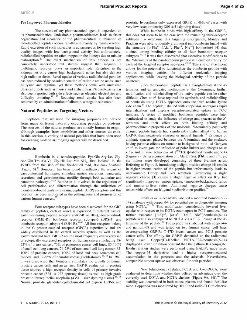
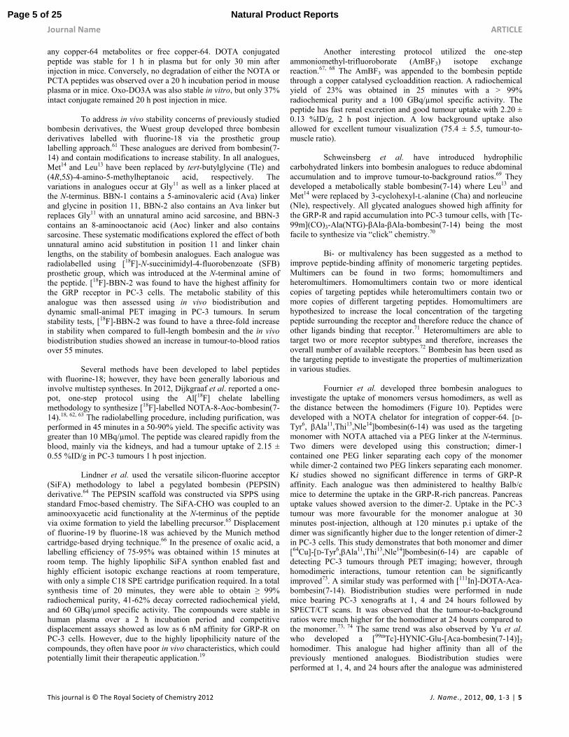
Since the bombesin peptide has a pyroglutamate at the N-terminus and an amidated methionine at the C-terminus, further modification and radiolabelling of the native peptide can be rather difficult. Chen et al. have reported the synthesis and radiolabelling of bombesin using DOTA appended onto the third residue lysine side chain.54 The peptide, labelled with copper-64, undergoes rapid internalization and displays receptor-mediated uptake in PC-3 tumours. A series of modified bombesin peptides were later synthesized to study the influence of charge and spacers at the N-terminus and their effect on both binding affinity and pharmacokinetic properties of the molecule. N-terminally positively charged peptide ligands had significantly higher affinity to human GRP-R than negatively charged or neutral ligands.55 Evidence of aliphatic spacers, placed between the N-terminus and the chelator, having positive effects on tumour-to-background ratio led Garayoa et al. to investigate the influence of polar linkers and charges on in vitro and in vivo behaviour of [99mTc(I)]-labelled bombesin(7-14) (Figure 7). Using a combination of βAla, β3hSer, β3hGlu and β3hLys, six linkers were developed consisting of three β-amino acids. Referring to Figure 8, introducing a slight positive charge (4) results in higher internalization of the peptide but unfortunately leads to unfavourable kidney and liver retention. Introducing a slight negative charge (3) causes a slight negative effect on IC50 but significantly improves tumour uptake, tumour-to-background ratios and tumour-to-liver ratios. Additional negative charges cause undesirable effects on IC50 and biodistribution profiles.56
Smith et al. successfully labelled a modified bombesin(7-
14) analogue with copper-64 for potential use in diagnostic imaging using NOTA.57, 58 This modification considerably lowered liver uptake with respect to its DOTA counterpart in PC-3 tumours. The further truncated [D-Tyr6, βAla11, Thi13, Nle14]bombesin(6-14) peptide was also conjugated to NOTA via a PEG linkage at the N-terminus of the peptide.59 The peptide was labelled with copper-64 and gallium-68 and was tested on two human cancer cell lines overexpressing GRP-R: T-47D breast cancer and PC-3 prostate cancer cells. The affinity for GRP-R depended on the radiometal being used. Copper(II)-labelled NOTA-PEG-bombesin(6-14) displayed a lower inhibition constant than the gallium(III) conjugate. Biodistribution studies were performed using BALB/c nude mice. The copper-64 derivative had a higher receptor-mediated accumulation in the pancreas and the adrenals. Nevertheless, comparable tumour uptake was observed for both peptides.
New bifunctional chelates, PCTA and Oxo-DO3A, were
evaluated to determine whether they offered an advantage over the currently used DOTA and NOTA chelates (Figure 9A, 9B).60 The stability was determined in both mouse plasma and female BALB/c mice. Copper-64 was monitored by HPLC and radio-TLC to observe
Page 4 of 25Natural Product Reports
Journal Name ARTICLE
This journal is © The Royal Society of Chemistry 2012 J. Name., 2012, 00, 1-3 | 5
any copper-64 metabolites or free copper-64. DOTA conjugated peptide was stable for 1 h in plasma but for only 30 min after injection in mice. Conversely, no degradation of either the NOTA or PCTA peptides was observed over a 20 h incubation period in mouse plasma or in mice. Oxo-DO3A was also stable in vitro, but only 37% intact conjugate remained 20 h post injection in mice.
To address in vivo stability concerns of previously studied
bombesin derivatives, the Wuest group developed three bombesin derivatives labelled with fluorine-18 via the prosthetic group labelling approach.61 These analogues are derived from bombesin(7-14) and contain modifications to increase stability. In all analogues, Met14 and Leu13 have been replaced by tert-butylglycine (Tle) and (4R,5S)-4-amino-5-methylheptanoic acid, respectively. The variations in analogues occur at Gly11 as well as a linker placed at the N-terminus. BBN-1 contains a 5-aminovaleric acid (Ava) linker and glycine in position 11, BBN-2 also contains an Ava linker but replaces Gly11 with an unnatural amino acid sarcosine, and BBN-3 contains an 8-aminooctanoic acid (Aoc) linker and also contains sarcosine. These systematic modifications explored the effect of both unnatural amino acid substitution in position 11 and linker chain lengths, on the stability of bombesin analogues. Each analogue was radiolabelled using [18F]-N-succinimidyl-4-fluorobenzoate (SFB) prosthetic group, which was introduced at the N-terminal amine of the peptide. [18F]-BBN-2 was found to have the highest affinity for the GRP receptor in PC-3 cells. The metabolic stability of this analogue was then assessed using in vivo biodistribution and dynamic small-animal PET imaging in PC-3 tumours. In serum stability tests, [18F]-BBN-2 was found to have a three-fold increase in stability when compared to full-length bombesin and the in vivo biodistribution studies showed an increase in tumour-to-blood ratios over 55 minutes.
Several methods have been developed to label peptides with fluorine-18; however, they have been generally laborious and involve multistep syntheses. In 2012, Dijkgraaf et al. reported a one-pot, one-step protocol using the Al[18F] chelate labelling methodology to synthesize [18F]-labelled NOTA-8-Aoc-bombesin(7-14).18, 62, 63 The radiolabelling procedure, including purification, was performed in 45 minutes in a 50-90% yield. The specific activity was greater than 10 MBq/µmol. The peptide was cleared rapidly from the blood, mainly via the kidneys, and had a tumour uptake of 2.15 ± 0.55 %ID/g in PC-3 tumours 1 h post injection.
Lindner et al. used the versatile silicon-fluorine acceptor (SiFA) methodology to label a pegylated bombesin (PEPSIN) derivative.64 The PEPSIN scaffold was constructed via SPPS using standard Fmoc-based chemistry. The SiFA-CHO was coupled to an aminooxyacetic acid functionality at the N-terminus of the peptide via oxime formation to yield the labelling precursor.65 Displacement of fluorine-19 by fluorine-18 was achieved by the Munich method cartridge-based drying technique.66 In the presence of oxalic acid, a labelling efficiency of 75-95% was obtained within 15 minutes at room temp. The highly lipophilic SiFA synthon enabled fast and highly efficient isotopic exchange reactions at room temperature, with only a simple C18 SPE cartridge purification required. In a total synthesis time of 20 minutes, they were able to obtain ≥ 99% radiochemical purity, 41-62% decay corrected radiochemical yield, and 60 GBq/µmol specific activity. The compounds were stable in human plasma over a 2 h incubation period and competitive displacement assays showed as low as 6 nM affinity for GRP-R on PC-3 cells. However, due to the highly lipophilicity nature of the compounds, they often have poor in vivo characteristics, which could potentially limit their therapeutic application.19
Another interesting protocol utilized the one-step ammoniomethyl-trifluoroborate (AmBF3) isotope exchange reaction.67, 68 The AmBF3 was appended to the bombesin peptide through a copper catalysed cycloaddition reaction. A radiochemical yield of 23% was obtained in 25 minutes with a > 99% radiochemical purity and a 100 GBq/µmol specific activity. The peptide has fast renal excretion and good tumour uptake with 2.20 ± 0.13 %ID/g, 2 h post injection. A low background uptake also allowed for excellent tumour visualization (75.4 ± 5.5, tumour-to-muscle ratio).
Schweinsberg et al. have introduced hydrophilic carbohydrated linkers into bombesin analogues to reduce abdominal accumulation and to improve tumour-to-background ratios.69 They developed a metabolically stable bombesin(7-14) where Leu13 and Met14 were replaced by 3-cyclohexyl-L-alanine (Cha) and norleucine (Nle), respectively. All glycated analogues showed high affinity for the GRP-R and rapid accumulation into PC-3 tumour cells, with [Tc-99m](CO)3-Ala(NTG)-βAla-βAla-bombesin(7-14) being the most facile to synthesize via “click” chemistry.70
Bi- or multivalency has been suggested as a method to improve peptide-binding affinity of monomeric targeting peptides. Multimers can be found in two forms; homomultimers and heteromultimers. Homomultimers contain two or more identical copies of targeting peptides while heteromultimers contain two or more copies of different targeting peptides. Homomultimers are hypothesized to increase the local concentration of the targeting peptide surrounding the receptor and therefore reduce the chance of other ligands binding that receptor.71 Heteromultimers are able to target two or more receptor subtypes and therefore, increases the overall number of available receptors.72 Bombesin has been used as the targeting peptide to investigate the properties of multimerization in various studies. Fournier et al. developed three bombesin analogues to investigate the uptake of monomers versus homodimers, as well as the distance between the homodimers (Figure 10). Peptides were developed with a NOTA chelator for integration of copper-64. [D-Tyr6, βAla11,Thi13,Nle14]bombesin(6-14) was used as the targeting monomer with NOTA attached via a PEG linker at the N-terminus. Two dimers were developed using this construction; dimer-1 contained one PEG linker separating each copy of the monomer while dimer-2 contained two PEG linkers separating each monomer. Ki studies showed no significant difference in terms of GRP-R affinity. Each analogue was then administered to healthy Balb/c mice to determine the uptake in the GRP-R-rich pancreas. Pancreas uptake values showed aversion to the dimer-2. Uptake in the PC-3 tumour was more favourable for the monomer analogue at 30 minutes post-injection, although at 120 minutes p.i uptake of the dimer was significantly higher due to the longer retention of dimer-2 in PC-3 cells. This study demonstrates that both monomer and dimer [64Cu]-[D-Tyr6,βAla11,Thi13,Nle14]bombesin(6-14) are capable of detecting PC-3 tumours through PET imaging; however, through homodimeric interactions, tumour retention can be significantly improved73. A similar study was performed with [111In]-DOTA-Aca-bombesin(7-14). Biodistribution studies were performed in nude mice bearing PC-3 xenografts at 1, 4 and 24 hours followed by SPECT/CT scans. It was observed that the tumour-to-background ratios were much higher for the homodimer at 24 hours compared to the monomer.73, 74 The same trend was also observed by Yu et al. who developed a [99mTc]-HYNIC-Glu-[Aca-bombesin(7-14)]2
homodimer. This analogue had higher affinity than all of the previously mentioned analogues. Biodistribution studies were performed at 1, 4, and 24 hours after the analogue was administered
Page 5 of 25 Natural Product Reports
ARTICLE Journal Name
6 | J. Name., 2012, 00, 1-3 This journal is © The Royal Society of Chemistry 2012
to nude mice bearing PC-3 tumours. Tumour to non-tumour ratios increased from 1 to 24 hours p.i.75 However, in all three cases, there is no evidence that two bombesin moieties are capable of binding two GRP-R’s simultaneously.
Generally agonists have been considered crucial for targeting receptors due to their attractive internalization properties, as they result in optimal tumour-to-background ratios. However, the development of antagonists with high tumour uptake despite their low internalization rate has disproved this notion.75 Many bombesin antagonists have been explored such as [99mTc]-demobesin 1 and [111In]-DOTA-amino-hexanoyl-[D-Phe6,Leu-NH(CH2)3CH3
13,des-Met14]-bombesin(6-14) supporting the use of radiolabelled bombesin antagonists. Mansi et al. describe a DOTA-conjugated bombesin antagonist, DOTA-4-amino-1-carboxymethyl-piperidine-D-Phe-Gln-Trp-Ala-Val-Gly-His-Sta-Leu-NH2, referred to as DOTA-RM2 that is capable of SPECT imaging with indium-111 and PET imaging with gallium-68. In vitro studies performed with PC-3 cells showed that surface bound activity greatly exceeded internalized activity with indium-111 as expected for an antagonist. Biodistribution studies were performed on nude mice bearing PC-3 xenografts with [111In] DOTA-RM2. The studies resulted in rapid blood clearance after four hours p.i. but more importantly, had tumour uptake values more than five-fold higher than their agonist counterparts (Figure 11).76 Respectable tumour uptake values at 72 hours were also observed, suggesting better tumour retention than agonist counterparts. The extended retention suggests antagonists could be useful for therapeutics when labelled with long half-life radioisotopes.
In 2013, a first-in-man study was performed by Ananias et al. using [99mTc]-HYNIC(tricine/TPPTS)-Aca-bombesin(7-14) ([99mTc]-HABBN) in eight men with biopsy-proven prostate cancer.77 No adverse side effects were reported; however, low metabolic stability was observed with metabolites forming as early as 10 minutes p.i. Researchers determined that at 30 minutes post-injection, less than 20% of [99mTc]-HABBN remained intact. This was in stark contrast to the initial in vitro study which showed that [99mTc]-HABBN was stable in human serum for 6 h with 77% of the compound remaining completely intact even after 24 h.78 Although immediate distribution via the vascular system and rapid excretion through the kidneys were observed from the dynamic images, no uptake of radioactivity in prostate or lymph node metastases was detected by SPECT/CT at any time point in any patient. This failure to visualise any prostate tumours was concluded to be due to the observed rapid in vivo degradation.
More recently, preclinical studies have demonstrated that radiolabelled antagonist-based bombesin peptides might be superior as targeting vectors compared to their agonist conterparts.79-81 BAY 86-7548 was the first bombesin antagonist analysed in healthy men in 2013.82 The first-in-human study investigated the safety, metabolism, pharmacokinetics, and biodistribution of the [68Ga]-DOTA-4-amino-1-carboxymethyl-piperidine-D-Phe-Gln-Trp-Ala-Val-Gly-His-Sta-Leu-NH2 agonist. Five healthy men (age: 52 ± 2 y, weight: 77 ± 10 kg, height: 172 ± 5 cm) were enrolled and underwent dynamic whole-body PET/CT after i.v. administration of BAY 86-7548 (138 ± 5 MBq). The mean effective dose was 0.051 mSv/MBq and the organs with the highest absorbed doses were the bladder (0.62 mSv/MBq) and the pancreas (0.51 mSv/MBq). The compound underwent rapid metabolism in vivo, with the proportion of unchanged antagonist dropping from 92% after 1 minute p.i to 19% at 65 minutes. Three main metabolites were observed, with the most prevalent being the cleaved [68Ga]-DOTA chelate.
Kähkönen et al. proceeded to investigate the use of the gallium-68 labelled BAY 86-7548 in vivo.83 The tracer showed high sensitivity and specificity for detection of primary prostate cancer and lymph node metastases, detecting organ-confined prostate cancer with an accuracy of 83%. Gallium-68 labelled BAY 86-4367 was able to detect the dominant tumour lesions in 10 of the 11 patients with primary prostate cancer. The scan also correctly detected local recurrence and lymph node metastases in two of the three patients with a biochemical relapse. Currently, the most commonly used tracers for imaging of biochemical relapse are 18F-fluoromethylcholine and 11C-acetate. However, these tracers have shown an inability to differentiate between benign hyperplastic prostate cells and malignant carcinoma cells, resulting in limited diagnostic value.84-86 Despite the relatively small number of patients, the accuracy of gallium-68 labelled BAY 86-4367 observed in this study is very encouraging for the detection and evaluation of organ-confined prostate cancers.
In 2015, fluorine-18 labelled BAY 86-4367 (3-cyano-4-[18F]-fluorobenzoyl-Ala(SO3H)-Ala(SO3H)-Ava-Gln-Trp-Ala-Val-NMeGly-His-Sta-Leu-NH2) underwent clinical evaluation in a small subset of patients with primary and recurrent prostate cancer.87 Whole body PET/CT scans were assessed at six time points, up to 110 minutes p.i. of 302 ± 11 MBq. The GRP-R antagonist was safe and well tolerated and had a mean effective dose of 0.014 mSv/MBq. This was lower than the gallium-68 labelled BAY 86-4367 analogue which had a mean effective dose of 0.051 mSv/MBq and is also lower than [18F]-fluoromethylcholine (0.031 mSv/MBq). The low gastrointestinal radiation dose observed represented a clear advantage over other literature-known labelled antagonists.88, 89 In 50% of the patients, fluorine-18 labelled BAY 86-4367 displayed accumulation in the malignant prostate tissue at a ratio of 4.4 ± 0.6 to normal prostate tissue. Three of the five patients with primary prostate cancer showed tumour delineation in the prostate. However, of the five recurrent disease cases only two suggestive lesions were detected by BAY 85-4367, whereas [18F]-fluoromethylcholine PET/CT depicted suggestive lesions in all five patients. The slight structural modifications between the gallium-68 labelled BAY 86-4367 and the fluorine-18 labelled BAY 86-4367 are most likely the reason for their substantially different diagnostic performances. In the fluorine-18 compound, the glycine residue has been replaced with an NMeGly. This structural modification could affect both peptide stability and excretion. Alternatively, the two –SO3H groups in the linker of the fluorine-18 labelled BAY 86-4367 could have decreased cell permeability at physiologic pH compared with the neutral 68Ga-compound and overall, decreasing tumour uptake.
As can be seen by the number of bombesin derivatives reported in the literature, significant effort has been expended to develop a bombesin based imaging agent. Both agonists and antagonists have been evaluated with high affinity ligands being identified. These agents remain as linear peptide sequences, where in vivo stability needs to be carefully considered in the development strategy. Clinical evaluation of some of these agents has shown promise and yet there is no marketed clinical product to date, possibly complicated by the limited ability to patent protect the entities due to the extensive number of structures that have been reported (prior art). Other targeting approaches to the imaging of prostate cancer have also shown promise. In particular, the imaging of prostate-specific membrane antigen (PSMA) by means of either small molecules (urea based inhibitors) or antibodies, are progressing to the clinic and it remains to be seen as to which targeting approach, bombesin versus PSMA, proves most beneficial for the patient. The potential to create a theranostic bombesin agent allowing for both imaging and therapy is particularly attractive, yet will require
Page 6 of 25Natural Product Reports
Journal Name ARTICLE
This journal is © The Royal Society of Chemistry 2012 J. Name., 2012, 00, 1-3 | 7
optimal pharmacokinetic properties for the ligand, in order to limit non-target toxicity as a radiotherapeutic.
Gastrin and Cholecystokinin
The existence of gastrin was proposed by Edkins in 1905 as a humoral mediator of gastric acid secretion, but remained controversial until 1964 when the structure of gastrin was determined.90, 91 Gastrin is a gastrointestinal peptide found in humans in two main forms, a 17 amino acid form (G17) and a 34 amino acid form (G34) that contains an amidated C-terminus and can be present in sulphated and non-sulphated forms. The primary physiological function of gastrin is to act on acid-secreting parietal cells and regulate expression of gastric Trefoil factors (TFFs). More recently, gastrin has been tied to the development of gastric tumours by causing epithelial remodelling, and epithelial-mesenchymal signalling or transition.92
Cholecystokinin (CCK) is another gastrointestinal peptide hormone originally discovered in 1928 that shares structural similarities to gastrin.93 The CCK sequence was originally reported to contain 33 amino acids but through further investigation of this peptide, it was discovered that CCK exists in various biologically active forms. The most common forms are CCK39, CCK33, CCK8, and CCK4 all of which are derived from a 115 amino acid precursor.94 CCK acts as a neurotransmitter/neuromodulator within the central nervous system (CNS), as well as controls various functions within the gastrointestinal tract such as gall bladder contraction, pancreatic enzyme secretion and gut motility.95, 96
CCK and gastrin share an amidated C-terminal receptor binding sequence, Trp-Met-Asp-Phe-NH2. These two groups of peptides differ based on the position of a tyrosyl residue with respect to the receptor binding sequence. Gastrin peptides generally contain one glycine amino acid between the Tyr residue on the N-terminal side of its receptor binding domain while CCK peptides contain two amino acids (generally Met-Gly or Thr-Gly).97 Considering their similarities, it is not surprising that gastrin and CCK peptides share the same set of receptors composed of three receptor subtypes; CCK1, CCK2 and CCK2i4sv.94
The previously mentioned tyrosine residue plays an important role in receptor specificity. When sulphated, CCK/gastrin analogues have high specificity for CCK1 and CCK2 receptors, while non-sulphated analogues show 1000-fold lower specificity for CCK1 with respect to CCK2.97 CCK2 can often be referred to as the “gastrin receptor” due to its affinity for gastrin. Overall, the CCK1 and CCK2 receptors have been identified in a large variety of human cancers. The more commonly found receptor, CCK2, has been located in medullary thyroid carcinoma (92%), astrocytoma (65%), stromal ovarian cancer (100%) and occasionally found in leiomyosarcoma. The CCK1 receptor was detected in meningioma (30%) as well as neuroblastoma. The presence of sufficiently high receptor density in various tumour types has gained a lot of attention and resulted in the development of various targeted gastrin and CCK radiopeptide analogues.
The first radiolabelled gastrin analogue was developed in 1998 by Behr, et al. in order to visualize medullary thyroid carcinoma (MTC). A G17 analogue (coined “little gastrin”) with the structure pGlu-Gly-Pro-Trp-Leu-(Glu)5-Ala-Tyr-Gly-Trp-Met-Asp-Phe-NH2 was radioiodinated at the tyrosine-12 moiety. The labelled peptide was evaluated in female nude mice bearing the MTC TT cell line and monitored over 24 hours. The [131I-Tyr12]G17 tracer had higher retention in tumour when compared to all other tissues with
the exception of the excretory organs (kidney, liver and gall bladder). Following successful diagnosis with the developed tracer, the radiolabelled gastrin was assessed for therapeutic treatment of CCK2 bearing tumours. Tumour size was found to decrease with respect to control mice and reached full remission with no tumour re-growth over a 22-week period.98 Although this analogue showed promising results in mice, imaging in human was less than optimal.
Subsequently, Behr performed an extensive study on gastrin and CCK analogues to optimize in vivo targeting and stability of the previously published [131I-Tyr12]G17 tracer (Table 3).99 Biodistribution studies were performed for all radioiodinated analogues at 1 h in MCT TT xenografts. Of the gastrin family, “minigastrin” (G14) showed the most promising results as well as favourable kinetics in excretory organs compared to other gastrin analogues. Following this discovery, various radiolabelled analogues of G14 arose bearing common isotopes for nuclear imaging such as 68Ga, 111In and 99mTc.100-108
The first technetium-99m analogue was developed by Von
Guggenberg et al. in 2004 by introducing a HYNIC bifunctional
chelator at the N-terminus of G14. Biodistribution studies showed
unfavourable kidney uptake requiring optimization for further
application.100 The following year, minigastrin was derivatized using
open-chain tetraamine chelators capable of stably binding
technetium-99m successfully in somatostatin and bombesin
analogues.109, 110 Of the three minigastrin analogues developed,
demogastrin-2 ([N40-1,Gly0-D-Glu1]-G14) (N4=
(H2NCH2CH2NHCH2)CH-(p-CH2C6H4)-NHCOCH2OCH2CO-) showed the most promise with rapid blood clearance, high tumour
uptake, rapid background clearance and lower liver uptake than the
other two analogues and therefore was subjected to MTC patient
imaging. [99mTc]-demogastrin 2 was injected into a patient with
MTC metastases in lymph nodes, lungs and bone. Within 90
minutes, all known lesions were detected and at four hours p.i.
images had increased signal-to-noise due to rapid background
clearance.101
In 2011, The European Journal of Nuclear Medicine and Molecular Imaging enlisted nine European research groups to develop standardized methods to investigate in vitro and in vivo characteristics of twelve indium-111 labelled CCK binding ligands. These studies were summarized into three publications focusing on in vivo biodistribution, internalization, as well as biological stability and metabolism of all twelve analogues.97, 111, 112 The study included two CCK analogues and ten gastrin analogues gathered from previous literature (Table 4). During biodistribution studies, overall tumour uptake was found to be much lower for CCK analogues than gastrin analogues. G14 had high tumour uptake values 4 h p.i but resulted in much higher kidney retention. Other modified analogues such as PP-F11, which simply replaces the L-Glu in G14 with D-Glu, retained tumour uptake while decreasing kidney uptake to one-tenth that of G14. Cyclo-MG1, a cyclized analogue of MG11, was found to have high tumour uptake at 1 h p.i and low kidney uptake but was found to have poor tumour retention causing a 40% lower tumour uptake 4 h p.i. Biological stability studies investigated peptide stability in human serum, as well as homogenized rat liver and kidney tissues. PP-F10 and MG11 were found to be the most stable in human serum, while PP-F11 was the most stable in homogenized tissues and had high stability in human serum. During identification of metabolites, all major cleavage sites were found in the C-terminal
Page 7 of 25 Natural Product Reports
ARTICLE Journal Name
8 | J. Name., 2012, 00, 1-3 This journal is © The Royal Society of Chemistry 2012
region of peptides. Therefore, modifications should focus on this structural region in order to enhance in vitro and in vivo stability.
Reubi et al. were the first to explore the occurrence of CCK2 receptors in human cancers and developed an iodinated non-sulphated CCK(26-33) analogue (known as CCK8).113 They developed a CCK analogue linked to a chelator to accommodate more clinically useful radioisotopes such as indium-111. Based on the parent peptide, H-Asp-Tyr-Met-Gly-Trp-Met-Asp-Phe-NH2, nine other analogues containing a chelator, unnatural amino acids, and D-amino acids were rationally designed and binding affinities of each analogue were compared to that of the parent peptide. The lead analogue, DTPA-D-Asp-Tyr-Nle-Gly-Trp-Nle-Asp-Phe-NH2, was designed by placing a DTPA chelator on the N-terminus and replacing both methionine residues with the unnatural amino acid norleucine (Nle). Biodistribution studies were performed on the indium-111 labelled compound in healthy female rats at 1, 4 and 24 hours to monitor uptake in the digestive tract known for higher CCK2 receptor densities.114 These studies showed localization of the lead analogue in the small and large intestines known to express the CCK2 receptor.
Ghrelin
The growth hormone secretagogue receptor (GHSR) is a seven-transmembrane receptor responsible for feeding behaviour. The natural ligand is ghrelin, a 28 amino acid peptide, which was discovered in 1999 to be the endogenous ligand for GHSR-1a, the predominant receptor sub-type (Figure 12).115
Multiple tumour types have been reported to have an overexpression of GHSR-1a which has led to the proposal that analogues of ghrelin may be useful as oncologic imaging agents.116 Recently, a fluorescent-ghrelin(1-18) analogue was developed and was able to distinguish prostate cancer from benign hyperplasia in ex vivo prostate tissue (Figure 13).117 Additionally, this optical agent was investigated for its ability to image the heart and may be useful for the imaging of cardiac myopathy, a complication of diabetes.118
The native ghrelin sequence is unlikely to be of value as an in vivo imaging agent, due to the susceptibility of the Ser3 octanoyl ester side chain to undergo enzymatic deacylation. To stabilize the ghrelin peptide, an amide linkage to the lipophilic side chain can be used instead of the ester, resulting in an improved biological half-life. Truncation of the C-terminal amino acids has allowed for a significant reduction in the molecular weight of the peptide.119, 120 The unique octanoyl side chain located at Ser3 is critical for ghrelin’s physiological function, however, this side chain can be readily replaced with other lipophilic moieties.119, 120
Two approaches to the radiolabelling of ghrelin have been explored. The first approach is the classical method of adding a metal chelator pendant to the peptide analogues at the C-terminus via a lysine residue. In one instance, a DOTA conjugated ghrelin(1-19), which also contained a diaminopropanoic acid residue in position three, was radiolabelled with gallium-68 for use as a PET imaging agent and the gallium-69/71 variant was determined to have an IC50 of 9.1 nM for the GHSR.121 In another instance, a monodentate isocyanide ligand conjugated ghrelin(1-6) was radiolabelled with technetium-99m and determined to have an IC50 of 45 nM for GHSR.122 The second approach is an integrated design whereby the radioisotope is attached as part of a lipophilic side chain, replacing the octanoyl side chain of native ghrelin. Fluorine-containing side chains, both in the form of an aliphatic chain and as an aromatic entity, have been reported and the addition of a bulky fluoro-napthyl
group appears the most promising to date for eventual use as a fluorine-18 labelled ghrelin analogue.123 In addition, a side chain containing a rhenium cyclopentadienyl tricarbonyl group has also been discovered to have GHSR affinity and is a unique discovery in that it is a rare example where an organometallic species is a key recognition element for a peptide-receptor interaction.116
Glucagon-Like Peptide-1
Glucagon-like peptide-1(GLP-1) is a peptide hormone that is responsible for the release of insulin from the β-cells in the pancreas. The GLP-1 receptor is a member of the GPCR B family and the binding of an agonist such as GLP-1 results in multiple downstream events including increased cAMP, activation of PI-3K and PKA, among others. Imaging of GLP-1R has potential utility for the imaging of insulinomas, a rare form of pancreatic cancer, and for the determination of β-cell mass.
The development of GLP-1 derived imaging agents has focused predominantly on two approaches, using either the human GLP-1(7-36)-NH2 peptide as the ligand or using the peptide exendin, which was discovered in the venom of the lizard Gila monster (Heloderma suspectum). Early efforts for creating a GLP-1 derived imaging agent were based upon simple radioiodination of the peptide, presumably on the Tyr residue in position 19, which resulted in a probe that demonstrated an ability to image insulinomas in vivo in a mouse model.124 However, the same authours have also commented that this radioiodinated probe had insufficient in vivo stability and a low radioiodination efficiency.125
In 2010, analogues of GLP-1(7-37) were reported as potential imaging agents, through placement of a DOTA chelator and radiolabelling with indium-111.126 A number of locations were explored for placement of the metal chelator complex, with positions 22 and 37 proving to be the most successful at retaining GLP-1R affinity. An important modification from the native sequence of GLP-1 was having the L-Ala8 be replaced with D-Ala8 resulting in a significant improvement in stability to serum proteases. Other reports have indicated that unnatural amino acids such as Aib (amino-isobutyric acid) can also be used for this purpose.127 This modification is critical in order to prevent degradation by DPP-IV, dipeptidyl peptidase-4. More recently a gallium-68 labelled version of GLP-1 has also been reported and PET imaging of insulinoma in a murine model was demonstrated.128 The GLP-1 peptide exists in a predominately alpha-helical structure, thus another approach to stabilizing the secondary structure is to create stapled peptides, whereby side chains are joined together at positions i/i+4 or i/i+7. Gao et al. reported the design of a GLP-1 analogue where two lactam bridges were installed, resulting in a stable GLP-1 derivative named FBEM-EM3106B.129 The PET radiolabel fluorine-18 was then installed through a C-terminal cysteine using thiol-maleimide conjugation chemistry.
While not the human ligand for GLP-1R, the 39 amino acid peptide exendin-4 has 53% homology with GLP-1 and has strong affinity for the receptor.130 Imaging agents based on exendin-4 have focused primarily on modifications to the C-terminus in order to radiolabel this peptide, typically through the addition of a Lys residue at position 40. An early report added the chelator DTPA to the Lys40 side chain, radiolabelled with indium-111 and described receptor-mediated uptake in GLP-1R expressing stomach, lung and pancreas.131 Since this early report, a variety of 68Ga, 18F and 64Cu exendin analogues have been reported, as detailed in Table 5. In general, the most promising approach appears to be the addition of a metal chelator at the C-terminus of the peptide, through the side
Page 8 of 25Natural Product Reports

Journal Name ARTICLE
This journal is © The Royal Society of Chemistry 2012 J. Name., 2012, 00, 1-3 | 9
chain of a lysine or cysteine. One study explored the conjugation of a fluorine-18 label through a cysteine at position 0 as compared to position 40 and while both of the analogues had receptor affinity, the authors concluded that modification at position 40 was preferred due to better in vivo targeting and high uptake in an insulinoma (INS-1) tumour.132 Replacing the Met14 with Nle14 is reported as a beneficial modification in order to avoid oxidation.133 Other exendin-4 C-terminal modifications include: [Lys40(Ahx-DOTA-Ga-68)NH2]exendin-4 where an amino hexanoic acid (Ahx) spacer was used to separate the peptide from the metal complex and a SPECT agent using the readily available isotope technetium-99m, [Lys40([99mTc]-HYNIC)NH2]exendin-4.134 One report compared the conjugation of a chelator at three different locations in exendin-4: Lys12, Lys27 and Lys40, and concluded that positions 12 and 40 are both suitable for modification.135 A paper from Merck reports that Cu-64 labelled [Lys40(DOTA)NH2]exendin-4 was taken up preferentially in GLP-1R expressing islets, as determined through ex vivo analysis of mouse pancreas, and thus could be useful for non-invasive PET imaging of beta cell mass, although the ability to carry out quantitative in vivo imaging of beta cell mass remains controversial (Figure 14).136-138
Within the last 8 years, clinical trials have been performed using exendin-4 analogues labelled with indium-111.139-142 The first example used [111In]-DOTA-exendin-4 to detect insulinoma in two patients with lesions poorly detected by conventional methods. Through SPECT/CT imaging, the small lesions were detected and confirmed by histological analysis although the highest localization was observed in the kidneys.142 A larger clinical study was performed in 2013 on 30 patients with hyperinsulinaemic hypoglycaemia. Patients were administered [111In]-[Lys40(Ahx-DTPA)NH2]-exendin-4 and SPECT/CT imaging was done 168 hours after injection. This tracer was able to correctly detect 23 positive lesions resulting in a positive predictive value of 83%.140 These results are promising for using radiolabelled exendin-4 analogues for detection of small insulinomas that are difficult to localize using the conventional methods.
Exendin-3 shares 95% homology with exendin-4 and has also been used as the basis for creating PET and SPECT imaging agents targeting GLP-1R. A study comparing both DTPA and DOTA chelators, as well as looking at 111In and 68Ga, determined that exendin-3 analogues are able to target GLP-1R in an INS-1 xenograft model, with little difference seen in the biodistribution results between these two exendin isoforms.143
The first example of a GLP-1 PET imaging analogue using fluorine-18 was published in 2011. This analogue, EM3106B, was designed to induce alpha helical structure found in the N and C terminal regions of natural GLP-1 by introducing 2 lactam bridges within the analogue. Fluorine-18 was incorporated using a fluorobenzamide prosthetic group, [18F]-FBEM, on the C terminal end through a cysteine side chain. When compared to natural GLP-1, [18F]-FBEM-E3106B had better affinity to GLP1-R in the insulinoma cell line INS-1. Preclinical evaluation in mouse INS-1 xenografts showed localization to the tumour and off-target uptake mainly present in the kidneys.129 Since GLP-1 has become known as an unstable peptide susceptible to degradation, the same group applied the developed methodology to exendin-4 by adding a cysteine the C-terminus of the peptide. [18F]-FBEM-[Cys40]-exendin-4 resulted in better affinity for GLP-1R than the native GLP-1 and similar affinity to [18F]-FBEM-E3106B.132 Given the difficulties associated with traditional prosthetic group labeling with fluorine-18, the exendin-4 analogue was further optimized to be radiolabelled using aluminum fluoride as it requires a more facile
labeling approach. In these analogues, a NOTA chelator replaced the prosthetic group on the C-terminal cysteine. During radiolabeling, cyclotron produced [18F]fluoride is introduced to the NOTA analogue in the presence of AlCl3 to yield a Al[18F]-NOTA analogue of exendin-4 in moderate radiochemical yield and specific activity. However, this method of synthesis resulted in a lower radiochemical yield, lower specific activity and slightly diminished affinity to GLP-1R than [18F]Al-NOTA-exendin-4.144
Melanocyte-stimulating Hormone
The melanocortin 1 receptor (MC1R) is a GPCR involved in the regulation of mammalian skin and hair colour through a process called melanogenesis.145, 146 MC1R is a cell membrane-embedded protein, which is activated by the α-melanocyte-stimulating hormone (α-MSH). Upon binding, the endogenous tridecapeptide hormone (Figure 15) initiates a complex signalling cascade that leads to the production of pigment. Melanoma contributes to more than 50% of all skin cancer deaths, making it the most lethal form of skin cancer and the most commonly diagnosed malignancy among young adults.147, 148 Due to its high metastatic potential, aggressive nature, and resistance to chemotherapeutics, improvement in patient survival rates heavily relies significantly on early diagnosis, therefore the development of melanoma-specific diagnostic agents is highly desirable. More than 80% of human metastatic melanoma samples have an overexpression of MC1R receptors, making this an ideal target for radiolabelled α-MSH peptides.149 Structure-activity studies have shown that the minimal sequence of α-MSH required for biological activity is His6-Phe7-Arg8-Trp9.149-151 The replacement of Met4 and Phe7 with the unnatural amino acids, Nle4 and D-Phe7 respectively, yielded a potent NDP-MSH analogue, which displays greater MC1R affinity, longer plasma half-life and increased enzymatic stability than the endogenous ligand.149 The truncated [Ac-Nle4, Asp5, D-Phe7, Lys11] α-MSH(4-11) (NAP-NH2) has been the most studied analogue for melanoma imaging. Recently, the multivalency concept was applied to the design of novel conjugates containing a pyrazolyl-diamine chelating the Re(CO)3
+ or [99mTc]-(CO)3+ core and two copies of the
targeting vector NAP-NH2. Binding affinity of the bivalent conjugates was found to be up to 19-fold higher than that of the monovalent NAP-NH2 conjugate.152
Cyclization has also been used as a means to improve binding affinity, in vivo stability, and receptor selectivity.149, 153, 154 Three different methods have been attempted: side chain to side chain disulphide bridge and lactam cyclization, as well as metal-based cyclization. The first disulphide bridge-containing α-MSH peptides were conjugated to DOTA and labelled with indium-111 (Figure 16A). The effect of cyclization on in vivo melanoma targeting was evaluated and compared to that of the corresponding linear peptide analogues.155 The disulphide-bridged compounds displayed moderate tumour uptake and high kidney accumulation at 2 h post injection in B16F1 murine melanoma-bearing mice, as well as increased receptor-binding affinity and resistance to proteolysis. The corresponding linear radiopeptide showed a decreased tumour uptake at 2 h post injection, underlining the benefit of cyclization.
The peptide, βAla-Nle-c[Asp-His-D-Phe-Arg-Trp-Lys]-NH2 (βAlaNleCycMSHhex), was synthesized via lactam bridge formation through the aspartic acid and lysine side chains (Figure 16B).156 Both βAlaNleCycMSHhex and the corresponding linear sequence, MSHoct, were conjugated to a PZL bifunctional chelator.156, 157 Lactam cyclization resulted in an enhancement in
Page 9 of 25 Natural Product Reports

ARTICLE Journal Name
10 | J. Name., 2012, 00, 1-3 This journal is © The Royal Society of Chemistry 2012
binding affinity over the linear conjugate. Both peptides were labelled with [99mTc]-(CO)3
+, with βAlaNleCycMSHhex displaying 30-fold higher internalization in B16F1 cells and 110-fold higher tumour uptake at 4 h post injection, compared to the linear MSHoct
conjugate.156 However, despite the promising tumour-targeting properties, the pharmacokinetic profile still needed improvement as cyclization negatively affected clearance and excretion rates. The effect of different pyrazolyl-diamine chelator substitution patterns on the pharmacokinetic properties was also investigated. In particular, the introduction of a carboxylate group in the 4-position of the azolyl ring. This lead to a large reduction in both kidney and liver accumulation for [99mTc]-(CO)3-Pz3-βAlaNleCycMSHhex and [99mTc]-(CO)3-Pz4-βAlaNleCycMSHhex when compared to the basic pzl chelator, where the 4-position of the azolyl ring is unsubstituted.158
Miao et al. also synthesized lactam bridge-based cyclizations using DOTA-CycMSH and DOTA-Gly-Glu-CycMSH conjugates (Figure 16C).159 Both indium-111 labelled peptides displayed high tumour uptake 2 h post injection. The accumulation in non-target organs was generally low, with the introduction of a negatively charged linker in a Gly-Glu peptidic sequence decreasing renal uptake by 44%, without affecting tumour accumulation.159 The DOTA-Gly-Glu-CycMSH was also labelled with Ga-67, which exhibited higher tumour uptake and prolonged tumour retention than the indium-111 labelled conjugate in B16F1 mice.160 Again, uptake was generally very low for non-target organs, except for the kidneys. More recently, a slight modification in the peptide sequence resulted in both enhanced melanoma uptake and reduced renal uptake. In fact, the tumour-to-kidney uptake ratio of [67Ga]DOTA-Gly-Gly-Nle-CycMSHhex was 4.6, 6.2, 8.3 and 5.6 times higher than those of [67Ga]DOTA-Gly-Glu-CycMSHhex at 0.5, 2, 4 and 24 h post injection, respectively.161 When changing from a DOTA chelator to a NOTA chelator, more favourable radiolabelling conditions, as well as higher tumour-to-kidney uptake ratios were observed.161
α-MSH analogues were also cyclized using the integrated labelling approach. An 11-amino acid α-MSH analogue, [Cys3,4,10, DPhe7]-α-MSH(4-13) was cyclized via technetium-99m and rhenium-188 coordination through three cysteine sulfhydyls and one cysteine amide nitrogen (Figure 16D).162 The resultant derivatives, [99mTc]-CCMSH and [188Re]-CCMSH, were resistant to chemical and proteolytic degradation and were also highly bioactive with binding affinities in the low nM range. The technetium-99m analogue displayed excellent tumour uptake and retention (10.88 ± 0.54 % ID/g after 1 h and 87% retention at 4 h post injection). The kidneys were the primary route of excretion with biodistribution reported to be 22.60 ± 2.70 % ID/g 1 h post injection. DOTA was subsequently conjugated to the N-terminus of Re-CCMSH to enable indium-111 labelling.155 Superior clearance kinetics was observed with this analogue, due to the greater hydrophilicity associated with the increased number of charged sites on DOTA. 92% of the injected dose was eliminated through the urine at 2 h post injection vs. 73% ID for [99mTc]-CCMSH. Superior tumour retention was also observed for the indium-111 DOTA derivative at 24 h post injection (4.86 ± 1.52 % ID/g vs. 1.38 ± 0.6 % ID/g).
In an attempt to decrease non-specific kidney uptake, two different routes were attempted.163 The first involved substitution of the Lys11 in CCMSH with Gly11 or Nle11. The second simply involved a lysine coinjection when administering the α-MSH analogue. The Lys11 replacement dramatically decreased kidney uptake, but also significantly lowered tumour uptake. Lysine coinjection, however, was able to significantly decrease kidney uptake (8.85 ± 2.25 % ID/g at 1 h post injection) without changing
tumour uptake. Alternatively, the Lys11 was swapped with Arg11 in [111In]-DOTA-Re-CCMSH.164 The Arg11 containing peptide had a slightly decreased IC50 of 2.1 nM vs. Lys11 at 1.2 nM. However, higher tumour uptake of 17.41 ± 5.61 % ID/g at 4 h post injection was observed with [111In]-DOTA-Re-CCMSH(Arg11), as well as lower kidney uptake and rapid clearance from non-target tissue.
Both NAP-NH2 and Re-CCMSH(Arg11) have been synthesized and radiolabelled with N-succinimidyl-4-[18F]-fluorobenzoate.6, 165 The resulting probes exhibited good tumour contrast at 1 h post injection in B16F1 mice. The rhenium analogue displayed higher tumour uptake and retention suggesting the advantages of the rhenium cyclized scaffold as opposed to the linear peptide chain for developing MC1R PET imaging agents. However, it also had higher liver (5.62 ± 2.14 % ID/g) and kidney (7.72 ± 1.19 % ID/g) uptake at 1 h post injection, as well as relatively high gall bladder uptake. Therefore, making it unfavourable for clinical translation. Ren et al. then used 4-nitrophenyl-2-[18F]-fluoroporpionate ([18F]-NFP) as a small prosthetic group with less hydrophobicity to label Ac-DLys-ReCCMSH(Arg11).166, 167 [18F]-NFP-RMSH-1 had higher tumour uptake and better tumour retention when compared with [18F]-SFB-RMSH-1, as well as lower accumulation in the kidneys and liver. This improved in vivo performance resulted in a 6.1 nM compound with high clinical translation potential for targeting MC1R.
Neuropeptide Y
Neuropeptide Y (NPY) is a 36 amino acid peptide neurotransmitter, which along with peptide YY (PYY) and pancreatic polypeptide (PP) comprise the pancreatic peptide family.168 NPY is the most abundant peptide present in the mammalian brain.169 Within the brain, it is found in high concentration in the striatum, nucleus accumbens, amygdala, frontal cortex, hypothalamus and hippocampus. The peptide has been involved in a wide spectrum of physiological processes, including feeding behaviour, learning and memory, emotional behaviour, cardiovascular homeostasis, hormone secretion, and circadian rhythms. 170 NPY has also been implicated in psychological disorders such as anxiety, depression, and epilepsy.171 Moreover, effects relevant to tumour progression have been demonstrated for these peptides, specifically on cell proliferation, matrix invasion, metastasization and angiogenesis. Recently, increasing evidence has been discovered on the oncological relevance of NPY to endocrine-related cancers including breast, ovarian and prostate cancers, and to endocrine (pituitary tumours, adrenocortical lesions) and neuroendocrine (pheocromocytoma, neuroblastoma, gastroenteropancreatic) tumours.172-174
The biological actions of NPY is conducted through interaction with a family of G protein-coupled receptors, of which five unique receptor subtypes (Y1, Y2, Y4, Y5 and Y6) have been identified and characterized.175 The Y2 and Y4 receptor subtypes are proposed to inhibit appetite, while the Y1 and Y5 subtypes have been implicated in stimulating appetite. Therefore, inhibition of Y1 or Y5 receptor subtypes has been pursued as a potential therapy for obesity.176 Thus, it appears that the NPY system could be exploited to study change of NPY receptors expression and how this change affects neurological conditions, hypertension or the progression of carcinoma.171, 172
In the 1990’s, exploration for the discovery of the pharmacophore of NPY became very active. These studies showed that the positions 33-36 play a critical role in the binding to receptors, namely the two positively charged Arg at positions 33 and
Page 10 of 25Natural Product Reports
Journal Name ARTICLE
This journal is © The Royal Society of Chemistry 2012 J. Name., 2012, 00, 1-3 | 11
35, and the Tyr-amide at position 36.177-179 This moiety is highly conserved within the neuropeptide family.180 Several NPY analogues have been reported including the truncated ones.181-184 This has been a breakthrough in the design of tracers having NPY as the lead peptide.
Development of tracers to target the Y1 receptor subtype has been attempted by several groups. Studies have mainly focused on radiolabelling of the parent NPY peptide and its analogues for imaging of neuroblastoma derived xenografts. Several tracers have been reported bearing different radioisotopes including gallium-68 and indium-111.185-187 Non-peptidic derivatives bearing fluorine-18 as the radioisotope have also been reported.188, 189
Langer et al. reported the first radiotracer related to NPY in 2001. In their investigation, they adopted two approaches, pre- and post-labelling with attachment of the chelator [pyridine-2-yl-methyl-amino]-diacetic acid (PADA) on the Lys4 side chain. The investigation involved two analogues, the full length analogue [Lys4([99mTc]-(CO)3-PADA), Ala26]-NPY and a centrally truncated NPY analogue Ac-[Ahx5-24,Lys4([99mTc]-(CO)3-PADA), Ala26]-NPY, designed according to the reported structure activity studies on NPY and its receptor Y1. It is worth noting that the histidine residue at position 26 has been replaced by an alanine residue to prevent any chelation of the radioisotope at that site. Thus, Ac-[Ahx5-24,Lys4([99mTc]-(CO)3-PADA,Ala26)]-NPY has proven to maintain high binding affinity to its receptor, being even able to exhibit receptor-mediated internalization (Figure 17A). It has interesting characteristics for future applications in tumour diagnosis and therapy, if using a suitable radioisotope as it is chemically and metabolically stable.185
In another investigation, Zwanziger et al., focused their attention on the development of gallium and indium labelled NPY receptor-selective analogues bearing DOTA as the chelation unit. In addition to NPY, three NPY analogues, [Phe7,Pro34]NPY, [Ahx5-
24]NPY, and [Ahx8-20]NPY were investigated (Figure 17B). Several conjugates were prepared by introducing DOTA either directly at the N-terminus or at the side chain of Lys4. The influences of both chelator locations and non-radioactive metal coordination have been studied with respect to binding affinity to the receptor. This investigation showed the potency of Y1 receptor selective NPY analogue [Lys(DOTA)4, Phe7, Pro34]NPY, thus it was labelled with indium-111 with the purpose to image in vivo NPY Y1 receptor in MCF-7 tumour-bearing mice. Although low uptake in tumour was observed, this study showed the potential offered by NPY analogues, selective for NPY Y1 receptor, for applications in medicine especially in tumour therapy and diagnosis.186
The Guerin Group focused their efforts in designing and synthesizing truncated NPY analogues for the imaging of breast cancer. Among reported truncated NPY analogues, [Pro30,Tyr32,Leu34]NPY(28-36)-NH2 (BVD15) was selected. This analogue has been reported to have affinity for receptor Y1, yet it is also a potent agonist for receptors Y2 and Y4.181 Guerin et al. substituted this peptide with DOTA at various positions and evaluated the effect on binding affinity for Y1 receptors. In their effort to design a suitable DOTA-NPY conjugate, the chelation moiety was first introduced at the N-terminus using an aminooctanoic acid spacer. However, the peptide derivative displayed poor affinity to NPY1R suggesting the N-terminal region is integral for peptide-substrate interactions. In the investigation of a suitable location for the chelating unit, Guerin et al. referred to BVD10, a methyl ester derivative of BVD15. It was reported that the Asn2 and Ile4 positions of BVD10 can be replaced by other residues
to yield selective NPY1R antagonists. Based on this affirmation, Guerin et al. substituted both amino acids with Lys and subsequently attached DOTA to each side chain. The binding affinity assays revealed that substitution at position four did not affect the binding, which was maintained even after DOTA attachment. Attempts to either elongate the C-terminus or truncate this analogue resulted in the loss of affinity. Thus, [Lys4(DOTA)]BVD15 arose as a potent NPY analogue suitable for radiolabelling, and was the first truncated peptide designed, synthesized and characterized as a target for NPY1 receptors.187
Recently, synthetic non-peptidic 2,4-diaminopyridine based NPY Y1 antagonist have been prepared and evaluated as potential candidates for NPY Y1 receptor PET tracers.188, 189 NPY Y1 distribution was carried using a synthetic non-peptidic antagonist radiolabelled with fluorine-18. The distribution was evaluated in rhesus monkey brain. The study showed that uptake was highest in the striatum and cortical regions and lowest in the pons, cerebellum nuclei, and brain stem. This result was consistent with the known NPY Y1 distribution.189
Neurotensin
Neurotensin (NT) is a linear tridecapeptide originally isolated in 1973 from bovine hypothalami.190 Acting as a peptide hormone, NT is a neuromodulator in the central nervous system. There are three known NT receptors NTR1, NTR2 and NTR3. NTR1 and NTR2 are GPCRs with the characteristic seven transmembrane alpha helices whereas NTR3 is a single transmembrane domain type-1 receptor. Mediation of most NT actions appears to be performed predominately through NTR1, while the roles of NTR2 and NTR3 are lesser known.191 NT and its derivatives are all agonists of the receptors. While small molecule antagonists exist, no NT derived antagonists have been reported. NTRs have been found to be differentially expressed in a variety of human cancers including pancreatic adenocarcinoma, small cell lung cancer and colon carcinoma, while the healthy tissues show no observable expression of NTRs.192 NT and its receptors have been linked to proliferation of various cancers, protection of breast cancer cells against apoptosis and induction of the proinvasive potential of colon cancer cells.193 As such, NT is a candidate for the development of molecular imaging probes for diagnostic and therapeutic treatment.
NT, as it exists naturally, has the following amino acid sequence: pGlu-Leu-Tyr-Glu-Asn-Lys-Pro-Arg-Arg-Pro-Tyr-Ile-Leu-OH (Figure 19B). However, NT is prone to biological degradation by endogenous peptidases, making it necessary to use modified NT analogues that exhibit improved biological stability. The C-terminal fragment of NT is the most important for affinity to NTRs, while NT(8-13) is the shortest analogue with affinity to NTRs. Studies have also shown that Ile12 and Leu13 are necessary for binding most likely through hydrophobic interactions and the positively charged Arg8 and Arg9 are also necessary due to electrostatic interactions.194 Alanine scans, truncation studies, biological stability and crystal structure analysis suggest that truncated NT analogues bearing amino acid modifications and N-terminally attached molecular imaging entities hold the most promise. Research in this area has produced a number of NT analogues with a variety of SPECT, PET and therapeutic radionuclides summarized into Table 7.
The development of [99mTc]-(CO)3NT(8-13) analogues with N-terminal tridentate Nα-histidine chelators has been extensively investigated, paying particular attention to the Arg8-Arg9
Page 11 of 25 Natural Product Reports
ARTICLE Journal Name
12 | J. Name., 2012, 00, 1-3 This journal is © The Royal Society of Chemistry 2012
and Tyr11-Ile12 peptide bonds.195 Such modifications have led to a collection of molecular imaging probes with increased biological stability and high binding affinity to NTR1. [99mTc]-NT-XI, where Arg8 and Ile12 were replaced by Lys(ψCH2-NH)8 and Tle12, respectively, was selected for a clinical trial in 2003 involving patients with ductal pancreatic carcinoma, an NTR expressing cancer.196 [99mTc]-NT-XI has shown high affinity to NTR1 with specific tumour uptake in HT-29 human colon carcinoma xenografts.197 Unfortunately, the digestive tract and kidneys show high uptake as well. NT-XII was developed by replacing Arg8 with (N-Me)Arg8 leading to increased tumour/non-tumour uptake ratios in vivo while maintaining biological affinity and stability.198 The additional replacement of Tyr11 with Dmt11 (NT-XIX) lead to a two-fold increase in tumour/kidney uptake ratio in comparison to the previously developed [99mTc]-NT-XII.199 This analogue has also been labelled with [188Re]-(CO)3 for evaluation of NT-XIX as a therapeutic agent. Preliminary results in nude mice bearing HT-29 tumour xenografts show significant inhibition of tumour growth over approximately 21 days. In addition to [188Re]-NT-XIX, another radiotherapeutic agent, [177Lu]-NT127, was recently reported containing the NT(8-13) peptide backbone and a [177Lu]-DOTA chelate. This therapeutic agent shows good specificity and has also been demonstrated to inhibit tumour progression.200 Similar lutetium-177 labelled NT(8-13) analogues have been reported recently in which 1,4-disubstituted 1,2,3-triazoles were introduced as stable trans amide bond mimics in the N-terminal region of the peptide. This replacement resulted in an up to 2-fold improved tumour uptake in vivo due to increased peptide stability.201
The demotension family of analogues, developed by Nock et al., consist of [99mTc(V)] NT substrates bearing N-terminal acyclic tetraamine chelators with amino acid linkers to previously mentioned NT(8-13) analogues.202 Demotensin 3 contains the same modifications as NT-XI and, not surprisingly, yields similar in vivo results. Demotensin 4 replaced Lys(ψCH2-NH)8 with Arg(ψCH2-NH)8 and observed improvement in binding affinity and increased tumour/kidney uptake ratio compared to Demotensin 3. Subsequent investigations of demotensin analogues have led to the development of demotensin 6 by replacing Arg9 with Dab9 resulting in similar binding affinity and improved biodistribution properties.203 Demotensin 6 was selected for a clinical trial in patients with a variety of cancers known to express NTRs.204 Unfortunately, tumour targeting was limited to brain metastases. Although high in vivo stability was observed in human serum, it is likely that demotensin 6 was quickly metabolized by the body, as suggested by ex vivo urine analysis.
Fluorine-based NT analogues have also been developed using the standard replacement of Ile12 with Tle12 and additionally the replacement of both arginines with lysines.205 An [18F]-NT(8-13) analogue utilized a unique glycosylated N-terminal addition in order to improve pharmacokinetic properties. However, as with other NT analogues, high kidney uptake is a serious problem. While in vitro stability in human serum was good, in vivo stability was poor, likely due to cleavage within the peptide sequence by peptidases.206 Nevertheless, NT analogues have been shown to be extremely tolerant of large N-terminal chelates and other prosthetic groups.
In addition to N-terminal modifications of NT, efforts have been made towards the development of branched multimeric NT derivatives. Tetrameric NT derivatives have been investigated using a macrocylic 1,4,8,11-tetraazacyclotetradecane (cyclam) chelation core, with four linear sequences of NT(8-13) attached via a propionic acid linker. The cyclam is used as a coordination sphere
for the positron emitting radioisotope copper-64 to create the first reported copper-64 NTR imaging agent.207 Results indicate an in vivo half-life of 34 min in rats, which is not of acceptable duration for imaging purposes. However, the stability of the NT(8-13) tetramer is significantly increased in comparison to unmodified NT(8-13) monomer. To increase in vivo stability, branched tetrameric NT(8-13) derivatives containing a series of glutamic acid side chain modifications have been investigated and show binding affinity to NTRs.208 Radiolabelling with fluorine-18 based prosthetic groups has led to acceptable radiochemical yields.209 The stabilization of the NT(8-13) amino acid sequence in the branched multimers might prove to be a viable method to improve the biological stability of these types of imaging agents. Multimeric NT(8-13) analogues show dramatically improved properties in comparison to unmodified NT(8-13) monomers and this approach is a promising methodology for developing peptide-based imaging agents.
Recent work using an N-terminal [99mTc]-HYNIC NT(7-13) derivative has shown promising results, producing an imaging agent with high serum stability, specific cell binding and accumulation in neurotensin receptor tissue. The peptide was stabilized by the replacement of Arg9 and Tyr11 with Lys9 and D-Tyr11, producing a labelled peptide with over 24 h of serum stability. HT-29 tumour bearing mice show specific accumulation in the tumour, as well as good pharmacokinetic properties due to the low hydrophobicity of the imaging agent. These qualities make [99mTc]-HYNIC NT(7-13) suitable for further investigation as a potentially useful imaging agent for NTR expressing cancers.210 For PET imaging, an N-terminal cysteine was added to NT in order to facilitate labelling with a thiol specific fluorine-18 containing labelling agent, ([18F]-(2-(2-(2-fluoroethoxy)ethoxy)ethylsulfonyl)-ethane), better known as [18F]DEG-VS. The [18F]DEG-VS-NT analogue showed high tumour uptake in HT-29 tumour xenografts with specificity to the NTR.211 The same Cys-NT peptide has also recently been reported where the thiol group and N-terminal amino group were chemically modified with a Cy5.5 dye and DOTA chelator respectively for multimodal imaging. Labelling with copper-64 allowed for visualization of HT-29 tumours in mouse models by PET and fluorescence imaging with the potential for application in image guided surgery.212
Overall, the development of truncated NT analogues for molecular imaging has proven to be successful, resulting in a wide variety of analogues with N-terminal radioisotopes. While a diverse range of radiometals have been explored, few fluorine-18 labelled NT analogues for PET imaging have been reported, especially in the recent literature. Biological stability has continually improved through a variety of modifications to protease sensitive amino acid peptide bonds. NT(8-13) analogues have been shown to have high tolerance for a wide variety of N-terminal modifications whilst maintaining binding affinity. Imaging experiments have proven that specific uptake in NTR positive tumours is possible; however, realistic use of any of the analogues is hindered by high non-specific uptake in other organs, such as the kidneys, possibly due to the overall positive charge of the analogues. Clinical studies have been performed, showing promise for using the technetium-based candidates, [99mTc]-NT-XI and demotensin 6. The imaging agents are tolerated well by patients, however, results indicate that further modifications in the structure of the imaging agents are necessary.
Somatostatin
Page 12 of 25Natural Product Reports
Journal Name ARTICLE
This journal is © The Royal Society of Chemistry 2012 J. Name., 2012, 00, 1-3 | 13
The somatostatin receptors are a family of seven-transmembrane receptors with five known sub-types, referred to as sst1 – sst5 receptors.213 The endogenous ligand for these receptors is somatostatin, a peptide hormone that exists in two forms as either a 28 amino acid or 14 amino acid variant. In both cases, the peptide has a Cys-Cys disulfide bond forming a conformational constraint, which is a key element for receptor affinity. While the natural ligand has poor stability in vivo with a half-life of only 2-3 minutes, a significant medicinal chemistry effort has resulted in modified peptide structures that maintain strong receptor affinity while minimizing molecular weight and improving the enzymatic stability. Most notably, researchers at Merck were able to create a cyclic hexapeptide c[Pro-Phe-D-Trp-Lys-Thr-Phe] that is a highly active somatostatin analogue.214, 215 This discovery eventually led to the development of MK-678, a cyclic hexapeptide containing two unnatural amino acids c[(N-Me)Ala-Tyr-D-Trp-Lys-Val-Phe], which had higher biological activity than that of native somatostatin and demonstrated a longer in vivo half-life.216 A similar approach to creating a constrained cyclic peptide was reported by Bauer et al. but in this instance the cyclic peptide was formed by a Cys-Cys disulphide bond.217 The optimized structure was D-Phe-Cys-Phe-D-Trp-Lys-Thr-Cys-Thr-ol, which has subsequently been referred to as octreotide or sandostatin. This cyclic octapeptide had greater potency than the native somatostatin and an in vivo half-life of nearly two hours.218 Both of these examples of potent and conformationally constrained somatostatin analogues, contain the somatostatin 7-10 residue sequence of Phe-D-Trp-Lys-Thr, which is the critical region for peptide receptor interaction. The discoveries of these cyclic derivatives of somatostatin for therapeutic use are the key breakthroughs that enabled the subsequent development of molecular imaging agents targeting the somatostatin receptor.
Somatostatin receptors are highly expressed on the cell surface of a variety of neoplastic tissues and in particular have been linked to neuroendocrine tumours such as carcinoids, insulinomas and gastrinomas.219 When assessing the expression of somatostatin receptors in normal and neuroendocrine tumour tissue, sst1,,sst3, sst4 and sst5 are strongly expressed in normal tissue and not the ideal receptor target. However, sst2 is predominately found in tumour subtypes and is not observed in normal tissue.220 The presence of a high density of sst2 receptors in primary tumours and metastases has led to the development of molecular imaging agents based on the somatostatin structure. To date these imaging agents have found clinical use primarily for the detection of primary and metastatic neuroendocrine tumours, as well as acromegaly, small cell lung cancer and pituitary adenomas.221
The first imaging agent targeting the somatostatin receptor family was reported in 1989 by Krenning et al. and was a radioiodinated version of the cyclic peptide octreotide, with a Tyr3 modification permitting the addition of iodine-123.222 Due to significant issues in stability and uptake in non-target tissues, a DTPA conjugated octreotide was prepared and radiolabelled with indium-111.223 This was accomplished through the addition of a D-Phe residue to the N-terminus of the peptide and subsequent addition of the DTPA chelator. Evaluation in humans demonstrated the ability to non-invasively image small endocrine gastroenteropancreatic tumours and carcinoid tumours.224, 225 This agent was approved for clinical use in the United States in 1994, for the imaging of patients with neuroendocrine tumours, and is known by the trade name OctreoScan (Figure 18A).
Further variation in the imaging agent has been reported using a DOTA chelator. For example, DOTA-TOC is a modified Tyr3 octreotide with DOTA at the N-terminus and is capable of
coordinating imaging isotopes such as 111In and 68Ga, as well as therapeutic isotopes 90Y and 177Lu (Figure 18B). A further modification involved replacing the threoninol C-terminus with the natural amino acid threonine and the resulting analogue is referred to as DOTA-TATE, Figure 18C, a compound with improved affinity for the sst2 receptor.226, 227 A further modification, with a 1-Nal3 unnatural amino acid, is called DOTA-NOC and has also been radiolabelled with a variety of radiometals.228 This ability to modify the N-terminus of octreotide and octreotate with a metal chelator has been exploited by many research groups to create analogues capable of SPECT and PET imaging.229 For example, technetium-99m analogues have been created using a variety of chelators and linkers to separate the cyclic peptide from the metal complex. [Tyr3]Octreotide (TOC) was conjugated to HYNIC, a tetraamine chelator, and other tetra-coordinated Tc-99m systems.109, 230 The technetium-99m tricarbonyl system has also been conjugated to TOC as cyclopentadienyl tricarbonyl technetium and more recently as a tridentate chelation system.231, 232
Somatostatin analogues have been cyclized using the integrated labelling approach that utilizes the metal isotope as a means for joining the two areas of the peptide and create the secondary structure conformation required for receptor recognition. Thus the radioisotope itself is a requirement in order for the compound to have receptor affinity, although the compounds were found to have only moderate somatostatin receptor affinity.233
While most of the somatostatin receptor targeted agents have been developed based on the structure of octreotide, another class of imaging agents were created using a slight variation on the cyclic hexapeptide MK-678 as the targeting peptide. A series of compounds containing c[hCys-(N-Me)Phe-Tyr-D-Trp-Lys-Val] as the targeting sequence, were conjugated to additional amino acids off the side chain thiol of homocysteine, which provided a site for chelation of technetium-99m.234 The best compounds of this class had sub-nanomolar binding affinities and further development resulted in the clinical product NeoTect, also referred to as NeoSpect.
Due to the success of radiometal-labelled somatostatin
derivatives, literature pertaining to fluorine-18 labelled derivatives is rather limited. More recently, research groups have been applying new fluorine-18 labelling approaches developed in the past decade to clinically relevant peptides, such as somatostatin.235-237 A notable example of this was demonstrated by Liu et. al. that appended a aryltrifluoroborate prosthetic group (ammoniomethyl-BF3, AMBF3) to the N-terminal end of TATE using copper catalyzed alkyne-azide click cycloaddition chemistry. This method of radiolabelling is an aqueous radiofluorination that produces high yields and specific activities in one facile step. [18F]-AMBF3-TATE was isolated in 20-25% radiochemical yield and impressive specific activities greater than 111 GBq/µmol. This is the highest recorded specific activity for all published octreotate derivatives. Preclinical evaluation of [18F]-AMBF3-TATE resulted in 10.2 %ID/g in AR42J tumours with low background activity in non-target tissues. 238 Following this work, Niedermoser et. al. developed an SiFA approach to the same TATE derivative using a new SiFAlin prosthetic group the contains a permanent positive charge for more promising in vivo characteristics. This method of radiofluorination resulted in a 52.5% radiochemical yield and 44-63 GBq/µmol. In an in vivo comparison study against [68Ga]-DOTATATE, [18F]-SiFAlin-TATE shows a comparable biodistribution profile and due to the lower maximum positron energy of fluorine-18, [18F]-SiFAlin-TATE is able to produce excellent image quality, resolution and tumour uptake.239
Page 13 of 25 Natural Product Reports
ARTICLE Journal Name
14 | J. Name., 2012, 00, 1-3 This journal is © The Royal Society of Chemistry 2012
In 2006, Reubi explored the similarities between
somatostatin agonists and antagonists with respect to binding affinity and tumour targeting properties. These findings lead to the discovery of a sst2 antagonist, [111/natIn]-sst2 –ANT ([111/natIn]-DOTA-[4-NO2-Phe-DCys-Tyr-DTrp-Lys-The-Cys)-DTyr-NH2), and a sst3
antagonist, [111/natIn]-sst3 –ODN-8 ([111/natIn]-DOTA-(NH2-CO-DCys-Phe-Tyr-DAgl8(Me,2-naphthoyl)-Lys-Thr-Phe-Cye)-OH). In in vivo studies, these antagonists afforded higher uptake and longer retention than any previously reported agonist.240 In the following years, these analogues underwent structural optimization by replacing residues 2, 3, 7-10, 14 and 15 with various natural, unnatural and D-amino acids.241 Interestingly enough, addition of the DOTA chelating moiety was found to be integral for selectivity to sst2 receptors making these analogues candidates for molecular imaging with radiometal isotopes. These analogues have been evaluated in vivo with radioisotopes such as 68Ga, 64Cu, 111In and 177Lu.240, 242, 243
Substance P
Tachykinins are a family of peptides that includes substance P and neurokinins A and B. These peptides share the same carboxyl terminal sequence and are produced from the same precursor. The tachykinin receptors exhibit rhodopsin-like membrane structure, consisting of seven hydrophobic transmembrane domains, connected by extra and intracellular loops.244-246 Currently accepted tachykinin receptor nomenclature defines three homologous receptor types, NK1, NK2 and NK3 presenting preferences for substance P, neurokinin A and neurokinin B, respectively.247
Substance P (SP) is an 11 amino acid residue neuropeptide, shown in Figure 19A, that is the most abundant neurokinin in the central nervous system (CNS).248-251 Substance P and its preferred receptor NK1, are widely distributed throughout the central and peripheral nervous systems, and are believed to be part of several physiological processes, including pain, inflammation, asthma, and emesis.252-257 Kramer et al. hypothesized that Substance P and NK1 receptors are implicated in pathogenesis of moderate to severe psychiatric disorders such as stress, anxiety and depression.258
NK1R can be found in several types of normal tissues including brain, salivary glands, thymus, lymphatic tissues, and smooth muscles of the gastrointestinal and respiratory tracts.259-261 Recently, evidence arose that substance P and NK1 are involved in cancer promotion and progression. NK1 receptors are frequently overexpressed on the plasma membranes of tumour cells from gliomas such astrocytomas and glioblastomas, and breast and pancreatic carcinomas. Also, its interaction with substance P induces tumour cell proliferation, angiogenesis, and migration of tumour cells for invasion and metastasis.262-267 To date, there have been no reported fluorine-18 labelling compounds for NK1.
For the imaging of NK1 receptor, the endogenous ligand could be considered as a good lead. Imaging of the NK1 receptor would be useful, not only in traditional exploratory and diagnostic imaging applications, but would also be useful in assays, both in vitro and in vivo, for labelling of the NK1 receptor and for competing with unlabelled NK1 receptor antagonists and agonists. Targeting SP-NK1 receptors could be a potential strategy for the development of radionuclide based anticancer therapy or new tumour-specific diagnostic entity.268
Willing to develop an innovative strategy for local control of malignant gliomas, Kneifel et al. proposed targeting radiotherapy using diffusible peptidic vectors. Using substance P as a template, the targeting vector has been synthesized by conjugating 1,4,7,10-etraazacyclododecan-1-glutaric acid-4,7,10-triacetic acid to the N-terminal arginine. The ligand showed high affinity, uptake and internalization at NK1R in brain tumours. This particular chelator is capable of chelating the α-particle emitter 213Bi, in addition to 90Y and 177Lu. The choice of radionuclides is dependent on the location of glioma cells and their accessibility to resection.269 Further, [90Y]-DOTAGA-substance P has been shown to be a potential neoadjuvant candidate for local treatment of malignant gliomas.270 In another study, substance P analogue, bearing DOTA at the N-terminus was synthesized (DOTA-[Thi8,Met(O2)11]-substance P) and bismuth-213 chelated as it has been hypothesized that this radioisotope has more favourable toxicity profile for the targeting functionally and critically located gliomas. This radiotracer has been designed and used to provide a proof of concept in the feasibility of targeting local therapy and may represent an innovative and effective treatment for critically located gliomas.271
Boschi et al took advantage of the fact that the two transition metals may form two structurally identical complexes that constitutes a true theranostatic pair and not simply a surrogate, technetium-99m and rhenium-188,.272-274 This idea led to a new chelating arrangement for the development of a theranostic pair of technetium and rhenium radiopharmaceuticals. The radiopharmaceuticals have been labelled with the [M≡N]2+ (M = 99mTc, 188Re) core using a combination of π-donor tridentate and π-acceptor monodentate ancillary ligands.275 This arrangement provides a suitable tridentate chelating system that can be conveniently formed by joining of amino acid or pseudo amino acid such cysteine-cysteine, cysteine- pseudocysteine or cysteine mercaptoacetic acid. Using this concept, new 188Re and 99mTc peptide conjugates with substance P were prepared and biologically evaluated. The radiopharmaceuticals have been obtained in high yield and their biological activities evaluated both in vitro using isolated expressing NK-1 receptors and in vivo using a small-animal SPECT. Both technetium-99m and rhenium-188 peptide radiopharmaceuticals exhibit high affinity for NK1 receptors, thus giving further evidence to the empirical rule that structurally related technetium-99m and rhenium-188 radiopharmaceuticals exhibit identical biological propertie.275
Another analogue of substance P was synthesized and evaluated for the detection of NK1R tumours is [6-hydrazinopyridine-3-carboxylic acid (HYNIC)-Tyr8-Met(O)11-SP] and radiolabelled with technetium-99m using ethylenediamine-N,N›-diacetic acid (EDDA) and tricine as coligands. The biological evaluation was performed in vitro using the cell line U373MG resulting in an acceptable uptake up to 4.91 ± 0.22% with the a tumour-to-background ratio of 60.21 ± 1.19% and increasing specific internalization over 4 h. The in vivo studies determined the specific tumour uptake in 3.36 %ID/g for U373MG cells and noticeable accumulations of activity in the intestines and lung. According to these results, the authors of the study concluded that this new radiolabeled peptide could be a promising radiotracer for detection of NK1R positive primary or secondary tumors.276
Tachyplesin and Polyphemusin
Tachyplesin I and II and polyphemusin I and II are antimicrobial self-defense peptides extracted from the hemocytes of horseshoe crabs Tachypleus tridentatus and Limulus polyphemus
Page 14 of 25Natural Product Reports
Journal Name ARTICLE
This journal is © The Royal Society of Chemistry 2012 J. Name., 2012, 00, 1-3 | 15
(Figure 20A).277, 278 All four peptides contain an amidated C-terminus, as well as two disulphide bridges to stabilize the antiparallel β-sheet structure. Tachyplesin and polyphemusin have been shown to inhibit human immunodeficiency virus type 1 (HIV-1) infection in T cells.279 A derivative, T22 ([Tyr5,12, Lys7]-polyphemusin II) was shown to have extremely high anti-HIV activity due to inhibition of the C-X-C chemokine receptor 4 (CXCR4), a well known co-receptor for HIV entry into T cells.280-282
CXCR4 and its natural ligand, C-X-C chemokine ligand 12 (CXCL12) are involved in migration of stem cells and immune cells in healthy tissues.283 CXCR4 has a significantly higher level of expression in cancer cells than in normal tissue, and its interaction with CXCL12 has been shown to play a role in progression and metastasis of various types of cancer.284-290 CXCR4 expression may correlate to poor prognosis in several cancers, making it a potential therapeutic target.291, 292 In vivo imaging of CXCR4 has received much interest recently as a non-invasive route to detect tumours, and their expression of CXCR4.
T140, a 14 amino acid derivative of T22 (Figure 20B) has been used as a scaffold for the synthesis of CXCR4 imaging probes due to its strong binding affinity for CXCR4.293, 294 T140 contains a single disulphide bridge, and a carboxyl group at the C-terminus. The first reported imaging probe for CXCR4 was published by Hanaoka et al. in 2006.295 They labelled the T140 derivative Ac-TZ14011 with indium-111, through chelation to a DTPA chelator attached to the side chain of D-Lys8. Compared to T140, Ac-TZ14011 displayed a higher binding affinity, and better in vivo stability. Attachment of the chelator did not significantly affect binding to the receptor as determined by competitive binding assays, however this was contradicted by determination of KD values with flow cytometry, which indicated decreased affinity.296 In vivo imaging and biodistribution studies of the labelled peptide showed rapid clearance from the blood, but accumulation and retention of the probe in the liver, kidneys and spleen. Though tumour uptake was low, tumour-to-muscle (T/M) and tumour-to-blood (T/B) ratios were adequate. They later reported an analogue displaying increased receptor affinity, In-DTPA-TF14016 which contained an N-terminal 4-fluorobenzoyl group.297
Kuil et al. developed a bimodal Ac-TZ14011 derivative containing a DTPA-In chelate as well as a fluorescent CyAL-5.5b employing a multi-functional single-attachment-point (MSAP) reagent for use in multimodal imaging applications.298 Ac-TZ14011-MSAP showed significantly lower binding to CXCR4 than Ac-TZ14011, though it retained specificity for the receptor. Dimeric and tetrameric derivatives showed improved receptor binding over the monomer.299 Indium-111 labelled Ac-TZ14011-MSAP displayed a similar T/M ratio as [111In]-DTPA-Ac-TZ14011, and shared the issue of high liver uptake. While the labelled dimer showed improved T/M ratios due to decreased nonspecific muscle uptake, the tetramer displayed lower T/M ratios than the dimer due to decreased tumour uptake.
The first peptide based PET imaging agent for the CXCR4 receptor was published by Jacobson et al. in 2010.300 They labelled a T140 derivative, 4F-Bn-TN14003 (which has come to be called T140 in recent literature) with fluorine-18. The peptide had been previously reported by Tamamura et al. and includes an N-terminal 4-fluorobenzoyl group, showing improved affinity for CXCR4 compared to T140.293 The peptide precursor had the lysine side-chain amino groups protected with 1-(4,4-dimethyl-2,6-dioxacyclohexylidene)ethyl (Dde) protecting groups allowing for selective labelling at the N-terminus. For initial in vivo studies, high
binding of [18F]-4F-Bn-TN14003 to red blood cells (RBCs) resulted in statistically insignificant differences between uptake in CXCR4 and non-CXCR4 expressing tumours. Co-injection of the labelled peptide with 4F-Bn-TN14003 resulted in specific uptake of the probe into CXCR4 expressing tumours with high T/M and T/B ratios.
In an effort to develop a fluorine-18 labelled CXCR imaging probe with decreased binding to RBCs, Zhang et al. reported two new fluorine-18 probes based on T140 derivative Ac-TC14012.301 Rather than labelling the N-terminus of the peptide, they labelled the Lys7 side chain, as it is non-essential for CXCR4 binding. The two new probes FB-Ac-TC14012 and FP-Ac-TC14012 included 4-fluorobenzoyl and 2-fluoropropionoyl groups respectively attached to the Lys7 side chain. Both peptides showed decreased binding to CXCR4 compared to Ac-TC14012 and N-terminally 4-FB labelled derivatives.300 In vivo PET imaging studies demonstrated decreased retention in the blood compared to [18F]-4F-Bn-TN14003, implying decreased binding to RBCs. Both peptides showed high liver and kidney uptake, however, co-injection with cold peptide reduced liver uptake, while increasing tumour uptake. Between the two radiotracers, the [18F]-FP-labelled peptide showed higher tumour uptake, lower nonspecific binding, and better tumour-to-background contrast, highlighting that smaller and less hydrophobic prosthetic groups are advantageous for in vivo imaging.
In order to reduce synthesis times, and to explore other options of reducing binding to RBCs, various T140 derivatives containing DOTA and NOTA chelators have been reported. Jacobson et al. reported a derivative of 4F-Bn-TN14003, T140-2D, which contains a 4-fluorobenzoyl group on the N-terminus, and two DOTA chelators attached to the Lys7 and D-Lys8 side chains.302 Addition of the two DOTA groups did not change the binding affinity to CXCR4 compared to 4F-Bn-TN14003. The peptide was coordinated to copper-64 for evaluation as an alternate PET imaging probe to [18F]-4F-Bn-TN14003. It was found that the [64Cu]-T140-2D bound to RBCs similarly to [18F]-4F-Bn-TN14003, and had much higher liver and kidney uptake that remained high over time. Use of gallium-68 instead of copper-64 showed similar metabolic organ uptake, which was thought to be due to transchelation of the radiometals. Hennrich et al. developed an alternate [18F]-4F-Bn-TN14003, [68Ga]-DOTA-4-FBn-TN14003, in which a single DOTA chelator was attached to D-Lys8.303 As with T140-2D, the addition of the DOTA chelator did not change the binding affinity compared to 4F-Bn-TN14003. Sano et al. have more recently published monomeric and dimeric derivatives of Ac-TZ14011, DOTA-TZ1 and DOTA-TZ2 in which a DOTA chelator is attached to one or two Ac-TZ14011 peptides at the D-Lys8 position.304 Both peptides were coordinated to gallium, and evaluated for their binding affinities to CXCR4. [Ga]-DOTA-TZ2 showed 20-fold greater binding to the receptor than [Ga]-DOTA-TZ1. Both peptides were then labelled with gallium-67, and cellular uptake studies showed higher uptake of the dimer compared to the monomer into CXCR4 expressing cells.
Jacobson et al. have also reported derivatives of 4F-Bn-TN14003 in which the N-terminal 4-fluorobenzoyl group is replaced with a DOTA or p-SCN-Bn-NOTA chelator (DOTA-NFB and NOTA-NFB), and coordinated with copper-64.305 DOTA-NFB and NOTA-NFB showed 30 and 60 times lower binding affinities respectively to CXCR4 compared to 4F-Bn-TN14003, indicating that large functional groups attached to the N-terminus are detrimental to receptor binding. In vivo imaging studies indicated that accumulation of the radioactivity in the liver and kidneys was still problematic, however both [64Cu]-DOTA-NFB and [64Cu]-NOTA-NFB displayed no binding to RBCs and gave remarkable
Page 15 of 25 Natural Product Reports

ARTICLE Journal Name
16 | J. Name., 2012, 00, 1-3 This journal is © The Royal Society of Chemistry 2012
T/M and T/B ratios without co-injection of cold peptide. A similar probe was more recently reported by George et al., as an improvement to the ones reported by Jacobson et al.306 Rather than using p-SCN-Bn-NOTA to introduce the NOTA chelator, NOTA-NHS ester was used to make the derivative CCIC16 or NO2A–TN14003. Gallium-68 was used in an effort to decrease residence time in metabolic organs. Clearance times were improved; however high uptake was still observed. Good T/M and T/B ratios were demonstrated as well. The same probe (designated as [68Ga]-NOTA-NFB) was later reported by Wang et al. for the preliminary evaluation of its use in glioma patients, as glioma has been shown to overexpress CXCR4.307 The radiation dosimetry profile was favourable, and the probe deemed safe for clinical imaging. The specific accumulation of [68Ga]-NOTA-NFB in glioma and the clean background in normal brain tissue suggest that [68Ga]-NOTA-NFB may be a valuable tracer in diagnosing and evaluating glioma patients compared to the currently used [18F]-FDG. The same group has also reported an Al[18F] derivative, Al[18F]-NOTA-T140.308 While this offered the advantage of an easily prepared fluorine-18 labelled imaging probe amenable for clinical translation, high liver and kidney uptake remains problematic.
In an effort to further reduce the molecular size of T140, Fujii et al. developed cyclic pentapeptide analogues using a library approach.309 Earlier studies by the group had determined that 4 residues of T140 were essential for binding, so these 4 residues along with a glycine linker were included in the pentapeptide library.310 One analogue FC131 [cyclo(-D-Tyr-Arg-Arg-Nal-Gly-)] was shown to have the same affinity to CXCR4 as T140 and another analogue FC122 [cyclo(-D-Tyr-D- MeArg-Arg-Nal-Gly-)] had better affinity.311 Demmer et al. have published the first imaging probe based on FC122, in which the D-MeArg was replaced by a D-MeOrn residue, and the side chain modified with a 4-(aminomethyl)benzoic acid (Amb) linker and a DOTA chelator (Figure 21).312 While the binding affinity of the free DOTA compound was only 150 nM, chelation of the compound to gallium increased the affinity to 5 nM, which is very close to the value for FC131 (4.3 nM).310 Biodistribution and in vivo imaging studies of mouse xenograft models with the gallium-68 labelled compound ([68Ga]-CPCR4-2/[68Ga]Pentixafor) showed high T/M and T/B ratios, and limited uptake and retention in the liver and kidneys.312, 313 A dimeric derivative has also been evaluated, but high liver and low tumour uptakes were observed in mouse xenograft models.314 With a suitable pharmacokinetic profile, [68Ga]Pentixafor has been evaluated for in vivo imaging in humans.315-317 With favourable radiation dosimetry and the ability to determine CXCR4 expression in multiple myeloma and other lymphoproliferative diseases, [68Ga]Pentixafor might provide a means to identify patients suffering from diseases where CXCR4-targeted treatment may be beneficial. Another FC131 derivative has been reported, in which the Arg2 was replaced with a propargylglycine (Prg), and labelled with fluorine-18 by Huisgen alkyne-azide cycloaddition with 2-[18F]-fluoroethylazide.308 In vitro binding assays show that the affinity of the unlabelled reference compound (CCIC15) for CXCR4 is comparable to that of FC131.
Vasoactive Intestinal Peptide (VIP)
In the early 1970s, Said and Mutt isolated a peptide from the duodenum and lung that display a potent vasodilatory activity and was named it vasoactive intestinal peptide (vasoactive intestinal polypeptide or VIP) referring to this primary biological role.318 VIP belongs to a family of gastrointestinal hormones that includes, but is not limited to, secretin, glucagon, glucagon-like peptide, and pituitary adenylate cyclase activating peptide (PACAP).319-321
VIP is a peptide hormone with 28 amino acid residues and exhibits very close similarities to PACAP. Furthermore, these two peptides exert their biological roles through the same set of receptors. All 28 amino acids are required for full biological activity of VIP.322 A few years after its discovery, VIP was identified in the central and peripheral nervous system and has since been recognized as a widely distributed neuropeptide, acting as a neurotransmitter or neuromodulator in many organs and tissues, including heart, lung, thyroid gland, kidney, immune system, urinary tract, and genital organs.323, 324 The widespread distribution of VIP is correlated with its involvement in a wide variety of biological activities including systemic vasodilation, increased cardiac output, bronchodilation, hyperglycemia, smooth muscle relaxation, promotion of growth, hormonal regulation, analgesia, hyperthermia, neurotrophic effects, learning and behaviour, bone metabolism, and some differential effects on secretory processes in the gastrointestinal tract and gastric motility.325
The biological actions of VIP and PACAP are mediated by the secretin receptor family, a class of GPCRs.326, 327 So far, three receptors that display high affinity for VIP and PACAP have been cloned and according to the established International Union of Pharmacology nomenclature are named VPAC1 and VPAC2 (high affinity for both VIP and PACAP) and PAC1 (selective affinity for PACAP).328 These receptors are present on cell membranes of normal intestinal and bronchial epithelial cells.329, 330 Exploration of human tumours revealed that these receptors are also located on plasma membranes of tumour cells.331, 332 VPAC1 in particular has been expressed in breast, prostate, urinary bladder (100%), colon (96%), pancreas (65%), lung (58%), stomach (54%) and liver (49%) cancers.333-335 Thus, in vivo visualization of these human tumours can be achieved by targeting these receptors using radiolabelled VIP.332
To better understand the biological role played by VIP, antagonists were designed and developed. VIP antagonists comprise structural similarity to VIP in order conserve the binding affinity to various VIP receptors in a competitive manner without agonistic properties.336, 337 The antagonists exhibit inhibition of the neoplastic growth of neuroblastoma, lung, breast and colon cancer cells.336-340
A hybrid peptide composed of truncated VIP (7-28) and neurotensin was designed to antagonize VIP's actions, neurotensin6-
11-VIP7-28.341-343 This particular design retains the VIP-receptor
binding site and has a suitable membrane permeability and hence peptide action.343 Specificity of this peptide to VIP receptors was retained in spinal cord cells with an affinity 10-fold greater than unmodified VIP. In contrast, 1000-fold higher concentrations of the antagonist were required to displaced VIP from its receptor on lymphoid cells, suggesting VIP receptor heterogeneity between immune and spinal cord cells. These results suggest that the antagonist could differentiate among immune vs. spinal cord VIP receptors. The data further suggest that there are pharmacological differences between VIP receptors in the periphery and central nervous system.344
In another study, the anti-cancer activity of this antagonist was assessed in vitro and in vivo. The cultured colon cancer cell line HCT-15 that exhibited VIP receptor expression was treated with the VIP hybrid antagonist neurotensin (6-11)-VIP (7-28). The antineoplastic activity was assessed by thymidine incorporation. Neurotensin (6-11)-VIP (7-28) efficiently inhibited cancer growth with a maximal effect at nanomolar concentrations. The in vivo curative effects were also analyzed on Sprague–Dawley rats were injected with azoxymethane (AOM) (15 mg/kg/week) for 2 weeks,
Page 16 of 25Natural Product Reports

Journal Name ARTICLE
This journal is © The Royal Society of Chemistry 2012 J. Name., 2012, 00, 1-3 | 17
providing artificial induction of colon tumours. The results showed that the antagonist treatment reduced the tumour volume, staging, lymphocyte infiltrate and the number of dysplastic crypts. Thus, neurotensin (6-11)-VIP (7-28) could serve as an effective cancer treatment and a preventing agent.345
For imaging, development of fluorine-18 labelled VIP has been carried out. (Arg15, Arg21) VIP was selected and radiolabelled with fluorine-18 using N-succinimidyl-4-[18F]-(fluoromethyl) benzoate. The obtained ([18F]-RR) VIP was evaluated in vitro and in vivo. ([18F]-RR) VIP bound with high affinity to T47D cells and was rapidly internalized. The in vivo evaluation was assessed using mice bearing breast cancer xenografts and after 4 h, the density of ([18F]-RR) VIP was elevated in the tumours relative to normal organs.346
To reduce proteolytic degradation of VIP, a new VIP analogue in the form of [R8,15,21, L17]-VIP was designed and synthesized. The designed peptide was radiolabeled with fluorine-18 using two different methods to produce N-4-[18F]-fluorobenzoyl-[R8,15,21, L17]-VIP ([18F]-FB-[R8,15,21, L17]-VIP) and N-4-[18F]-(fluoromethyl)-benzoyl-[R8,15,21, L17]-VIP ([18F]-FMB-[R8,15,21, L17]-VIP). The two products displayed good stability in HSA and low bone uptake in mice showed little defluorination in vivo. Moreover, the procedure for synthesizing [18F]-FMB-[R8,15,21, L17]-VIP is simpler than [18F]-FB-[R8,15,21, L17]-VIP, which exhibits advantages for automatization preparation.347
VIP contains Tyr residues at positions 10 and 22 suitable for radioiodination without further modification of the peptide. Using the iodogen method, Virgolini et al. successfully prepared radioiodinated VIP.348 The radiopeptide enabled the visualization of intestinal tumours and metastases. The studies concluded that the probe could be used to visualize any tumours involving VIP-receptor system.349
A couple of years later, Thakur et al. proceeded to radiolabel VIP with copper-64 and technetium-99m with the aim to produce radiotracers for PET and SPECT for the imaging of human colorectal cancer. To achieve this, VIP was modified by the introduction of an N2S2 chelation system located on a Lys side chain, present at the C-terminus, separated from the native VIP sequence by an aminobutyric acid spacer. Furthermore, the introduction of Gly-D-Ala-Gly-Gly as a chelating moiety strongly bound technetium-99m and the probe successfully imaged mouse tumours of the breast, colon, bone, and high grade spindle cell carcinoma.350
Based on a series of studies on the structure-activity of VIP, the Bolin group demonstrated that the VIP receptor affinity depends upon side chain functionality of three aromatic moieties Phe6, Tyr10 and Tyr22, as well as Asp3 and a lone pair at His1.351, 352 According to these results, several analogues of VIP with improved receptor affinity and in vivo stability have been synthesized, characterized and are summarized in Table 6. Thus Zhang et al. used four sequences to design potential radiotracers using VIP as the lead entity. The native VIP sequence, its congeners PACAP, and two additional sequences were selected as being the best analogues. The four peptides were derivatized using Lys and an aminobutyric acid as spacer to incorporate the N2S2 chelating moieties. The peptides were labelled with technetium-99m and with copper-64 to provide nearly quantitative yields of labelled compound without further purification. The studies demonstrated that these analogues maintained their biological activity, thus are candidates for further evaluation via PET imaging of human breast cancers and distinguishing between malignant and benign lesions.332
Discovering Novel Peptides as Targeting Vectors
While this review has focused on natural peptides as the starting point for the development of molecular imaging agents, there are many biological targets of interest for which there is no known endogenous ligand or for which the endogenous ligand is not amenable to being modified for imaging purposes. For this reason, de novo discovery of peptide ligands for a biological target is of value. Combinatorial Libraries
The modification of natural peptides for molecular imaging of diseases has proven to be a successful methodology in discovering new ligands. However, this process relies on the availability of information about the specific target of interest and the associated endogenous ligand(s). In many cases, there is not enough information available about the desired target. For example, obtaining crystal structures of active sites or protein-ligand interactions is a non-trivial exercise and of the structures available, comparatively few protein crystal structures are of high value therapeutic or diagnostic targets, as membrane bound proteins are particularly difficult and time-consuming to crystallize.353
An alternative to this process is the use of high-throughput peptide libraries. This methodology tests a large number of unique peptide sequences against a bioactive target to screen for affinity. In this manner, peptide sequences can be identified that might be suitable for further development into molecular imaging agents. There are many advantages to this process. First, the precondition of ligand and target information is dramatically lessened. Second, in comparison to a rational design process, a large number of potential peptide imaging agents can be discovered and developed simultaneously for one target. Third, the process is generally less time consuming than rational design, and potential peptides can be tested faster and more economically.
A number of methodologies can be used to generate libraries of peptides for screening of biological hits such as SPOT synthesis, light-directed, phage display and one-bead one-compound (OBOC) libraries.354-357 The methodologies associated with these different techniques are unique but all allow for a large number of peptides to be identified and further modified into potential imaging agents via the addition of a radioactive imaging isotope. Specifically, OBOC peptide libraries involve the chemical synthesis of peptides by sequentially adding amino acids to solid-phase support beads using the “mix-split” method. Depicted in Figure 22, a group of resin beads are split into a number of discrete wells, which correspond to the number of amino acids being used. The amino acids are then coupled to the beads. Subsequently, the beads from each well are then recombined and mixed together. This mix-split cycle is repeated as many times as necessary to produce the desired length of peptide. This type of library was first used by Lam et al. in 1991.358 With every mix-split cycle, the number of possible sequences increases exponentially. In this way, each bead will contain many copies of a single unique peptide sequence. The use of an OBOC octapeptide library to identify novel ligands for fluorine-18 labelling and PET imaging of the αvβ6 integrin is a recent example of this method.359 The authors were able to identify four promising fluorine-18 labelled peptide sequences to be developed as imaging agents.
Page 17 of 25 Natural Product Reports
ARTICLE Journal Name
18 | J. Name., 2012, 00, 1-3 This journal is © The Royal Society of Chemistry 2012
Phage display libraries are a broad classification of a type of biological library. The general process, depicted in Figure 23, for creating phage display libraries is as follows: bacteriophages, which are viruses that infect and replicate within bacterial cells, are engineered with DNA coding for specific peptide sequences.360 A phage display library has the potential to display billions of peptide sequences, of which bioactive peptides are identified based on DNA sequencing. A number of peptides have been isolated from phage display libraries that show promise as imaging agents, as discussed by Deutscher in an in depth review of phage display library contributions to molecular imaging.361 In a recent example, Kim et al. created [99mTc]-(CO)3-based peptide imaging agents as potential diagnostic tools for c-Met receptor kinase positive cancers.362 A [99mTc]-(CO)3-histidine chelate was attached to the peptide via a C-terminal Gly-βAla-Lys linker. Results have shown that the imaging entity has a negligible effect on binding affinity to the receptor. The peptide sequence had previously been discovered using a phage display library.363
Despite the ability of combinatorial science to quickly identify and develop a multitude of bioactive ligands, its full potential has not been realized in the development of imaging agents and comparatively few examples are reported of peptide libraries being utilized for molecular imaging agent development. This can primarily be attributed to the fact that imaging agents require the addition of radioisotope-containing imaging entities that often are large radiometal chelates. This post-screening modification can have a large effect on the bioactivity of the original peptide sequence through steric and electrostatic interactions in addition to potential change in peptide conformation. Efforts have been made to develop high-throughput OBOC peptide libraries containing imaging entity surrogates. It is hoped that this methodology would account for the effects of the radioisotope on binding affinity during the initial screen for bioactivity. Armstrong et al. created an OBOC metallopeptide library for the purpose of identifying technetium-99m peptide imaging agents.364 The library utilizes standard OBOC methodology with the addition of a C-terminal modified lysine with a side chain containing a rhenium chelate that acts as a surrogate for technetium-99m. Additionally, organometallic OBOC libraries containing a variety of N-terminal Re(CO)3 chelates have been developed. Methodology for utilizing MALDI tandem mass spectrometry has been established to determine the amino acid sequence of unknown organometallic octapeptides from OBOC libraries.365 Despite these advances in imaging agent development, modified OBOC libraries have not yet yielded clinical candidates for PET or SPECT imaging.
Conclusions
Natural peptides provide a bountiful source of biologically relevant precursors for those involved in creating targeted imaging agents. In this review, a broad range of peptides and their receptors were presented, demonstrating the magnitude of biosystems available for targeting purposes. These natural peptides require modifications in order to be of value for imaging purposes, which can include changes for stability or pharmacokinetic improvement. The addition of a short-lived radionuclide to these targeting peptides allows for the creation of a radiopharmaceutical for either SPECT or PET imaging.
The methodology of modifying a natural peptide to provide an imaging agent with in vivo stability and with a strong affinity to the receptor target, has advanced greatly over the past decade. Of particular importance is the stability improvement of the natural peptide towards the abundance of proteases present. More
elaborate modifications involving unnatural amino acids or macrocyclization can result in both improved stability and target affinity. Modern laboratory approaches of automated multi-well synthesis and combinatorial libraries provide higher throughput and increased likelihood of discovering an ideal candidate imaging probe.
Although many peptide receptor targets are available for exploration, it is the targets with relevance to a disease state that are important for further investigation in the context of translational medicine. An unmet clinical need must be identified in order for the discovery process to warrant the time and expense of developing a viable in vivo imaging agent. Validation of biological targets, in particular with an understanding of the relation of the target to the disease state, is an important step as part of the development pathway. However, of equal importance are comprehensive structure-activity studies for the peptide in order to provide an optimal clinical candidate. By carrying through all of these steps, valuable peptide-based imaging agents will continue to be discovered and ultimately will provide meaningful diagnostic tools for SPECT and PET imaging.
Acknowledgements The authours would like to thank all of their funding partners including: Canadian Breast Cancer Foundation; Canadian Institutes for Health Research (CIHR); Natural Sciences and Engineering Research Council of Canada (NSERC); Ontario Institute for Cancer Research; and Prostate Cancer Canada. Notes and references a Address here. b Address here. c Address here.
DOI: 10.1039/b000000x/
1. M. Fani, H. R. Maecke and S. M. Okarvi, Theranostics, 2012, 2,
481-501. 2. R. Schibli and P. A. Schubiger, Eur. J. Nucl. Med. Mol. Imaging,
2002, 29, 1529-1542. 3. S. Chakraborty, J. Shi, Y. S. Kim, Y. Zhou, B. Jia, F. Wang and S.
Liu, Bioconjugate Chem., 2010, 21, 969-978. 4. D. Psimadas, P. Georgoulias, V. Valotassiou and G. Loudos, J.
Pharm. Sci., 2012, 101, 2271-2280. 5. H. Zhang, J. Chen, C. Waldherr, K. Hinni, B. Waser, J. C. Reubi
and H. R. Maecke, Cancer Res., 2004, 64, 6707-6715. 6. Z. Cheng, L. Zhang, E. Graves, Z. Xiong, M. Dandekar, X. Chen
and S. S. Gambhir, J. Nucl. Med., 2007, 48, 987-994. 7. X. Zhang, Z. Xiong, Y. Wu, W. Cai, J. R. Tseng, S. S. Gambhir
and X. Chen, J. Nucl. Med., 2006, 47, 113-121. 8. M. Gabriel, C. Decristoforo, D. Kendler, G. Dobrozemsky, D.
Heute, C. Uprimny, P. Kovacs, E. Von Guggenberg, R. Bale and I. J. Virgolini, J. Nucl. Med., 2007, 48, 508-518.
9. I. Virgolini, V. Ambrosini, J. B. Bomanji, R. P. Baum, S. Fanti, M. Gabriel, N. D. Papathanasiou, G. Pepe, W. Oyen, C. De Cristoforo and A. Chiti, Eur. J. Nucl. Med. Mol. Imaging, 2010, 37, 2004-2010.
10. L. K. Meszaros, A. Dose, S. C. Biagini and P. J. Blower, Dalton Trans., 2011, 40, 6260-6267.
11. J. R. Morgan, D. V. Nguyen, A. R. Frohman, S. R. Rybka and J. A. Zebala, Bioconjugate Chem., 2012, 23, 2020-2024.
12. Y. Barda, N. Cohen, V. Lev, N. Ben-Aroya, Y. Koch, E. Mishani, M. Fridkin and C. Gilon, Nucl. Med. Biol., 2004, 31, 921-933.
13. G. Fridkin, T. A. Bonasera, P. Litman and C. Gilon, Nucl. Med. Biol., 2005, 32, 39-50.
14. J. Becaud, L. Mu, M. Karramkam, P. A. Schubiger, S. M. Ametamey, K. Graham, T. Stellfeld, L. Lehmann, S. Borkowski,
Page 18 of 25Natural Product Reports
Journal Name ARTICLE
This journal is © The Royal Society of Chemistry 2012 J. Name., 2012, 00, 1-3 | 19
D. Berndorff, L. Dinkelborg, A. Srinivasan, R. Smits and B. Koksch, Bioconjugate Chem., 2009, 20, 2254-2261.
15. P. Carberry, B. P. Lieberman, K. Ploessl, S. R. Choi, D. N. Haase and H. F. Kung, Bioconjugate Chem., 2011, 22, 642-653.
16. S. H. Hausner, J. Marik, M. K. Gagnon and J. L. Sutcliffe, J Med
Chem, 2008, 51, 5901-5904. 17. E. M. Sletten and C. R. Bertozzi, Angew. Chem. Int. Edit., 2009,
48, 6974-6998. 18. W. J. McBride, R. M. Sharkey, H. Karacay, C. A. D'Souza, E. A.
Rossi, P. Laverman, C. H. Chang, O. C. Boerman and D. M. Goldenberg, J. Nucl. Med., 2009, 50, 991-998.
19. A. Hohne, L. Mu, M. Honer, P. A. Schubiger, S. M. Ametamey, K. Graham, T. Stellfeld, S. Borkowski, D. Berndorff, U. Klar, U. Voigtmann, J. E. Cyr, M. Friebe, L. Dinkelborg and A. Srinivasan, Bioconjugate Chem., 2008, 19, 1871-1879.
20. O. Jacobson, L. Zhu, Y. Ma, I. D. Weiss, X. Sun, G. Niu, D. O. Kiesewetter and X. Chen, Bioconjugate Chem., 2011, 22, 422-428.
21. P. Wipf, J. Xiao and C. R. Stephenson, Chimia, 2009, 63, 764-775.
22. D. Boeglin and W. D. Lubell, J. Comb. Chem., 2005, 7, 864-878. 23. N. S. Freeman, Y. Tal-Gan, S. Klein, A. Levitzki and C. Gilon, J.
Org. Chem., 2011, 76, 3078-3085. 24. T. Narumi, R. Hayashi, K. Tomita, K. Kobayashi, N. Tanahara, H.
Ohno, T. Naito, E. Kodama, M. Matsuoka, S. Oishi and N. Fujii, Org. Biomol. Chem., 2010, 8, 616-621.
25. S. Oishi, K. Miyamoto, A. Niida, M. Yamamoto, K. Ajito, H. Tamamura, A. Otaka, Y. Kuroda, A. Asai and N. Fujii, Tetrahedron, 2006, 62, 1416-1424.
26. S. Borg, G. Estenne-Bouhtou, K. Luthman, I. Csoeregh, W. Hesselink and U. Hacksell, J. Org. Chem., 1995, 60, 3112-3120.
27. V. D. Bock, D. Speijer, H. Hiemstra and J. H. van Maarseveen, Org. Biomol. Chem., 2007, 5, 971-975.
28. J. Vagner, H. Qu and V. J. Hruby, Curr. Opin. Chem. Biol., 2008, 12, 292-296.
29. A. Choudhary and R. T. Raines, Chem Biochem, 2011, 12, 1801-1807.
30. P. C. Wynn, C. A. Suarez-Quian, G. V. Childs and K. J. Catt, Endocrinology, 1986, 119, 1852-1863.
31. E. Vegt, J. E. van Eerd, A. Eek, W. J. Oyen, J. F. Wetzels, M. de Jong, F. G. Russel, R. Masereeuw, M. Gotthardt and O. C. Boerman, J. Nucl. Med., 2008, 49, 1506-1511.
32. T. M. Behr, D. M. Goldenberg and W. Becker, Eur. J. Nucl. Med., 1998, 25, 201-212.
33. A. Anastasi, V. Erspamer and M. Bucci, Experientia, 1971, 27, 166-167.
34. R. Ekman, S. Ivarsson and L. Jansson, Regul. Peptides, 1985, 10, 99-105.
35. S. Mantey, H. Frucht, D. H. Coy and R. T. Jensen, Mol.
Pharmacol., 1993, 43, 762-774. 36. C. J. Smith, W. A. Volkert and T. J. Hoffman, Nucl. Med. Biol.,
2003, 30, 861-868. 37. G. S. Kroog, R. T. Jensen and J. F. Battey, Med. Res. Rev., 1995,
15, 389-417. 38. E. R. Spindel, E. Giladi, P. Brehm, R. H. Goodman and T. P.
Segerson, Mol. Endocrinol., 1990, 4, 1956-1963. 39. T. Von Schrenck, P. Heinz-Erian, T. Moran, S. A. Mantey, J. D.
Gardner and R. T. Jensen, Am. J. Physiol., 1989, 256, G747-758. 40. Z. Fathi, M. H. Corjay, H. Shapira, E. Wada, R. Benya, R. Jensen,
J. Viallet, E. A. Sausville and J. F. Battey, J. Biol. Chem., 1993, 268, 5979-5984.
41. T. K. Pradhan, T. Katsuno, J. E. Taylor, S. H. Kim, R. R. Ryan, S. A. Mantey, P. J. Donohue, H. C. Weber, E. Sainz, J. F. Battey, D. H. Coy and R. T. Jensen, Eur. J. Pharmacol., 1998, 343, 275-287.
42. S. R. Nagalla, B. J. Barry, K. C. Creswick, P. Eden, J. T. Taylor and E. R. Spindel, Proc. Natl. Acad. Sci. U. S. A., 1995, 92, 6205-6209.
43. R. T. Jensen, J. F. Battey, E. R. Spindel and R. V. Benya, Pharmacol. Rev., 2008, 60, 1-42.
44. O. Patel, A. Shulkes and G. S. Baldwin, Biochim. Biophys. Acta., 2006, 1766, 23-41.
45. M. Bologna, C. Festuccia, P. Muzi, L. Biordi and M. Ciomei, Cancer, 1989, 63, 1714-1720.
46. V. S. Smirnov, Voen Med Zh, 1976, 75-76. 47. T. K. Pradhan, T. Katsuno, J. E. Taylor, S. H. Kim, R. R. Ryan, S.
A. Mantey, P. J. Donohue, H. C. Weber, E. Sainz, J. F. Battey, D. H. Coy and R. T. Jensen, Eur. J. Pharmacol., 1998, 343, 275-287.
48. S. A. Mantey, H. C. Weber, E. Sainz, M. Akeson, R. R. Ryan, T. K. Pradhan, R. P. Searles, E. R. Spindel, J. F. Battey, D. H. Coy and R. T. Jensen, J. Biol. Chem., 1997, 272, 26062-26071.
49. R. La Bella, E. Garcia-Garayoa, M. Langer, P. Bläuenstein, A. G. Beck-Sickinger and P. August Schubiger, Nucl. Med. Biol., 2002, 29, 553-560.
50. C. Van de Wiele, F. Dumont, R. Vanden Broecke, W. Oosterlinck, V. Cocquyt, R. Serreyn, S. Peers, J. Thornback, G. Slegers and R. A. Dierckx, Eur. J. Nucl. Med., 2000, 27, 1694-1699.
51. K. E. Baidoo, K. S. Lin, Y. Zhan, P. Finley, U. Scheffel and H. N. Wagner, Jr., Bioconjugate Chem., 1998, 9, 218-225.
52. C. J. Smith, G. L. Sieckman, N. K. Owen, D. L. Hayes, D. G. Mazuru, R. Kannan, W. A. Volkert and T. J. Hoffman, Cancer Res., 2003, 63, 4082-4088.
53. C. J. Smith, W. A. Volkert and T. J. Hoffman, Nucl. Med. Biol., 2005, 32, 733-740.
54. X. Chen, R. Park, Y. Hou, M. Tohme, A. H. Shahinian, J. R. Bading and P. S. Conti, J. Nucl. Med., 2004, 45, 1390-1397.
55. H. Zhang, K. Abiraj, D. L. Thorek, B. Waser, P. M. Smith-Jones, M. Honer, J. C. Reubi and H. R. Maecke, PloS one, 2012, 7, e44046.
56. E. Garcia Garayoa, C. Schweinsberg, V. Maes, L. Brans, P. Blauenstein, D. A. Tourwe, R. Schibli and P. A. Schubiger, Bioconjugate Chem., 2008, 19, 2409-2416.
57. A. F. Prasanphanich, P. K. Nanda, T. L. Rold, L. Ma, M. R. Lewis, J. C. Garrison, T. J. Hoffman, G. L. Sieckman, S. D. Figueroa and C. J. Smith, Proc. Natl. Acad. Sci. U. S. A., 2007, 104, 12462-12467.
58. A. F. Prasanphanich, L. Retzloff, S. R. Lane, P. K. Nanda, G. L. Sieckman, T. L. Rold, L. Ma, S. D. Figueroa, S. V. Sublett, T. J. Hoffman and C. J. Smith, Nucl. Med. Biol., 2009, 36, 171-181.
59. P. Fournier, V. Dumulon-Perreault, S. Ait-Mohand, S. Tremblay, F. Benard, R. Lecomte and B. Guerin, Bioconjugate Chem., 2012, 23, 1687-1693.
60. S. Ait-Mohand, P. Fournier, V. Dumulon-Perreault, G. E. Kiefer, P. Jurek, C. L. Ferreira, F. Benard and B. Guerin, Bioconjugate Chem., 2011, 22, 1729-1735.
61. S. Richter, M. Wuest, S. S. Krieger, B. E. Rogers, M. Friebe, R. Bergmann and F. Wuest, Nucl. Med. Biol., 2013, 40, 1025-1034.
62. P. Laverman, W. J. McBride, R. M. Sharkey, A. Eek, L. Joosten, W. J. Oyen, D. M. Goldenberg and O. C. Boerman, J. Nucl. Med., 2010, 51, 454-461.
63. I. Dijkgraaf, G. M. Franssen, W. J. McBride, C. A. D'Souza, P. Laverman, C. J. Smith, D. M. Goldenberg, W. J. Oyen and O. C. Boerman, J. Nucl. Med., 2012, 53, 947-952.
64. S. Lindner, C. Michler, S. Leidner, C. Rensch, C. Wangler, R. Schirrmacher, P. Bartenstein and B. Wangler, Bioconjugate
Chem., 2014, 25, 738-749. 65. L. Iovkova, B. Wangler, E. Schirrmacher, R. Schirrmacher, G.
Quandt, G. Boening, M. Schurmann and K. Jurkschat, Chemistry, 2009, 15, 2140-2147.
66. S. H. Wessmann, G. Henriksen and H. J. Wester, Nuklearmedizin.
Nuclear medicine, 2012, 51, 1-8. 67. Z. Liu, M. Pourghiasian, M. A. Radtke, J. Lau, J. Pan, G. M. Dias,
D. Yapp, K. S. Lin, F. Benard and D. M. Perrin, Angew. Chem.
Int. Ed. Engl., 2014, 53, 11876-11880. 68. M. Pourghiasian, Z. Liu, J. Pan, Z. Zhang, N. Colpo, K. S. Lin, D.
M. Perrin and F. Benard, Bioorg. Med. Chem., 2015, 23, 1500-1506.
69. C. Schweinsberg, V. Maes, L. Brans, P. Blauenstein, D. A. Tourwe, P. A. Schubiger, R. Schibli and E. Garcia Garayoa, Bioconjugate Chem., 2008, 19, 2432-2439.
70. L. Brans, V. Maes, E. Garcia-Garayoa, C. Schweinsberg, S. Daepp, P. Blauenstein, P. A. Schubiger, R. Schibli and D. A. Tourwe, Chem. Biol. Drug. Des., 2008, 72, 496-506.
71. Z. B. Li, K. Chen and X. Chen, Eur J Nucl Med Mol Imaging, 2008, 35, 1100-1108.
Page 19 of 25 Natural Product Reports
ARTICLE Journal Name
20 | J. Name., 2012, 00, 1-3 This journal is © The Royal Society of Chemistry 2012
72. Z. B. Li, Z. Wu, K. Chen, E. K. Ryu and X. Chen, J Nucl Med, 2008, 49, 453-461.
73. P. Fournier, V. Dumulon-Perreault, S. Ait-Mohand, R. Langlois, F. Benard, R. Lecomte and B. Guerin, EJNMMI Res., 2012, 2, 8.
74. G. Carlucci, H. J. Ananias, Z. Yu, H. D. Hoving, W. Helfrich, R. A. Dierckx, S. Liu, I. J. de Jong and P. H. Elsinga, Mol. Pharm., 2013, 10, 1716-1724.
75. Z. Yu, G. Carlucci, H. J. Ananias, R. A. Dierckx, S. Liu, W. Helfrich, F. Wang, I. J. de Jong and P. H. Elsinga, Amino Acids, 2013, 44, 543-553.
76. R. Mansi, X. Wang, F. Forrer, B. Waser, R. Cescato, K. Graham, S. Borkowski, J. C. Reubi and H. R. Maecke, EJNMMI, 2011, 38, 97-107.
77. H. J. Ananias, Z. Yu, H. D. Hoving, S. Rosati, R. A. Dierckx, F. Wang, Y. Yan, X. Chen, J. Pruim, M. N. Lub-de Hooge, W. Helfrich, P. H. Elsinga and I. J. de Jong, Nucl. Med. Biol., 2013, 40, 933-938.
78. H. J. Ananias, Z. Yu, R. A. Dierckx, C. van der Wiele, W. Helfrich, F. Wang, Y. Yan, X. Chen, I. J. de Jong and P. H. Elsinga, Mol. Pharm., 2011, 8, 1165-1173.
79. R. Mansi, X. Wang, F. Forrer, S. Kneifel, M. L. Tamma, B. Waser, R. Cescato, J. C. Reubi and H. R. Maecke, Clin. Cancer Res., 2009, 15, 5240-5249.
80. K. Abiraj, R. Mansi, M. L. Tamma, M. Fani, F. Forrer, G. Nicolas, R. Cescato, J. C. Reubi and H. R. Maecke, J. Nucl. Med., 2011, 52, 1970-1978.
81. R. Cescato, T. Maina, B. Nock, A. Nikolopoulou, D. Charalambidis, V. Piccand and J. C. Reubi, J. Nucl. Med., 2008, 49, 318-326.
82. A. Roivainen, E. Kahkonen, P. Luoto, S. Borkowski, B. Hofmann, I. Jambor, K. Lehtio, T. Rantala, A. Rottmann, H. Sipila, R. Sparks, S. Suilamo, T. Tolvanen, R. Valencia and H. Minn, J. Nucl. Med., 2013, 54, 867-872.
83. E. Kahkonen, I. Jambor, J. Kemppainen, K. Lehtio, T. J. Gronroos, A. Kuisma, P. Luoto, H. J. Sipila, T. Tolvanen, K. Alanen, J. Silen, M. Kallajoki, A. Roivainen, N. Schafer, R. Schibli, M. Dragic, A. Johayem, R. Valencia, S. Borkowski and H. Minn, Clin. Cancer Res., 2013, 19, 5434-5443.
84. M. Souvatzoglou, G. Weirich, S. Schwarzenboeck, T. Maurer, T. Schuster, R. A. Bundschuh, M. Eiber, K. Herrmann, H. Kuebler, H. J. Wester, H. Hoefler, J. Gschwend, M. Schwaiger, U. Treiber and B. J. Krause, Clin. Cancer Res., 2011, 17, 3751-3759.
85. I. Jambor, R. Borra, J. Kemppainen, V. Lepomaki, R. Parkkola, K. Dean, K. Alanen, E. Arponen, M. Nurmi, H. J. Aronen and H. Minn, J. Nucl. Med., 2010, 51, 1676-1683.
86. E. Mena, B. Turkbey, H. Mani, S. Adler, V. A. Valera, M. Bernardo, V. Shah, T. Pohida, Y. McKinney, G. Kwarteng, D. Daar, M. L. Lindenberg, P. Eclarinal, R. Wade, W. M. Linehan, M. J. Merino, P. A. Pinto, P. L. Choyke and K. A. Kurdziel, J. Nucl. Med., 2012, 53, 538-545.
87. B. R. Sah, I. A. Burger, R. Schibli, M. Friebe, L. Dinkelborg, K. Graham, S. Borkowski, C. Bacher-Stier, R. Valencia, A. Srinivasan, T. F. Hany, L. Mu, P. J. Wild and N. G. Schaefer, J. Nucl. Med., 2015, 56, 372-378.
88. G. Wieser, R. Mansi, A. L. Grosu, W. Schultze-Seemann, R. A. Dumont-Walter, P. T. Meyer, H. R. Maecke, J. C. Reubi and W. A. Weber, Theranostics, 2014, 4, 412-419.
89. R. Mansi, A. Fleischmann, H. R. Macke and J. C. Reubi, Nature reviews. Urology, 2013, 10, 235-244.
90. J. S. Edkins, Lancet, 1905, 166, 156. 91. R. A. Gregory and H. J. Tracy, Gut, 1964, 5, 103-114. 92. R. Dimaline and A. Varro, J. Physiol., 2014, 592, 2951-2958. 93. A. C. Ivy and E. Oldberg, Am. J. Surg., 1928, 86, 599-613. 94. S. Roosenburg, P. Laverman, F. van Delft and O. Boerman,
Amino Acids, 2011, 41, 1049-1058. 95. T. Ertay, P. Unak, R. Bekis, F. Yurt, F. Z. Biber and H. Durak,
Nucl. Med. Biol., 2001, 28, 667-678. 96. S. M. Okarvi, Med. Res. Rev., 2004, 24, 357-397. 97. P. Laverman, L. Joosten, A. Eek, S. Roosenburg, P. Peitl, T.
Maina, H. Mäcke, L. Aloj, E. von Guggenberg, J. Sosabowski, M. de Jong, J.-C. Reubi, W. G. Oyen and O. Boerman, Eur. J. Nucl. Med. Mol. Imaging, 2011, 38, 1410-1416.
98. T. M. Behr, N. Jenner, S. Radetzky, M. Béhe, S. Gratz, S. Yücekent, F. Raue and W. Becker, Eur. J. Nucl. Med., 1998, 25, 424-430.
99. T. M. Behr, N. Jenner, M. Béhé, C. Angerstein, S. Gratz, F. Raue and W. Becker, J. Nucl. Med., 1999, 40, 1029-1044.
100. E. von Guggenberg, M. Behe, T. M. Behr, M. Saurer, T. Seppi and C. Decristoforo, Bioconjugate Chem., 2004, 15, 864-871.
101. B. A. Nock, T. Maina, M. Béhé, A. Nikolopoulou, M. Gotthardt, J. S. Schmitt, T. M. Behr and H. R. Mäcke, J. Nucl. Med., 2005, 46, 1727-1736.
102. M. Béhé, G. Kluge, W. Becker, M. Gotthardt and T. M. Behr, J. Nucl. Med., 2005, 46, 1012-1015.
103. E. von Guggenberg, H. Dietrich, I. Skvortsova, M. Gabriel, I. J. Virgolini and C. Decristoforo, Eur. J. Nucl. Med. Mol. Imaging, 2007, 34, 1209-1218.
104. P. Laverman, S. Roosenburg, M. Gotthardt, J. Park, W. G. Oyen, M. de Jong, M. Hellmich, F. J. T. Rutjes, F. van Delft and O. Boerman, Eur. J. Nucl. Med. Mol. Imaging, 2008, 35, 386-392.
105. S. Good, M. Walter, B. Waser, X. Wang, J. Müller-Brand, M. Béhé, J.-C. Reubi and H. Maecke, Eur. J. Nucl. Med. Mol.
Imaging, 2008, 35, 1868-1877. 106. E. von Guggenberg, W. Sallegger, A. Helbok, M. Ocak, R. King,
S. J. Mather and C. Decristoforo, J Med Chem, 2009, 52, 4786-4793.
107. M. Brom, L. Joosten, P. Laverman, W. J. G. Oyen, M. Béhé, M. Gotthardt and O. C. Boerman, Mol. Imaging, 2011, 10, 144-152.
108. E. von Guggenberg, C. Rangger, J. Sosabowski, P. Laverman, J.-C. Reubi, I. Virgolini and C. Decristoforo, Mol. Imaging Biol., 2012, 14, 366-375.
109. T. Maina, B. Nock, A. Nikolopoulou, P. Sotiriou, G. Loudos, D. Maintas, P. Cordopatis and E. Chiotellis, Eur. J. Nucl. Med. Mol.
Imaging, 2002, 29, 742-753. 110. B. A. Nock, A. Nikolopoulou, A. Galanis, P. Cordopatis, B.
Waser, J.-C. Reubi and T. Maina, J Med Chem, 2004, 48, 100-110.
111. M. Ocak, A. Helbok, C. Rangger, P. Peitl, B. Nock, G. Morelli, A. Eek, J. Sosabowski, W. P. Breeman, J. Reubi and C. Decristoforo, Eur. J. Nucl. Med. Mol. Imaging, 2011, 38, 1426-1435.
112. L. Aloj, M. Aurilio, V. Rinaldi, L. D’ambrosio, D. Tesauro, P. Peitl, T. Maina, R. Mansi, E. von Guggenberg, L. Joosten, J. Sosabowski, W. P. Breeman, E. De Blois, S. Koelewijn, M. Melis, B. Waser, K. Beetschen, J. Reubi and M. de Jong, Eur. J. Nucl.
Med. Mol. Imaging, 2011, 38, 1417-1425. 113. J. C. Reubi, J.-C. Schaer and B. Waser, Cancer Res., 1997, 57,
1377-1386. 114. J. C. Reubi, B. Waser, J. C. Schaer, U. Laederach, J. Erion, A.
Srinivasan, M. A. Schmidt and J. E. Bugaj, Eur. J. Nucl. Med., 1998, 25, 481-490.
115. M. Kojima, H. Hosoda, Y. Date, M. Nakazato, H. Matsuo and K. Kangawa, Nature, 1999, 402, 656-660.
116. D. Rosita, M. A. DeWit and L. G. Luyt, J Med Chem, 2009, 52, 2196-2203.
117. C. Lu, M. S. McFarland, R.-L. Nesbitt, A. K. Williams, S. Chan, J. Gomez-Lemus, A. M. Autran-Gomez, A. Al-Zahrani, J. L. Chin, J. I. Izawa, L. G. Luyt and J. D. Lewis, The Prostate, 2012, 72, 825-833.
118. R. McGirr, M. S. McFarland, J. McTavish, L. G. Luyt and S. Dhanvantari, Regul. Peptides, 2011, 172, 69-76.
119. M. A. Bednarek, S. D. Feighner, S.-S. Pong, K. K. McKee, D. L. Hreniuk, M. V. Silva, V. A. Warren, A. D. Howard, L. H. Y. Van der Ploeg and J. V. Heck, J Med Chem, 2000, 43, 4370-4376.
120. M. Matsumoto, H. Hosoda, Y. Kitajima, N. Morozumi, Y. Minamitake, S. Tanaka, H. Matsuo, M. Kojima, Y. Hayashi and K. Kangawa, Biochem. Biophys. Res. Commun., 2001, 287, 142-146.
121. C. L. Charlton, B. McGirr, S. Dhanvantari, M. Kovacs and L. G. Luyt, J. Labelled Comp. Radiopharm., 2013, 56, S192.
122. P. Koźmiński and E. Gniazdowska, Nucl. Med. Biol., 2015, 42, 28-37.
123. M. S. McFarland, J. McTavish, S. Dhanvantari, M. Kovacs and L. G. Luyt, J. Labelled Comp. Radiopharm., 2011, 54, S430.
Page 20 of 25Natural Product Reports
Journal Name ARTICLE
This journal is © The Royal Society of Chemistry 2012 J. Name., 2012, 00, 1-3 | 21
124. M. Gotthardt, M. Fischer, I. Naeher, J. Holz, H. Jungclas, H.-W. Fritsch, M. Béhé, B. Göke, K. Joseph and T. Behr, Eur. J. Nucl.
Med. Mol. Imaging, 2002, 29, 597-606. 125. D. Wild, M. Behe, A. Wicki, D. Storch, B. Waser, M. Gotthardt,
B. Keil, G. Christofori, J. C. Reubi and H. R. Macke, J. Nucl. Med., 2006, 47, 2025-2033.
126. B. Behnam Azad, V. A. Rota, D. Breadner, S. Dhanvantari and L. G. Luyt, Bioorg. Med. Chem., 2010, 18, 1265-1272.
127. J. Z. Dong, Y. Shen, J. Zhang, N. Tsomaia, D. F. Mierke and J. E. Taylor, Diabetes Obes. Metab., 2011, 13, 19-25.
128. B. Behnam Azad, V. Rota, L. Yu, R. McGirr, A. H. St Amant, T. Y. Lee, S. Dhanvantari and L. G. Luyt, Mol. Imaging, 2015, 14, 1-16.
129. H. Gao, G. Niu, M. Yang, Q. Quan, Y. Ma, E. N. Murage, J. M. Ahn, D. O. Kiesewetter and X. Chen, Mol. Pharm., 2011, 8, 1775-1782.
130. R. Goke, H. C. Fehmann, T. Linn, H. Schmidt, M. Krause, J. Eng and B. Goke, J. Biol. Chem., 1993, 268, 19650-19655.
131. M. Gotthardt, G. Lalyko, J. van Eerd-Vismale, B. Keil, T. Schurrat, M. Hower, P. Laverman, T. M. Behr, O. C. Boerman, B. Goke and M. Behe, Regul. Peptides, 2006, 137, 162-167.
132. D. O. Kiesewetter, H. Gao, Y. Ma, G. Niu, Q. Quan, N. Guo and X. Chen, Eur. J. Nucl. Med. Mol. Imaging, 2012, 39, 463-473.
133. K. Mikkola, C. B. Yim, V. Fagerholm, T. Ishizu, V. V. Elomaa, J. Rajander, J. Jurttila, T. Saanijoki, T. Tolvanen, M. Tirri, E. Gourni, M. Behe, M. Gotthardt, J. C. Reubi, H. Macke, A. Roivainen, O. Solin and P. Nuutila, Mol. Imaging Biol., 2014, 16, 255-263.
134. D. Wild, A. Wicki, R. Mansi, M. Behe, B. Keil, P. Bernhardt, G. Christofori, P. J. Ell and H. R. Macke, J. Nucl. Med., 2010, 51, 1059-1067.
135. A. Jodal, B. Lankat-Buttgereit, M. Brom, R. Schibli and M. Behe, EJNMMI Res., 2014, 4, 31.
136. B. M. Connolly, A. Vanko, P. McQuade, I. Guenther, X. Meng, D. Rubins, R. Waterhouse, R. Hargreaves, C. Sur and E. Hostetler, Mol. Imaging Biol., 2012, 14, 79-87.
137. D. R. Arifin and J. W. Bulte, Diabetes Metab. Res. Rev., 2011, 27, 761-766.
138. B. A. Blomberg, M. C. Moghbel and A. Alavi, Diabetes Metab. Res. Rev., 2012, 28, 601-602.
139. E. Christ, D. Wild, F. Forrer, M. Brandle, R. Sahli, T. Clerici, B. Gloor, F. Martius, H. Maecke and J. C. Reubi, J Clin Endocrinol Metab, 2009, 94, 4398-4405.
140. E. Christ, D. Wild, S. Ederer, M. Behe, G. Nicolas, M. E. Caplin, M. Brandle, T. Clerici, S. Fischli, C. Stettler, P. J. Ell, J. Seufert, B. Gloor, A. Perren, J. C. Reubi and F. Forrer, Lancet Diabetes
Endocrinol, 2013, 1, 115-122. 141. F. Pattou, J. Kerr-Conte and D. Wild, N Engl J Med, 2010, 363,
1289-1290. 142. D. Wild, H. Macke, E. Christ, B. Gloor and J. C. Reubi, N Engl J
Med, 2008, 359, 766-768. 143. M. Brom, W. J. Oyen, L. Joosten, M. Gotthardt and O. C.
Boerman, Eur. J. Nucl. Med. Mol. Imaging, 2010, 37, 1345-1355. 144. D. O. Kiesewetter, N. Guo, J. Guo, H. Gao, L. Zhu, Y. Ma, G. Niu
and X. Chen, Theranostics, 2012, 2, 999-1009. 145. A. B. Lerner and J. S. McGuire, Nature, 1961, 189, 176-179. 146. J. Y. Lin and D. E. Fisher, Nature, 2007, 445, 843-850. 147. A. Jemal, R. Siegel, E. Ward, Y. Hao, J. Xu and M. J. Thun, CA
Cancer J. Clin., 2009, 59, 225-249. 148. V. Gray-Schopfer, C. Wellbrock and R. Marais, Nature, 2007,
445, 851-857. 149. J. R. Holder and C. Haskell-Luevano, Med. Res. Rev., 2004, 24,
325-356. 150. V. J. Hruby, B. C. Wilkes, M. E. Hadley, F. Al-Obeidi, T. K.
Sawyer, D. J. Staples, A. E. de Vaux, O. Dym, A. M. Castrucci, M. F. Hintz and et al., J Med Chem, 1987, 30, 2126-2130.
151. C. Haskell-Luevano, T. K. Sawyer, S. Hendrata, C. North, L. Panahinia, M. Stum, D. J. Staples, A. M. Castrucci, M. F. Hadley and V. J. Hruby, Peptides, 1996, 17, 995-1002.
152. M. Morais, P. D. Raposinho, M. C. Oliveira, J. D. Correia and I. Santos, J. Biol. Inorg. Chem., 2012, 17, 491-505.
153. S. Froidevaux, M. Calame-Christe, J. Schuhmacher, H. Tanner, R. Saffrich, M. Henze and A. N. Eberle, J. Nucl. Med., 2004, 45, 116-123.
154. W. L. Cody, M. Mahoney, J. J. Knittel, V. J. Hruby, A. M. Castrucci and M. E. Hadley, J Med Chem, 1985, 28, 583-588.
155. J. Chen, Z. Cheng, N. K. Owen, T. J. Hoffman, Y. Miao, S. S. Jurisson and T. P. Quinn, J. Nucl. Med., 2001, 42, 1847-1855.
156. P. D. Raposinho, C. Xavier, J. D. Correia, S. Falcao, P. Gomes and I. Santos, J. Biol. Inorg. Chem., 2008, 13, 449-459.
157. M. Valldosera, M. Monsò, C. Xavier, P. Raposinho, J. G. Correia, I. Santos and P. Gomes, Int. J. Pept. Res. Ther, 2008, 14, 273-281.
158. M. Morais, B. L. Oliveira, J. D. Correia, M. C. Oliveira, M. A. Jimenez, I. Santos and P. D. Raposinho, J Med Chem, 2013, 56, 1961-1973.
159. Y. Miao, F. Gallazzi, H. Guo and T. P. Quinn, Bioconjugate Chem., 2008, 19, 539-547.
160. H. Guo, J. Yang, N. Shenoy and Y. Miao, Bioconjugate Chem., 2009, 20, 2356-2363.
161. H. Guo, F. Gallazzi and Y. Miao, Bioconjugate Chem., 2012, 23, 1341-1348.
162. M. F. Giblin, N. Wang, T. J. Hoffman, S. S. Jurisson and T. P. Quinn, Proc. Natl. Acad. Sci. U. S. A., 1998, 95, 12814-12818.
163. J. Chen, Z. Cheng, T. J. Hoffman, S. S. Jurisson and T. P. Quinn, Cancer Res., 2000, 60, 5649-5658.
164. Z. Cheng, J. Chen, Y. Miao, N. K. Owen, T. P. Quinn and S. S. Jurisson, J Med Chem, 2002, 45, 3048-3056.
165. G. Ren, Z. Liu, Z. Miao, H. Liu, M. Subbarayan, F. T. Chin, L. Zhang, S. S. Gambhir and Z. Cheng, J. Nucl. Med., 2009, 50, 1865-1872.
166. S. Liu, B. Shen, F. T. Chin and Z. Cheng, Curr. Org. Synth., 2011, 8, 584-592.
167. G. Ren, S. Liu, H. Liu, Z. Miao and Z. Cheng, Bioconjugate Chem., 2010, 21, 2355-2360.
168. K. Tatemoto, M. Carlquist and V. Mutt, Nature, 1982, 296, 659-660.
169. T. E. Adrian, J. M. Allen, S. R. Bloom, M. A. Ghatei, M. N. Rossor, G. W. Roberts, T. J. Crow, K. Tatemoto and J. M. Polak, Nature, 1983, 306, 584-586.
170. T. S. Gray and J. E. Morley, Life Sci., 1986, 38, 389-401. 171. C. Eva, M. Serra, P. Mele, G. Panzica and A. Oberto, Front.
Neuroendocrinol., 2006, 27, 308-339. 172. J. C. Reubi, M. Gugger, B. Waser and J.-C. Schaer, Cancer Res.,
2001, 61, 4636-4641. 173. J. Reubi, M. Gugger and B. Waser, Eur. J. Nucl. Med. Mol.
Imaging, 2002, 29, 855-862. 174. M. Korner, B. Waser and J. C. Reubi, Lab. Invest., 2003, 84, 71-
80. 175. D. Larhammar, A. Wraith, M. M. Berglund, S. K. S. Holmberg
and I. Lundell, Peptides, 2001, 22, 295-307. 176. D. J. MacNeil, Curr. Top. Med. Chem., 2007, 7, 1721-1733. 177. H. M. Cox, I. R. Tough, N. Ingenhoven and A. G. Beck-
Sickinger, Regul. Peptides, 1998, 75–76, 3-8. 178. A. G. Beck-Sickinger, G. Jung, W. Gaida, H. KÖPpen, G.
Schnorrenberg and R. Lang, Eur. J. Biochem., 1990, 194, 449-456.
179. D. A. Kirby, J. H. Boublik and J. E. Rivier, J Med Chem, 1993, 36, 3802-3808.
180. D. Larhammar, Regul. Peptides, 1996, 62, 1-11. 181. A. J. Daniels, J. E. Matthews, R. J. Slepetis, M. Jansen, O. H.
Viveros, A. Tadepalli, W. Harrington, D. Heyer, A. Landavazo, J. J. Leban and A. Spaltenstein, Proc. Natl. Acad. Sci. U. S. A., 1995, 92, 9067-9071.
182. S. D. S. Jois and A. Balasubramaniam, Peptides, 2003, 24, 1035-1043.
183. A. Balasubramaniam, V. C. Dhawan, D. E. Mullins, W. T. Chance, S. Sheriff, M. Guzzi, M. Prabhakaran and E. M. Parker, J Med Chem, 2001, 44, 1479-1482.
184. N. Koglin, C. Zorn, R. Beumer, C. Cabrele, C. Bubert, N. Sewald, O. Reiser and A. G. Beck-Sickinger, Angew. Chem. Int. Edit., 2003, 42, 202-205.
185. M. Langer, R. La Bella, E. Garcia-Garayoa and A. G. Beck-Sickinger, Bioconjugate Chem., 2001, 12, 1028-1034.
Page 21 of 25 Natural Product Reports

ARTICLE Journal Name
22 | J. Name., 2012, 00, 1-3 This journal is © The Royal Society of Chemistry 2012
186. D. Zwanziger, I. U. Khan, I. Neundorf, S. Sieger, L. Lehmann, M. Friebe, L. Dinkelborg and A. G. Beck-Sickinger, Bioconjugate Chem., 2008, 19, 1430-1438.
187. B. Guérin, V. Dumulon-Perreault, M.-C. Tremblay, S. Ait-Mohand, P. Fournier, C. Dubuc, S. Authier and F. Bénard, Bioorg. Med. Chem. Lett., 2010, 20, 950-953.
188. M. Kameda, M. Ando, C. Nakama, K. Kobayashi, H. Kawamoto, S. Ito, T. Suzuki, T. Tani, S. Ozaki, S. Tokita and N. Sato, Bioorg. Med. Chem. Lett., 2009, 19, 5124-5127.
189. E. D. Hostetler, S. Sanabria-Bohórquez, H. Fan, Z. Zeng, L. Gantert, M. Williams, P. Miller, S. O'Malley, M. Kameda, M. Ando, N. Sato, S. Ozaki, S. Tokita, H. Ohta, D. Williams, C. Sur, J. J. Cook, H. D. Burns and R. Hargreaves, NeuroImage, 2011, 54, 2635-2642.
190. R. Carraway and S. E. Leeman, J. Biol. Chem., 1973, 248, 6854-6861.
191. P. Kitabgi, Curr. Opin. Drug Discov. Devel., 2002, 5, 764-776. 192. C. Dal Farra, P. Sarret, V. Navarro, J. M. Botto, J. Mazella and J.
P. Vincent, Int. J. Cancer, 2001, 92, 503-509. 193. F. Souaze, S. Dupouy, V. Viardot-Foucault, E. Bruyneel, S.
Attoub, C. Gespach, A. Gompel and P. Forgez, Cancer Res., 2006, 66, 6243-6249.
194. P. P. Kitabgi, Claudine; Granier, Claude; Van Rietschoten, Jurphaas; Rivier, Jean; Morgat, Jean-Louis; Freychet, Pierre, Mol. Pharm., 1980, 18, 11-19.
195. E. García-Garayoa, P. Bläuenstein, M. Bruehlmeier, A. Blanc, K. Iterbeke, P. Conrath, D. Tourwé and P. A. Schubiger, J. Nucl. Med., 2002, 43, 374-383.
196. F. Buchegger, F. Bonvin, M. Kosinski, A. O. Schaffland, J. Prior, J. C. Reubi, P. Bläuenstein, D. Tourwé, E. García Garayoa and A. Bischof Delaloye, J. Nucl. Med., 2003, 44, 1649-1654.
197. M. Bruehlmeier, E. G. a. Garayoa, A. Blanc, B. Holzer, S. Gergely, D. Tourwé, P. A. Schubiger and P. Bläuenstein, Nucl. Med. Biol., 2002, 29, 321-327.
198. E. García-Garayoa, V. Maes, P. Bläuenstein, A. Blanc, A. Hohn, D. Tourwé and P. A. Schubiger, Nucl. Med. Biol., 2006, 33, 495-503.
199. E. García-Garayoa, P. Bläuenstein, A. Blanc, V. Maes, D. Tourwé and P. A. Schubiger, Eur. J. Nucl. Med. Mol. Imaging, 2009, 36, 37-47.
200. S. Maschauer, T. Ruckdeschel, P. Tripal, R. Haubner, J. Einsiedel, H. Hubner, P. Gmeiner, T. Kuwert and O. Prante, Pharmaceuticals, 2014, 7, 464-481.
201. A. Mascarin, I. E. Valverde, S. Vomstein and T. L. Mindt, Bioconjugate Chem., 2015, 2143-2152.
202. B. A. Nock, A. Nikolopoulou, J.-C. Reubi, V. Maes, P. Conrath, D. Tourwé and T. Maina, J Med Chem, 2006, 49, 4767-4776.
203. T. Maina, A. Nikolopoulou, E. Stathopoulou, A. S. Galanis, P. Cordopatis and B. A. Nock, Eur. J. Nucl. Med. Mol. Imaging, 2007, 34, 1804-1814.
204. M. Gabriel, C. Decristoforo, E. Wöll, W. Eisterer, B. Nock, T. Maina, R. Moncayo and I. Virgolini, Cancer Biother.
Radiopharm., 2011, 26, 557-563. 205. S. Maschauer, J. Einsiedel, R. Haubner, C. Hocke, M. Ocker, H.
Hübner, T. Kuwert, P. Gmeiner and O. Prante, Angew. Chem. Int.
Edit., 2010, 49, 976-979. 206. S. Maschauer, C. Greff, J. Einsiedel, J. Ott, P. Tripal, H. Hubner,
P. Gmeiner and O. Prante, Bioorg. Med. Chem., 2015, 23, 4026-4033.
207. A. Röhrich, R. Bergmann, A. Kretzschmann, S. Noll, J. Steinbach, J. Pietzsch and H. Stephan, J. Inorg. Biochem., 2011, 105, 821-832.
208. C. Hultsch, M. Berndt, R. Bergmann and F. Wuest, Appl. Radiat. Isotopes, 2007, 65, 818-826.
209. F. Wuest, C. Hultsch, M. Berndt and R. Bergmann, Bioorg. Med. Chem. Lett., 2009, 19, 5426-5428.
210. N. Ahrabi, M. Erfani, K. Parivar, D. Beiki and A. Jalilian, J. Radioanal. Nucl. Chem., 2014, 299, 461-469.
211. Z. Wu, L. Li, S. Liu, F. Yakushijin, K. Yakushijin, D. Horne, P. S. Conti, Z. Li, F. Kandeel and J. E. Shively, J. Nucl. Med., 2014, 55, 1178-1184.
212. H. Deng, H. Wang, M. Wang, Z. Li and Z. Wu, Mol. Pharm., 2015, 12, 3054-3061.
213. D. Hoyer, G. I. Bell, M. Berelowitz, J. Epelbaum, W. Feniuk, P. P. Humphrey, A. M. O'Carroll, Y. C. Patel, A. Schonbrunn, J. E. Taylor and et al., Trends Pharmacol. Sci., 1995, 16, 86-88.
214. D. F. Veber, F. W. Holly, R. F. Nutt, S. J. Bergstrand, S. F. Brady, R. Hirschmann, M. S. Glitzer and R. Saperstein, Nature, 1979, 280, 512-514.
215. D. F. Veber, R. M. Freidlinger, D. S. Perlow, W. J. Paleveda, Jr., F. W. Holly, R. G. Strachan, R. F. Nutt, B. H. Arison, C. Homnick, W. C. Randall, M. S. Glitzer, R. Saperstein and R. Hirschmann, Nature, 1981, 292, 55-58.
216. D. F. Veber, R. Saperstein, R. F. Nutt, R. M. Freidinger, S. F. Brady, P. Curley, D. S. Perlow, W. J. Paleveda, C. D. Colton, A. G. Zacchei and et al., Life Sci., 1984, 34, 1371-1378.
217. W. Bauer, U. Briner, W. Doepfner, R. Haller, R. Huguenin, P. Marbach, T. J. Petcher and Pless, Life Sci., 1982, 31, 1133-1140.
218. P. Marbach, U. Briner, M. Lemaire, A. Schweitzer and T. Terasaki, Metabolism, 1992, 41, 7-10.
219. A. V. Schally, Cancer Res., 1988, 48, 6977-6985. 220. H. Atkinson, J. A. England, A. Rafferty, V. Jesudason, K.
Bedford, L. Karsai and S. L. Atkin, Int. J. Exp. Pathol., 2013, 94, 226-229.
221. M. Theodoropoulou and G. K. Stalla, Front Neuroendocrinol., 2013, 34, 228-252.
222. E. P. Krenning, W. H. Bakker, W. A. Breeman, J. W. Koper, P. P. Kooij, L. Ausema, J. S. Lameris, J. C. Reubi and S. W. Lamberts, Lancet, 1989, 1, 242-244.
223. W. H. Bakker, R. Albert, C. Bruns, W. A. Breeman, L. J. Hofland, P. Marbach, J. Pless, D. Pralet, B. Stolz, J. W. Koper and et al., Life Sci., 1991, 49, 1583-1591.
224. E. P. Krenning, W. H. Bakker, P. P. Kooij, W. A. Breeman, H. Y. Oei, M. de Jong, J. C. Reubi, T. J. Visser, C. Bruns, D. J. Kwekkeboom and et al., J. Nucl. Med., 1992, 33, 652-658.
225. D. J. Kwekkeboom, E. P. Krenning, W. H. Bakker, H. Y. Oei, P. P. Kooij and S. W. Lamberts, Eur. J. Nucl. Med., 1993, 20, 283-292.
226. M. de Jong, W. A. Breeman, B. F. Bernard, W. H. Bakker, M. Schaar, A. van Gameren, J. E. Bugaj, J. Erion, M. Schmidt, A. Srinivasan and E. P. Krenning, Int. J. Cancer, 2001, 92, 628-633.
227. J. C. Reubi, J. C. Schar, B. Waser, S. Wenger, A. Heppeler, J. S. Schmitt and H. R. Macke, Eur. J. Nucl. Med., 2000, 27, 273-282.
228. D. Wild, J. S. Schmitt, M. Ginj, H. R. Macke, B. F. Bernard, E. Krenning, M. De Jong, S. Wenger and J. C. Reubi, Eur. J. Nucl. Med. Mol. Imaging, 2003, 30, 1338-1347.
229. M. de Jong, W. A. Breeman, D. J. Kwekkeboom, R. Valkema and E. P. Krenning, Acc. Chem. Res., 2009, 42, 873-880.
230. C. Decristoforo and S. J. Mather, Eur. J. Nucl. Med., 1999, 26, 869-876.
231. T. W. Spradau and J. A. Katzenellenbogen, Bioconjugate Chem., 1998, 9, 765-772.
232. K. P. Maresca, J. C. Marquis, S. M. Hillier, G. Lu, F. J. Femia, C. N. Zimmerman, W. C. Eckelman, J. L. Joyal and J. W. Babich, Bioconjugate Chem., 2010, 21, 1032-1042.
233. H. M. Bigott-Hennkens, S. F. Dannoon, S. M. Noll, V. C. Ruthengael, S. S. Jurisson and M. R. Lewis, Nucl. Med. Biol., 2011, 38, 549-555.
234. D. A. Pearson, J. Lister-James, W. J. McBride, D. M. Wilson, L. J. Martel, E. R. Civitello, J. E. Taylor, B. R. Moyer and R. T. Dean, J Med Chem, 1996, 39, 1361-1371.
235. C. Wangler, B. Waser, A. Alke, L. Iovkova, H. G. Buchholz, S. Niedermoser, K. Jurkschat, C. Fottner, P. Bartenstein, R. Schirrmacher, J. C. Reubi, H. J. Wester and B. Wangler, Bioconjug Chem, 2010, 21, 2289-2296.
236. P. Laverman, C. A. D'Souza, A. Eek, W. J. McBride, R. M. Sharkey, W. J. Oyen, D. M. Goldenberg and O. C. Boerman, Tumour biology : the journal of the International Society for Oncodevelopmental Biology and Medicine, 2012, 33, 427-434.
237. P. Laverman, W. J. McBride, R. M. Sharkey, A. Eek, L. Joosten, W. J. Oyen, D. M. Goldenberg and O. C. Boerman, J Nucl Med, 2010, 51, 454-461.
238. Z. Liu, M. Pourghiasian, F. Benard, J. Pan, K. S. Lin and D. M. Perrin, J Nucl Med, 2014, 55, 1499-1505.
Page 22 of 25Natural Product Reports
Journal Name ARTICLE
This journal is © The Royal Society of Chemistry 2012 J. Name., 2012, 00, 1-3 | 23
239. S. Niedermoser, J. Chin, C. Wangler, A. Kostikov, V. Bernard-Gauthier, N. Vogler, J. P. Soucy, A. J. McEwan, R. Schirrmacher and B. Wangler, J Nucl Med, 2015, 56, 1100-1105.
240. M. Ginj, H. Zhang, B. Waser, R. Cescato, D. Wild, X. Wang, J. Erchegyi, J. Rivier, H. R. Macke and J. C. Reubi, Proc. Natl. Acad. Sci. U. S. A., 2006, 103, 16436-16441.
241. R. Cescato, J. Erchegyi, B. Waser, V. Piccand, H. R. Maecke, J. E. Rivier and J. C. Reubi, J Med Chem, 2008, 51, 4030-4037.
242. M. Fani, L. Del Pozzo, K. Abiraj, R. Mansi, M. L. Tamma, R. Cescato, B. Waser, W. A. Weber, J. C. Reubi and H. R. Maecke, J. Nucl. Med., 2011, 52, 1110-1118.
243. R. Cescato, B. Waser, M. Fani and J. C. Reubi, J. Nucl. Med., 2011, 52, 1886-1890.
244. N. P. Gerard, L. Bao, H. Xiao-Ping and C. Gerard, Regul. Peptides, 1993, 43, 21-35.
245. S. Nakanishi, Annu. Rev. Neurosci., 1991, 14, 123-136. 246. C. A. Maggi and T. W. Schwartz, Trends Pharmacol. Sci., 1997,
18, 351-354. 247. D. Regoli, A. Boudon and J. L. Fauchére, Pharmacol. Rev., 1994,
46, 551-599. 248. S. Harrison and P. Geppetti, Int. J. Biochem. Cell Biol., 2001, 33,
555-576. 249. P. Datar, S. Srivastava, E. Coutinho and G. Govil, Curr. Top.
Med. Chem., 2004, 4, 75-103. 250. M. Otsuka and K. Yoshioka, Physiol. Rev., 1993, 73, 229-308. 251. V. Erspamer, Trends Neurosci., 1983, 6, 200-201. 252. W. D. Snider and S. B. McMahon, Neuron, 1998, 20, 629-632. 253. G. J. Sanger, Br. J. Pharmacol., 2004, 141, 1303-1312. 254. T. Kincy-Cain and K. L. Bost, J. Immunol., 1996, 157, 255-264. 255. A. Metwali, A. M. Blum, D. E. Elliott, T. Setiawan and J. V.
Weinstock, J. Immunol., 2004, 172, 6528-6532. 256. C. Wahlestedt, Science, 1998, 281, 1624-1625. 257. C. J. Gardner, D. R. Armour, D. T. Beattie, J. D. Gale, A. B.
Hawcock, G. J. Kilpatrick, D. J. Twissell and P. Ward, Regul. Peptides, 1996, 65, 45-53.
258. M. S. Kramer, N. Cutler, J. Feighner, R. Shrivastava, J. Carman, J. J. Sramek, S. A. Reines, G. Liu, D. Snavely, E. Wyatt-Knowles, J. J. Hale, S. G. Mills, M. MacCoss, C. J. Swain, T. Harrison, R. G. Hill, F. Hefti, E. M. Scolnick, M. A. Cascieri, G. G. Chicchi, S. Sadowski, A. R. Williams, L. Hewson, D. Smith, E. J. Carlson, R. J. Hargreaves and N. M. J. Rupniak, Science, 1998, 281, 1640-1645.
259. C. A. Maggi, Gen Pharmacol, 1995, 26, 911-944. 260. L. Caberlotto, Y. L. Hurd, P. Murdock, J. P. Wahlin, S. Melotto,
M. Corsi and R. Carletti, Eur. J. Neurosci., 2003, 17, 1736-1746. 261. H. Satake and T. Kawada, Curr. Drug Targets, 2006, 7, 963-974. 262. C. Palma and C. A. Maggi, Life Sci., 2000, 67, 985-1001. 263. F. Esteban, M. Muñoz, M. A. González-Moles and M. Rosso,
Cancer Metastasis Rev., 2006, 25, 137-145. 264. I. M. Hennig, J. A. Laissue, U. Horisberger and J.-C. Reubi, Int. J.
Cancer, 1995, 61, 786-792. 265. J. M. Allen, N. R. Hoyle, J. C. Yeats, M. A. Ghatei, D. G. T.
Thomas and S. R. Bloom, J. Neurooncol., 1985, 3, 197-202. 266. C. Palma, F. Nardelli, S. Manzini and C. A. Maggi, Br. J. Cancer,
1999, 79, 236-243. 267. M. Muñoz, M. Rosso and R. Coveñas, Curr. Med. Chem., 2010,
17, 504-516. 268. C. Ansquer, F. Kraeber-Bodere and J. F. Chatal, Curr. Pharm.
Des., 2009, 15, 2453-2462. 269. S. Kneifel, D. Cordier, S. Good, M. C. Ionescu, A. Ghaffari, S.
Hofer, M. Kretzschmar, M. Tolnay, C. Apostolidis, B. Waser, M. Arnold, J. Mueller-Brand, H. R. Maecke, J. C. Reubi and A. Merlo, Clin. Cancer Res., 2006, 12, 3843-3850.
270. D. Cordier, F. Forrer, S. Kneifel, M. Sailer, L. Mariani, H. Macke, J. Muller-Brand and A. Merlo, J. Neurooncol., 2010, 100, 129-136.
271. D. Cordier, F. Forrer, F. Bruchertseifer, A. Morgenstern, C. Apostolidis, S. Good, J. Muller-Brand, H. Macke, J. C. Reubi and A. Merlo, Eur. J. Nucl. Med. Mol. Imaging, 2010, 37, 1335-1344.
272. E. W. Price and C. Orvig, Chem. Soc. Rev., 2014, 43, 260-290. 273. S. S. Kelkar and T. M. Reineke, Bioconjugate Chem., 2011, 22,
1879-1903.
274. A. Boschi, L. Uccelli, M. Pasquali, R. Pasqualini, R. Guerrini and A. Duatti, Curr. Radiopharm., 2013, 6, 137-145.
275. K. Smilkov, E. Janevik, R. Guerrini, M. Pasquali, A. Boschi, L. Uccelli, G. Di Domenico and A. Duatti, Appl. Radiat. Isot., 2014, 92, 25-31.
276. S. Mozaffari, M. Erfani, D. Beiki, F. Johari Daha, F. Kobarfard, S. Balalaie and B. Fallahi, Iran J. Pharm. Res., 2015, 14, 97-110.
277. T. Nakamura, H. Furunaka, T. Miyata, F. Tokunaga, T. Muta, S. Iwanaga, M. Niwa, T. Takao and Y. Shimonishi, J Biol Chem, 1988, 263, 16709-16713.
278. T. Miyata, F. Tokunaga, T. Yoneya, K. Yoshikawa, S. Iwanaga, M. Niwa, T. Takao and Y. Shimonishi, J Biochem, 1989, 106, 663-668.
279. M. Morimoto, H. Mori, T. Otake, N. Ueba, N. Kunita, M. Niwa, T. Murakami and S. Iwanaga, Chemotherapy, 1991, 37, 206-211.
280. M. Masuda, H. Nakashima, T. Ueda, H. Naba, R. Ikoma, A. Otaka, Y. Terakawa, H. Tamamura, T. Ibuka, T. Murakami and et al., Biochem Biophys Res Commun, 1992, 189, 845-850.
281. T. Murakami, T. Nakajima, Y. Koyanagi, K. Tachibana, N. Fujii, H. Tamamura, N. Yoshida, M. Waki, A. Matsumoto, O. Yoshie, T. Kishimoto, N. Yamamoto and T. Nagasawa, J. Exp. Med., 1997, 186, 1389-1393.
282. Y. Feng, C. C. Broder, P. E. Kennedy and E. A. Berger, J. Immunol., 2011, 186, 6076-6081.
283. F. Balkwill, Semin. Cancer Biol., 2004, 14, 171-179. 284. J. Kuil, T. Buckle and F. W. van Leeuwen, Chem. Soc. Rev.,
2012, 41, 5239-5261. 285. T. Koshiba, R. Hosotani, Y. Miyamoto, J. Ida, S. Tsuji, S.
Nakajima, M. Kawaguchi, H. Kobayashi, R. Doi, T. Hori, N. Fujii and M. Imamura, Clin. Cancer Res., 2000, 6, 3530-3535.
286. A. Muller, B. Homey, H. Soto, N. Ge, D. Catron, M. E. Buchanan, T. McClanahan, E. Murphy, W. Yuan, S. N. Wagner, J. L. Barrera, A. Mohar, E. Verastegui and A. Zlotnik, Nature, 2001, 410, 50-56.
287. A. J. Schrader, O. Lechner, M. Templin, K. E. Dittmar, S. Machtens, M. Mengel, M. Probst-Kepper, A. Franzke, T. Wollensak, P. Gatzlaff, J. Atzpodien, J. Buer and J. Lauber, Br. J. Cancer, 2002, 86, 1250-1256.
288. R. J. Phillips, M. D. Burdick, M. Lutz, J. A. Belperio, M. P. Keane and R. M. Strieter, Am. J. Respir. Crit. Care Med., 2003, 167, 1676-1686.
289. D. Uchida, N. M. Begum, A. Almofti, K. Nakashiro, H. Kawamata, Y. Tateishi, H. Hamakawa, H. Yoshida and M. Sato, Exp. Cell. Res., 2003, 290, 289-302.
290. R. S. Taichman, C. Cooper, E. T. Keller, K. J. Pienta, N. S. Taichman and L. K. McCauley, Cancer Res., 2002, 62, 1832-1837.
291. H. J. Lee, S. W. Kim, H. Y. Kim, S. Li, H. J. Yun, K. S. Song, S. Kim and D. Y. Jo, Int. J. Oncol., 2009, 34, 473-480.
292. Y. Oda, N. Tateishi, H. Matono, S. Matsuura, H. Yamamaoto, S. Tamiya, R. Yokoyama, S. Matsuda, Y. Iwamoto and M. Tsuneyoshi, Int. J. Cancer, 2009, 124, 1852-1859.
293. H. Tamamura, A. Hori, N. Kanzaki, K. Hiramatsu, M. Mizumoto, H. Nakashima, N. Yamamoto, A. Otaka and N. Fujii, FEBS Lett., 2003, 550, 79-83.
294. H. Tamamura, Y. Xu, T. Hattori, X. Zhang, R. Arakaki, K. Kanbara, A. Omagari, A. Otaka, T. Ibuka, N. Yamamoto, H. Nakashima and N. Fujii, Biochem. Biophys. Res. Commun., 1998, 253, 877-882.
295. H. Hanaoka, T. Mukai, H. Tamamura, T. Mori, S. Ishino, K. Ogawa, Y. Iida, R. Doi, N. Fujii and H. Saji, Nucl. Med. Biol., 2006, 33, 489-494.
296. T. Buckle, N. S. van Berg, J. Kuil, A. Bunschoten, J. Oldenburg, A. D. Borowsky, J. Wesseling, R. Masada, S. Oishi, N. Fujii and F. W. van Leeuwen, Am. J. Nucl. Med. Mol. Imaging, 2012, 2, 99-109.
297. R. Masuda, S. Oishi, H. Ohno, H. Kimura, H. Saji and N. Fujii, Bioorg. Med. Chem., 2011, 19, 3216-3220.
298. J. Kuil, T. Buckle, H. Yuan, N. S. van den Berg, S. Oishi, N. Fujii, L. Josephson and F. W. van Leeuwen, Bioconjugate Chem., 2011, 22, 859-864.
Page 23 of 25 Natural Product Reports
ARTICLE Journal Name
24 | J. Name., 2012, 00, 1-3 This journal is © The Royal Society of Chemistry 2012
299. J. Kuil, T. Buckle, J. Oldenburg, H. Yuan, A. D. Borowsky, L. Josephson and F. W. van Leeuwen, Mol. Pharm., 2011, 8, 2444-2453.
300. O. Jacobson, I. D. Weiss, D. O. Kiesewetter, J. M. Farber and X. Chen, J. Nucl. Med., 2010, 51, 1796-1804.
301. X. X. Zhang, Z. Sun, J. Guo, Z. Wang, C. Wu, G. Niu, Y. Ma, D. O. Kiesewetter and X. Chen, Mol. Imaging Biol., 2013, 15, 758-767.
302. O. Jacobson, I. D. Weiss, L. P. Szajek, G. Niu, Y. Ma, D. O. Kiesewetter, J. M. Farber and X. Chen, Theranostics, 2011, 1, 251-262.
303. U. Hennrich, L. Seyler, M. Schafer, U. Bauder-Wust, M. Eisenhut, W. Semmler and T. Bauerle, Bioorg. Med. Chem., 2012, 20, 1502-1510.
304. K. Sano, R. Masuda, H. Hisada, S. Oishi, K. Shimokawa, M. Ono, N. Fujii, H. Saji and T. Mukai, Bioorg. Med. Chem. Lett., 2014, 24, 1386-1388.
305. O. Jacobson, I. D. Weiss, L. P. Szajek, G. Niu, Y. Ma, D. O. Kiesewetter, A. Peled, H. S. Eden, J. M. Farber and X. Chen, J. Control Release, 2012, 157, 216-223.
306. G. P. George, E. Stevens, O. Aberg, Q. D. Nguyen, F. Pisaneschi, A. C. Spivey and E. O. Aboagye, Bioorg. Med. Chem., 2014, 22, 796-803.
307. Z. Wang, M. Zhang, L. Wang, S. Wang, F. Kang, G. Li, O. Jacobson, G. Niu, W. Yang, J. Wang and X. Chen, Theranostics, 2015, 5, 882-889.
308. X. Yan, G. Niu, Z. Wang, X. Yang, D. O. Kiesewetter, O. Jacobson, B. Shen and X. Chen, Mol. Imaging Biol., 2015.
309. N. Fujii, S. Oishi, K. Hiramatsu, T. Araki, S. Ueda, H. Tamamura, A. Otaka, S. Kusano, S. Terakubo, H. Nakashima, J. A. Broach, J. O. Trent, Z. X. Wang and S. C. Peiper, Angew. Chem. Int. Ed.
Engl., 2003, 42, 3251-3253. 310. H. Tamamura, A. Omagari, S. Oishi, T. Kanamoto, N.
Yamamoto, S. C. Peiper, H. Nakashima, A. Otaka and N. Fujii, Bioorg. Med. Chem. Lett., 2000, 10, 2633-2637.
311. S. Ueda, S. Oishi, Z. X. Wang, T. Araki, H. Tamamura, J. Cluzeau, H. Ohno, S. Kusano, H. Nakashima, J. O. Trent, S. C. Peiper and N. Fujii, J Med Chem, 2007, 50, 192-198.
312. O. Demmer, E. Gourni, U. Schumacher, H. Kessler and H. J. Wester, ChemMedChem, 2011, 6, 1789-1791.
313. E. Gourni, O. Demmer, M. Schottelius, C. D'Alessandria, S. Schulz, I. Dijkgraaf, U. Schumacher, M. Schwaiger, H. Kessler and H. J. Wester, J. Nucl. Med., 2011, 52, 1803-1810.
314. O. Demmer, I. Dijkgraaf, U. Schumacher, L. Marinelli, S. Cosconati, E. Gourni, H. J. Wester and H. Kessler, J Med Chem, 2011, 54, 7648-7662.
315. K. Herrmann, C. Lapa, H. J. Wester, M. Schottelius, C. Schiepers, U. Eberlein, C. Bluemel, U. Keller, S. Knop, S. Kropf, A. Schirbel, A. K. Buck and M. Lassmann, J. Nucl. Med., 2015, 56, 410-416.
316. K. Philipp-Abbrederis, K. Herrmann, S. Knop, M. Schottelius, M. Eiber, K. Luckerath, E. Pietschmann, S. Habringer, C. Gerngross, K. Franke, M. Rudelius, A. Schirbel, C. Lapa, K. Schwamborn, S. Steidle, E. Hartmann, A. Rosenwald, S. Kropf, A. J. Beer, C. Peschel, H. Einsele, A. K. Buck, M. Schwaiger, K. Gotze, H. J. Wester and U. Keller, EMBO Mol. Med., 2015, 7, 477-487.
317. H. J. Wester, U. Keller, M. Schottelius, A. Beer, K. Philipp-Abbrederis, F. Hoffmann, J. Simecek, C. Gerngross, M. Lassmann, K. Herrmann, N. Pellegata, M. Rudelius, H. Kessler and M. Schwaiger, Theranostics, 2015, 5, 618-630.
318. S. I. Said and V. Mutt, Science, 1970, 169, 1217-1218. 319. I. Gozes, M. Fridkin, J. M. Hili and D. E. Brenneman, Curr. Med.
Chem., 1999, 6, 1019-1034. 320. C. Tourrel, D. Bailbe, M. Lacorne, M.-J. Meile, M. Kergoat and
B. Portha, Diabetes, 2002, 51, 1443-1452. 321. A. Arimura, Jpn. J. Physiol., 1998, 48, 301-331. 322. S. Chakder and S. Rattan, J. Pharmacol. Exp. Ther., 1993, 266,
392-399. 323. S. I. Said and R. N. Rosenberg, Science, 1976, 192, 907-908. 324. R. J. Henning and D. R. Sawmiller, Cardiovasc. Res., 2001, 49,
27-37. 325. M. Deschodt-Lanckman, P. Robberecht and J. Christophe, FEBS
Lett., 1977, 83, 76-80.
326. L. F. Kolakowski Jr, Recept. Channels, 1994, 2, 1-7. 327. A. J. Harmar, Genome Biol., 2001, 2, 3013.3011-3013.3010. 328. A. J. Harmar, A. Arimura, I. Gozes, L. Journot, M. Laburthe, J. R.
Pisegna, S. R. Rawlings, P. Robberecht, S. I. Said, S. P. Sreedharan, S. A. Wank and J. A. Waschek, Pharmacol. Rev., 1998, 50, 265-270.
329. A. M. Blum, R. Mathew, G. A. Cook, A. Metwali, R. Felman and J. V. Weinstock, J. Neuroimmunol., 1992
, 39, 101-108. 330. A. Couvineaum and M. Laburthe, J. Clin.Endocrinol. Metab.,
1985, 61, 50-55. 331. S. Schulz, C. Röcken, C. Mawrin, W. Weise, V. Höllt and S.
Schulz, Clin. Cancer Res., 2004, 10, 8235-8242. 332. K. Zhang, M. R. Aruva, N. Shanthly, C. A. Cardi, C. A. Patel, S.
Rattan, G. Cesarone, E. Wickstrom and M. L. Thakur, Regul. Peptides, 2007, 144, 91-100.
333. J. C. Reubi, J. Nucl. Med., 1995, 36, 1825-1835. 334. J. C. Reubi, Q. J. Nucl. Med., 1997, 41, 63-70. 335. J. C. Reubi, U. Läderach, B. Waser, J.-O. Gebbers, P. Robberecht
and J. A. Laissue, Cancer Res., 2000, 60, 3105-3112. 336. Ann. N. Y. Acad. Sci., 1998, 865, 1-611. 337. I. Gozes, M. Fridkin and D. E. Brenneman, Cell Mol Neurobiol,
1995, 15, 675-687. 338. H. Zia, T. Hida, S. Jakowlew, M. Birrer, Y. Gozes, J. C. Reubi,
M. Fridkin, I. Gozes and T. W. Moody, Cancer Res., 1996, 56, 3486-3489.
339. G. Lilling, Y. Wollman, M. N. Goldstein, S. Rubinraut, M. Fridkin, D. E. Brenneman and I. Gozes, J Mol Neurosci, 1994, 5, 231-239.
340. T. W. Moody, F. Zia, M. Draoui, D. E. Brenneman, M. Fridkin, A. Davidson and I. Gozes, Proc. Natl. Acad. Sci. U. S. A., 1993, 90, 4345-4349.
341. I. Gozes, H. Werner, M. Fawzi, A. Abdelatty, Y. Shani, M. Fridkin and Y. Koch, J Mol Neurosci, 1989, 1, 55-61.
342. I. Gozes, E. Meltzer, S. Rubinrout, D. E. Brenneman and M. Fridkin, Endocrinology, 1989, 125, 2945-2949.
343. I. Gozes, S. K. McCune, L. Jacobson, D. Warren, T. W. Moody, M. Fridkin and D. E. Brenneman, J Pharmacol Exp Ther, 1991, 257, 959-966.
344. Y. Gozes, D. E. Brenneman, M. Fridkin, R. Asofsky and I. Gozes, Brain Res, 1991, 540, 319-321.
345. A. Levy, R. Gal, R. Granoth, Z. Dreznik, M. Fridkin and I. Gozes, Regul. Peptides, 2002, 109, 127-133.
346. T. W. Moody, J. Leyton, E. Unsworth, C. John, L. Lang and W. C. Eckelman, Peptides, 1998, 19, 585-592.
347. D. Cheng, Y. Liu, H. Shen, L. Pang, D. Yin, Y. Wang, S. Li and H. Shi, Biomed. Res. Int., 2013, 2013, 420480.
348. I. Virgolini, P. Angelberger, S. R. Li, F. Koller, E. Koller, J. Pidlich, G. Lupattelli and H. Sinzinger, J. Nucl. Med., 1991, 32, 2132-2138.
349. I. Virgolini, M. Raderer, A. Kurtaran, P. Angelberger, S. Banyai, Q. Yang, S. Li, M. Banyai, J. Pidlich, B. Niederle, W. Scheithauer and P. Valent, New Engl. J. Med., 1994, 331, 1116-1121.
350. M. L. Thakur, M. R. Aruva, J. Gariepy, P. Acton, S. Rattan, S. Prasad, E. Wickstrom and A. Alavi, J. Nucl. Med., 2004, 45, 1381-1389.
351. D. R. Bolin, J. Cottrell, R. Garippa, J. Michalewsky, N. Rinaldi, B. Simko and M. O'Donnell, Int. J. Pept. Protein Res., 1995, 46, 279-289.
352. D. R. Bolin, J. Cottrell, R. Garippa, N. Rinaldi, R. Senda, B. Simko and M. O'Donnell, Drug Des. Discov., 1996, 13, 107-114.
353. L. Aloj and G. Morelli, Curr. Pharm. Des., 2004, 10, 3009-3031. 354. R. Frank, J. Immunol. Methods, 2002, 267, 13-26. 355. S. P. Fodor, J. L. Read, M. C. Pirrung, L. Stryer, A. T. Lu and D.
Solas, Science, 1991, 251, 767-773. 356. C. Rader and C. F. Barbas, 3rd, Curr. Opin. Biotechnol., 1997, 8,
503-508. 357. K. S. Lam, M. Lebl and V. Krchnak, Chem. Rev., 1997, 97, 411-
448. 358. K. S. Lam, S. E. Salmon, E. M. Hersh, V. J. Hruby, W. M.
Kazmierski and R. J. Knapp, Nature, 1991, 354, 82-84.
Page 24 of 25Natural Product Reports

Journal Name ARTICLE
This journal is © The Royal Society of Chemistry 2012 J. Name., 2012, 00, 1-3 | 25
359. M. K. Gagnon, S. H. Hausner, J. Marik, C. K. Abbey, J. F. Marshall and J. L. Sutcliffe, Proc. Natl. Acad. Sci. U. S. A., 2009, 106, 17904-17909.
360. G. P. Smith and V. A. Petrenko, Chem. Rev., 1997, 97, 391-410. 361. S. L. Deutscher, Chem. Rev., 2010, 110, 3196-3211. 362. E. M. Kim, M. H. Joung, C. M. Lee, H. J. Jeong, S. T. Lim, M. H.
Sohn and D. W. Kim, Bioorg. Med. Chem. Lett., 2010, 20, 4240-4243.
363. K. Kim, Y. Hur, E. K. Ryu, J. H. Rhim, C. Y. Choi, C. M. Baek, J. H. Lee and J. Chung, Biochem. Biophys. Res. Commun., 2007, 354, 115-121.
364. A. F. Armstrong, N. Oakley, S. Parker, P. W. Causey, J. Lemon, A. Capretta, C. Zimmerman, J. Joyal, F. Appoh, J. Zubieta, J. W. Babich, G. Singh and J. F. Valliant, Chem. Commun., 2008, 5532-5534.
365. D. R. Cruickshank and L. G. Luyt, Can. J. Chem., 2014, 93, 234-243.
Page 25 of 25 Natural Product Reports