Molecular Cloning, Characterization, and Regulation of the Human Mitochondrial Serine...
10
Eur. J. Biochem. 268, 3223–3232 (2001) q FEBS 2001 Molecular cloning, characterization and regulation by cadmium of a superoxide dismutase from the ectomycorrhizal fungus Paxillus involutus Christophe Jacob 1 , Mikae ¨ l Courbot 1 , Annick Brun 1 , Howard M. Steinman 2 , Jean-Pierre Jacquot 1 , Bernard Botton 1 and Michel Chalot 1 1 Universite ´ Henri Poincare ´ Nancy I, Faculte ´ des Sciences, UMR INRA–UHP 1136 Interactions Arbres/Micro-organismes, Vandœuvre-les-Nancy Cedex, France; 2 Department of Biochemistry, Albert Einstein College of Medicine, New York, USA The gene encoding a superoxide dismutase (PiSOD) was cloned by suppressive subtractive hybridization from cDNA library of the ectomycorrhizal fungus, Paxillus involutus, grown under cadmium-stress conditions. The encoded protein was presumed to be localized in the peroxisomes because it contained a C-terminal peroxisomal localization peptide (SKL) and lacked an N-terminal mitochondrial transit peptide. Complementation of an Escherichia coli SOD null strain that is unable to grow in the presence of paraquat or cadmium indicated that cloned Pisod encoded a functional superoxide dismutase. Sensitivity of PiSOD activity to H 2 O 2 but not KCN, and sequence homologies to other SODs strongly suggest that it is a manganese- containing superoxide dismutase. Monitoring PiSOD transcript, immunoreactive polypeptide and superoxide dismutase activity following cadmium stress suggests that the principal level of control is post-translational. This is, to our knowledge, the first insight in the characterization of molecular events that take place in an ectomycorrhizal fungus during exposure to heavy metals. Keywords: cadmium; complementation; expression analysis; Paxillus involutus; superoxide dismutase. A fraction of the molecular oxygen generated in respiring cells is subjected to sequential univalent reduction to superoxide radicals ( X O 2 2 ), hydrogen peroxide (H 2 O 2 ), and hydroxyl radicals ( X OH). Unless effectively removed, these highly reactive oxygen species (ROS) cause damage to almost all known biological molecules, including DNA, membrane lipids, and other vital cellular components. Superoxide dismutase (SOD; superoxide: superoxide oxi- doreductase, EC 1.15.1.1) functions as a defense mechan- ism against high concentrations of ROS by catalysing the conversion of superoxide radicals ( X O 2 2 ) to molecular oxygen and hydrogen peroxide (H 2 O 2 ) [1]: 2 X O 2 2 1 2H 1 ! H 2 O 2 1 O 2 SODs have been categorized into two major families on the basis of metal cofactors at the active site and their evolutionary homology: copper and zinc-containing SOD (Cu/ZnSOD), and manganese or iron-containing SOD (Mn or FeSOD) [2]. Recently, a novel type of SOD containing nickel as a cofactor has been found in several Streptomyces spp. [3–5]. Cu/ZnSOD is found almost exclusively in eukaryotic species, where it is often present as several isoforms. One of these is always present in the cytosol, and a chloroplastic isoform is frequently found in plant cells [6]. Some bacterial species contain a periplasmic Cu/ZnSOD [7]. FeSOD is present in prokaryotes and in some plants. MnSOD is widely distributed among prokaryotic and eukaryotic organisms; in eukaryotes it is found in mitochondria and peroxisomes [8–10]. Although ROS species are produced as by-products of normal cell metabolism [11], their levels are enhanced by stresses induced by exposure to chemical or adverse environmental conditions [1,12]. Tolerance to these environmental stresses correlates with increasing capacities of detoxifying enzymes, including SODs [13–17]. In some eukaryotic cells containing both Cu/ZnSOD and MnSOD activities, MnSOD is induced in response to oxygen toxicity threats [18]. SOD overproduction was found to be associated with enhanced tolerance to oxidative stress [19]. The heavy metal cadmium is a nonessential element for metabolic processes, which can be toxic at very low concentrations. It has been demonstrated that cadmium accumulation is involved in the generation of activated oxygen species in a sensitive clone of Holcus lanatus [20], in the induction of oxidative stress in Saccharomyces cerevisiae [21] and in germinating seedlings of mung bean [22]. Moreover, one possible mechanism, via which elevated concentrations of heavy metals may damage plant tissues, is the stimulation of free radical production and lipid peroxidation by induced oxidative stress [23–25]. P. involutus is an abundant species in many forest ecosystems and has been one of the most widely found Correspondence M. Chalot, Universite ´ Henri Poincare ´ Nancy I, Faculte ´ des Sciences, UMR INRA–UHP 1136 Interactions Arbres/Micro-organismes, F-54506 Vandœuvre-Les-Nancy Cedex, France. Fax: 1 33 3 83 91 22 43, Tel.: 1 33 3 83 91 27 38, E-mail: [email protected] Abbreviations: EST, expressed sequence tag; IPTG, isopropyl thio-b-d-galactopyranoside; ROS, reactive oxygen species; SOD, superoxide dismutase. Enzyme: superoxide dismutase (EC 1.15.1.1). (Received 1 February 2001, revised 5 April 2001, accepted 5 April 2001)
-
Upload
independent -
Category
Documents
-
view
0 -
download
0
Transcript of Molecular Cloning, Characterization, and Regulation of the Human Mitochondrial Serine...
Eur. J. Biochem. 268, 3223±3232 (2001) q FEBS 2001
Molecular cloning, characterization and regulation bycadmium of a superoxide dismutase from the ectomycorrhizalfungus Paxillus involutus
Christophe Jacob1, MikaeÈ l Courbot1, Annick Brun1, Howard M. Steinman2, Jean-Pierre Jacquot1,Bernard Botton1 and Michel Chalot1
1Universite Henri Poincare Nancy I, Faculte des Sciences, UMR INRA±UHP 1136 Interactions Arbres/Micro-organismes,
Vandúuvre-les-Nancy Cedex, France; 2Department of Biochemistry, Albert Einstein College of Medicine, New York, USA
The gene encoding a superoxide dismutase (PiSOD) was
cloned by suppressive subtractive hybridization from cDNA
library of the ectomycorrhizal fungus, Paxillus involutus,
grown under cadmium-stress conditions. The encoded
protein was presumed to be localized in the peroxisomes
because it contained a C-terminal peroxisomal localization
peptide (SKL) and lacked an N-terminal mitochondrial
transit peptide. Complementation of an Escherichia coli
SOD null strain that is unable to grow in the presence of
paraquat or cadmium indicated that cloned Pisod encoded a
functional superoxide dismutase. Sensitivity of PiSOD
activity to H2O2 but not KCN, and sequence homologies
to other SODs strongly suggest that it is a manganese-
containing superoxide dismutase. Monitoring PiSOD
transcript, immunoreactive polypeptide and superoxide
dismutase activity following cadmium stress suggests that
the principal level of control is post-translational. This is, to
our knowledge, the first insight in the characterization of
molecular events that take place in an ectomycorrhizal
fungus during exposure to heavy metals.
Keywords: cadmium; complementation; expression
analysis; Paxillus involutus; superoxide dismutase.
A fraction of the molecular oxygen generated in respiringcells is subjected to sequential univalent reduction tosuperoxide radicals (XO2
2), hydrogen peroxide (H2O2), andhydroxyl radicals (XOH). Unless effectively removed, thesehighly reactive oxygen species (ROS) cause damage toalmost all known biological molecules, including DNA,membrane lipids, and other vital cellular components.Superoxide dismutase (SOD; superoxide: superoxide oxi-doreductase, EC 1.15.1.1) functions as a defense mechan-ism against high concentrations of ROS by catalysing theconversion of superoxide radicals (XO2
2) to molecularoxygen and hydrogen peroxide (H2O2) [1]:
2XO22 1 2H1 ! H2O2 1 O2
SODs have been categorized into two major families onthe basis of metal cofactors at the active site and theirevolutionary homology: copper and zinc-containing SOD(Cu/ZnSOD), and manganese or iron-containing SOD (Mnor FeSOD) [2]. Recently, a novel type of SOD containingnickel as a cofactor has been found in several Streptomyces
spp. [3±5]. Cu/ZnSOD is found almost exclusively ineukaryotic species, where it is often present as severalisoforms. One of these is always present in the cytosol, anda chloroplastic isoform is frequently found in plant cells [6].Some bacterial species contain a periplasmic Cu/ZnSOD[7]. FeSOD is present in prokaryotes and in some plants.MnSOD is widely distributed among prokaryotic andeukaryotic organisms; in eukaryotes it is found inmitochondria and peroxisomes [8±10].
Although ROS species are produced as by-products ofnormal cell metabolism [11], their levels are enhanced bystresses induced by exposure to chemical or adverseenvironmental conditions [1,12]. Tolerance to theseenvironmental stresses correlates with increasing capacitiesof detoxifying enzymes, including SODs [13±17]. In someeukaryotic cells containing both Cu/ZnSOD and MnSODactivities, MnSOD is induced in response to oxygentoxicity threats [18]. SOD overproduction was found tobe associated with enhanced tolerance to oxidative stress[19].
The heavy metal cadmium is a nonessential element formetabolic processes, which can be toxic at very lowconcentrations. It has been demonstrated that cadmiumaccumulation is involved in the generation of activatedoxygen species in a sensitive clone of Holcus lanatus [20],in the induction of oxidative stress in Saccharomycescerevisiae [21] and in germinating seedlings of mung bean[22]. Moreover, one possible mechanism, via whichelevated concentrations of heavy metals may damageplant tissues, is the stimulation of free radical productionand lipid peroxidation by induced oxidative stress [23±25].
P. involutus is an abundant species in many forestecosystems and has been one of the most widely found
Correspondence M. Chalot, Universite Henri Poincare Nancy I,
Faculte des Sciences, UMR INRA±UHP 1136 Interactions
Arbres/Micro-organismes, F-54506 Vandúuvre-Les-Nancy Cedex,
France. Fax: 1 33 3 83 91 22 43, Tel.: 1 33 3 83 91 27 38,
E-mail: [email protected]
Abbreviations: EST, expressed sequence tag; IPTG, isopropyl
thio-b-d-galactopyranoside; ROS, reactive oxygen species; SOD,
superoxide dismutase.
Enzyme: superoxide dismutase (EC 1.15.1.1).
(Received 1 February 2001, revised 5 April 2001, accepted
5 April 2001)
species on industrial wastes polluted by heavy metals [26].The response of mycorrhizal fungi to toxic metals is ofimportance in view of their interest in the reclamation ofpolluted sites and their importance in tree growth [27]. Wehave recently demonstrated that P. involutus could tightlybind cadmium onto its cell walls or accumulate it in thevacuolar compartment, these two mechanisms being con-sidered as essential in the metal-detoxification processes[28]. However cadmium was also found in the cytoplasm[28], where it could induce an oxidative stress, as alreadydemonstrated for S. cerevisiae [21].
We set out to search for genes that might be involved inmetal tolerance in P. involutus. The acquisition of severalexpressed sequence tags (ESTs) in the laboratory facilitatesthe screening of a cDNA library to isolate a full-lengthcDNA clone from P. involutus encoding a SOD. ThisP. involutus SOD cDNA can efficiently complement theSOD deficiency of E. coli OX326A and rescue the trans-formed sod mutant from oxidative stress generated byparaquat or cadmium. This report also investigated therelationship between cadmium stress and regulation of thisSOD by analysing transcript level, polypeptide content andenzyme activation.
E X P E R I M E N T A L P R O C E D U R E S
Organisms and culture media
The ectomycorrhizal fungus used was P. involutus (Batsch)Fr. (ATCC 200175) strain which was originally isolatedfrom a fruitbody associated with 15- to 30-year-old Betulapendula trees growing on coal waste in Midlothian,Scotland. It was grown on cellophane covered agar mediumcontaining modified Melin±Norkrans medium [29]. Themedium contained (mg´L21): KH2PO4 (500), (NH4)2HPO4
(250), CaCl2 (50), NaCl (25), MgSO4´7H2O (150), thiaminehydrochloride (0.1), and FeCl3´6H2O (1). A glucoseconcentration of 10 g´L21 was used.
The E. coli strains BL21 (DE3) (Novagen) containingthe pSBET plasmid [30], DsodADsodB OX326A [31] andY1090r2 (Clontech) were maintained on Luria±Bertani(10 g´L21 bactotryptone, 5 g´L21 yeast extract and10 g´L21 NaCl) agar medium supplemented with either50 mg´mL21 kanamycin (BL21 and OX326A strains) or50 mg´mL21 ampicillin (Y1090r2 strain).
RNA isolation
Fungal colonies were fixed in liquid nitrogen. Total RNAisolation was performed with the RNeasy Plant Mini kit(Qiagen) from approximately 100 mg of frozen mycelium.According to the manufacturer's recommendations, a buffercontaining guanidium hydrochloride was used instead of abuffer containing guanidium isothiocyanate to avoid solidi-fication of samples due to secondary metabolites in myceliaof filamentous fungi. An average of 800 ng total RNA permg frozen material was isolated and stored in diethylpyrocarbonate-treated water at 270 8C until further use.
cDNA library construction and screening
A cDNA library was prepared using 2 mg of total RNAisolated from a 12-h cadmium-stressed mycelium using the
SMART PCR cDNA library construction kit (Clontech).cDNAs were cloned into lgt11 and phage DNA waspackaged using the Gigapack Gold III Plus (Stratagene) andplated using E. coli strain Y1090r2 (Clontech). A total of1.5 � 105 p.f.u. was screened using standard techniques[32]. Membranes were then prehybridized for 2 h at 42 8Cin buffer A consisting of 50% (v/v) formamide, 5 � NaCl/Cit (0.75 m NaCl, 0.075 m sodium citrate), 5 � Denhardt'ssolution (0.1% Ficoll/0.1% poly(vinylpyrrolidone)/0.1%bovine serum albumin), 0.1% SDS, 20 mm Tris/HClpH 7.6, and 400 mg´mL21 of denatured sonicated salmonsperm DNA. The probe was made of a partial length cDNA(accession no. AW064502) isolated using a suppressivesubtractive hybridization procedure [33] and a cadmium-treated mycelium as starting material (C. Jacob, unpublishedresults). The probe was generated by PCR amplification ofthe clone with the primer pair T3/T7and labelled by therandom priming procedure [34,35] using the NonaPrimerTM
kit (Appligene-Oncor) with [a-32P]dCTP (110 Tbq´mmol21)as the labelled nucleotide. Prehybridized membranes werehybridized with the denatured labelled probe in the bufferdescribed above for 16 h at 42 8C. Hybridized membraneswere subjected to three washes in 2 � NaCl/Cit/0.5%SDS at room temperature for 30 min, followed by onewash in 1 � NaCl/Cit/0.1% SDS at 65 8C for 45 min. Themembranes were then dried and exposed to X-ray films.
DNA and protein sequencing and sequence analysis
The cDNA inserts from isolated plaques were amplified byPCR using the reverse (RP) and forward (FP) primersdesigned from the lgt11 vector (Table 1). In addition tostandard components, each 25 mL reaction contained 1 mLof template (heat-denaturated phage suspension), and0.2 mm of each primer. Thermal cycling conditions were:94 8C for 1 min; 30 cycles at (94 8C for 30 s; 72 8C for2 min); and a final extension at 72 8C for 3 min. Thereaction mixture was purified using GFX PCR DNA andGel Band Purification Kit (Amersham Pharmacia BiotechInc.). Sequencing reactions were performed with the RPand FP primers and the PrismTM Ready Reaction AmpliTaq(R) FS kit (PerkinElmer). Data collection was done with anABI 310 Genetic Analyser. The full length cDNA wasnamed Pisod. N-terminal sequencing of the electroblottedprotein was performed on a Beckman Instrument LF 3000sequencer. Phenylthiohydantoin-amino-acids were identi-fied by chromatography on a micro-PTH ODS Spherogelcolumn (2 � 150 mm) using the system Gold analyticalmicrobore HPLC (Beckman Instruments). Homologysearches in databases were performed by using the blastprogram [36] of the National Center for Biotechnology
Table 1. Names and sequences of oligonucleotides used in this
work.
Oligonucleotide Sequence
FP 5 0-GGTACCTGCAGGACACCAGACCAACTGG-3 0
RP 5 0-GGTACCTCGAGGGTGGCGACGACTCCTGG-3 0
5 0-sod 5 0-CCCCCCCATGGCTGGCCAACACACTC-3 0
3 0-sod 5 0-CCCCCGGATCCTTACAGCTTGGAGCCAGA-3 0
3224 C. Jacob et al. (Eur. J. Biochem. 268) q FEBS 2001
Information and beauty postprocessing program providedby the Human Genome Sequencing Center [37,38].
Northern blot analysis
Total RNAs (7.5 mg) were separated on 1.5% agarose/formaldehyde gels, blotted to positive nylon membranes(Appligene-Oncor) by capillary elution according tostandard procedures [32] and fixed by UV irradiation for2.5 min. Membranes were then prehybridized for 2 h at37 8C in buffer A, the composition of which is describedabove. After the prehybridization step, the radioactiveprobe was denatured and added to the prehybridizationbuffer. After 20 h, membranes were subjected to threewashes in 2 � NaCl/Cit/0.5% SDS at room temperature for30 min, followed by one wash in 1� NaCl/Cit/0.1% SDS at55 8C for 20 min. After drying, membranes were exposedto X-ray films.
Cloning and functional complementation in E. coliOX326A sod deficient cells
PCR was performed on cDNA clone to amplify onlythe coding region. In addition to standard components,each 25 mL reaction contained 1 mL of template (heat-denatured phage suspension for Pisod), and 0.2 mm ofeach primer (5 0-sod and 3 0-sod). Thermal cycling condi-tions were: 92 8C for 2 min; 35 cycles at (92 8C for 1 min;68 8C for 1 min; 72 8C for 1 min); and a final extension at72 8C for 3 min. The reaction mixture was purified usingGFX PCR DNA and Gel Band Purification Kit (AmershamPharmacia Biotech Inc.), double-digested with BamHI andNcoI, and ligated to BamHI±NcoI digested pTrc99A(Amersham Pharmacia Biotech Inc.) in the presence of1 U of T4 DNA Ligase (Boehringer Mannheim Corp.).Positive clones were named pTrcsod. E. coli OX326A cellswere transformed with pTrc99A or pTrcsod and grown inLuria±Bertani medium with antibiotics supplemented with100 mm MnCl2. When an A600 of 0.5 was reached, 1 mmIPTG was added to induce cultures and growth wascontinued for 6 h. To test the sensitivity of the trans-formants to paraquat (methyl viologen, Sigma), equalnumbers of cells were streaked onto Luria±Bertani platessupplemented with antibiotics, 1 mm IPTG, 50 or 0 mmparaquat and incubated at 37 8C overnight. These cells werealso used in an experiment in which paraquat was replacedby cadmium at a concentration of 75 mm. Uninducedcontrols (0 mm IPTG) were performed separately.
Cloning and expression of recombinant PiSOD in E. coliBL21(DE3)
Cloning was performed as described above except thatthe double-digested fragment was inserted in pET3d(Novagen). Positive clones were named pETsod. E. coliBL21 (DE3) cells containing the pSBET plasmid [30] weretransformed with pET3d or pETsod and grown in Luria±Bertani medium with antibiotics supplemented with100 mm MnCl2. When an A600 of 0.5 was reached,0.4 mm IPTG was added to induce cultures and growthwas continued for 5 h. The total cell and the insolublecytoplasmic proteins were extracted according to themanufacturer's instructions (Novagen). The protein content
of the total cell extract was determined by a modification ofthe Bradford method [39]. The protein content of theinsoluble fraction was determined by using the BCA-200Protein Assay Kit (Pierce Chemical) using BSA as astandard.
Denaturating and nondenaturating PAGE
For native PAGE, proteins were extracted from comple-mented E. coli OX326A by centrifuging 100-mL samplesat 5000 g for 15 min and sonicating the pellet resuspendedin 10 mL buffer containing 50 mm potassium phosphateand 1 mM EDTA, pH 7.8. For SOD enzymatic activitystaining, protein samples were separated on 8% nativePAGE stained for superoxide dismutase using nitrobluetetrazolium [7,39], after electrophoresis performed in aMini-Protean II unit at 25 mA for about 1 h. SDS/PAGE(12.5%) [41] was performed in a Mini-Protean II unit at18 mA for about 2 h, and gels were stained with CoomassieBrilliant Blue R250 according to standard procedures [32].Molecular mass estimations of the subunits were carriedout by SDS/PAGE using standards of known molecularmass (phosphorylase b, 94 kDa; BSA 67 kDa; ovalbumin43 kDa; carbonic anhydrase, 30 kDa; soybean trypsininhibitor, 20 kDa; a-lactalbumin, 14 kDa).
Size estimation by gel filtration
The molecular mass of PiSOD was estimated by loading200 mL of sample (proteins from complemented E. coliOX326A cells) onto a Superose 12 (HR 10/30,10 � 300 mm) gel permeation FPLC column (LKBPharmacia) and eluted with 50 mm phosphate buffer,pH 8.2, and 150 mm KCl at a flow rate of 0.2 mL´min21
in 0.5-mL fractions. The column was previously calibratedwith calibration molecular mass markers (Bio-Rad Labora-tories: vitamin B-12, 1.35 kDa; myoglobin, 17 kDa; oval-bumin, 44 kDa; IgG 150 kDa; thyroglobin, 670 kDa).Twenty microliters of each fraction were used for SODactivity assay based on the method of Beauchamp andFridovich [40] as described by Donahue et al. [42], whichmeasures inhibition of the photochemical reduction ofnitroblue tetrazolium at 560 nm. The reaction mixturecontained 50 mm phosphate buffer, pH 7.8, 0.1 mm EDTA,13 mm methionine, 75 mm nitroblue tetrazolium and16.7 mm riboflavin.
Partial purification of SOD from P. involutus mycelium
All purification steps were carried out at 4 8C. Approxi-mately 100 mg of mycelium was ground in a chilled mortarand pestle with 10 vol. of 0.1 m Tris/HCl, pH 7.8,containing 5 mm MgSO4, 10% (v/v) glycerol, 2% (w/v)poly(vinyl pyrrolidone)-40, 50% (w/w) poly(vinyl poly-pyrrolidone), 2 mm EDTA and 0.8% Triton-X100. Thehomogenate was centrifuged at 16 000 g for 20 min and500 mL of the resulting supernatant were adjusted to 1 mNaCl, and 1.3 mL of a DEAE-cellulose (Merck Eurolab)suspension [consisting of 6% (w/v) DEAE-cellulose pre-pared in 0.1 m Tris/HCl, 1 m NaCl, pH 7.8] were added.After gently mixing, the suspension was centrifuged at16 000 g for 20 min. The supernatant was collected andconcentrated using a Microsep column 10 kDa (Pall Filtron
q FEBS 2001 SOD from the ectomycorrhizal fungus P. involutus (Eur. J. Biochem. 268) 3225

Corp.) to approximately 150 mL. The fractions were usedfor SOD activity assays.
Immunological techniques
The protein over-expressed in E. coli BL21 (DE3) cells wascut out from SDS polyacrylamide gel and used to elicitpolyclonal antibodies in rabbits according to a protocolgiven by the manufacturer (Eurogentec). For immuno-blotting, polyacrylamide gels were incubated for about10 min in 48 mm Tris/HCl, pH 8.3, 39 mm glycine,0.0375% (w/v) SDS, and 20% (v/v) methanol. Polypeptideswere transferred to an Immobilon poly(vinylidene difluo-ride) Transfer Membrane (Millipore Co.) at a current of80 mA per gel for about 1.5 h under semidry horizontalconditions on a Multiphor II Novablot unit (LKBPharmacia) according to Brun et al. [43]. After transfer,the membranes were blocked overnight with 10% (w/v)powdered milk in 10 mm Tris/HCl, pH 8 with 150 mmNaCl and reacted with rabbit immune serum diluted2500-fold in the blocking solution for 4 h at roomtemperature. Membranes were washed with the blockingsolution and probed for 1 h with purified goat anti-(rabbitIgG) Ig coupled to alkaline phosphatase (Sigma) diluted7500-fold. After washing, membranes were incubated for10±20 min in 0.015% (w/v) 5-bromo-4-chloro-3-indolyl-phosphate and 0.03% (w/v) nitroblue tetrazolium in 0.1 msodium bicarbonate buffer, pH 9.8, with 1 mm MgCl2 atroom temperature. Molecular mass of the polypeptide onmembranes was estimated using prestained standards ofknown molecular mass (phosphorylase b, 101 kDa; BSA79 kDa; ovalbumin 50.1 kDa; carbonic anhydrase, 34.7 kDa;soybean trypsin inhibitor, 28.4 kDa; a-lactalbumin, 20.8 kDa).
Statistical analyses
Differences in transcript level, polypeptide quantity andprotein activity of the SOD from P. involutus in neithercontrol or cadmium-treated cultures over time or in acomparison of control to cadmium-treated cultures at aspecific time were analysed using one-way analysis ofvariance (anova). Statistical analyses were performedusing statview (version 4.02) for Macintosh.
Sequence accession number
The nucleotide sequence of the Pisod cDNA fromP. involutus and the encoded amino-acid sequence havebeen deposited in the GenBank nucleotide database underaccession number AF114848. The partial length of thePisod cDNA had the accession number AW064502.
R E S U L T S A N D D I S C U S S I O N
Cloning and sequence analysis of P. involutus sod
The suppressive subtractive hybridization procedure identi-fied cDNAs that were differentially expressed on myceliagrown for 12 h on 0.05 p.p.m. cadmium. In particular, weisolated a partial sod cDNA (accession no. AW064502)from a subtracted cDNA library (C. Jacob, unpublishedresults) and used it to screen a lgt11 cDNA library. SevencDNA clones were isolated, the largest of which (770 bp)was sequenced with the two oligonucleotides FP and RPgiven in Table 1. This clone contained the complete sodORF, consisting of 618 bp (Fig. 1), which was most similar(88%) to the Ganoderma microsporum Mnsod (accessionno. U56403), and to other partial Mnsod cDNAs fromGanoderma species (G. lucidum, accession no. U56122;G. tsugae, accession no. U56116). Intron positions withinthe Pisod gene were determined by direct sequencing of aPCR product obtained from P. involutus genomic DNAusing specific primers for Pisod cDNA (5 0-sod and 3 0-sod,Table 1). The Pisod gene contained three introns: intron 1,113 bp (nucleotides 257±369); intron 2, 66 bp (nucleotides560±625); intron 3, 62 bp (nucleotides 763±825). Eachintron is flanked by 5 0(G/gtahgty) donor and 3 0(myag/G)acceptor intron/exon consensus splice sites found generallyin eukaryotes and particularly in fungi [44,45]. The putativesplice signals for lariat formation are indicated by a # abovethe line in Fig. 1. In contrast to introns in genes fromanimals, S. cerevisiae and plants, introns in the Pisod geneare relatively short as reported for introns in other genes offilamentous fungi [46]. The cDNA clone possessed 5 0 and3 0 UTRs of, respectively, 48 and 104 bp, as well as a polyAtail. The 5 0 UTR did not show any TATA or CAT boxpresumably because of its short size. The 3 0 UTR contains apolyadenylation signal with the sequence ATAA 59 bp
Fig. 1. Nucleotide sequence and deduced
amino-acid sequence of the sod from
P. involutus. In the nucleotide sequence,
introns are shown in lower case letters and
signals for lariat formation are indicated by a
# above the line. The complete sod open
reading frame (618 bp) is shown in upper
case and the TAA stop codon marked with an
asterisk. The putative polyadenylation signal
(ataa) appears at nucleotides 950±955. The
consensus amino-acid sequence for MnSOD
and FeSOD is underlined. Conserved
metal-binding residues are underlined, and
amino acids belonging to the Parker and
Blake signature are boxed. The consensus
peroxisomal targeting sequence (SKL) is
indicated by dashed underline.
3226 C. Jacob et al. (Eur. J. Biochem. 268) q FEBS 2001
upstream of polyA tail resembling the eukaryotic consensussequence for polyadenylation (AATAAA). This polyadeny-lation signal is found only in some homobasidiomycetes3 0 UTRs [47]. Moreover, the size of the 3 0 UTR is relativelysmall compared to other eukaryotic cDNAs [48].
Further analysis is consistent with the proposal that thegene encodes a putative MnSOD. The 206-amino-acidsequence deduced from the cDNA sequence (with a pre-dicted molecular mass of 22.7 kDa) was analysed using theProsite database, which identified it as a MnSOD with theconsensus pattern DXWEH[STA][FY]Y (residue Asp163 toTyr170, Fig. 1) [49], although this pattern cannot discrimi-nate between MnSOD and FeSOD sequences. The proteinsequence was aligned with other MnSOD and FeSODsequences which allowed identification of the putativemetal-binding residues as His29, His74, Asp163 andHis167. Furthermore, the protein sequence conformed tothe signature of Parker and Blake [49] that defines a SODas a MnSOD with the presence of the following aminoacids: Gly69, Gly70, Phe77, Gln150 and Asp151 (Fig. 1).Most of the eukaryotic MnSODs include a putative
mitochondrial transit peptide. However, Pisod lacked thissignal peptide as do bacterial [50], fungal [51,52], andchlorophycean [53] MnSODs. The presence of a C-terminalperoxisomal targeting sequence (SKL) matching the con-sensus motif of C-terminal peroxisomal targeting signal(PTS1) indicates a putative peroxisomal localization ofPiSOD [54].
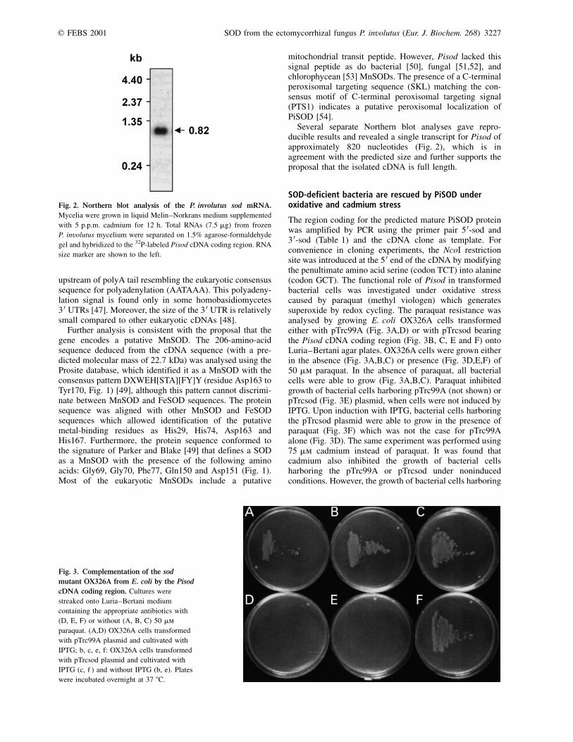
Several separate Northern blot analyses gave repro-ducible results and revealed a single transcript for Pisod ofapproximately 820 nucleotides (Fig. 2), which is inagreement with the predicted size and further supports theproposal that the isolated cDNA is full length.
SOD-deficient bacteria are rescued by PiSOD underoxidative and cadmium stress
The region coding for the predicted mature PiSOD proteinwas amplified by PCR using the primer pair 5 0-sod and3 0-sod (Table 1) and the cDNA clone as template. Forconvenience in cloning experiments, the NcoI restrictionsite was introduced at the 5 0 end of the cDNA by modifyingthe penultimate amino acid serine (codon TCT) into alanine(codon GCT). The functional role of Pisod in transformedbacterial cells was investigated under oxidative stresscaused by paraquat (methyl viologen) which generatessuperoxide by redox cycling. The paraquat resistance wasanalysed by growing E. coli OX326A cells transformedeither with pTrc99A (Fig. 3A,D) or with pTrcsod bearingthe Pisod cDNA coding region (Fig. 3B, C, E and F) ontoLuria±Bertani agar plates. OX326A cells were grown eitherin the absence (Fig. 3A,B,C) or presence (Fig. 3D,E,F) of50 mm paraquat. In the absence of paraquat, all bacterialcells were able to grow (Fig. 3A,B,C). Paraquat inhibitedgrowth of bacterial cells harboring pTrc99A (not shown) orpTrcsod (Fig. 3E) plasmid, when cells were not induced byIPTG. Upon induction with IPTG, bacterial cells harboringthe pTrcsod plasmid were able to grow in the presence ofparaquat (Fig. 3F) which was not the case for pTrc99Aalone (Fig. 3D). The same experiment was performed using75 mm cadmium instead of paraquat. It was found thatcadmium also inhibited the growth of bacterial cellsharboring the pTrc99A or pTrcsod under noninducedconditions. However, the growth of bacterial cells harboring
Fig. 2. Northern blot analysis of the P. involutus sod mRNA.
Mycelia were grown in liquid Melin±Norkrans medium supplemented
with 5 p.p.m. cadmium for 12 h. Total RNAs (7.5 mg) from frozen
P. involutus mycelium were separated on 1.5% agarose-formaldehyde
gel and hybridized to the 32P-labeled Pisod cDNA coding region. RNA
size marker are shown to the left.
Fig. 3. Complementation of the sod
mutant OX326A from E. coli by the Pisod
cDNA coding region. Cultures were
streaked onto Luria±Bertani medium
containing the appropriate antibiotics with
(D, E, F) or without (A, B, C) 50 mm
paraquat. (A,D) OX326A cells transformed
with pTrc99A plasmid and cultivated with
IPTG; b, c, e, f: OX326A cells transformed
with pTrcsod plasmid and cultivated with
IPTG (c, f ) and without IPTG (b, e). Plates
were incubated overnight at 37 8C.
q FEBS 2001 SOD from the ectomycorrhizal fungus P. involutus (Eur. J. Biochem. 268) 3227
the pTrcsod plasmid was restored under IPTG induction(not shown). These results indicate that the PiSOD canfunctionally substitute for the E. coli SODs and thereforeefficiently protect the cells from the detrimental effects ofsuperoxide ions generated by paraquat. In addition, a rolefor PiSOD in the detoxification processes against cadmiumwas demonstrated. As PiSOD is the only SOD activitydetected in crude extracts of P. involutus grown under oxida-tive or cadmium stress conditions (C. Jacob, unpublished
results), these results with E. coli suggested that PiSODplays similar roles in P. involutus.
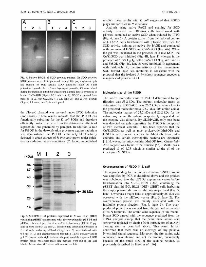
Analysis using native PAGE and staining for SODactivity revealed that OX326A cells transformed withpTrcsod contained an active SOD when induced by IPTG(Fig. 4, lane 2). A protein extract from the induced cultureof OX326A cells transformed with pTrcsod was used forSOD activity staining on native 8% PAGE and comparedwith commercial FeSOD and Cu/ZnSOD (Fig. 4A). Whenthe gel was incubated in the presence of 5 mm KCN, theCu/ZnSOD was inhibited (Fig. 4B, lane 1) whereas in thepresence of 5 mm H2O2, both Cu/ZnSOD (Fig. 4C, lane 1)and FeSOD (Fig. 4C, lane 3) were inhibited. In agreementwith Fridovich [7], the insensitivity of the recombinantSOD toward these two inhibitors is consistent with theproposal that the isolated P. involutus sequence encodes amanganese-dependent SOD.
Molecular size of the PiSOD
The native molecular mass of PiSOD determined by gelfiltration was 53.2 kDa. The subunit molecular mass, asdetermined by SDS/PAGE, was 26.2 kDa, a value close tothe predicted molecular mass (22.7 kDa, 206 amino acids).The molecular masses of 53.2 kDa and 26.2 kDa for thenative enzyme and the subunit, respectively, suggested thatthe enzyme was dimeric. By SDS/PAGE, only one bandwas detected on gels suggesting the PiSOD is composedof two identical subunits. It has been reported that theCu/ZnSODs, as well as most prokaryotic MnSODs andFeSODs, are dimeric whereas the MnSODs from mito-chondria and certain thermophilic bacteria are tetrameric[1]. However, the mitochondrial MnSOD from Caenorhab-ditis elegans was found to be dimeric [55]. PiSOD has apredicted pI of 6.75 which is similar to the pI of theC. elegans MnSODs.
Overexpression of PiSOD in E. coli
The region coding for the predicted mature PiSOD proteinwas amplified by PCR as described above and the productwas subcloned into the pET 3d expression vector beforetransformation into E. coli BL21 (DE3) containing thepSBET plasmid [30]. BL21 (DE3) pSBET cells harboringthe empty plasmid did not exhibit any major band (Fig. 5,lane 1), whereas a major band at approximately 26 kDa wasobserved with the pETsod vector (Fig. 5, lane 2). Theoverexpressed protein was mainly associated with theinsoluble protein fraction (Fig. 5, lane 3). The over-produced protein was excised from the gel and sequencedat its N-terminus. The amino-acid sequence of the recom-binant SOD agreed with the sequence predicted from thecDNA analysis except that the penultimate amino acidserine was replaced by alanine from introduction of an NcoIcloning site, as described above. This result furtherconfirmed that there was no cleavage of any putativeN-terminal signal sequence. Moreover, the first amino acidsequenced was alanine and not methionine, presumablybecause of the small size of the alanine residue, aspreviously described by Hirel et al. [56].
Fig. 4. Native PAGE of SOD proteins stained for SOD activity.
SOD proteins were electrophoresed through 8% polyacrylamide gels
and stained for SOD activity. SOD inhibitors (none, A; 5 mm
potassium cyanide, B; or 5 mm hydrogen peroxide, C) were added
during incubation in nitroblue tetrazolium. Sample lanes correspond to
bovine Cu/ZnSOD (Sigma, 0.21 unit, lane 1), PiSOD expressed from
pTrcsod in E. coli OX326A (10 mg, lane 2), and E. coli FeSOD
(Sigma, 1.1 units, lane 3) in each panel.
Fig. 5. SDS/PAGE of proteins expressed in E. coli BL21 (DE3)
containing pSBET transformed with the two plasmids pET 3d and
pETsod. Total cell proteins of E. coli cells harboring pET 3d (5 mg,
lane 1) or pETsod (5 mg, lane 2), and insoluble cytoplasmic proteins of
E. coli cells harboring pETsod (5 mg, lane 3) were induced with
0.4 mm IPTG and electrophoresed through a 12.5% polyacrylamide
gel. The arrow on the right indicates the position of the expressed SOD
protein bands. Molecular mass size markers were run in the lane
labeled M and sizes (kDa) are indicated on the left.
3228 C. Jacob et al. (Eur. J. Biochem. 268) q FEBS 2001
Immunological characterization
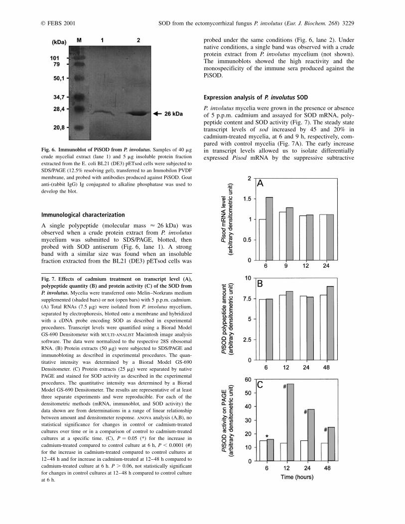
A single polypeptide (molecular mass < 26 kDa) wasobserved when a crude protein extract from P. involutusmycelium was submitted to SDS/PAGE, blotted, thenprobed with SOD antiserum (Fig. 6, lane 1). A strongband with a similar size was found when an insolublefraction extracted from the BL21 (DE3) pETsod cells was
probed under the same conditions (Fig. 6, lane 2). Undernative conditions, a single band was observed with a crudeprotein extract from P. involutus mycelium (not shown).The immunoblots showed the high reactivity and themonospecificity of the immune sera produced against thePiSOD.
Expression analysis of P. involutus SOD
P. involutus mycelia were grown in the presence or absenceof 5 p.p.m. cadmium and assayed for SOD mRNA, poly-peptide content and SOD activity (Fig. 7). The steady statetranscript levels of sod increased by 45 and 20% incadmium-treated mycelia, at 6 and 9 h, respectively, com-pared with control mycelia (Fig. 7A). The early increasein transcript levels allowed us to isolate differentiallyexpressed Pisod mRNA by the suppressive subtractive
Fig. 6. Immunoblot of PiSOD from P. involutus. Samples of 40 mg
crude mycelial extract (lane 1) and 5 mg insoluble protein fraction
extracted from the E. coli BL21 (DE3) pETsod cells were subjected to
SDS/PAGE (12.5% resolving gel), transferred to an Immobilon PVDF
membrane, and probed with antibodies produced against PiSOD. Goat
anti-(rabbit IgG) Ig conjugated to alkaline phosphatase was used to
develop the blot.
Fig. 7. Effects of cadmium treatment on transcript level (A),
polypeptide quantity (B) and protein activity (C) of the SOD from
P. involutus. Mycelia were transferred onto Melin±Norkrans medium
supplemented (shaded bars) or not (open bars) with 5 p.p.m. cadmium.
(A) Total RNAs (7.5 mg) were isolated from P. involutus mycelium,
separated by electrophoresis, blotted onto a membrane and hybridized
with a cDNA probe encoding SOD as described in experimental
procedures. Transcript levels were quantified using a Biorad Model
GS-690 Densitometer with multi-analist Macintosh image analysis
software. The data were normalized to the respective 28S ribosomal
RNA. (B) Protein extracts (50 mg) were subjected to SDS/PAGE and
immunobloting as described in experimental procedures. The quan-
titative intensity was determined by a Biorad Model GS-690
Densitometer. (C) Protein extracts (25 mg) were separated by native
PAGE and stained for SOD activity as described in the experimental
procedures. The quantitative intensity was determined by a Biorad
Model GS-690 Densitometer. The results are representative of at least
three separate experiments and were reproducible. For each of the
densitometric methods (mRNA, immunoblot, and SOD activity) the
data shown are from determinations in a range of linear relationship
between amount and densitometer response. anova analysis (A,B), no
statistical significance for changes in control or cadmium-treated
cultures over time or in a comparison of control to cadmium-treated
cultures at a specific time. (C), P � 0.05 (*) for the increase in
cadmium-treated compared to control culture at 6 h, P , 0.0001 (#)
for the increase in cadmium-treated compared to control cultures at
12±48 h and for increase in cadmium-treated at 12±48 h compared to
cadmium-treated culture at 6 h. P . 0.06, not statistically significant
for changes in control cultures at 12±48 h compared to control culture
at 6 h.
q FEBS 2001 SOD from the ectomycorrhizal fungus P. involutus (Eur. J. Biochem. 268) 3229

hybridization procedure. However these increases at 6 and9 h were not statistically different from control values(anova P � 0.11, 0.72). No significant changes weredetectable at stages 12 and 24 h with 5 p.p.m. cadmium(P � 0.93, 0.99, Fig. 7A). The amount of SOD polypeptidedid not vary significantly in this time period (P � 0.60±0.96, Fig. 7B). The absence of a correlation betweenmRNA and protein level at 6 and 9 h may be due todifferences in efficiency of translation in cadmium-treatedmycelia and control mycelia. After 48 h the amounts oftotal RNA and total polypeptide decreased dramatically incadmium-treated mycelia. Therefore, it was not possible tofollow SOD transcript and polypeptide levels at those laterstages. In contrast (Fig. 7C), the basal SOD activity wasgreatly increased following treatment with cadmium, themaximum being 4.5-fold increase after 12 h with cadmium(P , 0.0001 for SOD activity at 12, 24 and 48 h comparedto 6 h). In untreated mycelia, SOD activity remainedconstant (no significant change for SOD activity at 12, 24and 48 h compared to 6 h).
Induction of SODs in response to metals and oxidativestress has been studied in other species. In the marinedinoflagellate G. polyedra, exposure to lead, mercury,copper or cadmium induces MnSOD and to a lesser extentFeSOD [57]. In A. thaliana, levels of MnSOD mRNA,polypeptide and enzymatic activity were not affected bydifferent oxidative stresses [58]. However, other SODisoforms were highly responsive to these stresses in A.thaliana [58]. In the ectomycorrhizal fungus Rhizopogonroseolus, SOD inhibition assays similar to those performedin this study implicated the presence of a single SOD, anMnSOD [59]. Moreover, MnSOD but not Cu/ZnSODactivity was found in protein extracts from certain flagellatefungi [60]. In crude extracts of P. involutus a single SODisozyme, a putative MnSOD, was found following growthunder oxidative stress conditions or under cadmium stressconditions (C. Jacob, unpublished results). Whether thefailure to find Cu/ZnSOD, FeSOD or other putativeMnSODs in P. involutus reflects their absence, or instead,a low specific activity for those SODs is unclear. As theonly SOD identified in crude extracts, PiSOD is implicatedas essential in the detoxification mechanisms against ROSin the ectomycorrhizal fungus P. involutus.
Our results comparing PiSOD polypeptide and PiSODactivity suggest that post-translational mechanisms areinvolved in activating a pool of pre-existing SOD inresponse to cadmium stress. One possible mechanismwould be a competition between Fe and Mn for themetal-binding site as shown in E. coli [61]. In that case,MnSOD polypeptide is inactive when Fe is bound and isactive when Mn is bound. In the PiSOD case, addition ofoxidants or a change in redox balance may shift thecompetitive balance in favor of Mn and thus allow synthesisof the active PiSOD. Another possibility is that cadmiuminfluences the synthesis, metal binding or metal deliveryactivity of a metal chaperone which converts apo PiSOD toactive metal-bound PiSOD. Such a metal chaperone isinvolved in the delivery of copper to Cu/Zn superoxidedismutase in S. cerevisiae [62]. Thus far, no eukaryoticchaperones for metals other than copper have beenestablished; however, it is likely that analogous cofactortrafficking pathways exist for metals other than copper[63].
The increasing activity of the PiSOD as well as the factthat it can functionally substitute for the E. coli SODsunder cadmium stress suggests that this enzyme is involvedin the cellular response of P. involutus to cadmium. This isto our knowledge the first insight in the characterization ofmolecular events that take place in an ectomycorrhizalfungus during exposure to heavy metals. Alongside studieson SOD, we are currently investigating other antioxidantand detoxification enzymes such as those involved in bio-synthesis of glutathione and chelating agents. The isolationof genes and proteins involved in the response to cadmium-induced stress open new perspectives in the understandingof molecular mechanisms that promote tolerance in mycor-rhizal fungi, which are key organisms in remediationprogrammes [64].
A C K N O W L E D G E M E N T S
Financial support from the European Commission (project FAIR3-
CT961377) is gratefully acknowledged. This work was supported by
grants from the ReÂgion Lorraine to C. Jacob and from the Agence
de l'Environnment et de la MaõÃtrise de l'Energie (ADEME) to
M. Courbot. We thank SaõÈd Azza (University H. PoincareÂ, Nancy I) for
assistance with gel filtration, Jean-Louis Hilbert and Pierre-Eric
SautieÁre (University of Science and Technology, Lille, France) for
assistance with protein sequencing, ReÂgis Belleville and Dr ValeÂrie
Legue (University H. PoincareÂ, Nancy I) for assistance with photo-
graphy. We thank one referee for suggesting alternative mechanisms
for control of PiSOD in response to cadmium stress.
R E F E R E N C E S
1. Scandalios, J.G. (1997) Molecular genetics of superoxide
dismutases in plants. In Oxidative Stress and the Molecular
Biology of Antioxidant Defenses (Scandalios, J.G., ed.), pp.
527±568. Cold Spring Harbor Laboratory Press, Cold Spring
Harbor, New York.
2. Bannister, J.V., Bannister, W.H. & Rotillo, G. (1987) Aspects of
the structure, function, and applications of superoxide dismutase.
Crit. Rev. Biochem. 2, 111±180.
3. Kim, E.-J., Kim, H.P., Hah, Y.C. & Roe, J.H. (1996) Differential
expression of superoxide dismutases containing Ni and Fe/Zn in
Streptomyces coelicolor. Eur. J. Biochem. 241, 178±185.
4. Youn, H.D., Kim, E.J., Roe, J.H., Hah, Y.C. & Kang, S.O. (1996)
A novel nickel-containing superoxide dismutase from Strepto-
myces spp. Biochem. J. 318, 889±896.
5. Youn, H.D. Youn, H. Lee, J.W. Yim, Y.I.J.K. Lee, Hah, Y.C. &
Kang, S.O. (1996) Unique isozymes of superoxide dismutase in
Streptomyces griseus. Arch. Biochem. Biophys. 334, 341±348.
6. Halliwell, B. (1984) Chloroplast Metabolism: The Structure and
Function of Chloroplasts in Green Leaf Cells. Clarendon Press,
Oxford, UK.
7. Fridovich, I. (1986) Superoxide dismutases. Adv. Enzymol. Relat.
Areas Mol. Biol. 58, 61±97.
8. Baum, J.A. & Scandalios, J.G. (1981) Isolation and characteriz-
ation of the cytosolic and mitochondrial superoxide dismutases of
maize. Arch. Biochem. Biophys. 206, 249±264.
9. Bowler, C. De Alliotte, T. Loose, M. Van Montagu, M. & InzeÂ, D.
(1989) The induction of manganese superoxide dismutase in
response to stress in Nicotiana plumbaginifolia. EMBO J. 8,
31±38.
10. del Rio, L.A. Lyon, D.S. Olah, I. Glick, B. & Salin, M.L. (1983)
Immunocytochemical evidence for a peroxisomal localization of
manganese superoxide dismutase in leaf protoplasts from a higher
plant. Planta 158, 216±224.
3230 C. Jacob et al. (Eur. J. Biochem. 268) q FEBS 2001
11. Halliwell, B. & Gutteridge, J.M.C. (1989) Free Radicals in
Biology and Medicine. Clarendon Press, Oxford, UK.
12. Bowler, C. Van Montagu, M. & InzeÂ, D. (1992) Superoxide
dismutase and stress tolerance. Annu. Rev. Plant Physiol. Plant
Mol. Biol. 43, 83±116.
13. McKersie, B.D. Chen, Y. de Beus, M. Bowley, S.R. Bowler, C.
InzeÂ, D. D'Halluin, K. & Botterman, J. (1993) Superoxide
dismutase enhances tolerance of freezing stress in transgenic
alfalfa (Medicago sativa L.). Plant Physiol. 103, 1155±1163.
14. McKersie, B.D. Bowley, S.R. Harjanto, E. & Leprince, O. (1996)
Water-deficit tolerance and field performance of transgenic
alfalfa overexpressing superoxide dismutase. Plant Physiol. 111,
1177±1181.
15. Scandalios, J.G. (1993) Oxygen stress and superoxide dismutases.
Plant Physiol. 101, 7±12.
16. SeppaÈnen, M.M. & Fagerstedt, K. (2000) The role of superoxide
dismutase activity in response to cold acclimatation in potato.
Physiol. Plant. 108, 279±285.
17. Van Camp, W. Van Montagu, M. & InzeÂ, D. (1994) Superoxide
dismutases. In Causes of Photooxidative Stress and Amelioration
of Defense Systems in Plants (Foyer, C.H. & Mullineaux, P.M.,
eds), pp. 317±341. CRC Press, Boca Raton, FL, USA.
18. Henkle-DuÈhrsen, K. Tawe, W. Warnecke, C. & Walter, R.D.
(1995) Characterization of the manganese superoxide dismutase
cDNA and gene from the human parasite Onchocerca volvulvus.
Biochem. J. 308, 441±446.
19. Arisi, A.-C.M. Cornic, G. Jouanin, L. & Foyer, C.H. (1998)
Overexpression of iron superoxide dismutase in transformed
poplar modifies the regulation of photosynthesis at low CO2
partial pressures or following exposure to the prooxidant herbicide
methyl viologen. Plant Physiol. 117, 565±574.
20. Hendry, G.A.F. Baker, A.J.M. & Ewart, C.F. (1992) Cadmium
tolerance and toxicity, oxygen radical processes and molecular
damage in cadmium-tolerant and cadmium-sensitive clones of
Holcus lanatus L. Acta Bot. Neerl. 41, 271±281.
21. Brennan, R.J. & Schiestl, R.H. (1996) Cadmium is an inducer of
oxidative stress in yeast. Mutation Res. 356, 171±178.
22. Somashekaraiah, B.V. Padmaja, K. & Prasad, A.R.K. (1992)
Phytotoxicity of cadmium ions on germinating seedlings of mung
bean (Phaseolus vulgaris): involvement of lipid peroxides in
chlorophyll degradation. Physiol. Plant. 85, 85±89.
23. Foyer, C.H. Lopez-Delgado, H. Dat, J.F. & Scott, I.M. (1997)
Hydrogen peroxide- and glutathione-associated mechanisms of
acclimatory stress tolerance and signalling. Physiol. Plant. 100,
241±254.
24. Howlett, N.G. & Avery, S.V. (1997) Induction of lipid peroxidation
during heavy metal stress in Saccharomyces cerevisiae and
influence of plasma membrane fatty acid unsaturation. Appl.
Environ. Microbiol. 63, 2971±2976.
25. Stohs, S.J. & Bagchi, D. (1995) Oxidative mechanisms in the
toxicity of metal ions. Free Radic. Biol. Med. 18, 321±336.
26. Blaudez, D. Jacob, C. Turnau, K. Colpaert, J.V. Ahonen-Jonnarth,
U. Finlay, R. Botton, B. & Chalot, M. (2000) Differential
responses of ectomycorrhizal fungal isolates to heavy metals in
vitro. Mycol. Res. 104, 1366±1371.
27. Smith, S.E. & Read, D.J. (1997) Mycorrhizal Symbiosis. Academic
Press, London.
28. Blaudez, D. Botton, B. & Chalot, M. (2000) Cadmium uptake and
subcellular compartmentation in the ectomycorrhizal fungus
Paxillus involutus. Microbiol. 146, 1109±1117.
29. Marx, D.H. (1969) The influence of ectotrophic mycorrhizal fungi
on the resistance of pine roots to pathogenic infections. I.
Antagonism of mycorrhizal fungi to root pathogenic fungi and
soil bacteria. Phytopathol. 59, 159±163.
30. Schenk, P.M. Baumann, S. Mattes, R. & Steinbiss, H.-H. (1995)
Improved high-level expression system for eukaryotic genes in
Escherichia coli using T7 RNA polymerase and rare ArgtRNAs.
Biotechniques 19, 196±200.
31. Steinman, H.M. (1992) Construction of an Escherichia coli K-12
strain deleted for manganese and iron superoxide dismutase genes
and its use in cloning the iron superoxide dismutase gene of
Legionella pneumophila. Mol. Gen. Genet. 232, 427±430.
32. Sambrook, J. Fritsch, E.F. & Maniatis, T. (1989). Molecular
Cloning: a Laboratory Manual, 2nd edn. Cold Spring Harbor
Laboratory Press, Cold Spring Harbor, New York.
33. Diatchenko, L. Lau, Y.-F.C. Campbell, A.P. Chenchik, A.
Moqadam, F. Huang, B. Lukyanov, S. Lukyanov, K. Gurskaya,
N. Sverdlov, E.D. & Siebert, P.D. (1996) Suppression subtractive
hybridization: a method for generating differentially regulated or
tissue-specific cDNA probes and libraries. Proc. Natl Acad. Sci.
USA 93, 6025±6030.
34. Feinberg, A.P. & Vogelstein, B. (1983) A technique for radio-
labelling DNA restriction endonuclease fragments to high specific
activity. Anal. Biochem. 132, 6±13.
35. Feinberg, A.P. & Vogelstein, B. (1984) A technique for radio-
labelling DNA restriction endonuclease fragments to high specific
activity. Addendum Anal. Biochem. 137, 266±267.
36. Altschul, S.F. Gish, W. Miller, W. Myers, E.W. & Lipman, D.J.
(1990) Basic local alignment search tool. J. Mol. Biol. 215,
403±410.
37. Worley, K.C. Culpepper, P. Wiese, B.A. & Smith, R.F. (1998)
BEAUTY-X: enhanced BLAST searches for DNA queries.
Bioinformatics 14, 890±891.
38. Worley, K.C. Wiese, B.A. & Smith, R.F. (1995) BEAUTY: an
enhanced BLAST-based search tool that integrates multiple
biological information resources into sequence similarity search
results. Genome Res. 5, 173±184.
39. Bradford, M.M. (1976) A rapid and sensitive method for the
quantitation of microgram quantities of protein utilizing the
principle of protein-dye binding. Anal. Biochem. 72, 248±254.
40. Beauchamp, C. & Fridovich, I. (1971) Superoxide dismutase:
improved assays and an assay applicable to acrylamide gels. Anal.
Biochem. 44, 276±287.
41. Laemmli, U.K. (1970) Cleavage of structural proteins during the
assembly of the head of bacteriophage T4. Nature 227, 680±685.
42. Donahue, J.L., Okpodu, C.M., Cramer, C.L., Grabau, E.A. &
Alscher, R.G. (1997) Responses of antioxidants to paraquat in pea
leaves: relationships to resistance. Plant Physiol. 113, 249±257.
43. Brun, A., Chalot, M., Botton, B. & Martin, F. (1992) Purification
and characterization of glutamine synthetase and NADP-glutamate
dehydrogenase from the ectomycorrhizal fungus Laccaria laccata.
Plant Physiol. 99, 938±944.
44. Ballance, D.J. (1986) Sequences important for gene expression in
filamentous fungi. Yeast 2, 229±236.
45. Parker, R. & Patterson, B. (1987) Architecture of fungal intron
codons: implications for spliceosome assembly. In Molecular
Biology of RNA: New Perspectives (Inouye, M. & Dusdock, B.,
eds), pp. 133±149. Academic Press, New York.
46. Lugones, L.G., Scholtmeijer, K., Klootwijk, R. & Wessels, J.G.H.
(1999) Introns are necessary for mRNA accumulation in
Schizophyllum commune. Mol. Microbiol. 32, 681±689.
47. Schuren, F.H.J. (1992) Regulation of gene expression during fruit-
body development in Schizophyllum commune. PhD Thesis.
University of Groningen, the Netherlands.
48. Pesole, G., Liuni, S., Grillo, G. & Saccone, C. (1997) Structural
and compositional features of untranslated regions of eukaryotic
mRNAs. Gene 205, 95±102.
49. Parker, R. & Blake, C.C.F. (1988) Iron- and manganese-containing
superoxide dismutases can be distinguished by analysis of their
primary structures. FEBS Lett. 229, 377±382.
50. Takeda, Y. & Avila, H. (1986) Structure and gene expression of the
E. coli Mn-superoxide dismutase gene. Nucleic Acids Res. 14,
4577±4589.
q FEBS 2001 SOD from the ectomycorrhizal fungus P. involutus (Eur. J. Biochem. 268) 3231
51. Crameri, R., Faith, A., Hemmann, S., Jaussi, R., Ismail, C., Menz,
G. & Blaser, K. (1996) Humoral and cell-mediated autoimmunity
in allergy to Aspergillus fumigatus. J. Exp. Med. 184, 265±270.
52. Pan, S.M., Ye, J.S. & Hseu, R.S. (1997) Purification and
characterization of manganese superoxide dismutase from Gano-
derma microsporum. Biochem. Mol. Biol. Int. 42, 1035±1043.
53. Kitayama, K., Kitayama, M. & Togasaki, R.K. (1995) A cDNA
clone encoding a manganese-superoxide dismutase (GenBank
U24500) from Chlamydomonas reinhardtii. Plant Physiol. 108,
1748
54. Gould, S.J., Keller, G.A., Hosken, N., Wilkinson, J. & Subramani,
S. (1989) A conserved tripeptide sorts proteins to peroxisomes.
J. Cell. Biol. 108, 1657±1664.
55. Hunter, T., Bannister, W.H. & Hunter, G.J. (1997) Cloning,
expression, and characterization of two manganese superoxide
dismutases from Caenorhabditis elegans. J. Biol. Chem. 272,
28652±28659.
56. Hirel, P.H., Schmitter, M.J., Dessen, P., Fayat, G. & Blanquet, S.
(1989) Extent of N-terminal methionine excision from Escherichia
coli proteins is governed by the side-chain length of the penulti-
mate amino acid. Proc. Natl Acad. Sci. USA 86, 8247±8251.
57. Okamoto, O.K. & Colepicolo, P. (1998) Response of superoxide
dismutase to pollutant metal stress in the marine dinoflagellate
Gonyaulax polyedra. Comp. Biochem. Physiol. 119C, 67±73.
58. Kliebenstein, D.J., Monde, R.A. & Last, R.L. (1998) Superoxide
dismutase in Arabidopsis: an eclectic enzyme family with
disparate regulation and protein localization. Plant Physiol. 118,
637±650.
59. Miszalski, Z., Niewiadomska, E., Skoczowski, A. & Botton, B.
(1998) Chromatographic properties of the cadmium induced SOD
isoform in Rhizopogon roseolus. Acta Physiol. Plant. 20, 229±233.
60. Natvig, D.O., Dvorachek, W.H. & Sylvester, K. (1994) Evolution
and biological roles of fungal superoxide dismutase. In Metal Ions
in Fungi (Winkelmann, G. & Winge, D.R., eds), pp. 391±412.
Marcel Dekker Inc, New York.
61. Privalle, C.T. & Fridovich, I. (1992) Transcriptional and matura-
tional effects of manganese and iron on the biosynthesis of
manganese-superoxide dismutase in Escherichia coli. J. Biol.
Chem. 267, 9140±9145.
62. Culotta, V.C., Klomp, L.W.J., Strain, J., Casareno, R.L.B., Krems,
B. & Gitlin, J.D. (1997) The copper chaperone for superoxide
dismutase. J. Biol. Chem. 272, 23469±23472.
63. O'Halloran, T.V. & Culotta, V.C. (2000) Metallochaperones, an
intracellular shuttle service for metal ions. J. Biol. Chem. 275,
25057±25060.
64. Jasper, D.A. (1994) Management of mycorrhizas in revegetation.
In Management of Mycorrhizas in Agriculture, Horticulture and
Forestry (Robson, A.D.Abbott, L.K. & Malajczuk, N., eds), pp.
211±219. Kluwer Academic Publishers, Norwell, MA, USA.
3232 C. Jacob et al. (Eur. J. Biochem. 268) q FEBS 2001