Modulation of P2X 7 purinergic receptor in macrophages by Leishmania amazonensis and its role in...
10
Differential Modulation of ATP-Induced P2X7-Associated Permeabilities to Cations and Anions of Macrophages by Infection with Leishmania amazonensis Camila Marques-da-Silva 1 , Mariana Martins Chaves 1 , Juliany Cola Rodrigues 1 , Suzana Corte-Real 2 , Robson Coutinho-Silva 1 *, Pedro Muanis Persechini 1 1 Instituto de Biofisica Carlos Chagas Filho, Universidade Federal do Rio de Janeiro, Rio de Janeiro, Rio de Janeiro, Brazil, 2 Instituto Oswaldo Cruz, Fundac ¸a ˜ o Oswaldo Cruz, Rio de Janeiro, Rio de Janeiro, Brazil Abstract Leishmania and other parasites display several mechanisms to subvert host immune cell function in order to achieve successful infection. The ATP receptor P2X7, an agonist-gated cation channel widely expressed in macrophages and other cells of the immune system, is also coupled to inflammasome activation, IL-1 beta secretion, production of reactive oxygen species, cell death and the induction of the permeabilization of the plasma membrane to molecules of up to 900 Da. P2X7 receptors can function as an effective microbicidal triggering receptor in macrophages infected with several microorganisms including Mycobacteria tuberculosis, Chlamydia and Leishmania. We have previously shown that its expression is up-regulated in macrophages infected with L.amazonensis and that infected cells also display an increase in P2X7-induced apoptosis and membrane permeabilization to some anionic fluorescent dyes. In an independent study we recently showed that the phenomenon of macrophage membrane permeabilization can involve at least two distinct pathways for cations and anions respectively. Here, we re-addressed the effects of ATP-induced P2X7-associated phenomena in macrophages infected with L.amazonensis and demonstrated that the P2X7-associated dye uptake mechanisms are differentially modulated. While the membrane permeabilization for anionic dyes is up-modulated, as previously described, the uptake of cationic dyes is strongly down-modulated. These results unveil new characteristics of two distinct permeabilization mechanisms associated with P2X7 receptors in macrophages and provide the first evidence indicating that these pathways can be differentially modulated in an immunologically relevant situation. The possible importance of these results to the L.amazonensis escape mechanism is discussed. Citation: Marques-da-Silva C, Chaves MM, Rodrigues JC, Corte-Real S, Coutinho-Silva R, et al. (2011) Differential Modulation of ATP-Induced P2X7-Associated Permeabilities to Cations and Anions of Macrophages by Infection with Leishmania amazonensis. PLoS ONE 6(9): e25356. doi:10.1371/journal.pone.0025356 Editor: Jean Kanellopoulos, University Paris Sud, France Received May 8, 2011; Accepted September 1, 2011; Published September 23, 2011 Copyright: ß 2011 Marques-da-Silva et al. This is an open-access article distributed under the terms of the Creative Commons Attribution License, which permits unrestricted use, distribution, and reproduction in any medium, provided the original author and source are credited. Funding: This work was supported by the Conselho Nacional de Desenvolvimento Cientı ´fico e Tecnolo ´ gico (CNPq)(www.cnpq.br), Fundac ¸a ˜o Carlos Chagas Filho de Amparo a ` Pesquisa do Estado do Rio de Janeiro (FAPERJ)(www.faperj.br), Programa de Nu ´ cleos de Excele ˆ ncia (PRONEX-CNPq) and Instituto Nacional para Pesquisa Translacional em Sau ´ de e Ambiente na Regia ˜o Amazo ˆ nica, Conselho Nacional de Desenvolvimento Cientı ´fico e Tecnolo ´ gico/MCT (INCT-INPeTAm/CNPq/ MCT), Brazil.(http://www.cnpq.br/programas/inct/_apresentacao/index.html). The funders had no role in study design, data collection and analysis, decision to publish, or preparation of the manuscript. Competing Interests: The authors have declared that no competing interests exist. * E-mail: [email protected] Introduction Macrophages display strong differentiation plasticity character- ized by the modulation of the expression of several membrane receptors, cytokine production, and functions such as migration, phagocytosis, microorganism killing and antigen presentation [1]. These cells are also the host of a number of intracellular pathogens such as Mycobacterium tuberculosis, Leishmania, Trypanosoma cruzi, and HIV-1 [2–12]. Therefore, it is no surprise that receptor-agonist interactions are important for macrophage-mediated immune regulation involved in both the resolution of the infection and the survival strategies of the pathogens [13–15]. The P2X7 receptor is a member of the P2X ligand-gated cation- selective ion channels that are highly expressed in macrophages, dendritic cells and other cells of the immune system [16–21]. Besides its ion-channel activity, it also induces the so-called ‘‘permeabilization phenomenon’’ originally characterized by the uptake of fluorescent molecules of up to 900 Da [22–24]. In macrophages, P2X7 receptors have been implied in cell death, inflammasome activation, secretion of IL1b, modulation of regulatory T cell function, and in the killing of intracellular microorganisms, among other effects [21,25–30]. The protozoan Leishmania amazonensis (L.amazonensis) is an obligatory intracellular parasite that survives and proliferates inside parasitophorous vacuoles (PVs) that reside in the cytosol of its main host cell, the macrophage [31]. Inside PVs, the parasites must subvert several macrophage functions to provide a regular supply of nutrients and avoid being killed by the host cells. In order to do so, they modulate the traffic of endocytic-derived and intracellular vesicles and their fusion with the PVs, while also modulating the expression of membrane proteins derived from both the host and the parasites [32]. Our laboratory recently reported that macrophages infected with L.amazonensis up-regulate the expression of functional P2X7 receptors and the ATP-induced permeabilization to the vital dye Lucifer Yellow (LY) [33]. Extracellular ATP (ATPe) also induces macrophage apoptosis and parasite elimination in vitro [33]. PLoS ONE | www.plosone.org 1 September 2011 | Volume 6 | Issue 9 | e25356
-
Upload
independent -
Category
Documents
-
view
1 -
download
0
Transcript of Modulation of P2X 7 purinergic receptor in macrophages by Leishmania amazonensis and its role in...

Differential Modulation of ATP-Induced P2X7-AssociatedPermeabilities to Cations and Anions of Macrophages byInfection with Leishmania amazonensisCamila Marques-da-Silva1, Mariana Martins Chaves1, Juliany Cola Rodrigues1, Suzana Corte-Real2,
Robson Coutinho-Silva1*, Pedro Muanis Persechini1
1 Instituto de Biofisica Carlos Chagas Filho, Universidade Federal do Rio de Janeiro, Rio de Janeiro, Rio de Janeiro, Brazil, 2 Instituto Oswaldo Cruz, Fundacao Oswaldo Cruz,
Rio de Janeiro, Rio de Janeiro, Brazil
Abstract
Leishmania and other parasites display several mechanisms to subvert host immune cell function in order to achievesuccessful infection. The ATP receptor P2X7, an agonist-gated cation channel widely expressed in macrophages and othercells of the immune system, is also coupled to inflammasome activation, IL-1 beta secretion, production of reactive oxygenspecies, cell death and the induction of the permeabilization of the plasma membrane to molecules of up to 900 Da. P2X7receptors can function as an effective microbicidal triggering receptor in macrophages infected with severalmicroorganisms including Mycobacteria tuberculosis, Chlamydia and Leishmania. We have previously shown that itsexpression is up-regulated in macrophages infected with L.amazonensis and that infected cells also display an increase inP2X7-induced apoptosis and membrane permeabilization to some anionic fluorescent dyes. In an independent study werecently showed that the phenomenon of macrophage membrane permeabilization can involve at least two distinctpathways for cations and anions respectively. Here, we re-addressed the effects of ATP-induced P2X7-associatedphenomena in macrophages infected with L.amazonensis and demonstrated that the P2X7-associated dye uptakemechanisms are differentially modulated. While the membrane permeabilization for anionic dyes is up-modulated, aspreviously described, the uptake of cationic dyes is strongly down-modulated. These results unveil new characteristics oftwo distinct permeabilization mechanisms associated with P2X7 receptors in macrophages and provide the first evidenceindicating that these pathways can be differentially modulated in an immunologically relevant situation. The possibleimportance of these results to the L.amazonensis escape mechanism is discussed.
Citation: Marques-da-Silva C, Chaves MM, Rodrigues JC, Corte-Real S, Coutinho-Silva R, et al. (2011) Differential Modulation of ATP-Induced P2X7-AssociatedPermeabilities to Cations and Anions of Macrophages by Infection with Leishmania amazonensis. PLoS ONE 6(9): e25356. doi:10.1371/journal.pone.0025356
Editor: Jean Kanellopoulos, University Paris Sud, France
Received May 8, 2011; Accepted September 1, 2011; Published September 23, 2011
Copyright: � 2011 Marques-da-Silva et al. This is an open-access article distributed under the terms of the Creative Commons Attribution License, which permitsunrestricted use, distribution, and reproduction in any medium, provided the original author and source are credited.
Funding: This work was supported by the Conselho Nacional de Desenvolvimento Cientıfico e Tecnologico (CNPq)(www.cnpq.br), Fundacao Carlos Chagas Filhode Amparo a Pesquisa do Estado do Rio de Janeiro (FAPERJ)(www.faperj.br), Programa de Nucleos de Excelencia (PRONEX-CNPq) and Instituto Nacional paraPesquisa Translacional em Saude e Ambiente na Regiao Amazonica, Conselho Nacional de Desenvolvimento Cientıfico e Tecnologico/MCT (INCT-INPeTAm/CNPq/MCT), Brazil.(http://www.cnpq.br/programas/inct/_apresentacao/index.html). The funders had no role in study design, data collection and analysis, decision topublish, or preparation of the manuscript.
Competing Interests: The authors have declared that no competing interests exist.
* E-mail: [email protected]
Introduction
Macrophages display strong differentiation plasticity character-
ized by the modulation of the expression of several membrane
receptors, cytokine production, and functions such as migration,
phagocytosis, microorganism killing and antigen presentation [1].
These cells are also the host of a number of intracellular pathogens
such as Mycobacterium tuberculosis, Leishmania, Trypanosoma cruzi, and
HIV-1 [2–12]. Therefore, it is no surprise that receptor-agonist
interactions are important for macrophage-mediated immune
regulation involved in both the resolution of the infection and the
survival strategies of the pathogens [13–15].
The P2X7 receptor is a member of the P2X ligand-gated cation-
selective ion channels that are highly expressed in macrophages,
dendritic cells and other cells of the immune system [16–21]. Besides its
ion-channel activity, it also induces the so-called ‘‘permeabilization
phenomenon’’ originally characterized by the uptake of fluorescent
molecules of up to 900 Da [22–24]. In macrophages, P2X7 receptors
have been implied in cell death, inflammasome activation, secretion of
IL1b, modulation of regulatory T cell function, and in the killing of
intracellular microorganisms, among other effects [21,25–30].
The protozoan Leishmania amazonensis (L.amazonensis) is an
obligatory intracellular parasite that survives and proliferates
inside parasitophorous vacuoles (PVs) that reside in the cytosol of
its main host cell, the macrophage [31]. Inside PVs, the parasites
must subvert several macrophage functions to provide a regular
supply of nutrients and avoid being killed by the host cells. In
order to do so, they modulate the traffic of endocytic-derived and
intracellular vesicles and their fusion with the PVs, while also
modulating the expression of membrane proteins derived from
both the host and the parasites [32].
Our laboratory recently reported that macrophages infected
with L.amazonensis up-regulate the expression of functional P2X7
receptors and the ATP-induced permeabilization to the vital dye
Lucifer Yellow (LY) [33]. Extracellular ATP (ATPe) also induces
macrophage apoptosis and parasite elimination in vitro [33].
PLoS ONE | www.plosone.org 1 September 2011 | Volume 6 | Issue 9 | e25356
The increase in permeabilization, a P2X7-associated phenom-
ena that can potentially eliminate the parasite is in apparent
conflict with the development of successful escape mechanisms
that will ultimately guarantee their survival inside the host cell.
The resolution of this conflict may lead us to a better
understanding of the P2X7-associated phenomena such as the
ATP-induced permeabilization in macrophages and their modu-
lation by the parasite. Two mechanisms have been proposed to
explain how P2X7 receptors control the uptake of high weight
dyes: one involving the passage of large molecules through the
P2X7 channels after a structural modification called ‘‘pore
dilation’’, and the other involving the opening of a second, larger
and molecularly distinct pore such as the Z pores we have
previously described [22,23,34]. Recently, the independent pore
hypothesis was favored by the demonstration that in macrophages,
astrocytes, and some P2X7-transfected cells, pannexin-1 pores can
be formed under the control of agonist binding to P2X7 molecules
[35,36]. However, the permeabilization phenomenon of macro-
phages proved to be more complex than previously thought since
we and others have shown that large organic cationic and anionic
dyes are taken up by macrophages through at least two different
mechanisms [37,38]. These recent findings prompted us to
hypothesize that the P2X7 receptor and the different transport
mechanisms associated with its activation could be differentially
modulated during the course of many physiologically-relevant
immune-pathological or immunomodulatory processes. In this
work we investigated the effects of ATPe on the uptake of cationic
and anionic dyes by L.amazonensis-infected intraperitoneal murine
macrophages and describe for the first time a differential
modulation of the P2X7-mediated ATPe-induced uptake of
cationic and anionic dyes in an immunologically relevant situation.
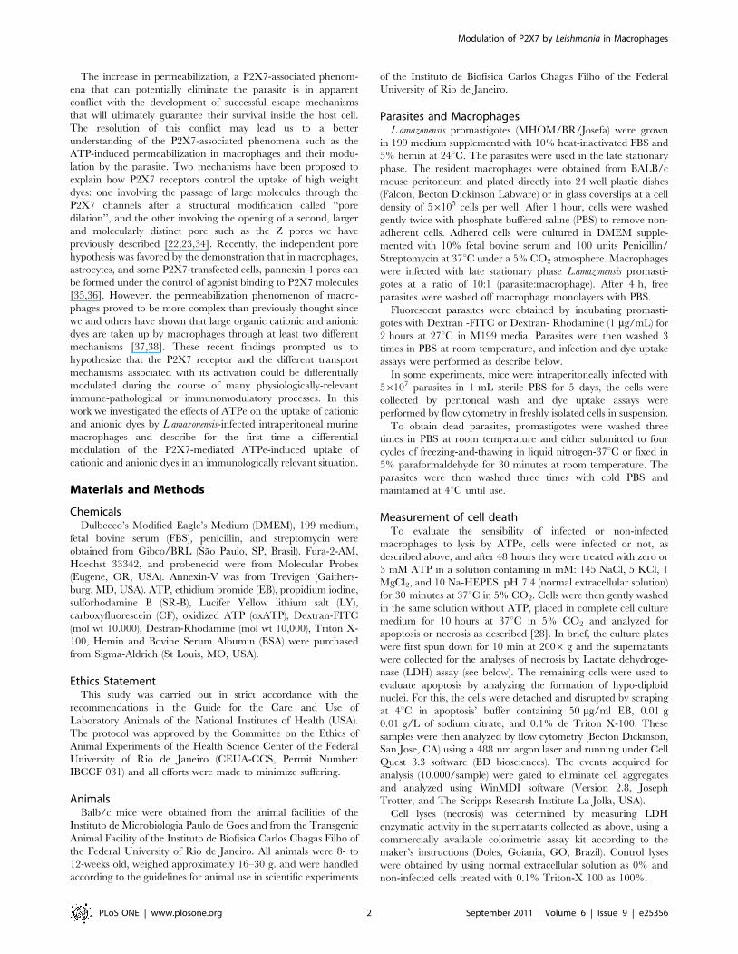
Materials and Methods
ChemicalsDulbecco’s Modified Eagle’s Medium (DMEM), 199 medium,
fetal bovine serum (FBS), penicillin, and streptomycin were
obtained from Gibco/BRL (Sao Paulo, SP, Brasil). Fura-2-AM,
Hoechst 33342, and probenecid were from Molecular Probes
(Eugene, OR, USA). Annexin-V was from Trevigen (Gaithers-
burg, MD, USA). ATP, ethidium bromide (EB), propidium iodine,
sulforhodamine B (SR-B), Lucifer Yellow lithium salt (LY),
carboxyfluorescein (CF), oxidized ATP (oxATP), Dextran-FITC
(mol wt 10.000), Destran-Rhodamine (mol wt 10,000), Triton X-
100, Hemin and Bovine Serum Albumin (BSA) were purchased
from Sigma-Aldrich (St Louis, MO, USA).
Ethics StatementThis study was carried out in strict accordance with the
recommendations in the Guide for the Care and Use of
Laboratory Animals of the National Institutes of Health (USA).
The protocol was approved by the Committee on the Ethics of
Animal Experiments of the Health Science Center of the Federal
University of Rio de Janeiro (CEUA-CCS, Permit Number:
IBCCF 031) and all efforts were made to minimize suffering.
AnimalsBalb/c mice were obtained from the animal facilities of the
Instituto de Microbiologia Paulo de Goes and from the Transgenic
Animal Facility of the Instituto de Biofısica Carlos Chagas Filho of
the Federal University of Rio de Janeiro. All animals were 8- to
12-weeks old, weighed approximately 16–30 g. and were handled
according to the guidelines for animal use in scientific experiments
of the Instituto de Biofısica Carlos Chagas Filho of the Federal
University of Rio de Janeiro.
Parasites and MacrophagesL.amazonensis promastigotes (MHOM/BR/Josefa) were grown
in 199 medium supplemented with 10% heat-inactivated FBS and
5% hemin at 24uC. The parasites were used in the late stationary
phase. The resident macrophages were obtained from BALB/c
mouse peritoneum and plated directly into 24-well plastic dishes
(Falcon, Becton Dickinson Labware) or in glass coverslips at a cell
density of 56105 cells per well. After 1 hour, cells were washed
gently twice with phosphate buffered saline (PBS) to remove non-
adherent cells. Adhered cells were cultured in DMEM supple-
mented with 10% fetal bovine serum and 100 units Penicillin/
Streptomycin at 37uC under a 5% CO2 atmosphere. Macrophages
were infected with late stationary phase L.amazonensis promasti-
gotes at a ratio of 10:1 (parasite:macrophage). After 4 h, free
parasites were washed off macrophage monolayers with PBS.
Fluorescent parasites were obtained by incubating promasti-
gotes with Dextran -FITC or Dextran- Rhodamine (1 mg/mL) for
2 hours at 27uC in M199 media. Parasites were then washed 3
times in PBS at room temperature, and infection and dye uptake
assays were performed as describe below.
In some experiments, mice were intraperitoneally infected with
56107 parasites in 1 mL sterile PBS for 5 days, the cells were
collected by peritoneal wash and dye uptake assays were
performed by flow cytometry in freshly isolated cells in suspension.
To obtain dead parasites, promastigotes were washed three
times in PBS at room temperature and either submitted to four
cycles of freezing-and-thawing in liquid nitrogen-37uC or fixed in
5% paraformaldehyde for 30 minutes at room temperature. The
parasites were then washed three times with cold PBS and
maintained at 4uC until use.
Measurement of cell deathTo evaluate the sensibility of infected or non-infected
macrophages to lysis by ATPe, cells were infected or not, as
described above, and after 48 hours they were treated with zero or
3 mM ATP in a solution containing in mM: 145 NaCl, 5 KCl, 1
MgCl2, and 10 Na-HEPES, pH 7.4 (normal extracellular solution)
for 30 minutes at 37uC in 5% CO2. Cells were then gently washed
in the same solution without ATP, placed in complete cell culture
medium for 10 hours at 37uC in 5% CO2 and analyzed for
apoptosis or necrosis as described [28]. In brief, the culture plates
were first spun down for 10 min at 2006 g and the supernatants
were collected for the analyses of necrosis by Lactate dehydroge-
nase (LDH) assay (see below). The remaining cells were used to
evaluate apoptosis by analyzing the formation of hypo-diploid
nuclei. For this, the cells were detached and disrupted by scraping
at 4uC in apoptosis’ buffer containing 50 mg/ml EB, 0.01 g
0.01 g/L of sodium citrate, and 0.1% de Triton X-100. These
samples were then analyzed by flow cytometry (Becton Dickinson,
San Jose, CA) using a 488 nm argon laser and running under Cell
Quest 3.3 software (BD biosciences). The events acquired for
analysis (10.000/sample) were gated to eliminate cell aggregates
and analyzed using WinMDI software (Version 2.8, Joseph
Trotter, and The Scripps Researsh Institute La Jolla, USA).
Cell lyses (necrosis) was determined by measuring LDH
enzymatic activity in the supernatants collected as above, using a
commercially available colorimetric assay kit according to the
maker’s instructions (Doles, Goiania, GO, Brazil). Control lyses
were obtained by using normal extracellular solution as 0% and
non-infected cells treated with 0.1% Triton-X 100 as 100%.
Modulation of P2X7 by Leishmania in Macrophages
PLoS ONE | www.plosone.org 2 September 2011 | Volume 6 | Issue 9 | e25356

Dye uptake assaysATPe-induced, dye uptake assays were performed as described
[38]. Cells were first removed from the incubator, gently washed
and kept at 37uC for 5 min, in normal extracellular solution. EB
(10 mM final concentration), CF (5 mM), LY (3 mM), or SR-B
(3 mM) and ATP (0–5 mM) were then added. These cells were
kept under the same conditions for an additional period of
10 minutes and then gently washed in normal extracellular
solution. Then cells were then incubated or not with Hoechst
33342 (1:1000) for an additional 5 minutes. The dye uptake was
determined by fluorescence microscopy using an Axiovert 100
microscope (Karl Zeiss, Oberkochen, Germany) equipped with an
HBO lamp, an Olympus digital camera (Olympus American Inc.,
PA, USA) and Image-pro plus v 6.2 software (Media Cybernetics,
Inc. Bethesda, MD, USA). Quantitative spectrofluorimetry was
performed in experiments with CF and SR-B using an FLX-800
plate reader (BioTek Instruments Inc., Winooski, VT, USA)
according to the following protocol [38]: The cells were gently
washed five times with PBS, lysed by the addition of 100 ml PBS
containing 0.01% BSA and 0.05% Triton X-100, scraped off the
plate, and used for fluorescence determination using the following
excitation and emission wavelengths ranges (nm): 420–450 and
528–520 for CF, and 516–520 and 620–640 for SR-B. Protein
concentrations were determined by the Bradford method and a
calibration curve was prepared for each dye in order to determine
the final amount of dye taken up by the cells expressed as mg of
dye/mg protein. The results were then normalized considering the
uptake of uninfected macrophages treated with 5 mM ATP as
100%. In some experiments, freshly isolated intraperitoneal cells in
suspension were exposed to ATP as above and dye uptake was
measured by flow cytometry as previously described [39].
Intracellular calcium measurementsThe intracellular calcium concentration of macrophages was
determined by Fura-2 fluorimetry as described [39]. Infected and
non-infected macrophages prepared as described above in glass
coverslips were loaded with 2.5 mM Fura-2-AM (Molecular
Probes) for 40 min at room temperature in culture medium.
The coverslips were then washed in PBS and mounted in a three-
compartment perfusion chamber attached to the stage of an
inverted microscope (NIKON DIAPHOT 300 TMD). Cells were
perfused with PBS supplemented with 1 mM CaCl2 at 37uC at a
rate of 1 ml/min. The intracellular calcium concentration of
groups of a minimum of 20 cells was monitored continuously with
the use of a fluorescence photometer (Photon Technology;
Princeton, NJ). Fura-2 was excited alternately at 340 and
380 nm, and the emission at 510 nm was measured. The ratio
measurement, which is proportional to the intracellular calcium
concentration, was determined every 100 ms. ATP was perfused
continuously while the temperature was kept constant at 37uC.
Electron microscopyIn order to examine nucleotide treated cells using electron
microscopy, uninfected and L. amazonensis-infected cells were
incubated or not with ATP 3 mM for 30 min at 37uC in PBS,
gently washed in PBS and incubated for an additional period of
8 hrs in complete DMEM medium at 37uC. The cells were then
fixed in 2.5% glutaraldehyde, 4% paraformaldehyde in 0.1 M
sodium cacodylate buffer containing 0.1 M sucrose and 3 mM
CaCl2, pH 7.4 at room temperature for 60 min. After this, the cells
were scraped off and transferred to an Eppendorf-tube for
continued fixation overnight at 4uC. Following this procedure the
cells were rinsed in PBS pH 7.4 and pelleted by centrifugation.
These pellets were then postfixed in 1% osmium tetroxide dissolved
in potassium ferrocianate for 1 hour; dehydrated sequentially in
acetone; and embedded in Epon 812. Ultrathin sections (90 nm)
were cut using LKB ultramicrotome and collected on copper grids.
Contrast on the sections was obtained by uranyl acetate followed by
lead citrate and examination performed in a Zeiss EM10C
transmission electron microscope at 80 kV.
Image acquisition and analysisDifferences between experimental groups were evaluated by the
two-tailed unpaired Student’s t-test. Each experiment was
performed at least three times in duplicate. Data were analyzed
using GraphPad InsTat software (GraphPad Software Inc., version
4.0). Values are mean 6 s.d. The quantification of the
fluorescence intensity was performed using the ImageJ 1.38X
program (National Institute of Health, USA). Electron micro-
graphs were digitalized employing an 8-bit gray scale at a
resolution of 5.5 nm/pixel in an EPSON series Scanjet 3970.
Results
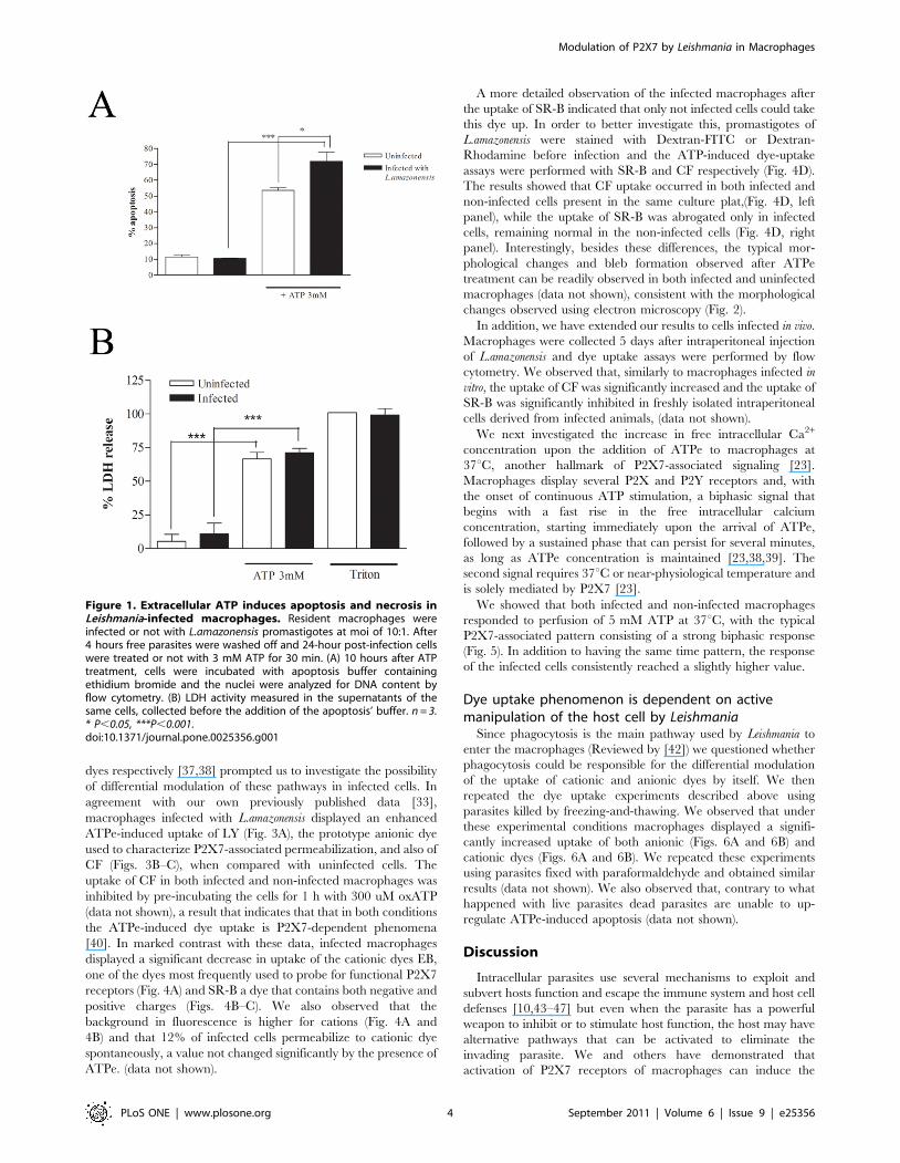
Cytotoxic effects of ATPe in macrophages andintracellular L.amazonensis
It is well established that ATPe, at millimolar concentrations,
induces apoptosis and necrosis through the activation of P2X7
receptors in different cell types such as macrophages, dendritic
cells and other cells [24,40,41]. We have recently shown that
L.amazonensis infection up-regulates P2X7 expression and renders
the cells more susceptible to ATPe-induced apoptosis and we have
also shown that even at sub-lytic doses, ATPe can induce parasite
elimination by macrophages [33]. At 3 mM, a condition that
induces more than 50% of macrophages to undergo necrosis and
apoptosis, a significant rise of 1866% was observed in apoptosis
(Fig. 1A) but not in necrosis (Fig. 1B) of infected cells. These results
were confirmed by staining with annexin-V and propidium iodide
6 hrs after ATPe (data not shown).
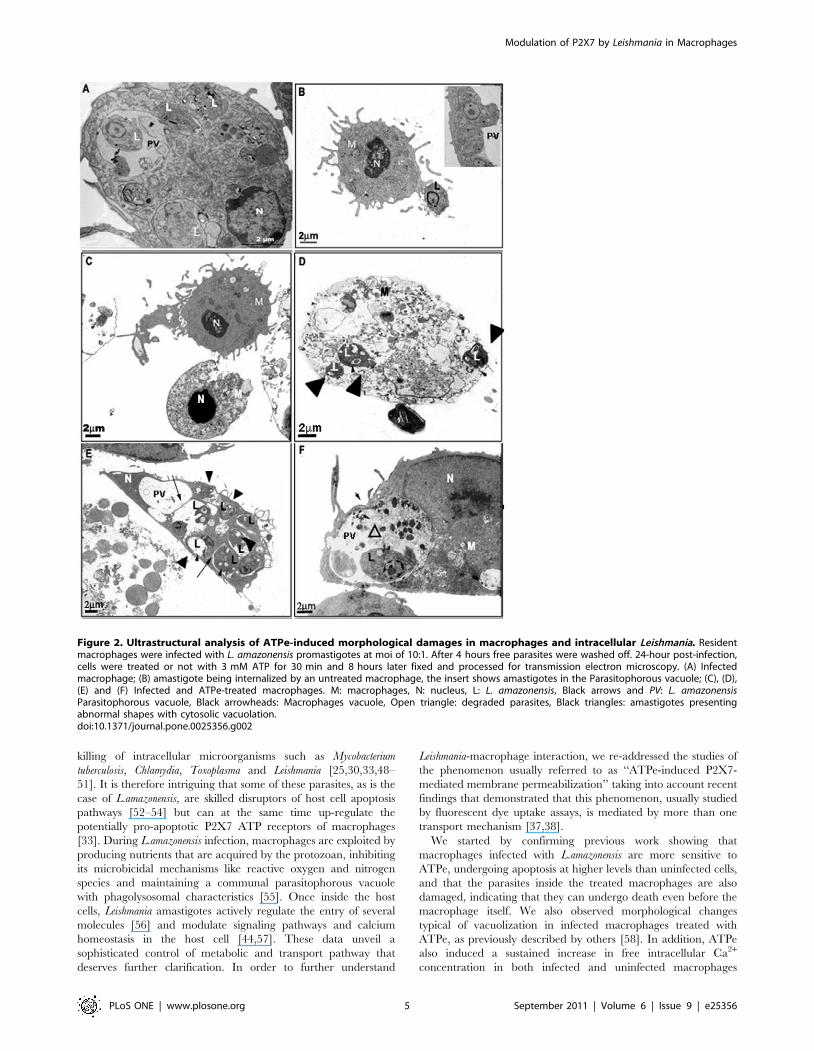
Cells were also observed by electron microscopy (Fig. 2). Infected
macrophages with morphological alterations characteristic of apop-
tosis - condensed nuclei, decreased cytosolic area/volume, loss of
cytosolic electron density, and difficult to identify organelles - were
clearly observed after ATP treatment (Fig. 2C) as well as cells
displaying typical characteristics of necrosis such as loss of shape,
shrinkage, and loss of plasma membrane structure (Fig. 2D),
characteristics not present in untreated cells (Figs. 2A–B). We also
observed macrophages undergoing extensive vacuolation (Figs. 2 E–
F). We observed that parasitophorous vacuoles (PV) of ATP-treated
cells often appear to be fusionating to each other and with other
vesicles (arrows in Fig. 2E). In some vacuoles we observed plasma
membrane profiles and electrondense structures resembling degraded
parasites (open triangle in Fig. 2F). In addition, we also observed loss of
integrity of parasitophorous and parasite membranes, indicative of
the induction of killing of both macrophages and parasites (black
triangles in Fig. 2D). We also noticed the presence of amastigotes
presenting abnormal shapes with cytosolic vacuolization (black triangles
in Figs. 2E and 2D) if compared to amastigotes inside untreated cells
(L) (Figs. 2A and 2B). These results are consistent with the strong
induction of cell death of both macrophages and amastigotes induced
by ATPe and the up-regulation of the expression of functional P2X7
receptors during the course of macrophage infection by L.amazonensis,
as we have already described [33].
Differential regulation of the uptake of anionic andcationic dyes by L.amazonensis
The recent demonstration that ATPe can induce at least two
distinct dye-uptake pathways for cationic and anionic fluorescent
Modulation of P2X7 by Leishmania in Macrophages
PLoS ONE | www.plosone.org 3 September 2011 | Volume 6 | Issue 9 | e25356
dyes respectively [37,38] prompted us to investigate the possibility
of differential modulation of these pathways in infected cells. In
agreement with our own previously published data [33],
macrophages infected with L.amazonensis displayed an enhanced
ATPe-induced uptake of LY (Fig. 3A), the prototype anionic dye
used to characterize P2X7-associated permeabilization, and also of
CF (Figs. 3B–C), when compared with uninfected cells. The
uptake of CF in both infected and non-infected macrophages was
inhibited by pre-incubating the cells for 1 h with 300 uM oxATP
(data not shown), a result that indicates that that in both conditions
the ATPe-induced dye uptake is P2X7-dependent phenomena
[40]. In marked contrast with these data, infected macrophages
displayed a significant decrease in uptake of the cationic dyes EB,
one of the dyes most frequently used to probe for functional P2X7
receptors (Fig. 4A) and SR-B a dye that contains both negative and
positive charges (Figs. 4B–C). We also observed that the
background in fluorescence is higher for cations (Fig. 4A and
4B) and that 12% of infected cells permeabilize to cationic dye
spontaneously, a value not changed significantly by the presence of
ATPe. (data not shown).
A more detailed observation of the infected macrophages after
the uptake of SR-B indicated that only not infected cells could take
this dye up. In order to better investigate this, promastigotes of
L.amazonensis were stained with Dextran-FITC or Dextran-
Rhodamine before infection and the ATP-induced dye-uptake
assays were performed with SR-B and CF respectively (Fig. 4D).
The results showed that CF uptake occurred in both infected and
non-infected cells present in the same culture plat,(Fig. 4D, left
panel), while the uptake of SR-B was abrogated only in infected
cells, remaining normal in the non-infected cells (Fig. 4D, right
panel). Interestingly, besides these differences, the typical mor-
phological changes and bleb formation observed after ATPe
treatment can be readily observed in both infected and uninfected
macrophages (data not shown), consistent with the morphological
changes observed using electron microscopy (Fig. 2).
In addition, we have extended our results to cells infected in vivo.
Macrophages were collected 5 days after intraperitoneal injection
of L.amazonensis and dye uptake assays were performed by flow
cytometry. We observed that, similarly to macrophages infected in
vitro, the uptake of CF was significantly increased and the uptake of
SR-B was significantly inhibited in freshly isolated intraperitoneal
cells derived from infected animals, (data not shown).
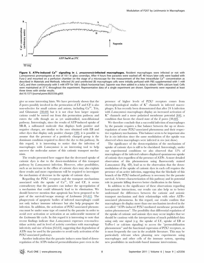
We next investigated the increase in free intracellular Ca2+
concentration upon the addition of ATPe to macrophages at
37uC, another hallmark of P2X7-associated signaling [23].
Macrophages display several P2X and P2Y receptors and, with
the onset of continuous ATP stimulation, a biphasic signal that
begins with a fast rise in the free intracellular calcium
concentration, starting immediately upon the arrival of ATPe,
followed by a sustained phase that can persist for several minutes,
as long as ATPe concentration is maintained [23,38,39]. The
second signal requires 37uC or near-physiological temperature and
is solely mediated by P2X7 [23].
We showed that both infected and non-infected macrophages
responded to perfusion of 5 mM ATP at 37uC, with the typical
P2X7-associated pattern consisting of a strong biphasic response
(Fig. 5). In addition to having the same time pattern, the response
of the infected cells consistently reached a slightly higher value.
Dye uptake phenomenon is dependent on activemanipulation of the host cell by Leishmania
Since phagocytosis is the main pathway used by Leishmania to
enter the macrophages (Reviewed by [42]) we questioned whether
phagocytosis could be responsible for the differential modulation
of the uptake of cationic and anionic dyes by itself. We then
repeated the dye uptake experiments described above using
parasites killed by freezing-and-thawing. We observed that under
these experimental conditions macrophages displayed a signifi-
cantly increased uptake of both anionic (Figs. 6A and 6B) and
cationic dyes (Figs. 6A and 6B). We repeated these experiments
using parasites fixed with paraformaldehyde and obtained similar
results (data not shown). We also observed that, contrary to what
happened with live parasites dead parasites are unable to up-
regulate ATPe-induced apoptosis (data not shown).
Discussion
Intracellular parasites use several mechanisms to exploit and
subvert hosts function and escape the immune system and host cell
defenses [10,43–47] but even when the parasite has a powerful
weapon to inhibit or to stimulate host function, the host may have
alternative pathways that can be activated to eliminate the
invading parasite. We and others have demonstrated that
activation of P2X7 receptors of macrophages can induce the
Figure 1. Extracellular ATP induces apoptosis and necrosis inLeishmania-infected macrophages. Resident macrophages wereinfected or not with L.amazonensis promastigotes at moi of 10:1. After4 hours free parasites were washed off and 24-hour post-infection cellswere treated or not with 3 mM ATP for 30 min. (A) 10 hours after ATPtreatment, cells were incubated with apoptosis buffer containingethidium bromide and the nuclei were analyzed for DNA content byflow cytometry. (B) LDH activity measured in the supernatants of thesame cells, collected before the addition of the apoptosis’ buffer. n = 3.* P,0.05, ***P,0.001.doi:10.1371/journal.pone.0025356.g001
Modulation of P2X7 by Leishmania in Macrophages
PLoS ONE | www.plosone.org 4 September 2011 | Volume 6 | Issue 9 | e25356
killing of intracellular microorganisms such as Mycobacterium
tuberculosis, Chlamydia, Toxoplasma and Leishmania [25,30,33,48–
51]. It is therefore intriguing that some of these parasites, as is the
case of L.amazonensis, are skilled disruptors of host cell apoptosis
pathways [52–54] but can at the same time up-regulate the
potentially pro-apoptotic P2X7 ATP receptors of macrophages
[33]. During L.amazonensis infection, macrophages are exploited by
producing nutrients that are acquired by the protozoan, inhibiting
its microbicidal mechanisms like reactive oxygen and nitrogen
species and maintaining a communal parasitophorous vacuole
with phagolysosomal characteristics [55]. Once inside the host
cells, Leishmania amastigotes actively regulate the entry of several
molecules [56] and modulate signaling pathways and calcium
homeostasis in the host cell [44,57]. These data unveil a
sophisticated control of metabolic and transport pathway that
deserves further clarification. In order to further understand
Leishmania-macrophage interaction, we re-addressed the studies of
the phenomenon usually referred to as ‘‘ATPe-induced P2X7-
mediated membrane permeabilization’’ taking into account recent
findings that demonstrated that this phenomenon, usually studied
by fluorescent dye uptake assays, is mediated by more than one
transport mechanism [37,38].
We started by confirming previous work showing that
macrophages infected with L.amazonensis are more sensitive to
ATPe, undergoing apoptosis at higher levels than uninfected cells,
and that the parasites inside the treated macrophages are also
damaged, indicating that they can undergo death even before the
macrophage itself. We also observed morphological changes
typical of vacuolization in infected macrophages treated with
ATPe, as previously described by others [58]. In addition, ATPe
also induced a sustained increase in free intracellular Ca2+
concentration in both infected and uninfected macrophages
Figure 2. Ultrastructural analysis of ATPe-induced morphological damages in macrophages and intracellular Leishmania. Residentmacrophages were infected with L. amazonensis promastigotes at moi of 10:1. After 4 hours free parasites were washed off. 24-hour post-infection,cells were treated or not with 3 mM ATP for 30 min and 8 hours later fixed and processed for transmission electron microscopy. (A) Infectedmacrophage; (B) amastigote being internalized by an untreated macrophage, the insert shows amastigotes in the Parasitophorous vacuole; (C), (D),(E) and (F) Infected and ATPe-treated macrophages. M: macrophages, N: nucleus, L: L. amazonensis, Black arrows and PV: L. amazonensisParasitophorous vacuole, Black arrowheads: Macrophages vacuole, Open triangle: degraded parasites, Black triangles: amastigotes presentingabnormal shapes with cytosolic vacuolation.doi:10.1371/journal.pone.0025356.g002
Modulation of P2X7 by Leishmania in Macrophages
PLoS ONE | www.plosone.org 5 September 2011 | Volume 6 | Issue 9 | e25356

maintained in the presence of ATPe for several minutes at 37uC,
one of the hallmarks of the ‘‘permeabilization’’ phenomenon [23].
ATPe-induced macrophage death, parasite killing, vacuoliza-
tion, and Ca2+ signaling have all been associated with the
expression of P2X7 receptors [23,33,58], known to be up-
regulated during macrophage infection by L.amazonensis [33].
Consistent with these observations we have also shown that uptake
of LY and CF are up-regulated in infected macrophages, a
phenomenon that has been associated with the formation of a
permeation pore [23,38,59,60], possibly due to pannexin mole-
cules in the membrane [35,36]. Surprisingly, we show here that
the uptake of the cationic dyes EB and SR-B is down-regulated
during infection. This result not only confirms that cationic dyes
can be taken up by a different P2X7-activated transport
mechanism in macrophages [37,38] but also demonstrates for
the first time that these transport mechanisms can be differentially
modulated in immunologically relevant conditions.
It is therefore reasonable to state that only the transport
mechanism accessed by the anionic dyes the LY and CF is involved
in macrophage death, vacuolization, blebbing, Ca2+ signaling, and
parasite killing. It is also clear that, at least in infected macrophages,
the mechanism of transport involved in the ATP-induced uptake of
cationic dyes is not important for triggering neither of these
phenomena. However, the precise role of the anionic and the
Figure 3. Increased uptake of anionic dyes by L.amasonensis-infected macrophages. Resident macrophages were infected or not withL.amazonensis promastigotes at moi of 10:1. After 4 hours free parasites were washed off and 24-hour post-infection cells were treated or not with3 mM ATP for 15 min. LY (A) or CF (B and C) was added 5 min after the addition of ATP. Cells were then washed 3 times with buffer and countedusing fluorescence microscopy (C) or lysed for fluorimetric analysis (A and B) n = 3. Macrophage nuclei are stained in blue (Hoechst). Calibration Bar:10 mm. * P,0.05, **P,0.01, ***P,0.001.doi:10.1371/journal.pone.0025356.g003
Modulation of P2X7 by Leishmania in Macrophages
PLoS ONE | www.plosone.org 6 September 2011 | Volume 6 | Issue 9 | e25356

cationic branches of the ATP-induced transport mechanisms in each
of the many P2X7-dependent cellular responses needs to be better
characterized. This is the case of apoptosis. We showed that infected
cells display increased ATP-induced apoptosis and increased uptake
of anionic dyes (Figs. 1 and 3). However, macrophages exposed dead
parasites also up-regulates the uptake of anionic dyes (Fig. 6), without
the corresponding increase in apoptosis (not shown). One possibility
to explain this apparent contradiction is may be the involvement of an
active participation of the live parasites. Further experiments are
needed to investigate this possibility.
The mechanism involved in EB and SR-B uptake remains
elusive as well as the pathway used by the parasites to down-
regulate it. At the present moment we can only say that this down-
regulation requires the active participation of live parasites.
Further experiments are required to clarify the nature of this
transport mechanism, its role in Leishmania infection and the
mechanism used by the parasite to induce its down-regulation
during infection.
The study of the selectivity of each of these transport
mechanisms it beyond the scope of this study, but our results
Figure 4. Decreased uptake of cationic dyes by L.amasonensis-infected macrophages. Resident macrophages were infected or not withL.amazonensis promastigotes at a moi of 10:1. After 4 hours free parasites were washed off and 24-hour post-infection cells were treated or not with3 mM ATP for 15 min. EB (A), SR-B (B, C, and right panel in D), and CF (left panel in D) was added 5 min after the addition of ATP. Cells were thenwashed 3 times with buffer and counted using fluorescence microscopy (C, D) or lysed for fluorimetric analysis (A and B) n = 3. Macrophage nuclei arestained in blue (Hoechst). White arrow in (D) indicate infected macrophage without SR-B uptake. Parasites are stained in red (D, left panel) or green(D, right panel) Calibration Bar: 10 mm. ** P,0.05, ***P,0.001.doi:10.1371/journal.pone.0025356.g004
Modulation of P2X7 by Leishmania in Macrophages
PLoS ONE | www.plosone.org 7 September 2011 | Volume 6 | Issue 9 | e25356
give us some interesting hints. We have previously shown that the
Z-pores possibly involved in the permeation of LY and CF is also
non-selective for small cations and anions, including Ca2+, Tris,
and Glutamate [38,60] but it is not clear how larger organic
cations could be sorted out from this permeation pathway and
enters the cells though an as yet unidentified, non-difusional
pathway. Interestingly, since the results of ATP-induced uptake of
SR-B, a sulfonated molecule that displays both positive and
negative charges, are similar to the ones obtained with EB and
other dyes that display only positive charges [38], it is possible to
assume that the presence of a positively charged group is the
dominant condition required to direct the dye to this pathway. In
this regard, it is interesting to notice that the infection of
macrophages with L.amazonensis is an interesting tool to help
uncover the molecular nature of this cationic transport mecha-
nism.
The results presented here suggest that the decreased uptake of
cationic dyes is due to the down-modulation of this transport
pathway by L.amazonensis infection. However, other possibilities,
such as an increase in the efflux of cationic dyes may also explain
these results and more experiments will be required to investigate
the mechanism of decrease in the uptake of cationic dyes.
Regarding the P2X7 receptors and the transport mechanisms
associated with the uptake of Ca2+, LY and CF, it seems
contradictory that the parasites can induce the up-regulation of
a mechanism that could ultimately lead to its elimination. We
should however mention that apoptosis has been proposed to be
part of the survival/escape strategy of Leishmania [45,61]. The
phagocytosis of apoptotic bodies of infected macrophages could
not only induce immune tolerance but also help propagate the
infection. In addition, the activation of P2X7-associated phenom-
ena must be under some type of control by the parasite in order to
avoid over activation or activation at an unfavorable moment of
the Leishmania life cycle. In this regard it is interesting to note that
recent findings indicate that L.amazonensis expresses ecto-nucleo-
tidase activity and its level of activity correlates with macrophage
infectivity and size of lesion [62,63], suggesting that degradation of
ATPe may be used by the parasites to avoid early activation of the
P2X7-associated pathways.
Another indication that the parasite induces some kind of down-
regulation of the ATPe-induced permeabilization pore even in the
presence of higher levels of P2X7 receptors comes from
electrophysiological studies of K+ channels in infected macro-
phages. It has recently been demonstrated that after 24 h infection
with L.amazonensis macrophages display an increased activation of
K+ channels and a more polarized membrane potential [64], a
condition that favors the closed state of the Z pores [38,60].
We therefore conclude that a successful infection of macrophages
by the parasite requires a fine balance between the up or down-
regulation of some P2X7-associated phenomena and their respec-
tive regulatory mechanisms. This balance seem to be important also
for in vivo infection since the same modulation of dye uptake was
observed when macrophages were infected in vivo (not show).
The significance of the down-regulation of the mechanism of
uptake of cationic dyes is still to be elucidated. Interestingly, under
our experimental conditions we also observed that 12% of
macrophages of the infected cultures displayed spontaneous uptake
of cationic dyes regardless of the presence of ATPe. A more detailed
observation of this phenomenon using fluorescently stained
L.amazonensis (Fig. 4D), lead us to the observation that the down-
modulation of the uptake of cationic dyes in each cell requires the
presence of an active infection, suggesting that the blockade of this
branch of the P2X7-induced pathway is necessary for the parasite
survival. A better characterization of this pathway and its potential
role in parasite killing deserves better clarification in the future.
In addition to the significance of these observations regarding
host-parasite interactions, our results can also help us to better
understand the differences between the cationic and anionic
transport mechanism and their relationships with other ATPe-
associated phenomena. In this regard, our results confirm that
macrophages do display more than one mechanism involved in the
so-called ‘‘ATPe-induced P2X7-mediated membrane permeabili-
zation phenomenon’’. The possibility that differential regulation of
the uptake of cationic and anionic dyes may occur implies that we
should be cautious with the interpretation of much published data
using only one signal (e.g. the uptake of LY, uptake of EB or
YoPro-1 or calcium signaling) to access the ‘‘permeabilization
phenomenon’’ and the functional expression of P2X7 receptors, as
is most frequently the case in the available literature. This may be
particularly relevant when planning new experiments using
macrophages and other cells of the immune system, opening
new possibilities in nucleotide-based immune interventions.
Figure 5. ATPe-induced Ca2+ signaling in L. amazonensis-infected macrophages. Resident macrophages were infected or not withL.amazonensis promastigotes at moi of 10:1 in glass coverslips. After 4 hours free parasites were washed off. 48 hours later cells were loaded withFura-2 and mounted on a perfusion chamber on the stage of a microscope for the measurement of the free intracellular Ca2+ concentration asdescribed in Materials and Methods. Infected (A) and uninfected (B) macrophages cells were initially perfused with PBS supplemented with 1 mMCaCl2 and then continuously with 5 mM ATP for 500 s (black horizontal bar). Saponin was then added in a bolus to obtain 100% calcium load. Cellswere maintained at 37uC throughout the experiment. Representative data of a single experiment are shown. Experiments were repeated at leastthree times with similar results.doi:10.1371/journal.pone.0025356.g005
Modulation of P2X7 by Leishmania in Macrophages
PLoS ONE | www.plosone.org 8 September 2011 | Volume 6 | Issue 9 | e25356

Acknowledgments
The authors wish to thank Suzana Passos Chaves for helpful discussions,
Regina Goldemberg for help with the use of fluorescence microscope,
Vandir da Costa and Pryscilla Braga for technical assistance, and Maria
Luisa Avila for carefully reviewing the manuscript.
Author Contributions
Conceived and designed the experiments: CMS RCS PMP. Performed the
experiments: CMS MMC JCR SCR. Analyzed the data: CMS RCS PMP.
Contributed reagents/materials/analysis tools: CMS JCR RCS PMP.
Wrote the paper: CMS RCS PMP.
References
1. Soehnlein O, Lindbom L (2010) Phagocyte partnership during the onset and
resolution of inflammation. Nat Rev Immunol 10: 427–439.
2. Dardanoni L (1956) In-vitro culture of guinea-pig macrophages; pro-
posed use in study of the biology of the tubercle bacillus. Riv Ist Sieroter Ital
31: 1–7.
3. Lamy L, Wonde T, Lamy H (1966) Action of amphotericin B on Leishmania
donovani during multiplication in mouse macrophages maintained in vitro. Bull
Soc Pathol Exot Filiales 59: 964–968.
4. Nogueira N, Cohn Z (1976) Trypanosoma cruzi: mechanism of entry and
intracellular fate in mammalian cells. J Exp Med 143: 1402–1420.
Figure 6. Modulation of the uptake of cationic dyes is dependent on parasite metabolism. Resident macrophages were exposed or notwith dead L.amazonensis promastigotes at a concentration equivalent to a moi of 10:1. After 4 hours free parasites were washed off and 24-hourpost-interaction cells were treated or not with 3 mM ATP for 15 min. CF (A and B) or SR-B (A and C) was added 5 min after the addition of ATP. Cellswere then washed 3 times with buffer and analyzed with fluorescence microscopy (A) or lysed for fluorimetric analysis (B and C) n = 3. Macrophagenuclei are stained in blue (Hoechst). Calibration Bar: 10 mm. ** P,0.01, ***P,0.001.doi:10.1371/journal.pone.0025356.g006
Modulation of P2X7 by Leishmania in Macrophages
PLoS ONE | www.plosone.org 9 September 2011 | Volume 6 | Issue 9 | e25356
5. Popovic M, Gartner S (1987) Isolation of HIV-1 from monocytes but not T
lymphocytes. Lancet 330: 916.6. Cassol E, Cassetta L, Alfano M, Poli G (2010) Macrophage polarization and
HIV-1 infection. J Leukoc Biol 87: 599–608.
7. Junqueira C, Caetano B, Bartholomeu DC, Melo MB, Ropert C, et al. (2010)The endless race between Trypanosoma cruzi and host immunity: lessons for
and beyond Chagas disease. Expert Rev Mol Med 12: e29.8. Ray K, Marteyn B, Sansonetti PJ, Tang CM (2009) Life on the inside: the
intracellular lifestyle of cytosolic bacteria. Nat Rev Microbiol 7: 333–340.
9. Benoit M, Desnues B, Mege JL (2008) Macrophage polarization in bacterialinfections. J Immunol 181: 3733–3739.
10. Bogdan C (2008) Mechanisms and consequences of persistence of intracellularpathogens: leishmaniasis as an example. Cell Microbiol 10: 1221–1234.
11. Denkers EY, Butcher BA (2005) Sabotage and exploitation in macrophagesparasitized by intracellular protozoans. Trends Parasitol 21: 35–41.
12. Kaufmann SH (2007) The contribution of immunology to the rational design of
novel antibacterial vaccines. Nat Rev Microbiol 5: 491–504.13. Bowdish DM, Loffredo MS, Mukhopadhyay S, Mantovani A, Gordon S (2007)
Macrophage receptors implicated in the ‘‘adaptive’’ form of innate immunity.Microbes Infect 9: 1680–1687.
14. Taylor PR, Martinez-Pomares L, Stacey M, Lin HH, Brown GD, et al. (2005)
Macrophage receptors and immune recognition. Annu Rev Immunol 23:901–944.
15. Peters-Golden M (2009) Putting on the brakes: cyclic AMP as a multiprongedcontroller of macrophage function. Sci Signal 2: e37.
16. Di Virgilio F, Chiozzi P, Ferrari D, Falzoni S, Sanz JM, et al. (2001) Nucleotidereceptors: an emerging family of regulatory molecules in blood cells. Blood 97:
587–600.
17. Burnstock G, Fredholm BB, North RA, Verkhratsky A (2010) The birth andpostnatal development of purinergic signalling. Acta Physiol (Oxf) 199: 93–147.
18. Abbracchio MP, Burnstock G, Verkhratsky A, Zimmermann H (2009)Purinergic signalling in the nervous system: an overview. Trends Neurosci 32:
19–29.
19. Surprenant A, North RA (2009) Signaling at Purinergic P2X Receptors. AnnuRev Physiol 71: 333–359.
20. Di Virgilio F (2007) Purinergic signalling in the immune system. A brief update.Purinergic Signal 3: 1–3.
21. Ferrari D, Pizzirani C, Adinolfi E, Lemoli RM, et al. (2006) The P2X7 receptor:a key player in IL-1 processing and release. J Immunol 176: 3877–3883.
22. North RA (2002) Molecular physiology of P2X receptors. Physiol Rev 82:
1013–1067.23. Persechini PM, Bisaggio RC, Alves-Neto JL, Coutinho-Silva R (1998)
Extracellular ATP in the lymphohematopoietic system: P2Z purinoceptorsand membrane permeabilization. Braz J Med Biol Res 31: 25–34.
24. Steinberg TH, Newman AS, Swanson JA, Silverstein SC (1987) ATP4-
permeabilizes the plasma membrane of mouse macrophages to fluorescentdyes. J Biol Chem 262: 8884–8888.
25. Coutinho-Silva R, Stahl L, Raymond M-N, Jungas T, Verbeke P, et al. (2003)Inhibition of chlamydial infectious activity due to P2X7R-dependent phospho-
lipase D activation. Immunity 19: 403–412.26. Mariathasan S, Monack DM (2007) Inflammasome adaptors and sensors:
intracellular regulators of infection and inflammation. Nat Rev Immunol 7:
31–40.27. Pelegrin P (2008) Targeting interleukin-1 signaling in chronic inflammation:
Focus on P2X(7) receptor and Pannexin-1. Drug News Perspect 21: 424–433.28. Costa-Junior HM, Mendes AN, Davis GHNG, Monteiro-da-Cruz C,
Ventura AL, et al. (2009) ATP-induced apoptosis involves a Ca2+-independent
phospholipase A2 and 5-lipoxygenase in macrophages. Prostaglandins OtherLipid Mediat 88: 51–61.
29. Hubert S, Rissiek B, Klages K, Huehn J, Sparwasser T, et al. (2010)Extracellular NAD+ shapes the Foxp3+ regulatory T cell compartment through
the ART2-P2X7 pathway. J Exp Med 207: 2561–2568.
30. Coutinho-Silva R, Correa G, Sater AA, Ojcius DM (2009) The P2X(7) receptorand intracellular pathogens: a continuing struggle. Purinergic Signal 5: 197–204.
31. Sharma U, Singh S (2009) Immunobiology of leishmaniasis. Indian J Exp Biol47: 412–423.
32. McConville MJ, de SD, Saunders E, Likic VA, Naderer T (2007) Living in aphagolysosome; metabolism of Leishmania amastigotes. Trends Parasitol 23:
368–375.
33. Chaves SP, Torres-Santos EC, Marques-da-Silva C, Figliuolo VR, Perse-chini PM, et al. (2009) Modulation of P2X7 purinergic receptor in macrophages
by Leishmania amazonensis and its role in parasite elimination. Microbes Infect11: 842–849.
34. Duan S, Neary JT (2006) P2X(7) receptors: properties and relevance to CNS
function. Glia 54: 738–746.35. Locovei S, Scemes E, Qiu F, Spray DC, Dahl G (2007) Pannexin1 is part of the
pore forming unit of the P2X(7) receptor death complex. FEBS Lett 581:483–488.
36. Pelegrin P, Surprenant A (2006) Pannexin-1 mediates large pore formation andinterleukin-1beta release by the ATP-gated P2X(7) receptor. EMBO J 25:
5071–5082.
37. Cankurtaran-Sayar S, Sayar K, Ugur M (2009) P2X7 receptor activates multiple
selective dye-permeation pathways in RAW 264.7 and human embryonic kidney293 cells. Mol Pharmacol 76: 1323–1332.
38. Schachter J, Motta AP, Zamorano AS, Silva-Souza HA, Guimaraes MZP, et al.
(2008) ATP-induced P2X7-associated uptake of large molecules involves distinctmechanisms for cations and anions in macrophages. J Cell Sci 121: 3261–3270.
39. Monteiro-da-Cruz C, Ventura AL, Schachter J, Costa-Junior HM, Silva-Souza HA, et al. (2006) Activation of ERK1/2 by extracellular nucleotides in
macrophages is mediated by multiple P2 receptors independently of P2X7-
associated pore or channel formation. Br J Pharmacol 147: 324–334.40. Di Virgilio F, Chiozzi P, Falzoni S, Ferrari D, Sanz JM, et al. (1998) Cytolytic
P2X purinoceptors. Cell Death Diff 5: 191–199.41. Coutinho-Silva R, Persechini PM, Bisaggio RC, Perfettini J-L, Sa-Neto ACT, et
al. (1999) P2Z/P2X7 receptor-dependent apoptosis of dendritic cells.Am J Physiol 276: C1139–C1147.
42. Mauel J (1990) Macrophage-parasite interactions in Leishmania infections.
J Leukoc Biol 47: 187–193.43. Sacks D, Sher A (2002) Evasion of innate immunity by parasitic protozoa. Nat
Immunol 3: 1041–1047.44. Bhardwaj S, Srivastava N, Sudan R, Saha B (2010) Leishmania interferes with
host cell signaling to devise a survival strategy. J Biomed Biotechnol 2010:
109189.45. Wanderley JL, Pinto da Silva LH, Deolindo P, Soong L, Borges VM, et al.
(2009) Cooperation between apoptotic and viable metacyclics enhances thepathogenesis of Leishmaniasis. PLoS ONE 4: e5733.
46. Fortea J, Prina E, de La LE, Lecoeur H, Lang T, et al. (2007) Unveilingpathways used by Leishmania amazonensis amastigotes to subvert macrophage
function. Immunol Rev 219: 66–74.
47. Wanderley JL, Benjamin A, Real F, Bonomo A, Moreira ME, et al. (2005)Apoptotic mimicry: an altruistic behavior in host/Leishmania interplay.
Braz J Med Biol Res 38: 807–812.48. Sikora A, Liu J, Brosnan C, Buell G, Chessell I, et al. (1999) Purinergic signaling
regulates radical-mediated bacterial killing mechanisms in macrophages through
a P2X7-independent mechanism. J Immunol 163: 558–561.49. Molloy A, Laochumroonvorapong P, Kaplan G (1994) Apoptosis, but not
necrosis, of infected monocytes is coupled with killing of intracellular bacillusCalmette- Guerin. J Exp Med 180: 1499–1509.
50. Lees MP, Fuller SJ, McLeod R, Boulter NR, Miller CM, et al. (2010) P2X7Receptor-Mediated Killing of an Intracellular Parasite, Toxoplasma gondii, by
Human and Murine Macrophages. J Immunol 184: 7040–7046.
51. Correa G, Marques-da-Silva C, Moreira-Souza AC, Vommaro RC, Coutinho-Silva R (2010) Activation of the P2X(7) receptor triggers the elimination of
Toxoplasma gondii tachyzoites from infected macrophages. Microbes Infect 12:497–504.
52. Donovan MJ, Maciuba BZ, Mahan CE, McDowell MA (2009) Leishmania
Infection Inhibits Cycloheximide-Induced Macrophage Apoptosis in a StrainDependent Manner. Exp Parasitol 123: 58–64.
53. Valdes-Reyes L, Argueta J, Moran J, Salaiza N, Hernandez J, et al. (2009)Leishmania mexicana: inhibition of camptothecin-induced apoptosis of
monocyte-derived dendritic cells. Exp Parasitol 121: 199–207.54. Heussler VT, Kuenzi P, Rottenberg S (2001) Inhibition of apoptosis by
intracellular protozoan parasites. Int J Parasitol 31: 1166–1176.
55. Russell DG, Xu S, Chakraborty P (1992) Intracellular trafficking and theparasitophorous vacuole of Leishmania mexicana-infected macrophages. J Cell
Sci 103(Pt 4): 1193–1210.56. Burchmore RJ, Barrett MP (2001) Life in vacuoles–nutrient acquisition by
Leishmania amastigotes. Int J Parasitol 31: 1311–1320.
57. Gregory DJ, Olivier M (2005) Subversion of host cell signalling by the protozoanparasite Leishmania. Parasitol 130 Suppl: S27–S35.
58. Verhoef PA, Estacion M, Schilling W, Dubyak GR (2003) P2X7 receptor-dependent blebbing and the activation of Rho-effector kinases, caspases, and IL-
1 beta release. J Immunol 170: 5728–5738.
59. Fortes FSA, Pecora IL, Persechini PM, Hurtado S, Costa V, et al. (2004)Modulation of intercellular communication in macrophages: possible interac-
tions between GAP junctions and P2 receptors. J Cell Sci 117: 4717–4726.60. Coutinho-Silva R, Persechini PM (1997) P2Z purinoceptor-associated pores
induced by extracellular ATP in macrophages and J774 cells. Am J Physiol 273:C1793–C1800.
61. Barcinski MA, DosReis GA (1999) Apoptosis in parasites and parasite-induced
apoptosis in the host immune system: a new approach to parasitic diseases.Braz J Med Biol Res 32: 395–401.
62. Souza MC, Assis EA, Gomes RS, Marques da Silva EA, Melo MN, et al. (2010)The influence of ecto-nucleotidases on Leishmania amazonensis infection and
immune response in C57B/6 mice. Acta Trop 115: 262–269.
63. Pinheiro CM, Martins-Duarte ES, Ferraro RB, Fonseca De Souza AL,Gomes MT, et al. (2006) Leishmania amazonensis: Biological and biochemical
characterization of ecto-nucleoside triphosphate diphosphohydrolase activities.Exp Parasitol 114: 16–25.
64. Forero ME, Marin M, Corrales A, Llano I, Moreno H, et al. (1999) Leishmaniaamazonensis infection induces changes in the electrophysiological properties of
macrophage-like cells. J Membr Biol 170: 173–180.
Modulation of P2X7 by Leishmania in Macrophages
PLoS ONE | www.plosone.org 10 September 2011 | Volume 6 | Issue 9 | e25356