Subcortical regional morphology correlates with fluid and spatial intelligence
Upload
independentCategory
view
4download
0
CPI
FFSa
Lb
Nc
Rd
7e
Ag(Afp(spcvonCoancdtdceicaPa
*L0EAncnfippts
Neuroscience 151 (2008) 791–801
0d
ORTICAL AND SUBCORTICAL DISTRIBUTION OF IONOTROPICURINERGIC RECEPTOR SUBUNIT TYPE 1 (P2X1R)
MMUNOREACTIVE NEURONS IN THE RAT FOREBRAIN
cP
Ksc
Aoaimd2hbrKtsl1tatwbchmcssausB2tapaw(
Csp
. FLORENZANO,a P. CARRIVE,b M. T. VISCOMI,a
. FERRARI,a,c L. LATINI,a D. CONVERSI,a,d
. CABIB,a,d,e C. BAGNIa,c AND M. MOLINARIa*
Istituto di Ricovero e Cura a Carattere Scientifico (I.R.C.C.S.) Santaucia Foundation, Via Fosso del Fiorano 64, Rome, Italy
Department of Anatomy, School of Medical Sciences, University ofew South Wales, Sydney, NSW 2052, Australia
Department of Biology, University of Roma Tor Vergata, Via dellaicerca Scientifica 1, 00133, Rome, Italy
Department of Psychology, University “La Sapienza,” Via dei Marsi8, 00185, Rome, Italy
Center D. Bovet, University “La Sapienza,” Rome, Italy
bstract—Ionotropic purinergic receptors (P2XR) are ATP-ated cationic channels composed of seven known subunitsP2X1–7R) and involved in different functions in neural tissue.lthough their presence has been demonstrated in the brain,
ew studies have investigated their expression pattern. Inarticular, ionotropic purinergic receptor subunit type 1P2X1R) has been observed in the cerebellum and in brain-tem nuclei. The present study investigates the P2X1R ex-ression pattern in the rat forebrain using immunohisto-hemistry. The specificity of the immunolabeling has beenerified by Western blotting and in situ hybridization meth-ds. P2X1R immunoreactivity was specifically localized ineurons, dendrites and axons throughout the forebrain.haracteristic differences in the distribution of P2X1R werebserved in different cortical areas. In prefrontal, cingulatend perirhinal cortices, very intense labeling was present ineuronal bodies. In frontal, parietal, temporal and occipitalortices, immunostaining was lighter and mainly found inendrites and axons. The hippocampal formation was in-ensely labeled. Labeling was present almost exclusively inendrites and axons and never in neuronal bodies. The dien-ephalon was devoid of P2X1R positive neurons or fibersxcept for the medial habenular nucleus, which showed veryntense P2X1R immunostaining. Furthermore, two subcorti-al regions, namely, the nucleus centralis of the amygdaland the bed nucleus of the stria terminalis, showed intense2X1R neuronal labeling. Present data indicate that P2X1Rre prevalent in forebrain areas involved in the integration of
Correspondence to: M. Molinari, Experimental Neurorehabilitationaboratory, I.R.C.C.S. Santa Lucia Foundation, Via Ardeatina 306,0179 Rome, Italy. Tel: �39-6-51501600; fax: �39-6-51501679.-mail address: [email protected] (M. Molinari).bbreviations: BST, bed nucleus of the stria terminalis; BSTLD, beducleus of the stria terminalis, lateral division, dorsal part; Ce, nucleusentralis; Cg2, posterior cingulate area; DAPI, 4=,6-diamidino-2-phe-ylindole dihydrochloride; DG, dentate gyrus; EtC, Entorhinal cortex;r, fasciculus retroflexus; GFAP, anti-glial fibrillary acid protein; IL,nfralimbic; NeuN, neuronal nuclei; PFC, prefrontal cortex; PB, phos-hate buffer; PRhC, perirhinal cortex; PrL, prelimbic; P2XR, ionotropicurinergic receptors; P2X1R, ionotropic purinergic receptor subunit
Pype 1; RSGC, retrosplenial granular cortex; RT, room temperature; S,ubiculum.
306-4522/08$32.00�0.00 © 2007 IBRO. Published by Elsevier Ltd. All rights reseroi:10.1016/j.neuroscience.2007.11.027
791
ognitive, limbic and autonomic functions. © 2007 IBRO.ublished by Elsevier Ltd. All rights reserved.
ey words: nucleus centralis of the amygdala, bed nucleus oftria terminalis, limbic cortex, prefrontal cortex, cingulateortex.
TP is a signaling molecule that acts through two classesf purinergic receptors (P2R), namely, ionotropic (P2XR)nd metabotropic (P2YR) purinergic receptors. P2XR are
onotropic channels involved in fast excitatory neurotrans-ission, neuromodulatory actions and as sensors of tissueamage (Khakh, 2001; North, 2002; Khakh and North,006). Until now, seven distinct P2XR subunits (P2XR1–7)ave been cloned from mammalian species. P2XR haveeen identified in virtually all tissues, where their functionalole has been demonstrated (Burnstock and Knight, 2004;hakh and North, 2006). Fewer studies have investigated
he distribution and function of P2XR in the CNS; however,ome of these have revealed unexpectedly wide, but se-
ective, CNS distribution (Xiang et al., 1998; Kanjhan et al.,999; Norenberg and Illes, 2000; Yao et al., 2000). Ana-omical characterization has provided some informationbout the expression of the different P2XR subunits. Forhe ionotropic purinergic receptor subunit type 2 (P2X2R),hich is the one most studied, a widespread pattern haseen reported that includes broad sectors of the cerebralortex, different rhinencephalic structures, many thalamic,ypothalamic and basal ganglia nuclei as well as sensory,otor and integration nuclei of the brainstem and the
erebellar cortex (Kanjhan et al., 1999). Co-localizationtudies have suggested an important role of the P2X2Rubunit in the regulation of hypothalamic functions (Yao etl., 2003; Florenzano et al., 2006). As to the other sub-nits, few studies have investigated the topographical andubcellular distribution of P2X1– 6R subunits (Loesch andurnstock, 1998; Yao et al., 2000, 2001; Rubio and Soto,001). In experimental lesion models, it has been foundhat ionotropic purinergic receptor subunit type 1 (P2X1R)nd P2X2R are up-regulated in neurons after central anderipheral axotomy (Florenzano et al., 2002; Atkinson etl., 2003; Kassa et al., 2007), and a functional interactionsith post-axotomy nNOS expression has been suggested
Viscomi et al., 2004; Kassa et al., 2007).Whether or not the P2X1R subunit is present in the
NS is still a matter of debate. Indeed, P2X1R RNA tran-cripts were demonstrated in the cerebral cortex, hip-ocampus and striatum (Norenberg and Illes, 2000);
2X1R protein expression was assessed in the cerebellumved.

aedo2Pesiiuc
A
T2sdetE(d12bcwssiCsistf(
W
RbdedcwvaawwPUpmBts
H
Ifl
r0s
miPiaPclwon
o(P(mlscaswiaor4wM
iP2a(rTs
F
Ibt
wmp
P5Pm2mtien(t
F. Florenzano et al / Neuroscience 151 (2008) 791–801792
nd in brainstem nuclei (Loesch and Burnstock, 1998; Yaot al., 2000, 2001); and the presence of P2X1R in pyrami-al neurons of the somatosensory cortex was suggestedn pharmacological grounds (Pankratov et al., 2002,003). Recently a wide debate arose on the specificity of2X antibodies (Ashour et al., 2006; Anderson and Ned-rgaard, 2006) and a multi-technical approach has beenuggested to support the specificity of immunohistochem-cal data (Rhodes and Trimmer, 2006). The present studynvestigates the pattern of expression of the P2X1R sub-nit in the forebrain by Western blotting, immunohisto-hemistry and in situ hybridization methods.
EXPERIMENTAL PROCEDURES
nimals and tissue preparation
welve adult male rats (Wistar, Harlan, Udine, Italy) weighing00–250 g were used in this study. They were group-housed intandard cages and kept under a 12-h light/dark cycle in a con-itioned facility. Food and water were provided ad libitum. Allxperiments were carried out in accordance with the Italian law onhe use and care of laboratory animals (DL 116\92) and with theuropean Communities Council Directive of 24 November 1986
86/609/EEC). The animals were transcardially perfused undereep anesthesia (Sodium pentobarbitol, 60 mg\kg, i.p.) with50 ml of 0.9% saline at room temperature (RT) followed by00 ml of cold 4% paraformaldehyde in a 0.1 M pH 7.4 phosphateuffer (PB). Brains were dissected, post-fixed for 2 h at RT andryoprotected in 30% PB/sucrose at 4 °C. Then, they were frozenith dry ice and cut into 40 �m transverse, sagittal or horizontalections with a sliding microtome. Three series of transverseections from the frontal pole to the occipital pole were collectedn PB and stored at 4 °C. The first series was Nissl stained withresyl Violet for anatomical reference. The second and thirderies were processed for immunoperoxidase detection, multiplemmunofluorescence or fluorescent in situ hybridization. We usedix animals for immunoperoxidase staining, three animals for mul-iple immunofluorescence in situ hybridization and three animalsor Western blotting. All chemicals were purchased from SigmaSt. Louis, MO, USA).
estern blotting
ats were deeply anesthetized by i.p. injections of sodium pento-arbital (60 mg/kg) and killed by decapitation. The brain wasissected, and the forebrain was taken. The tissues were homog-nized, extracted with RIPA buffer (1% Nonidet P-40, 0.5% so-ium deoxycolate, 0.1% SDS) in PB for 30 min at 4 °C andentrifuged for 10 min at 4 °C (14,000 r.p.m.). The supernatantas collected and sonicated, and the protein content was re-ealed by the Bradford method. Total protein (100 �g) from eachnimal was subjected to SDS–polyacrylamide gel electrophoresisnd transferred to a nitrocellulose membrane. The membranesere saturated with 5% nonfat dried milk and incubated overnightith the primary antibody at the appropriate dilution (rabbit anti-2X1R, 1:200; mouse anti-�-actin, (Chemicon, Temecula, CA,SA) 1:2000). Membranes were then incubated with horseradisheroxidase–conjugated goat anti-rabbit (1:5000) or horse anti-ouse (1:2000) secondary antibodies (Cell Signaling Technology,everly, MA, USA). Immunoreactive bands were detected using
he enhanced chemiluminescence kit (ECL, Amersham Bio-ciences, Little Chalfont, UK).
istology and immunohistochemistry
mmunohistochemical procedures were performed at RT on free-
oating sections. PB was used for both chemical dilution and winses. All primary antibody solutions were prepared in PB and.3% Triton X-100 and incubated overnight at RT. Each incubationtep was followed by three 5 min rinses in PB.
For immunoperoxidase staining, sections were treated for 5in with 0.3% H2O2 to block endogenous peroxidase. They were
ncubated overnight in primary antiserum (polyclonal rabbit anti-2X1R, Alomone, Jerusalem, Israel) diluted 1:500, and were then
ncubated for 2 h in secondary antiserum, diluted 1:200 (biotinyl-ted donkey anti-rabbit, Jackson Immunoresearch, West Grove,A, USA). Further, sections were incubated for 1 h in avidin–biotinomplex, diluted 1:100 (Vectastain elite, Vector Laboratories, Bur-ingame, CA, USA). As chromogen, 3,3= diaminobenzidine 0.05%ith nickel intensification was used. Finally, sections were mountedn chrome-alum–coated slides, air dried, dehydrated with etha-ol, cleared in xylene and coverslipped.
Double immunofluorescence was performed using a solutionf rabbit anti-P2X1R (1:100) and mouse anti-neuronal nucleiNeuN; 1:100, (Chemicon), lot number 24010098) or rabbit anti-2X1R receptor and mouse anti-glial fibrillary acid protein (GFAP)
1:600, Chemicon) or rabbit anti-P2X1R receptor subunits andouse anti-OX-42 (1:200; Serotec, Kidlington, Oford, UK). Fol-
owing incubation with the mix solution of primary antibodies, theections were incubated for 2 h at RT in a solution of Cy2-onjugated donkey anti-mouse IgG and Cy3-conjugated donkeynti-rabbit IgG secondary antibodies (1:100; Jackson Immunore-earch Laboratories, West Grove, PA, USA). Following incubationith the mix solution of primary antibodies, the sections were
ncubated for 2 h at RT in a solution of Cy2-conjugated donkeynti-mouse IgG and Cy3-conjugated donkey anti-rabbit IgG sec-ndary antibodies (1:100; Jackson Immunoresearch Laborato-ies). Before the last rinse, sections were counterstained with=,6-diamidino-2-phenylindole dihydrochloride (DAPI). Then theyere mounted on gelatin-coated slides and coverslipped in Gel/ount (Biomeda, Foster City, CA, USA).
Triple fluorescence was performed using sections derived byn situ hybridization procedures that were incubated with the anti-2X1R (1:100) antibody. Subsequently, they were incubated forh at RT in a solution of Cy3-conjugated mouse anti-digoxigenin
nd Cy2-conjugated donkey anti-rabbit IgG secondary antibodies1:100; Jackson Immunoresearch Laboratories). Before the lastinse, sections were DAPI counterstained (5 min, 1:1000; Sigma).hen they were mounted on gelatin-coated slides and cover-lipped in Gel/Mount (Biomeda).
luorescence in situ hybridization
n situ hybridization experiments were performed using probes inoth sense and antisense orientations. The primers used to clonehe partial cDNAs from rat total brain RNA were:
P2X1R forward: 5=-GGA CAG CTC CTT TGT AGT TAT-3=P2X1R reverse: 5=-TGG TAG ATG GGT TTG CAG TGC-3=The cDNA was cloned into the pGemTeasy vector, linearized
ith SacI or with SacII and transcribed with T7 or Sp6 poly-erases to obtain antisense or sense digoxigenin-labeled cRNArobes, respectively.
Sections were post-fixed with 4% paraformaldehyde in 0,1 MBS for 30 min, then rinsed with 1� PBS (0.1% DEPC treated) formin. Sections were permeabilized with 0.3% Triton X-100 in 1�BS for 30 min, rinsed with 0.5� SSC (0.1% DEPC treated) for 10in and incubated in 500 �l of pre-hybridization buffer (2� SSC,5% formamide, 1% Denhardt’s solution, 10% dextran sulfate, 0.5g/ml heparin, 0.5 mg/ml E. coli tRNA and 0.25 mg/ml of dena-
ured salmon sperm DNA) at 42 °C for 2–3 h. After the prehybrid-zation, 700 ng of digoxigenin-labeled cRNA probe was added toach section. Hybridization was performed overnight at 55 °C. Theext day, sections were washed twice with 2� SSC/1 mM EDTA10 min each), treated with 002 �g/ml RNAseA for 30 min andhen washed twice with 2� SSC/1 mM EDTA. The stringency
ash was performed at 55 °C for 2 h in 0.5� SSC/1 mM EDTA.
TRds
C
Tpsbfiscfdtpi
mwtt
D
Baawntl(rr
teinleal4paD
C
stnsmp1p
i
Pitw
ws(
fccp
FstPtacPtCcapS
F. Florenzano et al / Neuroscience 151 (2008) 791–801 793
hen, sections were washed with 0.5� SSC twice (10 min each atT). Sections were rinsed three times with 1� PBS and thenestined for multiple fluorescence of the P2X1R protein, as de-cribed above.
ontrols
he specificity of the P2X1R primary antiserum was verified byreadsorption with its own specific target peptide (Alomone, Jew-alem, Israel). For Western blotting, preadsorption was performedy mixing the P2X1R antiserum at the same concentration used
or membrane incubation with the peptide (3 �g/ml) for 2 h beforencubating the membrane. For immunoperoxidase staining, pread-orption was performed by mixing the antiserum at the sameoncentration used for immunostaining with the peptide (10 �g/ml)or 1 h before it was used. Adjacent sections were placed in twoifferent wells: one was incubated with P2X1R primary antiserum,he other with P2X1R primary antiserum, preadsorbed with theeptide. Sections were then transferred to the same well and
ncubated with a secondary antibody.The specificity of the in situ hybridization procedures and
ultiple immunofluorescence was verified by incubating sectionsith a sense digoxigenin-labeled cRNA probe. After rinses, sec-
ions were destined for multiple fluorescence of the P2X1R pro-ein, as described above.
ata collection and analysis
oundaries of cortical and subcortical structures were delineatedccording to Cresyl Violet staining, following Paxinos (Paxinosnd Watson, 1997; Paxinos, 2004). Some nuclear subdivisionsere not subdivided as such, and instead the name of the wholeucleus was used. Topography and nomenclature were accordingo Paxinos (Paxinos and Watson, 1997; Paxinos, 2004); habenu-ar subdivisions were identified with reference to Andres et al.1999). Sections were collected from selected rostrocaudal levels,anging approximately from �2.70 to �6.04 from bregma, witheference to the rat brain atlas of Paxinos and Watson (1997).
The immunoperoxidase material was examined under a light-ransmission microscope (Zeiss, Axioskop 2, Jena, Germany)quipped with a CCD camera (ProgRes C10 plus, Zeiss). Double
mmunofluorescence was examined under a confocal laser scan-ing microscope (CLSM; Zeiss, LSM 510) equipped with two laser
ines: an argon laser emitting at 488 nm and a helium/neon lasermitting at 543 nm. Triple fluorescent material was examined withCLSM (Leica, SP5, Wetzlar, Germany) equipped with three
aser lines: violet diode emitting at 405 nm, argon emitting at88 nm and helium/neon emitting at 543 nm. Images were ex-orted in JPEG format, contrast and brightness were adjusted,nd final plates were composed with Adobe Illustrator 9 or Corelraw 9.
RESULTS
ontrols
Western Blotting. Western blotting, performed on tis-ue extracts derived from the rat brain (Fig. 1A), assessedhe specificity of the polyclonal P2X1R antibody. An immu-oreactive band was detected at 60 kDa that corre-ponded to P2X1R (Fig. 1A, lane 1), as shown by theanufacturer. Preadsorption of the antiserum with theeptide antigen resulted in the absence of the band (Fig.A, lane 2), indicating that the antibody detects the appro-riate antigen sequence.
Immunohistochemistry. The specificity of the P2X R
1mmunoperoxidase staining was verified by a blocking test. s
readsorption with the P2X1R peptide completely abol-shed the staining in the sections (Fig. 1B, C), confirminghe specificity of the antibody for the P2X1R sequence alsohen used for immunohistochemistry.
In situ hybridization. In situ hybridization performedith a sense probe for P2X1R mRNA showed a fluorescentignal that was barely detectable above the backgroundFig. 1E).
In situ hybridization performed with an antisense probeor P2X1R mRNA demonstrated P2X1R mRNA positiveells in the forebrain (Fig. 2C, D). Labeled cells wereharacterized by a fluorescent rim in the perinuclear cyto-lasmatic domain and positive cytoplasmatic granular
ig. 1. Control panel; A–C: P2X1R antibody specificity tests; D–G: initu hybridization specificity test. (A) Western blotting from brain ex-racts. M, molecular weight marker line (SeeBleu, Invitrogen). Line 1,2X1R immunoreactive band is located at 60 kDa. Line 2, preadsorp-
ion of the P2X1R antisera with its peptide antigen resulted in thebsence of the band. (B–C) Preadsorption test. P2X1R immunohisto-hemistry in adjacent sections of the prefrontal cortex (PFC) using2X1R primary antibody alone (B) or after preadsorption with its pep-
ide antigen (C). Note the absence of immunostaining in C. (D–G)onfocal images of in situ hybridization with a sense P2X1R fluores-ent riboprobe (E, red) coupled with DAPI histofluorescence (D, blue)nd P2X1R protein immunofluorescence (F, green). Note that P2X1Rrotein positive neurons do not show sense mRNA positivity (arrows).cale bars�100 �m B, G�10 �m.
tructures.

sPsftpeep
P
lfiBitossaigtgaes
wa(PNOp
psoatilsrlaempdftF(ismdncdpwceost(tn
aBBB
B
BBBCCCCDEfGHi
F. Florenzano et al / Neuroscience 151 (2008) 791–801794
Multiple fluorescence of the P2X1R protein and mRNAhowed that all P2X1R protein positive cells were also2X1R mRNA positive (Fig. 2 filled arrows). On the otheride, P2X1R mRNA positive cells were often not positiveor the P2X1R protein (Fig. 2 arrowheads). However, theendency of the P2X1R protein to be located in neuronalrocesses rather than in the cell body and the almostxclusive cell body location of the P2X1R mRNA mayxplain the mismatch between P2X1R mRNA and P2X1Rrotein labeling.
2X1R immunostaining pattern
Overall pattern. P2X1R immunoreactivity appearedocalized in neurons, dendrites and axons throughout theorebrain of the rat. P2X1R immunostaining was distributedn the cytoplasm and was never observed in the nucleus.ased on differences in the immunoperoxidase staining
ntensity or in the expression within different cell domains,hree basic qualitative patterns of immunoreactivity werebserved. These basic intensity patterns were: i, lighttaining of axons and dendrites (Fig. 3A, B) ii, diffusetaining of the entire cell (Fig. 3C–F), iii, intense staining ofxons and dendrites (Fig. 3G, H). In addition, neuropil
mmunostaining was observed in several anatomical re-ions. These differences in intensity and cellular distribu-ion were apparent mainly when different anatomical re-ions were compared and were not evident in the samenatomical region. This indicates that the intensity of thexpression level and the cellular sites of expression werepecific for different anatomical areas and neuronal types.
The neuronal or glial origin of the staining observedas addressed by double NeuN and P2X1R (Fig. 4A–I)nd by double P2X1R and GFAP (Fig. 4J–L) or Ox-42data not shown) immunolabeling. In all areas studied,2X1R cell body labeling was always associated witheuN immunofluorescence, and never with GFAP orX-42 labeling. Taken together data demonstrated theresence of P2X1R only in neurons.
Abbreviation
c anterior commissureLA basolateral amygdaloid nucleus, anteriorMA basomedial amygdaloid nucleus, anteriorSTLD bed nucleus of the stria terminalis, lateral division, dors
partSTLP bed nucleus of the stria terminalis, lateral division, posteri
partSTM bed nucleus of the stria terminalis, medial divisionSTTA bed nucleus of the stria terminalis, intraamygdalar divisioSTV bed nucleus of the stria terminalis, ventral divisioneC nucleus centralis, capsulareL nucleus centralis, lateraleM nucleus centralis, medialPu caudate putamenG dentate gyrustC entorhinal cortex
r fasciculus retroflexusrl granular layeriF hippocampal fissure
c internal capsule
Throughout the cerebral cortex, a general distributionattern of P2X1R immunoreactivity was evident. Immuno-tained, radially-oriented processes, which could be rec-gnized as apical dendrites extending between layers Vnd III, were observed (Fig. 3A–F). The neuronal bodies of
hese apical dendrites were only occasionally stained,dentifying them as pyramidal neurons of layer V and, to aesser degree, of layer VI (Fig. 3C, E). In the absence ofomata labeling, confocal imaging of double immunofluo-escence for P2X1R and NeuN confirmed the prevalentayer V pyramidal origin of these processes (Fig. 4). Inddition, immunostained axons running toward the deep-st layers could often be followed as they entered the whiteatter. Within this general distribution pattern, differentatterns of intensity were observed in different corticalomains. In the isocortex, labeling was characterized byaint staining of the apical and basal dendrites as well as ofhe axons of layers V–VI pyramidal neurons (Fig. 3A, B;ig. 4A–C). No clear neuronal body labeling was presentFig. 3B; Fig. 4B). On the medial and lateral edges of thesocortex, in medial prefrontal and perirhinal areas, corre-ponding to proiso- and periallo-cortex transitional zones,ore intense labeling of neuronal bodies, axons and den-rites was present (Fig. 3C–F, Fig. 4D–F). In the RSGC,euronal labeling was prevalent in dendrites, with only faintytoplasmatic labeling (Fig. 3G–H; Fig. 4G–I). Intense den-ritic and axonal labeling was also present in the hip-ocampal formation, where no positive neuronal bodiesere found (Fig. 5C–F). The piriform cortex was the onlyortical structure in which labeling was not detected inither fibers or neuronal bodies. Finally, a striking featuref P2X1R immunoreactivity was the presence of Golgi-liketained neurons in the nucleus centralis (Ce; Fig. 6C–D) ofhe amygdala and in the bed nucleus of the stria terminalisBST; Fig. 6A–B). These two subcortical structures werehe only ones that displayed consistent immunoreactiveeuronal bodies.
the figures
lateral amygdaloid nucleuslateral habenulalateral septal nucleuslateral ventricule
A medial amygdaloid nucleus, anterior partb medial habenular nucleusl molecular layer
optic tractoriens stratumpyramidal cell layer of the hippocampus
l polymorphic layerhC perirhinal cortex
subiculumstratum lucidumstratum radiatumstria terminalistemporal cortex
B vertical limb of the diagonal bandthird ventricle
s used in
al
or
n
LaLhbLSLVMemHMooptOrPyPoPRSSlSrstTCVD3V
bcc(pnnwvpMsdita
ucoapd4nhmt
sngwwbnVlmpca
IaegegslcdslPaa
fenapbfilifibptr5s
FpbCpp(paPs
F. Florenzano et al / Neuroscience 151 (2008) 791–801 795
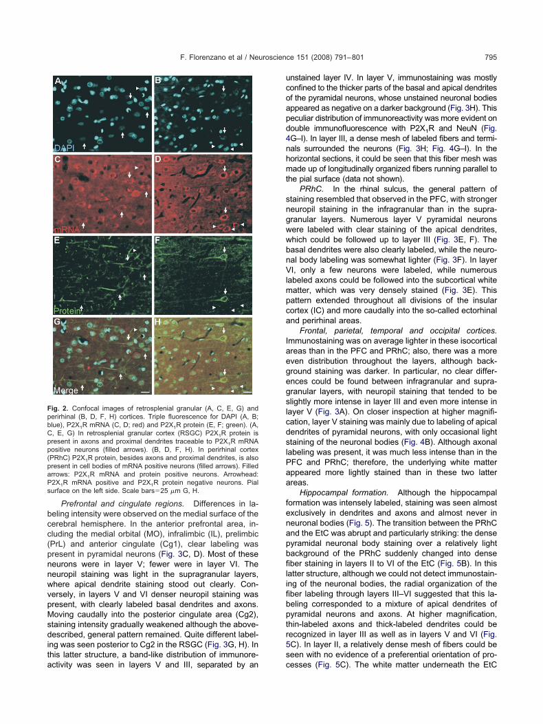
Prefrontal and cingulate regions. Differences in la-eling intensity were observed on the medial surface of theerebral hemisphere. In the anterior prefrontal area, in-luding the medial orbital (MO), infralimbic (IL), prelimbicPrL) and anterior cingulate (Cg1), clear labeling wasresent in pyramidal neurons (Fig. 3C, D). Most of theseeurons were in layer V; fewer were in layer VI. Theeuropil staining was light in the supragranular layers,here apical dendrite staining stood out clearly. Con-ersely, in layers V and VI denser neuropil staining wasresent, with clearly labeled basal dendrites and axons.oving caudally into the posterior cingulate area (Cg2),
taining intensity gradually weakened although the above-escribed, general pattern remained. Quite different label-
ng was seen posterior to Cg2 in the RSGC (Fig. 3G, H). Inhis latter structure, a band-like distribution of immunore-
ig. 2. Confocal images of retrosplenial granular (A, C, E, G) anderirhinal (B, D, F, H) cortices. Triple fluorescence for DAPI (A, B;lue), P2X1R mRNA (C, D; red) and P2X1R protein (E, F; green). (A,, E, G) In retrosplenial granular cortex (RSGC) P2X1R protein isresent in axons and proximal dendrites traceable to P2X1R mRNAositive neurons (filled arrows). (B, D, F, H). In perirhinal cortexPRhC) P2X1R protein, besides axons and proximal dendrites, is alsoresent in cell bodies of mRNA positive neurons (filled arrows). Filledrrows: P2X1R mRNA and protein positive neurons. Arrowhead:2X1R mRNA positive and P2X1R protein negative neurons. Pialurface on the left side. Scale bars�25 �m G, H.
ctivity was seen in layers V and III, separated by an c
nstained layer IV. In layer V, immunostaining was mostlyonfined to the thicker parts of the basal and apical dendritesf the pyramidal neurons, whose unstained neuronal bodiesppeared as negative on a darker background (Fig. 3H). Thiseculiar distribution of immunoreactivity was more evident onouble immunofluorescence with P2X1R and NeuN (Fig.G–I). In layer III, a dense mesh of labeled fibers and termi-als surrounded the neurons (Fig. 3H; Fig. 4G–I). In theorizontal sections, it could be seen that this fiber mesh wasade up of longitudinally organized fibers running parallel to
he pial surface (data not shown).PRhC. In the rhinal sulcus, the general pattern of
taining resembled that observed in the PFC, with strongereuropil staining in the infragranular than in the supra-ranular layers. Numerous layer V pyramidal neuronsere labeled with clear staining of the apical dendrites,hich could be followed up to layer III (Fig. 3E, F). Theasal dendrites were also clearly labeled, while the neuro-al body labeling was somewhat lighter (Fig. 3F). In layerI, only a few neurons were labeled, while numerous
abeled axons could be followed into the subcortical whiteatter, which was very densely stained (Fig. 3E). Thisattern extended throughout all divisions of the insularortex (IC) and more caudally into the so-called ectorhinalnd perirhinal areas.
Frontal, parietal, temporal and occipital cortices.mmunostaining was on average lighter in these isocorticalreas than in the PFC and PRhC; also, there was a moreven distribution throughout the layers, although back-round staining was darker. In particular, no clear differ-nces could be found between infragranular and supra-ranular layers, with neuropil staining that tended to belightly more intense in layer III and even more intense in
ayer V (Fig. 3A). On closer inspection at higher magnifi-ation, layer V staining was mainly due to labeling of apicalendrites of pyramidal neurons, with only occasional lighttaining of the neuronal bodies (Fig. 4B). Although axonal
abeling was present, it was much less intense than in theFC and PRhC; therefore, the underlying white matterppeared more lightly stained than in these two latterreas.
Hippocampal formation. Although the hippocampalormation was intensely labeled, staining was seen almostxclusively in dendrites and axons and almost never ineuronal bodies (Fig. 5). The transition between the PRhCnd the EtC was abrupt and particularly striking: the denseyramidal neuronal body staining over a relatively lightackground of the PRhC suddenly changed into denseber staining in layers II to VI of the EtC (Fig. 5B). In this
atter structure, although we could not detect immunostain-ng of the neuronal bodies, the radial organization of theber labeling through layers III–VI suggested that this la-eling corresponded to a mixture of apical dendrites ofyramidal neurons and axons. At higher magnification,hin-labeled axons and thick-labeled dendrites could beecognized in layer III as well as in layers V and VI (Fig.C). In layer II, a relatively dense mesh of fibers could beeen with no evidence of a preferential orientation of pro-
esses (Fig. 5C). The white matter underneath the EtC
wgfwttNc5sl
hsapclltgw
FIItdm 200 �m A
F. Florenzano et al / Neuroscience 151 (2008) 791–801796
as densely stained. Coming out of it, labeled axonsrouped into thick bundles could be followed into the per-orant path traversing the subiculum (S) and heading to-ard the molecular layer of the dentate gyrus (DG), where
hey terminated in a very fine mesh of terminal arboriza-ions. Granular cells of the DG were not immunoreactive.o staining was observed in this layer or in the pyramidalell layer of any of the CA3–CA1 hippocampal fields (Fig.B, D). In contrast, a dense mesh of labeled fibers could beeen in the polymorphic layer of the DG and in the strata
ig. 3. P2X1R immunolabeling in different cortical regions (A, C, Emmunostaining is mainly confined to dendrites in layer IV. (C, D) PrlII to I are essentially devoid of positive neurons. (E, F) PRhC. Note tho I are essentially devoid of positive neurons. (G, H) RSGC. Immunopevoid of immunostaining. Note the layer specific arrangement of the stesh of intensely immunostained processes in layer III. Scale bars�
ucidum, radiatum and oriens of the CA3–CA2 sector of the l
ippocampus (Fig. 5E). The densest labeling was in thetratum radiatum; axons could be seen emerging from itnd heading toward the CA1 sector. However, it was notossible to verify whether these fibers were Schaffer’sollaterals. By contrast, the CA1 sector was devoid of
abeling (Fig. 5B). Labeling reappeared in the S, whereabeled fibers could be seen between the fiber bundles ofhe perforant path (Fig. 5D). The density of subicular fibersradually increased and reached a maximum at the borderith the EtC (Fig. 5F). This was also the most densely
d relative higher magnifications (B, D, F, H). (A, B) Motor cortex.munopositive neurons can be found in layers V and VI, while layers
taining of immunopositive neurons in layers V and VI, while layers IIIeurons can be found in layers V and VI, while layers II and IV appearh apical dendrites in layer V, almost no staining in layer IV and a dense; B�40 �m; C, E, G�250 �m; D, F, H�60 �m.
, G) ancortex. Ime clear sositive n
aining wit
abeled area of the hippocampal formation. Finally, labeled

atd
nitrtpbssvst
r(na
dtitnvntio
F(cpl are P2X1
G
F. Florenzano et al / Neuroscience 151 (2008) 791–801 797
xons could be seen coming out of the S and headingoward the white matter and the alveus, which were botharkly stained (Fig. 5B).
Extended amygdala. P2X1R immunoreactive neuro-al bodies in subcortical telencephalic regions were found
n only two structures, namely, in the BST (Fig. 6A–B) andhe amygdala (Fig. 6C–D). Golgi-like immunostained neu-ons in the BST were selectively found in the dorsal part ofhe lateral division (BSTLD), where a population of looselyacked, medium size, intensely immunoreactive for P2X1R,ipolar and multipolar positive neurons displaying an ovoidhape was observed (Fig. 6B). No labeled neurons wereeen in the rest of the BST. However, immunopositivearicosities and puncta were detected in the medial divi-ion. The central amygdaloid nucleus was the other struc-
ig. 4. Confocal images of double immunofluorescence for NeuN (A,J; green) and P2X1R (K; red) together with DAPI counterstaining (K;onfined to proximal dendrites and possibly axons. (D–F) PRhC, layossibly axonal domains. (G–I) RSGC. P2X1R immunofluorescence i
amina III. (J–L) Cingulate cortex, note that all GFAP positive cells–I�35 �m; J–L�20 �m.
ure that displayed P2X1R, Golgi-like, immunostained neu- s
ons, and most of its labeling was located in the lateralCeL) and capsular (CeC) subdivisions (Fig. 6C, D). Theseeurons, like those of the BSTLD, were medium size, bipolarnd multipolar and displayed an ovoid shape (Fig. 6D).
Diencephalon. The thalamus appeared substantiallyevoid of P2X1R positive neurons or fibers with the excep-ion of the medial habenular nucleus (mHb), which showedntense P2X1R immunoreactivity (Fig. 6E). In this struc-ure, labeling was present in the neuropil and fibers, witho evident cell staining. Since the lateral habenular subdi-ision was not positive, an abrupt change in the stainingicely underlined the medial habenular borders. Althoughhe stria medullaris was not immunoreactive, axonal label-ng was present (Fig. 6E) in both the medial and lateral rootf the fr. In the hypothalamus, a few scattered immuno-
en) and P2X1R (B, E, H; red) in different cortical areas and for GFAP–C) Temporal cortex, layer III. P2X1R immunofluorescence is mainly
1R immunofluorescence is localized in cytoplasmatic, dendritic andd only to dendrites and possibly axons. Note the network of fibers inR negative. Pial surface on the left side. Scale bars�10 �m A–F;
D, G; greblue). (Aer V. P2Xs confine
tained neurons were present in the periventricular nu-

ccri
Ttcslbtdcepstra
M
Io
cdatds
PaFsasaoTc
pmPs3P
Fh . (E) CAC
F. Florenzano et al / Neuroscience 151 (2008) 791–801798
leus, beside the third ventricle, and more posteriorly in theentral part of the dorsomedial nucleus. Overall, the neu-opil staining in the hypothalamus (lateral and medial) wasntense with fibers and puncta.
DISCUSSION
he present data demonstrate the selective expression ofhe P2X1R subunit in the pyramidal neurons of the neo-ortex in layers V and VI and in two defined amygdaloidtructures, namely, BST and Ce. In general, at the cellularevel P2X1R expression tends to be more intense in theasal and apical dendrites of the pyramidal neurons than inhe soma. Axonal staining is also frequently found. In theifferent cortical areas, differences in the intensity and theellular location of the staining were observed. Particularlyvident was the soma staining in the medial prefrontal anderirhinal areas and the almost complete absence of theoma staining in the piriform cortex. These data supporthe role of P2X1R at both pre- and post-synaptic sites inegions involved in the integration of cognitive, limbic andutonomic functions.
ethodological considerations
nitial studies indicated the absence or limited expression
ig. 5. Nissl staining (A) and P2X1R immunolabeling (B–F) of the hippocampal formation. (C) Entorhinal cortex (EtC). (D) Perforant path, D, E�250 �m; F�400 �m.
f the P2X1R in the CNS. Later, based on immunohisto- (
hemical techniques P2X1R expression was reported inifferent brain nuclei (Yao et al., 2000, 2001; Florenzano etl., 2002; Viscomi et al., 2004). Different reports ques-ioned the validity of immunohistochemical techniques inetecting P2XR with particular attention to P2X7R (Ander-on and Nedergaard, 2006).
In the present study, we investigated the specificity of the2X1R immunolabeling using several approaches; the reli-bility of our data are supported by different observations.irst, the pattern of CNS immunostaining observed is highlypecific. P2X1R are present in neurons and not in glial cells,nd are confined to discrete, functionally-related, cortical andubcortical forebrain regions. Second, in situ hybridizationnd immunohistochemistry double labeling consistently dem-nstrated co-expression of the P2X1R protein and RNA.hird, the antibodies employed matched all the specificityontrols for Western blot and immunohistochemistry.
Support for the specificity of CNS P2X1R expression isrovided by studies demonstrating P2X1R mediated trans-ission in the same CNS areas in which we showed2X1R immunostaining. Pyramidal neurons of the somato-ensory cortex present P2X1R in dendrites and axons (Fig.), and fast excitatory postsynaptic current matching the2X1R characteristics were identified in the same neurons
pal formation from horizontal sections. (B) Low magnification of the3 subfield. (F) Border between S and EtC. Scale bars�600 �m A, B;
ippocam
Pankratov et al., 2002, 2003). Furthermore, in the medial

heec(
A
WiuppAipga
ctatpst
sahidrtu
FiH the meb
F. Florenzano et al / Neuroscience 151 (2008) 791–801 799
abenula, where we observed intense dendritic P2X1Rxpression (Fig. 6), early studies reported P2 mediatedxcitation (Edwards et al., 1992; Robertson et al., 1999)orresponding to the later described P2X1R kinetic profileNorth, 2002).
natomofunctional considerations
ithin the general framework of extensive P2X1R expressionn the forebrain, different major points of interest can benderlined. In PFC and PRhC areas, P2X1R expression isresent in the neurons of layers V–VI, depicting the cellrofile with soma as well as dendritic and axonal labeling.lthough cortical labeling is still present outside these areas
n the same classes of neurons, it is confined to axons androximal dendrites. Efferent neurons in layers V and VI arelutamatergic, mainly directed to subcortical structures,
ig. 6. P2X1R immunolabeling in subcortical regions. (A, B) Coronal sen the BST. (B) Inset of A. (C, D) Coronal section at the level of the aabenular complex. Note the intensity of P2X1R immunostaining inars�400 �m A, C; B, D�50 �m; E�200 �m; F�30 �m.
nd they make only a minor contribution to cortico-cortical e
onnections. This suggests a role for P2X1R in controllinghe information output from the cortex. Furthermore, PFCnd PRhC, which are considered the rat homologues ofhe human PFC, display differences in intensity when com-ared with the remaining subdivisions of the isocortex,uggesting prevalent purinergic control over the output ofhe former areas.
At the subcortical level, the main point of interest is theolitary presence of P2X1R positive neurons in the BSTnd Ce. These nuclei share reciprocal connections, and itas been suggested that they are part of a single anatom-
cal entity commonly referred to as the “extended amyg-ala” (Alheid, 2003). In particular, BST would represent theostrolateral end and Ce, the caudolateral end of the cen-ral division of the extended amygdala. This concept reliespon similarities between Ce and BST in terms of their
he level of the anterior commissure. Note the intense neuronal labelingcomplex. Note the selective staining of the Ce. (D) inset of C. (E, F)dial habenula and fasciculus retroflexus (fr). (F) Inset of E. Scale
ction at tmygdalar
xtrinsic and intrinsic connectivity, cell morphology and

ncanttherGfhCnnvMP(aptta(Ils
daeT(lscbisorbmsdsoannwdcpthts
AMPmI
A
A
A
A
A
B
E
E
F
F
G
K
K
K
K
L
N
N
N
P
F. Florenzano et al / Neuroscience 151 (2008) 791–801800
eurochemistry. Concerning connectivity, BST and Ce re-eive and send extrinsic projections from almost the samereas. They receive a major contribution from the entorhi-al, agranular insular, IL and perirhinal cortices and fromhe parabrachial nuclei, caudal medulla and nucleus of theractus solitary. BST and Ce appear to project mainly toypothalamic nuclei, brain stem nuclei and, to a lesserxtent, to thalamic territories. The BSTL and Ce shareeciprocal connections mainly via the stria terminalis, andABA has been suggested as the major neurotransmitter
or both BST and Ce. In addition, several neuropeptidesave been identified in both neurons and fibers of BST ande. Our results indicate that ATP can be another excitatoryeurotransmitter used in this circuit and they underline theeurochemical homogeneity of these two nuclei, thus pro-iding support for the functional identity of the BST and Ce.oderate to high immunoreactivity has been shown for2X1–6R subunits in the nucleus of the tractus solitarius
Yao et al., 2000, 2001), and functional responses medi-ted by the P2X1R have been found. This nucleus is therimary visceral relay station in the brainstem, which inurn issues projections to the amygdala and other struc-ures. In addition, all of the cortices that project to the BSTnd the Ce appear particularly enriched in the P2X1Rpresent study) and other P2XR subunits (Norenberg andlles, 2000). Thus, a circuit from the higher cognitive to theower autonomic centers appears to be enriched in P2XRubunits, particularly in the P2X1R subunit.
The hippocampal formation and the medial habenulaisplay high immunostaining of dendritic processes, axonsnd neuropil. In the hippocampal formation, all regionsxcept the CA1 display P2X1R intensely labeled fibers.he habenular complex is a phylogenetically old structureAndres et al., 1999) where, according to Nauta (1974),imbic and striatal mechanisms intermix directly. The dor-al diencephalic conduction system found in the habenularomplex is an anatomical relay between the limbic fore-rain and the midbrain (Sutherland, 1982) and has been
nvolved in complex behaviors, psychosis syndromes andchizophrenia (Ellison, 1994). Despite the high potentialityf this relay structure in the crosstalk between differentegions of the limbic system, the habenula still appears toe a neglected structure. The habenula receives afferentsainly from the septum through the stria medullaris and
ends efferences through the fr (Nauta, 1974) to the interpe-uncular nucleus, ventral tegmental area, raphe and sub-tantia nigra. The habenular cytological organization hasnly been studied recently (Andres et al., 1999; Geisler etl., 2003) and is characterized by a highly complex sub-uclear parcellation with differences in the expressions ofeuroactive molecules. In our material, P2X1R expressionas selectively confined to the medial habenula, whichisplayed homogeneous staining throughout the rostro-audal extension. The stria medullaris did not show anyarticular staining, suggesting that the P2X1R receptor isargeted mainly to postsynaptic locations in the medialabenula, while the fr was highly stained, indicating that inhe projection areas the receptor may be targeted to pre-
ynaptic locations.cknowledgments—English professional style editing of Claireontagna is gratefully acknowledged. The authors wish to thankrof. Giorgio Bernardi for his continuous support and encourage-ent. This work was supported by grants from MIUR and the
talian Ministry of Health to M.M.
REFERENCES
lheid GF (2003) Extended amygdala and basal forebrain. Ann N YAcad Sci 985:185–205.
nderson CM, Nedergaard M (2006) Emerging challenges of assign-ing P2X7 receptor function and immunoreactivity in neurons.Trends Neurosci 29:257–262.
ndres KH, von During M, Veh RW (1999) Subnuclear organization ofthe rat habenular complexes. J Comp Neurol 407:130–150.
shour F, Atterbury-Thomas M, Deuchars J, Evans RJ (2006) Anevaluation of antibody detection of the P2X1 receptor subunit in theCNS of wild type and P2X1-knockout mice. Neurosci Lett 397:120–125.
tkinson L, Shigetomi E, Kato F, Deuchars J (2003) Differential in-creases in P2X receptor levels in rat vagal efferent neuronesfollowing a vagal nerve section. Brain Res 977:112–118.
urnstock G, Knight GE (2004) Cellular distribution and functions ofP2 receptor subtypes in different systems. Int Rev Cytol 240:31–304.
dwards FA, Gibb AJ, Colquhoun D (1992) ATP receptor-mediatedsynaptic currents in the central nervous system. Nature 359:144–147.
llison G (1994) Stimulant-induced psychosis, the dopamine theory ofschizophrenia, and the habenula. Brain Res Brain Res Rev19:223–239.
lorenzano F, Viscomi MT, Cavaliere F, Volonté C, Molinari M (2002)Cerebellar lesion up-regulates P2X1 and P2X2 purinergic recep-tors in precerebellar nuclei. Neuroscience 115:425–434.
lorenzano F, Viscomi MT, Mercaldo V, Longone P, Bernardi G, BagniC, Molinari M, Carrive P (2006) P2X2R purinergic receptor subunitmRNA and protein are expressed by all hypothalamic hypocretin/orexin neurons. J Comp Neurol 498:58–67.
eisler S, Andres KH, Veh RW (2003) Morphologic and cytochemicalcriteria for the identification and delineation of individual subnucleiwithin the lateral habenular complex of the rat. J Comp Neurol458:78–97.
anjhan R, Housley GD, Burton LD, Christie DL, Kippenberger A,Thorne PR, Luo L, Ryan AF (1999) Distribution of the P2X2 re-ceptor subunit of the ATP-gated ion channels in the rat centralnervous system. J Comp Neurol 407:11–32.
assa RM, Bentivoglio M, Mariotti R (2007) Changes in the expressionof P2X(1) and P2X(2) purinergic receptors in facial motoneuronsafter nerve lesions in rodents and correlation with motoneurondegeneration. Neurobiol Dis 25:121–133.
hakh BS (2001) Molecular physiology of P2X receptors and ATPsignalling at synapses. Nat Rev Neurosci 2:165–174.
hakh BS, North RA (2006) P2X receptors as cell-surface ATP sen-sors in health and disease. Nature 442:527–532.
oesch A, Burnstock G (1998) Electron-immunocytochemical localiza-tion of P2X1 receptors in the rat cerebellum. Cell Tissue Res294:253–260.
auta HJ (1974) Evidence of a pallidohabenular pathway in the cat.J Comp Neurol 156:19–27.
orenberg W, Illes P (2000) Neuronal P2X receptors: localisation andfunctional properties. Naunyn Schmiedebergs Arch Pharmacol362:324–339.
orth RA (2002) Molecular physiology of P2X receptors. Physiol Rev82:1013–1067.
ankratov Y, Lalo U, Krishtal O, Verkhratsky A (2002) Ionotropic P2Xpurinoreceptors mediate synaptic transmission in rat pyramidalneurones of layer II/III of somato-sensory cortex. J Physiol 542:
529–536.
P
P
P
R
R
R
S
V
X
Y
Y
Y
F. Florenzano et al / Neuroscience 151 (2008) 791–801 801
ankratov Y, Lalo U, Krishtal O, Verkhratsky A (2003) P2X receptor-mediated excitatory synaptic currents in somatosensory cortex.Mol Cell Neurosci 24:842–849.
axinos G, Watson C (1997). The rat brain in stereotaxic coordinates,Third ed. New York: Academic Press.
axinos G (2004). The rat nervous system, Third edition. Oxford:Elsevier Academic Press.
hodes KJ, Trimmer JS (2006) Antibodies as valuable neuroscienceresearch tools versus reagents of mass distraction. J Neurosci26:8017–8020.
obertson SJ, Burnashev N, Edwards FA (1999) Ca2� permeabilityand kinetics of glutamate receptors in rat medial habenulaneurones: implications for purinergic transmission in this nucleus.J Physiol 518 (Pt 2):539–549.
ubio ME, Soto F (2001) Distinct localization of P2X receptors atexcitatory postsynaptic specializations. J Neurosci 21:641–653.
utherland RJ (1982) The dorsal diencephalic conduction system: areview of the anatomy and functions of the habenular complex.
Neurosci Biobehav Rev 6:1–13.iscomi MT, Florenzano F, Conversi D, Bernardi G, Molinari M (2004)Axotomy dependent purinergic and nitrergic co-expression. Neu-roscience 123:393–404.
iang Z, Bo X, Oglesby I, Ford A, Burnstock G (1998) Localization ofATP-gated P2X2 receptor immunoreactivity in the rat hypothala-mus. Brain Res 813:390–397.
ao ST, Barden JA, Finkelstein DI, Bennett MR, Lawrence AJ (2000)Comparative study on the distribution patterns of P2X(1)-P2X(6)receptor immunoreactivity in the brainstem of the rat and thecommon marmoset (Callithrix jacchus): association with catechol-amine cell groups. J Comp Neurol 427:485–507.
ao ST, Barden JA, Lawrence AJ (2001) On the immunohistochemicaldistribution of ionotropic P2X receptors in the nucleus tractus soli-tarius of the rat. Neuroscience 108:673–685.
ao ST, Gourine AV, Spyer KM, Barden JA, Lawrence AJ (2003)Localisation of P2X2 receptor subunit immunoreactivity on nitricoxide synthase expressing neurones in the brain stem and hypo-thalamus of the rat: a fluorescence immunohistochemical study.
Neuroscience 121:411–419.(Accepted 16 November 2007)
Copyright © 2022 FDOKUMEN