Endothelin1 increased immunoreactive von Willebrand factor in endothelial cells and induced micro...
8
Thrombosis Research. Vol. 76. No. I. DD. 71-78. 1994 Pergamon 0049-3848(94)EOOll9-7 Copy&ht Q 1% El&&r S&&e Ltd Printed in the USA. All rights resewed 0049-3848/94 $6.00 + .I0 ENDOTHELIN-1 INCREASED IMMUNOREACTIVE VON WILLEBRAND FACTOR IN ENDOTHELIAL CELLS AND INDUCED MICRO THROMBOSIS IN RATS. Abdul Haliml, Naohiro Kanayama l, Emad El Maradny l, Kayoko Maeharal , Hirano Masahikoz, Toshihiko Teraol. Department of Obstetrics and Gynaecologyt, Hamamatsu Photonics K.K.2, Hamamatsu University School of Medicine, 3600 Handa-cho, Hamamatsu 43 l-3 1, Japan. (Received 20 August 7 993 by Editor 0. N. U/utin; revised/accepted 72 July 7994) Abstract This study was performed (i) to investigate the interaction between ET-l and endothelial cells and (ii) to study the role of ET-l in in vivo thrombosis. Fura-2AM loaded human umbilical endothelial cell cultures were incubated with f, 25, 50 and 100 pmol of ET-1 for 24 hours (n = 6) at 37°C. Fura- released in the media was measured by spectroflurophotometer at wavelength of 350 nm excitation and 500 nm emission. We found significant (p < 0.01) and dose dependent decrease in Fura- release by the cells indicating increased intracellular calcium in HUVEC. Increased calcium by ET-l was also confirmed at single cell level by fluorescence digital image analysis using Fura-2AM. 5 ml solution of ET-l (100 pmollml) was injected within the venous lumen of umbilical cords (of normal pregnancy) clumped at both ends and incubated at a temperature 37°C for 3 hours (n = 7). We found intensely stained immunoreactive von Willebrand factor (vWF) on the endothelial cells of ET treated umbilical cords when compared with sham control (Umbilical cords incubated with phosphate buffer saline; n = 7). Intravenous ET-l infusions at a rate of 1 nmol/kg/hour for 2 hours (cases, n = 7) and 5% dextrose infusions (sham control, n = 7) were performed in rats. Aorta, kidney and liver tissues were obtained to perform immunostaining with polyclonal antibody to vWF and fibrinogen. ET-l treated rat tissues showed intense staining for vWF and fibrinogen intravascularly at the same sites. Simultaneous staining of vWF and fibrinogen suggested intravascular thrombosis. Flowcytometric measurement of vWF in normal platelets treated with 0, 10, 25 and 50 pmol of ET-l (per l-l.5 x 106 platelets) was performed. No significant change. in cellular fluorescence in m-1 treated platelets were observed. Therefore, this study suggested that endothelin-1 caused endothelial expression of vWF facilitating the attachment of platelets that might enhance thrombus formation in vivo by ET-l. Endothelin (ET) is one of the most potent vasoconstrictive peptide (1). High levels of endothelin were found to be associated with the states of hyper platelet activity and hypercoagulability like preeclampsia eclampsia (2), HELLP syndrome (2) and disseminated intravascular coagulation (DIC) (3). These clinical conditions are usually associated with thromboembolism (4). We obtained a DIC like phenomenon by bolus injections and infusion of ET- 1 in rabbits (5) and an in vitro Key words: Endothelin-1, Micro thrombosis, Intracellular calcium, vWF, Fibrinogen, Immunohistochemistry, Flowcytometry. Corresponding author: Md. Abdul Halim, Department of Obstetrics and Gynaecology, Hamamatsu University School of Medicine, 3600 Handa-cho, Hamamatsu 43 1-31, Japan. 71
-
Upload
independent -
Category
Documents
-
view
0 -
download
0
Transcript of Endothelin1 increased immunoreactive von Willebrand factor in endothelial cells and induced micro...

Thrombosis Research. Vol. 76. No. I. DD. 71-78. 1994
Pergamon
0049-3848(94)EOOll9-7
Copy&ht Q 1% El&&r S&&e Ltd Printed in the USA. All rights resewed
0049-3848/94 $6.00 + .I0
ENDOTHELIN-1 INCREASED IMMUNOREACTIVE VON WILLEBRAND FACTOR IN ENDOTHELIAL CELLS AND INDUCED MICRO THROMBOSIS
IN RATS.
Abdul Haliml, Naohiro Kanayama l, Emad El Maradny l, Kayoko Maeharal , Hirano Masahikoz, Toshihiko Teraol.
Department of Obstetrics and Gynaecologyt, Hamamatsu Photonics K.K.2, Hamamatsu University School of Medicine, 3600 Handa-cho, Hamamatsu 43 l-3 1, Japan.
(Received 20 August 7 993 by Editor 0. N. U/u tin; revised/accepted 72 July 7994)
Abstract This study was performed (i) to investigate the interaction between ET-l and endothelial cells and (ii) to study the role of ET-l in in vivo thrombosis. Fura-2AM loaded human umbilical endothelial cell cultures were incubated with f, 25, 50 and 100 pmol of ET-1 for 24 hours (n = 6) at 37°C. Fura- released in the media was measured by spectroflurophotometer at wavelength of 350 nm excitation and 500 nm emission. We found significant (p < 0.01) and dose dependent decrease in Fura- release by the cells indicating increased intracellular calcium in HUVEC. Increased calcium by ET-l was also confirmed at single cell level by fluorescence digital image analysis using Fura-2AM. 5 ml solution of ET-l (100 pmollml) was injected within the venous lumen of umbilical cords (of normal pregnancy) clumped at both ends and incubated at a temperature 37°C for 3 hours (n = 7). We found intensely stained immunoreactive von Willebrand factor (vWF) on the endothelial cells of ET treated umbilical cords when compared with sham control (Umbilical cords incubated with phosphate buffer saline; n = 7). Intravenous ET-l infusions at a rate of 1 nmol/kg/hour for 2 hours (cases, n = 7) and 5% dextrose infusions (sham control, n = 7) were performed in rats. Aorta, kidney and liver tissues were obtained to perform immunostaining with polyclonal antibody to vWF and fibrinogen. ET-l treated rat tissues showed intense staining for vWF and fibrinogen intravascularly at the same sites. Simultaneous staining of vWF and fibrinogen suggested intravascular thrombosis. Flowcytometric measurement of vWF in normal platelets treated with 0, 10, 25 and 50 pmol of ET-l (per l-l.5 x 106 platelets) was performed. No significant change. in cellular fluorescence in m-1 treated platelets were observed. Therefore, this study suggested that endothelin-1 caused endothelial expression of vWF facilitating the attachment of platelets that might enhance thrombus formation in vivo by ET-l.
Endothelin (ET) is one of the most potent vasoconstrictive peptide (1). High levels of endothelin were found to be associated with the states of hyper platelet activity and hypercoagulability like preeclampsia eclampsia (2), HELLP syndrome (2) and disseminated intravascular coagulation (DIC) (3). These clinical conditions are usually associated with thromboembolism (4). We obtained a DIC like phenomenon by bolus injections and infusion of ET- 1 in rabbits (5) and an in vitro
Key words: Endothelin-1, Micro thrombosis, Intracellular calcium, vWF, Fibrinogen, Immunohistochemistry, Flowcytometry.
Corresponding author: Md. Abdul Halim, Department of Obstetrics and Gynaecology, Hamamatsu University School of Medicine, 3600 Handa-cho, Hamamatsu 43 1-31, Japan.
71
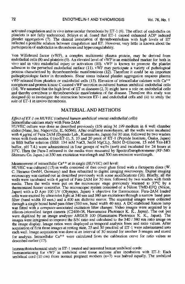
72 ENDOTHELIN-1 AND THROMBOSIS Vol. 76, No. 1
activated coagulation and in vivo intravascular thrombosis by ET-1 (6). The effect of endothelin on platelets is not fully understood. Sekiya et al. found that ET-1 caused enhanced ADP induced platelet aggregation (7). The clinical association of thromboembolism with high levels of ET reflected a possible relation between coagulation and ET. However, very little is known about the participation of endothelin in thrombosis and hypercoagulability.
Von Willebrand factor (vWF), a complex multimeric plasma protein, may be derived from endothelial cells (8) and platelets (9). An elevated level of vWF is an established marker for both in vivo and in vitro endothelial injury or activation (10). vWF is known to promote the platelet adhesion to the perturbed endothelial surface (11). vWF may participate a variety of pathological process characterized by thromboembolic manifestations (12). Therefore it could be an important pathophysiologic factor in thrombosis. Shear stress induced platelet aggregation requires plasma vWF released from platelets or endothelial cells (13). Elevation of intracellular calcium with Ca++ ionophore and protein kinase C caused vWF secretion in cultured human umbilical endothelial cells (14). We assumed that the high level of ET in diseases (2,3) might have a role on endothelial cells and thereby contribute to thromboembolic manifestation of the disease. Therefore this study was designed (i) to investigate the interaction between ET-l and endothelial cells and (ii) to study the role of ET- 1 in invivo thrombosis.
MATERIAL AND METHODS
Effect of ET-l on HUVEC (cultured human umbilical venous endothelial cells) Intracellular calcium study with Fura-2AM: HUVEC culture was done as described previously (15) using M 199 medium in 8 well chamber slides (Nunc, Inc. Naperville, IL 60566). After confluent monolayers, all the wells were incubated with 4 pg/ml of Fura-2AM (Dojindo Lab., Kumamoto, Japan) for 30 min. followed by two washes twice with fresh media. 0 (no ET-l), 5, 10 and 20 pmol of ET-l (Peptide Institute, Osaka, Japan) in BSS buffer solution (BSS: 134 mM NaCl, 3mM MgCL2, 5mM D-Glucose, 15 mM Tris-HCl buffer, pH 7.4.) were administered in four groups of wells (n=6) and incubated for 24 hours at 37°C. Then the Fura- released in the media were measured by Spectroflurophotometer (RF-540: Shimazu Co. Japan.) at 350 nm excitation wavelength and 500 nm emission wavelength.
Measurement of intracellular Ca ++ at in single (HUVEC) cell level: HUVEC was cultured (15) in wells consisted of thin cover glass fitted with a flexiperm discs (W. C. Heraeus GmbH, Germany) and then submitted to digital imaging microscopy. Digital imaging microscopy was carried out as described previously with some modifications (16). Briefly, all the wells were incubated with 4 rglml of Fura-2AM for 30 min. followed by two washes with fresh media. Then the wells were put on the microscope stage previously warmed to 37°C by a thermostated heater controller. The microscopic system consisted of a Nikon TMD-EFQ (Nikon, Japan) with a D Apo 100 UV (Olympus, Japan) x objective for fluorescence. Fura-2AM loaded cells were excited by ultraviolet light at 340 nm and 380 nm excitations through a narrow band pass filter (band width 10 mm.) and a 400 nm dichroic mirror. The sequential images were collected through a single broad band pass filter (510 nm, band width 40 nm). A DC-stabilized Xenon lamp was fitted with a computer-associated excitation filter changer. Video images were acquired by a silicon-intensified target camera (C2400-08, Hamamatsu Photonics K. K., Japan). The out put were digitized by an image analyzer ARGUS 100 (Hamamatsu Photonics K. K., Japan). The images were integrated to improve the S/N ratio and calculated to the 340 / 380 nm ratio image on the image display. Image ratios were displayed as temporal analysis lines and ratio values. After acquisition of first three images at resting state, 25 and 50 pmol/ml of ET- 1 were administered into each well. Image acquisition was done at an interval of 30 second for another 5 images and stored for analysis. Intracellular Ca++ were calculated from the calibration curve for ratio values as described earlier (17).
Immunohistochemical study in ET-1 treated and untreated human umbilical cords: Immunostaimng for vWF in umbilical cord tissue sections after incubation with ET-l: Each umbilical cord (10 cm) from normal pregnant mothers (n=7) was halved equally. The umbilical

Vol. 76, No. 1 ENDOTHELIN-1 AND THROMBOSIS 73
veins of both halves were washed 5 times with PBS (phosphate buffered saline, pH 7.2). 5 ml of PBS in one half (sham control) and 5 ml of ET- 1 solution in PBS (100 pmollml) in the other (case) were injected and kept within the venous lumen by clumping the both ends of the cords. They were incubated at a temperature of 37°C for 3 hours. Then all the umbilical cords were fixed in 3% paraformaldehyde for immunostaining with polyclonal anti vWF antibody (DAKO, USA). The method of immunostaining is described below with the method for rat tissues.
ET-1 infusions in rat and immunohistochemistry for vWF and fibrinogen in the tissues Wister rats (n = 7) weighing about 250 gm were infused with a solution of ET-l in 5% dextrose at a dose of 1 nmol/kg/hour for 2 hours and killed by intravenous 7.5% KCl. Liver, kidney, aorta from them and sham control rats (5% dextrose infusion for 2 hours, n = 7) were taken and fixed in 3% paraformaldehyde for hematoxylin and eosin staining. Immunostaining for polyclonal vWF and fibrinogen (anti rabbit serum, Behringwerke, Germany) were also done for all the tissues.
Immunostaining of the umbilical cord and rat tissue sections: The streptavidin-peroxidase technique of immunohistochemistry was performed as described previously (18). The tissue sections and umbilical cord sections were incubated with fresh 3% hydrogen peroxide in methanol (30 min., 23°C) and washed with PBS. Ten percent normal rabbit serum in PBS was applied (30 min., 23°C) for blocking. Primary polyclonal antibody at a dilution of 1 : 200 for vWF which reacts with both human and rat tissues and 1 : 75 for fibrinogen in PBS was applied to the tissue sections and incubated for 16 hours at 4°C then washed with PBS 5 times followed by superfusion of biotinated second antibody (7.0 @/ml in PBS, for 20 min., 23°C) and again washed 5 times with PBS. Then streptavidin-peroxidase (10 pllml in PBS) was added and washed 5 times with PBS. Finally slides were reacted with 3% 3-amino-9-ethylcarbazol substrate (5 min., 23°C DAKO) rinsed with tap water and mounted.
Flow cytometric assay of vWF in ET-l treated and untreated human platelets: How cytometric assay of vWF in ET-l treated and untreated human platelets was performed as described earlier (19, 20). PRP was made from venous blood in EDTA containing tubes centrifuged at 140 x gm for 10 minutes from apparently healthy persons. Those were washed twice with PBS. All washes were performed by centrifugation at 140 x gm. for 5 min. at 4” C. Platelets were re-suspended into 250 ~1 of PBS solution and incubated with 0, 10,25, 50 pmol of ET-l per l-l.5 x lo6 platelets for 3 hours at 25°C. Samples were washed twice with PBS followed by re suspension in PBS and were divided into equal halves. One of each samples were then incubated with 10 ~1 (protein concentration = 5.4glL) of (DAKO, USA) rabbit anti human von Willebrand factor for 30 min. at 4°C and washed twice with PBS by centrifugations and re suspended into 250 ,nl of PBS. Then all the samples were incubated with 10 ~1 of FITC (Sigma Chem. Co., USA) for 30 min. at 4°C followed by two washes with PBS. Finally all the samples were re suspended with 500 ~1 of PBS for analysis by a standard EPICS profile II instrument (Coulter, Japan) with a 15 mW air-cooled 488 nm argon laser. The instrument was also equipped with a fluorescence detector photo multiplier tube. Forward and side scatter as well as green (FITC) and red (phycoerythrin) signals were acquired with logarithm amplification with a 53080 nm band pass filter for collection of FITC. Both linear and logarithmic amplifier were used to process the fluorescence of antibodies. The data of lo- 20000 events were collected and stored in a list mode and analyzed. Statistical analysis was performed using students’ t test.
RESULTS
Fig. 1 shows the relative Fura- release measured by spectroflurophotometer at 350 nm excitation wavelength and 500 nm emission wavelength. Dose dependant inhibition of fura- release by cells treated with ET-l was observed (p>O.Ol). That suggested an increase in intracellular calcium in HUVEC by ET- 1. We confirmed that effect by digital image analysis of fura-2AM loaded HUVEC culture. We observed the changes selecting single endothelial cell and all the optic values were converted to real calcium ion concentration applying calibration curve. At resting level, endothelial cells showed an intracellular free calcium concentration of 95.5 f 8.7 nM and 93.8 f 8.5 nM. At resting state there was no significant changes. However immediately after administration of 25 and

74 ENDOTHELIN-1 AND THROMBOSIS Vol. 76, No. 1
50 pmol/ml of ET- 1, free calcium concentration increased at a level of 407.0 f 146 nM and 562.8 L 110 nM respectively. High level was maintained for at least next 3 minute in this expenment. Intracellular calcium changes by ET-l in HUVEC were statistically significant when compared with the initial three measurements at rest (p < 0.01 in all cases as shown in fig. 2).
0 No 5 10 20 Doses of endothelin-l(pmol)
0 5Opmollml 0 25 pmollml * p-zo.01
0 30 60 90 120 150 180 210 Time (seconds)
FIG. 1. FIG. 2.
Fura- release in HUVEC culture was 25 and 50 pmol/ml of ET-1 increased measured by spectroflurophotometer at intracellular calcium concentration at 350 nm excitation and 500 nm single cell level in HUVEC culture as emission. Fura- release was inhibited measured by fluorescence digital imaging dose dependantly by ET-l. * p < 0.01, analysis (n=20). * p < 0.01 compared to compared to No ET- 1 samples; n=7 resting level (0 _ 60 se&.)
Fig. 3 represented the immunostaining of the two halves of same umbilical cord from normal pregnancy with polyclonal antibody to vWF. Fig. 3a shows intensely stained immunoreactive vWF in the venous endothelium of umbilical cord incubated with intravenous ET-l solution. In contrast, the umbilical cords incubated with PBS solution (fig. 3b) were stained weakly.
a
FIG. 3.
Immunostaining for polyclonal vWF in two halves of same human umbilical cords incubated intravenously with ET-l solution (cases, fig. 3a) and PBS solution (sham control, fig. 3b) at 37” C for 3 hours. Arrows indicate the immunostaining for vWF.
Vol. 76, No. 1 ENDOTHELIN-1 AND THROMBOSIS 75
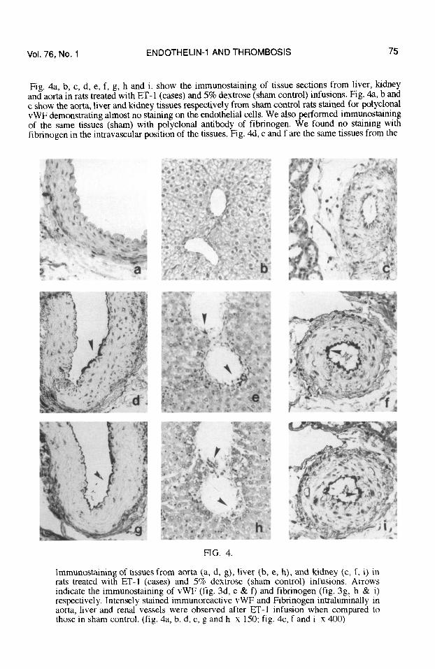
Fig. 4a, b, c, d, e, f, g, h and i. show the immunostaining of tissue sections from liver, kidney and aorta in rats treated with ET-1 (cases) and 5% dextrose (sham control) infusions. Fig. 4a, b and c show the aorta, liver and kidney tissues respectively from sham control rats stained for polyclonal vWF demonstrating almost no staining on the endothelial cells. We also performed immunostaining of the same tissues (sham) with polyclonal antibody of fibrinogen. We found no staining with fibrinogen in the intravascular position of the tissues. Fig. 4d, e and f are the same tissues from the
FIG. 4.
Immunostaining of tissues from aorta (a, d, g), liver (b, e, h), and kidney (c, f, i) in rats treated with ET-l (cases) and 5% dextrose (sham control) infusions. Arrows indicate the immunostaining of vWF (fig. 3d, e & f) and fibrinogen (fig. 3g, h & i) respectively. Intensely stained immunoreactive vWF and Fibrinogen intraluminally in aorta, liver and renal vessels were observed after ET-1 infusion when compared to those in sham control. (fig. 4a, b, d, e, g and h x 1.50; fig. 4-c, f and i x 400)

76 ENDOTHELIN-1 AND THROMBOSIS Vol. 76, No. 1
ET-1 treated rat demonstrating intense immunostained polyclonal vWF on the vascular endothelial cells. Fig. 4g, h and i. are the photographs of the same tissues as fig. 4d, e and f but stained for immunoreactive polyclonal fibrinogen. We found intensely stained fibrinogen at the same location of vWF as in fig. 4d, e and f. We also performed the haematoxylin and eosin staining of all the rats’ tissue sections (pictures are not shown here). ET-l infusions caused ischemic changes in the liver and renal tissues in all the cases when compared with those in sham control.
In flowcytometric measurement of vWF in normal platelets treated with 0, 10,25, M pmol of ET-1 per l-l.5 x 106 platelets for 3 hours at 25OC. No significant changes in cellular fluorescence in ET- 1 treated platelets were observed (figures are not shown) compared to that of ET-l untreated platelets.
DISCUSSION
High levels of endothelins were found to be associated with the states of hypercoagulability and hyper platelet activity (2, 3). Endothelin was thought to produce and/or release agents that may influence platelet function (21). ET-l could effect the second wave of ADP-induced platelet aggregation (7). Thrombin, a coagulation product could induce endothelin release from the endothelial cells (22). On the other hand, we found previously that ET-l induced a DIC like phenomenon in rabbits and thrombosis in rats’ microcirculation (6, 7). Therefore we investigated the interaction between ET-l and endothelial cells. We also studied the effect of ET-1 infusion on thrombus formation in rats.
In the HUVEC study, ET- 1 caused significant and dose dependant decrease in Fura- release by the cultured HUVEC. Fura-2AM, a calcium probe is known to represent the free intracellular calcium in different cells (23). Retained fura- in the HUVEC culture reflected an increase in intracellular calcium by ET- 1. We also confirmed that the ET- 1 induced increased intracellular calcium ion in cultured HUVEC (at single cell level) by fluorescence digital image analysis. That increased intracellular calcium could be a suggestion for increased synthetic or secretory activity in the endothelial cells. Carew et al. showed that increased intracellular calcium by calcium ionophore in HUVEC cultures were associated with increased vWF synthesis and secretion (12). In umbilical cord study, the samevolume of ET-l or PBS solution was injected into the washed venous lumen of the two halves of the same umbilical cord. They were incubated at 37°C for 3 hours (n=7). We found intensely stained immunoreactive vWF in the endothelial cells in ET-1 treated umbilical cords. The endothelial cells in sham control cords (incubated with PBS solution) showed faint staining for vWF. Expression of vWF by endothelial cells was also described as the endothelial injuries or activation (24). Von Willebrand factor, a complex multimeric plasma protein, have been shown to bind avidly both the blood platelets and the endothelial extracellular matrix (10). Therefore they provide an important co-factor for platelet adhesion to the purturbed vessel wall (10). From the in vitro study, we suggested that ET-1 could induce increased free cytosolic calcium ion in the endothelial cells associated with increased synthesis and expression of vWF. This result agreed with the findings of Carew et al. (7). We assumed that the expressed vWF could be one of the enhancing factor or the clue of thromboembolic manifestations in conditions with high level of endothelins.
With the idea that ET-1 increases vWF expression on vascular endothelial cells and enhances thrombus formation, we performed invivo rat study with intravenous ET-l infusions. In rat study, we found intensely stained intravascular immunoreactive vWF and fibrinogen in aorta, hepatic and renal micro vessels after ET-l infusion. Simultaneous staining of vWF and fibrinogen at the same intravascular position were also observed. That could suggest intravascular thrombus formation. ET-l infusions could cause generalized vasospasm including visceral and peripheral circulation in rats (25). Lamontange et al. mentioned that ET-l infusion could induce shear stress resulting increased cGMP in platelets in vivo (26). Platelet activation may also be resulted from shear force by ET-1 induced vasoconstriction. Therefore the vasospasm related thrombosis or enhanced coagulation may be one of the explanations for the in vivo intravascular thrombus formation. However, this study focused the interaction between ET-l and endothelial cells that might

Vol. 76, No. 1 ENDOTHELIN-1 AND THROMBOSIS 77
contribute to thrombosis. Because of the likely function of vWF in promoting the platelet adhesion to the perturbed endothelial surface, this protein and fibrinogen may be important pathophysiologic factor in thrombosis (27). The major sources of vWF are platelets and endothelial cells. However, we suggested that vWF might be derived from endothelial cells as an ET-1 effect. To exclude platelets as an alternative source of vWF, we performed flow cytometric measurement of vWF in normal platelets incubated with 0, 10,25,50 pmol of ET-l (per 1 - 1.5 x lo6 platelets) for 3 hours. No significant changes in cellular fluorescence in ET- 1 treated platelets were observed (figures are not shown) when compared to that of ET-1 untreated platelets. Therefore we assumed that the intensly stained vWF might be derived from ET- 1 stimulated endothelial cells. vWF and fibrinogen may be important pathophysiologic factor in in vivo thromboembolic manifestation. vWF promoted the platelet adhesion to the perturbed endothelial surface. Fibrinogen was deposited along with platelets and others as a component of in vivo thrombus. In summary, we suggest that endothelin- 1 increased vWF on the endothelial cells. This may facilitate the attachment of platelets to the endothelial cells. Accordingly ET- 1 played an important role in intravascular thrombus formation in vivo.
Many things are still to known about endothelin. However this study may find a clue for the co- association of high level of endothelin and thromboembolic conditions.
REFERENCES
1. YANAGISAWA, M., KURIHARA, H., KIMURA, S., TOMOBE, Y., KOBAYASHI, M., MITSUI, Y., YAZAKI, Y., GOTO, K., and MASAKI, T. A novel potent vasoconstrictor peptide produced by vascular endothelial cells, Nature, 332,411-415, 1988. 2. TAYLOR, RN., VARMA, M., NELSON, N.H. TENG and ROBERTS, JM. Women with preeclampsia have higher endothelin levels than women with normal pregnancies. J. Clin. Endocrinol. and Metab. 71(6), 1675-1677, 1990. 3. IHARA, Y., SAGAWA, N., HASEGAWA, M., OKAGAKI, A., LI, X.M., INAMORI, K., H., MORI, T., SAITO, Y., SHIRAKAMI, G., NAKAO, K. and IMURA, H. Concentration of endothelin in maternal and umbilical cord blood at various stages of pregnancy. J. Cardiovasc. Pharmacol. 17 Suppl 7, ~443-445, 1991. 4. MAKI, M. Haemostasis and thrombosis in obstetrics and gynecology, Marberg: Med. Verl- Ges., ISBN 3-92132018-6. pp, 124-199, 1991. 5. HALIM, A., KANAYAMA, N., MAEHARA, K., TAKAHASHI, M., TERAO, T. HELLP Syndrome-like biochemical parameters obtained with Endothelin-1 injections in rabbits. Gynecol. Obstet. Invest. 35: 193-198, 1993. 6. HALIM, A., KANAYAMA, N., MARADNY, E. El., MAEHARA, K., TAKAHASHI, M., TERAO, T. Coagulation in vivo microcirculation and in vitro caused by endothelin-1. Thromb. Res. 72; 203-209, 1993. 7. SEKIYA, F., KOZUKA, M., SHIBABE, S. and HAGIWARA, H. Effect of Endothelin on Platelets. Japanese journal of Thrombosis and haemostasis (Japanese) 2(4), 323-329, 1991. 8. JAFFE, EA., HOYER, LW., and NACHMAN, RL. Synthesis of von Willebrand factor by cultured human umbilical endothelial cells. Proc Nat1 Acad Sci USA 71, 1906, 1974. 9 SPORN, LA., CHAVIN, SI., MARDER, VJ., and WAGNER, DD. Biosynthesis of von Willebrand protein by human megakaryocytes. J Clin Invest. 76, 1102- 1106, 1985. 10. MATUCCI-CERINIC M., JAFFA A. and KAHALEH B. Angiotensine converting enzyme: an in vivo and in vitro marker of endothelial injury. J Lab Clin Med 120(3), 428-433, 1992. 11. RUGGERI, ZM., and ZIMMERMAN, TS. Variant von Willebrand’s disease: characterization of two subtypes by analysis of multimeric composition of factor VIIIlvon Willebrand factor in plasma and platelets. J. Clin Invest., 65 (6), 1140-l 143, 1980. 12. RUGGERI, Z., M. AND ZIMMERMAN, T., S. von Willebrand factor and von Willebrand disease, Blood, 70, 895904, 1987. 13. IKEDA, Y., HANDA, M., KAWANO, K., KAMATA, T., MURATA, M., ARAKI, Y., ANBO, H., KAWAI, Y., WATANABE, K., ATAGAKI, I., SAKAI, K., RUGGERI, Z., M. The role of von Willebrand factor and fibrinogen in platelet aggregation under varying shear stress, J. Clin. Invest., 87, 1234-1240, 1991.
78 ENDOTHELIN-1 AND THROMBOSIS Vol. 76. No. 1
14. CAREW, M. A, PALEOLOG, E.M., and PEARSON, J.D. The roles of proein kinase C and intracellular Ca2+ in the secretion of von Willebmnd factor from human vascular endothelial cells. Biochem. J. 286, 631-635, 1992. 15. JAFFE, E.A.(ed.), Method of culturing human umbilical endothelial cells. Biology of endothelial cells. ISBN O-89838-587-3, chapter 1,2-l 1; 1984. 16. NISHIO, H., IKEGAMI, Y., and SEGAWA, T. Fluorescence digital image analysis of Serotonin induced calcium oscillation in single blood platelets. Cell Calcium 12, 177-184, 1991. 17. MIYATA, H., HAYASHI, H., KOBAYASHI, A., and YAMAZAKI, N. Effect of Strophanthidin on intracellular Ca2+ concentration and cellular morphology of Guini pig myocyte. Cardiovasc Res. 23, 378-384, 1989. 18. SHI, Z.R., ITZKOWITZ, S.H., AND KIM, Y.S. A comparison of three immunoperoxidase technique for antigen detection in colorectal carcinoma tissues, J. Histochem. Cytochem. 36, 3 17- 322,1988. 19. SINTNICOLAAS, K., VRIES, W.D., LINDEN, R.V.D., GRATAMA, J.W. and BOLHUIS, R.L.H. Simultaneous flow cytometric detection of antibodies against platelets, granulocytes and lymphocytes. J. Immunol. Methods. 142, 215222, 1991. 20. MONIWA, N., TERAO, T., CHUCHOLOWSKI, N., SCHMITT, M. and GRAEFF, H. Flow cytometric analysis of tumor-associated proteases with special reference to uPA/PAI-1 complex. Current aspect of blood coagulation, fibrinolysis and platelets. p: 98- 103; M.-c. Shen.C.- Teng. A. Takada (Eds) 1993. 21. ELIOT, H. OHLSTEIN, STORER, B., NAMBI, P., GIVEN, M. and LIPPTON, H. Endothelin and platelet function. Thromb. Res. 57, 967-974, 1990. 22. SCHINI, V.B., HENDRICKSON, H., HEUBLEIN, D.M., BURNETT Jr, J.C., and VANHOUTTE, P.M. Thrombin enhances the release of endothelin from cultured porcine aortic endothelial cells. Eur J Pharmacol., 165333-334, 1989. 23. IKEGAMI, Y., NISHIO, H., FUKADA T., NAKATA, Y., and TOMIO, S.. Effect of Concanavalin A on intracellular calcium concentration in single blood platelets. Japan J. Pharmacol. 57, 233-241, 1991. 24. SEEBER, Ch., HILLER, E., HOLLER, E. and KOLB, H.J. Increased levels of tissue plasminogen (t-PA) and Tissue Plasminogen activator inhibitor (PAI) correlate with Tumor Necrosis Factor alpha (TNFa) release in patients suffering from microangiopathy following Allogenic bone marrow transplant (BMT). Thromb. Res. 66; 1373-1383, 1992. 25. GARDINER, S.M., COMPTON, A.M. and BENNETT, T. Regional haemodynamic effects of endothelin-1 and endothelin3 in concious Long Evans and Brattleboro rats. Br. J. Pharmacol. 99, 107-l 12, 1990. 26. LAMONTANGE D., POHL U., BUSSE R. Mechanical deformation of vessel wall and shear stress determine the basal release of endothelium derived relaxing factor in intact rabbit coronary vascular bed. Circ. Res. 70, 123-130, 1992. 27. BAUMGARTNER, H.R. The role of Blood flow in Platelet adhession, Fibrin Deposition, and Formation of Mural Thrombi., Microvasc. Res., 5, 167-176, 1973.