Characterization, Classification, and Treatment of von Willebrand Diseases: A Critical Appraisal of...
25
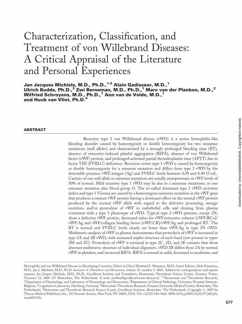
Characterization, Classification, and Treatment of von Willebrand Diseases: A Critical Appraisal of the Literature and Personal Experiences Jan Jacques Michiels, M.D., Ph.D., 1,5 Alain Gadisseur, M.D., 1 Ulrich Budde, Ph.D., 3 Zwi Berneman, M.D., Ph.D., 1 Marc van der Planken, M.D., 2 Wilfried Schroyens, M.D., Ph.D., 1 Ann van de Velde, M.D., 1 and Huub van Vliet, Ph.D. 4 ABSTRACT Recessive type 3 von Willebrand disease (vWD) is a severe hemophilia-like bleeding disorder caused by homozygosity or double heterozygosity for two nonsense mutations (null alleles) and characterized by a strongly prolonged bleeding time (BT), absence of ristocetin-induced platelet aggregation (RIPA), absence of von Willebrand factor (vWF) protein, and prolonged activated partial thromboplastin time (APTT) due to factor VIII (FVIII:C) deficiency. Recessive severe type 1 vWD is caused by homozygosity or double heterozygosity for a missense mutation and differs from type 3 vWD by the detectable presence vWF:antigen (Ag) and FVIII:C levels between 0.09 and 0.40 U/mL. Carriers of one null allele or missense mutations are usually asymptomatic at vWF levels of 50% of normal. Mild recessive type 1 vWD may be due to a missense mutations, or one missense mutation plus blood group O. The so-called dominant type 1 vWD secretion defect and type 1 Vicenza are caused by a heterozygous missense mutation in the vWF gene that produces a mutant vWF protein having a dominant effect on the normal vWF protein produced by the normal vWF allele with regard to the defective processing, storage secretion, and/or proteolysis of vWF in endothelial cells and clearing from plasma consistent with a type 2 phenotype of vWD. Typical type 2 vWD patients, except 2N, show a defective vWF protein, decreased ratios for vWF:ristocetin cofactor [vWF:RCo]/ vWF:Ag and vWF:collagen binding factor [vWF:CB]/vWF:Ag and prolonged BT. The BT is normal and FVIII:C levels clearly are lower than vWF:Ag in type 2N vWD. Multimeric analysis of vWF in plasma demonstrates that proteolysis of vWF is increased in type 2A and 2B vWD, with increased triplet structure of each band (not present in types 2M and 2U). Proteolysis of vWF is minimal in type 2C, 2D, and 2E variants that show aberrant multimeric structure of individual oligomers. vWD 2B differs from 2A by normal vWF in platelets, and increased RIPA. RIPA is normal in mild, decreased in moderate, and Hemophilia and von Willebrand Disease in Developing Countries; Editor in Chief, Eberhard F. Mammen, M.D.; Guest Editors, Alok Srivastava, M.D., Jan J. Michiels, M.D., Ph.D. Seminars in Thrombosis and Hemostasis, volume 31, number 5, 2005. Address for correspondence and reprint requests: Jan Jacques Michiels, M.D., Ph.D., Goodheart Institute and Foundation, Hemostasis Thrombosis Science Center, Erasmus Tower, Veenmos 13, 3069 AT Rotterdam, The Netherlands. E-mail: [email protected]. 1,2 Hemostasis and Thrombosis Research; 1 Department of Hematology, and Laboratory of Hematology and Hemostasis; 2 Department of Clinical Pathology, University Hospital Antwerp, Belgium; 3 Coagulation Laboratory, Hamburg, Germany; 4 Hemostasis Thrombosis Research, Erasmus University Medical Center, Rotterdam, The Netherlands; 5 Hemostasis and Thrombosis Research Science Center, Goodheart Institute, Rotterdam. The Netherlands. Copyright # 2005 by Thieme Medical Publishers, Inc., 333 Seventh Avenue, New York, NY 10001, USA. Tel: +1(212) 584-4662. 0094-6176,p;2005,31,05,577,601,ftx, en;sth01105x. 577 Downloaded by: Universitätsbibliothek. Copyrighted material.
Transcript of Characterization, Classification, and Treatment of von Willebrand Diseases: A Critical Appraisal of...
Characterization, Classification, andTreatment of von Willebrand Diseases:A Critical Appraisal of the Literatureand Personal ExperiencesJan Jacques Michiels, M.D., Ph.D.,1,5 Alain Gadisseur, M.D.,1
Ulrich Budde, Ph.D.,3 Zwi Berneman, M.D., Ph.D.,1 Marc van der Planken, M.D.,2
Wilfried Schroyens, M.D., Ph.D.,1 Ann van de Velde, M.D.,1
and Huub van Vliet, Ph.D.4
ABSTRACT
Recessive type 3 von Willebrand disease (vWD) is a severe hemophilia-likebleeding disorder caused by homozygosity or double heterozygosity for two nonsensemutations (null alleles) and characterized by a strongly prolonged bleeding time (BT),absence of ristocetin-induced platelet aggregation (RIPA), absence of von Willebrandfactor (vWF) protein, and prolonged activated partial thromboplastin time (APTT) due tofactor VIII (FVIII:C) deficiency. Recessive severe type 1 vWD is caused by homozygosityor double heterozygosity for a missense mutation and differs from type 3 vWD by thedetectable presence vWF:antigen (Ag) and FVIII:C levels between 0.09 and 0.40 U/mL.Carriers of one null allele or missense mutations are usually asymptomatic at vWF levels of50% of normal. Mild recessive type 1 vWD may be due to a missense mutations, or onemissense mutation plus blood group O. The so-called dominant type 1 vWD secretiondefect and type 1 Vicenza are caused by a heterozygous missense mutation in the vWF genethat produces a mutant vWF protein having a dominant effect on the normal vWF proteinproduced by the normal vWF allele with regard to the defective processing, storagesecretion, and/or proteolysis of vWF in endothelial cells and clearing from plasmaconsistent with a type 2 phenotype of vWD. Typical type 2 vWD patients, except 2N,show a defective vWF protein, decreased ratios for vWF:ristocetin cofactor [vWF:RCo]/vWF:Ag and vWF:collagen binding factor [vWF:CB]/vWF:Ag and prolonged BT. TheBT is normal and FVIII:C levels clearly are lower than vWF:Ag in type 2N vWD.Multimeric analysis of vWF in plasma demonstrates that proteolysis of vWF is increased intype 2A and 2B vWD, with increased triplet structure of each band (not present in types2M and 2U). Proteolysis of vWF is minimal in type 2C, 2D, and 2E variants that showaberrant multimeric structure of individual oligomers. vWD 2B differs from 2A by normalvWF in platelets, and increased RIPA. RIPA is normal in mild, decreased in moderate, and
Hemophilia and von Willebrand Disease in Developing Countries; Editor in Chief, Eberhard F. Mammen, M.D.; Guest Editors, Alok Srivastava,M.D., Jan J. Michiels, M.D., Ph.D. Seminars in Thrombosis and Hemostasis, volume 31, number 5, 2005. Address for correspondence and reprintrequests: Jan Jacques Michiels, M.D., Ph.D., Goodheart Institute and Foundation, Hemostasis Thrombosis Science Center, Erasmus Tower,Veenmos 13, 3069 AT Rotterdam, The Netherlands. E-mail: [email protected]. 1,2Hemostasis and Thrombosis Research;1Department of Hematology, and Laboratory of Hematology and Hemostasis; 2Department of Clinical Pathology, University Hospital Antwerp,Belgium; 3Coagulation Laboratory, Hamburg, Germany; 4Hemostasis Thrombosis Research, Erasmus University Medical Center, Rotterdam, TheNetherlands; 5Hemostasis and Thrombosis Research Science Center, Goodheart Institute, Rotterdam. The Netherlands. Copyright # 2005 byThieme Medical Publishers, Inc., 333 Seventh Avenue, New York, NY 10001, USA. Tel: +1(212) 584-4662. 0094-6176,p;2005,31,05,577,601,ftx,en;sth01105x.
577
Dow
nloa
ded
by: U
nive
rsitä
tsbi
blio
thek
. Cop
yrig
hted
mat
eria
l.
absent in severe type 2A vWD. RIPA is decreased or absent in 2M, 2U, 2C, and 2D;variable in 2E; and normal in 2N and dominant type 1. vWD 2M is usually mild andfeatures decreased vWF:RCo and RIPA, and a normal or near-normal vWF multimericpattern in a low-resolution agarose gel. vWD 2A-like or unclassifiable (2U) is distinct from2A and 2B and typically features low vWF:RCo and RIPA with the relative lack of largevWF multimers. vWD type 2C is recessive; the dominant type 2D is rare. The response todesmopressin acetate (DDAVP) of vWF parameters is normal in pseudo-vWD and mildtype 1. The responses to DDAVP of FVIII:C and vWF parameters in vWD 2M,Vincenza, 2E, and mild 2A, 2U, and 2N are transiently good for a variable number ofhours to arrest mucocutaneous bleeding episodes or to prevent bleeding during minorsurgery or trauma. However, the responses are not good enough to treat major bleedings orto prevent bleeding during major surgery or trauma. The response to DDAVP of vWFparameters is poor in recessive type 3, 1 and 2C, and dominant 2A, 2B, and 2U. Properrecommendations of FVIII/vWF concentrates using FVIII:C and vWF:RCo unit dosingfor the prophylaxis and treatment of bleeding episodes in type 2 disease that is non-responsive to DDAVP and in type 3 vWD are proposed.
KEYWORDS: von Willebrand factor, von Willebrand disease, ristocetin cofactor
activity, von Willebrand collagen binding activity, factor VIII:C, bleeding time,
desmopressin acetate
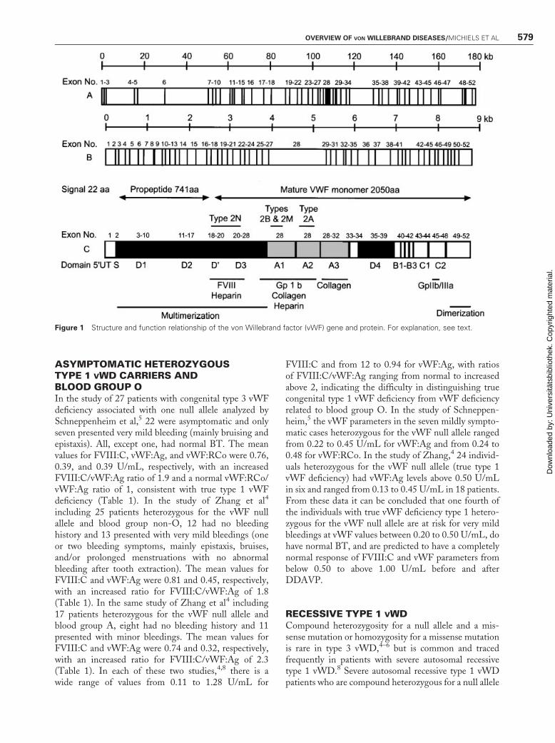
The von Willebrand factor (vWF) is a multi-meric plasma glycoprotein that plays a central role inhemostasis. vWF acts both as carrier for coagulationfactor VIII (FVIII) in the plasma and as a mediator ofplatelet adhesion to subendothelium after vascular in-jury. Several distinct functional domains have beenidentified within the vWF, including regions involvedin binding to factor VIII, to platelet receptor GPIb, toplatelet GPIIb-IIIa, to components of extracellular ma-trix such as collagen and heparin, regions involved inmultimerization and dimerization of vWF, and the A1and A2 domains involved in the majority of type 2 vonvWF defects (Fig. 1).1 The classification of congenitalvon Willebrand disease (vWD) as recommended bythe von Willebrand Factor Scientific StandardisationCommittee (vWF-SSC) of the International Societyon Thrombosis and Haemostasis (ISTH) is based onlaboratory phenotyping using the combination ofFVIII:C and vWF:antigen (Ag) levels, ristocetin-induced platelet aggregation (RIPA), and rather insen-sitive tests for vWF:ristocetin cofactor (RCo) and vWFmultimeric pattern in a low-resolution agarose gel.2,3
Accumulating data on the structure and function rela-tionship between phenotype and genotype of vWD andthe use of more specific and sensitive diagnostic toolsincluding vWF:collagen binding factor (CB) assay,FVIII binding to vWF, and improved multimeric anal-ysis will have a major impact on a correct diagnosis andproper classification of the vWDs. The contribution of aDDAVP challenge test has become available for clini-cians to better distinguish the various type 1 and type 2vWDs. In this study, we evaluated the clinical features,
laboratory phenotypes, and genotypes of severe autoso-mal recessive type 3 and type 1 vWD, asymptomaticcarriers of a nonsense or missense mutation in the vWFgene, blood group O-related vWF deficiency and mildor moderate dominant type 1 vWD, and all variantsof type 2A, 2B, 2C, 2D, 2E, 2M, 2N, and 2 unclassifi-able (U).
RECESSIVE TYPE 3 VWD ANDBLOOD GROUP OObligate heterozygous parents of type 3 vWF patients infact can be regarded as true type 1 quantitative vWFdeficiency heterozygous for the vWF null allele. Theinheritance of vWD type 3 is autosomal recessive.4–6
Type 3 vWD patients typically have strongly prolongedbleeding times (BT) and activated partial thromboplas-tin times; FVIII:C levels between 1 and 9%; undetect-able vWF:Ag, vWF:RCo, and vWF:CB levels; andabsence of RIPA.4–8 In 31 cases with type 3 vWD (age2 to 80 years; median, 15 years) described by Schnep-penheim et al,5 bleeding manifestations were recorded aseasy bruising and prolonged epistaxis in 31 (100%),spontaneous joint bleedings in 23 (76%), muscle bleed-ings in seven (22%), and gastrointestinal bleedings inthree (10%) patients. Type 3 vWDs are homozygous orcompound heterozygous for two null alleles (gene dele-tions, stop codons, frameshift mutations, splice sitemutations, and absence of MRNA) in the vast majorityof reported cases.4–6 Compound heterozygosity for a nullallele and a missense mutation or homozygosity for amissense mutation is rare in type 3 vWD.5
578 SEMINARS IN THROMBOSIS AND HEMOSTASIS/VOLUME 31, NUMBER 5 2005
Dow
nloa
ded
by: U
nive
rsitä
tsbi
blio
thek
. Cop
yrig
hted
mat
eria
l.
ASYMPTOMATIC HETEROZYGOUSTYPE 1 VWD CARRIERS ANDBLOOD GROUP OIn the study of 27 patients with congenital type 3 vWFdeficiency associated with one null allele analyzed bySchneppenheim et al,5 22 were asymptomatic and onlyseven presented very mild bleeding (mainly bruising andepistaxis). All, except one, had normal BT. The meanvalues for FVIII:C, vWF:Ag, and vWF:RCo were 0.76,0.39, and 0.39 U/mL, respectively, with an increasedFVIII:C/vWF:Ag ratio of 1.9 and a normal vWF:RCo/vWF:Ag ratio of 1, consistent with true type 1 vWFdeficiency (Table 1). In the study of Zhang et al4
including 25 patients heterozygous for the vWF nullallele and blood group non-O, 12 had no bleedinghistory and 13 presented with very mild bleedings (oneor two bleeding symptoms, mainly epistaxis, bruises,and/or prolonged menstruations with no abnormalbleeding after tooth extraction). The mean values forFVIII:C and vWF:Ag were 0.81 and 0.45, respectively,with an increased ratio for FVIII:C/vWF:Ag of 1.8(Table 1). In the same study of Zhang et al4 including17 patients heterozygous for the vWF null allele andblood group A, eight had no bleeding history and 11presented with minor bleedings. The mean values forFVIII:C and vWF:Ag were 0.74 and 0.32, respectively,with an increased ratio for FVIII:C/vWF:Ag of 2.3(Table 1). In each of these two studies,4,8 there is awide range of values from 0.11 to 1.28 U/mL for
FVIII:C and from 12 to 0.94 for vWF:Ag, with ratiosof FVIII:C/vWF:Ag ranging from normal to increasedabove 2, indicating the difficulty in distinguishing truecongenital type 1 vWF deficiency from vWF deficiencyrelated to blood group O. In the study of Schneppen-heim,5 the vWF parameters in the seven mildly sympto-matic cases heterozygous for the vWF null allele rangedfrom 0.22 to 0.45 U/mL for vWF:Ag and from 0.24 to0.48 for vWF:RCo. In the study of Zhang,4 24 individ-uals heterozygous for the vWF null allele (true type 1vWF deficiency) had vWF:Ag levels above 0.50 U/mLin six and ranged from 0.13 to 0.45 U/mL in 18 patients.From these data it can be concluded that one fourth ofthe individuals with true vWF deficiency type 1 hetero-zygous for the vWF null allele are at risk for very mildbleedings at vWF values between 0.20 to 0.50 U/mL, dohave normal BT, and are predicted to have a completelynormal response of FVIII:C and vWF parameters frombelow 0.50 to above 1.00 U/mL before and afterDDAVP.
RECESSIVE TYPE 1 VWDCompound heterozygosity for a null allele and a mis-sense mutation or homozygosity for a missense mutationis rare in type 3 vWD,4–6 but is common and tracedfrequently in patients with severe autosomal recessivetype 1 vWD.8 Severe autosomal recessive type 1 vWDpatients who are compound heterozygous for a null allele
Figure 1 Structure and function relationship of the von Willebrand factor (vWF) gene and protein. For explanation, see text.
OVERVIEW OF VON WILLEBRAND DISEASES/MICHIELS ET AL 579
Dow
nloa
ded
by: U
nive
rsitä
tsbi
blio
thek
. Cop
yrig
hted
mat
eria
l.
and a missense mutation, or homozygous or doubleheterozygous for a missense mutation have detectablebut very low vWF levels (Table 2).
Homozygotes for the missense mutationsW377C (Schneppenheim et al5) and for R273W (Allenet al9) in the propeptide D1 domain have been describedto be associated with severe autosomal recessive type 1or type 3 vWD phenotype. Reported cases of severe
recessive type 1 vWD are in fact severe autosomalrecessive type 2 vWD as demonstrated by vWFmultimeranalysis.9–13 The multimeric pattern of homozygousR273W clearly showed the absence of high molecularweight multimers and a pronounced monomeric bandconsistent with type 2A or 2E vWD.9 Homozygosity forthe missense mutation C2364F in a family and doubleheterozygosity for C2364F/null in three families has
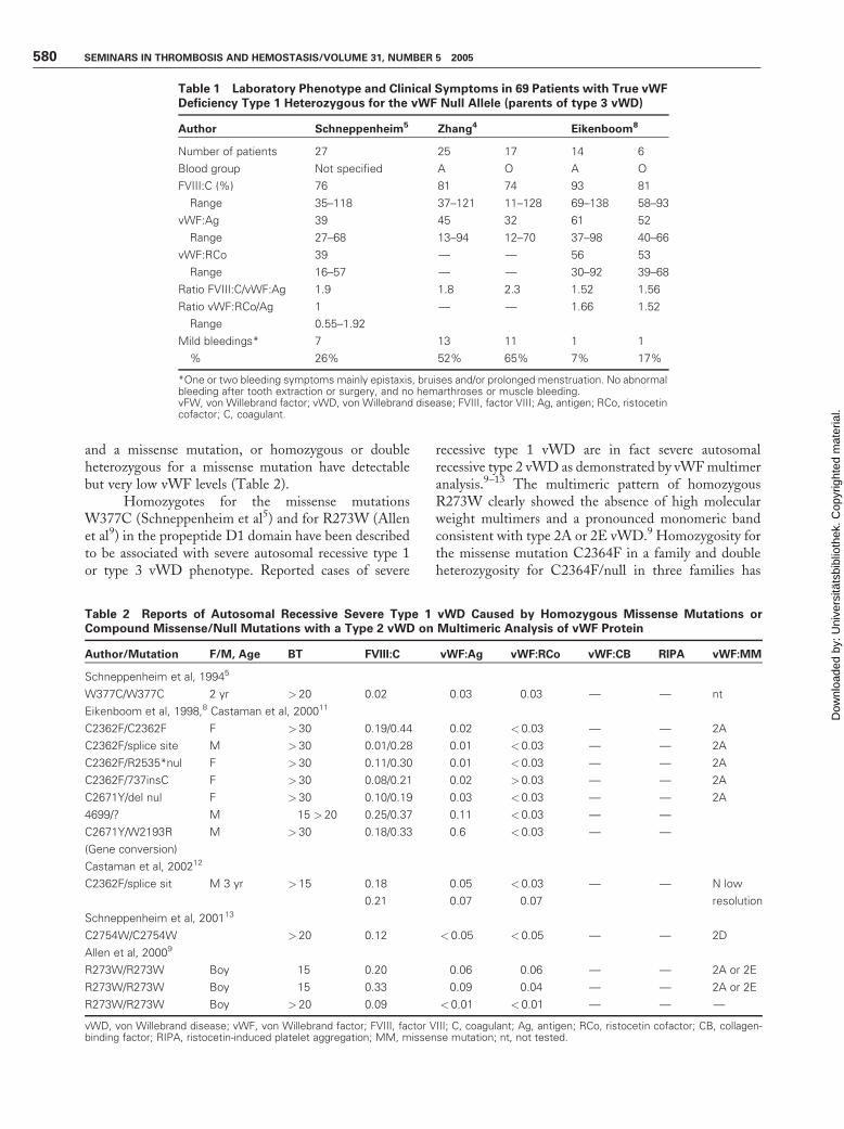
Table 1 Laboratory Phenotype and Clinical Symptoms in 69 Patients with True vWFDeficiency Type 1 Heterozygous for the vWF Null Allele (parents of type 3 vWD)
Author Schneppenheim5 Zhang4 Eikenboom8
Number of patients 27 25 17 14 6
Blood group Not specified A O A O
FVIII:C (%) 76 81 74 93 81
Range 35–118 37–121 11–128 69–138 58–93
vWF:Ag 39 45 32 61 52
Range 27–68 13–94 12–70 37–98 40–66
vWF:RCo 39 — — 56 53
Range 16–57 — — 30–92 39–68
Ratio FVIII:C/vWF:Ag 1.9 1.8 2.3 1.52 1.56
Ratio vWF:RCo/Ag 1 — — 1.66 1.52
Range 0.55–1.92
Mild bleedings* 7 13 11 1 1
% 26% 52% 65% 7% 17%
*One or two bleeding symptoms mainly epistaxis, bruises and/or prolonged menstruation. No abnormalbleeding after tooth extraction or surgery, and no hemarthroses or muscle bleeding.vFW, von Willebrand factor; vWD, von Willebrand disease; FVIII, factor VIII; Ag, antigen; RCo, ristocetincofactor; C, coagulant.
Table 2 Reports of Autosomal Recessive Severe Type 1 vWD Caused by Homozygous Missense Mutations orCompound Missense/Null Mutations with a Type 2 vWD on Multimeric Analysis of vWF Protein
Author/Mutation F/M, Age BT FVIII:C vWF:Ag vWF:RCo vWF:CB RIPA vWF:MM
Schneppenheim et al, 19945
W377C/W377C 2 yr > 20 0.02 0.03 0.03 — — nt
Eikenboom et al, 1998,8 Castaman et al, 200011
C2362F/C2362F F > 30 0.19/0.44 0.02 <0.03 — — 2A
C2362F/splice site M > 30 0.01/0.28 0.01 <0.03 — — 2A
C2362F/R2535*nul F > 30 0.11/0.30 0.01 <0.03 — — 2A
C2362F/737insC F > 30 0.08/0.21 0.02 >0.03 — — 2A
C2671Y/del nul F > 30 0.10/0.19 0.03 <0.03 — — 2A
4699/? M 15 > 20 0.25/0.37 0.11 <0.03 — —
C2671Y/W2193R M > 30 0.18/0.33 0.6 <0.03 — —
(Gene conversion)
Castaman et al, 200212
C2362F/splice sit M 3 yr > 15 0.18 0.05 <0.03 — — N low
0.21 0.07 0.07 resolution
Schneppenheim et al, 200113
C2754W/C2754W > 20 0.12 < 0.05 <0.05 — — 2D
Allen et al, 20009
R273W/R273W Boy 15 0.20 0.06 0.06 — — 2A or 2E
R273W/R273W Boy 15 0.33 0.09 0.04 — — 2A or 2E
R273W/R273W Boy > 20 0.09 < 0.01 <0.01 — — —
vWD, von Willebrand disease; vWF, von Willebrand factor; FVIII, factor VIII; C, coagulant; Ag, antigen; RCo, ristocetin cofactor; CB, collagen-binding factor; RIPA, ristocetin-induced platelet aggregation; MM, missense mutation; nt, not tested.
580 SEMINARS IN THROMBOSIS AND HEMOSTASIS/VOLUME 31, NUMBER 5 2005
Dow
nloa
ded
by: U
nive
rsitä
tsbi
blio
thek
. Cop
yrig
hted
mat
eria
l.
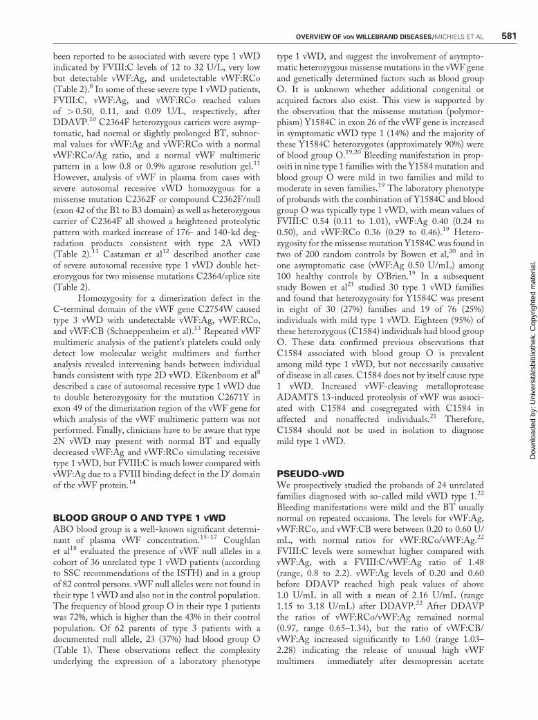
been reported to be associated with severe type 1 vWDindicated by FVIII:C levels of 12 to 32 U/L, very lowbut detectable vWF:Ag, and undetectable vWF:RCo(Table 2).8 In some of these severe type 1 vWD patients,FVIII:C, vWF:Ag, and vWF:RCo reached valuesof > 0.50, 0.11, and 0.09 U/L, respectively, afterDDAVP.10 C2364F heterozygous carriers were asymp-tomatic, had normal or slightly prolonged BT, subnor-mal values for vWF:Ag and vWF:RCo with a normalvWF:RCo/Ag ratio, and a normal vWF multimericpattern in a low 0.8 or 0.9% agarose resolution gel.11
However, analysis of vWF in plasma from cases withsevere autosomal recessive vWD homozygous for amissense mutation C2362F or compound C2362F/null(exon 42 of the B1 to B3 domain) as well as heterozygouscarrier of C2364F all showed a heightened proteolyticpattern with marked increase of 176- and 140-kd deg-radation products consistent with type 2A vWD(Table 2).11 Castaman et al12 described another caseof severe autosomal recessive type 1 vWD double het-erozygous for two missense mutations C2364/splice site(Table 2).
Homozygosity for a dimerization defect in theC-terminal domain of the vWF gene C2754W causedtype 3 vWD with undetectable vWF:Ag, vWF:RCo,and vWF:CB (Schneppenheim et al).13 Repeated vWFmultimeric analysis of the patient’s platelets could onlydetect low molecular weight multimers and furtheranalysis revealed intervening bands between individualbands consistent with type 2D vWD. Eikenboom et al8
described a case of autosomal recessive type 1 vWD dueto double heterozygosity for the mutation C2671Y inexon 49 of the dimerization region of the vWF gene forwhich analysis of the vWF multimeric pattern was notperformed. Finally, clinicians have to be aware that type2N vWD may present with normal BT and equallydecreased vWF:Ag and vWF:RCo simulating recessivetype 1 vWD, but FVIII:C is much lower compared withvWF:Ag due to a FVIII binding defect in the D0 domainof the vWF protein.14
BLOOD GROUP O AND TYPE 1 VWDABO blood group is a well-known significant determi-nant of plasma vWF concentration.15–17 Coughlanet al18 evaluated the presence of vWF null alleles in acohort of 36 unrelated type 1 vWD patients (accordingto SSC recommendations of the ISTH) and in a groupof 82 control persons. vWF null alleles were not found intheir type 1 vWD and also not in the control population.The frequency of blood group O in their type 1 patientswas 72%, which is higher than the 43% in their controlpopulation. Of 62 parents of type 3 patients with adocumented null allele, 23 (37%) had blood group O(Table 1). These observations reflect the complexityunderlying the expression of a laboratory phenotype
type 1 vWD, and suggest the involvement of asympto-matic heterozygous missense mutations in the vWF geneand genetically determined factors such as blood groupO. It is unknown whether additional congenital oracquired factors also exist. This view is supported bythe observation that the missense mutation (polymor-phism) Y1584C in exon 26 of the vWF gene is increasedin symptomatic vWD type 1 (14%) and the majority ofthese Y1584C heterozygotes (approximately 90%) wereof blood group O.19,20 Bleeding manifestation in prop-ositi in nine type 1 families with the Y1584 mutation andblood group O were mild in two families and mild tomoderate in seven families.19 The laboratory phenotypeof probands with the combination of Y1584C and bloodgroup O was typically type 1 vWD, with mean values ofFVIII:C 0.54 (0.11 to 1.01), vWF:Ag 0.40 (0.24 to0.50), and vWF:RCo 0.36 (0.29 to 0.46).19 Hetero-zygosity for the missense mutation Y1584C was found intwo of 200 random controls by Bowen et al,20 and inone asymptomatic case (vWF:Ag 0.50 U/mL) among100 healthy controls by O’Brien.19 In a subsequentstudy Bowen et al21 studied 30 type 1 vWD familiesand found that heterozygosity for Y1584C was presentin eight of 30 (27%) families and 19 of 76 (25%)individuals with mild type 1 vWD. Eighteen (95%) ofthese heterozygous (C1584) individuals had blood groupO. These data confirmed previous observations thatC1584 associated with blood group O is prevalentamong mild type 1 vWD, but not necessarily causativeof disease in all cases. C1584 does not by itself cause type1 vWD. Increased vWF-cleaving metalloproteaseADAMTS 13-induced proteolysis of vWF was associ-ated with C1584 and cosegregated with C1584 inaffected and nonaffected individuals.21 Therefore,C1584 should not be used in isolation to diagnosemild type 1 vWD.
PSEUDO-VWDWe prospectively studied the probands of 24 unrelatedfamilies diagnosed with so-called mild vWD type 1.22
Bleeding manifestations were mild and the BT usuallynormal on repeated occasions. The levels for vWF:Ag,vWF:RCo, and vWF:CB were between 0.20 to 0.60 U/mL, with normal ratios for vWF:RCo/vWF:Ag.22
FVIII:C levels were somewhat higher compared withvWF:Ag, with a FVIII:C/vWF:Ag ratio of 1.48(range, 0.8 to 2.2). vWF:Ag levels of 0.20 and 0.60before DDAVP reached high peak values of above1.0 U/mL in all with a mean of 2.16 U/mL (range1.15 to 3.18 U/mL) after DDAVP.22 After DDAVPthe ratios of vWF:RCo/vWF:Ag remained normal(0.97, range 0.65–1.34), but the ratio of vWF:CB/vWF:Ag increased significantly to 1.60 (range 1.03–2.28) indicating the release of unusual high vWFmultimers immediately after desmopressin acetate
OVERVIEW OF VON WILLEBRAND DISEASES/MICHIELS ET AL 581
Dow
nloa
ded
by: U
nive
rsitä
tsbi
blio
thek
. Cop
yrig
hted
mat
eria
l.

(DDAVP; Fig. 2).22 Interestingly, the ratio FVIII:C/vWF:Ag remained normal or corrected to normal afterDDAVP in nearly all probands (Fig. 2), which issuggestive of a pseudo-vWF deficiency rather than a
genetically determined true type 1 vWF deficiency.22
The values for FVIII:C and vWF parameters remainedin the low normal range for 24 hours after DDAVPinfusion (Fig. 2).
Figure 2 Normal responses to desmopressin acetate (DDVAP) of factor VIII coagulant activity (FVIIIc) and vonWillebrand factor (vWF)parameters in three patients with mild vWF deficiency consistent with pseudo-von Willebrand disease (vWD).
Figure 3 Normal response of factor VIII coagulant activity (FVIIIc) and decreased response of von Willebrand factor (vWF) parametersto desmopressin acetate (DDAVP) in three cases of dominant type 1 von Willebrand disease (vWD; one woman, left, and one brotherand sister, middle and right, respectively) showing an increased ratio of FVIII:C/vWF:antigen (Ag) and a normal vWF:ristocetin cofactor(RCo)/Ag ratio before and after DDAVP consistent with a secretion defect.
582 SEMINARS IN THROMBOSIS AND HEMOSTASIS/VOLUME 31, NUMBER 5 2005
Dow
nloa
ded
by: U
nive
rsitä
tsbi
blio
thek
. Cop
yrig
hted
mat
eria
l.
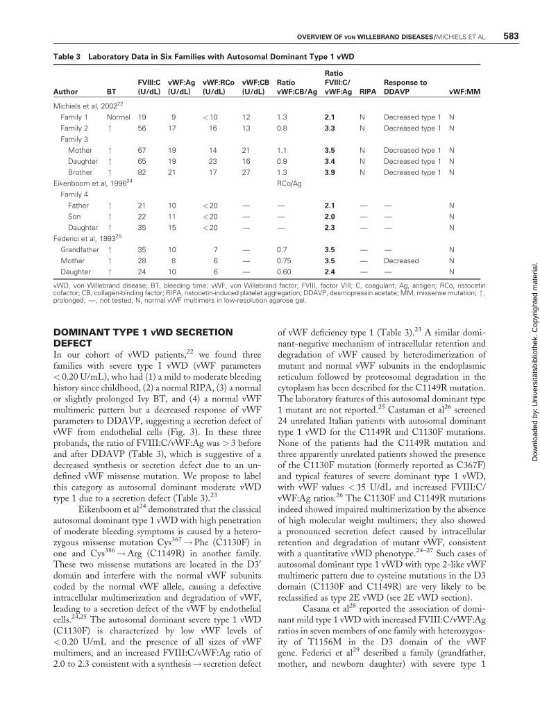
DOMINANT TYPE 1 VWD SECRETIONDEFECTIn our cohort of vWD patients,22 we found threefamilies with severe type I vWD (vWF parameters< 0.20 U/mL), who had (1) a mild to moderate bleedinghistory since childhood, (2) a normal RIPA, (3) a normalor slightly prolonged Ivy BT, and (4) a normal vWFmultimeric pattern but a decreased response of vWFparameters to DDAVP, suggesting a secretion defect ofvWF from endothelial cells (Fig. 3). In these threeprobands, the ratio of FVIII:C/vWF:Ag was > 3 beforeand after DDAVP (Table 3), which is suggestive of adecreased synthesis or secretion defect due to an un-defined vWF missense mutation. We propose to labelthis category as autosomal dominant moderate vWDtype 1 due to a secretion defect (Table 3).23
Eikenboom et al24 demonstrated that the classicalautosomal dominant type 1 vWD with high penetrationof moderate bleeding symptoms is caused by a hetero-zygous missense mutation Cys367!Phe (C1130F) inone and Cys386!Arg (C1149R) in another family.These two missense mutations are located in the D30
domain and interfere with the normal vWF subunitscoded by the normal vWF allele, causing a defectiveintracellular multimerization and degradation of vWF,leading to a secretion defect of the vWF by endothelialcells.24,25 The autosomal dominant severe type 1 vWD(C1130F) is characterized by low vWF levels of< 0.20 U/mL and the presence of all sizes of vWFmultimers, and an increased FVIII:C/vWF:Ag ratio of2.0 to 2.3 consistent with a synthesis! secretion defect
of vWF deficiency type 1 (Table 3).23 A similar domi-nant-negative mechanism of intracellular retention anddegradation of vWF caused by heterodimerization ofmutant and normal vWF subunits in the endoplasmicreticulum followed by proteosomal degradation in thecytoplasm has been described for the C1149R mutation.The laboratory features of this autosomal dominant type1 mutant are not reported.25 Castaman et al26 screened24 unrelated Italian patients with autosomal dominanttype 1 vWD for the C1149R and C1130F mutations.None of the patients had the C1149R mutation andthree apparently unrelated patients showed the presenceof the C1130F mutation (formerly reported as C367F)and typical features of severe dominant type 1 vWD,with vWF values < 15 U/dL and increased FVIII:C/vWF:Ag ratios.26 The C1130F and C1149R mutationsindeed showed impaired multimerization by the absenceof high molecular weight multimers; they also showeda pronounced secretion defect caused by intracellularretention and degradation of mutant vWF, consistentwith a quantitative vWD phenotype.24–27 Such cases ofautosomal dominant type 1 vWD with type 2-like vWFmultimeric pattern due to cysteine mutations in the D3domain (C1130F and C1149R) are very likely to bereclassified as type 2E vWD (see 2E vWD section).
Casana et al28 reported the association of domi-nant mild type 1 vWD with increased FVIII:C/vWF:Agratios in seven members of one family with heterozygos-ity of T1156M in the D3 domain of the vWFgene. Federici et al29 described a family (grandfather,mother, and newborn daughter) with severe type 1
Table 3 Laboratory Data in Six Families with Autosomal Dominant Type 1 vWD
Author BT
FVIII:C
(U/dL)
vWF:Ag
(U/dL)
vWF:RCo
(U/dL)
vWF:CB
(U/dL)
Ratio
vWF:CB/Ag
Ratio
FVIII:C/
vWF:Ag RIPA
Response to
DDAVP vWF:MM
Michiels et al, 200222
Family 1 Normal 19 9 <10 12 1.3 2.1 N Decreased type 1 N
Family 2 " 56 17 16 13 0.8 3.3 N Decreased type 1 N
Family 3
Mother " 67 19 14 21 1.1 3.5 N Decreased type 1 N
Daughter " 65 19 23 16 0.9 3.4 N Decreased type 1 N
Brother " 82 21 17 27 1.3 3.9 N Decreased type 1 N
Eikenboom et al, 199624 RCo/Ag
Family 4
Father " 21 10 <20 — — 2.1 — — N
Son " 22 11 <20 — — 2.0 — — N
Daughter " 35 15 <20 — — 2.3 — — N
Federici et al, 199329
Grandfather " 35 10 7 — 0.7 3.5 — — N
Mother " 28 8 6 — 0.75 3.5 — Decreased N
Daughter " 24 10 6 — 0.60 2.4 — — N
vWD, von Willebrand disease; BT, bleeding time; vWF, von Willebrand factor; FVIII, factor VIII; C, coagulant; Ag, antigen; RCo, ristocetincofactor; CB, collagen-binding factor; RIPA, ristocetin-induced platelet aggregation; DDAVP, desmopressin acetate; MM,missensemutation; " ,prolonged; —, not tested; N, normal vWF multimers in low-resolution agarose gel.
OVERVIEW OF VON WILLEBRAND DISEASES/MICHIELS ET AL 583
Dow
nloa
ded
by: U
nive
rsitä
tsbi
blio
thek
. Cop
yrig
hted
mat
eria
l.

vWD, subtype plasma low-platelet low that could beexplained by decreased synthesis of vWF in culturedendothelial cells isolated from the umbilical vein of thenewborn daughter. This family can readily be reclassifiedas autosomal dominant severe type 1 vWD (Table 3).
VWD TYPE 1 VWD VICENZA DIFFERSFROM TYPE 2MIn our cohort of vWD patients,22 we found two pro-bands with severe type 1 vWD and decreased RIPA, thatcould be classified as vWD type 2M because of a verycharacteristic response to DDAVP (Fig. 4). The typicallaboratory features in our vWD type 2 M patients are(1) severe type 1, (2) decreased RIPA in the presence ofa normal or near-normal vWF multimeric pattern ina low-resolution agarose gel, (3) a poor response ofvWF:RCo to DDAVP, and (4) a good response ofboth vWF:CB and vWF:Ag to DDAVP. This goodresponse of vWF:CB to DDAVP in 2M vWD isconsistent with the presence of all vWF multimers andcan explain the slightly prolonged and normal Ivy BTsbefore and after DDAVP (Fig. 4).22 In a recent study of317 patients previously registered as type 1 vWD, 30patients from 17 unrelated families with discrepantvWF:RCo/vWF:Ag ratios in plasma, normal vWF mul-timers could be reclassified as type 2M.30 RIPA assaywas previously performed in 26 of 30 2M patients and anabsent or decreased responsiveness to ristocetin withminor aggregation at 1.5 mg/mL was found in allcases.30 This simply may mean that all cases of severetype 1 vWD with a normal or near-normal vWF multi-meric pattern and a decreased or absent RIPA, and adecreased vWF:RCo/vWF:Ag ratio, can currently bereclassified as 2M vWD.
Congenital vWD Vicenza clearly differs from 2Mand 2U. vWD Vicenza is characterized by equally lowlevels of FVIII:C, vWF:Ag, and vWF:RCo; usuallynormal RIPA; a normal or slightly prolonged BT; andthe presence of unusually large vWF multimers inplasma.17,31,32 The response to DDAVP in several casesof vWD Vicenza32,33 was good for FVIII:C, vWF:Ag,and vWF:RCo, which was followed by unexplained veryshort half-life times of less than a few hours for FVIII:Cand all vWF parameters, consistent with a laboratorytype 1 phenotype. The ratios for FVIII:C/vWF:Ag,vWF:RCo/vWF:Ag, and vWF:CB/vWF:Ag remainednormal before and after DDAVP.31–33 A single muta-tion R1205H in the D3 (multimerization) domain wasfound as the probable cause of vWD type Vicenza.34
On the basis of this update and detailed analysisof the literature in a previous study,35 we propose a novelclassification and characterization of type 1 and type 3vWD patients (Table 4). Carriers of recessive type 1 andtype 3 vWD are in fact true type 1 vWD at the geneticlevel, but are usually asymptomatic or have mild bleed-
ing, particularly when associated with blood group O.Carriers of a missense mutation in the vWF gene maybe asymptomatic (polymorphism), for example C1584,and may be mildly symptomatic in particular whenassociated with blood group O. Variants of dominanttype 1 vWD result from a missense mutation in the vWFgene with dominant negative effect of the mutated vWFprotein on the normal vWF protein causing a defectiveintracellular processing and secretion of vWF by endo-thelial cells and defective clearing of the secreted vWFfrom plasma consistent with a type 2E or type VicenzavWD.
TYPE 2 VWDIn the plasma, vWF appears as a series of large andintermediate multimers of regularly decreasing molec-ular mass, from several thousand to 500 kd.36,37 The sizeof circulating vWFmultimers is controlled by proteolyticcleavage performed by a specific protease, ADAMTS 13,at cleavage site 1506 in the A2 domain. The 140-kdfragment corresponds to residues 763 to 1605aa, andthe 176-kd fragment to residue 1605 to 2813aa. ThevWF is an adhesive protein that mediates the initiationof platelet adhesion and aggregation through vWFsubendothelial and vWF–platelet interactions to controlprimary hemostasis (BT) at sites of vascular trauma orinjury. vWF binds coagulant FVIII (FVIII:C), contri-buting to its stability and, indirectly, to its function inthe generation of fibrin.37
Using high-resolution sodium dodecyl sulfate–agarose multimeric analysis of vWF in plasma in combi-nation with immunoblots of vWF proteolytic degrada-tion products, Zimmerman et al38 nicely demonstratedthat proteolysis of vWF is a normal event in normalindividuals, and is increased in types IIA (2A) and IIB(2B) vWD with increased triplet structure of each bandas the result of proteolysis, and that proteolysis of vWFis minimal in types IIC (2C), IID (2D), and IIE (2E)variants with aberrant multimeric structure of individualoligomers.38 In types IIA (2A) and IIB (2B), theproportion of 176- and 140-kd fragments was increasedrelated to the intact 225-kd subunit, and these degradedvWF fragment were not detected or were present in onlytrace amount in types IIC (2C), IID (2D), and IIE (2E)vWD.38,39 Zimmerman et al38 therefore postulatedadditional mechanisms to explain the aberrant structureof individual oligomers in types IIC, IID, and IIE vWDpatients. All types IIA, B, C, D, and E2,3,35 are reclassi-fied by Schneppenheim, Budde, and Ruggeri as 2A, 2B,2C, 2D, and 2E (Fig. 5; Table 5)39,40
DOMINANT TYPE 2A VWDThe pertinent findings in patients with type 2A vWDinclude prolonged BT; consistently low vWF:RCo/
584 SEMINARS IN THROMBOSIS AND HEMOSTASIS/VOLUME 31, NUMBER 5 2005
Dow
nloa
ded
by: U
nive
rsitä
tsbi
blio
thek
. Cop
yrig
hted
mat
eria
l.
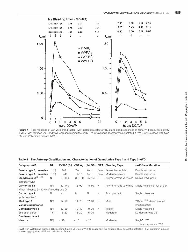
Table 4 The Antwerp Classification and Characterization of Quantitative Type 1 and Type 3 vWD
Category vWD BT FVIII:C (%) vWF:Ag (%) RCo RIPA Bleeding Type vWF Gene Mutation
Severe type 3, recessive " " " 1–9 Zero Zero Zero Severe hemophilia Double nonsense
Severe type 1, recessive " " " 9–40 1–10 0–6 Zero Moderate severe Double missense
Bloodgroup O15,16,17 N 35–150 35–150 35–150 N Asymptomatic very mild Normal vWF gene
(pseudo-vWD)
Carrier type 3 N/ " 30–140 15–90 15–90 N Asymptomatic very mild Single nonsense (null allele)
Minor influence (�10%) of blood group O
Carrier type 1 N N N N N Asymptomatic Single missense
(polymorphism)
Mild type 1 N/ " 12–70 14–70 12–80 N Mild Y1584C19–21/blood group O
Variable penetrance (multigenetic)
Dominant type 1 N/ " 20–80 10–40 0–30 N Mild or Single missense
Secretion defect " / " " 5–20 5–20 5–20 Moderate D3 domain type 2E
Dominant type 1
Vicenza N/ " <15 < 15 < 15 Moderate SingleR1205H
missense (variant 2M)
vWD, von Willebrand disease; BT, bleeding time; FVIII, factor VIII; C, coagulant; Ag, antigen; RCo, ristocetin cofactor; RIPA, ristocetin-inducedplatelet aggregation; vWF, von Willebrand factor.
Figure 4 Poor response of von Willebrand factor (vWF):ristocetin cofactor (RCo) and good responses of factor VIII coagulant activity(FVIIIc), vWF:antigen (Ag), and vWF:collagen-binding factor (CB) to intravenous desmopressin acetate (DDAVP) in two cases with type2M von Willebrand disease (vWD).
OVERVIEW OF VON WILLEBRAND DISEASES/MICHIELS ET AL 585
Dow
nloa
ded
by: U
nive
rsitä
tsbi
blio
thek
. Cop
yrig
hted
mat
eria
l.
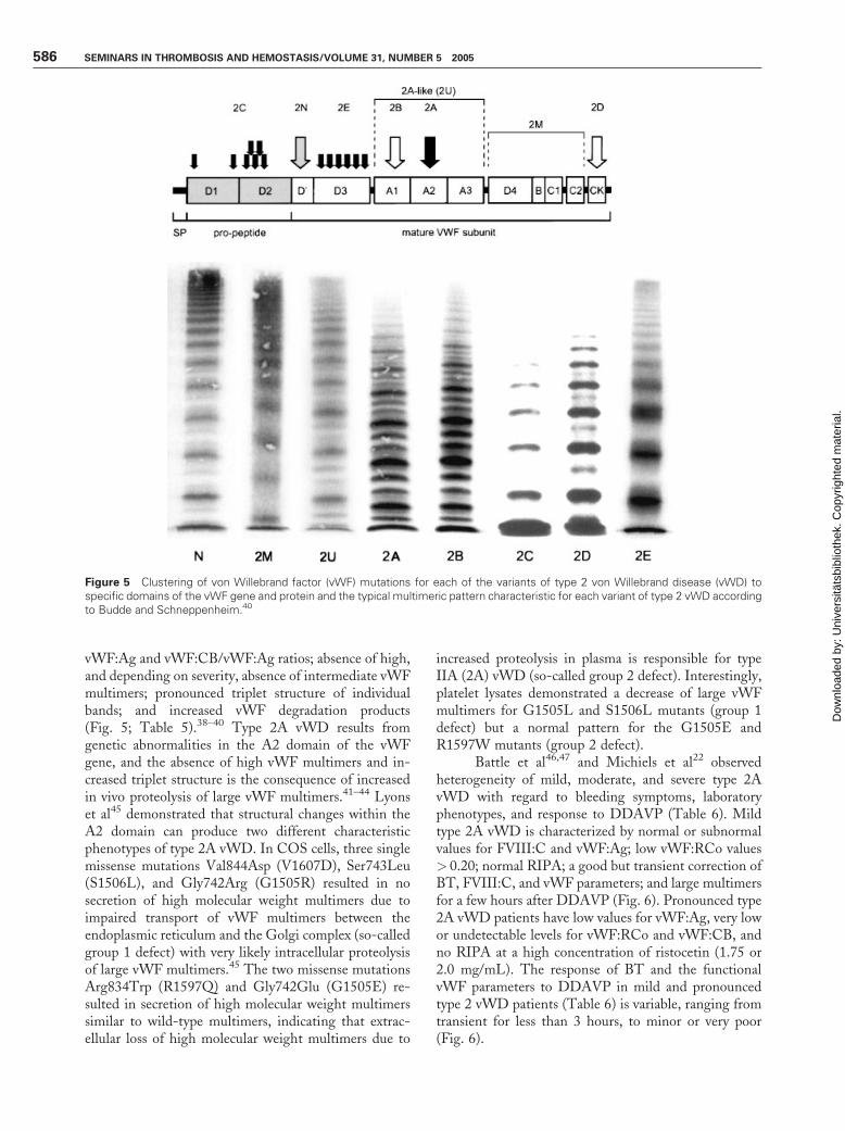
vWF:Ag and vWF:CB/vWF:Ag ratios; absence of high,and depending on severity, absence of intermediate vWFmultimers; pronounced triplet structure of individualbands; and increased vWF degradation products(Fig. 5; Table 5).38–40 Type 2A vWD results fromgenetic abnormalities in the A2 domain of the vWFgene, and the absence of high vWF multimers and in-creased triplet structure is the consequence of increasedin vivo proteolysis of large vWF multimers.41–44 Lyonset al45 demonstrated that structural changes within theA2 domain can produce two different characteristicphenotypes of type 2A vWD. In COS cells, three singlemissense mutations Val844Asp (V1607D), Ser743Leu(S1506L), and Gly742Arg (G1505R) resulted in nosecretion of high molecular weight multimers due toimpaired transport of vWF multimers between theendoplasmic reticulum and the Golgi complex (so-calledgroup 1 defect) with very likely intracellular proteolysisof large vWF multimers.45 The two missense mutationsArg834Trp (R1597Q) and Gly742Glu (G1505E) re-sulted in secretion of high molecular weight multimerssimilar to wild-type multimers, indicating that extrac-ellular loss of high molecular weight multimers due to
increased proteolysis in plasma is responsible for typeIIA (2A) vWD (so-called group 2 defect). Interestingly,platelet lysates demonstrated a decrease of large vWFmultimers for G1505L and S1506L mutants (group 1defect) but a normal pattern for the G1505E andR1597W mutants (group 2 defect).
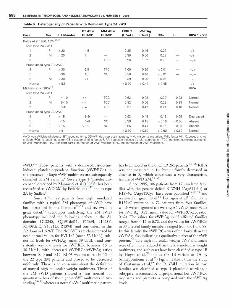
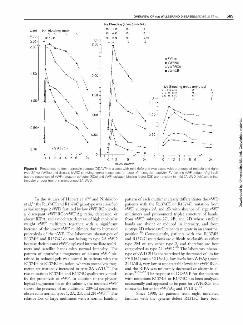
Battle et al46,47 and Michiels et al22 observedheterogeneity of mild, moderate, and severe type 2AvWD with regard to bleeding symptoms, laboratoryphenotypes, and response to DDAVP (Table 6). Mildtype 2A vWD is characterized by normal or subnormalvalues for FVIII:C and vWF:Ag; low vWF:RCo values> 0.20; normal RIPA; a good but transient correction ofBT, FVIII:C, and vWF parameters; and large multimersfor a few hours after DDAVP (Fig. 6). Pronounced type2A vWD patients have low values for vWF:Ag, very lowor undetectable levels for vWF:RCo and vWF:CB, andno RIPA at a high concentration of ristocetin (1.75 or2.0 mg/mL). The response of BT and the functionalvWF parameters to DDAVP in mild and pronouncedtype 2 vWD patients (Table 6) is variable, ranging fromtransient for less than 3 hours, to minor or very poor(Fig. 6).
Figure 5 Clustering of von Willebrand factor (vWF) mutations for each of the variants of type 2 von Willebrand disease (vWD) tospecific domains of the vWF gene and protein and the typical multimeric pattern characteristic for each variant of type 2 vWD accordingto Budde and Schneppenheim.40
586 SEMINARS IN THROMBOSIS AND HEMOSTASIS/VOLUME 31, NUMBER 5 2005
Dow
nloa
ded
by: U
nive
rsitä
tsbi
blio
thek
. Cop
yrig
hted
mat
eria
l.
DOMINANT TYPE 2B VWDThe laboratory phenotype of type 2B is indistinguishablefrom type 2A. Types 2A and 2B are characterized byprolonged BT, consistently low vWF:RCo/vWF:Ag andvWF:CB/vWF:Ag ratios, absence of high and some ofthe intermediate vWF multimers with pronounced trip-let structure of individual bands, and increased vWFdegradation products (Fig. 5; Table 5),38–44 but 2Bdiffers from 2A by a normal vWF multimeric patternin platelets, and increased RIPA. For type 2B vWD,the response to DDAVP is good for FVIII:C andvWF:Ag, but despite the fact that vWF:RCo increasesto > 1.0 U/mL, the vWF:CB does not increase tonormal levels, and there is no correction of BT and largevWF multimers after DDAVP.22 Patients with type 2BvWD may respond to an infusion with DDAVP withthrombocytopenia due to the appearance of higher vWFmultimers, causing reversible platelet aggregationin vivo.
DOMINANT VWD TYPE 2MAND TYPE 2 UThe determination and characterization of type 2MvWD in the literature is problematic. The very firstcase labeled as vWD 2M (previously labeled as type 1BvWD48,49) showed the mutation Gly561Ser (G1324S),a slight qualitative defect in multimer distribution,a completely normal vWFmultimer distribution, normalvalues of FVIII:C and vWF:Ag, but absence ofvWF:RCo and RIPA, a profound decrease of vWFbinding to platelets in the presence of ristocetin but anormal binding of vWF to platelets in the presence ofbotrocetin.48–50 There was no genetic evidence for eithera dominant or recessive inheritance because familymembers of the affected patient were not available, butcircumstantial evidence was produced that Gly561Ser(G1324S) may be a recessive mutation and that the otherallele may not be expressed (null allele).48–52 This singleobservation is at the origin of the definition of type 2M
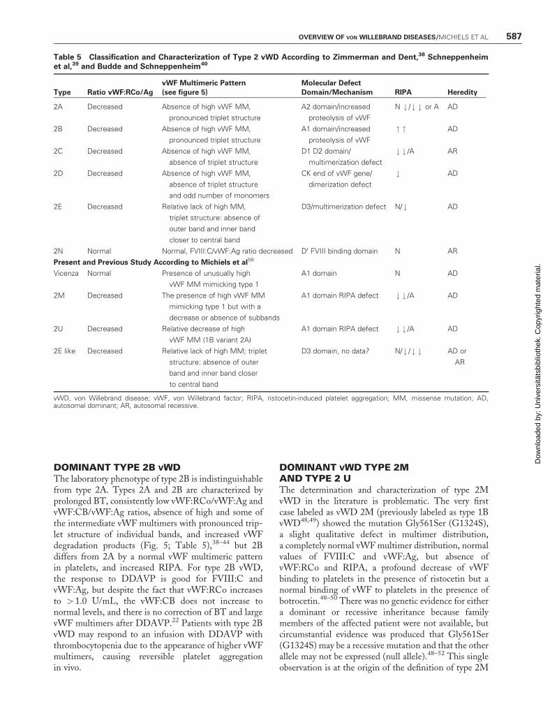
Table 5 Classification and Characterization of Type 2 vWD According to Zimmerman and Dent,38 Schneppenheimet al,39 and Budde and Schneppenheim40
Type Ratio vWF:RCo/Ag
vWF Multimeric Pattern
(see figure 5)
Molecular Defect
Domain/Mechanism RIPA Heredity
2A Decreased Absence of high vWF MM,
pronounced triplet structure
A2 domain/increased
proteolysis of vWF
N # / # # or A AD
2B Decreased Absence of high vWF MM,
pronounced triplet structure
A1 domain/increased
proteolysis of vWF
" " AD
2C Decreased Absence of high vWF MM,
absence of triplet structure
D1 D2 domain/
multimerization defect
# # /A AR
2D Decreased Absence of high vWF MM,
absence of triplet structure
and odd number of monomers
CK end of vWF gene/
dimerization defect
# AD
2E Decreased Relative lack of high MM,
triplet structure: absence of
outer band and inner band
closer to central band
D3/multimerization defect N/ # AD
2N Normal Normal, FVIII:C/vWF:Ag ratio decreased D0 FVIII binding domain N AR
Present and Previous Study According to Michiels et al56
Vicenza Normal Presence of unusually high
vWF MM mimicking type 1
A1 domain N AD
2M Decreased The presence of high vWF MM
mimicking type 1 but with a
decrease or absence of subbands
A1 domain RIPA defect # # /A AD
2U Decreased Relative decrease of high
vWF MM (1B variant 2A)
A1 domain RIPA defect # # /A AD
2E like Decreased Relative lack of high MM; triplet
structure: absence of outer
band and inner band closer
to central band
D3 domain, no data? N/ # / # # AD or
AR
vWD, von Willebrand disease; vWF, von Willebrand factor; RIPA, ristocetin-induced platelet aggregation; MM, missense mutation; AD,autosomal dominant; AR, autosomal recessive.
OVERVIEW OF VON WILLEBRAND DISEASES/MICHIELS ET AL 587
Dow
nloa
ded
by: U
nive
rsitä
tsbi
blio
thek
. Cop
yrig
hted
mat
eria
l.
vWD.2,3 Those patients with a decreased ristocetin-induced platelet-dependent function (vWF:RCo) inthe presence of large vWF multimers are subsequentlyclassified as 2M variants.2 Severe type 1 ‘‘platelet dis-crepant’’ described by Mannucci et al (1985)51 has beenreclassified as vWD 2M by Federici et al,52 and as type2A by Sadler.2
Since 1996, 22 patients from eight unrelatedfamilies with a typical 2M phenotype of vWD havebeen described in the literature53–55 and reviewed ingreat detail.56 Genotypes underlying the 2M vWDphenotype included the following defects in the A1domain: G1324S, Q1191del11, F1369I, I1425F,K1408delK, Y1321D, R1394I, and one defect in theA2 domain I1526T. The 2M vWDs are characterized bynear-normal values for FVIII:C (mean 53 U/dL), sub-normal levels for vWF:Ag (mean 39 U/dL), and con-sistently very low levels for vWF:RCo between < 5 to30 U/mL, with decreased vWF:RCo/vWF:Ag ratiosbetween 0.40 and 0.12. RIPA was measured in 13 ofthe 22 type 2M patients and proved to be decreaseduniformly. There is no consensus about the definitionof normal high molecular weight multimers. Three ofthe 2M vWD patients showed a near normal butquantitative loss of the highest vWF multimers in twostudies,54–56 whereas a normal vWF multimeric pattern
has been noted in the other 19 2M patients.53–56 RIPAwas not measured in 14, but uniformly decreased orabsence in 8, which constitutes a very characteristicfeature of vWD 2M.53–56
Since 1995, 106 patients from 12 unrelated fam-ilies with the genetic defect R1374H (Arg611His) orR1374C (Arg611Cys) have been published55,57–59 andreviewed in great detail.56 Lethagen et al57 found theR1374C mutation in 72 patients from four families,which were diagnosed as severe type 1 vWD (mean valuefor vWF:Ag, 0.21; mean value for vWF:RCo,13; ratio,0.62). The values for vWF:Ag in 63 affected familiesranged from 0.12 to 0.72, and the values for vWF:RCoin 33 affected family members ranged from 0.01 to 0.88.In this family, the vWF:RCo was often lower than thevWF:Ag, also indicating a qualitative defect of the vWFprotein.57 The high molecular weight vWF multimerswere often more reduced than the low molecular weightmultimers, and such cases have been classified as type 1Bby Hoyer et al,58 and as the 1B variant of 2A bySchneppenheim et al39 (Fig. 5; Table 5). In the studyof Castaman et al,59 the R1374H mutation in twofamilies was classified as type 1 platelet discordant, asubtype characterized by disproportional low vWF:RCoin plasma and platelets as compared with the vWF:Aglevels.
Table 6 Heterogeneity of Patients with Dominant Type 2A vWD
Case Sex BT Minutes
BT After
DDAVP
MM After
DDAVP
FVIII:C
(U/mL)
vWF:Ag
(U/mL) RCo CB RIPA 1.2/2.0
Battle et al 1986, 199446,47
Mild type 2A vWD
1 F > 20 4.5 — 0.35 0.46 0.22 — þ/þ2 M > 20 7 — 0.30 0.50 0.22 — þ/þ3 F 15 8 TCC 0.96 1.52 0.7 — � /þ
Pronounced type 2A vWD
4 F > 30 9.5 TPC 1.00 0.50 < 0.01 — � /þ5 F > 30 16 NC 0.50 0.40 < 0.01 — � /�6 M > 30 21 — 0.39 0.26 0.05 — � /�Normal < 8.5 > 0.50 >0.45 > 0.45 þ/þ
Michiels et al, 200222 RIPA
Mild type 2A vWD
1 F 4–10 <4 TCC 0.62 0.98 0.30 0.23 Normal
2 M 6–10 <4 TCC 0.92 0.56 0.28 0.23 Normal
3 F 3–6 <4 TCC 0.37 0.42 0.21 0.18 Normal
Pronounced type 2A vWD
4 F > 15 3–9 — 0.93 0.45 0.13 0.05 Decreased
5 F > 15 4–8 NC 0.38 0.15 < 0.10 <0.05 Absent
6 F > 15 >15 NC 0.66 0.41 0.15 0.05 Absent
Normal < 4 > 0.60 >0.60 > 0.60 >0.60 Normal
vWD, von Willebrand disease; BT, bleeding time; DDAVP, desmopressin acetate; MM, missense mutation; FVIll, factor VIII; C, coagulant; Ag,antigen; RCo, ristocetin cofactor; CB, collagen-binding factor; RIPA, ristocetin-induced platelet aggregation; TCC, transient complete correctionof vWF multimers; TPC, transient partial correction of vWF multimers; NC, no correction of vWF multimers.
588 SEMINARS IN THROMBOSIS AND HEMOSTASIS/VOLUME 31, NUMBER 5 2005
Dow
nloa
ded
by: U
nive
rsitä
tsbi
blio
thek
. Cop
yrig
hted
mat
eria
l.
In the studies of Hilbert et al60 and Nishikuboet al,61 the R1374H and R1374C genotype was classifiedas variant type 2 vWD featured by low vWF:RCo levels,a discrepant vWF:RCo/vWF:Ag ratio, decreased orabsent RIPA, and a moderate decrease of high molecularweight vWF multimers together with a significantincrease of the lower vWF multimers due to increasedproteolysis of the vWF. The laboratory phenotypes ofR1374H and R1374C do not belong to type 2A vWDbecause their plasma vWF displayed intermediate multi-mers and satellite bands with normal intensity. Thepattern of proteolytic fragments of plasma vWF ob-tained in reduced gels was normal in patients with theR1374H or R1374C mutation, whereas proteolytic frag-ments are markedly increased in type 2A vWD.39 Thetwo mutations R1374H and R1374C qualitatively mod-ify the proteolysis of vWF. In addition to the physio-logical fragmentation of the subunit, the mutated vWFshows the presence of an additional 209-kd species notobserved in normal types 1, 2A, 2B, and 2N vWF.61 Therelative loss of large multimers with a normal banding
pattern of each multimer clearly differentiates the vWDpatients with the R1374H or R1374C mutation fromvWD subtypes 2A and 2B with absence of large vWFmultimers and pronounced triplet structure of bands,from vWD subtypes 2C, 2E, and 2D where satellitebands are absent or reduced in intensity, and fromsubtype 2D where satellite bands migrate in an abnormalposition.39 Consequently, patients with the R1374Hand R1374C mutations are difficult to classify as eithertype 2M or any other type 2, and therefore are bestcategorized as type 2U vWD.60 The laboratory pheno-type of vWD 2U is characterized by decreased values forFVIII:C (mean 32 U/dL), low levels for vWF:Ag (mean21 U/dL), very low or undetectable levels for vWF:RCo,and the RIPA was uniformly decreased or absent in allcases.30,58–62 The response to DDAVP for the patientswith mutations R1374H or R1374C has been analyzedoccasionally and appeared to be poor for vWF:RCo andsomewhat better for vWF:Ag and FVIII:C.63
Since 1998, 23 patients from eight unrelatedfamilies with the genetic defect R1315C have been
Figure 6 Responses to desmopressin acetate (DDAVP) in a case with mild (left) and two cases with pronounced (middle and right)type 2A von Willebrand disease (vWD) showing normal responses for factor VIII coagulant activity (FVIIIc) and vWF:antigen (Ag) in all,but the responses of vWF:ristocetin cofactor (RCo) and vWF: collagen-binding factor (CB) are transient in mild 2A vWD (left) and minor(middle) or poor (right) in pronounced 2A vWD.
OVERVIEW OF VON WILLEBRAND DISEASES/MICHIELS ET AL 589
Dow
nloa
ded
by: U
nive
rsitä
tsbi
blio
thek
. Cop
yrig
hted
mat
eria
l.
described in the literature30,58,62 and reviewed in greatdetail.56 The laboratory phenotype of the R1315Cmutation is characterized by subnormal values forFVIII:C (mean 41 U/dL), low levels for vWF:Ag(mean 20 U/dL), very low or undetectable levels forvWF:RCo of less than 0.10, a decreased vWF:RCo/vWF:Ag ratio between and the RIPA was uniformlydecreased or absent in all cases. The R1315C mutationhas been labeled as type 2M in two cases by Nitu-Whalley et al.30 The R1315C mutation in 11 affectedmembers of one family has been described by Lethagenet al57 as severe type 1 vWD with a poor response toDDAVP and as type 2 variant or 2U by Ribba et al.62
During the 51st Annual Meeting of the ISTH(Sydney, Australia, August 2005), Budde and Schnep-penheim40 presented a state-of-the-art poster (P2057)entitled ‘‘Multimeric pattern in patients diagnosed withtype 1 von Willebrand disease in the European study,Molecular and Clinical Markers for the Diagnosis andManagement of Type 1 vWD (MCMDM-1vWD).’’About half of the patients diagnosed as type 1 havetype 2 vWD. So-called type 1 vWD patients are neverre-diagnosed as type 2A, 2B, 2C, or 2D, but as type 2E,2M, or 2A-like vWD. None of the heterozygous muta-tions of the D1, D2, and D0 domains show an abnormalvWF multimeric pattern. The multimerization defectscaused by mutations in the D1 or D2 domain areinherited recessively and correlate with a particularphenotype, namely, 2C (see Table 5; Fig. 5). Mutationsin the D3 domain (C1130R, C1130G, W1144G,Y1146C) show a type 2E pattern with relative loss oflarge vWF multimers and reduced triplet structure andproteolysis (Table 5; Fig. 5). Mutations in the A1 to A3domain (R1315C, R1374C, G1415D) show a relativeloss of large vWF multimers, no increase of tripletstructure, and a defect of the ristocetin cofactor activity
and RIPA, suggesting a type 2A-like or 2U vWD(Table 5; Fig. 5). Mutations in the D4-CK domain(G2441C, R2464C, R2464X, C2469P) show a type 2Mmultimeric pattern with the presence of high vWFmultimers, no triplet structure, and a smeary pattern.These new observations indicate that the reported casesof type 2M with a mutation in the A1-A3 domain areuniformly associated with reduced vWF:RCo and de-creased or absent RIPA, and therefore are to be relabeledas type 2A-like or 2U, and that type 2M is now muchbetter defined as a distinct laboratory phenotype of vWD(Table 5; Fig. 5). The results of the MCMDM-1vWDstudies surely will show the manifold phenotypic ex-pressions of types 1 and 2 vWDs, which have importantimplications with regard to classification and responsesof DDAVP with subsequent consequences for the pre-vention and treatment of bleeding complications.
RECESSIVE TYPE 2C VWDvWD type 2C shows a characteristic multimeric patternwith a lack of high molecular weight multimers, thepresence of one single-banded multimer instead oftriplets, and there is a pronounced first band thatprobably includes a dimer and a tetramer (Fig. 5;Table 5).39,40 Autosomal recessive vWD type 2C iscaused by homozygosity for a missense mutation (ordouble heterozygosity of a null allele and missensemutation) in the D1 and D2 domains of the vWFpropeptide that catalyzes the multimerization in theD3 domain at the N terminus of mature vWF.36 Thelaboratory phenotypes and the genetic defect of six casesof autosomal recessive vWD type 2C are shown inTable 7.64–69 Autosomal recessive type 2C vWD, whichaffects both males and females, is characterized by verylow levels of FVIII:C and vWF:Ag, unmeasurable
Table 7 Laboratory Phenotypes and Genetic Defects in the D2 Domain in Six Cases with Autosomal Recessive Type2C vWD
Author Age (yr) Gender BT (min) FVIII:C (%) vWF:Ag vWF:RCo
vWF:CB
(%) RIPA
MM
Pattern
Ruggeri et al63 39 M >30 0.67 0.50 0.10 nt # # 2C
Mutation Ins405AsnPro (F405insNP)( Holmberg et al59)
Mazurier et al65 19 F >20 0.24 0.16 < 0.03 nt Zero 2C
Mutation 625insGly/26pdelCT-stop (A625InsG/null) (Gaucher62)
Battle et al67 — M >20 0.09 0.13 < 0.01 nt Zero 2C
— F >20 0.20 0.15 < 0.01 nt Zero 2C
Schneppenheim et al69 Child F 10 >1.00 1.59 0.25 0.29 nt 2C
Mutation homozygous G550R
Gaucher et al66 64 F >30 0.20 0.10 < 0.10 nt nt 2C
Mutation homozygous Cys623TRP (C623W)
Mannucci et al70 10 F >30 0.85 1.25 0.14 nt Zero 2C?
Mutation Unknown
vWD, von Willebrand disease; BT, bleeding time; FVIII, factor VIII; C, coagulant; vWF, von Willebrand factor; Ag, antigen; RCo, ristocetincofactor; CB, collagen-binding factor; RIPA, ristocetin-induced platelet aggregation; MM, missense mutation; N, normal; nt, not tested; # ,decreased.
590 SEMINARS IN THROMBOSIS AND HEMOSTASIS/VOLUME 31, NUMBER 5 2005
Dow
nloa
ded
by: U
nive
rsitä
tsbi
blio
thek
. Cop
yrig
hted
mat
eria
l.
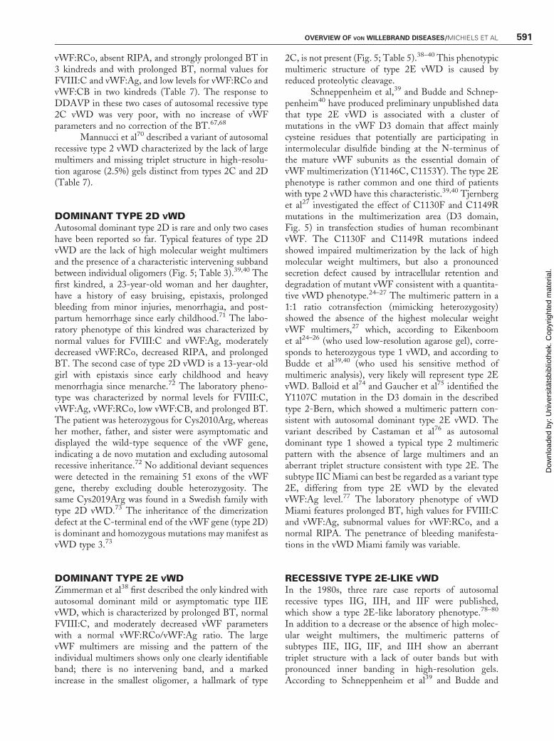
vWF:RCo, absent RIPA, and strongly prolonged BT in3 kindreds and with prolonged BT, normal values forFVIII:C and vWF:Ag, and low levels for vWF:RCo andvWF:CB in two kindreds (Table 7). The response toDDAVP in these two cases of autosomal recessive type2C vWD was very poor, with no increase of vWFparameters and no correction of the BT.67,68
Mannucci et al70 described a variant of autosomalrecessive type 2 vWD characterized by the lack of largemultimers and missing triplet structure in high-resolu-tion agarose (2.5%) gels distinct from types 2C and 2D(Table 7).
DOMINANT TYPE 2D VWDAutosomal dominant type 2D is rare and only two caseshave been reported so far. Typical features of type 2DvWD are the lack of high molecular weight multimersand the presence of a characteristic intervening subbandbetween individual oligomers (Fig. 5; Table 3).39,40 Thefirst kindred, a 23-year-old woman and her daughter,have a history of easy bruising, epistaxis, prolongedbleeding from minor injuries, menorrhagia, and post-partum hemorrhage since early childhood.71 The labo-ratory phenotype of this kindred was characterized bynormal values for FVIII:C and vWF:Ag, moderatelydecreased vWF:RCo, decreased RIPA, and prolongedBT. The second case of type 2D vWD is a 13-year-oldgirl with epistaxis since early childhood and heavymenorrhagia since menarche.72 The laboratory pheno-type was characterized by normal levels for FVIII:C,vWF:Ag, vWF:RCo, low vWF:CB, and prolonged BT.The patient was heterozygous for Cys2010Arg, whereasher mother, father, and sister were asymptomatic anddisplayed the wild-type sequence of the vWF gene,indicating a de novo mutation and excluding autosomalrecessive inheritance.72 No additional deviant sequenceswere detected in the remaining 51 exons of the vWFgene, thereby excluding double heterozygosity. Thesame Cys2019Arg was found in a Swedish family withtype 2D vWD.73 The inheritance of the dimerizationdefect at the C-terminal end of the vWF gene (type 2D)is dominant and homozygous mutations may manifest asvWD type 3.73
DOMINANT TYPE 2E VWDZimmerman et al38 first described the only kindred withautosomal dominant mild or asymptomatic type IIEvWD, which is characterized by prolonged BT, normalFVIII:C, and moderately decreased vWF parameterswith a normal vWF:RCo/vWF:Ag ratio. The largevWF multimers are missing and the pattern of theindividual multimers shows only one clearly identifiableband; there is no intervening band, and a markedincrease in the smallest oligomer, a hallmark of type
2C, is not present (Fig. 5; Table 5).38–40 This phenotypicmultimeric structure of type 2E vWD is caused byreduced proteolytic cleavage.
Schneppenheim et al,39 and Budde and Schnep-penheim40 have produced preliminary unpublished datathat type 2E vWD is associated with a cluster ofmutations in the vWF D3 domain that affect mainlycysteine residues that potentially are participating inintermolecular disulfide binding at the N-terminus ofthe mature vWF subunits as the essential domain ofvWF multimerization (Y1146C, C1153Y). The type 2Ephenotype is rather common and one third of patientswith type 2 vWD have this characteristic.39,40 Tjernberget al27 investigated the effect of C1130F and C1149Rmutations in the multimerization area (D3 domain,Fig. 5) in transfection studies of human recombinantvWF. The C1130F and C1149R mutations indeedshowed impaired multimerization by the lack of highmolecular weight multimers, but also a pronouncedsecretion defect caused by intracellular retention anddegradation of mutant vWF consistent with a quantita-tive vWD phenotype.24–27 The multimeric pattern in a1:1 ratio cotransfection (mimicking heterozygosity)showed the absence of the highest molecular weightvWF multimers,27 which, according to Eikenboomet al24–26 (who used low-resolution agarose gel), corre-sponds to heterozygous type 1 vWD, and according toBudde et al39,40 (who used his sensitive method ofmultimeric analysis), very likely will represent type 2EvWD. Balloid et al74 and Gaucher et al75 identified theY1107C mutation in the D3 domain in the describedtype 2-Bern, which showed a multimeric pattern con-sistent with autosomal dominant type 2E vWD. Thevariant described by Castaman et al76 as autosomaldominant type 1 showed a typical type 2 multimericpattern with the absence of large multimers and anaberrant triplet structure consistent with type 2E. Thesubtype IICMiami can best be regarded as a variant type2E, differing from type 2E vWD by the elevatedvWF:Ag level.77 The laboratory phenotype of vWDMiami features prolonged BT, high values for FVIII:Cand vWF:Ag, subnormal values for vWF:RCo, and anormal RIPA. The penetrance of bleeding manifesta-tions in the vWD Miami family was variable.
RECESSIVE TYPE 2E-LIKE VWDIn the 1980s, three rare case reports of autosomalrecessive types IIG, IIH, and IIF were published,which show a type 2E-like laboratory phenotype.78–80
In addition to a decrease or the absence of high molec-ular weight multimers, the multimeric patterns ofsubtypes IIE, IIG, IIF, and IIH show an aberranttriplet structure with a lack of outer bands but withpronounced inner banding in high-resolution gels.According to Schneppenheim et al39 and Budde and
OVERVIEW OF VON WILLEBRAND DISEASES/MICHIELS ET AL 591
Dow
nloa
ded
by: U
nive
rsitä
tsbi
blio
thek
. Cop
yrig
hted
mat
eria
l.
Schneppenheim,40 the phenotype IIG/IIF/IIH mayreadily be taken together as autosomal recessive 2Edifferent from type 2A (Fig. 5, Table 5). vWD type2E appears to be less well defined, is usually autosomaldominant, may be recessive, and accounts for about onethird of patients with 2A in a large cohort of vWDpatients diagnosed in the coagulation laboratory inHamburg, Germany.39,40
RECESSIVE TYPE 2N VWDType 2N vWD encompasses all patients with FVIII:Cdeficiency caused by a markedly decreased affinity ofvWF for FVIII:C.81,82 The laboratory phenotype ofclassical type 2N vWD is characterized typically byreduced FVIII:C levels despite normal or near-normalvWF:Ag, vWF:RCo, and vWF:CB levels; a normalvWF multimeric pattern; and normal vWF-dependentplatelet functions including RIPA and bleeding time.The FVIII:C levels range from 1 to 40 U/dL and dependon the severity of the FVIII:vWFCB defect. Althoughsome cases of have severe FVIII:C deficiency of 1 to 2 U/dL, the majority of 2N vWD patients have FVIII:Clevels above 5 U/dL. Consequently, type 2N vWD maybe misclassified as mild hemophilia. Such patients gen-erally are considered as sporadic cases of hemophilia A orhemophilia A carriers, and the only definitive way todistinguish between mild hemophilia and 2N is tomeasure FVIII:vWFCB.81–84 The bleeding manifesta-tions of type 2N vWD primarily depend on the degree ofdecreased FVIII:C levels, and therefore mimick those ofpatients with mild hemophilia A. The most frequentbleeding manifestations of type 2N vWD patients in-clude recurrent epistaxis; bruising, hematomas aftertrauma; excessive bleeding after minor injuries; bleedingafter tonsillectomy and tooth extraction; occasionallyhemarthrosis and muscle bleedings; and for affectedwomen, menorrhagia, menorrhagia after the first men-struation, and bleeding after delivery.80
The FVIII binding site has been located on theterminal part of the mature vWF subunit and corre-sponds to the D0 domain and to the N-terminal part ofthe D3 domain of the pre-pro vWF molecule synthe-sized by the vWF gene (Fig. 1).76,77 The vWF genedefects in type 2N vWD are missense mutations locatedin exons 18 to 27. Most of the type 2N missensemutations are located in exons 18 to 20 of the FVIIIbinding domain N-terminal region of the maturevWF, which include R782W, G785E, E787K,C788R, C788Y, T791M, Y795C, M800V, R816W,R816Q, H817Q, R854Q, R854W, C858F, andD879N.81,82 A few missense mutations of type 2NvWD (1053, 1061, and 1225) are located outside theFVIII binding domain, suggesting that such mutationsaffect the FVIII binding site, probably by changing itsconformation.82
The inheritance of vWD type 2N is autosomalrecessive. Type 2N vWD patients are homozygous ordouble homozygous for the 2N missense mutation ordouble heterozygous for one 2N and one null vWFallele.80–83 In type 2N vWD, the FVIII:vWF bindingis either nil or dramatically reduced.81–84
DDAVP IN TYPE 2 VWDThe responses to DDAVP of FVIII:C and vWF param-eters in patients with pseudo-vWD (Fig. 2), type 1 vWD(Fig. 3), and type 2M vWD (Fig. 4) have been describedin previous sections of this article. Mild type 2A vWDhas a transiently complete response to DDAVP that maybe good enough for the treatment and prophylaxis ofminor bleedings (Table 6; Fig. 6).22,46,47 Most patientswith type 2A vWD show only a transient minor or apoor response to DDAVP, with no correction of the BTdespite some increase of vWF:RCo (Fig. 6), and there-fore are candidates for FVIII/vWF concentrate substi-tution for the treatment and prophylaxis of bleedingsymptoms.22,46,47 Federici et al85 demonstrated that in
Table 8 FVIII:C and vWF Levels Contained in Cryoprecipitates and FVIII/vWF Concentrates; In Vitro Study 1994
FVIII-vWF Concentrate
FVIII:C
(U/mL)
vWF:Ag/
FVIII:C (U)
vWF:RCo/
FVIII:C (U)
vWF:CB/
FVIII:C (U)
Ratio vWF:
RCo/Ag
Ratio
vWF:CB/Ag
Wet cryoprecipitate
Blood bank 1 9.70�1.72 4.58�1.17 3.87� 0.56 3.99�0.47 0.84 0.87
Blood bank 2 2.34�0.15 2.76�0.21 2.69� 0.41 2.61�0.34 0.97 0.94
Blood bank 3 8.62�0.55 5.00�0.97 4.19� 0.54 4.28�0.50 0.84 0.85
Lyophilyzed cryoprecipitate 3.65�0.21 4.22�0.74 2.07� 0.36 2.40�0.57 0.49 0.58
FVIII/vWF concentrates
Biotransfusion 23.15�1.59 3.82�0.44 4.30� 0.30 3.81�1.13 1.13 0.99
Nordic 47.91�2.75 7.47�1.97 3.59� 0.33 4.76�0.96 0.48 0.63
Alpha VIII 250 55.44�4.53 7.11�1.13 3.79� 0.20 3.51�0.43 0.53 0.49
Hemate-P 27.33�4.08 2.74�0.14 2.31� 0.37 2.66�0.22 0.84 0.97
Monoclate 94.63�3.08 0.40�0.02 < 0.05 <0.10
FVIII, factor VIII; vWF, von Willebrand factor; C, Coagulant; Ag, antigen; RCo, ristocetin cofactor; CB, collagen-binding factor.
592 SEMINARS IN THROMBOSIS AND HEMOSTASIS/VOLUME 31, NUMBER 5 2005
Dow
nloa
ded
by: U
nive
rsitä
tsbi
blio
thek
. Cop
yrig
hted
mat
eria
l.
type 2A vWD patients with the mutations S1506L andV1665E causing group 1 defects, the response toDDAVP is poor, with no reappearance of the largemultimers and with persistence of a strongly prolongedBT and little increase of vWF:RCo. In contrast, in type2A vWD patients with the mutations R1597W andG1629R causing group 2 defects, DDAVP induced atransient increase of large multimers associated with atransient (1 or 2 hours) correction of BT and transientcorrection of vWF:RCo to low normal.
The response to DDAVP in type 2B is normal forFVIII:C and vWF:Ag, transiently good and reachingnormal values for vWF:RCo, and subnormal forvWF:CB without correction of BT and no reappearanceof large vWF multimers. Therefore type 2B patientsare candidates for FVIII/vWF concentrate substitu-tion for the prophylaxis and treatment of bleedingepisodes.22
Recessive type 1 and recessive type 2C vWD arecharacterized by variable FVIII:C levels, very lowvWF:RCo, decreased or absent RIPA, and a very poorresponse to DDAVP,10,67,68 and therefore are candidatesfor FVIII/vWF concentrates.
Federici et al85 evaluated the biological responsesin 20 patients with type 2M vWD. Gene mutations were
not known in six and known in 14 of 20 patients:R1315C in three, R1374C in nine, and R1384H intwo patients. These three mutations are reported in theliterature to be linked with type 2M or 2U vWD.48–58
The response to DDAVP was fairly good for FVIII:C,not evaluated for vWF:Ag and vWF:CB, and poor forvWF:RCo in 18 of the 20 so-called type 2M/U vWDpatients. The cohort of 20 patients with type 2M vWDhad borderline to slightly prolonged BT in seven, pro-longed BT in 10, and strongly prolonged (� 30 minutes)in three patients. The prolonged BTs were normal at2 hours after DDAVP in 13 of 20 patients with type 2MvWD.85
The response to DDAVP in recessive type 2NvWD of the vWF:Ag, vWF:RCo, and vWF:CB arenormal,84 but the response of FVIII:C to DDAVPdepends on the severity of the FVIII:vWF bindingdefect. The response to DDAVP of FVIII:C is fairlygood in type 2N patients homozygous for the R816Wmutation,22,84 but the response to DDAVP is poor intype 2N homozygous for the R854Q mutation, andtherefore are candidates for FVIII/vWF concentrates.
In conclusion, the responses to DDAVP ofFVIII:C and vWF parameters in types 2M, Vincenza,2E, and mild 2A, 2U, and 2N vWD are transient and
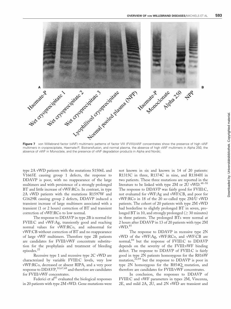
Figure 7 von Willebrand factor (vWF) multimeric patterns of factor VIII (FVIII)/vWF concentrates show the presence of high vWFmultimers in cryoprecipitate, Haemate-P, Biotransfusion, and normal plasma, the absence of high vWF multimers in Alpha 250, theabsence of vWF in Monoclate, and the presence of vWF degradation products in Alpha and Nordic.
OVERVIEW OF VON WILLEBRAND DISEASES/MICHIELS ET AL 593
Dow
nloa
ded
by: U
nive
rsitä
tsbi
blio
thek
. Cop
yrig
hted
mat
eria
l.
good for a few or some more hours to arrest mucocuta-neous bleeding episodes or to prevent bleeding duringminor surgery or trauma, but not good enough to treatmajor bleedings or to prevent bleeding during majorsurgery or trauma. The responses to DDAVP ofFVIII:C and/or vWF parameters in types 2B, 2C, andpronounced cases of 2A and 2U vWD are rather poor topoor, indicating the need to use FVIII/vWF concentrateto treat significant mucocutaneous bleeding episodes orto prevent bleeding during minor and major surgery ortrauma. No data on type 2D vWD are available.
FVIII/ VWF CONCENTRATES IN VWDTYPES 1, 2, AND 3 WITH A POORRESPONSE TO DDAVPvWD patients with recessive type 3, type 1, type 2C, andsevere type 2N, and dominant type 2A, 2B, and 2U thatrespond poorly or not at all to DDAVP are candidatesfor FVIII/vWF concentrate for the treatment of bleed-ing episodes and prophylaxis of bleeding during surgeryor after minor or major trauma. Cryoprecipitate was thekeystone for the treatment of vWD patients from the1960s until the late 1980s, when the virus-inactivatedFVIII/vWF concentrates appeared on the market.86,87
Table 8 shows the content of FVIII:C, vWF:Ag,vWF:RCo, and vWF:CB (values of three separate as-says) in cryoprecipitates and FVIII/vWF concentratesafter reconstitution in the amount of fluid recommendedby the manufacturer. For wet cryoprecipitates producedin the 1980s and early 1990s by local blood banks in TheNetherlands, the concentrations of FVIII:C were lowerthan those of vWF:Ag (Table 8). The values forvWF:Ag, vWF:RCo, and vWF:CB were similar, withratios of vWF:RCo/vWF:Ag and vWF:CB/vWF:Agranging from 0.84 to 0.97 for the three wet cryopreci-pitates and 0.49 and 0.58 for the lyophilized cryopreci-pitate (Table 8). The multimeric patterns of vWF inthe cryoprecipitates compared with a normal plasmarun showed a near-normal vWF multimeric pattern,with the presence of high vWF multimers and theabsence of vWF degradation products (Fig. 7A). Themultimeric patterns of Haemate-P (ZLB-Behring,Marburg, Germany) and normal plasma are quite sim-ilar, with the presence of the high-vWF multimers(Fig. 7B).
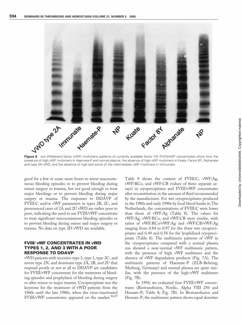
In 1994, we evaluated four FVIII/vWF concen-trates (Biotransfusion, Nordic, Alpha VIII 250 andHemate-P; Table 8; Fig. 7B). In Biotransfusion andHemate-P, the multimeric pattern shows equal densities
Figure 8 von Willebrand factor (vWF) multimeric patterns of currently available factor VIII (FVIII)/vWF concentrates show how thepresence of high vWF multimers in Haemate-P and normal plasma, the absence of high vWF multimers in Koate, Factor 8Y, Alphanateand type 2A vWD, and the absence of high and some of the intermediate vWF multimers in Immunate.
594 SEMINARS IN THROMBOSIS AND HEMOSTASIS/VOLUME 31, NUMBER 5 2005
Dow
nloa
ded
by: U
nive
rsitä
tsbi
blio
thek
. Cop
yrig
hted
mat
eria
l.

of the low-, intermediate-, and high-vWF multimers,and absence of vWF degradation products (Fig. 7B),which is consistent with normal ratios of vWF:RCo/vWF:Ag and vWF:CB/vWF:Ag ranging from 0.84 to1.13 (Table 8). In Nordic and Alpha VIII 250, there wasa relative excess of vWF:Ag over vWF:RCo andvWF:CB, but decreased ratios of vWF:RCo/vWF:Agand vWF:CB/vWF:Ag were observed, ranging from0.48 to 0.63 (Table 8). The multimeric pattern of thevWF in the concentrates Nordic and Alpha VIII 250(Fig. 7B) showed a relative decrease of the high-vWFmultimers together with an abnormal pattern with muchmore pronounced densities of the low vWF multimersand the presence of vWF degradation band, which isconsistent with the lower vWF:RCo/vWF:Ag andvWF:CB/vWF:Ag ratios (Table 8).
The multimeric structure of vWF of the currentlyavailable FVIII/vWF concentrates Immunate (Baxter-Immuno, Vienna, Austria), Koate and Factor 8Y (BioProducts Laboratory, Elstree, United Kingdom), andAlphanate (Alpha Therapeutic Corporation, LosAngelos, CA) is abnormal with the exception ofHaemate-P are shown in Fig. 8. The multimeric patternof vWF in the FVIII/vWF concentrate Haemate-P
compared with a normal plasma run shows a near-normal vWF multimeric pattern with equal densitiesof the low-, intermediate-, and high-vWF multimers(Fig. 8). The multimeric structure of vWF of Immunateshows the absence of the high- and some of the inter-mediate-vWF multimers, whereas the other currentlyavailable FVIII/vWF concentrates Koate, Factor 8Y,and Alphanate show the absence of the high-vWFmultimers (Fig. 8).
Prospective comparative pharmacokinetic studiesof replacement therapy with various FVIII/vWF con-centrates have been conducted for type 3 vWD but notfor type 2 vWD patients. FVIII/vWF replacementtherapy has been largely empirical, and not tailoredto different types of vWD patients, and is notbased on FVIII:C for dosing recommendations.88–91
The recommendations to treat vWD type 2 patientswith FVIII/vWF concentrates are derived from phar-macokinetic studies in type 3 vWD patients. As shownin Table 9, we compared the pharmacokinetic andhemostatic effect of 2 FVIII/von Willebrand factorconcentrates (Haemate-P and Immunate) for the treat-ment of patients with type 2 vWD in four recent studies(Table 9).92–95 The calculated in vivo recoveries of
Table 9 Comparative Analysis of the Calculated In Vivo Recoveries of FVIII:C and vWF:RCo and Their BiologicalHalf-Life Times after a Loading Dose of FVIII/vWF Concentrate for Bleeding Prophylaxis in vWD Patients
Parameter
Authors
Auerswald et al93 Van Elst et al92 Michiels et al95 Mannucci et al94
FVIII/vWF concentrate Immunate Immunate Hemate-P Alphanate SD
versus SDHT
Number of patients 5 7 5 13
vWD type Type 2A/B 2A/B 2A/B Type 3
Loading dose given in units
FVIII/kg body weight
54 40 60
50–86 46–63 25–50
Ratio vWF:RCo to FVIII:C nt 0.5 2.2 0.625 0.625
Ratio FVIII:C to VWF:RCo nt 2 0.45 1.6 1.6
Recovery per unit FVIII:C
Time after infusion
1 h 0.5 h 1 h 15 min*
FVIII:C 2.2% 2.1% 3.2% 2.1% 2.9%
1.6–2.9 1.6–3.8 1.8–4.4 — —
vWF:RCo 1.3% nt 3.9% 1.3% 1.8%
0.6–2.3 3.1–4.6 — —
vWF:CB (type 1 collagen) nt 0.65% 2.8% nt nt
0.4–1.0 2.4–3.0
Recovery per unit vWF:RCo
FVIII:C 4.4% 4.2% 1.3% 3.4% 4.6%
vWF:RCo nt nt 1.7% 2.5%* 2.9%*
vWF:CB nt 0.35 1.25% nt nt
Biological half-life times
T1/2 vWF:RCo
Normal 14 h >12 h 6.5 7.1 h
FVIII, factor VIII; C, coagulant; vWF, von Willebrand factor; RCo, ristocetin cofactor; CB, collagen-binding factor SD, solvent detergent; SDHT,solvent detergent, heat treated.
OVERVIEW OF VON WILLEBRAND DISEASES/MICHIELS ET AL 595
Dow
nloa
ded
by: U
nive
rsitä
tsbi
blio
thek
. Cop
yrig
hted
mat
eria
l.
Immunate per transfused unit of FVIII:C were 2.2%for FVIII:C and 1.3% vWF:RCo in five type 2 vWDpatients in the study of Auerswald et al,93 and 2.1%for FVIII:C and 0.65% for vWF:CB in seven type 2vWD patients in the study of Ver Elst.92 Mean in vivo
recoveries for Haemate-P per transfused unit ofFVIII:C/kg body weight were 3.2% for FVIII:C,3.9% for vWF:RCo, and 2.8% for vWF:CB, indicatinga clear superiority of Haemate-P over Immunate.95
Dosing of Immunate and Haemate-P with units of
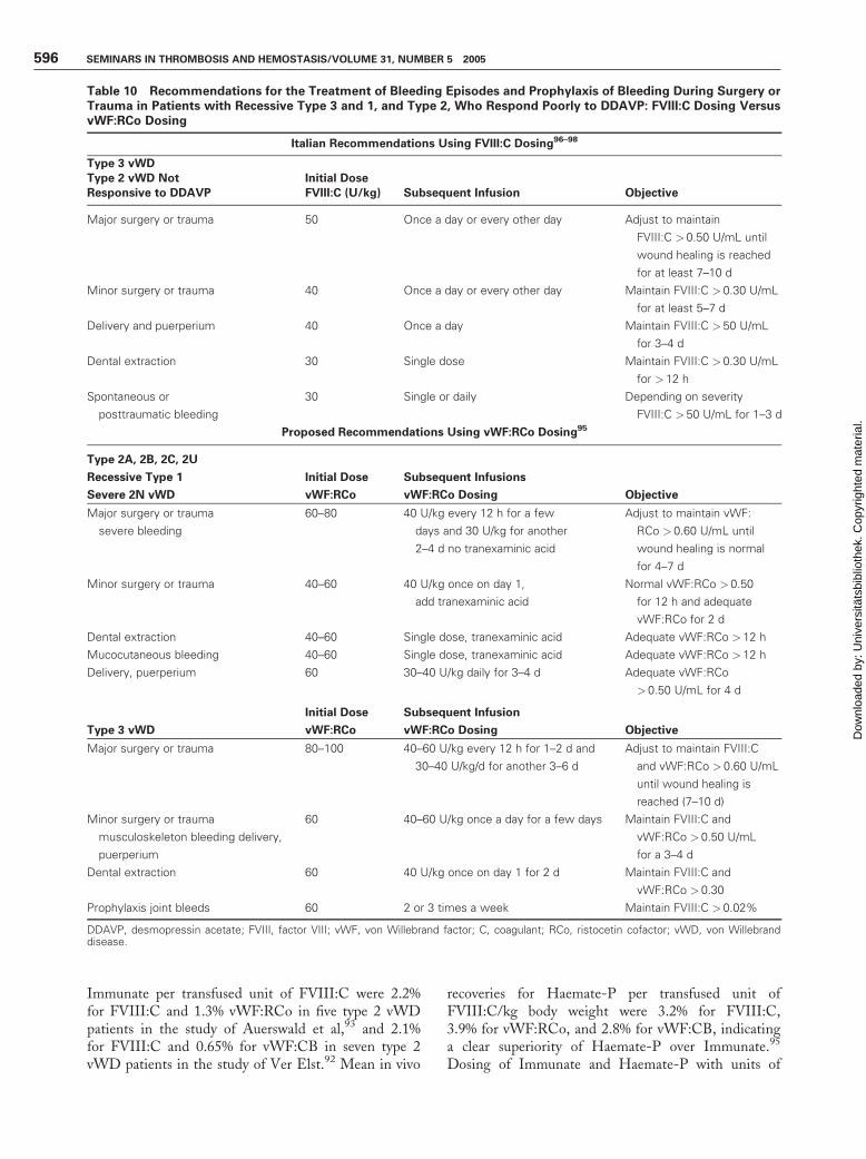
Table 10 Recommendations for the Treatment of Bleeding Episodes and Prophylaxis of Bleeding During Surgery orTrauma in Patients with Recessive Type 3 and 1, and Type 2, Who Respond Poorly to DDAVP: FVIII:C Dosing VersusvWF:RCo Dosing
Italian Recommendations Using FVIII:C Dosing96–98
Type 3 vWD
Type 2 vWD Not Initial Dose
Responsive to DDAVP FVIII:C (U/kg) Subsequent Infusion Objective
Major surgery or trauma 50 Once a day or every other day Adjust to maintain
FVIII:C >0.50 U/mL until
wound healing is reached
for at least 7–10 d
Minor surgery or trauma 40 Once a day or every other day Maintain FVIII:C > 0.30 U/mL
for at least 5–7 d
Delivery and puerperium 40 Once a day Maintain FVIII:C > 50 U/mL
for 3–4 d
Dental extraction 30 Single dose Maintain FVIII:C > 0.30 U/mL
for >12 h
Spontaneous or
posttraumatic bleeding
30 Single or daily Depending on severity
FVIII:C >50 U/mL for 1–3 d
Proposed Recommendations Using vWF:RCo Dosing95
Type 2A, 2B, 2C, 2U
Recessive Type 1 Initial Dose Subsequent Infusions
Severe 2N vWD vWF:RCo vWF:RCo Dosing Objective
Major surgery or trauma
severe bleeding
60–80 40 U/kg every 12 h for a few
days and 30 U/kg for another
2–4 d no tranexaminic acid
Adjust to maintain vWF:
RCo > 0.60 U/mL until
wound healing is normal
for 4–7 d
Minor surgery or trauma 40–60 40 U/kg once on day 1,
add tranexaminic acid
Normal vWF:RCo >0.50
for 12 h and adequate
vWF:RCo for 2 d
Dental extraction 40–60 Single dose, tranexaminic acid Adequate vWF:RCo > 12 h
Mucocutaneous bleeding 40–60 Single dose, tranexaminic acid Adequate vWF:RCo > 12 h
Delivery, puerperium 60 30–40 U/kg daily for 3–4 d Adequate vWF:RCo
>0.50 U/mL for 4 d
Type 3 vWD
Initial Dose
vWF:RCo
Subsequent Infusion
vWF:RCo Dosing Objective
Major surgery or trauma 80–100 40–60 U/kg every 12 h for 1–2 d and
30–40 U/kg/d for another 3–6 d
Adjust to maintain FVIII:C
and vWF:RCo > 0.60 U/mL
until wound healing is
reached (7–10 d)
Minor surgery or trauma
musculoskeleton bleeding delivery,
puerperium
60 40–60 U/kg once a day for a few days Maintain FVIII:C and
vWF:RCo > 0.50 U/mL
for a 3–4 d
Dental extraction 60 40 U/kg once on day 1 for 2 d Maintain FVIII:C and
vWF:RCo > 0.30
Prophylaxis joint bleeds 60 2 or 3 times a week Maintain FVIII:C > 0.02%
DDAVP, desmopressin acetate; FVIII, factor VIII; vWF, von Willebrand factor; C, coagulant; RCo, ristocetin cofactor; vWD, von Willebranddisease.
596 SEMINARS IN THROMBOSIS AND HEMOSTASIS/VOLUME 31, NUMBER 5 2005
Dow
nloa
ded
by: U
nive
rsitä
tsbi
blio
thek
. Cop
yrig
hted
mat
eria
l.
vWF:RCo/kg to reach an equal 1.7% in vivoincrease of vWF:RCo will induce an increase in FVIII:Cof more than 4.2% for Immunate and only 1.3%for Haemate-P (Table 9). These in vivo data aredirectly related to their in vitro characteristics of theabsence of high- and intermediate-vWF multimersin Immunate and the presence of the large-vWFmultimers in Haemate-P, and with the ratio ofvWF:RCo to FVIII:C of 0.5 in Immunate92,93 and 2.2in Hemate-P.95
The comparative analysis of data in Table 9demonstrate that treatment of vWD patients withAlphanate94 or Immunate92,93 using FVIII:C dosingwill lead to undertreatment with regard to vWF:RCo,and that treatment with Alphanate or Immunate usingvWF:RCo dosing in units per kilogram will result inovertreatment with regard to FVIII:C. When comparingthe shortened half-life times for vWF:RCo of 6.5 to7.1 hours after transfusion of Alphanate94 versus ap-proximately 12 hours for Haemate-P,95 it becomesclear to clinicians that sufficient hemostatic levels ofvWF:RCo persist with Haemate-P for a much longertime. For repeated doses (e.g., major surgery in vWD),clearance becomes even more important, leading toa significantly lower amount of overall Haemate-Prequired when compared with Alphanate.
The Italian recommendations in Table 1096–98 forthe treatment of severe types 1, 2, and 3 vWD that arenot responsive to DDAVP using FVIII:C dosing isproblematic and has different implications for the vari-ous FVIII/vWF concentrates; reaching the recom-mended adequate FVIII:C levels, with regard tovWF:RCo levels, will result in either overtreatmentwhen using Haemate-P (with a vWF:RCo/FVIII:Cratio of 2.1 to 2.5) or to undertreatment when usingAlphanate or Immunate (with a vWF:RCo/FVIII:Cratio of 0.625 and 0.5, respectively). Conversely, giventhat the average FVIII:C/vWF:RCo ratio is 1.6 forAlphanate94 and 2 for Immunate,92,93 treatment ofvWD patients with the recommended dose of 60 Uof vWF:RCo/kg to reach normal values for vWF:RCo ofapproximately 1.0 to 1.2 U/mL, will result in muchhigher and very high levels of FVIII:C above 2.0 U/mLup to 3.0 to 4.0 U/mL for both Alphanate94 andImmunate.92,93,96 Such high FVIII:C levels for morethan 1 week after treatment with FVIII/vWF concen-trate for major surgery in vWD patients is a plausibleexplanation for the increased risk of postoperative ve-nous thrombosis. Because the average FVIII:C/vWF:RCo ratio is 0.40 to 0.45 for Hemate-P,95 treat-ment of vWD patients with the recommended dose of60 U of vWF:RCo/kg to reach normal values forvWF:RCo of approximately 1.0 to 1.2 U/mL will resultin much lower recoveries of FVIII:C during prolongedprophylaxis after major surgery or trauma in type 2 andtype 3 vWD patients.99–101
Given that the functional vWF parametersvWF:RCo and vWF:CB are very low, and the levels ofvWF:Ag and FVIII:C are just below or in the normalrange in type 2 vWD (Figs. 3, 4, and 6), the Italianrecommendation using FVIII:C dosing cannot be ap-plied for these type 2 vWD patients. vWF:RCo dosingshould be used in type 2 vWD for proper monitoring ofthe effect of the various FVIII/vWF concentrates for thetreatment of bleeding episodes and bleeding prophylaxisduring and after surgery or trauma. Therefore, wepropose adjusted recommendations to dose FVIII/vWF concentrates in units of vWF:RCo/kg body weightfor the treatment and prophylaxis of bleeding in type 2vWD with a poor response to DDAVP, and extended itto recessive type 1 and type 3 vWD (Table 10).95 Theadjusted recommendations are tailored for the severityof bleeding complications, for the type of surgery (minoror major), and for type 2 and type 3 vWD patients(Table 10).95 Evaluation of these recommendations inprospective management studies together with labelingof the hemostatic potency of FVIII/vWF concentratesby the content of FVIII:C, vWF:Ag, vWF:RCo,vWF:CB, and vWF multimeric patterns are warrantedand are of enormous importance.
Finally, Goudemand et al102 evaluated the phar-macokinetic response of one loading dose of high-purityvWF concentrate (63 U/kg vWF:RCo and 4 U/kgFVIII:C) in one patient with type 3 vWD. Maximumlevels of vWF:RCo and vWF:Ag were observed 1 hourafter infusion, whereas maximum levels of FVIII:C wereattained from 6 to 12 hours after the first infusion. Thebiological half-life times (T1/2) of FVIII:C, vWF:Ag,and vWF:RCo were 74, 21, and 18 hours, respectively.In subsequent pharmacokinetic studies in type 3 vWDpatients infused with purified vWF product (LFB; Lille,France), 10 infusion studies at doses of 50 or 100 U ofvWF:RCo/kg body weight revealed that FVIII:C syn-thesized (was bound to the infused vWF) at 0.6 U/dL/hand decayed with a half-life of 16 hours, that the infusedvWF:Ag and vWF:RCo decayed with a half-life of12 hours.103,104 Wilfactin (LFB, Lille, France), a veryhigh purity (VHP) vWF concentrate, has been demon-strated to be effective in type 2 and type 3 vWD patients.A loading dose of 50 to 60 U of vWF:RCo/kg resulted invWF:RCo levels of> 1.00 U/mL followed by correctionof FVIII:C to> 0.50 after 12 hours. Surgery was coveredwith an infusion, 1 hour preoperatively (mean initialdose 51 to 55 U of vWF:RCo/kg), in type 2 vWD (withFVIII:C > 0.20 to > 0.30 U/mL) for surgery. The meanpostinfusion levels were 1.00 U/mL for vWF:RCo and> 0.50 U/mL for FVIII:C. Patients with FVIII:C below0.20 to 0.30 U/mL received either one infusion of VHPvWF concentrate given 12 to 24 hours preoperatively,or one infusion of FVIII 1 hour preoperatively. ThevWF:RCo was increased to > 1.00 U/mL and FVIII:Cwas > 0.60 in all patients. By maintaining vWF:RCo at
OVERVIEW OF VON WILLEBRAND DISEASES/MICHIELS ET AL 597
Dow
nloa
ded
by: U
nive
rsitä
tsbi
blio
thek
. Cop
yrig
hted
mat
eria
l.
approximately 1.00 U/mL for 1 to 16 days, depending onthe procedure, no additional FVIII was required and allpatients had adequate hemostasis.105
REFERENCES
1. Goodeve A, Peake I. A standard nomenclature for vonWillebrand factor gene mutations and polymorphisms.Baillieres Clin Haematol 2001;14:235–240
2. Sadler JE. A revised classification of von Willebrand disease.Baillieres Clin Haematol 1994;71:520–525
3. Sadler JE, Mannucci PM, Berntorp E, et al. Impact,diagnosis and treatment of vonWillebrand Disease. ThrombHaemost 2000;84:160–174
4. Zhang Z, Lindstedt M, Blomback M, Anvret M. Effects ofmutant von Willebrand factor gene in von Willebranddisease. Hum Genet 1995;96:388–394
5. Schneppenheim R, Krey S, Bergmann F, et al. Geneticheterogeneity of severe vonWillebrand disease type III in theGerman population. Hum Genet 1994;94:640–652
6. Eikenboom JCJ. Congenital von Willebrand disease type 3:clinical manifestations, pathophysiology and molecularbiology. Baillieres Clin Haematol 2001;14:365–379
7. Lak M, Peyvandi F, Mannucci PM. Clinical manifestationsand complications of childbirth and replacement therapy in385 Iranian patients with type 3 vonWillebrand disease. Br JHaematol 2000;111:1236–1239
8. Eikenboom JCJ, Castaman G, Vos HL, Bertina RM,Rodeghiero FL. Characterisation of genetic defects in re-cessive type 1 and type 3 von Willebrand disease patients ofItalian origin. Thromb Haemost 1998;79:709–717
9. Allen S, Abuzenadah AM, Hinks J, et al. A novelvon Willebrand disease-causing mutation (Arg273Trp) inthe vonWillebrand factor propeptide that results in defectivemultimerisation and secretion. Blood 2000;96:560–568
10. Castaman G, Lattuada A, Mannucci PM, Rodeghiero F.Factor VIII:C increases after desmopressin in a subgroup ofpatients with autosomal recessive severe von Willebranddisease. Br J Haematol 1995;89:147–151
11. Castaman G, Eikenboom JCJ, Lattuada A, Manucci PM,Rodeghiero F. Heightened proteolysis of the vonWillebrandfactor subunit in patients with von Willebrand diseasehemizygous or homozygous for the C2364F mutation. Br JHaematol 2000;108:188–190
12. Castaman G, Novella E, Castiglia E, Eikenboom JCJ,Rodeghiero F. A novel family with recessive von Willebranddisease due to compound heterozygosity for a splice sitemutation and a missense mutation in the von Willebrandfactor gene. Thromb Res 2002;105:135–138
13. Schneppenheim R, Budde U, Obser T, et al. Expression andcharacterization of von Willebrand factor dimerizationdefects in different types of von Willebrand disease. Blood2001;97:2059–2066
14. Eikenboom JCJ, Reitsma PH, Peerlinck KJM, Briet E.Recessive inheritance of von Willebrand’s disease. Lancet1993;341:982–986
15. Gill JC, Endres-Brooks J, Bauer PJ, Marks WJ, Montgom-ery JR. The effect of ABO bloodgroup on the diagnosis ofvon Willebrand dsease. Blood 1987;69:1691–1695
16. Sadler JE. Von Willebrand disease type 1: a diagnosis insearch of a disease. Blood 2003;101:2089–2093
17. Sadler JE. New concepts in von Willebrand Disease. AnnuRev Med 2005;56:173–191
18. Coughlan TC, Blagg JL, Alulola M, et al. Null alleles arenot a common cause of type 1 von Willebrand disease in theBritish population. Thromb Haemost 1999;82:1373–1375
19. O’Brien LA, James PD, Hemophilia Clinical DirectorsCanada, Lillicrap D. Founder von Willebrand factorhaplotype associated with type 1 von Willebrand disease.Blood 2003;102:549–557
20. Bowen D. Type 1 von Willebrand disease: a possible novelmechanism. Blood Coagul Fibrinolysis 2004;14(suppl 1):S21–S23
21. Bowen D, Collins PW, Lester W, et al. The prevalence ofthe cysteine 1584 variant of von Willebrand factor isincreased in type 1 von Willebrand disease: co-segregationwith increased susceptibility to 13 proteolysis but not clinicalphenotype. Br J Haematol 2005;128:830–836
22. Michiels JJ, Van De Velde A, Van Vliet HHDM, Van DerPlanken M, Schroyens W, Berneman ZN. Response of vonWillebrand factor parameters to desmopressin in patientswith type 1 and type 2 congenital von Willebrand disease:diagnostic and therapeutic implications. Semin ThrombHemost 2002;28:111–131
23. Eikenboom JCJ, Castaman G, Kamphuisen PW, RosendaalFR, Bertina RM. The factor VIII/von Willebrand factorratio discriminates between reduced synthesis and increasedclearance of von Willebrand factor. Thromb Haemost 2002;87:252
24. Eikenboom JCJ, Matsushita T, Reitsma PH, et al. Domi-nant type 1 von Willebrand disease caused by mutatedcysteine residues in the D3 domain of vonWillebrand factor.Blood 1996;88:2433–2441
25. Bodo I, Katsumi A, Tuley EA, Eikenboom JCJ, Dong Z,Sadler JE. Type 1 von Willebrand disease mutationCys1149Arg causes intracellular retention and degradationof heterodimers: a possible general mechanism for dominantmutations of oligomeric proteins. Blood 2001;98:2973–2978
26. Castaman G, Eikenboom JCJ, Missiaglia E, Rodeghiero F.Autosomal dominant type 1 von Willebrand disease due toG3639T mutation (C1130) in exon 26 of von Willebrandgene: description of five Italian families and evidence for afounder effect. Br J Haematol 2000;108:876–879
27. Tjernberg P, Vos HL, Castaman G, Bertina RM,Eikenboom JCJ. Dimerization and multimerization defectsof von Willebrand factor due to mutated cysteine residues.J Thromb Haemost 2004;2:257–265
28. Casana P, Martinez F, Haya S, Espinos C. Association ofthe 3467C>T mutation (T1156M) in the von Willebrand’sfactor gene with dominant type 1 von Willebrand’s disease.Ann Hematol 2001;80:381–383
29. Federici AB, De Groot PhG, Moia M, Ijssedijk MJW,Sixma JJ, Mannucci PM. Type 1 von Willebrand disease,subtype ‘platelet low’: decreased platelet adhesion can beexplained by low synthesis of von Willebrand factor inendothelial cells. Br J Haematol 1993;83:88–93
30. Nitu-Whalley IC, Ridell A, Lee C, et al. Identification oftype 2 von Willebrand disease in previously diagnosed type 1patients: a reappraisal using phenotypes, genotypes andmolecular modelling. Thromb Haemost 2000;84:998–1004
31. Mannucci PM, Lombardi R, Castaman G, et al. VonWillebrand disease ‘‘Vicenza’’ with larger-than-normal(Supranormal) von Willebrand factor multimers. Blood1988;71:65–70
598 SEMINARS IN THROMBOSIS AND HEMOSTASIS/VOLUME 31, NUMBER 5 2005
Dow
nloa
ded
by: U
nive
rsitä
tsbi
blio
thek
. Cop
yrig
hted
mat
eria
l.
32. Casonato A, Pontara E, Sartorelo F, et al. Reduced vonWillebrand factor survival in type Vicenza von Willebranddisease. Blood 2002;99:180–184
33. Cattaneo M, Federici AB, Lecchi A, et al. Evaluation ofthe PFA-1000 system in the diagnosis and therapeuticmonitoring of patients with vonWillebrand disease. ThrombHaemost 1999;82:35–39
34. Schneppenheim R, Federici A, Budde U, et al. vonWillebrand disease type 2M ‘‘Vicenza’’ in Italian andGerman patients: identification of the first candidate muta-tion (G3864A; R1205H) in 8 families. Thromb Haemost2000;82:136–140
35. Michiels JJ, Berneman Z, Gadisseur A, et al. Characteriza-tion of recessive severe type 1 and 3 von Willebrand disease(VWD), asymptomatic heterozygous carriers versus blood-group O related von Willebrand factor deficiency anddominant type 1 VWD. Clin Applied Thromb Hemost2005; (in press)
36. De Wit TR, Van Mourik JA. Biosynthesis, processing andsecretion of von Willebrand factor: biological implications.Baillieres Clin Haematol 2001;14:241–255
37. Ruggeri ZM. Structure of von Willebrand factor anditsfunction in platelet adhesion and thrombus formation.Baillieres Clin Haematol 2001;14:257–279
38. Zimmerman ThS, Dent JA, Ruggeri ZM, Nannini LH.Subunit composition of plasma vonWillebrand factor. J ClinInvest 1986;77:947–951
39. Schneppenheim R, Budde U, Ruggeri ZM. A molecularapproach to the classfication of von Willebrand disease.Baillieres Clin Haematol 2001;14:281–298
40. Budde U, Scheneppenheim R. Phenotypic and genotypicdiagnosis of von Willebrand disease: a 2004 update. SeminHematol 2005;42:15–28
41. Nichols W, Ginsburg D. von Willebrand disease. Medicine1997;76:1–20
42. Meyer D, Fressinaud E, Gaucher C, et al. Gene defects in150 unrelated French cases with type 2 von Willebranddisease: from the patient to the gene. Thromb Haemost1997;78:451–456
43. Meyer D, Fressinaud E, Hilbert L, Ribba A-S, LavergneJ-M, Mazurier C. Type 2 von Willebrand disease causingdefective vonWillebrand factor-dependent platelet function.Baillieres Clin Haematol 2001;14:349–364
44. Castaman G, Federici AB, Rodeghiero F, Mannucci PM.von Willebrand’s disease in the year 2003: towards thecomplete indentification of gene defects for correct diagnosisand treatment. Haematologica 2003;88:94–108
45. Lyons SE, Bruck ME, Bowie EJW, Ginsburg D. Impairedintracellular transport by a subset of type IIA vonWillebranddisease mutations. J Biol Chem 1992;267:4424–4430
46. Batlle J, Lopez-Fernandez MF, Campos M, et al. Theheterogeneity of type IIA von Willebrand’s disease. Blood1986;68:1207–1212
47. Batlle J, Lassieri J, Villamor AF, et al. Proteolyticprocessing of von Willebrand factor subunit: heterogeneityin type IIA von Willebrand disease. Ann Hematol 1994;68:111–115
48. Howard MA, Salem HH, Thomas KB, et al. Variantvon Willebrand’s disease type B—revisited. Blood 1982;60:1420–1428
49. Rabinowitz I, Tuley EA, Mancuso DJ, et al. von Willebranddisease type B: a missense mutation selectively abolishesristocetine-induced von Willebrand binding to platelet
glycoprotein Ib. Proc Natl Acad Sci USA 1992;89:9846–9849
50. Firkin B, Firkin F, Stott L. von Willebrand’s disease type B:a newly defined bleeding diathesis. Aust N Z J Med 1973;3:225–229
51. Mannucci PM, Lombardi R, Bader R, et al. Heterogeneity oftype I von Willebrand disease: evidence for a subgroup withabnormal von Willebrand factor. Blood 1985;66:796–802
52. Federici AB, Canciani MT, Forza I, Cozzi G. Ristocetincofactor and collagen binding activities normalized toantigen levels for a rapid diagnosis of type 2 von Willebranddisease. Thromb Haemost 2000;84:1127–1128
53. Mancusco DJ, Kroner PA, Christopherson PM, Vokac EA,Gill JC, Montgomery RR. Type 2M:Milwaukee-1 vonWillebrand disease with an in-frame deletion in the Cys509-Cys695 loop of the von Willebrand factor A1 domain causesdeficient binding of vonWillebrand to platelets. Blood 1996;88:2559–2568
54. Hillery CA, Mabcusco DJ, Sadler JE, et al. Type 2Mvon Willebrand disease: F606I and I662F mutations inthe glycoprotein Ib binding domain selectively impairristocetin—but not botrocetin-mediated binding of vonWillebrand factor to platelets. Blood 1998;91:1572–1581
55. Hilbert L, Jenkins PV, Gaucher G, et al. Type 2M vWDresulting from a lysine deletion within a four lysine residuerepeat in the A1 loop of von Willebrand factor. ThrombHaemost 2000;84:188–194
56. Michiels JJ, Berneman Z, Gadisseur A, et al. Classificationand characterization of type 2A, 2B, 2C, 2E, 2D, 2M, 2Nand 2U (unclassifiable) von Willebrand diseases. Clin ApplThromb Hemost 2005; (in press)
57. Lethagen S, Frick K, Isaksson C, Kristoffersson A-C,Holmberg L. Revised classification and treatment of vonWillebrand disease. Thromb Haemost 1998;80:199–200
58. Hoyer LW, Rizza CR, Tuddenham GD, Carta CA,Armitage H, Rotblatt F. von Willebrand factor multimerpatterns in vonWillebrand’s disease. Br J Haematol 1983;55:493–507
59. Castaman G, Eikenboom JCJ, Rodeghiero F, Briet E,Reitsma PH. A novel candidate mutation (Arg611!His) intype I ‘platelet discordant’ von Willebrand disease withdesopressine-induced thrombocytopenia. Br J Haematol1995;89:656–658
60. Hilbert L, Gaucher C, Mazurier C. Identification of twomutations (Arg611Cys and Arg611His) in the A1 loop ofvon Willebrand factor (VWF) responsible for type 2 vonWillebrand disease with decreased platelet-dependent func-tion of VWF. Blood 1995;86:1010–1018
61. Nishikubo T, Christophe O, Lavergne J-M, et al. Abnormalproteolytic processing of von Willebrand factor Arg611Cysand Arg611His. Thromb Haemost 1997;77:174–182
62. Ribba AN, Hilbert L, Lavergne JM, et al. The Arg552Cys(R552C) mutation within the A1 loop of von Willebrandfactor (VWF) induces abnormal folding with loss-of-function resulting in 2A-like phenotype of von Willebranddisease. Study of ten patients and of mutated recombinantVWF. Blood 2001;97:952–959
63. Ruggeri ZM, Nilsson IM, Lombardi R, Holmberg L,Zimmerman TS. Aberrant multimeric structure of vonWillebrand factor in a new variant of von Willebrand’sdisease (type IIC). J Clin Invest 1982;70:1124–1127
64. Holmberg L, Karpman D, Isakson C, Kristofferson AC,Lethagen S, Schneppenheim R. Ins405AsnPro mutation in
OVERVIEW OF VON WILLEBRAND DISEASES/MICHIELS ET AL 599
Dow
nloa
ded
by: U
nive
rsitä
tsbi
blio
thek
. Cop
yrig
hted
mat
eria
l.
the von Willebrand factor propeptide in recessive type 2A(IIC) von Willebrand’s disease. Thromb Haemost 1998;79:718–722
65. Mazurier C, Manucci PM, Parquet-Gernez A, GoudemandM, Meyer D. Investigation of a case of subtype IIC vonWillebrand disease: characterisation of the variability of thissubtype. Am J Hematol 1986;22:301–311
66. Gaucher C, Dieval J, Mazurier C. Characterization of vonWillebrand factor gene defects in two unrelated patients withtype IIC von Willebrand disease. Blood 1994;84:1024–1030
67. Batlle J, Lopez-Fernandez MF, Lasiera J, et al. vonWillebrand disease type IIC with different abnormalities ofvon Willebrand factor in the same sibship. Am J Hematol1986;21:177–188
68. Batlle J, Lopez Gernandez MF, Fernandez Villamor A,Lopez Berges C, Zimmerman TS. Multimeric patterndiscrepancy between platelet and plasma von Willebrandfactor in type IIC von Willebrand disease. Am J Hematol1986;22:87–88
69. Schneppenheim R, Thomas KB, Krey S, et al. Identificationof a candidate missense mutation in a family withvon Willebrand disease type IIC. Hum Genet 1995;95:681–686
70. Mannucci PM, Lombardi R, Pareti FI, Solinas S, Mazzuc-coni MG, Mariani G. A variant of von Willebrand’s diseasecharacterized by recessive inheritance and missing tripletstructure of von Willebrand factor multimers. Blood 1983;62:1000–1005
71. Kinoshita S, Harrison J, Lazerson J, Abildgaard CF. A newvariant of dominant type II von Willebrand’s disease withaberrant multimeric pattern of factor VIII-related antigen(type IID). Blood 1984;63:1369–1371
72. Schneppenheim R, Brassard J, Krey S, et al. Defectivedimerization of von Willebrand factor subunits due to aCys!Arg mutation in type IID von Willebrand disease.Proc Natl Acad Sci USA 1996;93:3581–3586
73. Schneppenheim R, Budde U, Obser T, et al. Expression andcharacterization of von Willebrand factor dimerizationdefects in different types of von Willebrand disease. Blood2001;97:2059–2066
74. Baillod P, Gaucher C, Affolter B, Mazurier C, PflugshauptR. New variant of type II von Willebrand’s disease withstructural abnormality of plasma von Willebrand factor in apatient with very mild bleeding history. Am J Hematol1995;49:21–28
75. Gaucher C, Parquet A, Baillod P, Hanss M, Mazurier C.Mutations in the D3 domain of von Willebrand factor areidentified in patients classified as 1 or 2A von Willebranddisease. Thromb Haemost 1997;(suppl):388 (abst OC-1583)
76. Castaman G, Rodeghiero F, Lattuada A, Mannucci PM. Anew variant of von Willebrand disease (type II I) with anormal degree of proteolytic cleavage of von Willebrandfactor. Thromb Res 1992;65:343–351
77. Ledford M, Rabinowitz I, Sadler JE, Kent JW, Civantos F.New variant of vonWillebrand disease type II with markedlyincreased levels of von Willebrand antigen and dominantmode of inheritance: von Willebrand disease type IICMiami. Blood 1993;82:169–175
78. Mannucci PM, Lombardi R, Federici AB, Dent JA,Zimmerman TS, Ruggeri ZM. A new variant of type IIvonWillebrand disease with aberrant multimeric structure ofplasma but not platelet von Willebrand factor (type IIF).Blood 1986;68:269–274
79. Gralnick HR, Williams SB, McKeown LP, Maisonneuve P,Jeneau C, Sultan Y. A variant of type II von Willebranddisease with an abnormal triplet structure and discordanteffects of protease inhibitors on plasma and platelet vonWillebrand factor structure. Am J Hematol 1987;24:259–266
80. Federici AB, Manuccio PM, Lombardi R, et al. Type II Hvon Willebrand disease: new structural abnormality ofplasma and platelet von Willebrand factor in a patients withprolonged bleeding time and borderline levels of ristocetincofactor activity. Am J Hematol 1989;32:287–293
81. Mazurier C. von Willebrand disease masquerading ashaemophilia A. Thromb Haemost 1992;67:391–396
82. Mazurier C, Goudemand J, Hilbert L, Caron C, FressinaudE, Meyer D. Type 2N von Willebrand disease: clinicalmanifestations, pathophysiology. Laboratory diagnosis andmolecular biology. Baillieres Clin Haematol 2001;14:337–347
83. Schneppenheim R, Budde U, Krey S, et al. Results of ascreening for von Willebrand disease type 2N in patientswith suspected haemophilia A or von Willebrand diseasetype 1. Thromb Haemost 1996;76:598–602
84. Mazurier C, Gaucher C, Joriuex S, Goudemand M.Biological effect of desmopressin in eight patients with type2N (‘Normandy’) von Willebrand disease. Br J Haematol1994;88:849–854
85. Federici AB, Mazurier C, Berntorp E, et al. Biologicresponse to desmopressin in patients with severe type 1and type 2 von Willebrand disease: results of a multicenterEuropean study. Blood 2004;103:2032–2038
86. Mannucci PM, Moia M, Rebulla P, Altieri D, MonteagudaJ, Castillo R. Correction of the bleeding time in treatedpatients with severe von Willebrand disease is not solelydependent on the normal multimeric structure of plasma vonWillebrand factor. Am J Hematol 1987;25:55–65
87. Castillo R, Monteaguda J, Escolar G, Ordinas A, MagallonM, Villar JM. Hemostatic effect of normal platelet transfu-sion in severe von Willebrand disease patients. Blood 1991;77:1901–1905
88. Mannucci PM. Recommended protocol for the study of exvivo biological effects of virus-inactivated plasma concen-trates with von Willebrand disease. Thromb Haemost 1992;68:84–88
89. Mannucci PM, Tenconi PM, Castaman G, Rodeghiero R.Comparison of four virus-inactivated plasma concentratesfor treatment of severe von Willebrand disease: a cross-overrandomized trial. Blood 1992;79:3130–3137
90. Lethagen S, Berntorp E, Nilsson IM. Pharmacokinetics andhemostatic effect of different factor VIII-von Willebrandfactor concentrates in von Willebrand’s disease type III. AnnHematol 1992;65:253–259
91. Menache D, Aronson DL, Darr F, Montgomery RR,The Cooperative Study Group. Pharmacokinetics of vonWillebrand factor and factor VIIIC in patients with severevon Willebrand disease (type 3): estimation of the rate offactor VIIIC synthesis. Br J Haematol 1996;94:740–745
92. Ver Elst K, Van Vliet HHDM, Kappers-Klunne MC,Leebeek FWG. In vitro studies, pharmacokinetic studies andclinical use of a high purity double virus inactivated FVIII/VWF concentrate (Immunate) in the treatment of vonWillebrand disease. Thromb Haemost 2004;92:67–74
93. Auerswald G, Eberspacher B, Engl W, et al. Successfultreatment of patients with von Willebrand disease using
600 SEMINARS IN THROMBOSIS AND HEMOSTASIS/VOLUME 31, NUMBER 5 2005
Dow
nloa
ded
by: U
nive
rsitä
tsbi
blio
thek
. Cop
yrig
hted
mat
eria
l.
a high-purity double-virus inactivated factor VIII/vonWillebrand factor concentrate (Immunate). Semin ThrombHemost 2002;28:203–213
94. Mannucci PM, Chediak J, Bymes W, et al. Treatment ofvon Willebrand disease with high-purity factor VIII/vonWillebrand factor concentrate: a prospective, multicenterstudy. Blood 2002;99:450–456
95. Michiels JJ, Berneman ZN, Van Der Planken M, SchroyensW, Budde U, Van Vliet HHDM. Bleeding prophylaxis formajor surgery in patients with type 2 von Willebrand diseasewith an intermediate purity factor VIII-von Willebrandfactor concentrate (Haemate-P). Blood Coagul Fibrinolysis2004;15:323–330
96. Mannucci PM, Federici AB. Management of inherited vonWillebrand disease. Baillieres Clin Haematol 2001;14:455–462
97. Mannucci PM. Treatment of von Willebrand’s Disease.N Engl J Med 2004;351:683–694
98. Rodeghiero F, Castaman G. Treatment of von Willebranddisease. Semin Hematol 2005;42:29–35
99. Pasi KJ, Collins PW, Keeling DM, et al. Management of vonWillebrand disease: a guideline from the UK HaemophiliaCentre Doctor’s Organization. Haemophilia 2004;10:218–231
100. Gill JC, Ewenstein BM, Thompson AR, Mueller-VeltensG, Schwartz BA. The Humate-P Study Group. Successful
treatment of urgent bleeding in von Willebrand disease withfactor VIII/VWF concentrate (VWF:RCo) to measurepotency and to guide therapy. Haemophilia 2003;9:688–695
101. Thompson AR, Gill JC, Ewenstein BM, Mueller-VeltensG, Schwartz BA. Successful treatment for patients with vonWillebrand disease undergoing urgent surgery using factorVIII/VWF concentrate (Humate-P). Haemophilia 2004;10:42–51
102. Goudemand J, Mazurier C, Marey A, et al. Clinical andbiological evaluation in von Willebrand’s disease of a vonWillebrand factor concentrate with low factor VIII activity.Br J Haematol 1992;80:214–221
103. Menache D, Aronson DL, Darr F, Montgomery RR.The Cooperative Study Group. Pharmacokinetics of vonWillebrand factor and factor VIIIC in patients withsevere von Willebrand disease (type 3): estimation of therate of factor VIII synthesis. Br J Haematol 1996;94:740–745
104. Menache D, Aronson DL. New treatments of vonWillebrand disease: plasma derived von Willebrand factorconcentrates. Thromb Haemost 1997;78:566–570
105. Goudemand J, Negrier C, Ounmnoughene N, Sultan Y.Clinical management of patients with von Willebrand’sdisease with a VHP VWF concentrate: the Frenchexperience. Haemophilia 1998;4(suppl 3):48–52
OVERVIEW OF VON WILLEBRAND DISEASES/MICHIELS ET AL 601
Dow
nloa
ded
by: U
nive
rsitä
tsbi
blio
thek
. Cop
yrig
hted
mat
eria
l.