Spatial shift of spatially modulated light projected on turbid media
Upload
independentCategory
view
2download
0
ORIGINAL ARTICLE
The von Willebrand factor self-association is modulated by amultiple domain interaction
H. ULR ICHTS , K . VANHOORELBEKE , J . P . G IRMA,* P . J . L ENT ING,� S . VAUTER IN and H . DECKMYNLaboratory for Thrombosis Research, KU Leuven Campus Kortrijk, Kortrijk, Belgium; *Inserm U143, Le Kremlin-Bicetre, France; �Laboratory for
Thrombosis and Haemostasis, Department of Haematology, University Medical Center Utrecht, Utrecht, the Netherlands
To cite this article: Ulrichts H, Vanhoorelbeke K, Girma JP, Lenting PJ, Vauterin S, Deckmyn H. The von Willebrand factor self-association is
modulated by a multiple domain interaction. J Thromb Haemost 2005; 3: 552–61.
Summary. Background: Platelet adhesion and aggregation at
sites of vascular injury exposed to rapid blood flow require
von Willebrand factor (VWF). VWF becomes immobilized by
binding to subendothelial components or by a self-association
at the interface of soluble and surface-bound VWF.
Objectives: As this self-association has been demonstrated
only under shear conditions, our first goal was to determine
whether the same interaction could be observed under static
conditions. Furthermore, we wanted to identify VWF
domain(s) important for this self-association. Results: Biotin-
ylated VWF (b-VWF) interacted dose-dependently and
specifically with immobilized VWF in an enzyme-linked
immunosorbent assay (ELISA) assay, showing that shear is
not necessary to induce the VWF self-association. Whereas
anti-VWF monoclonal antibodies (mAbs) had no effect on the
self-association, the proteolytic VWF-fragments SpII(1366–
2050) and SpIII(1–1365) inhibited the b-VWF–VWF interac-
tion by 70 and 80%, respectively. Moreover, a specific binding
of b-VWF to immobilized Sp-fragments was demonstrated.
Finally, both biotinylated SpII and SpIII were able to bind
specifically to both immobilized SpII and SpIII. Similar results
were observed under flow conditions, which confirmed the
functional relevance of our ELISA system. Conclusion: We
have developed an ELISA binding assay in which a specific
VWF self-association under static conditions can be demon-
strated. Our results suggest a multiple domain interaction
between immobilized and soluble VWF.
Keywords: von Willebrand factor, platelet adhesion,
A-domains.
Introduction
Vascular damage is followed rapidly by platelet responses
leading to the firm attachment of platelets at the site of injury.
At sites of high blood flow, the vonWillebrand factor (VWF) is
essential for this process [1]. VWF modulates platelet-surface
and platelet–platelet interactions by linking platelet receptors to
the extracellular matrix and to one another [2]. Moreover,
VWF exerts a carrier function for the plasma coagulation
factor VIII [3], protecting it thereby from proteolytic inactiva-
tion. Soluble VWF is not able to interact with non-activated
platelets. However, when immobilized to subendothelial struc-
tures, VWF forms a reactive surface capable of capturing
platelets from flowing blood [4,5]. Earlier studies showed a
shear-dependent conformational change of VWF from a
globular state to an extended chain upon immobilization [6].
However, we recently failed in showing such a large overall
conformational change but did not exclude small-scale effects
of shear on one or more VWF domains [7]. Surface properties
itself have an effect on VWF shape [8]. Therefore, the
discrepancy between these studies can reside in the different
strategy of immobilizing VWF.
Transient interactions of the platelet GPIb-IX-V receptor
complex with the A1-domain of immobilized VWF [9] result in
an initial attachment, characterized by a continuous surface
translocation of the platelets [10]. This leads to stable platelet
adhesion through the platelet collagen receptors GPVI and
GPIa/IIa [11], activation of the platelet GPIIb/IIIa receptor
complex [12] and finally to platelet aggregation [13] and
thrombus growth.
In addition to circulating in plasma, VWF is deposited into
the extracellular matrix of the subendothelium [14]. Regardless
of the presence of this subendothelial VWF, several extracel-
lular matrix components bind plasma VWF rapidly upon
exposure to circulating blood due to vascular injury. VWF
binds directly to collagen types I, III and VI, and also interacts
with collagen type IV [15,16]. In addition, VWF contains two
distinct binding sites for heparin [17,18], which could possibly
interact with matrix proteoglycans that contain sulfated
sugars. The major collagen-binding site resides in the VWF
A3-domain [19]. Sites other than the VWF A3-domain could
Correspondence: Hans Deckmyn, Laboratory for Thrombosis
Research, IRC, KU Leuven Campus Kortrijk, E. Sabbelaan 53,
8500 Kortrijk, Belgium.
Tel.: +32 56 24 64 22; fax: +32 56 24 69 97; e-mail: Hans.Deckmyn@
kulak.ac.be
Received 15 March 2004, accepted 30 November 2004
Journal of Thrombosis and Haemostasis, 3: 552–561
� 2005 International Society on Thrombosis and Haemostasis

bind to extracellular matrix components, such as the VWF
A1-domain which is the primary interaction site for collagen
type VI [16]. However, we showed that the monoclonal anti-
VWF antibody 82D6A3, which inhibits the VWF interaction
with fibrillar collagen, inhibits arterial platelet thrombus
formation in vivo in baboons [20], suggesting that VWF sites
other than the A3-domain are less significant for binding to
the extracellular matrix. Moreover, these findings confirm that
the relevant thrombogenic connective tissue ligand is fibrillar
collagen.
Upon immobilization, VWF sustains a functionally relevant
homotypic self-association with soluble VWF multimers [21],
demonstrating that direct immobilization of VWF is not
strictly needed for interaction with the platelet GPIb-IX–V
complex. This function of circulating VWF in the initiation of
platelet adhesion was demonstrated in flow chamber studies:
in spite of the fact that the A1-domain of immobilized VWF is
essential for platelet translocation under shear [5], platelets
were still able to interact with immobilized DA1-VWF when
soluble plasma VWF was present. Moreover, a shear-
dependent self-association of VWF in suspension was demo-
nstrated in a cone-plate viscometer [22]. The goal of this study
was to elucidate, first, if shear is a prerequisite for the
interaction of soluble VWF with immobilized VWF. Sec-
ondly, we wanted to identify regions in VWF important for
this self-association.
Methods
Materials
The three fragments SpI (monomer, AA911–1365), SpII
(dimer, AA1366–2050) and SpIII (dimer, AA1–1365) were
produced by digestion of purified VWF with Staphyloccocus
aureus V8 protease [23]. Amino acids were numbered from the
first amino acid of the mature VWF subunit. Different
monoclonal antibodies (mAbs) to VWF have been described
previously: mAb 82D6A3 binds to the VWF A3 domain and
inhibits VWF binding to collagen types I, III and IV [16], mAb
1C1E7 recognizes theN-terminal part of VWF (AA1–272) [24].
mAb 724 and mAb 701 recognize the VWF A1 domain. mAb
724 blocks VWF binding to botrocetin and botrocetin-induced
VWF binding to GPIb [25], while mAb 701 inhibits ristocetin-
and botrocetin-induced platelet agglutination [26]. Recombin-
ant DA3-VWF (D909-1113) and recombinant DA1-VWF
(D478–716) were purified as described previously [19,27].
VWF (Red Cross, Brussels, Belgium), SpII, SpIII, DA3-VWF
and 82D6A3 were biotinylated using EZ-link Sulfo-NHS-
SS-Biotin (Perbio, Helsingberg, Sweden). Botrocetin was
purified from crude Bothrops jararaca venom (Sigma, St Louis,
MO, USA), as described previously [28]. A recombinant
N-terminal, VWF-binding GPIba fragment (AA1-289)
(rGPIba) was expressed in Chinese hamster ovary cells and
purified as described [29,30]. Heparin (Clexane) was obtained
from Aventis (Brussels, Belgium). Fibrinogen was from
Calbiochem (La Jolla, CA, USA).
Binding of VWF and biotinylated VWF to human collagen
type III
Ninety-six-well microtiter plates (Greiner, Frickenhausen,
Germany) were coated overnight (ON) at 4 �C with 25 lgmL)1 human collagen type III (Sigma), in phosphate-buffered
saline (PBS). Wells were blocked with PBS containing 3%
milk powder and incubated with a dilution series of purified
VWF or biotinylated VWF (b-VWF). Bound VWF or
b-VWF was detected for 45 min at RT with a 1/3000 dilution
of anti-VWF polyclonal antibodies labeled with horseradish
peroxidase (anti-VWF-Ig-HRP, Dako, Glostrup, Denmark).
Visualization was obtained with ortho-phenylenediamine
(Sigma) and H2O2 and the coloring reaction was stopped
with 4 mol L)1 H2SO4, after which the absorbance was
determined at 490 nm.
Botrocetin-induced binding of b-VWF and VWF to a
recombinant fragment of GPIba
The botrocetin-induced binding of b-VWF and VWF to
rGPIba was performed as described previously [30,31]. Briefly,
microtiter plates were coated ON at 4 �C with anti-GPIbamAb 2D4 (5 lg mL)1 in PBS) and blocked with Tris-buffered
saline (TBS) containing 3%milk powder.Wells were incubated
for 2 h at 37 �Cwith rGPIba (1 lgmL)1 in TBS, 0.1%Tween-
20) and incubated for 2 h at 37 �C with a dilution series of
VWF or b-VWF in TBS, 0.1% Tween-20 containing 0.01 lgmL)1 botrocetin. Bound VWF or b-VWF was detected with
anti-VWF-Ig-HRP as described above.
Multimeric analysis
The multimeric pattern of VWF and b-VWF was determined
essentially as described [32]. Briefly, VWF and b-VWF were
separated on sodium-dodecyl sulfate (SDS) 0.8–1.5% IEF
agarose gels. Gels were fixed on Gelbond (Cambrex Bio
Science Rockland, Inc., ME, USA) and VWF was immuno-
detected using anti-VWF-Ig-HRP andH2O2 and 3–3¢ diamino-
benzidine as substrate (DAB, Sigma).
Binding of soluble biotinylated VWF to immobilized VWF,
DA3-VWF, SpI, SpII or SpIII
Ninety-six-well microtiter polystyrene plates were directly
coated ON at 4 �C with VWF, DA3-VWF, bovine serum
albumin (BSA, Sigma), human serum albumin (HSA, Sigma),
SpI, SpII or SpIII (10 lg mL)1 in PBS) and blocked for 2 h at
room temperature (RT) with PBS containing 3%milk powder.
Wells were incubated for 2 h at 37 �C with a dilution series of
b-VWF in PBS containing 0.3% milk powder. Bound b-VWF
was detected for 45 min at RT with a 1/10 000 dilution of
streptavidin-HRP (Roche Molecular Biochemicals, Mann-
heim, Germany). Visualization was performed as described
above. The binding of soluble b-BSA to immobilized BSA or
VWF was performed in parallel.
Functional VWF self-association 553
� 2005 International Society on Thrombosis and Haemostasis

Kinetic binding assays
Ninety-six-well microtiter plates were coated ON at 4 �C with
VWF and blocked as described above. For the association
experiments, wells were incubated at 37 �C for different
intervals with b-VWF (50 lg mL)1 in PBS containing 0.3%
milk powder). For the dissociation experiments, wells were first
equilibrated for 1.5 h with b-VWF (25 lg mL)1 in PBS
containing 0.3% milk powder), after which the supernatant
was removed and exchanged by a 10 · excess of unlabeled
VWF in PBS containing 0.3% milk powder. Bound b-VWF
was detected with streptavidin-HRP and visualization was
performed as described above.
Binding of soluble VWF to immobilized DA3-VWF: detection
of bound VWF with anti-VWF mAb 82D6A3
Ninety-six-well microtiter plates were coated ON at 4 �C with
DA3-VWF and blocked as described above. Wells were
incubated for 2 h at 37 �C with a dilution series of VWF in
PBS containing 0.3%milk powder, followed by the addition of
1 lgmL)1 of biotinylatedmAb 82D6A3 (b-82D6A3) for 1 h at
RT. Bound b-82D6A3 was detected with streptavidin-HRP
and visualization was performed as described above.
Inhibition of association of soluble VWF with immobilized
VWF, SpI, SpII or SpIII
Ninety-six-well microtiter plates were coated ON with VWF,
SpI, SpII or SpIII and blocked as described above. Wells were
incubated at RT with a dilution series of VWF, SpI, SpII,
SpIII, BSA, heparin, fibrinogen or the anti-VWF mAbs
1C1E7, 82D6A3, 701 or 724 in PBS containing 0.3% milk
powder to which a constant amount of b-VWFwas next added
after 30 min (final concentration of 25 lg mL)1 in PBS
containing 0.3% milk powder). After 2 h incubation at 37 �C,residual bound b-VWF was detected with streptavidin-HRP
and visualization was performed as described above.
Binding of soluble biotinylated SpII- and SpIII-fragment to
immobilized VWF, SpII- and SpIII-fragment
Ninety-six-well microtiter plates were coated ON with SpII- or
SpIII-fragment and blocked as described above. Wells were
incubated for 2 h at 37 �C with a dilution series of biotinylated
SpII- and SpIII-fragment (b-SpII and b-SpIII) in PBS
containing 0.3% milk powder. Bound b-SpII and b-SpIII
was detected with streptavidin-HRP and visualization was
performed as described above.
Inhibition of binding of soluble biotinylated SpII- and
SpIII-fragment to immobilized SpII- and SpIII-fragment by
soluble VWF
Ninety-six-well microtiter plates were coated ON with SpII-
and SpIII-fragment and blocked as described above. Wells
were incubated at RT with a dilution series of VWF in PBS
containing 0.3% milk powder to which a constant amount of
b-SpII or b-SpIII was next added after 30 min (final concen-
tration of 50 lg mL)1 in PBS containing 0.3% milk powder).
Residual bound b-SpII and b-SpIII was detected with
streptavidin-HRP and visualization was performed as des-
cribed above.
Flow studies
Glass coverslips were coated overnight at 4 �CwithDA1-VWF
or SpII fragment (40 lg mL)1 in PBS). Reconstituted perfu-
sates were prepared as described [27]. Briefly, platelet rich
plasma was obtained from whole blood by centrifugation and
platelets were washed in Krebs–Ringer buffer (4 mmol L)1
KCL, 107 mmol L)1 NaCl, 20 mmol L)1 NaHCO3, 2 mmol
L)1 Na2SO4) containing 19 mmol L)1 sodium citrate, pH 5.0.
Platelets were resuspended in Krebs–Ringer buffer containing
4% albumin to a platelet count of 190 000 lL)1. Autologous
red blood cells were washed with PBS containing 5 mM
glucose. At 5 min before perfusion, washed red blood cells
were added in a volume fraction of 40% of total volume.
Platelet adhesion was studied at 1600 s)1 at 37 �C for 5 min.
Perfusions were performed in the absence or presence of 20 lgmL)1 VWF, SpIII fragment orDA1-VWF.After perfusion, the
coverslips were rinsed in Hepes buffered saline (10 mmol L)1
Hepes, 145 mmol L)1 NaCl, pH 7.35), fixated by 0.5%
glutaraldehyde and stained with May–Grunwald/Giemsa.
Platelet adhesion was evaluated with a light microscope at
400 · magnification, connected to an Image Analyzer (Lucia,
Laboratory Imaging Ltd, Analis, Namur, Belgium). Platelet
adhesion was expressed as percentage of surface covered with
platelets. For each experiment, the mean ± SE of 10 images
was determined.
Results
Functional self-association of soluble and immobilized
VWF under static conditions
Our first goal was to determine whether soluble VWFwas able
to interact with immobilized VWF under static conditions. An
enzyme-linked immunosorbent assay (ELISA) assay was set up
in which the binding of soluble biotinylated VWF (b-VWF) to
immobilized VWF was determined. Soluble b-VWF bound
dose-dependently to immobilized VWF in this assay, demon-
strating that shear is not essential for the VWF self-association
(Fig. 1). The data were fitted using the specific binding model
characterized by the equation y ¼ Bmax x/(Kd,app + x), where
y represents the absorbance at 490 nm · the soluble b-VWF
concentration and Kd,app the apparent dissociation constant
[33]. In order to compare individual experiments, OD values
were recalculated as percentage binding using the extrapolated
parameter Bmax as 100% binding. Soluble b-VWF interacted
with a Kd,app of 93 ± 3 nmol L)1 using aMr of 270 kDa for a
VWF monomer [34]. Biotinylation had no major influence on
554 H. Ulrichts et al
� 2005 International Society on Thrombosis and Haemostasis

VWF function as neither the binding to human collagen type
III nor the botrocetin-induced binding to rGPIba was altered
significantly (Fig. 2A). Ristocetin could not be used in the assay
as amodulator as this resulted in a too high background signal.
Moreover, themultimericVWFstructurewas not influenced by
the biotinylation process (Fig. 2B).
Specificity of the VWF self-association
In order to be sure that the binding of soluble b-VWF to
immobilized VWF was not caused by putative conformational
changes induced by the biotinylation process, the binding of
soluble b-VWF and soluble VWF to immobilized DA3-VWF
was compared: bound b-VWF was detected with streptavidin-
HRP, whereas bound VWF was measured with b-mAb
82D6A3 followed by streptavidin-HRP. As mAb 82D6A3
recognizes an epitope in the A3-domain of VWF, it does not
bind to immobilized DA3-VWF. Both VWF as well as b-VWF
interacted comparably with immobilized DA3-VWF (Fig. 3A),
showing that the measured self-association of VWF in the
assay is not caused by the biotinylation.
The interaction of soluble b-VWF with immobilized VWF
under static conditions was shown to be specific as soluble
b-VWF did not interact with immobilized BSA or immobilized
HSA, nor could soluble biotinylated BSA (b-BSA) interact
with immobilized VWF (Fig. 3B). Also, no self-association of
soluble b-BSA with immobilized BSA could be demonstrated.
Moreover, the binding of soluble b-VWF to immobilized VWF
could already be inhibited by at least 80% at a 12 molar excess
of unlabeled soluble VWF, whereas soluble BSA or fibrinogen
had no effect (Fig. 3C), suggesting that the measured self-
association is specific.
Reversibility of the VWF self-association
In order to verify if the relatively rapid reversibility of the VWF
self-interaction under flow can also be observed in our ELISA
system, we determined the association rate and the dissociation
rate of the binding of b-VWF to immobilized VWF. Compar-
able experiments were performed for the binding of FVIII to
immobilized VWF [35].
100
80
60
40
% b
indi
ng
20
00 20 40 60 80
350nM
280210140
b-VWF
700
100µg/mL
Fig. 1. Binding of soluble b-VWF to immobilized VWF under static
conditions. Microtiter plates were coated with VWF and incubated
subsequently with a dilution series of soluble b-VWF after which
bound b-VWF was detected with streptavidin-HRP (n ¼ 3).
1.6A
B
1.4
1.2
1.0
0.8
OD
490
nm
0.6
0.4
0.2
0.00 2
VWF b-VWF
4VWF or b-VWF (µg/ml)
6 8 10
Fig. 2. Effect of biotinylation on VWF function and multimeric structure.
(A) Binding of VWF and b-VWF to human collagen type III and to
rGPIba. Microtiter plates were coated with (i) human collagen type III
and were incubated with a dilution series of VWF (d) or b-VWF (s) or
(ii) were coated with anti-GPIba mAb 2D4, followed by incubation with
rGPIba and incubated with a dilution series of VWF (j) or b-VWF (h) in
the presence of 0.01 lg mL)1 botrocetin. Bound VWF and b-VWF were
detected with anti-VWF-Ig-HRP. Representative of three independent
experiments. (B) Purified VWF and b-VWF were separated on SDS-IEF
agarose gels. Gels were fixed on Gelbond and VWF or b-VWF multimers
were immunostained with anti-VWF-Ig-HRP and DAB as substrate.
Functional VWF self-association 555
� 2005 International Society on Thrombosis and Haemostasis

For the association experiments, we observed the time-
dependent binding of b-VWF to immobilized VWF (Fig. 4A).
In order to compare the results of the different repeats of this
experiment, we recalculated the OD values as percentage
binding, using the OD value for the binding of b-VWF after
1.5 h incubation as 100% binding. These results demonstrated
that binding of b-VWF increased with time and that maximal
binding was reached after 45 min incubation. After 10 min
incubation, more than 50% of the maximal binding was
reached.
For the dissociation experiments, the removal of bound
b-VWF by a 10 · excess of unlabeled VWF was measured
as a function of time (Fig. 4B). Unlabeled VWF could
completely remove bound b-VWF from the surface, clearly
demonstrating the reversibility and specificity of the VWF
self-interaction measured in the ELISA assay. Comparable
with the association data, bound b-VWF was almost
completely removed after 45 min incubation with unlabeled
VWF, and after 10 min incubation more than 50% bound
b-VWF was removed.
80A
B
C
60
40
% b
indi
ngO
D 4
90nm
% in
hibi
tion
20
00
0.8
0.6
0.4
0.2
0.00
100
80
60
40
20
0VWF BSA Fibrinogen
20b-VWF or b-BSA (µg/mL)
40 60 80
20 40VWF or b-VWF (µg/mL)
60 80 100
Fig. 3. Specificity of the VWF self-association. (A) Interaction of VWF
and b-VWF with immobilized DA3-VWF. Microtiter plates were coated
with DA3-VWF and were incubated with a dilution series of VWF (j) or
b-VWF (d). Bound VWF was detected with b-82D6A3 followed by
incubation with streptavidin-HRP. Bound b-VWF was detected with
streptavidin-HRP; representative of three independent experiments. (B)
Interaction of soluble b-VWF or b-BSA with immobilized VWF or BSA.
Microtiter plates were coated with VWF (squares), BSA (circles) or HSA
(triangles) and incubated with a dilution series of soluble b-BSA (open
symbols) or b-VWF (filled symbols). Bound b-BSA or b-VWF was
detected with streptavidin-HRP; representative of three independent
experiments. (C) Inhibition of VWF self-association by soluble VWF,
BSA or fibrinogen. Microtiter plates were coated with VWF and prein-
cubated with soluble unlabeled VWF (300 lg mL)1), BSA (300 lg mL)1)
or fibrinogen (300 lgmL)1). Soluble b-VWFwas added (25 lgmL)1 final
concentration) for 1.5 h after which residual bound b-VWF was detected
with streptavidin-HRP. The percentage inhibition is shown. As 0% inhi-
bition reference, the binding of 25 lg mL)1 soluble b-VWF in the absence
of inhibitor was chosen (n ¼ 3).
125
100
100
75
50
25
0
75
% b
indi
ng%
bin
ding
50
25
0
0 30
time (min)
60 90
0 30
time (min)
60 90
Fig. 4. Binding kinetics of VWF self-association. (A) Association studies.
Microtiter plates were coated with VWF and incubated with 50 lg mL)1
soluble b-VWF for different time intervals. Bound b-VWF was detected
with streptavidin-HRP (n ¼ 4). (B) Dissociation studies. Microtiter plates
were coated with VWF and equilibrated with soluble 25 lg mL)1 b-VWF
for 1.5 h after which bound b-VWF was removed by excess unlabeled
VWF (250 lg mL)1) for different time intervals. The residual bound
b-VWF was detected with streptavidin-HRP. The percentage binding is
shown. As 100% binding reference, the binding of 25 lg mL)1 soluble
b-VWF in the absence of unlabeled VWF was chosen (n ¼ 5).
556 H. Ulrichts et al
� 2005 International Society on Thrombosis and Haemostasis
These kinetic binding experiments of b-VWF to immobilized
VWF revealed a comparable association and dissociation rate,
reflecting a rapid reversibility. Moreover, the time-scale of this
VWF self-interaction in the ELISA system is comparable with
the self-association observed under flow conditions [21].
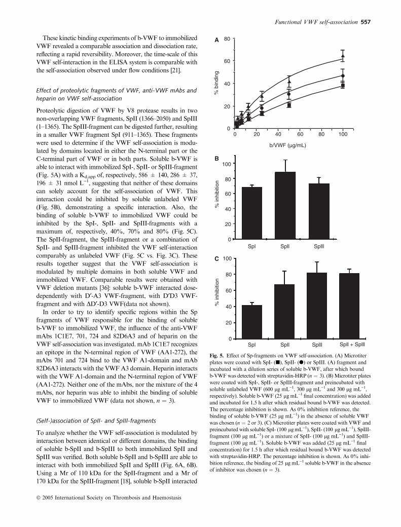
Effect of proteolytic fragments of VWF, anti-VWF mAbs and
heparin on VWF self-association
Proteolytic digestion of VWF by V8 protease results in two
non-overlapping VWF fragments, SpII (1366–2050) and SpIII
(1–1365). The SpIII-fragment can be digested further, resulting
in a smaller VWF fragment SpI (911–1365). These fragments
were used to determine if the VWF self-association is modu-
lated by domains located in either the N-terminal part or the
C-terminal part of VWF or in both parts. Soluble b-VWF is
able to interact with immobilized SpI-, SpII- or SpIII-fragment
(Fig. 5A) with a Kd,app of, respectively, 586 ± 140, 286 ± 37,
196 ± 31 nmol L)1, suggesting that neither of these domains
can solely account for the self-association of VWF. This
interaction could be inhibited by soluble unlabeled VWF
(Fig. 5B), demonstrating a specific interaction. Also, the
binding of soluble b-VWF to immobilized VWF could be
inhibited by the SpI-, SpII- and SpIII-fragments with a
maximum of, respectively, 40%, 70% and 80% (Fig. 5C).
The SpII-fragment, the SpIII-fragment or a combination of
SpII- and SpIII-fragment inhibited the VWF self-interaction
comparably as unlabeled VWF (Fig. 5C vs. Fig. 3C). These
results together suggest that the VWF self-association is
modulated by multiple domains in both soluble VWF and
immobilized VWF. Comparable results were obtained with
VWF deletion mutants [36]: soluble b-VWF interacted dose-
dependently with D¢-A3 VWF-fragment, with D¢D3 VWF-
fragment and with DD¢-D3 VWF(data not shown).
In order to try to identify specific regions within the Sp
fragments of VWF responsible for the binding of soluble
b-VWF to immobilized VWF, the influence of the anti-VWF
mAbs 1C1E7, 701, 724 and 82D6A3 and of heparin on the
VWF self-association was investigated. mAb 1C1E7 recognizes
an epitope in the N-terminal region of VWF (AA1-272), the
mAbs 701 and 724 bind to the VWF A1-domain and mAb
82D6A3 interacts with the VWFA3 domain. Heparin interacts
with the VWF A1-domain and the N-terminal region of VWF
(AA1-272). Neither one of the mAbs, nor the mixture of the 4
mAbs, nor heparin was able to inhibit the binding of soluble
VWF to immobilized VWF (data not shown, n ¼ 3).
(Self-)association of SpII- and SpIII-fragments
To analyze whether the VWF self-association is modulated by
interaction between identical or different domains, the binding
of soluble b-SpII and b-SpIII to both immobilized SpII and
SpIII was verified. Both soluble b-SpII and b-SpIII are able to
interact with both immobilized SpII and SpIII (Fig. 6A, 6B).
Using a Mr of 110 kDa for the SpII-fragment and a Mr of
170 kDa for the SpIII-fragment [18], soluble b-SpII interacted
80A
B
C
100
80
60
40
20
0
100
80
60
40
20
0
Spl Spll Splll
Spl Spll Splll Spll + Splll
60
40
% b
indi
ng%
inhi
bitio
n%
inhi
bitio
n
20
00 20 40
b/VWF (µg/mL)
60 80 100
Fig. 5. Effect of Sp-fragments on VWF self-association. (A) Microtiter
plates were coated with SpI- (j), SpII- (d) or SpIII. (A) fragment and
incubated with a dilution series of soluble b-VWF, after which bound
b-VWFwas detected with streptavidin-HRP (n ¼ 3). (B)Microtiter plates
were coated with SpI-, SpII- or SpIII-fragment and preincubated with
soluble unlabeled VWF (600 lg mL)1, 300 lg mL)1 and 300 lg mL)1,
respectively). Soluble b-VWF (25 lg mL)1 final concentration) was added
and incubated for 1.5 h after which residual bound b-VWF was detected.
The percentage inhibition is shown. As 0% inhibition reference, the
binding of soluble b-VWF (25 lg mL)1) in the absence of soluble VWF
was chosen (n ¼ 2 or 3). (C) Microtiter plates were coated with VWF and
preincubated with soluble SpI- (100 lgmL)1), SpII- (100 lgmL)1), SpIII-
fragment (100 lg mL)1) or a mixture of SpII- (100 lg mL)1) and SpIII-
fragment (100 lg mL)1). Soluble b-VWF was added (25 lg mL)1 final
concentration) for 1.5 h after which residual bound b-VWF was detected
with streptavidin-HRP. The percentage inhibition is shown. As 0% inhi-
bition reference, the binding of 25 lg mL)1 soluble b-VWF in the absence
of inhibitor was chosen (n ¼ 3).
Functional VWF self-association 557
� 2005 International Society on Thrombosis and Haemostasis

with immobilized SpII and SpIII with a Kd,app of
62 ± 10 nmol L)1 and 96 ± 19 nmol L)1, respectively, while
soluble b-SpIII interacted with immobilized SpII and SpIII
with a Kd,app of 136 ± 35 nmol L)1 and 121 ± 24 nmol L)1,
respectively. VWF is able to inhibit these interactions, showing
that the measured binding is specific (Fig. 6C).
Functional self-association of soluble and immobilized VWF
and VWF-fragments under flow conditions
In order to verify whether the interaction of Sp-fragments can
also be observed under flow conditions, perfusion experiments
were performed. Coverslips were coated with DA1-VWF
(Fig. 7A) or SpII (Fig. 7B), which both lack the A1-domain
essential for platelet interaction. Plasma-free blood was
perfused over these surfaces at 1600 s)1 for 5 min in the
presence of VWF, DA1-VWF or SpIII fragment after which
adhered platelets were fixed, stained and analyzed postper-
fusion.
Similar to previous experiments [21], platelets failed to
interact with immobilized DA1-VWF unless soluble VWF was
provided (P < 0.01). Moreover, soluble SpIII fragment was
able to sustain platelet adhesion to immobilized DA1-VWF
(P < 0.01). When SpII was immobilized, platelets did not
interact with this surface unless soluble VWFor SpIII fragment
was provided (P < 0.05). In the presence of soluble DA1-VWF, no platelet adhesion to immobilized SpII was observed,
as expected.
Discussion
Platelet adhesion is initiated at regions of high shear stress by
interaction of the GPIb-IX–V platelet receptor complex of
flowing platelets with immobilized VWF. It is believed that
collagen-bound VWF is able to modulate platelet transloca-
tion. However, recent flow chamber studies demonstrated that
immobilized VWF is able to support a homotypic self-
association [21]. Soluble VWF is not only able to become
14
12
10
8
6
4
2
0
Sur
face
cov
erag
e (%
)
12B
A
10
8
6
4
2
0
Sur
face
cov
erag
e (%
)
/ VWF Splll
/ VWF Splll ∆A1-VWF
Fig. 7. Flow studies. Cover slips were coated with DA1-VWF (A) or SpII
(B) and perfused for 5 min at 1600 s)1 with washed blood cells in the
absence or presence of soluble VWF, SpIII or DA1-VWF (20 lg mL)1).
Cover slips were fixated with glutaraldehyde and stained with May–
Grunwald/Giemsa (n ¼ 3 or 4).
100A
80
60
40% b
indi
ng
20
0
100
80
60
40
20
0
0 10 20 30b-Spll (µg/mL)
40 50
B
C
80
60
40
% b
indi
ng
% in
hibi
tion
20
00
Immobilized Spllb-Spll
Splllb-Spll
Splllb-Splll
Spllb-SplllSoluble
10 20 30b-Spll (µg/mL)
40 50
Fig. 6. Binding of soluble b-SpII- and b-SpIII-fragment to immobilized
SpII- and SpIII- fragment. Microtiter plates were coated with SpII (j) or
SpIII (d) and incubated with a dilution series of soluble b-SpII (A) or
b-SpIII (B). Bound b-SpII or b-SpIII was detected with streptavidin-HRP
(n ¼ 3). (C)Microtiter plates were coated with SpII or SpIII fragment and
preincubated with soluble unlabeled VWF (300 lg mL)1). Soluble b-SpII
(50 lg mL)1 final concentration) or soluble b-SpIII (50 lg mL)1 final
concentration) was added and incubated for 1.5 h after which residual
bound b-SpII or b-SpIII was detected. The percentage inhibition is shown.
As 0% inhibition reference, the binding of soluble b-SpII (50 lg mL)1) or
soluble b-SpIII (50 lg mL)1) in the absence of soluble VWF was chosen
(n ¼ 2 or 3).
558 H. Ulrichts et al
� 2005 International Society on Thrombosis and Haemostasis

immobilized on subendothelial collagen, but also on collagen-
bound VWF. This self-interaction was shown to be specific and
was demonstrated only in the presence of shear. Also, VWF in
suspension is able to self-associate in the presence of high shear
[22].
In the present study, we demonstrated that shear is not a
prerequisite for homotypic self-association of VWF as we
could show that this interaction also occurred under static
conditions. This was demonstrated using a solid-phase assay
where soluble b-VWF interacted dose-dependently with immo-
bilized VWF. The observation seen in this ELISA assay was
not due to the biotinylation, as this modification did not alter
the VWF function: b-VWF interacted normally with a
recombinant GPIba fragment or with human collagen type
III and the multimeric structure of VWFwas not influenced by
the biotinylation process. Moreover, a comparable interaction
of soluble VWF and b-VWF with immobilized DA3-VWF
could also be demonstrated. These observations indicate that
biotinylation of VWF does not induce major changes in VWF
structure or function. This is in agreement with earlier results
[37].
The measured VWF self-association in the ELISA setup
was, moreover, specific and reversible as (i) no interaction of
VWF with BSA or HSA could be demonstrated, (ii) unlabeled
VWF could almost completely remove bound b-VWF from
the surface and (iii) kinetic binding experiments showed
comparable association and dissociation rates for the self-
interaction. Taken together, these results show that our
ELISA is a good tool to further investigate the VWF self-
association. To try to elucidate the different domains or
regions necessary for this self-association, mAbs and proteo-
lytic fragments of VWF were used. In agreement with earlier
results [21], our studies consolidate the observation that the
measured VWF self-association does not involve a strictly
homotypic association of the A3-domain, as (i) soluble VWF
could interact with immobilized DA3-VWF and (ii) the
SpI-fragment which contains the A3-domain could inhibit
the VWF self-interaction with a maximum of 40% only. We
extended these results by demonstrating that the VWF self-
interaction is modulated by a multiple domain interaction.
First, none of the mAbs directed against well-defined regions
in VWF were able to inhibit the self-association. Secondly,
neither the N-terminal part (SpIII, AA1-1365) or the
C-terminal part (SpII, AA1366–2050) or a central part (SpI,
AA911–1365) of VWF could account solely for this self-
association, as (i) soluble VWF is able to interact with
immobilized SpI(911–1365), SpII(1366–2050) and SpIII(1–
1365) fragment and (ii) soluble SpI-, SpII- and SpIII-fragment
are able to inhibit the VWF self-association. In agreement
with this hypothesis is the observation that larger VWF-
fragments are able to inhibit the self-interaction more strongly
and soluble b-VWF is able to bind with greater affinity/avidity
to larger VWF-fragments. Indeed, soluble unlabeled VWF
could inhibit the binding of b-VWF to immobilized VWF
with a maximum of 80% while the dimeric SpIII-, dimeric
SpII- and monomeric SpI-fragment could inhibit maximally
by 80%, 70% or 40%, respectively. Soluble b-VWF interacted
with immobilized VWF with a Kd,app of 93 ± 3 nmol L)1,
while the binding of soluble b-VWF with immobilized SpIII-,
SpII- and SpI-fragment was characterized with a Kd,app of,
respectively, 196 ± 31 nmol L)1, 286 ± 37 nmol L)1 and
586 ± 140 nmol L)1.
We next studied whether this multiple domain interaction
between different VWF molecules occurred through a homo-
typic or heterotypic domain association by using N-terminal
parts (SpIII) and C-terminal parts (SpII) of VWF and their
biotinylated counterparts. Both soluble b-SpII and b-SpIII
fragments were able to interact with both immobilized SpII-
and SpIII-fragments. This demonstrates that VWF can
self-associate in different ways combining different regions.
Moreover, this Sp-fragment association was specific as it could
be inhibited by soluble VWF.
A
B
C (i) (ii)
ss
ss ss ss
ssssss
ssss
Spll
SplIl
Fig. 8. Possible model of VWF self-association. (A) In solution, the globular structure of VWF is maintained by multiple interactions between SpII–
SpII, SpIII–SpIII and SpII–SpIII domains. (B) Immobilization of VWF disrupts the multiple domain interactions present in the globular VWF structure.
(C) VWF self-association is mediated by the same interactions which are present in globular VWF. Homotypic (i) and heterotypic (ii) domain interac-
tions from the N- and C-terminal portion of VWF modulate VWF self-interaction.
Functional VWF self-association 559
� 2005 International Society on Thrombosis and Haemostasis

Finally, we were able to demonstrate that Sp-fragments can
associate under shear conditions as well. These results corro-
borate the data of our ELISA system and give indirect proof
for the functionality and specificity of the results obtained in
the static system. The surface coverage observed when recon-
stituted bloodwas perfused over immobilizedDA1-VWF in the
presence of VWF, was however, significantly higher than what
was observed previously by Lankhof et al. in an analogous
system [27]. In that study, a surface coverage of approximately
3% was observed when whole blood was perfused over
immobilized DA1-VWF. However, in our study a higher
concentration of both immobilized and soluble VWFwas used,
which might explain the higher surface coverage observed. The
analogy between the results under static and under flow
conditions suggests that the ELISA system is a good tool to
investigate further the VWF self-association. On one hand, the
ELISA system is more easy to perform and requires less
materials; on the other hand, several interactions can be studied
in the ELISA system which cannot be studied in the flow
system as this system requires the absence of the A1-domain in
the immobilized VWF fragment or mutant and the presence of
the A1-domain in the soluble VWF.
It has to be noted that other groups were not able to
demonstrate VWF self-association using surface plasmon
resonance [21]. These different results are due possibly to the
different coating surfaces that were used. In the present study,
VWF was immobilized on a plastic surface (polystyrene
microtiter plates), while in surface plasmon resonance mainly
carboxymethylated dextran matrices or streptavidin-deriva-
tized surfaces are used [38]. As immobilization of VWF onto a
plastic surface is known to be a prerequisite for platelet
adhesion [39], due probably to a conformational change in
VWF, the same immobilization of VWF (and thus conform-
ational change) might be necessary to observe the VWF self-
association. Moreover, to observe self-interaction of VWF in
solution, a certain threshold of shear stress was needed,
suggesting that also here a conformational change in VWF is
favorable [22,40].
In conclusion, we have demonstrated that soluble VWF is
able to interact with immobilized VWF and that this associ-
ation can be induced in a shear-independent way and
modulated by a multiple domain interaction. A tempting
hypothesis is that the same multiple domain interactions are
responsible for maintaining the globular structure of VWF
under resting conditions (Fig. 8). Once these are disrupted by,
for example, coating onto plastic or shear, homotypic and/or
heterotypic region interaction might result in self-association.
This mechanism might be similar to the process of 3D domain
swapping [41].
Acknowledgements
H.U. and K.V. are supported by a postdoctoral fellowship of
the F.W.O.-Flanders. We thank Hendrik Feys for performing
the multimer analysis. This study is supported by the F.W.O.-
Flanders grant G.0168.02 and GOA-grant 2004/09.
References
1 Sakariassen KS, Bolhuis PA, Sixma JJ. Human blood platelet adhe-
sion to artery subendothelium is mediated by factor VIII-von
Willebrand factor bound to the subendothelium. Nature 1979; 279:
636–8.
2 Ruggeri ZM. Structure of von Willebrand factor and its function in
platelet adhesion and thrombus formation. Best Pract Res Clin
Haematol 2001; 14: 257–79.
3 Foster PA, Fulcher CA, Marti T, Titani K, Zimmerman TS. A major
factor VIII binding domain resides within the amino-terminal 272
amino acid residues of von Willebrand factor. J Biol Chem 1987; 262:
8443–6.
4 SugimotoM,Miyata S. Functional property of vonWillebrand factor
under flowing blood. Int J Hematol 2002; 75: 19–24.
5 Savage B, Almus-Jacobs F, Ruggeri ZM. Specific synergy of multiple
substrate–receptor interactions in platelet thrombus formation under
flow. Cell 1998; 94: 657–66.
6 SiedleckiCA,LestiniBJ,Kottke-MarchantKK,EppellSJ,WilsonDL,
Marchant RE. Shear-dependent changes in the three-dimensional
structure of human von Willebrand factor. Blood 1996; 88: 2939–50.
7 Novak L, Deckmyn H, Damjanovich S, Harsfalvi J. Shear-dependent
morphology of vonWillebrand factor bound to immobilized collagen.
Blood 2002; 99: 2070–6.
8 Raghavachari M, Tsai HM, Kottke-Marchant KK, Marchant RE.
Surface dependent structures of von Willebrand factor observed by
AFM under aqueous conditions. Colloids Surf B Biointerfaces 2003;
19: 315–24.
9 Miura S, Li CQ, Cao Z,WangH,WardellMR, Sadler JE. Interaction
of von Willebrand factor domain A1 with platelet glycoprotein Ibal-
pha-(1-289). Slow intrinsic binding kinetics mediate rapid platelet
adhesion. J Biol Chem 2000; 275: 7539–46.
10 Savage B, Saldivar E, Ruggeri ZM. Initiation of platelet adhesion by
arrest onto fibrinogen or translocation on von Willebrand factor.
Cell 1996; 84: 289–97.
11 Nieswandt B, Watson SP. Platelet–collagen interaction: is GPVI the
central receptor? Blood 2003; 102: 449–61.
12 Savage B, Cattaneo M, Ruggeri ZM. Mechanisms of platelet aggre-
gation. Curr Opin Hematol 2001; 8: 270–6.
13 Yap CL, Hughan SC, Cranmer SL, Nesbitt WS, Rooney MM,
Giuliano S, Kulkarni S, Dopheide SM, Yuan Y, Salem HH, Jackson
SP. Synergistic adhesive interactions and signaling mechanisms
operating between platelet glycoprotein Ib/IX and integrin alpha
IIbbeta 3. Studies in human platelets ans transfected Chinese hamster
ovary cells. J Biol Chem 2000; 275: 41377–88.
14 Rand JH, Glanville RW,WuXX, Ross JM, Zangari M, Gordon RE,
Schwartz E, Potter BJ. The significance of subendothelial von Wille-
brand factor. Thromb Haemost 1997; 78: 445–50.
15 Sixma JJ, van Zanten GH, Huizinga EG, van der Plas RM, Verkley
M, Wu YP, Gros P, de Groot PG. Platelet adhesion to collagen: an
update. Thromb Haemost 1997; 78: 434–8.
16 Hoylaerts MF, Yamamoto H, Nuyts K, Vreys I, Deckmyn H,
Vermylen J. von Willebrand factor binds to native collagen VI pri-
marily via its A1 domain. Biochem J 1997; 324: 185–91.
17 Fujimura Y, Titani K, Holland LZ, Roberts JR, Kostel P, Ruggeri
ZM, Zimmerman TS. A heparin-binding domain of human von
Willebrand factor. Characterization and localization to a tryptic
fragment extending from amino acid residue Val-449 to Lys-728.
J Biol Chem 1987; 262: 1734–9.
18 Fretto LJ, Fowler WE, McCaslin DR, Erickson HP, McKee PA.
Substructure of human von Willebrand factor. Proteolysis by V8 and
characterization of two functional domains. J Biol Chem 1986; 261:
15679–89.
19 Lankhof H, van Hoeij M, Schiphorst ME, Bracke M, Wu YP,
Ijsseldijk MJ, Vink T, de Groot PG, Sixma JJ. A3 domain is essential
for interaction of von Willebrand factor with collagen type III.
Thromb Haemost 1996; 75: 950–8.
560 H. Ulrichts et al
� 2005 International Society on Thrombosis and Haemostasis

20 Wu D, Vanhoorelbeke K, Cauwenberghs N, Meiring M, Depraetere
H, Kotze HF, DeckmynH. Inhibition of the vonWillebrand (VWF)–
collagen interaction by an antihuman VWF monoclonal antibody
results in abolition of in vivo arterial platelet thrombus formation in
baboons. Blood 2002; 99: 3623–8.
21 Savage B, Sixma JJ, Ruggeri ZM. Functional self-association of von
Willebrand factor during platelet adhesion under flow. Proc Natl
Acad Sci USA 2002; 99: 425–30.
22 Shankaran H, Alexandridis P, Neelamegham S. Aspects of hydro-
dynamic shear regulating shear-induced platelet activation and self-
association of von Willebrand factor in suspension. Blood 2003; 101:
2637–45.
23 Girma JP, Chopek MW, Titani K, Davie EW. Limited proteolysis of
human vonWillebrand factor by Staphylococcus aureus V-8 protease:
isolation and partial characterization of a platelet-binding domain.
Biochemistry 1986; 25: 3156–63.
24 Tornai I, Arnout J, Deckmyn H, Peerlinck K, Vermylen J. A
monoclonal antibody recognizes a von Willebrand factor domain
within the amino-terminal portion of the subunit that modulates the
function of the glycoprotein IB- and IIB/IIIA-binding domains. J Clin
Invest 1993; 91: 273–82.
25 Depraetere H, Ajzenberg N, Girma JP, Lacombe C, Meyer D,
DeckmynH, BaruchD. Platelet aggregation induced by amonoclonal
antibody to the A1 domain of von Willebrand factor. Blood 1998; 91:
3792–9.
26 Obert B, Houllier A, Meyer D, Girma JP. Conformational changes in
the A3 domain of von Willebrand factor modulate the interaction of
the A1 domain with platelet glycoprotein Ib. Blood 1999; 93: 1959–68.
27 Lankhof H, Wu YP, Vink T, Schiphorst ME, Zerwes HG, de Groot
PG, Sixma JJ. Role of the glycoprotein Ib-binding A1 repeat and the
RGD sequence in platelet adhesion to human recombinant von
Willebrand factor. Blood 1995; 86: 1035–42.
28 Fujimura Y, Titani K, Usami Y, Suzuki M, Oyama R, Matsui T,
Fukui H, Sugimoto M, Ruggeri ZM. Isolation and chemical char-
acterization of two structurally and functionally distinct forms of
botrocetin, the platelet coagglutinin isolated from the venom of
Bothrops jararaca. Biochemistry 1991; 30: 1957–64.
29 Ulrichts H, Vanhoorelbeke K, Cauwenberghs S, Vauterin S, Kroll H,
Santoso S, Deckmyn H. Von Willebrand factor but not a-thrombin
binding to platelet glycoprotein Iba is influenced by the HPA-2
polymorphism. Arterioscler Thromb Vasc Biol 2003; 23: 1302–7.
30 Vanhoorelbeke K, Cauwenberghs N, Vauterin S, Schlammadinger A,
Mazurier C, DeckmynH. A reliable and reproducible ELISAmethod
to measure ristocetin cofactor activity of von Willebrand factor.
Thromb Haemost 2000; 83: 107–13.
31 Cauwenberghs N, Vanhoorelbeke K, Vauterin S, Westra DF, Romo
G, Huizinga EG, Lopez JA, Berndt MC, Harsfalvi J, Deckmyn H.
Epitope mapping of inhibitory antibodies against platelet glycopro-
tein Ibalpha reveals interaction between the leucine-rich repeat
N-terminal and C-terminal flanking domains of glycoprotein Ibalpha.
Blood 2001; 98: 652–60.
32 Ruggeri ZM, Zimmerman TS. The complex multimeric com-
position of factor VIII/von Willebrand factor. Blood 1981; 57:
1140–3.
33 Li CQYeP, Cao Z, Wang H, Lu L, Nicastro P, Wood E, Robert JJ,
Ouwehand WH, Hill F, Lopez JA, Wardell MR. Expression of the
amino-terminal domain of platelet glycoprotein Ib alpha: exploitation
of a calmodulin tag for determination of its functional activity.Protein
Expr Purif 2001; 22: 200–10.
34 Budde U, Schneppenheim R. Von Willebrand factor and von Wille-
brand disease. Rev Clin Exp Hematol 2001; 5: 335–68.
35 Bendetowicz AV, Wise RJ, Gilbert GE. Collagen-bound von Wille-
brand factor has reduced affinity for factor VIII. J Biol Chem 1999;
274: 12300–7.
36 Lenting PJ, Westein E, Terraube V, Ribba AS, Huizinga EG, Meyer
D, de Groot PG, Denis CV. An experimental model to study the
in vivo survival of Von Willebrand Factor: basic aspects and
application to the Arg1205His mutation. J Biol Chem 2004; 279:
12102–09.
37 Miura S, Nishida S, Makita K, Sakurai Y, Shimoyama T, Sugimoto
M, Yoshioka A, Ishii K, Kito M, Kobayashi T, Fujimura Y. Inhi-
bition assay for the binding of biotinylated von Willebrand factor to
platelet-bound microtiter wells in the presence of ristocetin or botr-
ocetin. Anal Biochem 1996; 236: 215–20.
38 McDonnell JM. Surface plasmon resonance: towards an under-
standing of the mechanisms of biological molecular recognition.
Curr Opin Chem Biol 2001; 5: 572–7.
39 Van Os E, Wu YP, Pouwels JG, Ijsseldijk MJ, Sixma JJ, Akkerman
JW, de Groot PG, VanWilligenG. Thrombopoietin increases platelet
adhesion under flow and decreases rolling. Br J Haematol 2003; 121:
482–90.
40 Loscalzo J, Fisch M, Handin RI. Solution studies of the quaternary
structure and assembly of human vonWillebrand factor.Biochemistry
1985; 24: 4468–75.
41 Bennett MJ, Schlunegger MP, Eisenberg D. 3D domain swapping:
a mechanism for oligomer assembly. Protein Sci 1995; 4: 2455–68.
Functional VWF self-association 561
� 2005 International Society on Thrombosis and Haemostasis
Copyright © 2022 FDOKUMEN


![[- 200 [ PROVIDING MODULATED COMMUNICATION SIGNALS ]](https://static.fdokumen.com/doc/165x107/6328adc85c2c3bbfa804c60f/-200-providing-modulated-communication-signals-.jpg)