Vitamin a deficiency modifies antioxidant defenses and essential element contents in rat heart
Upload
independentCategory
view
0download
0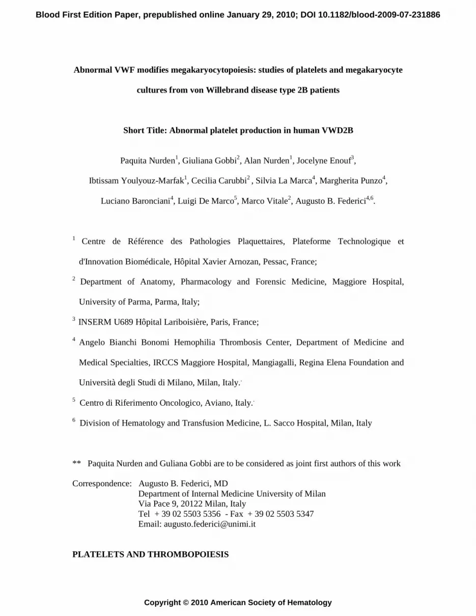
Abnormal VWF modifies megakaryocytopoiesis: studies of platelets and megakaryocyte
cultures from von Willebrand disease type 2B patients
Short Title: Abnormal platelet production in human VWD2B
Paquita Nurden1, Giuliana Gobbi2, Alan Nurden1, Jocelyne Enouf3,
Ibtissam Youlyouz-Marfak1, Cecilia Carubbi2 , Silvia La Marca4, Margherita Punzo4,
Luciano Baronciani4, Luigi De Marco5, Marco Vitale2, Augusto B. Federici4,6.
1 Centre de Référence des Pathologies Plaquettaires, Plateforme Technologique et
d'Innovation Biomédicale, Hôpital Xavier Arnozan, Pessac, France;
2 Department of Anatomy, Pharmacology and Forensic Medicine, Maggiore Hospital,
University of Parma, Parma, Italy;
3 INSERM U689 Hôpital Lariboisière, Paris, France;
4 Angelo Bianchi Bonomi Hemophilia Thrombosis Center, Department of Medicine and
Medical Specialties, IRCCS Maggiore Hospital, Mangiagalli, Regina Elena Foundation and
Università degli Studi di Milano, Milan, Italy..
5 Centro di Riferimento Oncologico, Aviano, Italy..
6 Division of Hematology and Transfusion Medicine, L. Sacco Hospital, Milan, Italy
** Paquita Nurden and Guliana Gobbi are to be considered as joint first authors of this work
Correspondence: Augusto B. Federici, MD Department of Internal Medicine University of Milan Via Pace 9, 20122 Milan, Italy Tel + 39 02 5503 5356 - Fax + 39 02 5503 5347 Email: [email protected]
PLATELETS AND THROMBOPOIESIS
Blood First Edition Paper, prepublished online January 29, 2010; DOI 10.1182/blood-2009-07-231886
Copyright © 2010 American Society of Hematology

2
Abstract Von Willebrand factor (VWF) is an essential mediator of platelet adhesion to the vessel wall,
but little is known about its role in megakaryocytopoiesis. VWF and its platelet receptor,
glycoprotein Ibα (GPIbα) are both expressed during megakaryocyte (MK) maturation. This
study was designed to evaluate whether the enhanced VWF-GPIbα interactions typical of
patients with von Willebrand disease type 2B (VWD2B) modify platelet production. Platelets
from 9 VWD2B patients with 7 different gain-of-function mutations were examined by
electron microscopy (EM) and immunofluorescence labelling. For the VWD2B patients, EM
characteristically showed variable numbers of structurally abnormal giant platelets,
sometimes in agglutinates. Cultures of MK from controls performed with or without purified
VWF confirmed a positive influence of VWF on platelet production with specific inhibition
by an antibody blocking VWF binding to GPIbα. VWD2B MK cultures examined by EM
showed a disorganized demarcation membrane system, and abnormal granule distribution.
They produced platelets with structural abnormalities typical of VWD2B. Confocal
examination of MK revealed limited extension of pseudopods with few large proplatelets.
These results confirm that megakaryocytopoiesis is modified by the enhanced VWF-
GPIbα interactions. These data obtained for controls and VWD2B patients suggest a novel
regulatory role of VWF-GPIbα interactions in platelet production.

3
Introduction
Von Willebrand Factor (VWF) is a large multimeric adhesive glycoprotein that circulates in
plasma and is also found in platelets, megakaryocytes (MK), endothelial cells, and the
subendothelial matrix.1,2 VWF is synthesized both in endothelial cells and MK where under
normal conditions it is stored in secretory organelles as ultralarge multimers (ULVWF) that
are processed on secretion by ADAMTS13 (a disintegrin and metalloproteinase with
thrombospondin type I motif-13).3 VWF is a well-known mediator of platelet adhesion to the
vessel wall and of platelet-platelet interactions under high shear-stress conditions.1 VWF has
also been identified as a sensitive and distinct marker for early MK4 and, more recently,
exposure of human MK to VWF at high shear rates was reported to accelerate platelet
production.5
Reduced or dysfunctional levels of VWF cause inherited von Willebrand disease (VWD),
currently classified within six different types.6 VWD type 2B (VWD2B) results from a gain-
of-function of VWF that has an increased affinity for platelet glycoprotein(GP)Ibα.7,8 The
mutations responsible for the gain of function are located in exon 28 of the VWF gene coding
for the A1 domain. Thrombocytopenia is often present in VWD2B and the presence of giant
platelets has been reported in isolated cases.9-12 Circulating platelet agglutinates have been
found in rare families.12-14 Studies using a nanobody recognizing soluble VWF in its gain-of-
function state confirmed that soluble VWF in its GPIbα-binding conformation was present in
greater amounts in most VWD2B patients with thrombocytopenia.13
We recently reported that impaired megakaryocytopoiesis results from an abnormal
interaction between GPIbα with newly synthesized VWF in MK of a family with VWD2B
due to a R1308P mutation.14 Severe thrombocytopenia and enlarged platelets were
accompanied by the presence of platelet agglutinates in freshly drawn blood. Our observation
that the impaired interaction between GPIbα and VWF R1308P was seen in maturing MK
suggested a direct effect of VWF on MK maturation and platelet production. Previously,

4
thrombocytopenia in VWD2B had been considered to result from premature platelet
elimination after the abnormal VWF had bound to the platelet surface. But, our work
suggested that the abnormal VWF structure in VWD2B was directly influencing
megakaryocytopoiesis. More recently, the very rare Montreal platelet syndrome, an autosomal
dominant form of inherited thrombocytopenia associated with lifelong mucocutaneous
bleeding, giant platelets and spontaneous platelet agglutination, has been recognized to be
associated with the V1316M VWF mutation typical of VWD2B.15
Given this complex background, we tested here the hypothesis of a role for VWF in human
megakaryocytopoiesis. Studies were performed to evaluate the role on platelet morphology
and production of the enhanced VWF-GPIb interaction using the human model of patients
with VWD2B who are characterized by a gain-of-function of VWF. Platelets were first
examined by electron microscopy; then in in vitro studies, we assessed the role of native
VWF and mutated VWF on platelet production by MK derived from peripheral blood
precursors. Not only were platelet ultrastructural abnormalities found to be widespread,
suggestive of a generalized abnormal megakaryocytopoiesis in VWD2B patients; but cultured
CD34+ or CD45+ MK from VWD2B patients have an altered morphology and produced
abnormal and fewer platelets than those observed from control donors suggesting that the
presence of abnormal VWF during MK maturation influenced the formation of platelets.
Significantly, the newly produced platelets in vitro have the same abnormal morphological
abnormalities as found in peripheral blood. These data suggest a novel and relevant regulatory
role of VWF-GPIb interactions in megakaryocytopoiesis.

5
Materials and Methods
Patients and blood collection
Nine VWD patients were enrolled in the study after informed consent was obtained. The
study was performed according to the Declaration of Helsinski. Diagnosis of VWD was based
on the criteria of the ISTH-SSC VWF Subcommittee.6 The methods used to obtain clinical
and laboratory information were as previously described.8 The characteristics of the patients,
bleeding score, levels of VWF activities, values of RIPA and distribution of VWF multimers
are reported in Table 1. All cases were symptomatic with a bleeding score >5. Included were
9 VWD2B patients from 8 families with the following mutations (Table 1): P1266L (n=1),
R1306W (n=1), R1308C (n=1), I1309V (n=1), V1316M (n=2), R1341Q (n=2) and R1341W
(n=1). The platelet count was decreased at the time of examination for 5/9 VWD2B patients
and normal for the remainder (Table 1).
All VWD2B patients gave an enhanced ristocetin-induced platelet aggregation (RIPA) in
platelet-rich plasma (PRP). The mutation P1266L corresponds to VWD2B “Malmo or New
York” that is associated with increased RIPA, but with normal multimeric distribution in
plasma and a normal platelet count.8 While all patients enrolled in the study could be tested
for platelet morphology, only three VWD2B subjects [one case with the R1306W mutation
(P2) and 2 cases with the V1316M mutation (P5 and P6) found also in the Montreal Platelet
Syndrome] were available for in vitro studies performed in MK cultures. Since a relative large
amount of venous blood was required for these in vitro studies on platelet production by MK,
only male patients with normal levels of hemoglobin were studied after obtaining ethical
board approval from all participating institutions and informed consent. The in vitro studies
on CD34+/MK were performed at the Department of Anatomy in Parma using blood obtained
from healthy donors (HD) and patients and were done blind (i.e., without knowing in advance
samples from VWD2B donors) whereas in Bordeaux cultures were performed from CD45+
cells from controls or patients.

6
Proteins
Native VWF was purified from human cryoprecipitate obtained from the Division of
Hematology and Transfusion Medicine at the L. Sacco Hospital, as previously described16 it
was characterized by VWF activities and multimer composition typical of normal plasma: no
fibrinogen or fibronectin were present. Recombinant mutant R1306W-VWF (VWF2B) was
obtained using stable cell lines as described.17 Fibrinogen (FG) was purified from human
plasma as reported.18 Human fibronectin (FN) was obtained from Sigma (St Louis, MO).
Antibodies
A MoAb directed against αIIbβ3 (AP-2 from Dr. T. Kunicki, Scripps Research Institute, La
Jolla, CA) as reported previously was used.19 LJIb1 (anti-GPIbα inhibitor antibody) and
LJIb10 (anti-GPIbα antibody control) were prepared from hybridomas made with splenocytes
harvested from the same mouse (a kind gift of Dr. Z.M. Ruggeri, The Scripps Research
Institute, La Jolla, CA). Both are IgG1k and have been characterized in detail. They each
recognize a distinct epitope located in the 45-kDa- amino-terminal domain of GPIbα. Purifid
IgG was stored at - 70 °C. The antibody LJ-Ib10 has only a minimal inhibitory effect on VWF
binding to platelets, but obliterates completely the high affinity α-thrombin- binding site,
while LJ-Ib1 inhibits both binding sites.20,21 Tirofiban (αIIbβ3 inhibitor) was obtained from
Merck Sharp Dohme-Chibret and was used according to previously reported
recommendations.22
Electron microscopy: Standard procedure
Venous blood was taken in ACD-A (1vol/7vol) and centrifuged for 10 min at 80g, and the
PRP up to the buffy coat carefully aspirated. After incubation for 20 min at 37° C, platelets
were fixed in 1.25% glutaraldehyde (Fluka Chemie, Buchs, Switzerland) and diluted in 0.1 M

7
phosphate buffer (pH 7.2) for 1 h at room temperature. For MK cultures, at day 14 cells were
fixed under the same conditions after their concentration by centrifugation at 120g for 10 min.
Samples were processed for EM as previously described.14 Sections were observed with a Jeol
JEM-1010 transmission electron microscope (Jeol, Croissy-sur-Seine, France) at 80 kV. In
morphometric studies, a minimum of 100 sections of platelets obtained using standard EM
procedures were analysed for each subject. Platelet diameters and platelet surface area were
measured using the Software Image J (NIH, Bethesda, MD). Platelets from 4 healthy control
donors have been examined under the same conditions.
Immunofluorescence studies
Smears on glass slides were prepared using the same preparations of PRP as used for EM for
each patient. They were fixed with cold acetone, washed in PBS-albumin 0.5% and then
incubated with a predetermined optimal dilution of AP-2 directed against αIIbβ3. Highly cross-
adsorbed polyclonal antibody to mouse IgG conjugated to ALEXA-Fluor 488 (green)
(Interchim, Montluçon, France) was then used to reveal bound mouse IgG. After further
washings, a drop of a solution of 4,6-diamidino-2-phenylindole (DAPI) was used to visualize
DNA. The slides were examined with a Nikon Eclipse 80i fluorescent microscope (Nikon
France SAS, Champigny-sur-Marne, France) equipped with a fluorescence illumination
system EXFO Xcite and a Nikon DXM1200F camera. The objectives were Nikon Plan Fluor
x100/1.30 (oil) and Nikon Plan Fluor x40/0.75. Images were analyzed using Nikon NIS
Elements D2.20 imaging software. To study the formation of proplatelets from MK in culture,
cells were re-plated on a poly-L-lysine coated coverslip in a 4-well plate. The MK extending
proplatelets were studied after 24 hours with a Leica SP2 confocal microscope equipped with
Leica confocal software using a 40x/1.25 or a 63x/1.40, objective (Leica, Nanterre, France),
after labelling with AP-2 directed against αIIbβ3 and a species-specific goat anti-mouse
8
antibody conjugated with FITC (DakoCytomation, Trappes, France) and by DAPI to visualize
DNA as described below.
CD34+ or CD45+ purification and MK differentiation
Primary CD34+ cells were isolated from peripheral blood of healthy donors (HD) or three
VWD2B patients by immunomagnetic positive selection using the CD34+ (Parma, 150-200
mL blood) or CD45+ (Bordeaux, 40 mL blood) cell isolation Kit (Milteny Biotech, Gladbach,
Germany) in the magnetic field of a Vario-MACS apparatus (Milteny Biotech), according to
manufacturer’s protocols.23 Purity of CD34+ cell preparations was immediately verified by a
R-phycoerythrin (RPE)-conjugated anti-CD34 MoAb (Beckman Coulter, Miami, FL) and
flow cytometry. Only samples exceeding 95% purity were used for subsequent experiments.
Purified human CD34+ cells were cultured up to 7 days (or 14 days in some experiments), at
an optimal cell density of 1x10⁶ cells/ml, in serum free X-vivo medium (BioWhittaker,
Walkersville, MD) supplemented every 3 days with 3 ng/ml of recombinant human
interleukin-3 (IL-3), 40 ng/ml of recombinant human stem cell factor (SCF) and 100 ng/ml of
recombinant human thrombopoietin (TPO), in the presence or absence (controls) of 10 µg/ml
of purified VWF. To test if the effects of VWF were dependent on its concentration, we
cultured CD34+ cells from HD with increasing doses of native purified VWF (2.5; 5; 10; 20;
40 µg/ml). In some experiments we also used FG (200 μg/ml) or FN (200 μg/ml) instead of
VWF. To test the specificity of exogenous VWF in its interactions with GPIbα, in some
experiments purified CD34+ cells were cultured in the presence of 80 μg/ml LJIb1 or 50
μg/ml of LJIb10 (both anti-GPIbα MoAbs), or 20 μg/ml of Tirofiban (αIIbβ3 inhibitor). In
other types of experiments (in Bordeaux) the cultures were performed under the same
conditions but from CD45+ isolated cells. In case of morphological studies, the MK cultures
were performed without addition of exogenous VWF.

9
Flow cytometry analysis
To quantify the platelet production in culture, fixed volumes of the culture supernatants were
collected, incubated with anti-CD41-RPE and calcein AM (Sigma, St Louis, MO) (to exclude
fragments) and added to a fixed volume of calibration beads (DAKO, Glostrup, Denmark) at
known concentration, as described previously.23,24 Both the platelet and bead populations
were simultaneously identified in flow cytometry on the Forward Scatter (FS) versus
logarithmic Side Scatter (SS) dot plot. The absolute platelet count was performed on the
CD41+/Calcein AM+ platelet population. Analysis of the samples was performed by an Epics
XL flow cytometer (Beckman Coulter) and the Expo ADC software (Beckman Coulter). The
instrument was calibrated daily with a set of standardized beads (DAKO).23,24
Statistics
To evaluate differences among various groups of samples, a Bonferroni-type analysis was
applied to the data obtained in different experiments: P values < 0.05 were considered
significant. Error bars represent the standard error of the mean (SEM) or the standard
deviation (SD), as indicated in the Figure Legends.

10
Results
Ultrastructure of platelets in VWD2B
For each patient, platelets with an abnormal morphology were found within the normal
population. Figure 1 shows a gallery of electron micrographs of platelets chosen to illustrate
the morphological changes that were detected in VWD2B. Illustrations are given for each
mutation. Globally, the characteristics included platelet agglutinates without signs of
activation and sometimes with what appear to be fused surface membranes (a, b, e), while
enlarged but not totally spherical platelets with an altered distribution of membranes and
granules were abundant (c, d, g). Even when not giant, platelet size and shape was often
heterogeneous and occasional enlarged α-granules were seen (h). These observations led us to
perform a morphometric analysis.
Morphometric studies of platelet size in VWD2B
Results summarized in Table 2 show that mean maximal platelet diameter was increased for
the majority of patients with VWD2B, nonetheless results were variable and remained at the
upper limit of normal values for P3, P4 and P8. This heterogeneity extended to platelet
surface area where mean values were always greater than seen for normal platelets and
sometimes by several fold. We show in Figure 2 the proportion of platelets with increased
size for each patient. Results confirm that for each VWD2B patient platelet maximal diameter
and minimal diameter increased almost proportionally, with the largest platelets found for P2
(R1306W), P6 (V1316M) and P9 (R1341W). Notably, quite marked size differences were
seen between P5 and P6, both of whom possessed the V1316M mutation.

11
Further evidence for an abnormal megakaryocytopoiesis using immunofluorescence
labelling
Figure 3 illustrates the results for platelets from the VWD2B patient (P2) with the R1306W
mutation and was typical of the results seen for all the patients. Selected images for αIIbβ3 are
shown in panels (a-e). Compared to normal platelets (f), there was considerable size
heterogeneity with the presence of giant platelets. Occasional nucleated cells were identified
as MKs by their labelling for αIIbβ3.
Effects on platelet production of exogenous VWF and other ligands or inhibitors
We first studied the effects of exogenous VWF on platelet production following in vitro MK
differentiation. Primary CD34+ cells from HD cultured for 7 days in the presence of TPO and
VWF released a higher number of platelets when compared to TPO alone (Figure 4,a,b). In
contrast, αIIbβ3 ligands, such as FG and FN did not boost platelet production. VWF effects on
platelet production were indeed related to GPIbα because these effects were selectively
blocked by its functional inhibition by the antibody LJIb1 but not by LJIb10 reactive with the
thrombin-binding site. Conversely, tirofiban, that inhibits αIIbβ3, was not able to block the
VWF-induced enhancement of platelet production in TPO-treated CD34+ cell cultures. Of
note, the addition of exogenous R1306W-recombinant VWF to CD34+ cells from HD strongly
stimulated platelet production (Figure 4, a).
The presence of VWF in the TPO-treated CD34+ cell cultures not only boosted the
production of platelets in vitro at 7 days, but continued to promote terminal MK
differentiation even after 14 days of culture (Figure 4, c). Moreover, the effects of VWF on
platelet production were dose-dependent, as shown in Figure 5.

12
Platelet production by normal versus VWD2B MK (endogenous VWF)
Quantitative aspects (Figure 6A): We finally tested the effects of purified native VWF on
CD34+ cells isolated from peripheral blood of three VWD2B patients and of three HD
(Figure 6 A). In the absence of exogenous VWF, platelet production from CD34+ cells from
VWD2B patients cultured for 7 days with 100 ng/ml of TPO, was reduced as compared to
CD34+ cells from the control donors. However, the addition of native exogenous VWF with
the TPO was able to increase platelet production levels from CD34+ VWD2B cells, even if
the global level of platelet production remained lower than that of VWF-treated CD34 from
healthy controls (Figure 6A).
Morphological aspects (Figure 6 B,C): We used confocal microscopy to examine MK in
culture at day 14 at a late stage of maturation (Figure 6B). First illustrated are control MK
typically showing very thin and long extensions with small swellings (a, b); beads extending
for a long distance can be also observed in c. Conversely, in MK cultures from the V1316M
patients the extensions are shorter and the swellings larger (a-f), with proplatelets remaining
distinct but associated (g). MK cultures were also performed until day 14 and examined by
EM (Figure 6C). For the controls, EM showed a normal MK morphology and granules were
uniformly distributed while the Demarcation Membrane System (DMS) uniformly invaded
the cytoplasm (a). For VWD2B V1316M cultures, it can be seen that granules are distributed
heterogeneously within most of the the MK while the DMS is poorly developed (b). At higher
magnification, in mature MK, platelet territories are small for controls and delimitated by a
well-developed membrane system (c) whereas for the VWD2B V1316M a defective DMS
delimits large platelet territories containing varying numbers of granules while other parts of
the cytoplasm contains little DMS (d). Similar observations were made for MK from the
R1306W patient (results not shown). In (e, f) platelets produced in vitro from the VWD2B
R1306W are shown: note the heterogeneity in size, in granule distribution and again the
presence of giant platelets as illustrated in f).

13
Discussion
VWF plays a major role in hemostasis, and molecular abnormalities or deficiency of
this protein cause inherited VWD.1,2 By associating with collagen in the vascular matrix,
VWF permits the anchoring of platelets at the site of vascular lesions under shear and then to
the arrest of bleeding.25,26 In VWD2B, conformational changes in the mutated VWF are
responsible for an up-regulated binding to GPIbα.27 A consequence of this gain-of-function is
the selective binding of the larger multimers to the platelets and the loss of VWF multimers
from the circulation. The concentration of circulating VWF multimers in a GPIbα-binding
conformation is particularly elevated in patients with VWD2B and thrombocytopenia.8 Such a
result suggests that accelerated platelet loss from the circulation is linked to the presence of
platelet-reactive VWF multimers.8 Our current working hypotheses is that an altered platelet
production in addition to the loss of the larger VWF multimers may intervene to cause a low
platelet count in VWD2B and, more generally, that the VWF-GPIb interactions is crucial for
normal megakaryocytopoiesis. Earlier reports showing i) impaired megakaryocytopoiesis in a
family with the R1308P mutation of VWF, ii) altered megakaryocytopoiesis for patients with
Bernard-Soulier Syndrome (BSS) and heterozygotes for the Bolzano mutation of GPIbα, and
iii) that platelet production was accelerated by high shear forces in presence of VWF
constitute additional reasons to suggest a role for the interaction between GPIbα and VWF in
megakaryocytopoiesis. 5,14,28,29
Now we report that in a large series of VWD2B patients with different mutations,
variable populations of platelets have increased size with ultrastructural characteristics of
giant platelet syndromes. Giant granules were occasionally found and independently of the
nature of the mutation, these granules were confirmed to contain VWF by immunogold
labelling (data not shown). Giant platelets were regularly present in higher numbers than in
control donors; a finding seen whether or not the patient was thrombocytopenic. Giant

14
platelets have also been reported in the Montreal platelet syndrome first considered to be a
platelet disease.30 but now shown to be VWD2B associated with the presence of platelet
agglutinates.15 Giant granules are rare but well described in the Paris-Trousseau syndrome31
given by mutations of the Fli gene but the mechanism responsible for their formation is
unknown. Our results implied that enhanced binding of endogenous VWF to GPIbα observed
in VWD2B patients can interfere with the fine regulation of megakaryocytopoiesis.
First confirmation of this came from cultures of CD34+ cells obtained from healthy
donors when platelet production was shown to be increased by the addition of VWF with
more platelets liberated at day 7 and day 14 of culture than in the presence of TPO alone. The
potentiating effect of VWF was specifically mediated by the VWF-binding domain of GPIbα.
In fact, the use of the antibody against GPIbα (LJIb1) could block such a VWF-dependent
accelerated platelet production and this was specific since another MoAb against the
thrombin-binding site of GPIbα (LJIb10) did not interfere with this positive effect. The lack
of inhibition with tirofiban further showed that VWF was not mediating this effect through
αIIbβ3. In our sets of experiments we could confirm that the VWF-dependent positive effect
on platelet production does require an intact and functional GPIbα. Moreover other ligands
of αIIbβ3 (fibronectin and fibrinogen) were not able to boost MK differentiation. Interestingly,
antibodies against GPIbα, that selectively block VWF effects on MK cells, have been
previously shown to interfere with MK maturation and platelet production in ITP.32
Further evidence came when we found that not only native VWF, but also
recombinant R1306W-VWF mutant protein was able to boost in vitro MK differentiation
from normal CD34+ cells, and even more efficiently than its native form when platelet
production was studied after 7 and also 14 days maturation. This observation suggested that
interaction of exogenous VWF to membrane GPIbα was able to promote MK differentiation
and that an increased VWF/GPIb interaction can accelerate the last step of platelet release.

15
The fact that (i) the same morphological characteristics were present in these platelets as well
as platelets derived from the peripheral blood, and (ii) that abnormalities were present during
megakaryocytopoiesis implied a role for newly synthesized VWF and GPIbα. Performing the
opposite experiments using VWD2B-MK, we could also show that the exposure of MK
cultures from VWD2B patients to normal purified VWF was able to improve platelet
production, suggesting the same VWF-dependent positive effect on platelet production
independently of the source of the MK (normal donors or VWD2B patient). In our hands,
VWF accelerated platelet production when added to the media and this effect was dose-
dependent and independent of high shear rate. Recent evidence from studies performed in
cultures point to a role for interactions between mature MK and stromal adhesive proteins in
proplatelet formation.25,26 Interestingly, GPIbα is a signalling molecule,33 offering the
possibility that the GPIb-VWF axis may play a regulatory role in MK maturation. GPIbα
serves as an anchor between extracellular matrix proteins and the actin network and
significantly macrothrombocytopenia was improved in a BSS mouse model in which the
GPIbα cytoplasmic domain was retained. 34
In parallel to the work performed with BSS patients (see review 28) or GPIbα
deficient mice35, or with the patients with Bolzano heterozygote mutation of GPIbα
characterized by an abnormal GPIbα29 , we could demonstrate that the CD34+/MK obtained
from VWD2B patients characterized by two relatively common mutations (R1306W and
V1316M) showed significantly abnormal platelet formation. Although GPIbα is expressed
later than αIIbβ3,36 it is present in maturing MK and may play a role in the interaction in the
development of internal MK membranes and formation of pseudopods. Using confocal
microscopy, it was possible to visualize the extending pseudopods and we confirmed with
cultures from CD34+ or CD45+ cells that major differences were present for the patients.
Whereas for controls the proplatelets were mostly long and thin and decorated by protrusions

16
or beads corresponding to the small future platelets, for VWD2B the pseudopods remained
thick, were fewer in number and the protrusions larger. EM of the cultured MKs confirmed
the presence of morphological abnormalities in the cytoplasm with an abnormal distribution
of the DMS present in decreased amounts at the later stages of maturation. In VWD2B the
interaction between newly synthesized VWF and GPIbα begins at an early stage of
differentiation and has the potential to also occur intracellularly.14 Confirming our original
work, platelet agglutinates have recently been shown in a marrow aspirate taken from a new
unrelated VWD patient with the R1308P mutation.37 Circulating platelet agglutinates were
also a feature of VWD2B Tampa.12 and they are also present in the Montreal Platelet
Syndrome recently ascribed to a V1316M mutation as discussed above.15
BSS and VWD2B are two syndromes with giant platelets and thrombocytopenia; but
the platelet morphology differs, in BSS, with round giant platelets deficient in internal
membranes.33 In VWD2B the platelets although of large size often did not appear to be
spheroid. It is possible that the binding of the gain-of-function VWF in the circulation while
promoting platelet elimination may also exert a negative influence during
megakaryocytopoiesis modifying the organization of intracellular DMS, and reducing the
extension of pseudopods so important for platelet formation. Platelet formation is a complex
and well orchestrated phenomenon.38 In VWD2B the fine balance of platelet production is
interfered with.
In conclusion, our data show that megakaryocytopoiesis requires a normal VWF-GPIb
interaction. Taken together our results suggest a cytokine-like function of VWF as a
physiological promoter of MK differentiation. However, the pathophysiology of altered
megakaryocytopoiesis in VWD2B may also be due to an altered intracellular interaction
between the abnormal VWF2B and GPIb�, while extracellular VWF2B maintains its
promoting effects on proplatelet formation. .It will be studied in detail in the near future
17
whether such a difference might be due to the activation of specific signalling pathways
downstream of membrane GPIb�,
Acknowledgements.. This study was supported by grants from the Italian Ministry of
Health to ABF and from the Bayer Hemophilia Awards Program to LB and by the DIATROC
“Hospital Project of Clinical Research” to PN. AN and PN acknowledge support from the
French network 'Gis-Maladies Rares", from the French Health Ministry to the CRPP and from
INSERM (ANR-08-GENO-028-03). Preliminary data were presented in part at ASH 2008.
We wish to thank Rosanna Garavaglia for her help with the patients and Jean-Max Pasquet for
his technical help. We are grateful to Drs Z. M. Ruggeri and T. Kunicki, for the gift of their
Monoclonals antibodies.
Authorship
ABF and PN were responsible for study initiation and coordination. PN, GG, AN, JE, IM,
EC, CC, SLM, MP, LB, LDM, MV and ABF were involved in study design, data
collection and performing laboratory analyses. ABF, AN, PN wrote the manuscript; GG,
LB, MP, LDM and MV were responsible for revisions of draft manuscripts. All authors
were responsible for approval of the final manuscript. Authors report no conflict of
interest.

18
References
1. Ruggeri ZM. Von Willebrand Factor. Looking back and looking forward. Thromb
Haemost. 2007;98(1):55-62.
2. Sadler JE. Von Willebrand factor, ADAMTS13, and thrombotic thrombocytopenic
purpura. Blood. 2008;112(1):11-18.
3. Rayes J, Hommais A, Legendre P, et al. Effect of von Willebrand disease type 2B and
type 2M mutations on the susceptibility of von Willebrand factor to ADAMTS-13. J
Thromb Haemost. 2007;5(2):321-328.
4. Tomer A. Human marrow megakaryocyte differentiation: multiparameter correlative
analysis identifies von Willeband factor as a sentitive and distinctive marker for early
(2N and 4N) megakaryocytes. Blood. 2004;104(9): 2722-2727.
5. Dunois-Larde C, Capron C, Fichelson S, Bauer T, Cramer-Borde E, Baruch D. Exposure
of human megakaryocytes to high shear rates accelerates platelet production. Blood.
2009;114(9):1875-1883.
6. Sadler JE, Budde U, Eikenboom JC, et al. Update on the pathophysiology and
classification of von Willebrand disease: a report of the subcommittee on von
Willebrand factor. J Thromb Haemost. 2006;4(10):2103-2114.
7. Ruggeri ZM, Pareti FI, Mannucci PM, Ciaverella N, Zimmerman TS. Heightened
interaction between platelets and factor VIII/von Willebrand factor in a new subtype of
von Willebrand disease. N Engl J Med. 1980;302(19):1047-1051.
8. Federici AB, Mannucci PM, Castaman G, et al. Clinical and molecular predictors of
thrombocytopenia and risk of bleeding in patients with von Willebrand disease type 2B:
a cohort study of 67 patients. Blood. 2009;113(3):526-534.
9. Moll S, Lazarowski AR, White GC 2nd. Giant platelets in a patient with type 2B von
Willebrand's disease. Am J Hematol. 1998;57(1):62-67.
10. Lopez-Fernandez MF, Lopez-Berges C, Martin-Bernal JA, et al. Type 2B von
Willebrand's disease associated with a complex thrombocytopenic thrombocytopathy.
Am J Hematol. 1988;27(4):291-298.
11. Nurden P, Chretien F, Poujol C, Winckler J, Borel-Derlon A, Nurden A. Platelet
ultrastructural abnormalities in 3 patients with type 2B Von Willebrand's disease. Br J
Haematol. 2000;110(3):704-714.
19
12. Saba HI, Saba SR, Dent J, Ruggeri ZM, Zimmerman TS. Type IIB Tampa: a variant of
von Willebrand disease with chronic thrombocytopenia, circulating platelet aggregates,
and spontaneous platelet aggregation. Blood. 1985;66(2):282-286.
13. Hulstein JJ, de Groot PG, Silence K, Veyradier A, Fijnheer R, Lenting PJ. A novel
nanobody that detects the gain-of-function phenotype of von Willebrand factor in
ADAMTS 13 deficiency and von Willebrand disease type 2B. Blood. 2005;106(9):3035-
3042.
14. Nurden P, Debili N, Vainchenker W, et al. Impaired megakaryocytopoiesis in type 2B
von Willebrand disease with severe thrombocytopenia. Blood. 2006;108(8):2587-2595.
15. Jackson SC, Sinclair GD, Cloutier S, Duan Z, Rand ML, Poon MC. The Montreal
platelet syndrome kindred has type 2B von Willebrand disease with the VWF V1316M
mutation. Blood 2009;113(14):3348-3351.
16. Federici AB, Bader R, Pagani S, Colibretti ML, De Marco L, Mannucci PM. Binding of
von Willebrand factor to glycoproteins Ib and IIb/IIIa complex: affinity is related to
multimeric size. Br J Haematol. 1989;73(1):93-99.
17. Baronciani L, Federici AB, Punzo M, et al. Type 2A (IIH) von Willebrand disease is
due to mutations that affect von Willebrand factor multimerization. J Thromb Haemost.
2009;7(7):1114-1122.
18. De Marco L, Girolami A, Zimmerman TS, Ruggeri ZM. Von Willebrand factor
interaction with the glycoprotein IIb/IIIa complex. Its role in platelet function as
demonstrated in patients with congenital afibrinogenemia. J Clin Invest.
1986;77(4):1272-1277.
19. Pidard D, Montgomery RR, Bennett JS, Kunicki TJ. Interaction of AP-2, a monoclonal
antibody specific for the human platelet glycoprotein IIb-IIIa complex, with intact
platelets. J Biol Chem. 1983;258(20):12582-12586.
20. Handa M, Titani K, Holland LZ, Roberts JR, and Ruggeri ZM. The von Willebrand
factor-binding domain of platelet membrane glycoprotein Ib. Characterization by
monoclonal antibodies and partial amino acid sequence analysis of proteolytic
fragments. J Biol Chem. 1986;261(27):12579-12585.
21. De Marco L, Mazzucato M, Masotti A, Ruggeri ZM. Localization and characterization
of an alpha-thrombin-binding site on platelet glycoprotein Ib alpha. J Biol Chem.
1994;269(9):6478-6484.
22. Coller BS. Anti-GPIIb/IIIa drugs: current strategies and future directions. Thromb
Haemost. 2001;86(1):427-443.

20
23. Gobbi G, Mirandola P, Sponzilli I, et al. Timing and expression level of protein kinase
C epsilon regulate the megakaryocytic differentiation of human CD34 cells. Stem Cells.
2007;25(9):2322-2329.
24. Gobbi G, Mirandola P, Carrubbi C, et al. Phorbol ester-induced PKCepsilon down-
modulation sensitizes AML cells to TRAIL-induced apoptosis and cell differentiation.
Blood. 2009;113(13):3080-3087.
25. Wagner DD, Frenette PS. The vessel wall and its interactions. Blood.
2008;111(11):5271-5281.
26. Balduini A, Pallotta, I, Malara A, et al. Adhesive receptors, extracellular proteins and
myosin IIA orchestrate proplatelet formation by human megakaryocytes. J Thromb
Haemost. 2008;6(11):1900-1907.
27. Dumas JJ, Kumar R, McDonagh T, et al. Crystal structure of the wild-type von
Willebrand factor A1-glycoprotein Ibα complex reveals conformation differences with a
complex bearing von Willebrand disease. J. Biol Chem. 2004;279(22):23327-23334.
28. Nurden AT, George JN. Inherited abnormalities of the platelet membrane: Glanzmann
Thrombasthenia, Bernard-Soulier syndrome and other disorders. In: Colman RW,
Marder VJ, Clowes AW, George JN, Goldhaber S, eds. Hemostasis and Thrombosis,
Edition V. Philadelphia, PA: Lippincott, Williams & Wilkins; 2006:987-1011.
29. Balduini A, Malara A, Pecci A, et al. Proplatelet formation in heterozygous Bernard-
Soulier syndrome type Bolzano. J Thromb Haemost. 2009;7(3):478-484.
30. Milton JG, Frojmovic MM, Tang SS, White JG. Spontaneous platelet aggregation in a
hereditary giant platelet syndrome (MPS). Am J Pathol. 1984;114(2):336-345.
31. Nurden P, George JN, Nurden AT. Inherited Thrombocytopenias. In: Colman RW,
Marder VJ, Clowes AW, George JN, Goldhaber S, eds. Hemostasis and Thrombosis,
Edition V. Philadelphia, PA: Lippincott, Williams & Wilkins; 2006:975-986.
32. Chang M, Nakagawa PA, Williams SA, et al. Immune thrombocytopenic purpura (ITP)
plasma and purified ITP monoclonal autoantibodies inhibit megakaryocytopoiesis in vitro.
Blood. 2003;102(3):887-895.
33. Mu FT, Andrews RK, Arthur JF, et al. A functional 14-3-3zeta-independent association of
PI3-kinase with glycoprotein Ibα, the major ligand-binding subunit of the platelet
glycoprotein Ib-IX-V complex. Blood. 2008;111(9):4580-4587.
34. Kanaji T, Russell S, Ware J. Amelioration of the macrothrombocytopenia associated with the
murine Bernard-Soulier syndrome. Blood. 2002;100(6):2102-2107.

21
35. Poujol C, Ware J, Nieswandt B, Nurden AT, Nurden, P. Absence of GPIbα is responsible for
aberrant membrane development during megakaryocyte maturation: ultrastructural study
using a transgenic model. Exp Hematol. 2002;30(4):352-360.
36. Norol F, Vitrat N, Cramer E, et al. Effects of cytokines on platelet production from blood
and marrow CD34+ cells. Blood. 1998;91(3):830-843.
37. Slayton WB, Patel M, Sola-Visner M, et al. Type 2B von Willebrand disease associated with
the release of platelet agglutinates from megakaryocytes in the bone marrow. J Pediatr
Hematol Oncol. 2008;30(9):708-711.
38. Italiano JE Jr, Patel-Hett S, Hartwig JH. Mechanisms of proplatelet elaboration. J Thromb
Haemost. 2007;5(Suppl. 1):18-23.
.

22
Table 1: Clinical and laboratory parameters of the VWD2B patients enrolled in the
study listed according to mutations of the VWF A1 domain
Patients (P)
Mutation Bleeding score
Platelet count
(x1000 /µ L)
RIPA (mg/ml)
VWF:Ag (U/dL)
VWF:RCo (U/dL)
FVIII:C (U/dL)
VWF:RCo/Ag (ratio)
HMW/ Intermediate
multimers
P 1
P1266L 6 251 0.4 59 45 57 0.76 Present/ normal
P 2 R1306W
12 175 0.6 45 28 51 0.62 Absent/partial loss
P 3
R1308C 9 121 0.5 54 21 45 0.39 Absent/complete loss
P 4
I1309V 9 96 0.3 160 125 140 0.78 Absent//normal
P 5 V1316M 13 274 0.6 33 12 6 0.21 Absent/complete loss
P 6
V1316M 17 60 0.25 70 25 52 0.4 Absent/complete loss
P 7
R1341Q 9 98 0.25 95 70 69 0.7 Absent/partial loss
P 8
R1341Q 10 120 0.5 79 48 47 0.6 Absent/partial loss
P 9
R1341W 9 195 0.4 72 42 73 0.58 Absent/partial loss
Abbreviations: RIPA: ristocetin-induced platelet aggregation in platelet-rich plasma VWF:Ag: VWF antigen VWF:RCo: VWF as ristocetin cofactor activity FVIII:C: pro-coagulant activity of FVIII VWF:RCo/Ag: calculated ratio with antigen HMW/Intermediate: high molecular weight versus intermediate multimers

23
Table 2: Morphometric studies of the platelets: Mutations
Max D (µm)
Min D (µm)
Area (µm²)
C(n=4)
2.7 (+/-0.5)
1.1 (+/-0.5)
2.4 (+/-1.3)
P1
P1266L
3.7 (+/-1.7)
1.6 (+/-1.0)
5.5 (+/-5.7)
P2
R1306W
3.5 (+/-0.7)
1.8 (+/-0.7)
4.7 (+/-2.4)
P3
R1308C
2.9 (+/-0.6)
1.4 (+/-0.6)
3.2 (+/-2)
P4
I1309V
2.9 (+/-1.0)
1.5 (+/-0.6)
6.8 (+/-6.8)
P5
V1316M
3.1 (+/-1.1)
1.5 (+/-0.6)
6.0 (+/-6.1)
P6
V1316M
3.7 (+/-1.2)
2.4 (+/-1.0)
10.4 (+/-11.2)
P7
R1341Q
3.2 (+/-0.6)
1.8 (+/-0.7)
9.9 (+/-8.7)
P8
R1341Q
3.1 (+/-0.7)
1.8 (+/-0.7)
7.6 (+/-7.7)
P9
R1341W
3.6 (+/-1.0)
1.9 (+/-0.9)
5.6 (+/-5.4)
C: control P: Patient VWD2B

24
FIGURE LEGEND:
Figure 1. A gallery of electron micrographs of platelets from patients with VWD2B. Platelet
agglutinates are illustrated in panels a), b) and e). Membrane complexes (MC) are to be seen
in an enlarged platelet in panel a). Panel d) shows a giant platelet from P1 with the P1266L
mutation where HMW multimers are present despite a positive RIPA, while panel h)
highlights a α-granule of increased size for P1. A giant platelet from P6 with the V1316M
mutation is shown in panel g); enlarged platelets of abnormal form are shown for P7 in panel
c) and for P4 in panel f). Many of the illustrated platelets contain increased amounts of SCCS
and the granules are not homogeneously distributed. Bar = 1µm for all the electron
micrographs (a-h).
Figure 2. Quantitative morphometric evaluation of platelet size parameters in patients with
VWD. The results are given as percentage of platelets with maximal diameter >3µm in a),
with minimal diameter >2µm in b) and with surface area >5µm2 in c) compared to controls
(C).
Figure 3. Detection of circulating MK or their fragments by immunofluorescence labelling of
blood smears from VWD2B using a MoAb specific for the αIIbβ3 complex. Results for P2
with the R1306W VWD2B mutation show enlarged platelets confirming size diversity with
occasional MK staining both with DAPI and for αIIbβ3 (a-e). Control platelets are shown for
comparison (panel f).
Figure 4. Platelet production from healthy donors (HD) CD34+ cell cultures. Relative
number of platelets at day 7 of treatment with TPO or TPO and VWF (panel a). The number
of platelets was calculated by flow cytometry on a CD41+/calcein AM+ labelled population (a
representative dot plot is shown in panel b) analyzed in combination with a known number of
calibration beads. The effects of fibrinogen (FG) and fibronectin (FN) and different inhibitors
on platelet production in the presence or absence of VWF is shown in panel a. Time course
kinetics of platelet production after 7 and 14 days of treatment with TPO (control) or TPO
and VWF (panel c). Data from 4 independent experiments are shown. The statistical analyses
according to Bonferroni test were grouped as follows: In Group 1, samples from A to G were
all calculated against A. In this group only sample D show statistical significance (means ±
SD; * P<0.05 versus TPO treatment); VWF promote platelet production. In Group 2,
samples from A to G were all calculated against D. In this group samples A, B and E were

25
significantly different; GPIba inhibition selectively counteracts VWF stimulation (means ±
SD; # P<0.05 versus TPO and VWF treatment). In Group 3, samples A, D and H were all
calculated against A and D. In this group the sample H shows statistical significance;
recombinant mutated VWF strongly promote platelet production from normal CD34+ cells
(means ± SD; * P<0.05 versus TPO treatment; # P<0.05 versus TPO and VWF treatment).
Figure 5. Platelet production from healthy donor (HD)+ CD34+ cell cultures in the presence
of increasing concentrations of native VWF. Relative number of platelets at day 7 of
treatment with TPO or TPO and VWF at the indicated doses. The number of platelets was
calculated by flow cytometry on a CD41+/calcein AM+ labelled population analyzed in
combination with a known number of calibration beads. Data from 2 independent
experiments. All the samples were analysed with Bonferroni test against TPO treatment
(means ± SD; * P<0.05 vs TPO treatment).
Figure 6. Platelet production and megakaryocytopoiesis in culture. A). Platelet
production from cultured CD34+ MK obtained from three different healthy donors (HD,
empty histograms) used as controls and from three patients with VWD2B, one with mutation
R1306W (light grey histograms) and two with mutation V1316M (dark grey histograms).
Relative number of platelets at day 7 of treatment with TPO (left) or TPO and VWF (right).
The number of platelets was calculated by flow cytometry on a CD41+/calcein AM+ labelled
population analyzed in combination with a known number of calibration beads.
Data from 2 independent experiments (means ± SD; * P<0.05 HD versus each mutant VWF
as well as HD and each mutant VWF exposed to VWF versus TPO treatment only). The
statistical analyses according to Bonferroni test were grouped as follows: In Group 1, all
samples were calculated against TPO-treated HD. In this group the TPO- treated VWD2B
samples show statistical significance; endogenous VWD2B inhibits platelet production. In
Group 2, samples from VWD2B were calculated against TPO-treated VWD2B. In this
group, samples treated with normal exogenous VWF are significantly different as compared
to TPO-treatment; normal exogenous VWF normalizes platelet production from VWD2B
CD34+ cells.
B). Morphological studies of the formation of proplatelets as seen by confocal microscopy of
MK cultures labelled with an anti-αIIbβ3 (green) antibody and by DAPI (blue). In panel a)
and b) the progressive disruption of MK cytoplasm and formation of proplatelets can be seen,
MK appeared with long pseudopods decorated with small blisters corresponding to

26
proplatelets. In panel c) the very long pseudopods have a bead-like appearance. For the
VWD2B MK instead of thin pseudopods the cytoplasm forms thick protusions (panels a-f)
with interwoven membranes (panels d,f). In panel g) 3 distinct but adjacent proplatelets are to
be seen.
C). Examination of culture MK from controls and patients by EM. On the left side panels a)
and c) we illustrate the morphology of control MK and in panels b) and d) VWD2B V1316M
MK. In panel a) is shown the homogeneous presence of granules and DMS. In panel b), all
the granules (G) are concentrated in one part of the MK cytoplasm, most of the DMS is also
seen in this same area suggesting a defect in the process of partitioning the components. In
panel c), at a higher magnification, is shown the cytoplasm of another control mature MK
where small platelet territories are well delimited by abundant and well-organized DMS. In
panel d) at a high magnification is shown part of a mature VWD2B MK where an abnormal
large platelet territory (arrows) surrounded by DMS can be seen. Globally abnormal
distribution of DMS suggests a defect in the organisation of membranes during maturation.
In panels e, f) are shown the newly produced platelets from the R1306W MK. In panel e) the
platelets are heterogeneous in size not separated/or agglutinated and the distribution of
granules heterogeneous. In panel f) an illustration of a giant platelet as found in the
circulation.


Copyright © 2022 FDOKUMEN