Nanomedicines based americium nanoparticles drug delivery ...
Upload
independentCategory
view
1download
0
After online publication, subscribers (personal/institutional) to this journal will haveaccess to the complete article via the DOI using the URL:
If you would like to know when your article has been published online, take advantageof our free alert service. For registration and further information, go to:
.
Due to the electronic nature of the procedure, the manuscript and the original figureswill only be returned to you on special request. When you return your corrections,please inform us, if you would like to have these documents returned.
Dear Author
Here are the proofs of your article.
• You can submit your corrections online, via e-mail or by fax.
• For online submission please insert your corrections in the online correction form.
Always indicate the line number to which the correction refers.
• You can also insert your corrections in the proof PDF and email the annotated PDF.
• For fax submission, please ensure that your corrections are clearly legible. Use a fine
black pen and write the correction in the margin, not too close to the edge of the page.
• Remember to note the journal title, article number, and your name when sending your
response via e-mail or fax.
• Check the metadata sheet to make sure that the header information, especially author
names and the corresponding affiliations are correctly shown.
• Check the questions that may have arisen during copy editing and insert your
answers/corrections.
• Check that the text is complete and that all figures, tables and their legends are included.
Also check the accuracy of special characters, equations, and electronic supplementary
material if applicable. If necessary refer to the Edited manuscript.
• The publication of inaccurate data such as dosages and units can have serious
consequences. Please take particular care that all such details are correct.
• Please do not make changes that involve only matters of style. We have generally
introduced forms that follow the journal’s style.
• Substantial changes in content, e.g., new results, corrected values, title and authorship are
not allowed without the approval of the responsible editor. In such a case, please contact
the Editorial Office and return his/her consent together with the proof.
• If we do not receive your corrections within 48 hours, we will send you a reminder.
• Your article will be published Online First approximately one week after receipt of your
corrected proofs. This is the official first publication citable with the DOI. Further
changes are, therefore, not possible.
• The printed version will follow in a forthcoming issue.
Please note
http://www.link.springer.com
http://dx.doi.org/10.1007/s00709-014-0701-5

AUTHOR'S PROOF!
Metadata of the article that will be visualized in OnlineFirst
1 Article Title Modulation of liv er and kidney toxicity by herb Withania
somnifera for silv er nanoparticles: a nov el approach forharmonizing between safety and use of nanoparticles
2 Article Sub- Title
3 Article Copyright -Year
Springer-Verlag Wien 2014(This will be the copyright line in the final PDF)
4 Journal Name Protoplasma
5
Corresponding
Author
Family Name Samim
6 Particle
7 Given Name Mohd
8 Suffix
9 Organization Jamia Hamdard
10 Division Department of Chemistry, Faculty of Science
11 Address Hamdard Nagar, New Delhi 110062, India
12 Organization Hamdard University
13 Division Department of Chemistry, Faculty of science
14 Address New Delhi 110062, India
15 e-mail [email protected]
16
Author
Family Name Anwar
17 Particle
18 Given Name Mohammad F.
19 Suffix
20 Organization Hamdard University
21 Division Department of Chemistry, Faculty of science
22 Address New Delhi 110062, India
23 e-mail
24
Author
Family Name Yadav
25 Particle
26 Given Name Deepak
27 Suffix
28 Organization Hamdard University
29 Division Faculty of Medicine
30 Address New Delhi 110062, India

AUTHOR'S PROOF!
31 e-mail
32
Author
Family Name Rastogi
33 Particle
34 Given Name Shweta
35 Suffix
36 Organization Hamdard University
37 Division Department of Chemistry, Faculty of science
38 Address New Delhi 110062, India
39 e-mail
40
Author
Family Name Arora
41 Particle
42 Given Name Indu
43 Suffix
44 Organization University of Delhi
45 Division Department of Biomedical Sciences, ShaeedRajguru College of Applied Sciences
46 Address Vasundhra Enclave, New Delhi 110095, India
47 e-mail
48
Author
Family Name Khar
49 Particle
50 Given Name Roop K.
51 Suffix
52 Organization B.S. Anangpuria Institute of Health Sciences
53 Division Department of Pharmacy
54 Address Faridabad, Haryana, India
55 e-mail
56
Author
Family Name Chander
57 Particle
58 Given Name Jagdish
59 Suffix
60 Organization Technology Bhawan
61 Division Department of Science and Technology
62 Address New Delhi 110016, India
63 e-mail
64
Schedule
Received 29 April 2014
65 Revised
66 Accepted 10 September 2014
67 Abstract

AUTHOR'S PROOF!
68 Keywordsseparated by ' - '
Silver nanoparticles - Oral acute toxicity - Confocal microscopy -Cytotoxicity - In vivo - Withania somnifera
69 Foot noteinformation
Handling Editor: Reimer Stick
The online version of this article (doi:10.1007/s00709-014-0701-5)contains supplementary material, which is available to authorizedusers.
Electronic supplementary material
Fig. S1(DOCX 52 kb)
Fig. S2(DOCX 528 kb)

AUTHOR'S PROOF!
UNCORRECTEDPROOF
1
23 ORIGINAL ARTICLE
4 Modulation of liver and kidney toxicity by herb Withania5 somnifera for silver nanoparticles: a novel approach6 for harmonizing between safety and use of nanoparticles
8 Mohammad F. Anwar & Deepak Yadav & Shweta Rastogi &9 Indu Arora & Roop K. Khar & Jagdish Chander &
10
Q2
Mohd Samim
11
12
13 Received: 29 April 2014 /Accepted: 10 September 201414 # Springer-Verlag Wien 2014
15 Abstract In the present study, toxicity of nanoparticles is16 evaluated for assessing their effect on liver and kidney. We17 have synthesized highly mono-disperse spherical and rod-18 shaped silver nanoparticles using reverse microemulsion and19 aqueous phase methods. These were characterized by UV–vis20 spectrophotometer, dynamic light scattering, and transmission21 electron microscope confirming the formation of different22 sizes of spherical-shaped and rod-shaped silver nanoparticles23 (Ag NPs). Acute toxicity of different shapes and sizes of Ag24 NPs and their modulations by usingWithania somnifera were25 evaluated through biochemical and histopathological changes
26in liver and kidney tissues of Wistar rats. We also evaluated27cytotoxicity in specific murin macrophages through confocal28microscopy. Cytotoxicity analysis indicates that median lethal29dose (LD50) for 20, 50, and 100-nm size spherical and 100-nm30rod-shaped Ag NPs was 0.25, 0.35, 0.35, and 0.35 mg/ml,31respectively. We also calculated clinically important protein32concentration to illustrate the efficacy of Ag nanomaterials.33These studies indicated that 20, 50, and 100-nm spherical Ag34NPs (35 mg/kg, 23 days) increased the biochemically impor-35tant enzymes and substrate levels glutamate oxaloacetate36transaminase (GOT), glutamate pyruvate transaminase37(GPT), alkaline phosphatase (ALP), creatinine, and urea con-38centration in serum, showing liver and kidney tissue damage.39After 23 days of treatment of Ag NPs (20, 50, and 100 nm40spherical), along with W. somnifera, toxicity of Ag NPs sig-41nificantly decreased and marginalized. However, no signifi-42cant changes were observed for 100-nm rod-shaped Ag NPs43on normal liver and kidney architecture. Given their low toxic44effects and high uptake efficiency, these have a promising45potential as to lower the toxicity of Ag NPs.
46Keywords Silver nanoparticles . Oral acute toxicity .
47Confocal microscopy . Cytotoxicity . In vivo .Withania48somnifera
49Abbreviations51SGOT 52Serum glutamic oxaloacetic transaminase53SGPT 54Serum glutamic pyruvic transaminase55ALP 56Alkaline phosphatase57BUN 58Blood urea nitrogen59TEM 60Transmission electron microscopy61MTT 623-(4,5-Dimethylthiazol-2-yl)-2,5-63diphenyltetrazolium bromide64Ag NPs 65Silver nanoparticles66LD50 67Median lethal dose
Handling Editor: Reimer Stick
Electronic supplementary material The online version of this article(doi:10.1007/s00709-014-0701-5) contains supplementary material,which is available to authorized users.
M. F. Anwar : S. Rastogi :M. SamimDepartment of Chemistry, Faculty of science, Hamdard University,New Delhi 110062, India
D. YadavFaculty of Medicine, Hamdard University, New Delhi 110062, India
R. K. KharDepartment of Pharmacy, B.S. Anangpuria Institute of HealthSciences, Faridabad, Haryana, India
J. Chander
Q3 Department of Science and Technology, Technology Bhawan,New Delhi 110016, India
I. AroraDepartment of Biomedical Sciences, Shaeed Rajguru College ofApplied Sciences, University of Delhi, Vasundhra Enclave,New Delhi 110095, India
M. Samim (*)Department of Chemistry, Faculty of Science, Jamia Hamdard,Hamdard Nagar, New Delhi 110062, Indiae-mail: [email protected]
ProtoplasmaDOI 10.1007/s00709-014-0701-5
JrnlID 709_ArtID 701_Proof# 1 - 20/09/2014
AUTHOR'S PROOF!
UNCORRECTEDPROOF
68 DLS69 Dynamic light scattering70 AOT71 Sodium bis(2-ethylhexyl) sulfosuccinate72 EDTA73 Ethylenediaminetetracetic acid74
75 Introduction
76 Withania somnifera (L) Dunal, commonly known as77 Ashwagandha, is a perennial medicinal herb belonging to78 the order Solanaceae and, from ancient times, widely used in79 Ayurvedic medicine as a tonic to rejuvenate the body and80 prevent diseases (Kumar et al. 2011; Sharma et al. 2011).81 The biologically active chemical constituents ofW. somnifera82 are alkaloids (isopellertierine, anferine), steroidal lactones83 (withanolides, withaferins), saponins (sitoindoside VII and84 VIII), withanolides (sitonidoside XI and X), and iron85 (Budhiraja et al. 1987;Niyaz andSiddiqui 2014).W. somnifera86 has extensive pharmacological activities in the field of tradi-87 tional medicinal system and used as anti-tumor (Oberpichler88 et al. 1990), anti-inflammatory (Nittala and Lavie 1988),89 immunity stimulator (Kandil et al. 1994), anti-depressant90 (Abou-Douh 2002), anti-aging (Wagner et al. 1994), and91 anti-oxidant activities (Singh et al. 2003; Bhattacharya et al.92 2001; Dhalla et al. 1961). In other reports, W. somnifera has93 also shown anti-osteoarthritic (Kulkarni et al. 1991) and anti-94 stroke activities (Chaudhary et al. 2003). Its two compounds,95 sitoindosides VII–X and withaferin A (glycowithanolides),96 have been studied for anti-oxidant activities which act with97 the help of free radical scavenging enzymes, superoxide dis-98 mutase (SOD), catalase (CAT), and glutathione peroxidase99 (GPX) (Panda and Kar 1997). Therefore, in the present study,100 W. somnifera was used to evaluate the change in the toxicity101 created by silver nanoparticles into the liver and kidney102 tissues.103 There are several reports stating that silver nanoparticles104 (Ag NPs) have wide application for human uses. Hence, the105 toxicity studies of Ag NPs become a topic of important106 research. Researchers are focused on procedures used for the107 production of size and shaped-controlled of Ag NPs due to108 their distinct properties and potential use in various fields such109 as anti-microbial, optical, electromagnetic, catalytic proper-110 ties, and other biological characteristics in comparisons of111 bulk silver (Wagner et al. 1994; Alarco et al. 2012; Choi112 et al. 2009). The anti-microbial properties of AgNPs are being113 evaluated for their use in daily household products like wash-114 ing machines; water purification; toothpaste; shampoos; and115 disinfecting bedding, nipples, nursing bottles, fabrics, deodor-116 ants, filters, kitchen utensils, toys, and humidifiers etc. (Anwar117 et al. 2013; Cheng et al. 2004; Lankveld et al. 2010; Ahamed118 et al. 2010). Ag NPs also have been found application in119 surgical instruments as well for in wound dressings and con-120 traceptive devices (Muangman et al. 2006; Cohen et al. 2007;
121Lansdown 2006; Zhang and Sun 2007; Lu et al. 2010). Q4The122differences in shape and size of Ag NPs are being controlled123with the help of reducing agent, stabilizer, and various124methods (He et al. 2004; Wang et al. 2005).125AgNPs have toxicity due to their size, structure, shape, and126chemical–physical environment (Carlson et al. 2008; Park127et al. 2011; Huiliang et al. 2011; Nel et al. 2012). As utilization128of Ag NPs is continuously expanding, the risks regarding the129health implications due to lack of information on the basic130toxicity of size and shape dependence of Ag NPs are enor-131mous (Yong et al. 2010). Therefore, the objective of this study132was designed to investigate the acute toxicity effects associ-133ated with various sizes and shape of Ag NPs and its toxicity134modulation by W. somnifera. Considering the applications of135W. somnifera, it was used as herbal remedy to reduce the toxic136effect of various Ag NPs in vivo model on Wistar rats. Q5Before137conducting in vivo study, we evaluate the adverse effect of Ag138NPs against murin macrophages using 3-(4,5-dimethyl-139thiazol-2-yl)-2,5-diphenyltetrazolium bromide (MTT) assay.
140Materials and methods
141Materials
142Aerosol OT (AOT; sodium, bis(2-ethylhexyl) sulfosuccinate),143hexane, silver nitrate (AgNO3), tri-sodium citrate, sodium144borohydride (NaBH4), ascorbic acid, and aqueous solution145of hydrazine were purchased from Sigma-Aldrich, USA.146Q6Roswell Park Memorial Institute (RPMI) 1640 andMTTwere147purchased from Merck, India. Q7Glutamate oxaloacetate trans-148aminase (GOT), glutamate pyruvate transaminase (GPT), al-149kaline phosphatase (ALP), creatinine, and blood urea nitrogen150(BUN) assay kits were purchased from Span Diagnostic Pvt.151Ltd., Surat, India. W. somnifera was procured from Green152Earth Products Pvt Ltd, India.
153Methodology
154Synthesis of spherical-shaped silver nanoparticles155by microemulsion method
156Reverse micelles of AOTwere prepared by dissolving 0.444 g157AOT in 10 ml hexane and adding precise volumes of water158containing metal precursor or hydrazine. The amount of sol-159ubilized water is usually expressed as a molar ratio of water to160AOT, i.e., W0=[H2O]/[AOT]. For preparation of Ag NPs,16175 μl of 0.1, 1.5, and 0.2-M aqueous solution of silver nitrate162was added to 5 ml 0.1-MAOTsolution. The resulting solution163was stirred for 6 h on magnetic stirrer to form microemulsion164(A). In another set of reaction, 75 μl (0.3 M), 75 μl (0.45 M),165and 75 μl (0.6 M) of hydrazine hydrate solution were added166drop wise to 5-ml AOT (0.1 M) solution. Similarly, this
M.F. Anwar et al.
JrnlID 709_ArtID 701_Proof# 1 - 20/09/2014

AUTHOR'S PROOF!
UNCORRECTEDPROOF
167 solution was also stirred for 6 h on magnetic stirrer to form168 microemulsion, named B. The molar ratio of hydrazine hy-169 drate and silver nitrate was held constant for all experiment at170 a value of 3. In order to reduce the silver ions, microemulsion171 (B) containing hydrazine hydrate was added slowly into172 microemulsion (A) containing silver nitrate drop by drop173 and was subjected to continuous stirring for 6 h. Subsequently,174 the color of the microemulsion changed from colorless to a175 yellowish brown color, indicating the formation of spherical-176 shaped Ag NPs.
177 Synthesis of rod-shaped silver nanoparticles178 by aqueous-phase method
179 Aqueous solution of 2×10⁻⁴ M silver nitrate and anhydrous180 solut ion of 1 × 10⁻³ M sodium bis(2-ethylhexyl)181 sulfosuccinate (AOT) in hexane were prepared. In 100 ml of182 round bottom flask, 50 ml of silver nitrate solution and 35 μl183 of anhydrous AOTsolution were mixed by stirring for 1 h. Six184 hundred-microliter aqueous solution of 1×10⁻² M tri-sodium185 citrate was added to the above solution and was refluxed at186 100 °C with constant stirring for 70 min. After refluxing,187 reaction mixture becomes turbid and turns yellow-green.188 The final reaction mixture contained both spherical as well189 as rod-shaped Ag NPs. The silver nanorods were separated190 from the spherical particles by centrifuging the sample at191 2,000 rpm for 30 min at 25 °C. The silver nanorod settled at192 the bottom of their container after centrifugation. The AOT193 and spherical-shaped Ag NPs were removed using a pipette.194 This separation procedure was repeated several times until the195 supernatant becomes colorless.
196 Characterization of Ag nanoparticles
197 UV–vis spectroscopy
198 UV–vis absorption spectra (UV-1601 Shimadzu, Japan) were199 use to find characteristic plasma band of the Ag nanoparticles.
200 Dynamic light scattering
201 Dynamic light scattering was used to determine the size202 distribution profile of small particles in suspension solution203 (Malvern Zetasizer ver. 6.01) at 25 °C with light scattering at204 90° in the aqueous and nonaqueous medium.
205 High-resolution transmission electron microscopy
206 Field-emission transmission electron microscope (FE-TEM)207 [JEOL, JEM-2100, and 200 kV] was used for analysis. Sam-208 ples were prepared by placing small drops of aqueous disper-209 sions lyophilized Ag NPs on carbon-coated copper grids and210 allowing the solvent to slowly evaporate at room temperature.
2113-(4,5-Dimethylthiazol-2-yl)-2,5-diphenyltetrazolium212bromide assay
213Macrophages were collected by peritoneal lavage from sodi-214um thioglycolate-stimulated mice. The peritoneal cells were215collected in RPMI 1640 medium (incomplete), centrifuged at2161,400×g for 10 min at 4 °C, washed twice, and finally resus-217pended in complete medium. Macrophages at a density of 1×218106 cells ml─1 were seeded in 96-well tissue culture plates219(0.1 ml per well) and exposed to various sizes of Ag NPs (20,22050, and 100 nm spherical shape and 100 nm rod shape) at a221concentration of 5, 15, 25, and 35 μg/ml for 12 h in a CO2
222incubator (5%CO2, 37 °C) to calculate the median lethal dose223(LD50). Cell viability was evaluated using a modified MTT224assay, where the conversion of MTT to formazan by mito-225chondrial enzymes served as an indicator of cell viability and226amount of dye produced is directly proportional to the number227of metabolically active cells (24) Accordingly, absorbance at228492 nm represented the number of live cells. LD50 value was229calculated from the dose–response curve and used as a mea-230sure for drug resistance, and the mean percentage viability was231calculated as follows:
Meanspecificabsorbanceof treatedmacrophages
Meanspecific absorbanceof untreatedmacrophages� 100
232233
234Cellular uptake of Ag NPs
235Cellular uptakes of Ag NPs (20, 50, and 100 nm spherical236shape and 100 nm rod shape) after 4-h incubation in micro-237phages obtained from peritoneal lavage were visualized in238TCS-SPE confocal laser scanning microscopy, equipped with239an DMI4000 microscope (Leica Microsystems CMS GmbH,240Germany) with a ×40 objective and 532-nm wavelength of241laser radiation class 3B at scanning speed 800 Hz. LAS-AF242software (Leica Microsystems) was used for data acquisition243and processing.
244In vivo study
245Animals
246Forty-five healthy Wistar albino rats of 7 to 8 weeks age, each247weighing 250–350 g, were obtained and maintained in the248Animal House of the Jamia Hamdard, New Delhi. They were249kept under 12-h light–dark cycle at a temperature 24±1 °C250and relative humidity 55±10 % and were acclimatized for2511 week prior to the experiment with animal feed (Hindustan252Pvt. Ltd., India) and water supplied ad libitum. All animal253experiments were performed according to the procedures
Q1Modulation of liver and kidney toxicity by herb Withania somnifera
JrnlID 709_ArtID 701_Proof# 1 - 20/09/2014

AUTHOR'S PROOF!
UNCORRECTEDPROOF
254 established by the Ethical Committee of Jamia Hamdard, New255 Delhi, India (CPCSEA/173).
256 Preparation of the W. somnifera root extract
257 AQ8 pproximately, 1 g of dried powder of W. somnifera root is258 poured into a 500-ml Erlenmeyer flask with 100 ml triple-259 distilled water, and the solution was kept overnight; then, the260 mixture was boiled up to one fourth of its volume. The261 solution is cooled at room temperature and finally filtered with262 Whatman filter paper to obtain a light-brown-colored aqueous263 extract of the W. somnifera root.264 The obtained extract was further concentrated under vacu-265 um to dry form. The residue obtained was then redissolved in266 water to the desired final concentration of 50 mg/kg body267 weight of W. somnifera.
268 Acute toxicity and treatment withW. somnifera
269 TheQ9 acute toxicity of Ag nanoparticles on rats was examined270 following the protocol of Yong et al. 2010. Forty-five healthy271 rats were divided randomly into nine groups designated as272 groups 1–9, with each group comprising five rats. Rats in273 group 1 were given saline orally for 23 days daily and used274 as a control. Groups 2, 3, and 4 were administered 20, 50, and275 100-nm spherical-shaped Ag NPs, respectively, whereas276 group 5 was given 100-nm rod-shaped Ag NPs. Fixed con-277 centration of 35 mg/kg doses was given orally to groups 2–5278 to evaluate acute toxicity in rats. Groups 6–9 were adminis-279 tered 35mg/kg doses of AgNPs (20, 50, and 100 nm spherical280 shape, and 100 nm rod shape, respectively) withW. somnifera281 (35 mg/kg BW) for 23 days to examine modulated toxicity.
282 Biochemical analysis
283 Blood samples were collected from cardiac puncture of rats284 into clean dry bottles containing ethylenediaminetetracetic285 acid (EDTA). Plasma was separated by centrifugation at286 3,000 rpm for 5 min and stored at −20 °C until analysis for287 biochemical parameters. GOT, GPT (Reitman and Frankel288 1957), and ALP (King and King 1954) activities were deter-289 mined in plasma at 570, 340, and 405 nm, respectively, by290 UV–vis spectrophotometer according to the method described291 by Kind and King’s. Creatinine and urea concentrations were292 determined at 490 and 340 nm following the Husdan and293 Rapoport method (Husdan and Rapoport 1968), and urease294 and Berthlot method (Allain et al. 1978), respectively.
295 Histopathological examination
296 Ag NP-treated groups were compared with control group to297 observe liver and kidney tissue histopathology. On the last day298 of experiment, rats were anesthetized with ether and were
299perfuse transcardinally with saline. Samples of the liver and300kidney were removed, postfixed in 10 % neutral buffered301formalin for 24 h and processed for histopathological exami-302nations (Wang et al. 2005). After fixation, 3–4-mm slices of303tissues were dehydrated and embedded in paraffin. Cross304sections of 5-μm thickness were cut washed with xylene,305and tissues were mounted with DPX. Hematoxylin and eosin306(H and E) stains were used to visualize and differentiate307between normal tissue and Ag NP-treated tissues, and micro-308photographs were taken using an Olympus BX50 microscope309(Olympus, Japan).
310Statistical analysis
311Cytotoxicity data was fitted in a parameter nonlinear logistic312model from add-on options in Microsoft Excel, to calculate the313lethal dose of Ag NPs that caused a 50 % inhibition in com-314parison with untreated controls. All LD50 values were calcu-315lated using the average cytotoxicity data of the three indepen-316dent experimental results along with their associated errors,317and the values are reported in ±95 % confidence intervals318(±95%CI) for the triplicate set of each experimental condition.319Data are represented as mean±standard deviation (SD), and320statistical analysis were carried out to know the variance of321results using ANOVA analysis followed by Dunnett’s test.322
323Results
324Characterization of Ag nanoparticles
325Dynamic light scattering analysis
326Dynamic light scattering is a technique in physics which can327be used to determine the size distribution profile of small328particles in suspension. The hydrodynamic diameters (d) of329Ag NPs were calculated by using the Stokes–Einstein equa-330tion d=kBT/3πηD, where kB is the Boltzmann constant, T is331the absolute temperature, η is the solvent viscosity, and D is332the diffusion coefficient. CONTIN algorithms were used in333the Laplace inversion of the autocorrelation function to obtain334the size distribution (Wilhelm et al. 1991). The number aver-335age mean hydrodynamic sizes of Ag NPs atW0 corresponding336to 10, 12.5, and 15 were 20.21, 50.21, and 100 nm337(Supporting Information Fig. S1a–c), respectively, with a338narrow polydispersity index (0.22–0.29).
339UV–vis spectrophotometer analysis
340The UV–vis spectra of the synthesized spherical-shaped and341rod-shaped Ag NPs are given in Supporting Information342Fig. S2. The spectra illustrate that with increasing water
M.F. Anwar et al.
JrnlID 709_ArtID 701_Proof# 1 - 20/09/2014

AUTHOR'S PROOF!
UNCORRECTEDPROOF
Fig. 1 HR-TEMmicrograph of a20 nm at 75 μl silver salt, b 50 nmat 100 μl silver salt, and c 100 nmat 150 μl silver salt with sphericalshape synthesized through AOTmicroemulsion and d HR-TEMmicrograph of 100-nm rod-shaped silver nanoparticles
Fig. 2 Plots of cell viabilityMTTassay studied for Ag NPs after12-h incubation period at differentdoses (5, 15, 25, and 35 μg/ml). a20-nm spherical-shaped Ag NPstreated showing more toxic. b 50-nm spherical-shaped Ag NPstreated. c 100-nm spherical-shaped Ag NPs treated. d 100-nmrod-shaped Ag NPs treated
Q1Modulation of liver and kidney toxicity by herb Withania somnifera
JrnlID 709_ArtID 701_Proof# 1 - 20/09/2014
AUTHOR'S PROOF!
UNCORRECTEDPROOF
343 contents in microemulsion, core size microemulsion increased344 and formed bigger nanoparticles than that with lower water345 contents, and accordingly, a red shift in plasma band of Ag
346NPs was observed. The absorption spectra were recorded at347407, 423, and 429 nm for Ag NPs synthesized at water content348W0 10, 12.5, and 15, respectively. The silver nanorods showed
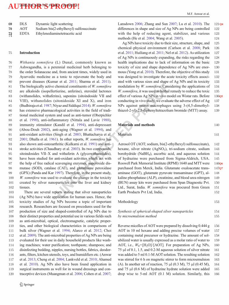
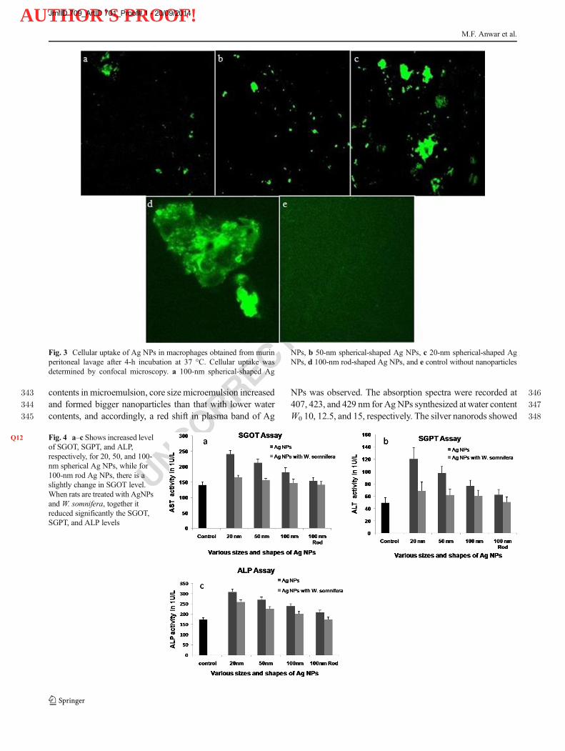
Fig. 3 Cellular uptake of Ag NPs in macrophages obtained from murinperitoneal lavage after 4-h incubation at 37 °C. Cellular uptake wasdetermined by confocal microscopy. a 100-nm spherical-shaped Ag
NPs, b 50-nm spherical-shaped Ag NPs, c 20-nm spherical-shaped AgNPs, d 100-nm rod-shaped Ag NPs, and e control without nanoparticles
Fig. 4Q12 a–c Shows increased levelof SGOT, SGPT, and ALP,respectively, for 20, 50, and 100-nm spherical Ag NPs, while for100-nm rod Ag NPs, there is aslightly change in SGOT level.When rats are treated with AgNPsand W. somnifera, together itreduced significantly the SGOT,SGPT, and ALP levels
M.F. Anwar et al.
JrnlID 709_ArtID 701_Proof# 1 - 20/09/2014

AUTHOR'S PROOF!
UNCORRECTEDPROOF
349 two plasma bands in absorption spectrum at 584 nm due to350 transverse plasmon band and the other at 435 nm due to351 longitudinal plasmon band. This confirmed that we were able352 to synthesize rod-shaped Ag nanomaterials from second syn-353 thetic route.
354 High-resolution transmission electron microscope analysis
355 The average sizes of Ag NPs synthesized by microemulsion356 synthesis route were calculated from HR-TEM image as 20,357 50, and 100 nm at 10, 12.5, and 15W0 (Fig. 1a–c), all having358 spherical shape. The second route generated rod-shaped nano-359 particles as seen from TEM image (Fig. 1d) which were in360 correlation with data from both dynamic light scattering361 (DLS) and UV–vis spectra.
362 Toxicity analysis
363 MTT assay
364 A dose-dependent response was observed in MTT assay for365 calculating LD50 values, and cytotoxicity data for Ag NPs and366 macrophages is presented in Fig. 2a–d. LD50 values for 20-nm367 Ag NPs were the most sensitive among all nanoparticles368 (Fig. 2a) inmacrophages. One hundred-nanometer rod-shaped369 Ag NPs (Fig. 2d) were found to be least sensitive in macro-370 phages, while 100-nm spherical-shaped Ag NPs were found371 to be more toxic. Cell viability was found to decrease with the372 decreasing size of Ag NPs and exposure time in macrophages.373 However, viability was always above than 80 % for treated374 groups with W. somnifera (23 days) and various sizes and375 shapes of Ag NPs (Fig. 2).376 LD50 values of 20-nm Ag NPs are much lower than those377 of 100-nm spherical Ag NPs at all exposure time points which378 has slightly higher values as compared to 100-nm rod-shaped379 Ag NPs after 12 h of exposure time. The LD50 values were380 0.25 mg/ml (confidence interval ±2.97), 0.35 mg/ml (confi-381 dence interval ±1.09), 0.35mg/ml (confidence interval ±1.09),382 and 0.35 mg/ml (confidence interval ±1.09) for 20, 50, and383 100-nm spherical-shaped Ag NPs and 100-nm rod-shaped Ag384 NPs, respectively. This shows that the small-sized Ag NPs385 have much cytotoxicity in comparison with 100-nm Ag NPs,386 and 100-nm rod-shaped Ag NPs have least toxicity in com-387 parison with spherical-shaped Ag NPs.
388 Cellular uptake of Ag NPs
389 Confocal laser scanning microphotographs of cell containing390 Ag NPs were taken, and the diameter of diffraction-limited391 spot corresponds to dB=1.22*ë/NA, with ë representing the392 excitation wavelength and NA numerical aperture of the ob-393 jective used with illuminations of specimens that are focused394 on the smallest possible spot.Q10 After 4-h exposure, 20, 50, and
395100-nm spherical-shaped and 100-nm rod-shaped Ag NPs396were visualized together with control without nanoparticles397(Fig. 3a–e). These analyses showed that high cellular uptake398in macrophages was obtained from murin peritoneal lavage399after 4-h incubation at 37 °C. With decreasing size of Ag NPs,400there had been rapid penetration in the cells that were dying401out of the population (Liu et al. 2013).
402Biochemical analysis
403Results of biochemical assay showed that serum glutamic404oxaloacetic transaminase (SGOT), serum glutamic pyruvic405transaminase (SGPT), and ALP levels increased with 20, 50,406and 100-nm Ag spherical NP-treated Wistar rats in compari-407son with control group. On the other hand, 100-nm rod Ag408NP-treated rats had slightly increased SGOT levels, and no409changes in SGPT and ALP levels were observed. Wistar rats410treated with Ag NPs along with W. somnifera significantly
Fig. 5 a Graph Q13showing that only 20-nm Ag NPs slightly increased thecreatinine, while 50/100-nm (spherical) and 100-nm Ag NPs (rod) andAg NPs with W. somnifera-treated groups have no effect on creatinineconcentration. b Graph showing that 20, 50, and 100-nm Ag NPs (spher-ical) increased the BUN level while Ag NPs (rod 100 nm) slightlychanged the BUN level. When rats are treated with Ag NPs andW. somnifera, together it reduced the BUN level
Q1Modulation of liver and kidney toxicity by herb Withania somnifera
JrnlID 709_ArtID 701_Proof# 1 - 20/09/2014

AUTHOR'S PROOF!
UNCORRECTEDPROOF
411 reduced SGOT, SGPT, and ALP levels in control group412 (Fig. 4a–c). Creatinine values were increased only for 20-nm413 Ag NPs while no changes were observed for 50 or 100 nm414 (spherical) and rod 100-nm Ag NPs alone and with415 W. somnifera-treated groups (Fig. 5a). Spherical-shaped 20,416 50, and 100-nm Ag NPs increased the BUN level.Q11 In rats417 treated with Ag NPs, along withW. somnifera, the BUN level418 reduced to normal level as in control group (Fig. 5b).
419 Histopathological examination
420 Photomicrographs of liver and kidney tissues showed histo-421 pathological changes in liver tissue on treatment with Ag NPs422 and are illustrated in Fig. 6. Control group showed normal423 liver architecture, portal triad (PT), and central vein (CV)424 whereas 20-nm Ag NP-treated animal showed vacuolation425 of hepatocytes, focal necrosis in the centrizonal area, inflam-426 mation, and dilated CV. Animals treated with 50-nm Ag NPs
427havemoderate sinusoidal dilatation in the centrizonal area and428no necrosis, and hepatocytic vacuolation has been seen in429portal triad (Fig. 6c). One hundred-nanometer spherical and430100-nm rod Ag NPs showed sinusoidal dilatation in the431centrizonal area, dilated CV, and moderate sinusoidal dilata-432tion in the centrizonal area, and normal PT and CV, respec-433tively (Fig. 6d, e).W. somnifera resulted in reduced toxicity of43420, 50, and 100-nmAgNPs as depicted with mild vacuolation435in centrizonal area and dilated CVand PT, large vacuolation of436hepatocytes in centrizonal area, inflammation with intact CV,437mild vacuolation, and normal PT and CV, respectively438(Fig. 6f–h). Rod-shaped 100-nm Ag NPs with W. somnifera439showed normal liver architecture with PT and CV (Fig. 6i).440Histopathological changes in kidney tissue were also ex-441amined; control group showed normal kidney architecture442with glomerula and normal cells, and treated groups by 20-443nm Ag NPs showed nuclear loss, karyolysis, hyalinization,444glomerular destruction, and necrosis and by 50-nm Ag NPs,
Fig. 6 Photomicrographsshowing histopathologicalchanges in liver tissue. a Controlgroup (×40) showing normal liverarchitecture, portal triad (PT), andcentral vein (CV). b Treated group(20-nm Ag NPs) (×40) showingvacuolation of hepatocytes andfocal necrosis in the centrizonalarea and inflammation and dilatedCV. c Treated group (50-nm AgNPs) (×40) showing moderatesinusoidal dilatation in thecentrizonal area and no necrosis,and hepatocytic vacuolation isseen PT. d Treated group (100-nmAg NPs) (×40) showingsinusoidal dilatation in thecentrizonal area and dilated CV. eTreated group (rod 100-nm AgNPs) (×40) showing moderatesinusoidal dilatation in thecentrizonal area and normal PTand CV. f Treated group (20-nmAg NPs with W. somnifera) (×40)showing mild vacuolation in thecentrizonal area and dilated CVand PT. g Treated group (50-nmAg NPs with W. somnifera) (×40)showing large vacuolation ofhepatocytes in centrizonal areaand inflammation with intact CV.h Treated group (Ag NPs 100 nmwithW. somnifera) (×40) showingmild vacuolation and normal PTand CV. i Treated group (rod 100-nm Ag NPs with W. somnifera)(×40) showing normal liverarchitecture with PT and CV
M.F. Anwar et al.
JrnlID 709_ArtID 701_Proof# 1 - 20/09/2014

AUTHOR'S PROOF!
UNCORRECTEDPROOF
445 mild hyalinization, eosinophilia, and mild glomerular destruc-446 tion (Fig. 7a–c.) Spherical 100 nm and rod-shaped 100-nmAg
447NPs showed normal glomerula and mild hyalinization448(Fig. 7d, e). Q14W. somnifera significantly normalized the
Fig. 7 Photomicrographsshowing histopathologicalchanges in kidney tissue. aControl group (×40) showingnormal kidney architecture withglomerula and normal cells. bTreated group (20-nm Ag NPs)(×40) showing nuclear loss,karyolysis, hyalinization,glomerular destruction, andnecrosis. c Treated group (50-nmAg NPs) (×40) showing mildhyalinization, eosinophilia, andmild glomerular destruction. dTreated group (100-nm Ag NPs)(×40) showing normal glomerulaand mild hyalinization. e Treatedgroup (rod 100-nm Ag NPs)(×40) showing normal glomerulaand mild hyalinization. f Treatedgroup (20-nm Ag NPs withW. somnifera) (×40) showinghyalinization, eosinophilia, mildglomerular destruction, and nonecrosis. g Treated group (50-nmAg NPs withW. somnifera) (×40)showing mild hyalinization, noglomerular destruction, and nonecrosis. h Treated group (AgNPs 100 nm with W. somnifera)(×40) showing normal kidneyarchitecture and glomerula. iTreated group (rod 100-nm AgNPs with W. somnifera) (×40)showing normal kidneyarchitecture and glomerula
Q1Modulation of liver and kidney toxicity by herb Withania somnifera
JrnlID 709_ArtID 701_Proof# 1 - 20/09/2014

AUTHOR'S PROOF!
UNCORRECTEDPROOF
449 changes with 20-nm Ag NPs and 50-nm Ag NPs, resulting in450 mild to negligible hyalinization, eosinophilia, glomerular de-451 struction, and necrosis for both (Fig. 7f, g).W. somnifera with452 100-nm spherical-shaped and 100-nm rod-shaped Ag NPs453 showed normal kidney architecture, glomerula, and cells454 (Fig. 7h, i).455
456 Discussion
457 Ag NPs have been used in health implications as anti-458 microbial agents and disinfectants for a very long time (Choi459 et al. 2009). As information presented in the literature, differ-460 ent sizes and shapes of Ag NPs have different toxic effects461 (Edwards-Jones 2009;Q15 Kim et al. 2013; Nel et al. 2006). In the462 present study, a comparison was done between different sizes463 and shapes of Ag NPs for their acute oral toxicity (Park and464 Bae 2010) for their potential application as anti-microbial465 agents, and acute toxicity modulation by W. somnifera was466 observed. In a previous study, bare Ag NPs (42 nm) in467 distilled water with doses of 0.25, 0.5, and 1 mg/kg induce468 hepatotoxicity by repeated oral administration (Kim et al.469 2009; Oberdorster et al. 2005). Particle morphology plays a470 significant role in the toxicology of various kinds of nanopar-471 ticles (Q16 Larissa et al. 2011; Sharma 2010).472 TheQ17 absorbance spectroscopy data of Ag NP solution was473 explored, and a comparison between various sizes and shapes474 of Ag NPs investigated single peaks for 20, 50, and 100 nm475 spherical-shaped Ag NPs at different wavelengths and two476 peaks for 100-nm rod-shaped Ag NPs due to the distribution477 of radii and orientation of the particles in the solution,478 respectively.479 The different sizes and shapes of nanoparticles can produce480 different biological responses and severe toxicity to tissue481 causing cell injury (Lam et al. 2004). Many studies suggested482 that crystalline small particles can inducemore toxic to cells as483 well as being more persistent in the tissues (Q18 Linda et al. 2011).484 In this study, various sizes and shapes of Ag NPs produce485 different cell viability in macrophages.Q19 From the results ob-486 tained, it appears that 100-nm spherical- and rod-shaped Ag487 NPs caused no significant change, while at 20- and 50-nm488 spherical shapes of Ag NPs, there is a significant increase in489 plasma SGOT, SGPT, and ALP activities. The elevated levels490 of SGOT, SGPT, and ALP indicated a diseased level.Q20 SGPT491 and SGOT both are the liver-specific enzymes and used to492 diagnose liver diseases (Yong et al. 2010; Yadav et al. 2013;493 Shayestech et al. 2014).Q21 The studies showed that silver dis-494 rupts the activity of mitochondria, and this causes a rise in495 plasma transaminase (GOT, GPT) activity which is a sensitive496 indicator of damage to cytoplasmic and/or mitochondrial497 membranes (Oberdorster et al. 2005; Fabian et al. 2013).498 Slight effect of BUN and creatinine concentration was
499measured. Acute oral toxicity was also confirmed by histo-500pathological changes in liver and kidney tissues that revealed501marked mononuclear cell infiltration, nuclear degeneration,502architectural disarray, glomerular destruction, and necrosis.503Q22On the other hand, the associated anatomy of these structures504causes the damage of a part leading to disruption of the other505parts (Yadav et al. 2013; Fabian et al. 2013; Singh et al. 2001).506Spherical-shaped Ag NPs increased glomerular destruc-507tion, and central vein and portal triad destruction causes severe508hepatorenal toxicity. Q23Hepatorenal and renal toxicity of Ag NPs509was reduced significantly when the rats were treated with510W. somnifera in the present case which indicated that511W. somnifera has free radical scavenging enzymes that512worked as anti-oxidant and normalizes all enzymatic activities513and improvesBUNand creatinine concentration.W. somnifera514should be used and worked further as a toxicity modulator.
515Conclusion
516It is demonstrated that Ag NPs show toxicity in macrophages517and Wistar rats. The toxicity of spherical-shaped Ag NPs is518inversely proportional to their size which is due to deeper and519higher penetration of small-sized nanoparticles; 100-nm rod-520shaped Ag NPs showed negligible toxicity in comparison to521100-nm spherical-shaped nanoparticles in macrophages under522in vitro conditions and in Wistar rats. When Ag NPs of523different shapes and sizes were treated with extracts of524W. somnifera administered to rats, the toxic effects were found525to be reduced to a significant extent. This study indicates the526potential use of W. somnifera extract as an anti-oxidant and527anti-inflammatory against the toxic effects of different shapes528and sizes of Ag NPs. The prospective applications529W. somnifera will necessitate significant consideration by530scientific community in the future.
531Acknowledgments Authors are thankful to Dr. G. N. Qazi, Vice Chan-532cellor, Jamia Hamdard, New Delhi, for providing working facilities.533Authors are also grateful to the Department of Science and Technology534(DST), Government of India, for financial support.
535536Conflict of interests The author(s) declare that they have no competing537interests.
538539Authors’ contributions Ag NPs were synthesized, characterized, and540studied in vivo by MFA and DY. Statistical analyses were done by MFA,541DY, RKK, JC, SR, IA, and MS that were involved in the inception and542planning of the project and in the preparation of the manuscript. All543authors read and approved the final manuscript.
544References 545
546Abou-Douh AM (2002) New withanolides and other constituents from547the fruit of Withania somnifera. Arch Pharm 335:267–276
M.F. Anwar et al.
JrnlID 709_ArtID 701_Proof# 1 - 20/09/2014

AUTHOR'S PROOF!
UNCORRECTEDPROOF
548 Ahamed M, Alsalhi MS, Siddiqui MKJ (2010) Silver nanoparticle appli-549 cations and human health. Clin Chim Acta 411:1841550 Alarco EI, Udekwu K, Skog M et al (2012) The biocompatibility and551 antibacterial properties of collagen-stabilized photochemically pre-552 pared silver nanoparticles. Biomaterials 33:4947553 Allain CC, Poon LS, Chan CSG et al (1978) Enzymatic determination of554 total serum urea. Clin Chem 20:470–475555 Anwar MF, Yadav D, Kapoor S et al (2013) Comparison of antibacterial556 activity of Ag nanoparticles synthesized from leaf extract of557 Parthenium hystrophorus L in aqueous media and gentamicin sul-558 phate: in-vitro. Drug Dev Ind Pharm. doi:10.3109/03639045.2013.559 845840560 Bhattacharya A, Ghosal S, Bhattacharya SK (2001) Anti-oxidant effect of561 Withania somnifera glycol withanolides in chronic foot shock stress-562 induced perturbations of oxidative free radical scavenging enzymes563 and lipid peroxidation in rat frontal cortex and striatum. J Ethno564 Pharmacol 74:1–6565 Budhiraja RD, Sudhir S, Garg KN et al (1987) BC: review of biological566 activity of withanolides. J Sci Ind Res 46:488–491567 Carlson C, Hussain SM, Schrand AM et al (2008) Unique cellular568 interaction of silver nanoparticles: size-dependent generation of569 reactive oxygen species. The J Physic Chem B 112:13608–570 13619571 Chaudhary G, Sharma U, Jagannathan NR et al (2003) Evaluation of572 Withania somnifera in a middle cerebral artery occlusion model of573 stroke in rats. Clin Exp Pharmacol Physiol 30:399–404574 Cheng D, Yang J, Zhao Y (2004) Antibacterial materials of silver nano-575 particles application in medical appliances and appliances for daily576 use. Chin Med Equip J 4:26–32577 Choi O, Clevenger TE, Deng B et al (2009) Role of sulfide and ligand578 strength in controlling nanosilver toxicity. Water Res 43:1879–579 1886580 Cohen MS, Stern JM, Vanni AJ et al (2007) In vitro analysis of a581 nanocrystalline silver-coated surgical mesh. Surg Infect 8:397–403582 Dhalla NS, Sastry NS, Malhotra CL (1961) Chemical studies of the583 Withania somnifera. J Pharm Sci 50:876–877584 Edwards-Jones V (2009) The benefits of silver in hygiene, personal care585 and health care. Lett Appl Microbiol 49:147–152586 Fabian H, Martin JD, Rothen-Rutishauser CB et al (2013) Exposure of587 silver-nanoparticles and silver-ions to lung cells in vitro at the air-588 liquid interface. Part Fibre Toxicol 10:11589 He BL, Tanm JJ, Kong YL et al (2004) Synthesis of size controlled Ag590 nanoparticles. J Mol Catal A: Chem 221:121–126591 Huiliang C, Xuanyong L, Fanhao M et al (2011) Biological actions of592 silver nanoparticles embedded in titanium controlled by micro-593 galvanic effects. Biomaterials 32:693–705594 Husdan H, Rapoport A (1968) Estimation of creatinine by the Jaffe595 reaction: a comparison of three methods. Clin Chem 14:222–238596 Kandil FE, Elsayeh NH, Abou-Douh AM et al (1994) Flavonol glyco-597 sides and phenolics from Withania somnifera. Phytochemistry 37:598 1215–1216599 Kim WY, Kim J, Park JD et al (2009) Histological study of gender600 differences in accumulation of silver nanoparticles in kidneys of601 Fischer 344 rats. J Toxicol Environ Health A 72:1279–84602 King PRM, King EJ (1954) Estimation of plasma phosphatase by deter-603 mination of hydrolysed phenol with amino-antipyrene. J Clin Path604 7:322–326605 Kulkarni RR, Patki PS, Jog VP et al (1991) Treatment of osteoarthritis606 with a herbomineral formulation: a double-blind, placebo-con-607 trolled, cross-over study. J Ethnopharmacol 33:91–95608 Kumar OA, Jyothirmayee G, Tata SS (2011) In vitro plant regeneration609 from leaf explants of Withania somnifera (L) Dunal610 (Ashwaganda)—an important medicinal plant. Res Biotech 2:34–3611 Lam PK, Chan ES, Ho WS et al (2004) In vitro cytotoxicity testing of a612 nanocrystalline silver dressing (Acticoat) on cultured keratinocytes.613 Br J Biomed Sci 61:125–127
614Lankveld DP, Oomen AG, Krystek P et al (2010) The kinetics of the615tissue distribution of silver nanoparticles of different sizes.616Biomaterials 31:8350–8361617Lansdown A (2006) Silver in health care: antimicrobial effects and safety618in use. Curr Probl Dermatol 33:17–34619Larissa VS, Andrea AD, Jong SK et al (2011) Nanosilver induces620minimal lung toxicity or inflammation in a subacute murine inhala-621tion model. Part Fibre Toxicol 8:5622Linda CS, Edgar G, Andreas S et al (2011) Shapematters: effects of silver623nanospheres and wires on human alveolar epithelial cells. Part Fibre624Toxicol 8:36625Liu L, Yang J, Xie J et al (2013) The potent antimicrobial properties of626cell penetrating peptide-conjugated silver nanoparticles with excel-627lent selectivity for gram-positive bacteria over erythrocytes.628Nanoscale 5:3834–40629Lu W, Senapati D, Wang S et al (2010) Effect of surface coating on the630toxicity of silver nanomaterials on human skin keratinocytes. Chem631Phys Lett 487:92632Muangman P, Chuntrasakul C, Silthram S et al (2006) Comparison of633efficacy of 1% silver sulfadiazine and Acticoat for treatment634of partial-thickness burn wounds. J Med Assoc Thai 89:953–635958636Nel A, Xia T, Madler L et al (2006) Toxic potential of materials at the637nanolevel. Science 311:622–627638Nel A, Xia T, Meng H et al (2012) Nanomaterial toxicity testing in639the 21st century: use of a predictive toxicological approach and640high throughput screening. Acc Chemi Res. doi:10.1021/641ar300022h642Nittala SS, Lavie S (1988) Chemistry and genetics of withanolides in643Withania somnifera hybrids. Phytochemistry 20:2741–2748644Niyaz A, Siddiqui EN (2014) Seed germination of Withania somnifera645(L.) Dunal. Eur J Med Plants 4:920–926646Oberdorster G, Maynard A, Donaldson K et al (2005) Principles for647characterizing the potential human health effects from exposure to648nanomaterials: elements of a screening strategy. Part Fibre Toxicol6492:8650Oberpichler H, Sauer D, Rossberg C et al (1990) PAF antagonist651ginkgolide B reduces postischemic neuronal damage in rat brain652hippocampus. J Cereb Blood Flow Metab 10:133653Panda S, Kar A (1997) Evidence for free radical scavenging activity of654Ashwagandha root powder in mice. Ind J Physio Pharmaco 41:424–655426656Park E, Bae E (2010) Repeated-dose toxicity and inflammatory responses657in mice by oral administration of silver nanoparticles. Environ658Toxicol Pharm 30:162–168659Park J, Lim DH, Lim HJ et al (2011) Size dependent macrophage660responses and toxicological effects of Ag nanoparticles. Chem661Commun 47:4382662Reitman S, Frankel S (1957) Glutamic—pyruvate transaminase assay by663colorimetric method. Am J Clin Path 28:56664Sharma M (2010) Understanding the mechanism of toxicity of carbon665nanoparticles in humans in the new millennium: a systemic review.666Indian J Occup Environ Med 14:3–5667Sharma V, Sharma S, Pracheta et al (2011) Withania somniforma: a668rejuvinating Ayurvedic medicinal herb for the treatment of various669human ailments. Intl J Pharm Tech Res 3:187–192670Shayestech TH, Khajavi F, Ghasemi H et al (2014) Effects of silver671nanoparticle (Ag NP) on oxidative stress, liver function in rat:672hepatotoxic or hepatoprotective? Issues Biol Sci Pharma Res 2:67340–44674Singh B, Saxena AK, Chandan BK et al (2001) Hepatoprotective activity675of indigtone—a bioactive fraction from Indigo feratinctoria Linn.676Phytother Res 15:294–7677Singh B, Chandan BK, Gupta DK (2003) Adaptogenic activity of a novel678withanolide-free aqueous fraction from the roots of Withania679somnifera Dun. (Part II). Phytother Res 17:531–6
Q1Modulation of liver and kidney toxicity by herb Withania somnifera
JrnlID 709_ArtID 701_Proof# 1 - 20/09/2014

AUTHOR'S PROOF!
UNCORRECTEDPROOF
680 Wagner H, Norr H,Winterhoff H (1994) Plantadaptogens. Phytomedicine681 1:63–76682 Wang H, Qiao X, Chen J, Ding S (2005) Preparation of silver nanopar-683 ticles by chemical reduction method. Coll Surf A Physico Chem684 Eng Asp 256:111–115685 Wilhelm M, Zhao CL, Wang Yet al (1991) Poly(styrene-ethylene oxide)686 block copolymer micelle formation in water: a fluorescence probe687 study. Macromolecules 24:1033–1040688Q24 Wim H, De J, Leo TM, Van DV et al (2013) Systemic and689 immunotoxicity of silver nanoparticles in an intravenous 28
690days repeated dose toxicity study in rats. Biomaterials 34:6918333–8343692Yadav D, Chaudhary AA, Garg V et al (2013) In vitro toxicity and693antidiabetic activity of a newly developed polyherbal formulation694(MAC-ST/001) in streptozotocin-induced diabetic Wistar rats.695Protoplasma 250:741–749696Yong SK, Moon YS, Jung DP et al (2010) Subchronic oral toxicity of697silver nanoparticles. Part Fibre Toxicol 7:20698Zhang Y, Sun J (2007) A study on the bio-safety for nano-silver as anti-699bacterial materials. Chin J Med Instrum 31:35–38
700
M.F. Anwar et al.
JrnlID 709_ArtID 701_Proof# 1 - 20/09/2014

AUTHOR'S PROOF!
UNCORRECTEDPROOF
AUTHOR QUERIES
AUTHOR PLEASE ANSWER ALL QUERIES.
Q1. Please check the suggested running page title if appropriate. Otherwise, please provide shortrunning title with maximum of 65 characters including spaces.
Q2. Please check if the captured Email address is correct.Q3. Please check if affiliation 4 is presented correctly.Q4. The sentence “The differences in shape and size of Ag…” has been edited for clarity. Please
check that the intended meaning was retained.Q5. The sentence “Before conducting in vivo study…” has been edited for clarity. Please check that
the intended meaning was retained.Q6. The term “Roswell Park Memorial Institute” was provided as the definition for “RPMI.” Please
check and amend as necessary.Q7. “Span Diagonist Pvt. Ltd” was changed to “Span Diagnostic Pvt. Ltd.” Please check and amend
as necessaryQ8. The sentence “Approximately, 1 g of dried powder ofW. somnifera root is…” has been edited for
clarity. Please check that the intended meaning was retained.Q9. The sentence “The acute toxicity of Ag nanoparticles…” has been edited for clarity. Please check
that the intended meaning was retained.Q10. The sentence “After 4 h exposure, 20, 50, and 100-nm spherical-shaped…” has been edited for
clarity. Please check that the intended meaning was retained.Q11. The sentence “In rats treated with Ag NPs, along withW. somnifera...” has been edited for clarity.
Please check that the intended meaning was retained.Q12. In Fig. 4 caption, “(d) while Ag NPs (100 nm rod) have slightly change in SGOT level” was
changed to “while for 100-nm rod Ag NPs, there is a slightly change in SGOT level” as there isno panel d found. Please check that the intended meaning was retained.
Q13. Figure 5 caption has been edited for clarity. Please check that the intended meaning was retained.Q14. The citation of “Fig. 9f and g” was changed to “Fig. 7f, g” as there was no Fig. 9 artwork. Please
check and amend as necessary.Q15. “Kim et al. 2013” is cited in the body but its bibliographic information is missing. Kindly provide
its bibliographic information. Otherwise, please delete it from the body.Q16. The citation “Larissa et al. 2001” (original) has been changed to “Larissa et al. 2011”. Please
check if appropriate.Q17. The sentence “The absorbance spectroscopy data of Ag NP…” has been edited for clarity. Please
check that the intended meaning was retained.Q18. The citation “Linda et al. 2001” (original) has been changed to “Linda et al. 2011”. Please check
if appropriate.Q19. The sentence “From the results obtained, it appears that…” has been edited for clarity. Please
check that the intended meaning was retained.Q20. The sentence “SGPT and SGOT both are the liver-specific enzymes…” has been edited for
clarity. Please check that the intended meaning was retained.Q21. The sentence “The studies showed that silver disrupts..” has been edited for clarity. Please check
that the intended meaning was retained.Q22. The sentence “On the other hand, the associated…” has been edited for clarity. Please check that
the intended meaning was retained.Q23. The sentence “Hepatorenal and renal toxicity of Ag NPs was…” has been edited for clarity.
Please check that the intended meaning was retained.Q24. Wim et al. (2013) was not cited anywhere in the text. Please provide a citation. Alternatively,
delete the item from the list.
Copyright © 2022 FDOKUMEN