Advanced nickel nanoparticles technology - De Gruyter
25
Review Article Nuru-Deen Jaji, Hooi Ling Lee, Mohd Hazwan Hussin, Hazizan Md Akil, Muhammad Razlan Zakaria, and Muhammad Bisyrul Hafi Othman* Advanced nickel nanoparticles technology: From synthesis to applications https://doi.org/10.1515/ntrev-2020-0109 received November 1, 2020; accepted November 19, 2020 Abstract: Over the last decade, nickel nanoparticles (NiNPs) have been investigated for various potential applications due to their superior ferromagnetic proper- ties such as magneto-crystalline anisotropy, high coer- cive forces, and chemical stability. Therefore, there has been a tremendous enhancement in the synthesis tech- niques, proposed reaction mechanisms, and applications of NiNPs. This paper presents a recent overview of the synthesis, reaction mechanisms, and applications of NiNPs. NiNPs in the size range of 1–100 nm are synthesized by various methods for research and commercial applica- tions. The synthesis techniques are classified into three main types, namely, top-down, bottom-up, and hybrids of top-down and bottom-up protocols including sol- vothermal, physical, and chemical approaches. The detailed reaction mechanisms in the formation of NiNPs, especially for biosynthesis techniques, are extensively described. Trends in NiNP applications in fields such as biomedical, catalysis, supercapacitors, and dye-sensitized solar cells are explored. The basic advantages and role of NiNPs as a catalyst for various reactions are illu- strated here. Keywords: nickel, nanoparticle, synthesis, mechanism, application 1 Introduction In the last decade, nanotechnology has broadened the scope for researchers, producers, and consumers in almost all sectors by enabling the engineering of functional sys- tems at the nanoscale level mostly in the form of nano- particles [1]. Nanoparticles are the raw materials used in nanotechnology [2]. NiNPs have gained much attention due to their unique magnetic, chemical, and physical properties as well as their potential applications in various technological fields such as catalysis [3], battery manufac- ture [4], novel ink for nanotube-printing [5], incorporation in textile [6], enhanced pseudo-capacitance [7], field- modulated gratings and optical switches [8], direct immo- bilization of biomolecules through magnetic force of NiNPs [9], and adsorption of yellow dyes [10]. In compar- ison to other magnetic nanoparticles, Ni nanoparticles possess great potential as a catalyst in reactions and pro- pellant and sintering additive in coatings, plastics, and fibres [11]. Due to its relative abundance in the earth’s crust, Ni is more cost-effective than most of the metals in use as a catalyst [12]. The electrical conductivity of Ni enables its use in several applications [13]. NiNPs can be used as nanofluids in high purity, ultra-high purity, pas- sivated, coated, and distributed forms [14]. Review articles on nickel abound in literature such as machinability of nickel-based superalloys [15], overview of nanostructured metal oxides and pure nickel oxides electrodes for supercapacitors [16], nickel-based mate- rials for supercapacitors [17], on the performance of com- mercially available corrosion-resistant nickel alloys [18], recent advances in the stabilization of nickel and nickel oxide nanoparticles [19], and a brief overview on grain growth of bulk electrodeposited nanocrystalline nickel Nuru-Deen Jaji: School of Chemical Sciences, Universiti Sains Malaysia, 11800 Minden, Penang, Malaysia; Department of Chemistry, Federal College of Education Technical Gombe, P.M.B. 060, Gombe, Gombe State, Nigeria Hooi Ling Lee: Nanomaterials Research Group, School of Chemical Sciences, University Sains Malaysia, 11800 Minden, Penang, Malaysia Mohd Hazwan Hussin: School of Chemical Sciences, Universiti Sains Malaysia, 11800 Minden, Penang, Malaysia Hazizan Md Akil: School of Materials and Minerals Resources Engineering, Engineering Campus, Universiti Sains Malaysia, 14300 Nibong Tebal, Pulau Pinang, Malaysia Muhammad Razlan Zakaria: Faculty of Chemical Engineering Technology, Universiti Malaysia Perlis (UniMAP), Perlis, Malaysia * Corresponding author: Muhammad Bisyrul Hafi Othman, School of Chemical Sciences, Universiti Sains Malaysia, 11800 Minden, Penang, Malaysia, e-mail: [email protected], tel: +604-653-4032; fax: +604-657-4854 Nanotechnology Reviews 2020; 9: 1456–1480 Open Access. © 2020 Nuru-Deen Jaji et al., published by De Gruyter. This work is licensed under the Creative Commons Attribution 4.0 International License.
-
Upload
khangminh22 -
Category
Documents
-
view
0 -
download
0
Transcript of Advanced nickel nanoparticles technology - De Gruyter

Review Article
Nuru-Deen Jaji, Hooi Ling Lee, Mohd Hazwan Hussin, Hazizan Md Akil,Muhammad Razlan Zakaria, and Muhammad Bisyrul Hafi Othman*
Advanced nickel nanoparticles technology: Fromsynthesis to applications
https://doi.org/10.1515/ntrev-2020-0109received November 1, 2020; accepted November 19, 2020
Abstract: Over the last decade, nickel nanoparticles(NiNPs) have been investigated for various potentialapplications due to their superior ferromagnetic proper-ties such as magneto-crystalline anisotropy, high coer-cive forces, and chemical stability. Therefore, there hasbeen a tremendous enhancement in the synthesis tech-niques, proposed reaction mechanisms, and applicationsof NiNPs. This paper presents a recent overview of thesynthesis, reaction mechanisms, and applications ofNiNPs. NiNPs in the size range of 1–100 nmare synthesizedby various methods for research and commercial applica-tions. The synthesis techniques are classified into threemain types, namely, top-down, bottom-up, and hybridsof top-down and bottom-up protocols including sol-vothermal, physical, and chemical approaches. Thedetailed reaction mechanisms in the formation of NiNPs,especially for biosynthesis techniques, are extensivelydescribed. Trends in NiNP applications in fields such asbiomedical, catalysis, supercapacitors, and dye-sensitizedsolar cells are explored. The basic advantages and role of
NiNPs as a catalyst for various reactions are illu-strated here.
Keywords: nickel, nanoparticle, synthesis, mechanism,application
1 Introduction
In the last decade, nanotechnology has broadened thescope for researchers, producers, and consumers in almostall sectors by enabling the engineering of functional sys-tems at the nanoscale level mostly in the form of nano-particles [1]. Nanoparticles are the raw materials used innanotechnology [2]. NiNPs have gained much attentiondue to their unique magnetic, chemical, and physicalproperties as well as their potential applications in varioustechnological fields such as catalysis [3], battery manufac-ture [4], novel ink for nanotube-printing [5], incorporationin textile [6], enhanced pseudo-capacitance [7], field-modulated gratings and optical switches [8], direct immo-bilization of biomolecules through magnetic force ofNiNPs [9], and adsorption of yellow dyes [10]. In compar-ison to other magnetic nanoparticles, Ni nanoparticlespossess great potential as a catalyst in reactions and pro-pellant and sintering additive in coatings, plastics, andfibres [11]. Due to its relative abundance in the earth’scrust, Ni is more cost-effective than most of the metals inuse as a catalyst [12]. The electrical conductivity of Nienables its use in several applications [13]. NiNPs can beused as nanofluids in high purity, ultra-high purity, pas-sivated, coated, and distributed forms [14].
Review articles on nickel abound in literature such asmachinability of nickel-based superalloys [15], overviewof nanostructured metal oxides and pure nickel oxideselectrodes for supercapacitors [16], nickel-based mate-rials for supercapacitors [17], on the performance of com-mercially available corrosion-resistant nickel alloys [18],recent advances in the stabilization of nickel and nickeloxide nanoparticles [19], and a brief overview on graingrowth of bulk electrodeposited nanocrystalline nickel
Nuru-Deen Jaji: School of Chemical Sciences, Universiti SainsMalaysia, 11800 Minden, Penang, Malaysia; Department ofChemistry, Federal College of Education Technical Gombe,P.M.B. 060, Gombe, Gombe State, NigeriaHooi Ling Lee: Nanomaterials Research Group, School of ChemicalSciences, University Sains Malaysia, 11800 Minden, Penang,MalaysiaMohd Hazwan Hussin: School of Chemical Sciences, UniversitiSains Malaysia, 11800 Minden, Penang, MalaysiaHazizan Md Akil: School of Materials and Minerals ResourcesEngineering, Engineering Campus, Universiti Sains Malaysia, 14300Nibong Tebal, Pulau Pinang, MalaysiaMuhammad Razlan Zakaria: Faculty of Chemical EngineeringTechnology, Universiti Malaysia Perlis (UniMAP), Perlis, Malaysia
* Corresponding author: Muhammad Bisyrul Hafi Othman, Schoolof Chemical Sciences, Universiti Sains Malaysia, 11800 Minden,Penang, Malaysia, e-mail: [email protected], tel: +604-653-4032;fax: +604-657-4854
Nanotechnology Reviews 2020; 9: 1456–1480
Open Access. © 2020 Nuru-Deen Jaji et al., published by De Gruyter. This work is licensed under the Creative Commons Attribution 4.0International License.

and nickel–iron alloys [7]. However, these reviews didnot focus on NiNPs; they discussed nickel oxides andalloys. Hence, there is the need for thorough analysesof synthesis, reaction mechanisms, and applications ofNiNPs.
The synthesis of Ni nanoparticles has often beenassociated with various challenges. These may includethe difficulty of reducing Ni(II) to Ni(0) at room tempera-ture [20]. Most synthetic protocols involve the use of dis-tilled water to wash the Ni nanoparticles, but Ni nano-particles are easily oxidized in air to form NiO, Ni2O3, Ni(OH)2, or NiOOH, making the synthesis difficult [21]. Houet al. reported that, in comparison to other magneticmetals such as Co, Pt, and Fe, there are few reports onthe synthesis of monodispersed Ni nanoparticles [22].Similar observation elsewhere constitutes a challenge inthe fabrication of Ni nanoparticles [23]. In compliancewith Chen and Hsieh [24], pure Ni nanoparticles in anonmagnetic environment undergo oxidation. Despitethe high electrocatalytic performance exhibited by Ninanoparticles, its poor stability poses a real challenge[25]. The magnetic properties of Ni nanoparticles interferewith magnets of microwave systems during synthesisusing the microwave. This makes it difficult to obtain agood yield of the Ni nanoparticles [26]. NiNPs tend tocluster on formation, due to their magnetic properties,and hence, to obtain monodispersed Ni nanoparticleswill require special techniques [27]. NiNPs must be char-acterized immediately after production to avoid conver-sion to Ni compounds like oxides and hydroxides. Thesefactors must be taken into consideration in planning thesynthesis of Ni nanoparticles [28].
Global rising of environmental carbon costs havemade it necessary for scientific research and industrialenterprise to invest in energy-saving and environmen-tally friendly alternative synthesis of Ni nanoparticlesby plant-mediated pathways. Green plant productionof Ni Nanoparticles is the use of plant parts, plantextracts, plant products, and organisms, leading toenvironmentally friendly products that are harmless tothe ecosystem. The green production of metallic nano-particles employing plant extracts as a reducing agent isconsidered economically viable and environmentallyfriendly.
The present study was designed to analyse recentsynthesis, reaction mechanisms, and applications of Ninanoparticles as well as the specific advantages of usingNi as a catalyst. The objectives of this article are based onhighlighting recent techniques in the synthesis of Ninanoparticles, ranging from green chemical methods to
physical methods; in addition, to discuss the reactionmechanisms leading to the formation of Ni nanoparticles,as proposed and reported in recent literature. Furthermore,the scope of this study will be limited to addressing theapplications of Ni nanoparticles in various fields of nano-technology, science, and biomedical sciences as well as todiscuss the specific advantages of Ni nanoparticles ascatalysts in reactions, as compared to other magneticnanoparticles.
2 Techniques for the synthesisof NiNPs
Top-down synthesis methods and bottom-up synthesisprotocols have traditionally been adopted for the synth-esis of Ni nanoparticles. Top-down synthesis protocolsinclude breaking down bulk materials into nanoscalematerials. The most widely used top-down nanoparticlesynthesis methods are mechanical milling, laser ablation,nanolithography, thermal decomposition, and sputtering[29]. Bottom-up protocols include the utilization ofmetallic oxides and metallic salts as precursors for thereactions. These salts or oxides are eventually reduced tometallic nanoparticles using an appropriate solvent andreducing agent. In these techniques, the disperse mode,shape, and dimensions of the nanoparticles can bemanipulated by varying the concentration of the pre-cursor and the reducing agent, the pH, temperature,heating time, and the type and choice of the stabilizingagent [30]. Several bottom-up approaches for the fabri-cation of Ni nanosized materials have been reported.These are spatting, solvent-gel, gas evaporation, copre-cipitation techniques, and plant-mediated synthesis[21]. Similarly, Ni nanoparticles have been fabricated innonaqueous and aqueous media by chemical reaction,sonication [31], radiolytic [32], and chemical reductionand freeze-drying reactions [33].
2.1 Top-down methods for the fabricationof NiNPs
2.1.1 Mechanical milling
Mechanical milling is the most common method of gen-erating diverse nanoparticles among the different top-down methods. Mechanical milling is used during
Advanced nickel nanoparticles technology: from synthesis to applications 1457
synthesis for milling as well as post-annealing of nano-particles where many elements are milled in an inertatmosphere. Mechanical milling is influenced by plasticdeformation, contributing to a specific shape, whereasfracture leads to a decrease in the particle size whilecold welding increases the particle size [34].
2.1.2 Laser ablation
Laser ablation synthesis is a method of production ofnanoparticles from different solvents. Nanoparticles aregenerated by irradiation of a metal immersed in a liquidsolution by a laser beam which condenses the plasmaplume to produce the nanoparticles. Laser ablationsynthesis is a reliable technique as well as an alternativeto a chemical metal reduction in the synthesis of nano-particles. This method provides a stable synthesis ofnanoparticles in organic solvents and water withoutrequiring stabilizing or capping agent [35].
2.1.3 Thermal decomposition
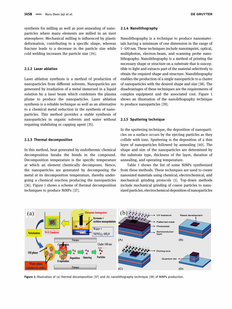
In this method, heat generated by endothermic chemicaldecomposition breaks the bonds in the compound.Decomposition temperature is the specific temperatureat which an element chemically decomposes. Hence,the nanoparticles are generated by decomposing themetal at its decomposition temperature, thereby under-going a chemical reaction producing the nanoparticles[36]. Figure 1 shows a scheme of thermal decompositiontechniques to produce NiNPs [37].
2.1.4 Nanolithography
Nanolithography is a technique to produce nanomater-ials having a minimum of one dimension in the range of1–100 nm. These techniques include nanoimprint, optical,multiphoton, electron-beam, and scanning probe nano-lithography. Nanolithography is a method of printing thenecessary shape or structure on a substrate that is suscep-tible to light and extracts part of the material selectively toobtain the required shape and structure. Nanolithographyenables the production of a single nanoparticle to a clusterof nanoparticles with the desired shape and size [38]. Thedisadvantages of these techniques are the requirements ofcomplex equipment and the associated cost. Figure 1shows an illustration of the nanolithography techniqueto produce nanoparticles [39].
2.1.5 Sputtering technique
In the sputtering technique, the deposition of nanoparti-cles on a surface occurs by the ejecting particles as theycollide with ions. Sputtering is the deposition of a thinlayer of nanoparticles followed by annealing [40]. Theshape and size of the nanoparticles are determined bythe substrate type, thickness of the layer, duration ofannealing, and operating temperature.
Table 1 shows the list of some NiNPs synthesizedfrom these methods. These techniques are used to createnanosized materials using chemical, electrochemical, andmechanical grinding protocols [1]. Top-down methodsinclude mechanical grinding of coarse particles to nano-sizedparticles, electrochemical deposition ofnanoparticles
Figure 1: Illustration of (a) thermal decomposition [37] and (b) nanolithography technique [39] of NiNPs production.
1458 Nuru-Deen Jaji et al.

fromametal electrode [2], laser ablation [3], and sputtering[4]. Selected top-down techniques for the fabrication of Ninanoparticles are presented in Table 1.
2.2 Bottom-up synthesis protocols infabricating Ni nanomaterials
2.2.1 Sol–gel technique
This technique involves the sol – a colloidal solution ofsolids suspended in a liquid phase and the gel – a solidmacromolecule submerged in a solvent. Most of themetallic nanoparticles can be synthesized by this methodand it is the most preferred bottom-up method due to itssimplicity. This is a wet-chemical process in which theprecursor is a chemical solution containing an integratedsystem of discrete particles [52]. Metal oxides and saltsare the typically used precursors in the sol–gel process.The precursor is then dispersed in a host liquid either bysonication, shaking, or stirring and the resultant system
contains a liquid and a solid phase. Various methodssuch as filtration, sedimentation, and centrifugation areused to recover the nanoparticles. Figure 2 shows anillustration of the sol–gel technique [53].
2.2.2 Spinning fabrication
In this technique, the synthesis of nanoparticles is carriedout by a spinning disc reactor. The rotating disc containsan inner chamber where the physical parameters such astemperature are controlled. The reactor is filled with inertgases or nitrogen to remove oxygen and avoid chemicalreactions. The liquid precursor and water are pumpedinto the chamber and the disc is rotated at a differentspeed. The spinning allows the atoms or molecules tofuse together and are precipitated, collected, and dried[54]. The characteristics of the synthesized nanoparticlesare determined by various operating parameters such asthe disc rotation speed, liquid flow rate, location of feed,disc surface, and liquid/precursor ratio. Figure 2 showsan illustration of a spinning disc reactor [55].
Table 1: Top-down techniques for the fabrication of Ni nanoparticles
Top-down technique Application of Ni nanoparticles Reference
Lithography Biomedical Fu et al. [45]Chemical etching Catalyst Heilmann et al. [46]Laser ablation Heterogeneous catalyst Marzun et al. [32]Mechanical milling Material softening Liu et al. [47]Ball milling Catalyst for CO2 hydrogenation Ochirkhuyag et al. [48]Sputtering Magnetic biocatalyst Bussamara et al. [49]Robust catalytic reactions Catalyst Li et al. [50]Plasma processing electrocatalyst Kim et al. [51]
Figure 2: Illustration of (a) sol–gel procedure [13] and (b) biosynthesis techniques [14] for NiNPs production.
Advanced nickel nanoparticles technology: from synthesis to applications 1459
2.2.3 Chemical vapour deposition (CVD) technique
In this technique, a thin film of gaseous reactants isdeposited onto a substrate. The combination of gas mole-cules occurs in the reaction chamber at ambient tempera-ture. The heated substrate then undergoes a chemicalreaction with the combined gas. A thin film of productsis deposited on the substrate surface which is recoveredand used. In CVD, the influencing factor is the substratetemperature [56]. CVD advantages include the produc-tion of uniform, highly pure, hard, and strong nanopar-ticles. The disadvantages of CVD are highly toxic gaseousby-products and the prerequisite of unique equipment.
2.2.4 Pyrolysis technique
Pyrolysis is the most employed technique in industriesfor large scale production of the nanoparticle. The pre-cursor is burned in a flame. The liquid or vapour pre-cursor is fed into the furnace through a small hole whereit burns at high pressure. The nanoparticles are then recov-ered from the combustion or by-product gases [57]. In somefurnaces, laser and plasma are used rather than a flame toproduce high temperature enabling easy evaporation. Pyro-lysis employs simple methodology, high yield with eco-nomic efficiency as well as a continuous operation.
2.2.5 Biosynthesis
These techniques are used in the synthesis of nanoparti-cles that are nontoxic and biodegradable. Biosynthesis isa renewable and environmentally friendly approach. Plantextracts, bacteria, and fungi, together with the precursors,are used toproducenanoparticles rather than conventionalchemicals for bio-reduction and capping purposes [59].The nanoparticles synthesized by this approach are uniqueand possess enhanced properties that satisfy the
requirements of biomedical applications. Figure 2 showsan illustration of the biosynthesis of NiNPs [58]. Table 2shows some selected bottom-up techniques for the synth-esis of Ni nanoparticles.
2.2.6 Comparison of the advantages and disadvantagesof top-down and bottom-up synthesis protocols
Top-down techniques have been successfully employedin nanotechnology for the fabrication of nanomaterials.Among the top-down protocols, nanolithography pro-vides opportunity for fabrication of smaller nanomater-ials. The techniques can easily be upgraded for largescale production. However, bottom-up techniques whichrely on self-assembly of nanomaterials are difficult toupgrade for large scale production. While top-down tech-niques are well-established over the decades, bottom-upprotocols offer unlimited possibilities in the design andfabrication of nanomaterials. Both top-down and bottom-up techniques have their advantages and disadvantagesas presented in Table 3.
3 Green plant-mediated productionof Ni nanoparticles
Global rising of environmental carbon costs have made itnecessary for scientific research and industrial enterpriseto invest in energy-saving and environmentally friendlyalternative [67] synthesis of Ni nanoparticles by plant-mediated pathways. Green plant production of Ni nano-particles is the use of plant parts, plant extracts, plantproducts, and organisms, leading to environmentallyfriendly products that are harmless to the ecosystem.The green production of metallic nanoparticles employ-ing plant extracts as a reducing agent is considered eco-nomically viable and environmentally friendly. These
Table 2: Selected bottom-up techniques for Ni nanoparticles production
Bottom-up technique Application of Ni nanoparticles Reference
Chemical vapour deposition Antimicrobial Chaudhary et al. [60]Sol–gel process Superparamagnetic material Li et al. [61]Laser pyrolysis Advanced functionalized material Mckeown et al. [62]Spray-pyrolysis Water splitting Li et al. [63]Atomic condensation Electrocatalyst Fadil et al. [64]Molecular condensation Antibacterial activity Bhattacharjee et al. [65]Aerosol process Enhanced electrocatalytic activity Khalid et al. [66]
1460 Nuru-Deen Jaji et al.
techniques have the advantage of being eco-friendly, lowcost, and mostly single-step reactions [68]. Angajala andcoworkers [68] reported that most plants are rich in sec-ondary metabolites and responsible for the reduction ofmetal salts or metal oxides to metallic nanoparticles.Plant metabolites are widely available, free of contami-nation, and cost-effective and the process of reduction isa one-step reaction. Green production of nanoparticlescan be easily upgraded to large scale production. Metalnanoparticles produced this way have characteristicallybeen found to be of distinct sizes [69] and morphologies[70]. The organic functional groups in plant metabolitesplay an active role in the production of nanoparticles, asreducing, capping, and stabilizing agents [71]. The nano-materials fabricated using plant metabolites have highpolydispersity, well-defined size, and thermodynamic sta-bility [72]. Green-based synthesis methods such as leafextract, stem bark root as well as plant secretions are con-sidered viable substitutes to physical and chemical proto-cols for the synthesis of metallic nanomaterials [73].
3.1 Synthesis of Ni nanoparticles using leafextracts as reducing agent
The production of Ni nanoparticles using aqueous leafextract of Ocimum sanctum has been reported by Pandianandcoworkers [74]. They concluded that thegreen synthesisofnanoparticlesprovidesmoreadvancement inpharmaceu-tical and biomedical applications than chemical and phy-sical methods due to its cost-effectiveness and eco-friendli-ness. Similarly, Nouneh and coworkers [75] reported thesynthesis of spherical NiNPs deposited on indium tin oxidesurfaceusingawet-chemicalmethod.Theyclaimed that thiswas thefirst in situattachmentofNinanoparticleson indiumtinoxide substrate. Sudhasreeandcoworkers [76]publishedNi nanoparticle synthesis by green and chemical routes.They compared the biological activity and toxicology of Ninanoparticles synthesized by the two routes. They con-cluded that the green-synthesized Ni nanoparticles showed
reduced size and better monodispersity compared to thechemically synthesized nanoparticles. The green-synthe-sized Ni nanoparticles were found to possess reliable anti-oxidant and antibacterial activity as well as being nontoxicto animal cells, as compared to chemically synthesized Ninanoparticles.
Ni nanoparticle synthesis with Azadirachta andPsidium guajava leaves has been documented by Mariamand coworkers [77]. They discovered that the Ni mesh valuewas higher than the bulk value. They also stated that the Ninanoparticles in HT29 cell lines were spherical and toxic.Chen and coworkers [78] reported the use of Medicagosativa, commonly called alfalfa plant extract, to synthesizeNi nanoparticles. They concluded that the flavonoids andreducing sugars in the extract were responsible for the bio-reduction of Ni. They further reported that the biosynthesisapproach is more cost-effective and is a promising alterna-tive to conventional methods of Ni nanoparticle synthesis.Mamuru and coworkers [79] documented the production ofNi nanoparticles by Annona squamosa (sugar apple) plantextract. Their study reveals that aryl amine in the phyto-chemicals of the leave extractwas responsible for the reduc-tion ofNi oxide toNi nanoparticles. Angajala and coworkers[68] documented the fabrication of Ni nanoparticles usingAegle marmelos Correa plant leaf extract as a reducing, sta-bilizing, and capping agent. They also compared the as-synthesized Ni nanoparticles with Aegle marmelos Correacrude leaf extract for their in vitro anti-inflammatory, larvi-cidal mosquito effectiveness against three blood eatingparasites. The results showed that the Ni nanoparticles pos-sess an enhanced anti-inflammatory and larvicidal activitywhen compared to the crude leaf extract.
3.2 Synthesis of Ni nanoparticles usingstarch and plant secretion as reducingagent
Fardood and coworkers [80] fabricated NiO nanoparticlesusing Arabic gum by the solvent–gel technique. They
Table 3: Comparison of top-down and bottom-up protocols
Top-down protocols Bottom-up protocols
Advantages Techniques are suitable for large scale production Low cost of productionProtocols are well-established High precision in designing size and shape of particlesTechniques provide surface control and precision Production of wide range of particle size and shape
Disadvantages High cost of production Highly specific; cannot be easily generalizedStronger resistance as feature becomes smaller Difficulty in large scale productionDefects become pronounced with decrease in size Unsuitable for integrated devices
Advanced nickel nanoparticles technology: from synthesis to applications 1461

reported the first green synthesis of NiO nanoparticleswith Arabic gum gel as a bio-polymeric template. Theyclaimed that the method can be employed for the fabrica-tion of transition metallic nanoparticles from their oxidesand other materials with low production cost. Similarly,several other reports on the synthesis of Ni nanoparticlesemploying green agents include those of Yu and Qiu [81]who fabricated nanosized nickel material using starch.They reported the synthesis of unique core–shell-struc-tured Ni nanoparticles from starch and the metal salt bycarbonization in flowing hydrogen. This apparently pro-vides a new approach to the synthesis of carbon-encap-sulated metal nanoparticles.
3.3 Synthesis of Ni nanoparticles usingmicroorganisms as reducing agent
Dias and coworkers [82] synthesized Ni nanoparticlesusing strains of Aspergillus terreus immobilized in
polyurethane foam. They demonstrate the feasibility ofreplacing synthetic adsorbents with bio-absorbents inthe treatment of industrial waste. Furthermore, the bio-absorbents can be recycled and are cheaper than syn-thetic resins. Various reports in the literature show thatalgae, both living and dead, can reduce noble metals tometal nanoparticles [83]. This provides an opportunityfor utilizing organisms [84] as an environmentally greenapproach to synthesize metal nanoparticles [85]. Figure 3is an illustration of the pathway for the synthesis of metalnanoparticles from microorganisms [86].
Based on these reports, green synthesis using plantparts has significantly improved over the past decade.Extracts of new plant species are constantly added tothe literature as reducing agents in the fabrication of Ninanoparticles. Green synthetic protocols are environmen-tally friendly, with low-cost startingmaterials [87]. Selectedreports in which green reducing agents were used in thesynthesis of Ni nanoparticles are summarized in Table 4.
The sizes reflected in Table 4 show that all the nano-particles produced via plant-assisted are within the sizerange 1–100 nm, except for Annona squamosa and Chlor-ella vulgaris. Therefore, they can be employed as catalystin appropriate reactions. The green-synthesized Ni nano-particles were found to possess reliable antioxidant andantibacterial activity as well as being nontoxic to animalcells, as compared to chemically synthesized Ni nano-particles. Ni nanoparticles synthesized using leaf extractof Aegle marmelos Correa as reducing agent possess anenhanced anti-inflammatory and larvicidal activity whencompared to the crude leaf extract. NiNPs synthesized byAzadirachta indica and Psidium guajava were furtherevaluated for the cytotoxic activity using HT-29 coloncancer cell lines and the results showed significantchanges in the cell morphology such as swelling of cellsand cell breakage, when treated with the synthesized NiOand Ni nanoparticles.
Biosorption studies using Aspergillus terreus to gene-rate Ni nanoparticles showed that due to its affinity for
Figure 3: Illustration of metal nanoparticles synthesis procedurefrom microorganisms [86].
Table 4: Green reducing agents in the synthesis of Ni nanoparticles with shapes and sizes
Green reducing agent Shapes Sizes (nm) Reference
Ocimum sactum leaf extract Spherical 12–36 Jeyaraj Pandian et al. [74]Aegle marmelos leaf extract Triangular 80–100 Angajala et al. [68]Azadirachta indica and Psidium guajava leaf extract Cubic 22–44 Mariam et al. [77]Medicago sativa leaf extract Cubic 1–10 Chen et al. [78]Annona squamosa leaf extract — — Mamuru et al. [79]Acacia senegal latex secretion Cubic 34 Fardood et al. [80]Starch Ellipsoidal 30–50 Yu and Qiu [81]Microalgae Colloidal 3 Song et al. [88]Microalgae Chlorella vulgaris Crystalline 200–500 Gong et al. [89]
1462 Nuru-Deen Jaji et al.
binding simultaneously to a mixture of three heavymetals in solution, this strain of A. terreus can be con-sidered as a good candidate for application at an indus-trial level for removing iron, chromium, and NiNPs.Extracts of new plant species are constantly added toliterature as reducing agents in the fabrication of Ninanoparticles. Similarly, more organisms are reportedfor the synthesis of Ni nanoparticles. This provides anopportunity for utilizing both plant extracts and organ-isms as an environmentally green approach to synthesizemetal nanoparticles.
4 Proposed reaction mechanismsof Ni nanoparticle formation inplant-mediated green synthesis
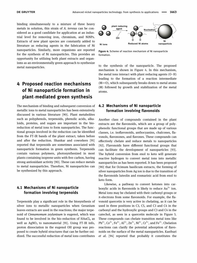
The mechanism of binding and subsequent conversion ofmetallic ions to metal nanoparticles has been extensivelydiscussed in various literature [90]. Plant metabolitessuch as polyphenols, terpenoids, phenolic acids, alka-loids, proteins, and sugars are important in the bio-reduction of metal ions to form nanoparticles. The func-tional groups involved in the reduction can be identifiedfrom the FT-IR bands of the plant extract, taken beforeand after the reduction. Shankar and coworkers [91]reported that terpenoids are sometimes associated withnanoparticle formation in green synthesis. Terpenoidscontain various polymers, photosynthesized in mostplants containing isoprene units with five-carbon, havingstrong antioxidant activity [90]. These can reduce metalsto metal nanoparticles. Therefore, Ni nanoparticles canbe synthesized by this approach.
4.1 Mechanisms of Ni nanoparticleformation involving terpenoids
Terpenoids play a significant role in the biosynthesis ofsilver ions to metallic nanoparticles when Geraniumleaves extracts are used in the reactions; the major terpe-noid of Cinnamomum zeylanisum is eugenol, which wasfound to be involved in the bio-reduction of HAuCl4 aswell as AgNO3 to nanomaterial [91]. Using FT-IR info,proton dissociation in the eugenol OH group was pro-posed to create hybrid structures that can be further oxi-dized. The successful reduction of metal ions contributes
to the synthesis of the nanoparticle. The proposedmechanism is shown in Figure 4. In this mechanism,the metal ions interact with plant reducing agents (O–H)leading to the formation of a reaction intermediate(M]O), which subsequently breaks down to metal atoms(M) followed by growth and stabilization of the metalatoms.
4.2 Mechanisms of Ni nanoparticleformation involving flavonoids
Another class of compounds contained in the plantextracts are the flavonoids, which are a group of poly-phenolic functional groups that are made up of variousclasses, i.e. isoflavonoids, anthocyanins, chalcones, fla-vonols, flavonones, and flavones. These compounds caneffectively chelate and reduce metals to nanoparticles[92]. Flavonoids have different functional groups thatcan facilitate the development of nanoparticles [93].The hybrid conversion from enol to keto will generatereactive hydrogen to convert metal ions into metallicnanoparticles as has been reported. It has been proposed[90] that for Ocimum basilicum extracts, the forming ofsilver nanoparticles from Ag ion is due to the transition ofthe flavonoids luteolin and rosmarinic acid from enol toketo form.
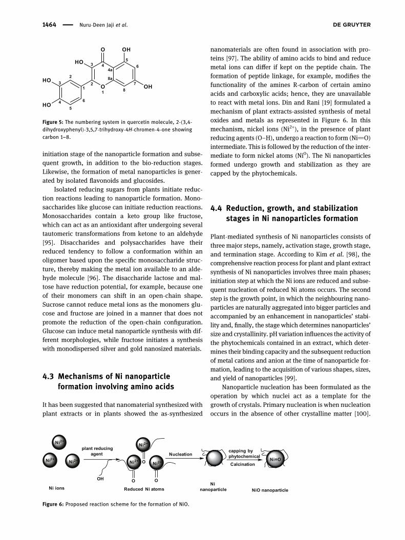
Likewise, a pathway to convert ketones into car-boxylic acids in flavonoids is likely to reduce Au3+ ion.Metal ions may be chelated with their carbonyl groups orπ-electrons from some flavonoids. For example, the fla-vonoid quercetin is very active in chelating, as it can beused in three positions in C3, C5, and C3 and C4 in thecarbonyl and the hydroxylic groups and C3 and C4 in thecatechol, as seen in a quercetin molecule in Figure 5.These compounds can chelate transition metal ions likePb2+, Co2+, Fe2+, Al3+, Zn2+, Ni2+, Cr3+, and Fe3+. Chelationreactions can clarify the potential adsorption of flavo-noids on the surface of the metal nanoparticles. Kasthuriet al. [94] reported that probably it constitutes the
Ni2+ Ni2+
Ni2+
Ni2+ Ni2+
Ni2+
Ninanoparticle
OO
O
OH
plant reducingagent Nucleation
Reduced Ni atomsNi ions
Figure 4: Scheme of reaction mechanism of Ni nanoparticleformation.
Advanced nickel nanoparticles technology: from synthesis to applications 1463
initiation stage of the nanoparticle formation and subse-quent growth, in addition to the bio-reduction stages.Likewise, the formation of metal nanoparticles is gener-ated by isolated flavonoids and glucosides.
Isolated reducing sugars from plants initiate reduc-tion reactions leading to nanoparticle formation. Mono-saccharides like glucose can initiate reduction reactions.Monosaccharides contain a keto group like fructose,which can act as an antioxidant after undergoing severaltautomeric transformations from ketone to an aldehyde[95]. Disaccharides and polysaccharides have theirreduced tendency to follow a conformation within anoligomer based upon the specific monosaccharide struc-ture, thereby making the metal ion available to an alde-hyde molecule [96]. The disaccharide lactose and mal-tose have reduction potential, for example, because oneof their monomers can shift in an open-chain shape.Sucrose cannot reduce metal ions as the monomers glu-cose and fructose are joined in a manner that does notpromote the reduction of the open-chain configuration.Glucose can induce metal nanoparticle synthesis with dif-ferent morphologies, while fructose initiates a synthesiswith monodispersed silver and gold nanosized materials.
4.3 Mechanisms of Ni nanoparticleformation involving amino acids
It has been suggested that nanomaterial synthesized withplant extracts or in plants showed the as-synthesized
nanomaterials are often found in association with pro-teins [97]. The ability of amino acids to bind and reducemetal ions can differ if kept on the peptide chain. Theformation of peptide linkage, for example, modifies thefunctionality of the amines R-carbon of certain aminoacids and carboxylic acids; hence, they are unavailableto react with metal ions. Din and Rani [19] formulated amechanism of plant extracts-assisted synthesis of metaloxides and metals as represented in Figure 6. In thismechanism, nickel ions (Ni2+), in the presence of plantreducing agents (O–H), undergo a reaction to form (Ni]O)intermediate. This is followed by the reduction of the inter-mediate to form nickel atoms (Ni0). The Ni nanoparticlesformed undergo growth and stabilization as they arecapped by the phytochemicals.
4.4 Reduction, growth, and stabilizationstages in Ni nanoparticles formation
Plant-mediated synthesis of Ni nanoparticles consists ofthree major steps, namely, activation stage, growth stage,and termination stage. According to Kim et al. [98], thecomprehensive reaction process for plant and plant extractsynthesis of Ni nanoparticles involves three main phases;initiation step at which the Ni ions are reduced and subse-quent nucleation of reduced Ni atoms occurs. The secondstep is the growth point, in which the neighbouring nano-particles are naturally aggregated into bigger particles andaccompanied by an enhancement in nanoparticles’ stabi-lity and, finally, the stage which determines nanoparticles’size and crystallinity. pH variation influences the activity ofthe phytochemicals contained in an extract, which deter-mines their binding capacity and the subsequent reductionof metal cations and anion at the time of nanoparticle for-mation, leading to the acquisition of various shapes, sizes,and yield of nanoparticles [99].
Nanoparticle nucleation has been formulated as theoperation by which nuclei act as a template for thegrowth of crystals. Primary nucleation is when nucleationoccurs in the absence of other crystalline matter [100].
7
65
4a
8a
8
43
2 O1
1
O
23
45
6HO
HO
HO
OH
OH
Figure 5: The numbering system in quercetin molecule, 2-(3,4-dihydroxyphenyl)-3,5,7-trihydroxy-4H-chromen-4-one showingcarbon 1–8.
Ni2+ Ni2+
Ni2+
Ni2+ Ni2+
Ni2+
Ninanoparticle
OO
O
OH
plant reducingagent Nucleation
Reduced Ni atomsNi ions
capping by phytochemical
CalcinationNi=O
NiO nanoparticle
Figure 6: Proposed reaction scheme for the formation of NiO.
1464 Nuru-Deen Jaji et al.

Homogeneous nucleation is said to occur when there is auniform formation of nuclei all over the phase, whileheterogeneous nucleation occurs at fundamental inho-mogeneity, dislocations, and grain boundaries [100]. Thetotal energy possessed by a nanoparticle is given by thesum of the bulk-free energy and the surface-free energy[71]. Mittal et al. [101] suggested a scheme of nanoparticleformation through the stages of reduction of Ni ions toNi atoms, growth of Ni atoms, and stabilization of theatoms during synthesis. The detailed mechanism of metalnanoparticle formation can be summarized as shown inFigure 7, in which the metal ions (Ni+) are reduced tometal atoms (Ni0). The nickel atoms then aggregate andgrow. Finally, the metal atoms are capped with stabi-lizing agents to form stable NiNPs.
Pandian et al. specifically formulated the mechanismof reaction in Ni nanoparticles formation employingOcimum sanctum leaf extract [74]. The mechanism
involves oxidative damage, surface bond, and the releaseof Ni ions, and finally, the reduction of Ni ions to Ni, asrepresented in Figure 8. A summary of the reported reac-tion mechanisms contains the three main phases ofreduction, growth, and stabilization.
5 Solvothermal synthesis of Ninanoparticles
Solvothermal synthesis of Ni nanoparticles, in whichsolvent at high temperature and pressure acts as redu-cing agent, has been reported. Several reports describinghydrothermal fabrication of Ni nanoparticles are obtain-able in literature [21,102–104]. Libor and Zhang [103]described the procedure for the synthesis of Ni nanopar-ticles in which an aqueous solution of Ni acetate washeated and then hydrazine was added to the solutionwith continuous stirring. They concluded that the crystalshape can be controlled by controlling various Ni-ionsreduction parameters to adjust the solution pH of coreand shell particles. They also reported the successfulcoating of Ni nanoparticles onto SiO2 particles. Liuet al. [102] reported the synthesis of Ni nanosized beltsusing a hydrothermal reduction technique involving acomplex surface-active reagent. Figure 9 shows a schematicdiagram of the solvothermal synthesis of NiNPS [105].
Chen and Wu synthesized nanoparticles of Ni using amicroemulsion of water-in-oil as a reducing agent and Nichloride as a precursor. They concluded that the mainfactor in the synthesis of smaller Ni nanoparticles was
Ni2+ Ni2+
Ni2+
Ni2+ Ni2+
Ni2+
Ni2+phytochemical Redaction
Ni
Growth
Ni2+ Ni2+
Ni2+
Ni2+ Ni2+
Ni2+Carpping stablization
Step 1
Step 2
Step 3
Figure 7: Mechanism of nanoparticle formation.
Figure 8: The mechanism of reaction for NiNP formation [74].
Advanced nickel nanoparticles technology: from synthesis to applications 1465

the higher ratio of the solvent system [21]. The depen-dence of particle sizes on the concentration of hydrazineand Ni chloride could be explained by the reduction,nucleation, and growth process. Figure 9 shows a sche-matic diagram of the microemulsion solvothermal synth-esis of NiNPs [106].
Kumar et al. described the fabrication of size-con-trolled Ni and NiO nanoparticles by employing micro-emulsions of water-in-oil. The size of the Ni nanoparticlescan easily be managed by adjusting the water-to-surfac-tant molar ratio or adjusting the reducing agent concen-tration [107]. Hou et al. reported the fabrication of size-controlled Ni nanoparticles using a standard airless pro-cedure in the synthesis. It was found that the introductionof hexadecylamine and trioctylphosphineoxide (TOPO)is an effective approach to obtain smaller monodispersedNi nanoparticles. They believed this method provided agood approach for the synthesis of other metal nanoparti-cles [22].
Roselina and Azizan fabricated Ni nanoparticles byapplying adjusted polyol by employing ethylene glycoland N2H4·6H2O. It was found that the molar ratio ofN2H4/Ni was significant in controlling the rate of reaction.They reported that the reaction rate increases with theincrease in N2H4/Ni molar ratio. However, an increasein agglomeration was observed as a rather unfavourablecondition in the synthesis of Ni nanoparticles [108]. Theseresults can be generalized to other metal nanoparticles,notably magnetic nanoparticles such as Ni nanoparticles.
The above reports show that solvothermal techniquesfor the synthesis of Ni nanoparticles are well-documented.In solvothermal synthesis, the crystal size and shape of thenanoparticles can be controlled by reaction parameterssuch as concentration of reactants, reaction temperature,reaction time, pH of reactants, and finally concentration of
stabilizing and capping agents. Specific size can be pro-duced by controlling the surface charge on the nanoparti-cles and the pH of the precursor, while the crystal shapecan be controlled by adjusting the crystal nucleation andgrowth process.
In solvothermal synthesis by water in oil procedure,the main factor to produce smaller Ni nanoparticles wasthe ratio of water to cetyltrimethylammonium bromide ton-hexanol system. Higher ratio of cetyltrimethylammo-nium bromide to n-hexanol favoured the production ofsmaller nanoparticles. The average diameter of Ni nano-particles was determined by the number of nuclei formedat the beginning of the reduction process. The magneti-zation of Ni nanoparticles was found to increase withdecreasing temperature.
In surfactant-assisted solvothermal synthesis, a slowreaction rate is favourable for isolating the growth stepfrom the nucleation step. In the solution phase synthesis,the main factor determining the size and shape of thefinal products was the kinetic control of the nucleationand growth step. The shape of the nanoparticles can beeffectively modulated by adding chemical capping agentsto the synthesis system. Thus, the selective interaction ofthe capping agents on a facet of the first-formed Ni nano-particles is the key to its growth.
The polyol assisted solvothermal synthesis using Nisalt and hydrazine hydrate as precursors, sodium hydro-xide as stabilizing agent, and ethylene glycol as cappingagent; the molar ratio of N2H4/Ni was found to play animportant role in controlling the reaction rate. However,the increase in reaction rate led to agglomeration of theNi nanoparticles which is a drawback in the fabrication ofthe nanoparticles.
In the standard airless solvothermal synthesis of Ninanoparticles using trioctylphosphine oxide and pure
Figure 9: Schematic diagram of (a) polyol [105], and (b) microemulsion solvothermal [106] synthesis of NiNPs.
1466 Nuru-Deen Jaji et al.

hexadecylamine as the stabilizing agents, the shape ofthe nanocrystal is significantly affected by reaction para-meters such as reaction temperature, time, solvent, andthe chemical potential between the reacting species. Thesurfactant trioctylphosphine oxide was effective in con-trolling the particle size. Hence, in solvothermal synth-esis various factors play important role in determiningthe final size and shape of the Ni nanoparticles.
6 Hybrid approach of top-down andbottom-up methods for thesynthesis of Ni nanoparticles
Several physical techniques abound in literature for thefabrication of Ni nanoparticles, among which are radi-olysis techniques such as ultrasonic sonication, micro-wave, and laser methods, and mechanical milling. Laserablation protocols for the fabrication of nanosized metalalloys and oxides in liquid medium employing solid targetare available in the literature. This technique is cost-effective with no need for high vacuum pumps andexpensive chambers and is considered environmentallyfriendly.
6.1 Electrochemical pulse-currentdeposition technique for the fabricationof Ni nanoparticles
Tu et al. reported an electrochemical deposition tech-nique for the synthesis of metallic nanoparticles. Theyadopted electrochemical deposition with pulse-currentto synthesize Ni nanoparticles for application as catalystsin the development of aligned nanotubes of carbon. Thedensity of the nanoparticle nucleation site was monitoredthrough the alteration of the pulse-current period andmagnitude [109]. The plasma-enhanced hot filamentCVD generated aligned carbon nanotubes of Ni nanopar-ticles. The goal was to produce many particles on a largearea with low density. To attain this goal, they employedthe technique of electrochemical deposition by pulse cur-rent. A two-electrode system was adopted for the experi-ment, and eventually, Ni nanoparticles were obtained.Bao et al. reported the successful preparation of a highlyordered Ni nanotube array by electrodeposition into theholes of an alumina membrane altered with an organo-
amine as a pore-wall altering agent. The resulting mag-netic property was analysed [110].
It is fundamental to the Ni nanotube assembly pro-cedure that the pore wall of the alumina template isaltered with methyl-γ-diethyltriaminopropyldimethoxy-silane. When Ni is electrochemically deposited in thepores, the presence of the amino group preferentiallydeposits the Ni on the walls of the unmodified aluminamembrane to form magnetic Ni nanotubes. At higher cur-rent density, nickel hydroxide was produced on the alu-mina membrane at the time of electrodeposition. Thesemetal nanotubes with open ends could be used for thecreation of materials with special magnetic, optical, andelectrical properties [110].
Pirota et al. synthesized novel magnetic materialsby electrochemical deposition techniques. NiNPs weredeposited inside alumina nanopores by applying a steadycurrent pulse in a sequence [111]. Lee et al. developed aunique process to synthesize nanotubes of metallic ele-ments by electrodeposition of preferential metal on thesurface of membrane walls of the nanoparticles. In thisnano synthesis process, many metals such as Fe, Au, Pl,Ag, Pt, Ni, and Co could be incorporated into the nanotubestructure, thereby enabling the synthesis of barcode-typenanomaterial [112]. Clusters of staked Ni nanotubes andmultisegmented nanotubes can be analysed by a quantuminterference magnetometer to compare their magneticproperties. At lower fields of saturation, the clusters of dis-connected nanotubes were effectively magnetized as com-pared to the array of continuous Ni nanotubes [112] asshown in Figure 10.
Figure 10: The images of multisegmented nickel nanotubes usingSEM Lee et al. [112].
Advanced nickel nanoparticles technology: from synthesis to applications 1467

6.2 Synthesis of Ni nanoparticles employingpulsed laser ablation in liquidstechnique
Gondal et al. synthesized nanoparticles of NiO by apply-ing pulsed laser ablation (PLA) in liquid. They reported ahomemade PLA setup consisting of a laser and a cell witha pure Ni metal plate dipped in liquid. Before laser irra-diation, a pure Ni metal target was held on a magneticrotator at the basement of a glass container. A colloidalmixture of NiO was produced, and Ni nanoparticles wereisolated from water by centrifugation [113].
6.3 Fabrication of Ni nanoparticles by arcdischarge method
The fabrication of Ni nanoparticles with unique structuraland magnetic properties was reported by El-Khatib et al.They employed an arc and discharge technique with anultrasonic nebulizer in an argon atmosphere to generateNiNPs. They suggested that the technique is economicaland environmentally friendly. Similarly, the techniqueleads to the production of the small size distribution ofNiNPs with high purity [114].
6.4 Production of Ni nanoparticles bymicrowave combustion technique
The production of nanometal by microwave combustionhas the advantage of being highly exothermic, whichleads to the uniform distribution of temperature withinthe bulk material and the surface, and hence, the fastproduction of nanoparticles. Ragupathi et al. reported atechnique of microwave combustion in the fabrication ofnanoparticles of Ni aluminate. Aluminium nitrate and Ninitrate were used as precursors. The microwave oven wasprogrammed such that it led to the formation of Ni nano-particles as the product [115]. The microwave combustionleads to the formation of fine particles with uniform mor-phology. The microwave combustion technique is a rapidand economic technique for the fabrication of Ni nano-composites within terms of time, simplicity, and energy.The NiAl2O4 nanoparticles generated by the microwavecombustion technique have been used in the catalysis ofbenzyl alcohol in the liquid phase. It resulted in excellentreactivity, good recyclability, and high stability and wasenvironmentally favourable.
Similarly, LaGrow and coworkers fabricated highlymonodispersed NiNPs. They stated that face-canteredcubic crystals were obtained by employing trioctylpho-sphine surfactants under a reducing hydrogen atmo-sphere to favour thermodynamic growth stabilization ofthe nanoparticles [116]. They reported that changing thenickel precursor concentration to trioctylphosphine ratiowas found to alter the face shape and size from sphericalat 5 nm to cubic at 12 nm.
7 Applications of Ni nanoparticles
Generally, nanostructures such as nanoparticles, nano-tubes, nanowires, nano-springs, nanobelts, and nano-rings are utilized in the development of flat-panel dis-play, nonlinear optical devices, light-emitting diodes,transistors, and logic gates. Ni-based materials were alsoapplied in turbines, automobile molds, and aerospace[117]. The application of Ni nanoparticles includes medicalapplications, as a catalyst, applications in sensor develop-ment, enhancement of materials, and adsorption of dyes.These are discussed in the following sections.
7.1 Biomedical applications of Ninanoparticles
The use of Ni nanoparticles in biomedical applicationsand as an antibacterial agent has been reported in theliterature. These include drug and gene delivery, mag-netic resonance imaging, cell separation, biomedicaldetection, and diagnostics [118]. Guo et al. reported thatfunctionally charged NiNPs could increase cell mem-brane permeability and promote cellular absorption intocancer cells of the outer target molecules [119]. Theseresults show that NiNPs may have a possible mechanismfor targeting the cytotoxicity of leukaemia cancer cellsand that NiNPs may be implemented in related bio-medical and clinical fields.
Ivanov et al. noted the synergistic effect of Ni nano-particles with reduced graphene oxide to enable thedesigning of high-performance applications in bioin-spired microelectronics for medical therapy [120].Angajala et al. showed that Ni nanoparticles possess highlarvicidal efficacy against Culex quinquefasciatus andexcellent anti-inflammatory activity comparable to that ofa standard essay. They suggested that the Ni nanoparticlescan also be used as a good drug carrier as well as for the
1468 Nuru-Deen Jaji et al.

control of C. quinquefasciatus, a vector in the transmissionof lymphatic filariasis, and dengue fever [68]. Figure 11shows an illustration of biomedical applications of mag-netic functionalized metal nanoparticles [121].
Similarly, Helan et al. suggested the potential appli-cation of Ni nanoparticles in the suppression of microbialpathogens such as Staphylococcus aureus and Escherichiacoli [122]. Roselina and coworkers noted the excellentcatalytic and magnetic properties of Ni nanoparticles,given its tremendous potential in many applicationsincluding biomedical and medical fields [108]. Gonget al. reported that algae can accelerate the aggregationof nanoparticles as well as reduce NiO to Ni. They
suggested that green algae may be promising for biore-mediation of nano-pollution [89]. Sudhasree et al.reported that green Ni nanoparticles exhibit colloidal sta-bility with antioxidant property, thereby making themgood antimicrobial agents [76]. Table 5 presents the bio-medical applications of Ni nanoparticles.
7.2 Applications of Ni nanoparticles ascatalyst
The application of Ni nanoparticles as a catalyst hasappeared in several reports. Simonsen et al. reported
Figure 11: Biomedical applications of magnetic functionalized metal nanoparticles [121].
Table 5: Biomedical applications of Ni nanoparticles
Type of medical application Structure of Ni nanoparticles Size of Ni nanoparticles (nm) Reference
Cancer treatment Colloidal 38.2 Gorgizadeh et al. [123]Integrated biomaterial hcp 0.5–24 Ivanov et al. [120]Antibacterial activity hcp 12 Helan et al. [122]Anti-inflammatory fcc 80–100 Angajala et al. [68]Absorbent of safranin-O Amorphous 320 Ghaedi et al. [124]Hydrodeoxygenation of microalgae Colloidal 3 Song et al. [88]Biomedical fcc 2–600 Roselina et al. [108]Thermo-therapeutic Crystalline 28 Hoque et al. [125]Electrochemical sensor hcp 8.9 Neiva et al. [125]Glucose sensor fcc 15 Liu et al. [126]High bioimaging resolution Ni2+ ions doped NaYF4:Yb
3+/Er3+ 100 nm Jia et al. [127]
Advanced nickel nanoparticles technology: from synthesis to applications 1469
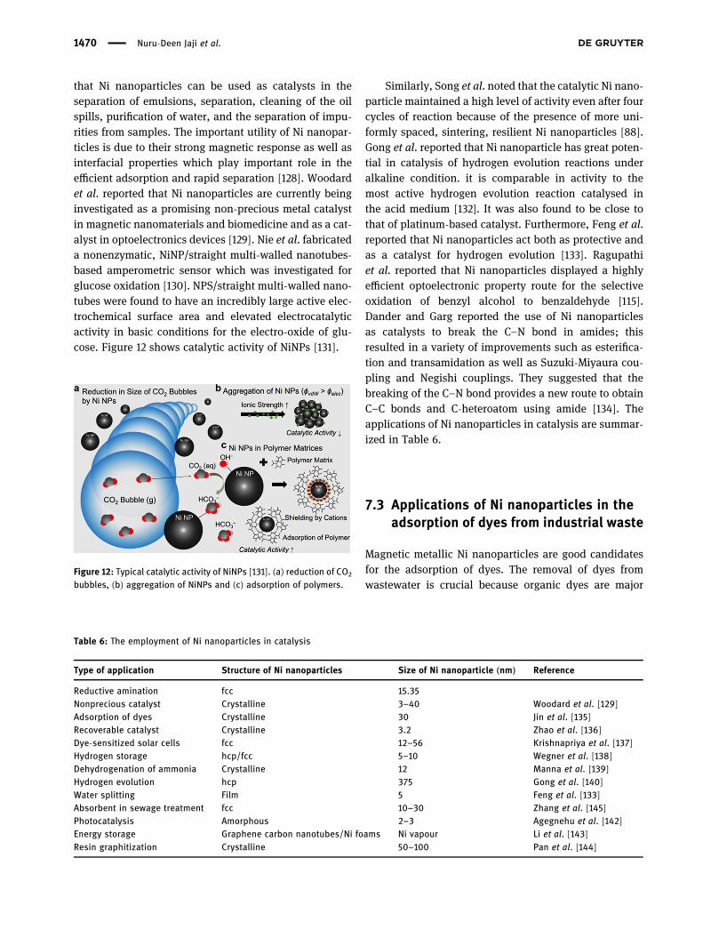
that Ni nanoparticles can be used as catalysts in theseparation of emulsions, separation, cleaning of the oilspills, purification of water, and the separation of impu-rities from samples. The important utility of Ni nanopar-ticles is due to their strong magnetic response as well asinterfacial properties which play important role in theefficient adsorption and rapid separation [128]. Woodardet al. reported that Ni nanoparticles are currently beinginvestigated as a promising non-precious metal catalystin magnetic nanomaterials and biomedicine and as a cat-alyst in optoelectronics devices [129]. Nie et al. fabricateda nonenzymatic, NiNP/straight multi-walled nanotubes-based amperometric sensor which was investigated forglucose oxidation [130]. NPS/straight multi-walled nano-tubes were found to have an incredibly large active elec-trochemical surface area and elevated electrocatalyticactivity in basic conditions for the electro-oxide of glu-cose. Figure 12 shows catalytic activity of NiNPs [131].
Similarly, Song et al. noted that the catalytic Ni nano-particle maintained a high level of activity even after fourcycles of reaction because of the presence of more uni-formly spaced, sintering, resilient Ni nanoparticles [88].Gong et al. reported that Ni nanoparticle has great poten-tial in catalysis of hydrogen evolution reactions underalkaline condition. it is comparable in activity to themost active hydrogen evolution reaction catalysed inthe acid medium [132]. It was also found to be close tothat of platinum-based catalyst. Furthermore, Feng et al.reported that Ni nanoparticles act both as protective andas a catalyst for hydrogen evolution [133]. Ragupathiet al. reported that Ni nanoparticles displayed a highlyefficient optoelectronic property route for the selectiveoxidation of benzyl alcohol to benzaldehyde [115].Dander and Garg reported the use of Ni nanoparticlesas catalysts to break the C–N bond in amides; thisresulted in a variety of improvements such as esterifica-tion and transamidation as well as Suzuki-Miyaura cou-pling and Negishi couplings. They suggested that thebreaking of the C–N bond provides a new route to obtainC–C bonds and C-heteroatom using amide [134]. Theapplications of Ni nanoparticles in catalysis are summar-ized in Table 6.
7.3 Applications of Ni nanoparticles in theadsorption of dyes from industrial waste
Magnetic metallic Ni nanoparticles are good candidatesfor the adsorption of dyes. The removal of dyes fromwastewater is crucial because organic dyes are major
Figure 12: Typical catalytic activity of NiNPs [131]. (a) reduction of CO2
bubbles, (b) aggregation of NiNPs and (c) adsorption of polymers.
Table 6: The employment of Ni nanoparticles in catalysis
Type of application Structure of Ni nanoparticles Size of Ni nanoparticle (nm) Reference
Reductive amination fcc 15.35Nonprecious catalyst Crystalline 3–40 Woodard et al. [129]Adsorption of dyes Crystalline 30 Jin et al. [135]Recoverable catalyst Crystalline 3.2 Zhao et al. [136]Dye-sensitized solar cells fcc 12–56 Krishnapriya et al. [137]Hydrogen storage hcp/fcc 5–10 Wegner et al. [138]Dehydrogenation of ammonia Crystalline 12 Manna et al. [139]Hydrogen evolution hcp 375 Gong et al. [140]Water splitting Film 5 Feng et al. [133]Absorbent in sewage treatment fcc 10–30 Zhang et al. [145]Photocatalysis Amorphous 2–3 Agegnehu et al. [142]Energy storage Graphene carbon nanotubes/Ni foams Ni vapour Li et al. [143]Resin graphitization Crystalline 50–100 Pan et al. [144]
1470 Nuru-Deen Jaji et al.

pollutants in wastewater. Organic dyes reduce the qualityof water, thereby posing a significant impact on humanhealth. Most organic dyes are toxic, mutagenic, and car-cinogenic. Jin et al. confirmed the use of composites of Ninanoparticles for the isolation of dyes from aqueous solu-tion. They found that Ni nanocomposite with a large porevolume and high surface area can be easily separatedfrom the aqueous solution by an external magnet [135].Ghaedi et al. reported the use of Ni sulphide nanoparti-cles loaded on activated carbon as a novel adsorbent forthe individual and simultaneous adsorption of methyleneblue and safranin-O [124].
Furthermore, Sudhasree et al. reported the fabrica-tion of Ni nanoparticles free of surfactants which weresubsequently used in the removal of Congo red, an azodye from industrial wastewater [76]. Zhang and coworkersconfirmed the fabrication of Ni nanoparticles having excel-lent magnetic properties and crystallite size of 10–30 nm.These synthesized Ni nanoparticles were subsequentlyused as adsorbents of Congo red from industrial waste-water [141].
7.4 Application of Ni nanoparticles in dye-sensitized solar cells and sensors
Krishnapriya et al. described the fabrication of dye-sen-sitized solar cells with various nanostructured TiO2. Itwas incorporated with Ni nanocomposites with morphol-ogies like interconnected bead-like, spindle shape-like,square platelets-like, and porous sphere-like to yield dye-sensitized solar cells. The nanostructures were synthe-sized in different solvents, namely ethanol, a mixture ofethanol and water, as well as HF in conjunction withshape- and size-tuned Ni nanocomposites of mixed trian-gular and hexagonal morphological crystals. These hadsizes ranging from 15 to 62 nm. They reported that theincorporation of Ni nanocomposites effectively traps inci-dent light and successively improves the rate of electron-hole pair formation and short circuit current. The fabri-cated dye-sensitized solar cells were found to exhibitexcellent stability in conventional electrolytes over aduration of a month [137].
Furthermore, Liu et al. developed a high-performancenonenzymatic sensor on Ni nanoparticle-chitosan nano-composite. They suggested that nonenzymatic sensorscan be used for monitoring blood glucose due to theirlow cost, simple preparation, and excellent detection ofglucose [25].
7.5 Application of Ni nanoparticles insuperconductors and enhancement ofmaterials
Zhang et al. reported the fabrication of electrodes employ-ing Ni nanocomposite materials. The electrodes showedlow charge-transfer resistance, outstanding cycle stabi-lity, high specific capacitance, and good rate perfor-mance. The high capacitation of the electrode nano-composite was due to enhanced conductivity, regularlyscattered nanoparticles of Ni(OH)2, low interfacial resis-tance, and synergetic effects of each portion. They sug-gested the composite as a promising material for highenergy supercapacitor application with the improvedelectrochemical performance [145].
Similarly, Li et al. developed a high-performanceelectrochemical supercapacitor from amorphous Ni (OH)2nanospheres. The developed electrode was found toexhibit high capacitance, high energy density, andlong life. They suggested that the electrode can be usedin advanced electrochemical pseudocapacitor material[146]. Agegnehu et al. reported the use of Ni and NiOnanoparticles for the enhancement of photocatalytichydrogen evolution from aqueous methanol. They sug-gested that the Ni/graphene oxide exhibits high activityattributed to ease of trapping photogenerated electronsby Ni and NiO nanoparticles [142].
7.6 Tabulation of other applicationsof NiNPs
The other applications of Ni nanoparticles in variousfields are hereby reported in Table 7. It shows the fieldof application of the Ni nanoparticles, the structure ofthe nanoparticles, the size of the nanoparticles, and thereferences. The nanoparticle crystallinity influencesnanomaterials applications [69].
8 The advantages of using Ni ascatalyst
The advantages of using Ni nanoparticles as catalysts arehereby reported with specific reactions and conversiontechniques. The physical and chemical characteristicsof Ni nanoparticles determine their potential applications
Advanced nickel nanoparticles technology: from synthesis to applications 1471

in research as well as in the industries. Various measure-ment techniques [157] are employed in the characteriza-tion of Ni nanoparticles as illustrated in Table 8.
8.1 The advantages of Ni nanoparticles as acheap catalyst
Nickel in comparison to commonly used metal catalystssuch as some transition metals and noble metals is rela-tively cheap. Dander and Garg reported the potential ben-efits of Ni catalysis due to the abundance of Ni, resultingin low cost, acceptable levels of toxicological require-ment for orally administered medication, opportunity in
green chemistry, and the ability to draw on alternativereactivity profiles to create novel chemical transitions[134]. According to Tobias and Chatani, arene prepara-tion can be improved by the application of Ni catalyst incross-coupling reactions. The aryl halogens can becoupled with organic nucleophiles and organometallic.Ni-catalysed reductive cleavage of aryl ethers in theabsence of an external reducing agent provides strongsupport for this oxidative addition process [158]. Martin-dale et al. documented the complex nickel bis-dipho-sphine as a catalyst to produce solar hydrogen [159].That is because nickel has the advantages of being anactive non-noble metal catalyst to produce hydrogen inacidic, organic, and aqueous solutions. Nickel catalyst
Table 7: Other applications of Ni nanoparticles
Application of Ni nanoparticles Structures Sizes in (nm) Reference
Ultra-high compressive strength Crystalline 100–1,000 Sharma et al. [147]Catalyst for p-nitrophenol reduction Crystalline 34.8–143.2 Jiang et al. [148]Magnetic separation Crystalline 5–20 Simonsen et al. [128]Voltammetric determination of Rifampicin fcc 3 Oliveira et al. [149]Added to polyester fabric Crystalline 40 Afshari et al. [50]Microwave absorption fcc 200–800 Guo et al. [105]Catalyst for hydrodecylchlorination hcp 6 Duraisamy et al. [151]Catalyst for the oxidation of benzyl alcohol fcc 19.8 Ragupathi et al. [115]Free radical scavenging activity fcc 2,453–2,695 Sudhasree et al. [76]Catalyst for hydrodesulfurization Crystalline 5–20 Layan Savithra et al. [152]Electrochemical pseudocapacitor material Amorphous 100 Li et al. [146]Catalyst for hydrogen evolution Crystalline 10–20 Karami et al. [153]Electroless Ni plating Amorphous 10–60 Wu et al. [154]Optoelectronic applications fcc 80–400 Libor et al. [103]Magnetic data storage application fcc 30–50 Yu et al. [81]Conducting material fcc/hcp 12.9 Chen et al. [155]Photocatalyst Crystalline 22 Asaithambi et al. [156]
Table 8: Characterization techniques for NiNPs
Technique Acronym Utility
UV-Visible spectroscopy UV-Vis Chemical analysisElectron probe microanalysis EPMA Particle size/chemical analysisTransmission electron microscopy TEM Imaging particle/size/shapeScanning electron microscopy SEM Imaging/topology/sizeEnergy dispersive spectroscopy EDS Elemental compositionHigh resolution transmission electron microscopy HRTEM Imaging structure/chemical analysisX-ray diffraction XRD Crystallinity/size/shapeBrunauer–Emmett–Teller analysis BET Porosity/surface areaFourier transform infrared spectroscopy FT-IR Organic functional groupsAtomic force microscopy AFM Topology/imaging/surface structureThermal gravimetric analysis TGA Mass loss vs temperatureDifferential scanning calorimetry DSC Reaction heat/phase changesAuger electron spectroscopy AES Chemical structure analysis
1472 Nuru-Deen Jaji et al.

has a low overpotential for the reduction of protonsin water. Furthermore, with solar light irradiation, Nicatalyses ruthenium dye inhomogeneous schemes aswell as hybrid systems with nanoparticulate lightabsorbers [159].
8.2 Advantages of Ni nanoparticles as acatalyst in the conversion of biomass tofuels; Ni catalyst is being employed invarious reactions involving theconversion of biomass to fuels
Lignin is a polymer of monomeric aromatic compoundsand has the potential to be a source for liquid fuels andvaluable chemicals. Luo et al. found Ni to be an efficientcatalyst for catalytic depolymerization of lignin, leadingto its conversion into four phenolic products. The conver-sion of carbon dioxide to methane is a famous reactioncalled the Sabatier reaction [160]. Fukuhara et al. foundthat the conversion of carbon dioxide to methane usingNi catalyst showed steady catalytic performance. It main-tained a high activity and high selectivity during a dur-ability test. It was found that Ni catalyst has the potentialfor producing energy from carbon dioxide [161]. Sweeneyet al. described the use of Ni catalyst for the allylationreactions using readily available, inexpensive, andair-insensitive catalysts and reagents [162]. The use ofallyl alcohols as substrates and an inexpensive Nisalt as a precursor of the catalyst offers significantadvantages.
8.3 Advantages of using Ni catalyst inSuzuki-Miyaura reactions
Guard et al. described the advantages of using Ni catalystin Suzuki-Miyaura reactions. Suzuki-Miyaura reactionsare generally employed in the formation of new carbon-carbon bonds [163]. Currently, the best catalyst in use isbased on precious metals such as Palladium. The use ofcheap Ni-based catalyst results in affordable systems.Nickel has a smaller size compared to palladium, leadingto weaker binding and increased nucleophilicity in het-erocyclic coordinating atoms. The chemical advantagesof using Ni over palladium are that the former can beused in Suzuki-Miyaura coupling in acyliminium, carbo-nate, carbamate, sulfamate, and sp3-generated substrates.The catalytic application of NiNPs in the Suzuki-Miyauracross-coupling reaction [164] is illustrated in Figure 13.
8.4 Advantages of using Ni nanoparticle dueto its various oxidation states
Tasker and Jamison reported that, among the manyadvantages of using Ni-catalyst, one is the availabilityof multiple oxidation states, including Ni0, Ni1+, Ni2+,and Ni3+, and recently reported a well-defined Ni4+ spe-cies. Nickel-catalysed reactions either proceed via a Ni0/Ni2+ pathway or via a Ni+/Ni3+ pathway. The formation ofproducts is likely enabled by a mechanistically distinctdirect oxidation of Ni intermediates [165]. The featuresdescribed above help in the wide use of Ni as a catalystand in its commercialization in the fields of organic
Figure 13: Illustration of the application of NiNPs in Suzuki-Miyaura cross-coupling reaction [164].
Advanced nickel nanoparticles technology: from synthesis to applications 1473

synthesis, inorganic synthesis, environmental cleaning,and medicinal drug administration. The electrochemicaloxidation of urea based on Ni(II/III) sites [166] is shown inFigure 14.
Mirzaei et al. suggested that the Voltammogramsobtained with nickel-rich compounds indicate the pre-sence of a new peak at a lower positive potential (−50mV)and a comparable strength than the urea oxidation peaknormally found with nickel [166]. Further studies arerequired to explain its nature.
9 Conclusion
In this review, several NiNPs synthetic techniques havebeen reported, ranging from the use of plant extracts,plant secretions, fungi, algae, and microorganisms, aswell as some physical and chemical methods of synthesis.Plant extracts are economically viable, and it can beupgraded and environmentally harmless. Biogeni-cally synthesized nickel nanomaterials are free of toxicchemicals and are especially suitable for applications inbiomedicine. Various physical and chemical methods forthe fabrication of nickel nanomaterials, such as mechanicalmilling, microwave, sonochemical, and electrochemicaltechniques, have been described. These methods yieldNiNPs with high quality magnetic properties, controlledsize, and monodispersed population. Proposed reactionmechanisms of NiNPs synthesis based on plant extractsand physical methods as reported in literature have beenoutlined with the specific functional groups involved inthe reduction of nickel to NiNPs. The applications of
nickel nanosized materials in various fields such as cata-lysis, biomedical applications, adsorption of dyes fromindustrial waste, fabrication of dye-sensitized solar cellsand sensors, and in the development of supercapacitorshave also been discussed. The specific advantages ofusing Ni nanoparticles as a cheap catalyst in the conver-sion of biomass to fuels, in Suzuki-Miyaura reactions, aswell as the advantages of Ni nanoparticles due to its mul-tiple oxidation states have been reported.
Extensive research has been published on the synth-esis, characterization, and applications of Ni nanoparti-cles. However, there are no universal conclusions [167]made on the biomedical effects of Ni nanoparticles onhuman, animal, and plant lives. The reported reactionmechanisms in literature are inconclusive; hence, theneed to ascertain the mechanisms leading to the forma-tion of Ni nanoparticles more especially in plant-mediated synthesis. While the advantages of using Ninanoparticles as catalyst have been stressed, the disad-vantages have not been enumerated.
Acknowledgments: The authors wish to acknowledgeUniversiti Sains Malaysia for sponsoring this projectunder USM-STG-6315076 and RUI-1001/P Kimia/8011086,School of Chemical Sciences, Universiti Sains Malaysia fortechnical support. N. J. wish to thank Federal College ofEducation Technical Gombe, Nigeria, for TETFUNDPostgraduate Scholarship.
Author contributions: Muhammad Bisyrul Hafi Othman,Hooi Ling Lee, Mohd Hazwan Hussin: Conceptualiza-tion; Nuru-Deen Jaji: Writing-original reviewer draft
Figure 14: (a) Voltammograms recorded for Ni/C in KOH 1mol L−1 and urea 0.1 mol L−1, at 10mV s−1. (b) Voltammograms of Ni100−xRhx/C inKOH 1mol L−1 and urea 0.1 mol L−1 (10mV s−1 [166]).
1474 Nuru-Deen Jaji et al.
preparation, visualization, investigation; MuhammadBisyrul Hafi Othman, Hazizan Md Akil, MuhammadRazlan Zakaria: Reviewing and Editing; MuhammadBisyrul Hafi Othman, Hooi Ling Lee: Supervision, fundingacquisition. This article has been read and approved by alllisted authors.
Conflict of interest: The authors declare no conflict ofinterest regarding the publication of this paper.
References
[1] Paramo LA, Feregrino-Perez AA, Guevara R, Mendoza S,Esquivel K. Nanoparticles in agroindustry: applications,toxicity, challenges, and trends. Nanomaterials.2020;10(9):1654–86.
[2] Magaye R, Zhao J. Recent progress in studies of metallicnickel and nickel-based nanoparticles’ genotoxicity andcarcinogenicity. Environ Toxicol Pharmacol.2012;34(3):644–50.
[3] Bibi I, Kamal S, Ahmed A, Iqbal M, Nouren S, Jilani K, et al.Nickel nanoparticle synthesis using camellia sinensis asreducing and capping agent: growth mechanism and photo-catalytic activity evaluation. Int J Biol Macromol.2017;103:783–90.
[4] Cheng Y, Guo M, Zhai M, Yu Y, Hu J. Nickel nanoparticlesanchored onto Ni foam for supercapacitors with high specificcapacitance. J Nanosci Nanotechnol. 2020;20(4):2402–7.
[5] Abdel Fattah AR, Majdi T, Abdalla AM, Ghosh S, Puri IK. Nickelnanoparticles entangled in carbon nanotubes: novel ink fornanotube printing. ACS Appl Mater Interfaces.2016;8(3):1589–93.
[6] Jiao M, Yao Y, Pastel G, Li T, Liang Z, Xie H, et al. Fly-throughsynthesis of nanoparticles on textile and paper substrates.Nanoscale. 2019;11(13):6174–81.
[7] Ni H, Zhu J, Wang Z, Lv H, Su Y, Zhang X. A brief overview ongrain growth of bulk electrodeposited nanocrystalline nickeland nickel-iron alloys. Rev Adv Mater Sci.2019;58(1):98–106.
[8] Reena Mary AP, Suchand Sandeep CS, Narayanan TN,Philip R, Moloney P, Ajayan PM, et al. Nonlinear and mag-neto-optical transmission studies on magnetic nanofluids ofnon-interacting metallic nickel nanoparticles. Materials.2011;22(37):375702–8.
[9] Barsan MM, Enache TA, Preda N, Stan G, Apostol NG, Matei E,et al. Direct immobilization of biomolecules through mag-netic forces on Ni electrodes via Ni nanoparticles: applica-tions in electrochemical biosensors. ACS Appl MaterInterfaces. 2019;11(22):19867–77.
[10] Kiran S, Rafique MA, Iqbal S, Nosheen S, Naz S, Rasheed A.Synthesis of nickel nanoparticles using citrullus colocynthisstem extract for remediation of reactive yellow 160 dye.Environ Sci Pollut Res. 2020;27(26):32998–33007.
[11] Hill D, Barron AR, Alexander S. Comparison of hydrophobicityand durability of functionalized aluminium oxide
nanoparticle coatings with magnetite nanoparticles-linksbetween morphology and wettability. J Colloid Interface Sci.2019;555:323–30.
[12] Bian Z, Das S, Wai MH, Hongmanorom P, Kawi S. A review onbimetallic nickel-based catalysts for CO2 reforming ofmethane. Chemphyschem. 2017;18(22):3117–34.
[13] Sagasti A, Palomares V, Porro JM, Orue I, Sanchez-Ilarduya MB, Lopes AC, et al. Magnetic, magnetoelastic andcorrosion resistant properties of (Fe-Ni)-based metallicglasses for structural health monitoring applications.Materials. 2019;13(1):57–70.
[14] Wang D, Jia Y, He Y, Wang L, Fan J, Xie H, et al. Enhancedphotothermal conversion properties of magnetic nanofluidsthrough rotating magnetic field for direct absorption solarcollector. J Colloid Interface Sci. 2019;557:266–75.
[15] Thellaputta GR, Chandra PS, Rao C. Machinability of nickel-based superalloys: a review. Mater Today Proc.2017;4(2):3712–21.
[16] Kate RS, Khalate SA, Deokate RJ. Overview of nanostructuredmetal oxides and pure nickel oxide (NiO) electrodes forsupercapacitors: a review. J Alloy Compd. 2018;734:89–111.
[17] Zhang L, Shi D, Liu T, Jaroniec M, Yu J. Nickel-based materialsfor supercapacitors. Mater Today. 2019;25:35–65.
[18] Sequeira CA, Cardoso DS, Amaral L, Šljukić B, Santos DM.On the performance of commercially available corrosion-resistant nickel alloys: a review. Corros Rev.2016;34(4):187–200.
[19] Imran Din M, Rani A. Recent advances in the synthesis andstabilization of nickel and nickel oxide nanoparticles: a greenadeptness. Int J Anal Chem. 2016;2016:1–4.
[20] Hossain MD, Mayanovic RA, Dey S, Sakidja R, Benamara M.Room-temperature ferromagnetism in Ni(ii)-chromia basedcore–shell nanoparticles: experiment and first principlescalculations. Phys Chem Chem Phys.2018;20(15):10396–406.
[21] Chen DH, Wu SH. Synthesis of nickel nanoparticles in water-in-oil microemulsions. Chem Mater. 2000;12(5):1354–60.
[22] Hou Y, Kondoh H, Ohta T, Gao S. Size-controlled synthesis ofnickel nanoparticles. Appl Surf Sci. 2005;241(1–2):218–22.
[23] Ely TO, Amiens C, Chaudret B, Snoeck E, Verelst M,Respaud M, et al. Synthesis of nickel nanoparticles.Influence of aggregation induced by modification of poly(vinylpyrrolidone) chain length on their magnetic properties.Chem Mater. 1999;11(3):526–9.
[24] Chen DH, Hsieh CH. Synthesis of nickel nanoparticles inaqueous cationic surfactant solutions. J Mater Chem.2002;12(8):2412–5.
[25] Liu Z, Guo Y, Dong C. A high performance nonenzymaticelectrochemical glucose sensor based on polyvinylpyrroli-done–graphene nanosheets–nickel nanoparticles–chitosannanocomposite. Talanta. 2015;137:87–93.
[26] You W, Che R. Excellent NiO-Ni nanoplate microwaveabsorber via pinning effect of antiferromagnetic-ferromag-netic interface. ACS Appl Mater Interfaces.2018;10(17):15104–11.
[27] Karam L, Reboul J, El Hassan N, Nelayah J, Massiani P.Nanostructured nickel aluminate as a key intermediate forthe production of highly dispersed and stable nickel nano-particles supported within mesoporous alumina for dryreforming of methane. Molecules. 2019;24(22):4107–19.
Advanced nickel nanoparticles technology: from synthesis to applications 1475

[28] Lyu F, Wang Q, Choi SM, Yin Y. Noble-metal-free electroca-talysts for oxygen evolution. Small. 2019;15(1):1804201–17.
[29] Ealias AM, Saravanakumar M. A review on the classification,characterisation, synthesis of nanoparticles and their appli-cation. IOP Conf Ser Mater Sci Eng. 2017;263:32019–32.
[30] Basu JK, Sengupta S. Catalytic reduction of nitrobenzeneusing silver nanoparticles embedded calcium alginate film.J Nanosci Nanotechnol. 2019;19(11):7487–92.
[31] Adam AA, Szabados M, Varga G, Papp A, Musza K, Konya Z,et al. Ultrasound-assisted hydrazine reduction method forthe preparation of nickel nanoparticles, physicochemicalcharacterization and catalytic application in Suzuki-Miyauracross-coupling reaction. Nanomaterials. 2020;10(4):632–49.
[32] Marzun G, Levish A, Mackert V, Kallio T, Barcikowski S,Wagener P. Laser synthesis, structure and chemical proper-ties of colloidal nickel-molybdenum nanoparticles for thesubstitution of noble metals in heterogeneous catalysis.J Colloid Interface Sci. 2017;489:57–67.
[33] Menezes JC, Ferreira NS, Abracado LG, Macedo MA.Synthesis and characterization of nickel nanoparticlesprepared using the aquolif approach. J Nanosci Nanotechnol.2014;14(8):5903–10.
[34] Molnarova O, Malek P, Vesely J, Minarik P, Lukac F,Chraska T, et al. The influence of milling and spark plasmasintering on the microstructure and properties of the Al7075alloy. Materials. 2018;11(4):547–64.
[35] Oujja M, Izquierdo JG, Banares L, de Nalda R, Castillejo M.Observation of middle-sized metal clusters in femtosecondlaser ablation plasmas through nonlinear optics. Phys ChemChem Phys. 2018;20(25):16956–65.
[36] Yao Z, Wang G, Shi Y, Zhao Y, Jiang J, Zhang Y, et al. One-stepsynthesis of nickel and cobalt phosphide nanomaterials viadecomposition of hexamethylenetetramine-containing pre-cursors. Dalton Trans. 2015;44(31):14122–9.
[37] Kobayashi J, Kobayashi N, Itaya Y, Hasatani M. Synthesis ofNi base nanoparticles by W/O emulsion combustion.J Nanostruct Chem. 2019;9(3):189–95.
[38] Reena Mary AP, Suchand Sandeep CS, Narayanan TN,Philip R, Moloney P, Ajayan PM, et al. Nonlinear and mag-neto-optical transmission studies on magnetic nanofluids ofnon-interacting metallic nickel nanoparticles.Nanotechnology. 2011;22(37):375702375702–8.
[39] Kumar DS, Kumar BJ, Mahesh H. Quantum nanostructures(QDs): an overview in synthesis of inorganic. Nanomaterials.2018;15:59–88.
[40] Rodrigues MS, Borges J, Proença M, Pedrosa P, Martin N,Romanyuk K, et al. Nanoplasmonic response of porous Au-TiO2 thin films prepared by oblique angle deposition.Nanotechnology. 2019;30(22):1–29.
[41] Park J, Kang E, Son SU, Park HM, Lee MK, Kim J, et al.Monodisperse nanoparticles of Ni and NiO: synthesis, char-acterization, self-assembled superlattices, and catalyticapplications in the Suzuki coupling reaction. Adv Mater.2005;17(4):429–34.
[42] Pandey RK, Chen L, Teraji S, Nakanishi H, Soh S. Eco-friendly,direct deposition of metal nanoparticles on graphite forelectrochemical energy conversion and storage. ACS ApplMater Interfaces. 2019;11(40):36525–34.
[43] Gonzalez-Martinez IG, Bachmatiuk A, Gemming T,Cuniberti G, Trzebicka B, Rummeli MH. Room temperature
single-step synthesis of metal decorated boron-richnanowires via laser ablation. Nano Convergence.2019;6(1):8–21.
[44] Kylian O, Shelemin A, Solar P, Pleskunov P, Nikitin D,Kuzminova A, et al. Magnetron sputtering of polymerictargets: from thin films to heterogeneous metal/plasmapolymer nanoparticles. Materials. 2019;12(15):2366–82.
[45] Fu X, Cai J, Zhang X, Li WD, Ge H, Hu Y. Twn fabrication ofshape-controlled, monodisperse nanoparticles for biome-dical applications. Adv Drug Deliv Rev. 2018;132:169–87.
[46] Heilmann M, Kulla H, Prinz C, Bienert R, Reinholz U,Guilherme Buzanich A, et al. Advances in nickel nanoparticlesynthesis via oleylamine route. Nanomaterials.2020;10(4):713.
[47] Liu Y, Xu X, Guo C, Kong H. Analysis on machining perfor-mance of nickel-base superalloy by electrochemical micro-milling with high-speed spiral electrode. Micromachines.2019;10(7):476–90.
[48] Ochirkhuyag A, Sapi A, Szamosvolgyi A, Kozma G,Kukovecz A, Konya Z. One-pot mechanochemical ball millingsynthesis of the MnOx nanostructures as efficient catalystsfor CO2 hydrogenation reactions. Phys Chem Chem Phys.2020;22(25):13999–4012.
[49] Bussamara R, Eberhardt D, Feil AF, Migowski P, Wender H,de Moraes DP, et al. Sputtering deposition of magnetic Ninanoparticles directly onto an enzyme surface: a novelmethod to obtain a magnetic biocatalyst. Chem Commun.2013;49(13):1273–5.
[50] Li B, Zeng HC. Formation combined with intercalation of Niand its alloy nanoparticles within mesoporous silica forrobust catalytic reactions. ACS Appl Mater Interfaces.2018;10(35):29435–47.
[51] Kim SJ, Kim BH, Chung MC, Ahn HG, Kim SC, Kim HG, et al.The synthesis of nickel nanoparticles by liquid phase plasmaprocessing. J Nanosci Nanotechnol. 2013;13(3):1997–2000.
[52] Li FL, Zhang HJ. Synthesis of hollow sphere and 1D structuralmaterials by sol–gel process. Materials.2017;10(9):995–1011.
[53] Parashar M, Shukla VK, Singh R. Metal oxides nanoparticlesvia sol–gel method: a review on synthesis, characterizationand applications. J Mater Sci. 2020;31(2):1–21.
[54] Manzano Martínez AN, van Eeten KM, Schouten JC, van derSchaaf J. Micromixing in a rotor–stator spinning disc reactor.Ind Eng Chem Res. 2017;56(45):13454–60.
[55] Khodashenas B, Zadghaffari R, Jafari SD. Process intensifi-cation approach for the synthesis of metal nanoparticles:a mini review. Orient J Chem. 2015;31(1):249–57.
[56] Riikonen S, Krasheninnikov A, Halonen L, Nieminen R. Therole of stable and mobile carbon adspecies in copper-promoted graphene growth. J Phys Chem C.2012;116(9):5802–9.
[57] Clemente A, Lobera MP, Balas F, Santamaria J. A versatilegenerator of nanoparticle aerosols. A novel tool in environ-mental and occupational exposure assessment. Sci TotalEnv. 2018;625:978–86.
[58] Ali M, Ahmed T, Wu W, Hossain A, Hafeez R, Islam Masum M,et al. Advancements in plant and microbe-based synthesis ofmetallic nanoparticles and their antimicrobial activityagainst plant pathogens. Nanomaterials.2020;10(6):1146–79.
1476 Nuru-Deen Jaji et al.

[59] Alsammarraie FK, Wang W, Zhou P, Mustapha A, Lin M. Greensynthesis of silver nanoparticles using turmeric extractsand investigation of their antibacterial activities. ColloidsSurf B. 2018;171:398–405.
[60] Chaudhary J, Tailor G, Yadav B, Michael O. Synthesis andbiological function of nickel and copper nanoparticles.Heliyon. 2019;5(6):e01878.
[61] Li PY, Cao ZH, Meng XK. Facile synthesis of superparamag-netic Ni-Fe ultrafine nanoalloy nanoparticles with equilibriumordered phase structure via a sol–gel process. Dalton Trans.2012;41(39):12101–5.
[62] McKeown JT, Roberts NA, Fowlkes JD, Wu Y, LaGrange T,Reed BW, et al. Real-time observation of nanosecond liquid-phase assembly of nickel nanoparticles via pulsed-laserheating. Langmuir. 2012;28(49):17168–75.
[63] Li H, Ci S, Zhang M, Chen J, Lai K, Wen Z. Facile spray-pyrolysis synthesis of yolk-shell earth-abundant elementalnickel-iron-based nanohybrid electrocatalysts for full watersplitting. ChemSusChem. 2017;10(23):4756–63.
[64] Fadil NA, Saravanan G, Ramesh GV, Matsumoto F,Yoshikawa H, Ueda S, et al. Synthesis and electrocatalyticperformance of atomically ordered nickel carbide (Ni3C)nanoparticles. Chem Commun. 2014;50(49):6451–3.
[65] Bhattacharjee D, Sheet SK, Khatua S, Biswas K, Joshi S,Myrboh B. A reusable magnetic nickel nanoparticle-basedcatalyst for the aqueous synthesis of diverse heterocyclesand their evaluation as potential anti-bacterial agent. BioorgMed Chem. 2018;26(18):5018–28.
[66] Khalid M, Hassan A, Honorato AMB, Crespilho FN, Varela H.Nano-flocks of a bimetallic organic framework for efficienthydrogen evolution electrocatalysis. Chem Commun.2018;54(78):11048–51.
[67] Machado IM, Atsumi S. Cyanobacterial biofuel production.J Biotechnol. 2012;162(1):50–6.
[68] Angajala G, Ramya R, Subashini R. In-vitro anti-inflammatoryand mosquito larvicidal efficacy of nickel nanoparticlesphytofabricated from aqueous leaf extracts of Aegle mar-melos Correa. Acta Trop. 2014;135:19–26.
[69] Khatami M, Sarani M, Mosazadeh F, Rajabalipour M, Izadi A,Abdollahpour-Alitappeh M, et al. Nickel-doped cerium oxidenanoparticles: green synthesis using stevia and protectiveeffect against harmful ultraviolet rays. Molecules.2019;24(24):4424.
[70] Nguyen DH, Lee JS, Park KD, Ching YC, Nguyen XT, Phan VHG,et al. Green silver nanoparticles formed by phyllanthus uri-naria, pouzolzia zeylanica, and scoparia dulcis leaf extractsand the antifungal activity. Nanomaterials.2020;10(3):542–54.
[71] Thanh NT, Maclean N, Mahiddine S. Mechanisms of nuclea-tion and growth of nanoparticles in solution. Chem Rev.2014;114(15):7610–30.
[72] Cohen JM, Beltran-Huarac J, Pyrgiotakis G, Demokritou P.Effective delivery of sonication energy to fast settling andagglomerating nanomaterial suspensions for cellular stu-dies: implications for stability, particle kinetics, dosimetryand toxicity. NanoImpact. 2018;10:81–6.
[73] Huang J, Lin L, Sun D, Chen H, Yang D, Li Q. Bio-inspiredsynthesis of metal nanomaterials and applications. ChemSoc Rev. 2015;44(17):6330–74.
[74] Jeyaraj Pandian C, Palanivel R, Dhanasekaran S. Screeningantimicrobial activity of nickel nanoparticles synthesizedusing Ocimum sanctum leaf extract. J Nanopart.2016;2016:1–13.
[75] Nouneh K, Oyama M, Diaz R, Abd-Lefdil M, Kityk I,Bousmina M. Nanoscale synthesis and optical features ofmetallic nickel nanoparticles by wet chemical approaches.J Alloy Compd. 2011;509(19):5882–6.
[76] Sudhasree S, Shakila Banu A, Brindha P, Kurian GA.Synthesis of nickel nanoparticles by chemical and greenroute and their comparison in respect to biological effect andtoxicity. Toxicol Env Chem. 2014;96(5):743–54.
[77] Mariam AA, Kashif M, Arokiyaraj S, Bououdina M,Sankaracharyulu M, Jayachandran M, et al. Bio-synthesis ofNiO and Ni nanoparticles and their characterization. Dig JNanomater Biostruct. 2014;9(3):1007–19.
[78] Chen H, Wang J, Huang D, Chen X, Zhu J, Sun D, et al. Plant-mediated synthesis of size-controllable Ni nanoparticleswith alfalfa extract. Mater Lett. 2014;122:166–9.
[79] Mamuru SA, Bello AS, Hamman SB. Annona squamosa leafextract as an efficient bioreducing agent in the synthesis ofchromium and nickel nanoparticles. IJASBT. 2015;3(2):167–9.
[80] Fardood ST, Ramazani A, Moradi S. A novel green synthesisof nickel oxide nanoparticles using Arabic Gum. Chem J Mold.2017;12(1):115–8.
[81] Yu C, Qiu J. Preparation and magnetic behavior of carbon-encapsulated cobalt and nickel nanoparticles from starch.Chem Eng Res Des. 2008;86(8):904–8.
[82] Dias M, Lacerda I, Pimentel P, De Castro H, Rosa C. Removalof heavy metals by an Aspergillus terreus strain immobilizedin a polyurethane matrix. Lett Appl Microbiol.2002;34(1):46–50.
[83] Hulkoti NI, Taranath T. Biosynthesis of nanoparticles usingmicrobes—a review. Colloids Surf B. 2014;121:474–83.
[84] Konishi Y, Tsukiyama T, Tachimi T, Saitoh N, Nomura T,Nagamine S. Microbial deposition of gold nanoparticles bythe metal-reducing bacterium Shewanella algae. ElectrochimActa. 2007;53(1):186–92.
[85] Narayanan KB, Sakthivel N. Biological synthesis of metalnanoparticles by microbes. Adv Colloid Interface Sci.2010;156(1–2):1–13.
[86] Gahlawat G, Choudhury AR. A review on the biosynthesis ofmetal and metal salt nanoparticles by microbes. RSC Adv.2019;9(23):12944–67.
[87] Garcia-Muelas R, Li Q, Lopez N. Initial stages in the formationof nickel phosphides. J Phys Chem B. 2018;122(2):672–8.
[88] Song W, Zhao C, Lercher JA. Importance of size and distri-bution of Ni nanoparticles for the hydrodeoxygenation ofmicroalgae oil. Chem Eur J. 2013;19(30):9833–42.
[89] Gong N, Shao K, Feng W, Lin Z, Liang C, Sun Y. Biotoxicity ofnickel oxide nanoparticles and bio-remediation by micro-algae Chlorella vulgaris. Chemosphere. 2011;83(4):510–6.
[90] Makarov V, Love A, Sinitsyna O, Makarova S, Yaminsky I,Taliansky M, et al. Green nanotechnologies: synthesis ofmetal nanoparticles using plants. Acta Nat. 2014;6(1):20.
[91] Shankar SS, Ahmad A, Pasricha R, Sastry M. Bioreduction ofchloroaurate ions by geranium leaves and its endophyticfungus yields gold nanoparticles of different shapes. J MaterChem. 2003;13(7):1822–6.
Advanced nickel nanoparticles technology: from synthesis to applications 1477

[92] Fardoun MM, Maaliki D, Halabi N, Iratni R, Bitto A, Baydoun E,et al. Flavonoids in adipose tissue inflammation and athero-sclerosis: one arrow, two targets. Clin Sci.2020;134(12):1403–32.
[93] Jo S, Kim H, Kim S, Shin DH, Kim MS. Characteristics offlavonoids as potent MERS-CoV 3C-like protease inhibitors.Chem Biol Drug Des. 2019;94(6):2023–30.
[94] Kasthuri J, Veerapandian S, Rajendiran N. Biological synth-esis of silver and gold nanoparticles using apiin as reducingagent. Colloids Surf B. 2009;68(1):55–60.
[95] Cao H, Ma S, Guo H, Cui X, Wang S, Zhong X, et al.Comparative study on the monosaccharide compositions,antioxidant and hypoglycemic activities in vitro of intra-cellular and extracellular polysaccharides of liquid fermentedCoprinus comatus. Int J Biol Macromol. 2019;139:543–9.
[96] Frihart CR, Pizzi A, Xi X, Lorenz LF. Reactions of soy flour andsoy protein by non-volatile aldehydes generation by specificoxidation. Polymers. 2019;11(9):1478–95.
[97] Zayed MF, Eisa WH, Shabaka A. Malva parviflora extractassisted green synthesis of silver nanoparticles.Spectrochim Acta A Mol Biomol Spectrosc. 2012;98:423–8.
[98] Kim J, Rheem Y, Yoo B, Chong Y, Bozhilov KN, Kim D, et al.Peptide-mediated shape-and size-tunable synthesis of goldnanostructures. Acta Biomater. 2010;6(7):2681–9.
[99] Sathishkumar M, Sneha K, Yun YS. Immobilization of silvernanoparticles synthesized using Curcuma longa tuberpowder and extract on cotton cloth for bactericidal activity.Bioresour Technol. 2010;101(20):7958–65.
[100] Johari GP. Effects of microstructure and sample’s surface tovolume ratio on pressure-induced nucleation and transfor-mation to crystalline and apparently amorphous solids.J Phys Chem B. 2019;123(46):9992–9.
[101] Mittal AK, Chisti Y, Banerjee UC. Synthesis of metallicnanoparticles using plant extracts. Biotechnol Adv.2013;31(2):346–56.
[102] Liu ZP, Li S, Yang Y, Peng S, Hu ZK, Qian Y, et al. Complex‐surfactant‐assisted hydrothermal route to ferromagneticnickel nanobelts. Adv Mater. 2003;15(22):1946–8.
[103] Libor Z, Zhang Q. The synthesis of nickel nanoparticles withcontrolled morphology and SiO2/Ni core–shell structures.Mater Chem Phys. 2009;114(2–3):902–7.
[104] Krishnamoorthy K, Veerasubramani GK, Radhakrishnan S,Kim SJ. One pot hydrothermal growth of hierarchical nano-structured Ni3S2 on Ni foam for supercapacitor application.Chem Eng J. 2014;251:116–22.
[105] Guo H, Pu B, Chen H, Yang J, Zhou Y, Yang J, et al. Surfactant-assisted solvothermal synthesis of pure nickel submicronspheres with microwave-absorbing properties. NanoscaleRes Lett. 2016;11(1):352–67.
[106] Dhand C, Dwivedi N, Loh XJ, Ying ANJ, Verma NK,Beuerman RW, et al. Methods and strategies for the synth-esis of diverse nanoparticles and their applications: a com-prehensive overview. RSC Adv. 2015;5(127):105003–37.
[107] Kumar A, Saxena A, De A, Shankar R, Mozumdar S. Controlledsynthesis of size-tunable nickel and nickel oxide nanoparti-cles using water-in-oil microemulsions. Adv Nat Sci NanosciNanotechnol. 2013;4(2):25009–17.
[108] Roselina NN, Azizan A. Ni nanoparticles: study of particlesformation and agglomeration. Procedia Eng.2012;41:1620–6.
[109] Tu Y, Huang Z, Wang D, Wen J, Ren Z. Growth of alignedcarbon nanotubes with controlled site density. Appl PhysLett. 2002;80(21):4018–20.
[110] Bao J, Tie C, Xu Z, Zhou Q, Shen D, Ma Q. Template synthesisof an array of nickel nanotubules and its magnetic behavior.Adv Mater. 2001;13(21):1631–3.
[111] Pirota K, Navas D, Hernández-Vélez M, Nielsch K, Vázquez M.Novel magnetic materials prepared by electrodepositiontechniques: arrays of nanowires and multi-layered micro-wires. J Alloy Compd. 2004;369(1–2):18–26.
[112] Lee W, Scholz R, Nielsch K, Gösele U. A template‐basedelectrochemical method for the synthesis of multisegmentedmetallic nanotubes. Angew Chem Int Ed.2005;44(37):6050–4.
[113] Gondal M, Saleh TA, Drmosh Q. Synthesis of nickel oxidenanoparticles using pulsed laser ablation in liquids and theiroptical characterization. Appl Surf Sci.2012;258(18):6982–6.
[114] Yu Y, Yang W, Sun X, Zhu W, Li XZ, Sellmyer DJ, et al.Monodisperse MPt (M]Fe, Co, Ni, Cu, Zn) nanoparticlesprepared from a facile oleylamine reduction of metal salts.Nano Lett. 2014;14(5):2778–82.
[115] Ragupathi C, Vijaya JJ, Kennedy LJ. Synthesis, characteriza-tion of nickel aluminate nanoparticles by microwave com-bustion method and their catalytic properties. Mater Sci Eng.2014;184:18–25.
[116] LaGrow AP, Ingham B, Cheong S, Williams GV, Dotzler C,Toney MF, et al. Synthesis, alignment, and magnetic pro-perties of monodisperse nickel nanocubes. J Am Chem Soc.2012;134(2):855–8.
[117] Yinghua L, Xuelong P, Jiacai K, Yingjun D. Improving themicrostructure and mechanical properties of laser claddedNi-based alloy coatings by changing their composition:a review. Rev Adv Mater Sci. 2020;59(1):340–51.
[118] Ban I, Stergar J, Maver U. NiCu magnetic nanoparticles:review of synthesis methods, surface functionalizationapproaches, and biomedical applications. Nanotechnol Rev.2018;7(2):187–207.
[119] Guo D, Wu C, Li X, Jiang H, Wang X, Chen B. In vitro cellularuptake and cytotoxic effect of functionalized nickel nano-particles on leukemia cancer cells. J Nanosci Nanotechnol.2008;8(5):2301–7.
[120] Ivanov M, Khomchenko V, Salimian M, Nikitin T, Kopyl S,Buryakov A, et al. Self-assembled diphenylalanine peptidemicrotubes covered by reduced graphene oxide/spikynickel nanocomposite: an integrated nanobiomaterialfor multifunctional applications. Mater Des.2018;142:149–57.
[121] Anderson SD, Gwenin VV, Gwenin CD. Magnetic functiona-lized nanoparticles for biomedical, drug delivery and imagingapplications. Nanoscale Res Lett. 2019;14(1):1–16.
[122] Helan V, Prince JJ, Al-Dhabi NA, Arasu MV, Ayeshamariam A,Madhumitha G, et al. Neem leaves mediated preparation ofNiO nanoparticles and its magnetization, coercivity andantibacterial analysis. Results Phys. 2016;6:712–8.
[123] Gorgizadeh M, Azarpira N, Lotfi M, Daneshvar F, Salehi F,Sattarahmady N. Sonodynamic cancer therapy by a nickelferrite/carbon nanocomposite on melanoma tumor: in vitroand in vivo studies. Photodiagn Photodyn Ther.2019;27:27–33.
1478 Nuru-Deen Jaji et al.

[124] Ghaedi M, Pakniat M, Mahmoudi Z, Hajati S, Sahraei R,Daneshfar A. Synthesis of nickel sulfide nanoparticles loadedon activated carbon as a novel adsorbent for the competitiveremoval of methylene blue and safranin-O. Spectrochim ActaA. 2014;123:402–9.
[125] Hoque SM, Tariq M, Liba SI, Salehin F, Mahmood ZH,Khan MN, et al. Thermo-therapeutic applications of chitosan-and PEG-coated NiFe2O4 nanoparticles. Nanotechnology.2016;27(28):285702–11.
[126] Neiva EG, Bergamini MF, Oliveira MM, Marcolino Jr, LH,Zarbin AJ. PVP-capped nickel nanoparticles: synthesis,characterization and utilization as a glycerol electrosensor.Sens Actuators B Chem. 2014;196:574–81.
[127] Jia F, Li G, Yang B, Yu B, Shen Y, Cong H. Investigation of rareearth upconversion fluorescent nanoparticles in biomedicalfield. Nanotechnol Rev. 2019;8(1):1–17.
[128] Simonsen G, Strand M, Øye G. Potential applications ofmagnetic nanoparticles within separation in the petroleumindustry. J Pet Sci Eng. 2018;165:488–95.
[129] Woodard A, Xu L, Barragan AA, Nava G, Wong BM,Mangolini L. On the non-thermal plasma synthesis of nickelnanoparticles. Plasma Process Polym. 2018;15(1):1700104–8.
[130] Nie H, Yao Z, Zhou X, Yang Z, Huang S. Nonenzymatic electro-chemical detection of glucose using well-distributed nickelnanoparticles on straight multi-walled carbon nanotubes.Biosens Bioelectron. 2011;30(1):28–34.
[131] Seo S, Perez GA, Tewari K, Comas X, Kim M. Catalytic activityof nickel nanoparticles stabilized by adsorbing polymers forenhanced carbon sequestration. Sci Rep. 2018;8(1):1–11.
[132] Gong M, Wang DY, Chen CC, Hwang BJ, Dai H. A mini reviewon nickel-based electrocatalysts for alkaline hydrogen evo-lution reaction. Nano Res. 2016;9(1):28–46.
[133] Feng J, Gong M, Kenney MJ, Wu JZ, Zhang B, Li Y, et al. Nickel-coated silicon photocathode for water splitting in alkalineelectrolytes. Nano Res. 2015;8(5):1577–83.
[134] Dander JE, Garg NK. Breaking amides using nickel catalysis.ACS Catal. 2017;7(2):1413–23.
[135] Jin L, Zhao X, Qian X, Dong M. Nickel nanoparticles encap-sulated in porous carbon and carbon nanotube hybrids frombimetallic metal-organic-frameworks for highly efficientadsorption of dyes. J Colloid Interface Sci. 2018;509:245–53.
[136] Zhao X, Chen H, Wang S, Wu Q, Xia N, Kong F. Electrolessdecoration of cellulose paper with nickel nanoparticles:a hybrid carbon fiber for supercapacitors. Mater Chem Phys.2018;215:157–62.
[137] Krishnapriya R, Praneetha S, Murugan AV. Microwave-sol-vothermal synthesis of various TiO2 nano-morphologies withenhanced efficiency by incorporating Ni nanoparticles in anelectrolyte for dye-sensitized solar cells. Inorg Chem Front.2017;4(10):1665–78.
[138] Wegner S, Rutz C, Schütte K, Barthel J, Bushmelev A,Schmidt A, et al. Soft, wet‐chemical synthesis of metastablesuperparamagnetic hexagonal close‐packed nickel nano-particles in different ionic liquids. Chem Eur J.2017;23(26):6330–40.
[139] Manna J, Akbayrak S, Ozkar S. Nickel(0) nanoparticles sup-ported on bare or coated cobalt ferrite as highly active,magnetically isolable and reusable catalyst for hydrolyticdehydrogenation of ammonia borane. J Colloid Interface Sci.2017;508:359–68.
[140] Gong M, Zhou W, Tsai MC, Zhou J, Guan M, Lin MC, et al.Nanoscale nickel oxide/nickel heterostructures for activehydrogen evolution electrocatalysis. Nat Commun.2014;5:4695–700.
[141] Zhang G, Li J, Zhang G, Zhao L. Room-temperature synthesisof Ni nanoparticles as the absorbent used for sewage treat-ment. Adv Mater Sci Eng. 2015;2015:1–4.
[142] Agegnehu AK, Pan CJ, Rick J, Lee JF, Su WN, Hwang BJ.Enhanced hydrogen generation by cocatalytic Ni and NiOnanoparticles loaded on graphene oxide sheets. J MaterChem. 2012;22(27):13849–54.
[143] Li Z, Xu K, Pan Y. Recent development of supercapacitorelectrode based on carbon materials. Nanotechnol Rev.2019;8(1):35–49.
[144] Pan Y, Xu K, Wu C. Recent progress in supercapacitors basedon the advanced carbon electrodes. Nanotechnol Rev.2019;8(1):299–314.
[145] Zhang J, Liu Y, Guan H, Zhao Y, Zhang B. Decoration of nickelhydroxide nanoparticles onto polypyrrole nanotubes withenhanced electrochemical performance for supercapacitors.J Alloy Compd. 2017;721:731–40.
[146] Li H, Yu M, Wang F, Liu P, Liang Y, Xiao J, et al. Amorphousnickel hydroxide nanospheres with ultrahigh capacitanceand energy density as electrochemical pseudocapacitormaterials. Nat Commun. 2013;4:1894–900.
[147] Sharma A, Hickman J, Gazit N, Rabkin E, Mishin Y. Nickelnanoparticles set a new record of strength. Nat Commun.2018;9(1):4102–9.
[148] Jiang Z, Xie J, Jiang D, Wei X, Chen M. Modifiers-assistedformation of nickel nanoparticles and their catalytic appli-cation to p-nitrophenol reduction. CrystEngComm.2013;15(3):560–9.
[149] Oliveira PR, Schibelbain AF, Neiva EG, Zarbin AJ, Marcolino-Junior LH, Bergamini MF. Nickel hexacyanoferrate supportedat nickel nanoparticles for voltammetric determination ofRifampicin. Sens Actuators B Chem. 2018;260:816–23.
[150] Afshari S, Montazer M. In-Situ sonosynthesis of Hedgehog-like nickel nanoparticles on polyester fabric producingmagnetic properties. Ultrason Sonochem. 2018;42:679–88.
[151] Duraisamy N, Numan A, Fatin SO, Ramesh K, Ramesh S.Facile sonochemical synthesis of nanostructured NiO withdifferent particle sizes and its electrochemical properties forsupercapacitor application. J Colloid Interface Sci.2016;471:136–44.
[152] Layan Savithra GH, Muthuswamy E, Bowker RH, Carrillo BA,Bussell ME, Brock SL. Rational design of nickel phosphidehydrodesulfurization catalysts: controlling particle size andpreventing sintering. Chem Mater. 2013;25(6):825–33.
[153] Karami H, Mohammadi S. Pulsed current electrochemicalsynthesis of nickel nanoclusters and application as catalystfor hydrogen and oxygen revolution. J Clust Sci.2010;21(4):739–52.
[154] Wu Z, Ge S, Zhang M, Li W, Tao K. Synthesis of nickelnanoparticles supported on metal oxides using electrolessplating: controlling the dispersion and size of nickel nano-particles. J Colloid Interface Sci. 2009;330(2):359–66.
[155] Chen Y, Peng DL, Lin D, Luo X. Preparation and magneticproperties of nickel nanoparticles via the thermal decompo-sition of nickel organometallic precursor in alkylamines.Nanotechnology. 2007;18(50):505703–8.
Advanced nickel nanoparticles technology: from synthesis to applications 1479

[156] Asaithambi S, Murugan R, Sakthivel P, Karuppaiah M,Rajendran S, Ravi G. Influence of Ni doping in SnO(2) nano-particles with enhanced visible light photocatalytic activityfor degradation of methylene blue dye. J NanosciNanotechnol. 2019;19(8):4438–46.
[157] Baranwal A, Mahato K, Srivastava A, Maurya PK, Chandra P.Phytofabricated metallic nanoparticles and their clinicalapplications. RSC Adv. 2016;6(107):105996–6010.
[158] Tobisu M, Chatani N. Cross-couplings using aryl ethers viaC–O bond activation enabled by nickel catalysts. Acc ChemRes. 2015;48(6):1717–26.
[159] Martindale BC, Hutton GA, Caputo CA, Reisner E. Solarhydrogen production using carbon quantum dots and amolecular nickel catalyst. J Am Chem Soc.2015;137(18):6018–25.
[160] Luo H, Klein IM, Jiang Y, Zhu H, Liu B, Kenttämaa HI, et al.Total utilization of miscanthus biomass, lignin and carbo-hydrates, using earth abundant nickel catalyst. ACS SustainChem Eng. 2016;4(4):2316–22.
[161] Fukuhara C, Hayakawa K, Suzuki Y, Kawasaki W, Watanabe R.A novel nickel-based structured catalyst for CO2 methana-tion: a honeycomb-type Ni/CeO2 catalyst to transformgreenhouse gas into useful resources. Appl Catal A-Gen.2017;532:12–8.
[162] Sweeney JB, Ball AK, Lawrence PA, Sinclair MC, Smith LJ. Asimple, broad-scope nickel (0) precatalyst system for thedirect amination of allyl alcohols. Angew Chem.2018;130(32):10359–63.
[163] Guard LM, Mohadjer Beromi M, Brudvig GW, Hazari N,Vinyard DJ. Comparison of dppf-supported nickel precata-lysts for the Suzuki–Miyaura reaction: the observation andactivity of nickel (I). Angew Chem. 2015;127(45):13550–4.
[164] Ádám AA, Szabados M, Varga G, Papp Á, Musza K, Kónya Z,et al. Ultrasound-assisted hydrazine reduction method forthe preparation of nickel nanoparticles, physicochemicalcharacterization and catalytic application in Suzuki-Miyaura cross-coupling reaction. Nanomaterials.2020;10(4):632–49.
[165] Tasker SZ, Jamison TF. Highly regioselective indoline synth-esis under nickel/photoredox dual catalysis. J Am Chem Soc.2015;137(30):9531–4.
[166] Mirzaei P, Bastide S, Dassy A, Bensimon R, Bourgon J,Aghajani A, et al. Electrochemical oxidation of urea on nickel-rhodium nanoparticles/carbon composites. ElectrochimActa. 2019;297:715–24.
[167] Kin-Tak L, David H. The revolutionary creation of newadvanced material-carbon nanotube composite. Compos BEng. 2002;33:263–77.
1480 Nuru-Deen Jaji et al.