Cytotoxicity of nickel zinc ferrite nanoparticles on cancer cells of epithelial origin
12
AUTHOR PROOF COPY Not for publication © 2013 Al-Qubaisi et al, publisher and licensee Dove Medical Press Ltd. This is an Open Access article which permits unrestricted noncommercial use, provided the original work is properly cited. International Journal of Nanomedicine 2013:8 1–12 International Journal of Nanomedicine Cytotoxicity of NiZn ferrite nanoparticles on cancer cells of epithelial origin Mothanna Sadiq Al-Qubaisi 1 Rasedee Abdullah 1,2 Moayad Husein Flaifel 5 Sahrim HJ Ahmad 5 Samer Hussein-Al-Ali 1 Mohd Zobir Hussein 3 Eltayeb EM Eid 6 Zulkarnain Zainal 3 Mohd Saeed 1 Muna Ilowefah 4 Sharida Fakurazi 1 Norhaszalina Mohd Isa 1 Mohamed El Zowalaty 1 1 Institute of Bioscience, 2 Faculty of Veterinary Medicine, 3 Department of Chemistry, Faculty of Science, 4 Faculty of Food Science and Technology, Universiti Putra Malaysia, Selangor, Malaysia; 5 School of Applied Physics, Faculty of Science and Technology, Universiti Kebangsaan Malaysia, Selangor, Malaysia; 6 College of Pharmacy, Qassim University, Buraidah, Saudi Arabia Correspondence: Rasedee Abdullah Faculty of Veterinary Medicine, Universiti Putra Malaysia, 43400 UPM Serdang, Selangor, Malaysia Tel +603 8946 3455 Fax +603 8946 1971 Email [email protected] Abstract: In this study, in vitro cytotoxicity of NiZn ferrite nanoparticles against human colon cancer HT29, breast cancer MCF7, and liver cancer HepG2 cells was examined. The morphology, homogeneity, and elemental composition of NiZn ferrite nanoparticles were investigated by scanning electron microscopy, transmission electron microscopy, and energy dispersive X-ray spectroscopy, respectively. The exposure of cancer cells into NiZn ferrite nanoparticles (15.6–1,000 µg/mL; 72 hours) has resulted in a dose-dependent inhibition of cell growth determined by MTT assay. The quantification of caspase-3 and -9 activities and DNA fragmentation to assess the cell death pathway of the treated cells showed that both were stimulated when exposed to NiZn ferrite nanoparticles. Light microscopy examination of the cells exposed to NiZn ferrite nanoparticles demonstrated significant changes in cellular morphology. The HepG2 cells were most prone to apoptosis among the three cells lines examined, as the result of treatment with NiZn nanoparticles. In conclusion, NiZn ferrite nanoparticles are suggested to have potential cytotoxicity against cancer cells. Keywords: NiZn ferrite nanoparticles, cancer cells lines, anticancer, apoptosis Introduction Magnetic nanoparticles are used in many biological and medical applications due to their interesting properties such as superparamagnetic behavior, high surface-to-volume ratio, and external magnetic force. 1 For instance, their high surface area with ability to bind with suspended antibiotic-resistant bacteria has encouraged environmental researchers to use them in the treatment of polluted waste water. 2 Magnetic nanoparticles also represent a new era of promising applications in counteracting nosocomial infections, where microorganisms tend to attach and subsequently grow on solid surfaces including medical devices and form biofilms. 3 The presence of these microbial biofilms is a critical problem in the biomedical field. Microbial biofilms act as barriers against the action of antimicrobial agents, which become refractory to antimicrobial therapy. Different approaches have been applied to protect solid surfaces against colonization and biofilm formation, such as the use of nanoparticle-coated surfaces. 4 In anticancer studies, magnetic nanoparticles are widely used in medical examinations, targeting, and treatment. 5 For detection purposes, incorporating magnetic nanoparticles into imaging modalities, such as magnetic resonance imaging MRI, confers enhanced performance to cancer diagnosis. 6 In another advanced study, magnetic nanoparticles were utilized to detect tumor with diameters as small as 10 mm, which enabled medical doctors to discover cancers at early stages of malignancy. 7 Dovepress submit your manuscript | www.dovepress.com Dovepress 1 ORIGINAL RESEARCH open access to scientific and medical research Open Access Full Text Article 42367
Transcript of Cytotoxicity of nickel zinc ferrite nanoparticles on cancer cells of epithelial origin
AUTHOR
PROOF
COPY
Not for
publication
© 2013 Al-Qubaisi et al, publisher and licensee Dove Medical Press Ltd. This is an Open Access article which permits unrestricted noncommercial use, provided the original work is properly cited.
International Journal of Nanomedicine 2013:8 1–12
International Journal of Nanomedicine
Cytotoxicity of NiZn ferrite nanoparticles on cancer cells of epithelial origin
Mothanna Sadiq Al-Qubaisi1
Rasedee Abdullah1,2
Moayad Husein Flaifel5
Sahrim HJ Ahmad5
Samer Hussein-Al-Ali1
Mohd Zobir Hussein3
Eltayeb EM Eid6
Zulkarnain Zainal3
Mohd Saeed1
Muna Ilowefah4
Sharida Fakurazi1
Norhaszalina Mohd Isa1
Mohamed El Zowalaty1
1Institute of Bioscience, 2Faculty of Veterinary Medicine, 3Department of Chemistry, Faculty of Science, 4Faculty of Food Science and Technology, Universiti Putra Malaysia, Selangor, Malaysia; 5School of Applied Physics, Faculty of Science and Technology, Universiti Kebangsaan Malaysia, Selangor, Malaysia; 6College of Pharmacy, Qassim University, Buraidah, Saudi Arabia
Correspondence: Rasedee Abdullah Faculty of Veterinary Medicine, Universiti Putra Malaysia, 43400 UPM Serdang, Selangor, Malaysia Tel +603 8946 3455 Fax +603 8946 1971 Email [email protected]
Abstract: In this study, in vitro cytotoxicity of NiZn ferrite nanoparticles against human
colon cancer HT29, breast cancer MCF7, and liver cancer HepG2 cells was examined.
The morphology, homogeneity, and elemental composition of NiZn ferrite nanoparticles were
investigated by scanning electron microscopy, transmission electron microscopy, and energy
dispersive X-ray spectroscopy, respectively. The exposure of cancer cells into NiZn ferrite
nanoparticles (15.6–1,000 µg/mL; 72 hours) has resulted in a dose-dependent inhibition of
cell growth determined by MTT assay. The quantification of caspase-3 and -9 activities and
DNA fragmentation to assess the cell death pathway of the treated cells showed that both were
s timulated when exposed to NiZn ferrite nanoparticles. Light microscopy examination of the cells
exposed to NiZn ferrite nanoparticles demonstrated significant changes in cellular morphology.
The HepG2 cells were most prone to apoptosis among the three cells lines examined, as the result
of treatment with NiZn nanoparticles. In conclusion, NiZn ferrite nanoparticles are suggested
to have potential cytotoxicity against cancer cells.
Keywords: NiZn ferrite nanoparticles, cancer cells lines, anticancer, apoptosis
IntroductionMagnetic nanoparticles are used in many biological and medical applications due to
their interesting properties such as superparamagnetic behavior, high surface-to-volume
ratio, and external magnetic force.1 For instance, their high surface area with ability
to bind with suspended antibiotic-resistant bacteria has encouraged environmental
researchers to use them in the treatment of polluted waste water.2
Magnetic nanoparticles also represent a new era of promising applications in
counteracting nosocomial infections, where microorganisms tend to attach and
subsequently grow on solid surfaces including medical devices and form biofilms.3
The presence of these microbial biofilms is a critical problem in the biomedical field.
Microbial biofilms act as barriers against the action of antimicrobial agents, which
become refractory to antimicrobial therapy. Different approaches have been applied
to protect solid surfaces against colonization and biofilm formation, such as the use
of nanoparticle-coated surfaces.4
In anticancer studies, magnetic nanoparticles are widely used in medical
examinations, targeting, and treatment.5 For detection purposes, incorporating
magnetic nanoparticles into imaging modalities, such as magnetic resonance imaging
MRI, confers enhanced performance to cancer diagnosis.6 In another advanced study,
magnetic nanoparticles were utilized to detect tumor with diameters as small as 10 mm,
which enabled medical doctors to discover cancers at early stages of malignancy.7
Dovepress
submit your manuscript | www.dovepress.com
Dovepress 1
O R I g I N A L R E S E A R C H
open access to scientific and medical research
Open Access Full Text Article
42367
10403
Rectangle
Dear author, is this authors name correctly represented here?
International Journal of Nanomedicine 2013:8
This diagnosis became known as the targeted-detection
technique.8,9
Magnetic fluid hyperthermia is another medical application
intended to inhibit tumor cell growth.10 This application
allows the insertion of magnetic nanoparticles into solid
tumors followed by exposure to an alternating current
(AC) magnetic field, which increases the temperature and
subsequently kills the cancer cells with fewer side effects
on normal cells.11
Magnetic nanoparticles are considered good carriers for
many chemotherapeutic agents.12,13 For example, in vivo
injection of doxorubicin-loaded magnetic nanoparticles
intratumorally into the mice implanted subcutaneously with
lung carcinoma has resulted in an increase in the efficacy of
doxorubicin against tumors.14
HepG2, HT29, and MCF7 are three cell lines of epithelial
origin were isolated from the hepatocellular carcinoma of a
15-year-old male adolescent, the colorectal adenocarcinoma
of a 44-year-old female adult, and the breast carcinoma
of a 69-year-old female adult, respectively. MCF10a is
a nontumorigenic epithelial cell line isolated from the
mammary gland of a 36-year-old female adult (American
Type Culture Collection [ATCC], Rockville, MD, USA).
Doxorubicin is one of the best drugs for systemic
chemotherapy, which works against breast cancer. For colon
cancer treatment, oxaliplatin is commonly used, while
tamoxifen are the most common drug used as for liver cancer.
Most chemotherapies are expensive and cause serious side
effects ranging from nausea, vomiting, mucositis, ulceration
and necrosis of the colon to acute myeloid leukemia with a
preleukemic phase and heart failure.15
Several reports clearly show that different types of
magnetic nanoparticles are toxic to different types of cancer
cells cultured in vitro including human SK-MEL-37 melanoma
cells,16 human osteosarcoma Saos-2 cells,17 and cervical
adenocarcinoma HeLa cells.18 Current paradigms of apop-
tosis suggest that in mammalian cancer cells, the magnetic
nanoparticles activate the oxidative stress by increasing reac-
tive oxygen species (ROS) level and depleting antioxidant
glutathione, which result in the induction of apoptosis via
p53, survivin, bax/bcl-2, and caspase pathways.19
Very few studies have reported on the role of magnetic
nanoparticles as anticancer materials. To our knowledge,
this is the first report of the in vitro anticancer effect of
NiZn ferrite nanoparticles against three cancer cell lines.
The magnetic, structural, and morphological characteristics
of NiZn ferrite nanoparticles were revealed. Further, the
toxicity of NiZn ferrite nanoparticles against human colon
cancer HT29, human breast cancer MCF7, and human
liver cancer HepG2 cell lines was assessed and compared
with the effects of oxaliplatin, doxorubicin, and tamoxifen,
respectively. The mechanism of cytotoxicity of NiZn ferrite
nanoparticles in these cancer cell lines is also discussed.
MethodologyMaterials and preparationTrypsin/EDTA solution was purchased from Invitrogen
(Carlsbad, CA, USA). Dimethylsulfoxide (DMSO),
phosphate-buffered saline (PBS), 3-(4,5-dimethylthiazol-
2-yl)-2,5-diphenyltetrazolium bromide (MTT), Dulbecco’s
modif ied Eagle’s medium (DMEM), diphenylamine
reagent (100 mL glacial acetic acid, 1.5 g diphenylamine,
1.5 mL concentrated sulfuric acid, 0.5 mL and 16 mg/mL
acetaldehyde stock) and trypan blue dye were purchased
from Sigma Chemical Company (Perth, WA, Australia).
NiZn ferrite nanoparticles powder of the chemical formula
Ni0.5
Zn0.5
Fe2O
4 with 98.5% purity was procured from
Nanostructured and Amorphous Materials Inc (Houston,
TX, USA). The powder was formulated following techniques
published previously.20
CharacterizationStructure and morphologyPowder X-ray diffraction patterns of NiZn ferrite nanoparticles
were recorded with a Shimadzu XRD-6000 instrument
(Shimadzu Corporation, Kyoto, Japan) instrument in the range
of 10°–70° using CuKα as a radiation source (λ = 1.5418 Å)
generated at 30 kV and 30 mA. Fourier transform infrared
(FTIR) spectra of NiZn ferrite nanoparticles powder were
recorded over the range of 400–4000 cm−1 on a Thermo
Nicolet Nexus, Smart Orbit spectrometer using a sample of
approximately 1% in 200 mg of spectroscopic-grade KBr
with 10 tons of pressure. Scanning electron microscopy
(SEM, Model LEO 1450VP), with an accelerating voltage
of 30 kV, and energy dispersive X-ray spectroscopy (EDX)
were used in order to investigate the morphology and elemen-
tal composition of NiZn ferrite nanoparticles, respectively.
In addition, transmission electron microscopy (TEM, Model
CM12; Philips) with an accelerating voltage of 120 kV and
a maximum magnification limit of 660 k times, was used to
determine the homogeneity of the nanoparticles.
Cell cultureThree human cell lines were obtained from ATCC. The
cell lines comprise human breast cancer (MCF7), human
colon cancer (HT29), human liver cancer (HepG2), and
submit your manuscript | www.dovepress.com
Dovepress
Dovepress
2
Al-Qubaisi et al
10403
Highlight
Dear author, this is the international headquarters for Sigma chemicals, please confirm.
10403
Highlight
Dear Author, Please include the details of the manufacturer. Please consider including the manufacturer details for each item of equipment that does not include the name of the manufacturer.
10403
Highlight
Dear Author, Please include the details of the manufacturer. Thank's
10403
Highlight
Dear Author, Please include the details of the manufacturer. Thank's
10403
Highlight
Dear Author, Please confirm that this is correct. Thank's
International Journal of Nanomedicine 2013:8
normal breast (MCF10a) cells, which are characterized as
virus-negative. They grow as an adherent monolayer of
tightly knit epithelial cells.
Cytotoxicity MTT assayHT29, MCF7, and HepG2 cells lines were plated at
2 × 10³ cells/well by adding 200 µL of a 1 × 10⁴ cells/mL
suspension to each well of a 96-well tissue culture plate. The
plates were incubated for sufficient time to ensure attach-
ment at 30% to 40% confluency. The media was aspirated
off and replaced with fresh media (200 µL) containing NiZn
ferrite of different concentrations (15.6 to 1,000 µg/mL) and
chemotherapeutic agents at 0.156 to 10.0 µg/mL (oxaliplatin
for HT29 cells, doxorubicin for both MCF7 and MCF10a
cells, and tamoxifen for HepG2 cells). The last row was left
as an untreated control. The plates were incubated at 37°C,
5% CO2, for 72 hours. After incubation with the compounds,
the media was aspirated off and the cells were washed by PBS
buffer three times to ensure that all NiZn ferrite nanoparticles
were removed, and then replaced with a fresh media. MTT
solution (20 µL) in a total volume of 200 µL was added to
every well and mixed gently with the media, which was later
incubated for 4 to 6 hours at 37°C with 5% CO2. The MTT-
containing medium was then removed carefully and replaced
with DMSO (200 µL per well) to dissolve the formazin
crystals. The plates were read in a microtiter plate reader
at 570 nm. The concentration of drug needed to inhibit cell
growth by 50% (IC50
) was generated from the dose-response
curves for each compound and each cell line.
Bromodeoxyuridine (BrdU) cell proliferation assayTo confirm antiproliferative effects in NiZn ferrite-treated
cells, a BrdU cell proliferation kit was used. HepG2, MCF7,
MCF10a, and HT29 cells were seeded at 1.5 × 10⁵ cells/mL
in a final volume of 100 µL in a 96-well culture dish and
incubated overnight. The cells were treated with NiZn fer-
rite for 24, 48, and 72 hours, respectively. Then, BrdU label
(20 µL, diluted by 1/2,000 in media) was added to each well
and incubated for 24 hours at 37°C. The media was discarded
and replaced by fix/denaturing solution (200 µL). The plate
was incubated for 30 minutes at room temperature. The fix/
denaturing solution was removed by inverting the plate.
Then, BrdU antibody (100 µL, diluted by 1/100 in dilution
buffer) was added to each well and left for 1 hour at room
temperature. The cells were washed three times with wash
buffer. After that, secondary antibody (100 µL, anti-mouse
immunoglobulin G horseradish peroxidase-conjugated) was
added to each well for 30 minutes. The washing step was
done three times with wash buffer and once with distilled
water. The distilled water was removed and substrate solution
(100 µL) was added to each well. The plate was left for
15 minute at room temperature in the dark. Finally, a stop
solution (100 µL) was added to the wells and absorbance
measured at 450–540 nm.
Microscopic examination of cell morphologyHepG2, HT29, and MCF7 cells (1 × 10⁴) were seeded in the
6-well plates and incubated overnight to allow the cells to
adhere to the dish. The cells were exposed to NiZn ferrite
nanoparticles for 72 hours at IC50
concentrations (calculated
from MTT results). General morphology and membrane
changes were examined via light-inverted microscope.
Caspase-3 and -9 assayThe extent of caspase-3 and -9 activation in HepG2, HT29,
MCF10a, and MCF7 cells treated with 10, 50, 100, and
1000 µg/mL NiZn ferrite was assessed using the commer-
cially available colorimetric assay kit in accordance with the
protocol supplied by the manufacturer (Promega, Madison,
WI, USA). The caspase activity in a sample is proportional to
the amount of p-nitroaniline (pNA) product detected spectro-
photometrically. This assay makes use of a caspase-specific
substrate L-aspartic-L-glutamic-L-valyl-L-aspartic acid
para-nitroaniline (DEVD-pNA) and L-leucine-L-glutamyl-
L-histidyl-L-aspartic-p-nitroaniline acid amide (LEHD-pNA)
labeled with pNA for caspase-3 and -9, respectively. Cleavage
of the substrate by the specific cellular caspase yields free pNA
that can be detected by spectrophotometer at 405 nm.
The cells were plated at a density of 1 × 10⁶ cells/culture
dish. After treatment with nanoparticles, the cells were har-
vested by centrifugation. The pellets were washed with PBS
and lysed in 50 µL of chilled cell lyses buffer and left on ice
for 10 minutes. The lysate was centrifuged at 10,000 g for
1 minute at 4°C, and the supernatant was used for caspase
assay. The caspase-3 and -9 activities were measured by
the cleavage of colorimetric DEVD-pNA and LEHD-pNA.
The production of pNA was estimated at 405 nm.
DNA fragmentationDNA fragmentation was quantitatively determined using
diphenylamine reagent. The cells were treated with NiZn
f errite nanoparticles at different concentrations then
harvested 12 and 24 hours after treatment. One hundred
eight microliters of 5 molar perchloric acid was added to
the samples, which were then heated at 70°C for 15 minutes.
Later, two volumes of a solution containing diphenylamine
submit your manuscript | www.dovepress.com
Dovepress
Dovepress
3
Ferrite nanoparticles in epithelial cancer cells
10403
Highlight
Dear Author, Please check the running header. Thank's
10403
Highlight
10403
Highlight
Dear Author, Please check whether this should be small caps ‘l’ here and throughout. Thank's
10403
Highlight
Dear Author, Please include the name of the centrifuge as rotor diameters vary. Thank's
International Journal of Nanomedicine 2013:8
reagent were added and then the samples were stored at
4oC for 48 hours. The colorimetric reaction was quantified
at 575 nm using an ultraviolet-visible Thermo Smart Orbit
spectrophotometer (Perkin Elmer, Waltham, MA, USA).
DNA from both pellet and supernatant were quantified. The
degree of DNA fragmentation is given by the following
equation:
Degree of DNA fragmentation
= (DNAsupernatant
/DNA(pellet + supernatant)
) × 100% (1)
Statistical analysisAll experiments were done at least three different times
unless otherwise indicated. Data are expressed as means ±
standard deviation (SD). All statistical analyses were per-
formed using Minitab statistical software (Minitab Inc, State
College, PA, USA). Treatment effects were determined using
one-way analysis of variance followed by Tukey’s post-hoc
analysis. A value of P , 0.05 was considered significant
unless indicated otherwise.
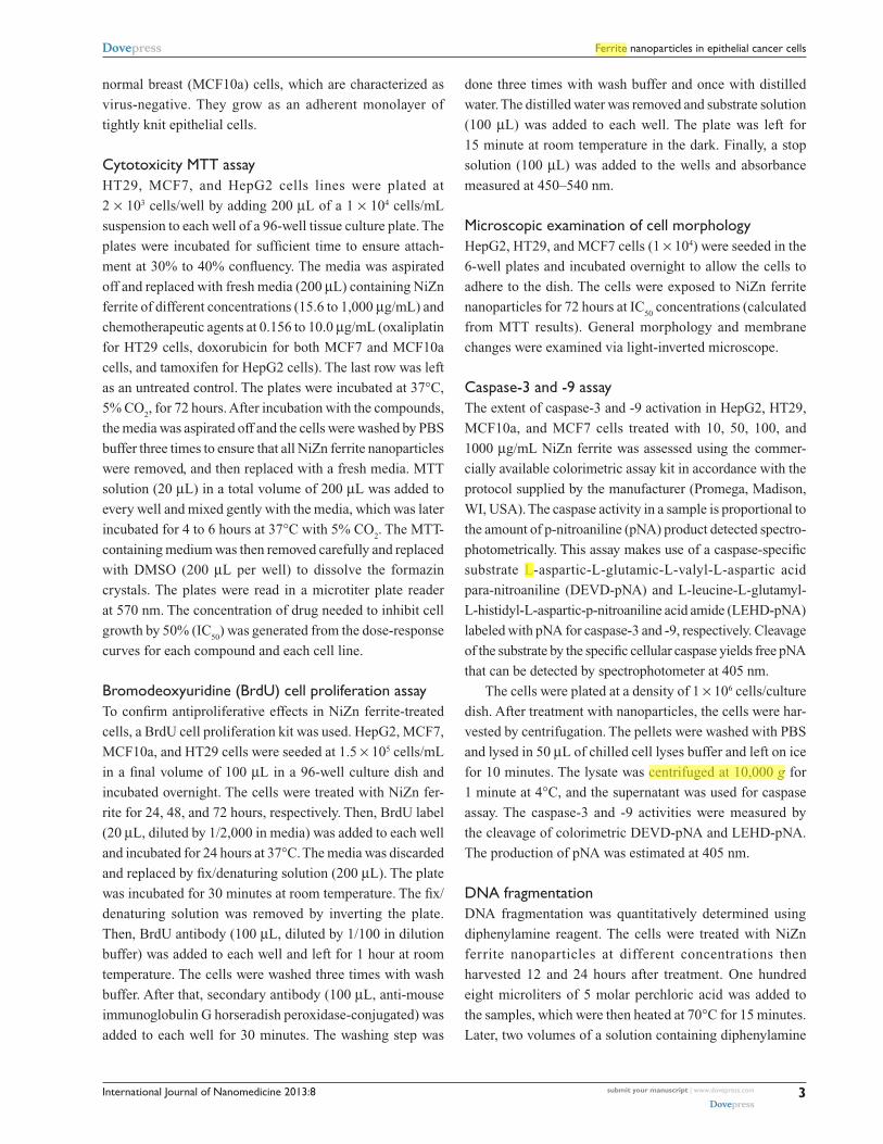
ResultsXRD analysisThe XRD diffraction patterns of NiZn ferrite nanoparticles
are depicted in Figure 1. In the studied angle range of
2θ = 10o–70o, it is clearly seen that the Ni0.5
Zn0.5
Fe2O
4 powder
has a crystalline phase with nine intense peaks which corre-
spond to diffractions due to (111), (220), (311), (222), (400),
(422), (511), (440), and (531) planes. These planes are well
indexed to a cubic spinel structure of a lattice parameter 8.4 Å
with no impurity phase detected. The most intense peak of
pure NiZn ferrite powder is assigned to the (311) index plane
at 2θ = 35.4o. The particle size was calculated to be 12 nm
using Debye–Scherrer’s equation:
D = 0.9 λ/βcosθ (2)
D is the crystallite size, λ is the incident X-ray wavelength,
β is the full width at half-maximum, and θ is the diffraction
angle.
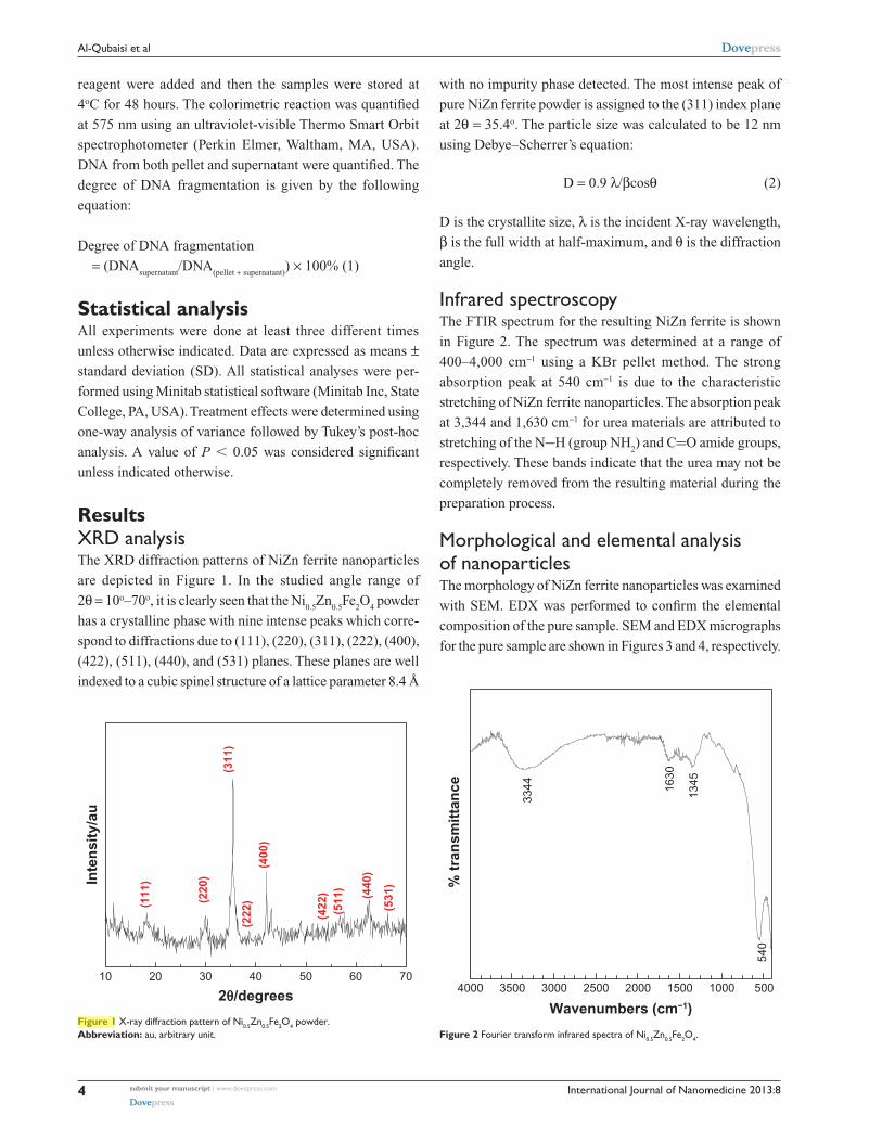
Infrared spectroscopyThe FTIR spectrum for the resulting NiZn ferrite is shown
in Figure 2. The spectrum was determined at a range of
400–4,000 cm−1 using a KBr pellet method. The strong
absorption peak at 540 cm−1 is due to the characteristic
stretching of NiZn ferrite nanoparticles. The absorption peak
at 3,344 and 1,630 cm−1 for urea materials are attributed to
stretching of the N−H (group NH2) and C=O amide groups,
respectively. These bands indicate that the urea may not be
completely removed from the resulting material during the
preparation process.
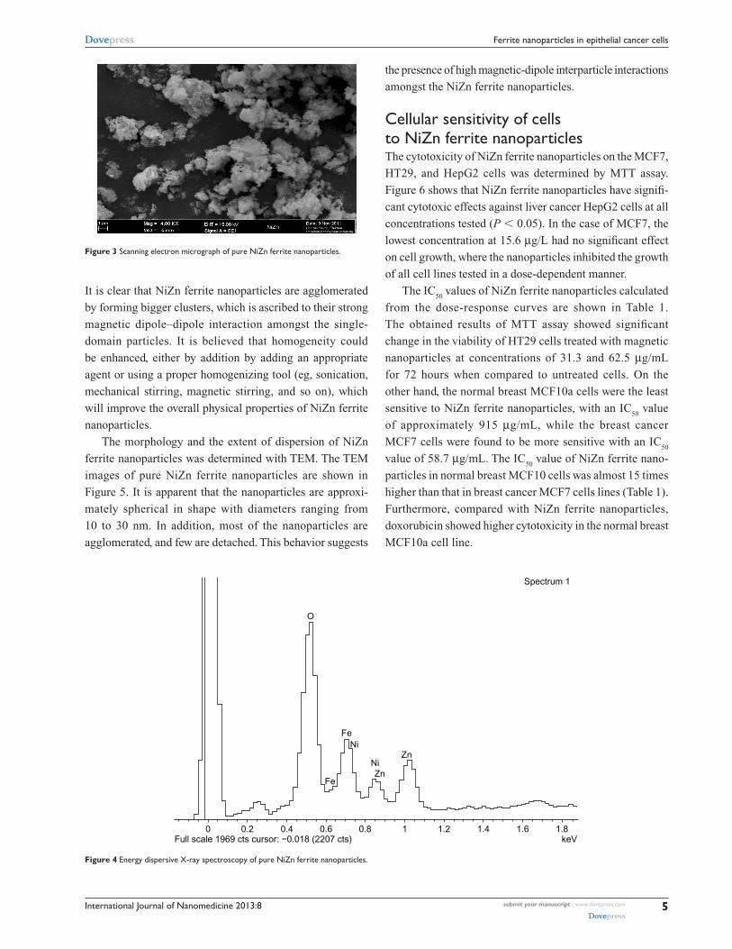
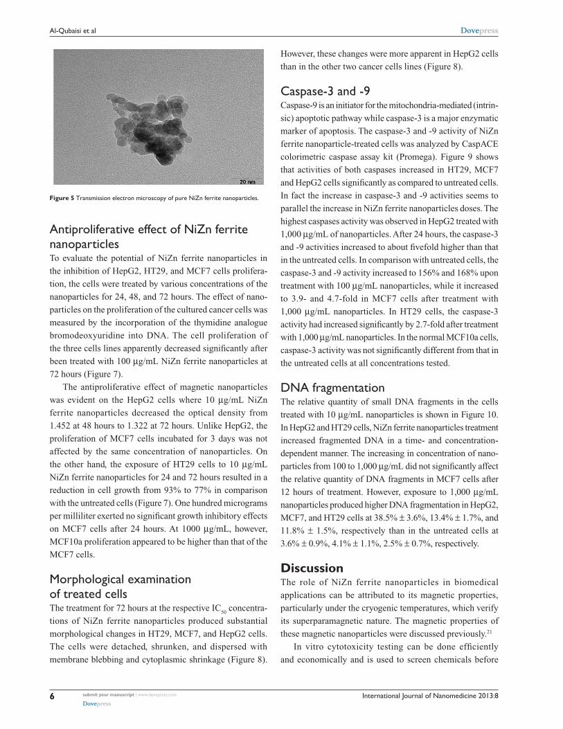
Morphological and elemental analysis of nanoparticlesThe morphology of NiZn ferrite nanoparticles was examined
with SEM. EDX was performed to confirm the elemental
composition of the pure sample. SEM and EDX micrographs
for the pure sample are shown in Figures 3 and 4, respectively.
10 20 30 40 50 60 70
(531
)
(422
) (440
)
(511
)
(400
)(2
22)
(311
)
(220
)Inte
nsi
ty/a
u
2θ/degrees
(111
)
Figure 1 X-ray diffraction pattern of Ni0.5Zn0.5Fe2O4 powder.Abbreviation: au, arbitrary unit.
4000 3500 3000 2500 2000 1500 1000 500
1345
540
1630
% t
ran
smit
tan
ce
Wavenumbers (cm−1)
3344
Figure 2 Fourier transform infrared spectra of Ni0.5Zn0.5Fe2O4.
submit your manuscript | www.dovepress.com
Dovepress
Dovepress
4
Al-Qubaisi et al
10403
Highlight
Dear author, please defined the abbreviation correctly. confirm I have defined the abbreviation correctly.
International Journal of Nanomedicine 2013:8
It is clear that NiZn ferrite nanoparticles are agglomerated
by forming bigger clusters, which is ascribed to their strong
magnetic dipole–dipole interaction amongst the single-
domain particles. It is believed that homogeneity could
be enhanced, either by addition by adding an appropriate
agent or using a proper homogenizing tool (eg, sonication,
mechanical stirring, magnetic stirring, and so on), which
will improve the overall physical properties of NiZn ferrite
nanoparticles.
The morphology and the extent of dispersion of NiZn
ferrite nanoparticles was determined with TEM. The TEM
images of pure NiZn ferrite nanoparticles are shown in
Figure 5. It is apparent that the nanoparticles are approxi-
mately spherical in shape with diameters ranging from
10 to 30 nm. In addition, most of the nanoparticles are
agglomerated, and few are detached. This behavior suggests
the presence of high magnetic-dipole interparticle interactions
amongst the NiZn ferrite nanoparticles.
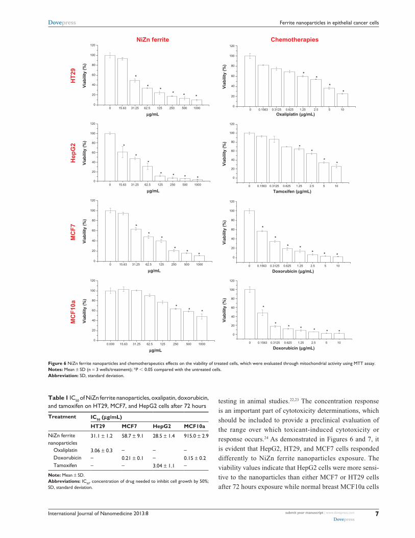
Cellular sensitivity of cells to NiZn ferrite nanoparticlesThe cytotoxicity of NiZn ferrite nanoparticles on the MCF7,
HT29, and HepG2 cells was determined by MTT assay.
Figure 6 shows that NiZn ferrite nanoparticles have signifi-
cant cytotoxic effects against liver cancer HepG2 cells at all
concentrations tested (P , 0.05). In the case of MCF7, the
lowest concentration at 15.6 µg/L had no significant effect
on cell growth, where the nanoparticles inhibited the growth
of all cell lines tested in a dose-dependent manner.
The IC50
values of NiZn ferrite nanoparticles calculated
from the dose-response curves are shown in Table 1.
The obtained results of MTT assay showed significant
change in the viability of HT29 cells treated with magnetic
nanoparticles at concentrations of 31.3 and 62.5 µg/mL
for 72 hours when compared to untreated cells. On the
other hand, the normal breast MCF10a cells were the least
sensitive to NiZn ferrite nanoparticles, with an IC50
value
of approximately 915 µg/mL, while the breast cancer
MCF7 cells were found to be more sensitive with an IC50
value of 58.7 µg/mL. The IC50
value of NiZn ferrite nano-
particles in normal breast MCF10 cells was almost 15 times
higher than that in breast cancer MCF7 cells lines (Table 1).
Furthermore, compared with NiZn ferrite nanoparticles,
doxorubicin showed higher cytotoxicity in the normal breast
MCF10a cell line.
Figure 3 Scanning electron micrograph of pure NiZn ferrite nanoparticles.
0 0.2Full scale 1969 cts cursor: −0.018 (2207 cts) keV
Spectrum 1
0.4 0.6 0.8 1 1.2 1.4 1.6 1.8
O
Fe
Fe
Ni
NiZn
Zn
Figure 4 Energy dispersive X-ray spectroscopy of pure NiZn ferrite nanoparticles.
submit your manuscript | www.dovepress.com
Dovepress
Dovepress
5
Ferrite nanoparticles in epithelial cancer cells
International Journal of Nanomedicine 2013:8
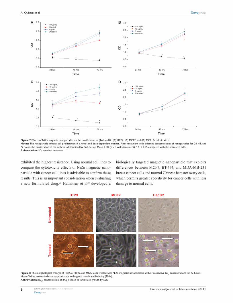
Antiproliferative effect of NiZn ferrite nanoparticlesTo evaluate the potential of NiZn ferrite nanoparticles in
the inhibition of HepG2, HT29, and MCF7 cells prolifera-
tion, the cells were treated by various concentrations of the
nanoparticles for 24, 48, and 72 hours. The effect of nano-
particles on the proliferation of the cultured cancer cells was
measured by the incorporation of the thymidine analogue
bromodeoxyuridine into DNA. The cell proliferation of
the three cells lines apparently decreased significantly after
been treated with 100 µg/mL NiZn ferrite nanoparticles at
72 hours (Figure 7).
The antiproliferative effect of magnetic nanoparticles
was evident on the HepG2 cells where 10 µg/mL NiZn
ferrite nanoparticles decreased the optical density from
1.452 at 48 hours to 1.322 at 72 hours. Unlike HepG2, the
proliferation of MCF7 cells incubated for 3 days was not
affected by the same concentration of nanoparticles. On
the other hand, the exposure of HT29 cells to 10 µg/mL
NiZn ferrite nanoparticles for 24 and 72 hours resulted in a
reduction in cell growth from 93% to 77% in comparison
with the untreated cells (Figure 7). One hundred micrograms
per milliliter exerted no significant growth inhibitory effects
on MCF7 cells after 24 hours. At 1000 µg/mL, however,
MCF10a proliferation appeared to be higher than that of the
MCF7 cells.
Morphological examination of treated cellsThe treatment for 72 hours at the respective IC
50 concentra-
tions of NiZn ferrite nanoparticles produced substantial
morphological changes in HT29, MCF7, and HepG2 cells.
The cells were detached, shrunken, and dispersed with
membrane blebbing and cytoplasmic shrinkage (Figure 8).
However, these changes were more apparent in HepG2 cells
than in the other two cancer cells lines (Figure 8).
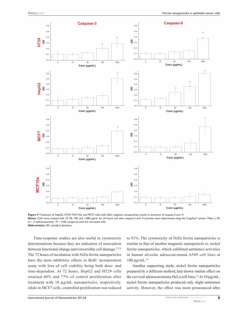
Caspase-3 and -9Caspase-9 is an initiator for the mitochondria-mediated (intrin-
sic) apoptotic pathway while caspase-3 is a major enzymatic
marker of apoptosis. The caspase-3 and -9 activity of NiZn
ferrite nanoparticle-treated cells was analyzed by CaspACE
colorimetric caspase assay kit (Promega). Figure 9 shows
that activities of both caspases increased in HT29, MCF7
and HepG2 cells significantly as compared to untreated cells.
In fact the increase in caspase-3 and -9 activities seems to
parallel the increase in NiZn ferrite nanoparticles doses. The
highest caspases activity was observed in HepG2 treated with
1,000 µg/mL of nanoparticles. After 24 hours, the caspase-3
and -9 activities increased to about fivefold higher than that
in the untreated cells. In comparison with untreated cells, the
caspase-3 and -9 activity increased to 156% and 168% upon
treatment with 100 µg/mL nanoparticles, while it increased
to 3.9- and 4.7-fold in MCF7 cells after treatment with
1,000 µg/mL nanoparticles. In HT29 cells, the caspase-3
activity had increased significantly by 2.7-fold after treatment
with 1,000 µg/mL nanoparticles. In the normal MCF10a cells,
caspase-3 activity was not significantly different from that in
the untreated cells at all concentrations tested.
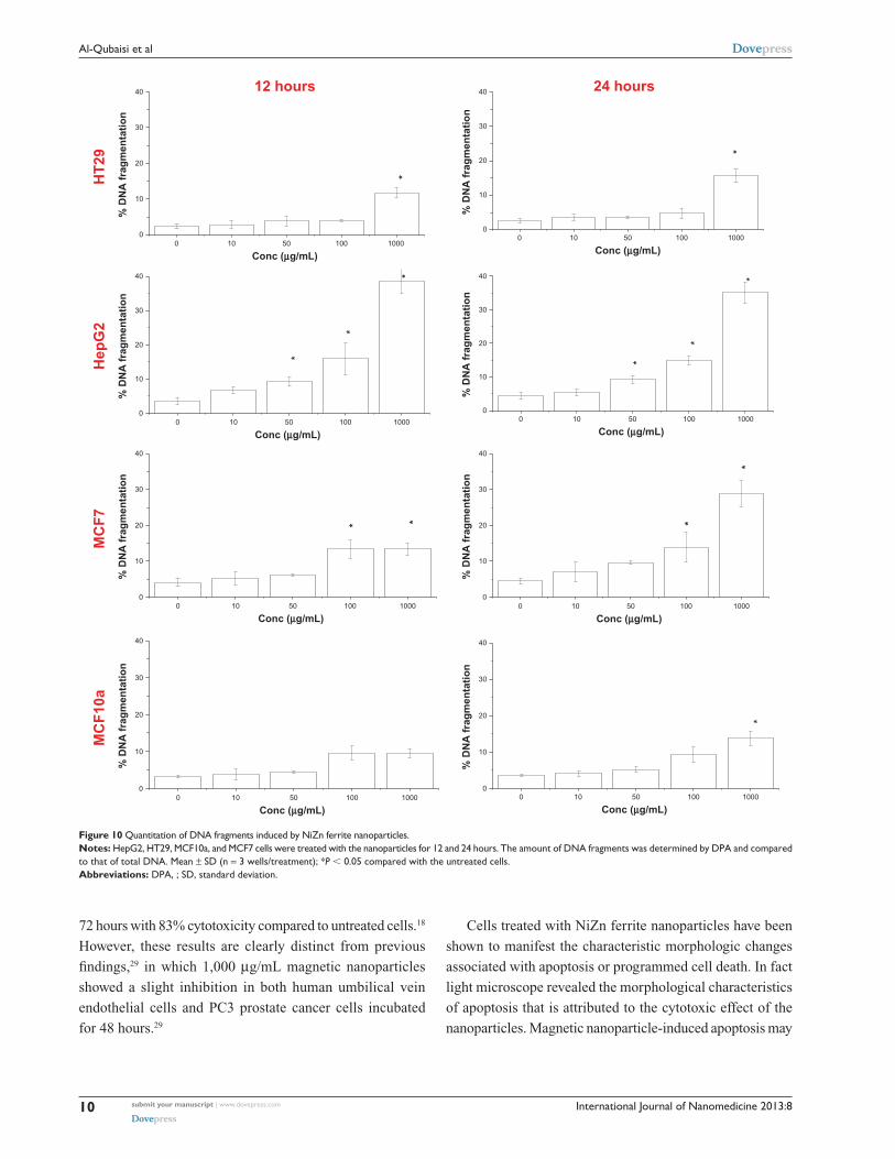
DNA fragmentationThe relative quantity of small DNA fragments in the cells
treated with 10 µg/mL nanoparticles is shown in Figure 10.
In HepG2 and HT29 cells, NiZn ferrite nanoparticles treatment
increased fragmented DNA in a time- and concentration-
dependent manner. The increasing in concentration of nano-
particles from 100 to 1,000 µg/mL did not significantly affect
the relative quantity of DNA fragments in MCF7 cells after
12 hours of treatment. However, exposure to 1,000 µg/mL
nanoparticles produced higher DNA fragmentation in HepG2,
MCF7, and HT29 cells at 38.5% ± 3.6%, 13.4% ± 1.7%, and
11.8% ± 1.5%, respectively than in the untreated cells at
3.6% ± 0.9%, 4.1% ± 1.1%, 2.5% ± 0.7%, respectively.
DiscussionThe role of NiZn ferrite nanoparticles in biomedical
applications can be attributed to its magnetic properties,
particularly under the cryogenic temperatures, which verify
its superparamagnetic nature. The magnetic properties of
these magnetic nanoparticles were discussed previously.21
In vitro cytotoxicity testing can be done efficiently
and economically and is used to screen chemicals before
Figure 5 Transmission electron microscopy of pure NiZn ferrite nanoparticles.
submit your manuscript | www.dovepress.com
Dovepress
Dovepress
6
Al-Qubaisi et al
International Journal of Nanomedicine 2013:8
testing in animal studies.22,23 The concentration response
is an important part of cytotoxicity determinations, which
should be included to provide a preclinical evaluation of
the range over which toxicant-induced cytotoxicity or
response occurs.24 As demonstrated in Figures 6 and 7, it
is evident that HepG2, HT29, and MCF7 cells responded
differently to NiZn ferrite nanoparticles exposure. The
viability values indicate that HepG2 cells were more sensi-
tive to the nanoparticles than either MCF7 or HT29 cells
after 72 hours exposure while normal breast MCF10a cells
NiZn ferrite Chemotherapies
HT
29
120
***
*
*
*
Via
bili
ty (
%)
µg/mL Oxaliplatin (µg/mL)
Tamoxifen (µg/mL)µg/mL
µg/mL
µg/mL
Doxorubicin (µg/mL)
Doxorubicin (µg/mL)*
*
*
*
Via
bili
ty (
%)
Hep
G2
****
*
*
*
Via
bili
ty (
%)
*
*
*
*
Via
bili
ty (
%)
MC
F7
*
*
*
*
*
*
Via
bili
ty (
%)
***
**
*
*
Via
bili
ty (
%)
MC
F10
a
*
* *
Via
bili
ty (
%)
0 15.63 31.25 62.5 125 250 500 10000
20
40
60
80
100
120
0
20
40
60
80
100
120
0
20
40
60
80
100
0
20
40
60
80
100
120
0
20
40
60
80
100
120
0
20
40
60
80
100
120
0
20
40
60
80
100
120
0
20
40
60
80
100
120
0 15.63 31.25 62.5 125 250 500 1000
0 15.63 31.25 62.5 125 250 500 1000
0.000 15.63 31.25 62.5 125 250 500 1000
0 0.1563 0.3125 0.625 1.25 2.5 5 10
0 0.1563 0.3125 0.625 1.25 2.5 5 10
0 0.1563 0.3125 0.625 1.25 2.5 5 10
0 0.1563 0.3125 0.625 1.25 2.5 5 10*****
*
*
Via
bili
ty (
%)
Figure 6 NiZn ferrite nanoparticles and chemotherapeutics effects on the viability of treated cells, which were evaluated through mitochondrial activity using MTT assay.Notes: Mean ± SD (n = 3 wells/treatment); *P , 0.05 compared with the untreated cells.Abbreviation: SD, standard deviation.
Table 1 IC50 of NiZn ferrite nanoparticles, oxalipatin, doxorubicin, and tamoxifen on HT29, MCF7, and Hepg2 cells after 72 hours
Treatment IC50 (μg/mL)
HT29 MCF7 HepG2 MCF10a
NiZn ferrite nanoparticles
31.1 ± 1.2 58.7 ± 9.1 28.5 ± 1.4 915.0 ± 2.9
Oxaliplatin 3.06 ± 0.3 – – – Doxorubicin – 0.21 ± 0.1 – 0.15 ± 0.2 Tamoxifen – – 3.04 ± 1.1 –
Note: Mean ± SD.Abbreviations: IC50, concentration of drug needed to inhibit cell growth by 50%; SD, standard deviation.
submit your manuscript | www.dovepress.com
Dovepress
Dovepress
7
Ferrite nanoparticles in epithelial cancer cells
International Journal of Nanomedicine 2013:8
24 hrs 48 hrs 72 hrs0.0
0.5
1.0
1.5
2.0
2.5
*
**
OD
Time
24 hrs 48 hrs 72 hrs
Time
24 hrs 48 hrs 72 hrs
Time
24 hrs 48 hrs 72 hrs
Time
100 µg/mL10 µg/mL5 µg/mL Untreated
100 µg/mL10 µg/mL5 µg/mL Untreated
100 µg/mL10 µg/mL5 µg/mL Untreated
100 µg/mL10 µg/mL5 µg/mL Untreated
A
C
B
D
0.0
0.5
1.0
1.5
2.0
2.5
3.0
*
*
OD
0.0
0.5
1.0
1.5
2.0
2.5
*
*
*
**
OD
0.0
0.5
1.0
1.5
2.0
2.5
3.0
OD
Figure 7 Effects of NiZn magnetic nanoparticles on the proliferation of (A) Hepg2, (B) HT29, (C) MCF7, and (D) MCF10a cells in vitro. Notes: The nanoparticle inhibits cell proliferation in a time- and dose-dependent manner. After treatment with different concentrations of nanoparticles for 24, 48, and 72 hours, the proliferation of the cells was determined by BrdU assay. Mean ± SD (n = 3 wells/treatment); * P , 0.05 compared with the untreated cells.Abbreviation: SD, standard deviation.
HT29 MCF7 HepG2
Un
trea
ted
Tre
ated
Figure 8 The morphological changes of Hepg2, HT29, and MCF7 cells treated with NiZn magnetic nanoparticles at their respective IC50 concentrations for 72 hours. Note: White arrows indicate apoptotic cells with typical membrane blebbing (200×).Abbreviation: IC50, concentration of drug needed to inhibit cell growth by 50%.
exhibited the highest resistance. Using normal cell lines to
compare the cytotoxicity effects of NiZn magnetic nano-
particle with cancer cell lines is advisable to confirm these
results. This is an important consideration when evaluating
a new formulated drug.25 Hathaway et al26 developed a
biologically targeted magnetic nanoparticle that exploits
differences between MCF7, BT-474, and MDA-MB-231
breast cancer cells and normal Chinese hamster ovary cells,
which permits greater specificity for cancer cells with less
damage to normal cells.
submit your manuscript | www.dovepress.com
Dovepress
Dovepress
8
Al-Qubaisi et al
International Journal of Nanomedicine 2013:8
Time-response studies are also useful in cytotoxicity
determinations because they are indicators of association
between functional change and irreversible cell damage.27,28
The 72 hours of incubation with NiZn ferrite nanoparticles
have the most inhibitory effects in BrdU incorporation
assay with loss of cell viability being both dose- and
time- dependent. At 72 hours, HepG2 and HT29 cells
retained 60% and 77% of control proliferation after
treatment with 10 µg/mL nanoparticles, respectively,
while in MCF7 cells, controlled proliferation was reduced
to 91%. The cytotoxicity of NiZn ferrite nanoparticles is
similar to that of another magnetic nanoparticle ie, nickel
ferrite nanoparticles, which exhibited antitumor activities
in human alveolar adenocarcinoma A549 cell lines at
100 µg/mL.19
Another supporting study, nickel ferrite nanoparticles
prepared by a different method, had shown similar effect on
the cervical adenocarcinoma HeLa cell lines.18 At 10 µg/mL,
nickel ferrite nanoparticles produced only slight antitumor
activity. However, the effect was more pronounced after
Caspase-3 Caspase-9
HT
29
*
*
OD
Conc (µg/mL)
0 10 50 100 1000
Conc (µg/mL)
0 10 50 100 1000
Conc (µg/mL)
0 10 50 100 1000
Conc (µg/mL)
0 10 50 100 1000
Conc (µg/mL)
Conc (µg/mL)
Conc (µg/mL)
Conc (µg/mL)
0.0
0.1
0.2
0.3
0.4
0.5
0.6
*
OD
Hep
G2
*
OD
0.0
0.1
0.2
0.3
0.4
0.5
0.6
*
*
*
OD
MC
F7
*
OD
0.0
0.1
0.2
0.3
0.4
0.5
0.6
*
OD
MC
F10
a
0 10 50 100 10000.0
0.1
0.2
0.3
0.4
0.5
0.6
0 10 50 100 1000
0 10 50 100 1000
0 10 50 100 1000
0.0
0.1
0.2
0.3
0.4
0.5
0.6
0.0
0.1
0.2
0.3
0.4
0.5
0.6
0.0
0.1
0.2
0.3
0.4
0.5
0.6
OD
0.0
0.1
0.2
0.3
0.4
0.5
0.6
OD
Figure 9 Treatment of Hepg2, HT29, MCF10a, and MCF7 cells with NiZn magnetic nanoparticles results in activation of caspase-3 and -9. Notes: Cells were treated with 10, 50, 100, and 1,000 µg/mL for 24 hours and then caspase-3 and -9 activities were determined using the CaspAce® system. Mean ± SD (n = 3 wells/treatment). *P , 0.05 compared with the untreated cells.Abbreviation: SD, standard deviation.
submit your manuscript | www.dovepress.com
Dovepress
Dovepress
9
Ferrite nanoparticles in epithelial cancer cells
International Journal of Nanomedicine 2013:8
72 hours with 83% cytotoxicity compared to untreated cells.18
However, these results are clearly distinct from previous
findings,29 in which 1,000 µg/mL magnetic nanoparticles
showed a slight inhibition in both human umbilical vein
endothelial cells and PC3 prostate cancer cells incubated
for 48 hours.29
Cells treated with NiZn ferrite nanoparticles have been
shown to manifest the characteristic morphologic changes
associated with apoptosis or programmed cell death. In fact
light microscope revealed the morphological characteristics
of apoptosis that is attributed to the cytotoxic effect of the
nanoparticles. Magnetic nanoparticle-induced apoptosis may
12 hours 24 hours H
T29
0
10
20
30
40
*
% D
NA
fra
gm
enta
tio
n%
DN
A f
rag
men
tati
on
% D
NA
fra
gm
enta
tio
n%
DN
A f
rag
men
tati
on
*
Hep
G2
0
10
20
30
40 *
*
*
*
*
*
MC
F7
0
10
20
30
40
* *
*
*
MC
F10
a
0
10
20
30
40
0
10
20
30
40
% D
NA
fra
gm
enta
tio
n%
DN
A f
rag
men
tati
on
% D
NA
fra
gm
enta
tio
n%
DN
A f
rag
men
tati
on
0
10
20
30
40
0
10
20
30
40
0
10
20
30
40
*
Conc (µg/mL)0 10 50 100 1000
Conc (µg/mL)0 10 50 100 1000
Conc (µg/mL)0 10 50 100 1000
Conc (µg/mL)0 10 50 100 1000
Conc (µg/mL)0 10 50 100 1000
Conc (µg/mL)0 10 50 100 1000
Conc (µg/mL)0 10 50 100 1000
Conc (µg/mL)0 10 50 100 1000
Figure 10 Quantitation of DNA fragments induced by NiZn ferrite nanoparticles.Notes: Hepg2, HT29, MCF10a, and MCF7 cells were treated with the nanoparticles for 12 and 24 hours. The amount of DNA fragments was determined by DPA and compared to that of total DNA. Mean ± SD (n = 3 wells/treatment); *P , 0.05 compared with the untreated cells.Abbreviations: DPA, ; SD, standard deviation.
submit your manuscript | www.dovepress.com
Dovepress
Dovepress
10
Al-Qubaisi et al
International Journal of Nanomedicine 2013:8
be an integral component of the cellular mechanism of action
relating to its therapeutic effects and cytotoxicities. The
magnetic nanoparticles are rapidly distributed in epithelial
tissue with strong binding to plasma proteins, principally
albumin.30
Activation of endogenous nuclease enzymes is considered
to be a key biochemical event in apoptosis, leading to the
cleavage of DNA into nucleosomesized fragments and it
is well-known that caspase-3 is a key mediator of nuclease
activation.31 The three cell lines, HT29, MCF7, and HepG2,
were treated with increasing concentrations of NiZn ferrite
nanoparticles to determine conditions that could induce
apoptosis measured by a standard interchromosomal DNA
fragmentation assay. NiZn ferrite nanoparticles induced
dose-dependent apoptosis in the treated cells with maximal
effective dose of about 100 µg/mL in HepG2 cells and
1,000 µg/mL for both HT29 and MCF7 cells after 12 hours.
This effect is similar to that produced with nickel ferrite
nanoparticles, but in the human alveolar adenocarcinoma
A549 cell line with similar effective concentration of
100 µg/mL or less.19 Nevertheless, previous data had
provided evidence that the magnetic nanoparticles fulfill
two basic criteria for an effective chemotherapeutic agent,
ie, tumor specificity and minimal toxicity to the normal
cells.26
ConclusionNiZn ferrite nanoparticles exhibited greater cytotoxicity
on liver cancer HepG2 cells among the cancer cell lines
examined. The normal breast MCF10a cells seem to show the
lowest sensitivity to the cytotoxic effect of the nanoparticles.
NiZn ferrite nanoparticles selectively killed liver cancer
cell through suppression of proliferation and induction of
apoptosis. Our magnetic nanoparticles induced apoptosis in
cancer cells via a caspase-9-dependent pathway. In fact, a
significant increase of caspase-3 activity, which correlates
with the DPA assay findings, a marker of DNA fragmentation,
was observed in HT29, MCF7, and HepG2 cell lines as a
result of treatment with NiZn ferrite nanoparticles.
Extrapolation of the in vitro cytotoxic effects of our NiZn
ferrite nanoparticles to in vivo anticancer application would
require further studies to determine its stability, half-life,
and biologically significant concentrations in the induction
of the apoptotic pathway in vivo. Therefore, taking this
into consideration, our findings strongly suggest that NiZn
ferrite nanoparticles merit further investigation as a cancer
chemotherapeutic agent.
DisclosureThe authors report no conflicts of interest in this work.
References 1. Sun C, Lee JSH, Zhang M. Magnetic nanoparticles in MR imaging and
drug delivery. Adv Drug Deliv Rev. 2008;60(11):1252–1265. 2. Gu H, Ho PL, Tsang KWT, Wang L, Xu B. Using biofunctional mag-
netic nanoparticles to capture vancomycin-resistant enterococci and other gram-positive bacteria at ultralow concentration. J Am Chem Soc. 2003;125(51):15702–15703.
3. Taylor EN, Webster TJ. The use of superparamagnetic nanoparticles for prosthetic biofilm prevention. Int J Nanomedicine. 2009;4:145–152.
4. Peyre J, Humblot V, Méthivier C, Berjeauc JM, Pradier CM. Co-grafting of amino-poly-ethylene-glycol and magainin I on a TiO
2 surface:
Tests of antifouling and antibacterial activities. J Phys Chem B. 2012;116(47):13839–13847.
5. Lee KJ, An JH, Shin JS, Kim DH. Synthesis and characterization of bicalutamide-loaded magnetic nanoparticles as anti-tumor drug carriers. J Nanosci Nanotechnol. 2012;12(2):1611–1615.
6. Kircher MF, Mahmood U, King RS, Weissleder R, Josephson L. A multimodal nanoparticle for preoperative magnetic resonance imaging and intraoperative optical brain tumor delineation. Cancer Res. 2003;63(23):8122–8125.
7. Brunke O, Odenbach S, Jurgons R, Alexiou C, Hilger I, Beckmann F. Determination of the magnetic particle distribution in tumour tissue by means of x-ray tomography. J Phys Condens Matter. 2006;18(38):S2903.
8. Pray L. Molecular Diagnostics: New Growth, New Markets. Waltham, MA: Cambridge Healthtech Advisors; 2005.
9. Leuschner C, Kumar CSSR, Hansel W, Soboyejo W, Zhou J, Hormes J. LHRH-conjugated magnetic iron oxide nanoparticles for detection of breast cancer metastases. Breast Cancer Res Treat. 2006;99(2): 163–176.
10. Zhao Q, Wang L, Cheng R, et al. Magnetic nanoparticle-based hyper-thermia for head and neck cancer in mouse models. Theranostics. 2012;2(1):113–121.
11. Thomas LA, Dekker L, Kallumadil M, et al. Carboxylic acid-stabilised iron oxide nanoparticles for use in magnetic hyperthermia. J Mater Chem. 2009;19(36):6529–6535.
12. Babincov M, Altanerov V, Altaner C, Bergemann C, Babinec P. In vitro analysis of cisplatin functionalized magnetic nanoparticles in combined cancer chemotherapy and electromagnetic hyperthermia. IEEE Trans Nanobiosci. 2008;7(1):15–19.
13. Maeng JH, Lee DH, Jung KH, et al. Multifunctional doxorubicin-loaded superparamagnetic iron oxide nanoparticles for chemotherapy and magnetic resonance imaging in liver cancer. Biomaterials. 2010;31(18): 4995–5006.
14. Jia Y, Yuan M, Yuan H, et al. Co-encapsulation of magnetic Fe3O4 nano-particles and doxorubicin into biodegradable PLGA nanocarriers for intratumoral drug delivery. Int J Nanomedicine. 2012;63:1697–1708.
15. Martin J. British National Formulary: September 2010. BMJ Group and RPS Publishing; 2010.
16. de Freitas ERL, Soares PRO, de Paula Santos R, et al. In vitro biological activities of anionic-Fe
2O
3 nanoparticles on human melanoma cells.
J Nanosci Nanotechnol. 2008;8(5):2385–2391. 17. Yang G, Li X, Zhao Z, Wang W. Preparation, characterization,
in vivo and in vitro studies of arsenic trioxide Mg-Fe ferrite magnetic nanoparticles. Acta Pharmacol Sin. 2009;30(12):1688–1693.
18. Tomitaka A, Koshi T, Hatsugai S, Yamada T, Takemura Y. Magnetic characterization of surface-coated magnetic nanoparticles for biomedical application. J Magn Magn Mater. 2011;323(10):1398–1403.
19. Ahamed M, Akhtar MJ, Siddiqui MA, et al. Oxidative stress mediated apoptosis induced by nickel ferrite nanoparticles in cultured A549 cells. Toxicology. 2011;283(2–3):101–108.
submit your manuscript | www.dovepress.com
Dovepress
Dovepress
11
Ferrite nanoparticles in epithelial cancer cells
10403
Highlight
Dear Author, Please define this abbreviation in full here and in the figure legend. Thank's
10403
Highlight
Dear Author, Please confirm this statement. Thank's
10403
Highlight
Dear aurthor, please include the location details for the publisher (city, state, country)
International Journal of Nanomedicine
Publish your work in this journal
Submit your manuscript here: http://www.dovepress.com/international-journal-of-nanomedicine-journal
The International Journal of Nanomedicine is an international, peer-reviewed journal focusing on the application of nanotechnology in diagnostics, therapeutics, and drug delivery systems throughout the biomedical field. This journal is indexed on PubMed Central, MedLine, CAS, SciSearch®, Current Contents®/Clinical Medicine,
Journal Citation Reports/Science Edition, EMBase, Scopus and the Elsevier Bibliographic databases. The manuscript management system is completely online and includes a very quick and fair peer-review system, which is all easy to use. Visit http://www.dovepress.com/ testimonials.php to read real quotes from published authors.
International Journal of Nanomedicine 2013:8
20. Modak S, Ammar M, Mazaleyrat F, Das S, Chakrabarti PK. XRD, HRTEM and magnetic properties of mixed spinel nanocrystalline Ni−Zn−Cu−ferrite. J Alloys Compd. 2009;473(1–2):15–19.
21. Flaifel MH, Ahmad SH, Abdullah MH, Al-Asbahi BA. NiZn Ferrite filled thermoplastic natural rubber nanocomposites: effect of low temperature on their magnetic behaviour. Cryogenics. 2012;52(10):523–529.
22. Malich G, Markovic B, Winder C. The sensitivity and specificity of the MTS tetrazolium assay for detecting the in vitro cytotoxicity of 20 chemicals using human cell lines. Toxicology. 1997;124(3):179–192.
23. Shokri F, Heidari M, Gharagozloo S, Ghazi-Khansari M. In vitro inhibitory effects of antioxidants on cytotoxicity of T-2 toxin. Toxicology. 2000;146(2):171–176.
24. Miller FJ, Schlosser PM, Janszen DB. Haber’s rule: a special case in a family of curves relating concentration and duration of exposure to a fixed level of response for a given endpoint. Toxicology. 2000;149(1): 21–34.
25. Al-Qubaisi M, Rozita R, Yeap SK, Omar AR, Ali AM, Alitheen NB. Selective cytotoxicity of goniothalamin against hepato-blastoma HepG2 cells. Molecules. 2011;16(4):2944–2959.
26. Hathaway HJ, Butler KS, Adolphi NL, et al. Detection of breast cancer cells using targeted magnetic nanoparticles and ultra-sensitive magnetic field sensors. Breast Cancer Res. 2011;13(5):R108.
27. Rozman KK, Doull J. Dose and time as variables of toxicity. Toxicology. 2000;144(1):169–178.
28. Tennekes HA. The significance of the Druckrey–Küpfmüller equa-tion for risk assessment – The toxicity of neonicotinoid insecticides to arthropods is reinforced by exposure time: Responding to a Letter to the Editor by Drs C Maus and R Nauen of Bayer CropScience AG. Toxicology. 2011;280(3):173.
29. Hafeli UO, Riffle JS, Harris-Shekhawat L, et al. Cell uptake and in vitro toxicity of magnetic nanoparticles suitable for drug delivery. Mol Pharm. 2009;6(5):1417–1428.
30. Lartigue L, Wilhelm C, Servais J, et al. Nanomagnetic sensing of blood plasma protein interactions with iron oxide nanoparticles: impact on macrophage uptake. ACS Nano. 2012;6(3):2665–2678.
31. Mitamura S, Ikawa H, Mizuno N, Kaziro Y, Itoh H. Cytosolic nuclease activated by caspase-3 and inhibited by DFF-45. Biochem Biophys Res Comm. 1998;243(2):480–484.
submit your manuscript | www.dovepress.com
Dovepress
Dovepress
Dovepress
12
Al-Qubaisi et al