Redox behaviour of Menadiol derivative at glassy carbon electrode
Upload
khangminh22Category
view
0download
0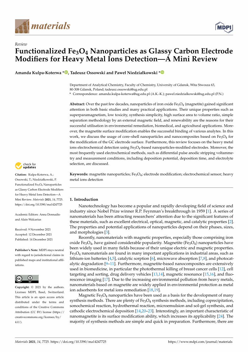
materials
Review
Functionalized Fe3O4 Nanoparticles as Glassy Carbon ElectrodeModifiers for Heavy Metal Ions Detection—A Mini Review
Amanda Kulpa-Koterwa * , Tadeusz Ossowski and Paweł Niedziałkowski *
�����������������
Citation: Kulpa-Koterwa, A.;
Ossowski, T.; Niedziałkowski, P.
Functionalized Fe3O4 Nanoparticles
as Glassy Carbon Electrode Modifiers
for Heavy Metal Ions Detection—A
Mini Review. Materials 2021, 14, 7725.
https://doi.org/10.3390/ma14247725
Academic Editors: Anna Donnadio
and Alain Walcarius
Received: 9 November 2021
Accepted: 12 December 2021
Published: 14 December 2021
Publisher’s Note: MDPI stays neutral
with regard to jurisdictional claims in
published maps and institutional affil-
iations.
Copyright: © 2021 by the authors.
Licensee MDPI, Basel, Switzerland.
This article is an open access article
distributed under the terms and
conditions of the Creative Commons
Attribution (CC BY) license (https://
creativecommons.org/licenses/by/
4.0/).
Department of Analytical Chemistry, Faculty of Chemistry, University of Gdansk, Wita Stwosza 63,80-308 Gdansk, Poland; [email protected]* Correspondence: [email protected] (A.K.-K.); [email protected] (P.N.)
Abstract: Over the past few decades, nanoparticles of iron oxide Fe3O4 (magnetite) gained significantattention in both basic studies and many practical applications. Their unique properties such assuperparamagnetism, low toxicity, synthesis simplicity, high surface area to volume ratio, simpleseparation methodology by an external magnetic field, and renewability are the reasons for theirsuccessful utilisation in environmental remediation, biomedical, and agricultural applications. More-over, the magnetite surface modification enables the successful binding of various analytes. In thiswork, we discuss the usage of core–shell nanoparticles and nanocomposites based on Fe3O4 forthe modification of the GC electrode surface. Furthermore, this review focuses on the heavy metalions electrochemical detection using Fe3O4-based nanoparticles-modified electrodes. Moreover, themost frequently used electrochemical methods, such as differential pulse anodic stripping voltamme-try and measurement conditions, including deposition potential, deposition time, and electrolyteselection, are discussed.
Keywords: magnetite nanoparticles; Fe3O4; electrode modification; electrochemical sensor; heavymetal ions detection
1. Introduction
Nanotechnology has become a popular and rapidly developing field of science andindustry since Nobel Prize winner R.P. Feynman’s breakthrough in 1959 [1]. A series ofnanomaterials has been attracting researchers’ attention due to the significant features ofthese materials, such as excellent electrical, optical, magnetic, and catalytic properties [2].The properties and potential applications of nanoparticles depend on their phases, sizes,and morphologies [3].
Recently, nanomaterials with magnetic properties, especially those comprising ironoxide Fe3O4, have gained considerable popularity. Magnetite (Fe3O4) nanoparticles havebeen widely used in many fields because of their unique electric and magnetic properties.Fe3O4 nanomaterials are found in many important applications in industrial areas, such aslithium-ion batteries [4,5], catalytic sorption [6], microwave absorption [7,8], and photocat-alytic degradation [9–11]. Furthermore, magnetite-based nanocomposites are extensivelyused in biomedicine, in particular the photothermal killing of breast cancer cells [12], celltargeting and sorting, drug delivery vehicles [13,14], magnetic resonance [15,16], and fluo-rescence imaging [17]. Due to the increasing environmental pollution from heavy metals,nanomaterials based on magnetite are widely applied in environmental protection as metalion adsorbents for metal ions remediation [18,19].
Magnetic Fe3O4 nanoparticles have been used as a basis for the development of manysynthesis methods. There are plenty of Fe3O4 synthesis methods, including coprecipitation,sonochemical reaction, hydrothermal reaction, microemulsion and sol-gel synthesis, andcathodic electrochemical deposition [14,20–23]. Interestingly, an important characteristic ofnanomagnetite is its surface modification ability, which increases its applicability [24]. Themajority of synthesis methods are simple and quick in preparation. Furthermore, there are
Materials 2021, 14, 7725. https://doi.org/10.3390/ma14247725 https://www.mdpi.com/journal/materials
Materials 2021, 14, 7725 2 of 14
many synthetic methods that can be used to obtain different nanoparticle sizes [24–28] andshapes [28–30]. Nanomagnetite can be obtained in various sizes and shapes, including themost popular spherical nanoparticles and in cuboids, octahedrons, plates, tetrahedrons,concaves, octapods, multibranches, and nanorods [31–34]. Fe3O4 nanoparticles are thebasic material for subsequent surface modifications creating core–shell structures, whichaffect the further extension of their applications in many fields.
To the authors’ knowledge, the very first paper covering Fe3O4 was published in1916 by the Americans, Sosman and Hostetter, and focused on iron oxides in general [35].Over the next few decades, several articles appeared each year. Since the 1990s, we haveobserved a growing interest in nanomagnetites. The highest number of publications in thefield with “Fe3O4” in the title appeared in 2020, totalling 1930 papers (Figure 1).
Materials 2021, 14, x FOR PEER REVIEW 2 of 16
[24]. The majority of synthesis methods are simple and quick in preparation. Furthermore,
there are many synthetic methods that can be used to obtain different nanoparticle sizes
[24–28] and shapes [28–30]. Nanomagnetite can be obtained in various sizes and shapes,
including the most popular spherical nanoparticles and in cuboids, octahedrons, plates,
tetrahedrons, concaves, octapods, multibranches, and nanorods [31–34]. Fe3O4 nanoparti-
cles are the basic material for subsequent surface modifications creating core–shell struc-
tures, which affect the further extension of their applications in many fields.
To the authors’ knowledge, the very first paper covering Fe3O4 was published in 1916
by the Americans, Sosman and Hostetter, and focused on iron oxides in general [35]. Over
the next few decades, several articles appeared each year. Since the 1990s, we have ob-
served a growing interest in nanomagnetites. The highest number of publications in the
field with “Fe3O4” in the title appeared in 2020, totalling 1930 papers (Figure 1).
Figure 1. Scheme of the number of publications with “Fe3O4” in title based on the year of publication
using the Scopus base.
The data presented in Figure 1 shows the popularity of Fe3O4 and its dynamics as a
topic of publication, especially during the most recent years. Additionally, composites
based on Fe3O4 are used in many fields of science and industry, including magnetic sepa-
ration, magnetic catalysis, environmental treatment, food analysis, target drug delivery
systems, biosensors, magnetic resonance imagining, hyperthermia, and tissue engineer-
ing [36].
There are plenty of magnetite nanoparticles and hybrid structures used in the elec-
trochemical detection of heavy metal ions providing an excellent basis for further func-
tionalisation. The surface of Fe3O4 nanoparticles can be combined with nanoparticles of
other metals (Au [37]); oxides (SiO2 [38,39] and TiO2 [40]) and additional conductive ma-
terials (GO [41]); complicated, organic functional groups (dendrimers [42] and polymers
[43]); and biological particles fragments (DNA [44]). Fe3O4-based nanomaterials possess a
high adsorption capacity, which makes them suitable for the electrochemical detection of
metals [45].
Figure 1. Scheme of the number of publications with “Fe3O4” in title based on the year of publicationusing the Scopus base.
The data presented in Figure 1 shows the popularity of Fe3O4 and its dynamics as atopic of publication, especially during the most recent years. Additionally, composites basedon Fe3O4 are used in many fields of science and industry, including magnetic separation,magnetic catalysis, environmental treatment, food analysis, target drug delivery systems,biosensors, magnetic resonance imagining, hyperthermia, and tissue engineering [36].
There are plenty of magnetite nanoparticles and hybrid structures used in the elec-trochemical detection of heavy metal ions providing an excellent basis for further func-tionalisation. The surface of Fe3O4 nanoparticles can be combined with nanoparticlesof other metals (Au [37]); oxides (SiO2 [38,39] and TiO2 [40]) and additional conductivematerials (GO [41]); complicated, organic functional groups (dendrimers [42] and poly-mers [43]); and biological particles fragments (DNA [44]). Fe3O4-based nanomaterialspossess a high adsorption capacity, which makes them suitable for the electrochemicaldetection of metals [45].
The Fe3O4 nanoparticles are easy to oxidise and aggregate, which results in their lowmagnetic properties [46]; therefore, there is a need to coat bare nanomagnetite with polymeror inorganic shells. Additionally, the modification can increase the biocompatibility ofthese material [47].
In this work, we describe the recently published applications of a variety of function-alised Fe3O4 nanoparticles to electrode surface modifications to create a sensor for heavy
Materials 2021, 14, 7725 3 of 14
metal ions. We discuss the preparation procedure of GCE for a sensor and the methodsof electrode modification using Fe3O4 nanoparticles. We also present the most frequentlyused electrochemical techniques with their characteristic measurement parameters. Finally,we present a performance comparison of the recently developed heavy metal ion sensors.
2. Nano-Fe3O4 as Electrode Modifiers
Based on several decades of intensive research, Fe3O4 has become one of the bestcharacterised metal oxides. Its cubic crystallographic system contains both Fe3+ and Fe2+
ions. Fe3O4 is a black solid with a density of 5.18 g·cm−3, Mohs hardness of 5, melting pointrange of 1583–1597 ◦C, and boiling point of 2623 ◦C. Its characteristic magnetic feature isthe ferrimagnetism at room temperature and Neel (Curie) temperature of 850 ◦C [48].
In the past few years, Fe3O4 nanoparticles have become a focus of interest for nu-merous scientific groups. In the nano range (in diameter from 1 to 100 nm) smaller than6 nm, magnetite particles indicate superparamagnetic properties, although their magneticfeatures strongly depend on the synthesis method [49]. Based on the gathered evidence, thenanomagnetites in most applications show the best characteristics in the range of 10–20 nm.Decreasing the nanoparticles’ size leads to an increase in the specific surface. Furthermore,the nanoparticles’ size strongly influences their magnetic moment and reaction to themagnetic field and depends on their size and shape [50]. Electrochemistry, and electrodemodifications for the generation of a highly sensitive sensor, is one of the most rapidlydeveloping fields of science. Electrochemical sensing is focused on the development ofnew electrode materials with better properties compared to commercial electrodes. Theperfect sensor should exhibit a signal output proportional to the number of target species,high selectivity, sensitivity, repeatability, and rapid response [50].
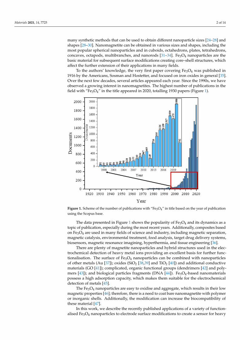
Nanomagnetite has been widely employed as a promising modifier due to its uniqueproperties, low-cost, easy preparation, non-toxicity, excellent absorption capacity, catalyticproperties, and inherent electrical conductivity [51]. The electrochemical performance of anelectrode is closely related to the absorption capacity and the conductivity of the modifiedmaterial. The imposition of Fe3O4 nanocomposites on the electrode surface causes theenhancement of the electrode area, enhancement of the rate of mass and electron transfer,improved selectivity and sensitivity, and, most importantly, increased response to the noiseratio [50]. Furthermore, Fe3O4-based electrochemical detection systems are characterisedby small dimensions, costlessness, sensitivity, flexibility, and quickness in use [52]. Theadvantages of Fe3O4 usage as an electro-sensor are described in Figure 2.
Materials 2021, 14, x FOR PEER REVIEW 4 of 16
Figure 2. Scheme of the important electro-sensor features.
3. Recent Electrode Modifications with Magnetic Nanoparticles to Heavy Metal Ions
Detection
The glassy carbon (GC) electrode is the most commonly used electrode for electroan-
alytical purposes due to its unique electrical conductivity, chemical stability, biocompati-
bility, and wide potential range and extremely low gas permeability [53]. Therefore, the
GC electrode is an excellent material for modification to obtain a stable surface used as a
biosensor. First of all, sensor development requires proper preparation of the electrode
for further modification. Before each modification, the GC electrode usually needs to be
polished to a shiny, mirror-like surface with wet alumina slurry—Al2O3 powder of differ-
ent sizes, 1.0 μm, 0.3 μm, and 0.05 μm, using a polishing cloth and rinsing with water.
Then, successive washing or sonications in absolute ethanol and ultrapure water are usu-
ally conducted, sometimes in a 1:1 (v/v) HNO3 solution, lasting at least a few minutes each
[54]. Subsequently, the electrode surface is dried with nitrogen or at room temperature,
and the electrode is ready for further use. As an exception, Miao et al. started the modifi-
cation with GCE soaking in piranha solution (98% H2SO4:30% H2O2 = 3:1) for about 5 min
to remove any adsorbed materials [44].
A homogeneous suspension of nanoparticles is necessary to modify the electrode,
most often by sonication in deionized water, absolute ethanol, and sometimes an ethanol
solution containing 0.25 wt% Nafion® [55] or IPA [56], or DMF [57] in a concentration of
1 mg/mL. Ultrasonic bath sonication lasts from 5 min to 2 h, but most often 30 min or until
a uniform suspension is obtained. The most common method of electrode modification is
drop-casting while the nanoparticles’ suspension is pipped on the electrode surface (Fig-
ure 3) [58]. The amount of applied nanoparticles depends on the active surface of the
working electrode. After the modification, the electrode was dried at room temperature
until the solvent completely evaporated, which usually takes from several minutes to a
few hours. A different approach was presented by Kong et al., where 6 mL of Fe3O4@PANI
nanoparticles suspension was pipetted onto an electrode, and after drying at 4 °C in a
refrigerator, the electrode was coated with 3 mL of Nafion® solution (0.5 wt%) [59]. More-
over, Wang et al. created an unconventional sensor by adding the Fe3O4@PDA@MnO2 NPs
homogenous suspension to the HCl solution (pH 3.0) with various concentrations of Pb2+.
Then, nanoparticles with already adsorbed Pb2+ ions were completely transferred onto the
mGCE for immediate electrochemical measurements (Figure 3.) [60]. Recent findings con-
cerning heavy metal ion detection with the GC electrode modified using Fe3O4-based
Figure 2. Scheme of the important electro-sensor features.
Materials 2021, 14, 7725 4 of 14
3. Recent Electrode Modifications with Magnetic Nanoparticles to Heavy MetalIons Detection
The glassy carbon (GC) electrode is the most commonly used electrode for electroana-lytical purposes due to its unique electrical conductivity, chemical stability, biocompatibility,and wide potential range and extremely low gas permeability [53]. Therefore, the GC elec-trode is an excellent material for modification to obtain a stable surface used as a biosensor.First of all, sensor development requires proper preparation of the electrode for furthermodification. Before each modification, the GC electrode usually needs to be polishedto a shiny, mirror-like surface with wet alumina slurry—Al2O3 powder of different sizes,1.0 µm, 0.3 µm, and 0.05 µm, using a polishing cloth and rinsing with water. Then, succes-sive washing or sonications in absolute ethanol and ultrapure water are usually conducted,sometimes in a 1:1 (v/v) HNO3 solution, lasting at least a few minutes each [54]. Sub-sequently, the electrode surface is dried with nitrogen or at room temperature, and theelectrode is ready for further use. As an exception, Miao et al. started the modificationwith GCE soaking in piranha solution (98% H2SO4:30% H2O2 = 3:1) for about 5 min toremove any adsorbed materials [44].
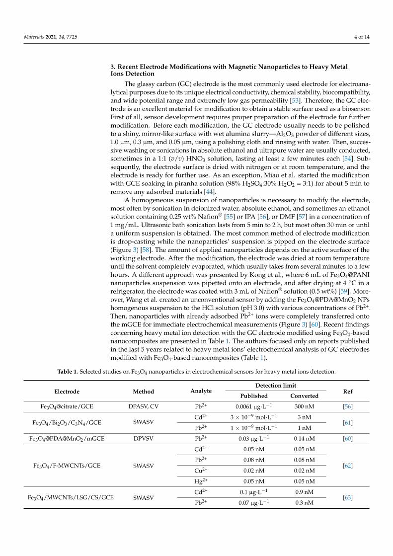
A homogeneous suspension of nanoparticles is necessary to modify the electrode,most often by sonication in deionized water, absolute ethanol, and sometimes an ethanolsolution containing 0.25 wt% Nafion® [55] or IPA [56], or DMF [57] in a concentration of1 mg/mL. Ultrasonic bath sonication lasts from 5 min to 2 h, but most often 30 min or untila uniform suspension is obtained. The most common method of electrode modificationis drop-casting while the nanoparticles’ suspension is pipped on the electrode surface(Figure 3) [58]. The amount of applied nanoparticles depends on the active surface of theworking electrode. After the modification, the electrode was dried at room temperatureuntil the solvent completely evaporated, which usually takes from several minutes to a fewhours. A different approach was presented by Kong et al., where 6 mL of Fe3O4@PANInanoparticles suspension was pipetted onto an electrode, and after drying at 4 ◦C in arefrigerator, the electrode was coated with 3 mL of Nafion® solution (0.5 wt%) [59]. More-over, Wang et al. created an unconventional sensor by adding the Fe3O4@PDA@MnO2 NPshomogenous suspension to the HCl solution (pH 3.0) with various concentrations of Pb2+.Then, nanoparticles with already adsorbed Pb2+ ions were completely transferred ontothe mGCE for immediate electrochemical measurements (Figure 3) [60]. Recent findingsconcerning heavy metal ion detection with the GC electrode modified using Fe3O4-basednanocomposites are presented in Table 1. The authors focused only on reports publishedin the last 5 years related to heavy metal ions’ electrochemical analysis of GC electrodesmodified with Fe3O4-based nanocomposites (Table 1).
Table 1. Selected studies on Fe3O4 nanoparticles in electrochemical sensors for heavy metal ions detection.
Electrode Method AnalyteDetection limit
RefPublished Converted
Fe3O4@citrate/GCE DPASV, CV Pb2+ 0.0061 µg·L−1 300 nM [56]
Fe3O4/Bi2O3/C3N4/GCE SWASVCd2+ 3 × 10⁻⁹ mol·L−1 3 nM
[61]Pb2+ 1 × 10⁻⁹ mol·L−1 1 nM
Fe3O4@PDA@MnO2/mGCE DPVSV Pb2+ 0.03 µg·L−1 0.14 nM [60]
Fe3O4/F-MWCNTs/GCE SWASV
Cd2+ 0.05 nM 0.05 nM
[62]Pb2+ 0.08 nM 0.08 nM
Cu2+ 0.02 nM 0.02 nM
Hg2+ 0.05 nM 0.05 nM
Fe3O4/MWCNTs/LSG/CS/GCE SWASVCd2+ 0.1 µg·L−1 0.9 nM
[63]Pb2+ 0.07 µg·L−1 0.3 nM

Materials 2021, 14, 7725 5 of 14
Table 1. Cont.
Electrode Method AnalyteDetection limit
RefPublished Converted
Fe3O4/SiO2/CS/Nafion/GCE DPASV Cu2+ 5 nmol·L−1 5 nM [55]
GCE/GO/Fe3O4@PMDA/AuNPs SWASVAs3+ 0.15 ppb 2 nM
[64]Cu2+ 0.11 ppb 2.4 nM
Fe3O4@PANI/MGCE DPASVCd2+ 0.3 nmol·L−1 0.3 nM
[59]Pb2+ 0.03 nmol·L−1 0.03 nM
Fe3O4/F-MWCNTs/GCE SWASV
Cd2+ 0.014 µM 14 nM
[65]
Pb2+ 0.0084 µM 8.4 nM
Hg2+ 0.0039 µM 3.9 nM
Zn2+ 0.012 µM 12 nM
Cu2+ 0.0053 µM 5.3 nM
GO@Fe3O4@2-CBT/GCE SWASVCd2+ 0.03 ng·mL−1 0.27 nM
[66]Pb2+ 0.02 ng·mL−1 0.1 nM
Fe3O4/GCE SWASV
Pb2+ 0.119 µM 119 nM
[54]Cd2+ 0.154 µM 154 nM
Hg2+ 0.0839 µM 83.9 nM
Cu2+ 0.0765 µM 76.5 nM
DNA/Fe3O4@Au/MGCE SWVAg+ 3.4 nM 3.4 nM [44]
Hg2+ 1.7 nM 1.7 nM
Fe3O4@C/GCE SWASV
Cd2+ 40.9 nM 40.9 nM
[57]
Pb2+ 20.7 nM 20.7 nM
Cu2+ 79.3 nM 79.3 nM
NH2-Fe3O4@C/GCE SWASV
Cd2+ 23.1 nM 23.1 nM
Pb2+ 28.5 nM 28.5 nM
Cu2+ 38.4 nM 38.4 nM
Fe3O4/GN/GE/GCE SWASV Pb2+ 0.0123 pM 0.0123 pM [67]
TA/Fe3O4/GCE SWASV
Pb2+ 0.04 µM 40 nM
[68]Hg2+ 0.3 µM 300 nM
Cd2+ 0.2 µM 200 nM
GSH@Fe3O4/MGCE SWASVPb2+ 0.182 µg·L−1 0.9 nM
[69]Cd2+ 0.172 µg·L−1 1.5 nM
Electrochemical techniques, especially voltammetry, include electroanalytical methodsfor the determination of one or more analytes by measuring the current as a function of thepotential. There are a few component techniques used to obtain information on the analyte,including CV, DPV, SWV, and stripping voltammetry [70]. Voltammetric techniques arewidely used in heavy metal ions detection due to their precision and sensitivity. The mostfrequently chosen are DPV or alternative SWV techniques (Table 1.) due to their highsensitivity and lower detection limits, which are suitable for trace level analysis. However,square wave voltammetry is preferable for obtaining the response rate. The most frequentlyused method for quantitative analysis is stripping voltammetry. There are two types ofstripping voltammetry, ASV and CSV, depending on the chosen concentration potential [71].The two steps of stripping analysis include analyte deposition at the electrode surface (or in

Materials 2021, 14, 7725 6 of 14
its volume, e.g., HDME [72]) and analyte quantification by potential sweeping [73]. Duringstripping experiments, a certain voltage is applied to the GC electrode to reduce the metalions on the electrode surface into the elemental metal, following which linear voltammetryis performed from negative to positive to oxidate the preconcentrated metal back into ions(Figure 3). The ions detection is determined according to the oxidation current producedby the process. According to the literature, the ions detection mechanism can be illustratedwith the following equations (M, metal; n, number of exchanged electrons) (Figure 3) [59]:
ne− + Mn+ →M0 (1)
M0 →Mn+ + ne− (2)
Materials 2021, 14, x FOR PEER REVIEW 6 of 16
Electrochemical techniques, especially voltammetry, include electroanalytical meth-
ods for the determination of one or more analytes by measuring the current as a function
of the potential. There are a few component techniques used to obtain information on the
analyte, including CV, DPV, SWV, and stripping voltammetry [70]. Voltammetric tech-
niques are widely used in heavy metal ions detection due to their precision and sensitivity.
The most frequently chosen are DPV or alternative SWV techniques (Table 1.) due to their
high sensitivity and lower detection limits, which are suitable for trace level analysis.
However, square wave voltammetry is preferable for obtaining the response rate. The
most frequently used method for quantitative analysis is stripping voltammetry. There
are two types of stripping voltammetry, ASV and CSV, depending on the chosen concen-
tration potential [71]. The two steps of stripping analysis include analyte deposition at the
electrode surface (or in its volume, e.g., HDME [72]) and analyte quantification by poten-
tial sweeping [73]. During stripping experiments, a certain voltage is applied to the GC
electrode to reduce the metal ions on the electrode surface into the elemental metal, fol-
lowing which linear voltammetry is performed from negative to positive to oxidate the
preconcentrated metal back into ions (Figure 3). The ions detection is determined accord-
ing to the oxidation current produced by the process. According to the literature, the ions
detection mechanism can be illustrated with the following equations (M, metal; n, number
of exchanged electrons) (Figure 3) [59]:
ne− + Mn+ → M0 (1)
M0 → Mn+ + ne− (2)
Figure 3. Schematic visualisation of the sensor development and heavy metal ions electrochemical
detection.
In the stripping analysis, the most significant parameters are potential and time of
accumulation. Deposition potential should be slightly lower than the oxidation potentials
of analytes. Obviously, each experiment is preceded by the optimisation of the measure-
ment conditions. Nevertheless, in the case of metal ions, the anodic range with an opti-
mum potential of −1.2 V, and sometimes lower to −1.4 V, is most commonly used [65,69].
Moreover, with an accumulation potential more negative than −1.2 V, a decrease in the
Figure 3. Schematic visualisation of the sensor development and heavy metal ions electrochemicaldetection.
In the stripping analysis, the most significant parameters are potential and time ofaccumulation. Deposition potential should be slightly lower than the oxidation potentials ofanalytes. Obviously, each experiment is preceded by the optimisation of the measurementconditions. Nevertheless, in the case of metal ions, the anodic range with an optimumpotential of −1.2 V, and sometimes lower to −1.4 V, is most commonly used [65,69]. More-over, with an accumulation potential more negative than −1.2 V, a decrease in the currentintensity was observed (Figure 4) [66]. The current intensity weakening can be attributed tothe H2 evolution that deteriorates the working electrode surface activity [64,68].

Materials 2021, 14, 7725 7 of 14
Materials 2021, 14, x FOR PEER REVIEW 7 of 16
current intensity was observed (Figure 4) [66]. The current intensity weakening can be
attributed to the H2 evolution that deteriorates the working electrode surface activity
[64,68].
Figure 4. Optimisation of deposition potential used in electrochemical determination of different ions such as Cd2+, Pb2+,
and Hg2+ presented by Pu et al. [61] (A), Xu et al. [63] (B), Wei et al. [55] (C), Dahaghin et al. [66] (D), Deshmukh et al. [68]
(E), and Baghayeri et al. [69] (F). All Figures are adapted from references [55,61,63,66,68,69] with permission from Elsevier.
The metal ions’ electro-reduction time on the electrode surface starts from 120 s and
reaches up to 480 s (Figure 5) [54,60]. However, the most common concentration time is
within 180 s. For example, Pu et al. examined the effect of accumulation times within the
range of 180 s to 420 s and observed that the peak currents of Cd2+ and Pb2+ increase line-
arly as the deposition time increases from 180 s to 300 s, after which the peak currents
achieved plateau. Consequently, the deposition time of 300 s was used in all subsequent
experiments [61]. This is caused by the saturation of selective sites on the electrode surface
with the ions. Because of this phenomenon, the electrode surface does not tend to absorb
more species [64].
Figure 4. Optimisation of deposition potential used in electrochemical determination of different ions such as Cd2+, Pb2+,and Hg2+ presented by Pu et al. [61] (A), Xu et al. [63] (B), Wei et al. [55] (C), Dahaghin et al. [66] (D), Deshmukh et al. [68] (E),and Baghayeri et al. [69] (F). All Figures are adapted from references [55,61,63,66,68,69] with permission from Elsevier.
The metal ions’ electro-reduction time on the electrode surface starts from 120 s andreaches up to 480 s (Figure 5) [54,60]. However, the most common concentration time iswithin 180 s. For example, Pu et al. examined the effect of accumulation times withinthe range of 180 s to 420 s and observed that the peak currents of Cd2+ and Pb2+ increaselinearly as the deposition time increases from 180 s to 300 s, after which the peak currentsachieved plateau. Consequently, the deposition time of 300 s was used in all subsequentexperiments [61]. This is caused by the saturation of selective sites on the electrode surfacewith the ions. Because of this phenomenon, the electrode surface does not tend to absorbmore species [64].
Additionally, after the stripping experiment, Fan et al., Wu et al., and Pu et al. in-troduced heavy metal ions oxidation in the measurement method to remove the residualmetals and clean the electrode surface by applying the desorption potential: 0.9 V for 150 s,1.0 V for 210 s, and 0.2 V for 120 s [54,61,62].
Another extremely important parameter optimised during the analysis of metal ions isthe selection of an appropriate electrolyte. The selection of a suitable supporting electrolyteand its pH guarantees the achievement of excellent electrochemical responses as well-formed, high-intensity current peaks. The electrolyte type affects the formation of variousmetals’ peaks (Figure 6). In heavy metal ions analysis, 0.1 M NaNO3 [56], 1 M HCl [60],and PBS [55] were selected, but a 0.1 M acetate buffer solution NaAc/HAc was the mostcommonly used and delivered the best results. The highest and best-defined peaks of metalions are observed in the acetate buffer (Figure 6). The explanation of this phenomenon iscomplex and affected by many factors. Firstly, different electrodes may exhibit differentelectrochemical properties in the same electrolyte because an electrical double-layer isformed on the electrode as a result of an interaction between the cations or anions present inthe solution. The double-layer model is described by many papers, including the Helmholtz,Gouy–Chapman, and Stern–Grahame models [74–76]. The authors of this review suggestthat the increase of electrochemical signals of measured ions observed in the acetate buffersolution (NaAc/HAc) is caused not only by the appropriate pH but also because the acetatebuffer enables the binding reaction as a result of intermolecular ion binding on the surfaceof the Fe3O4-modified electrode. The acetate buffer enables the reduction of ions by theintermolecular ion binding both the positively [55] and negatively charged species [69] anddue to their interaction with the organic ligand [66]. Additionally, the authors suggest that

Materials 2021, 14, 7725 8 of 14
the hydroxyl groups in the carboxylic group of acetic acid serve as active sites to adsorbheavy metal ions on the modified surface.
Materials 2021, 14, x FOR PEER REVIEW 8 of 16
Figure 5. Optimisation of deposition time of different ions such as Cd2+, Pb2+, and Hg2+ determination
by electrochemical methods presented by Pu et al. [61] (A), Wang et al. [60] (B), Xu et al. [63] (C),
Wei et al. [55] (D), Dahaghin et al. [66] (E), Deshmukh et al. [68] (F), and Baghayeri et al. [69] (G).
All Figures are adapted from references [55, 60, 61, 63, 66, 68, 69] with permission from Elsevier.
Figure 5. Optimisation of deposition time of different ions such as Cd2+, Pb2+, and Hg2+ determina-tion by electrochemical methods presented by Pu et al. [61] (A), Wang et al. [60] (B), Xu et al. [63] (C),Wei et al. [55] (D), Dahaghin et al. [66] (E), Deshmukh et al. [68] (F), and Baghayeri et al. [69] (G). AllFigures are adapted from references [55,60,61,63,66,68,69] with permission from Elsevier.

Materials 2021, 14, 7725 9 of 14
Materials 2021, 14, x FOR PEER REVIEW 9 of 16
Additionally, after the stripping experiment, Fan et al., Wu et al., and Pu et al. intro-
duced heavy metal ions oxidation in the measurement method to remove the residual
metals and clean the electrode surface by applying the desorption potential: 0.9 V for 150
s, 1.0 V for 210 s, and 0.2 V for 120 s [54,61,62].
Another extremely important parameter optimised during the analysis of metal ions
is the selection of an appropriate electrolyte. The selection of a suitable supporting elec-
trolyte and its pH guarantees the achievement of excellent electrochemical responses as
well-formed, high-intensity current peaks. The electrolyte type affects the formation of
various metals’ peaks (Figure 6.). In heavy metal ions analysis, 0.1 M NaNO3 [56], 1 M HCl
[60], and PBS [55] were selected, but a 0.1 M acetate buffer solution NaAc/HAc was the
most commonly used and delivered the best results. The highest and best-defined peaks
of metal ions are observed in the acetate buffer (Figure 6). The explanation of this phe-
nomenon is complex and affected by many factors. Firstly, different electrodes may ex-
hibit different electrochemical properties in the same electrolyte because an electrical dou-
ble-layer is formed on the electrode as a result of an interaction between the cations or
anions present in the solution. The double-layer model is described by many papers, in-
cluding the Helmholtz, Gouy–Chapman, and Stern–Grahame models [74–76]. The au-
thors of this review suggest that the increase of electrochemical signals of measured ions
observed in the acetate buffer solution (NaAc/HAc) is caused not only by the appropriate
pH but also because the acetate buffer enables the binding reaction as a result of intermo-
lecular ion binding on the surface of the Fe3O4-modified electrode. The acetate buffer en-
ables the reduction of ions by the intermolecular ion binding both the positively [55] and
negatively charged species [69] and due to their interaction with the organic ligand [66].
Additionally, the authors suggest that the hydroxyl groups in the carboxylic group of ace-
tic acid serve as active sites to adsorb heavy metal ions on the modified surface.
Figure 6. Electrolyte selection in determination of Pb2+ and Cu2+, by Dahaghin et al. [66] (A), Pb2+ by
Wei et al. [55] (B), and Cd2+ and Pb2+ detection by Bagahayeri et al. [69] (C). All Figures are adapted
from references [55,66,69] with permission from Elsevier.
The pH value of the supporting electrolyte is the factor that inherently affects the
intensity of the peaks of metal ions. The optimisation of the pH value is usually carried
out in an acidic environment because above pH 7, the vast majority of metals form hy-
droxides, with the highest probability in the range of pH 4 and 6. However, the peaks with
the highest intensity are obtained at pH 5 to 5.5 and less often within the range of pH 4 to
4.5. Qureashi et al. described the formation of Pb2+ ions occurring in a solution depending
on the pH (Figure 7) [56].
Figure 6. Electrolyte selection in determination of Pb2+ and Cu2+, by Dahaghin et al. [66] (A), Pb2+ by Wei et al. [55] (B), andCd2+ and Pb2+ detection by Bagahayeri et al. [69] (C). All Figures are adapted from references [55,66,69] with permissionfrom Elsevier.
The pH value of the supporting electrolyte is the factor that inherently affects theintensity of the peaks of metal ions. The optimisation of the pH value is usually carried outin an acidic environment because above pH 7, the vast majority of metals form hydroxides,with the highest probability in the range of pH 4 and 6. However, the peaks with thehighest intensity are obtained at pH 5 to 5.5 and less often within the range of pH 4 to 4.5.Qureashi et al. described the formation of Pb2+ ions occurring in a solution depending onthe pH (Figure 7) [56].
Materials 2021, 14, x FOR PEER REVIEW 10 of 16
Figure 7. Distribution of the lead species as a function of pH presented by Qureashi et al. [56]. Figure
is adapted from reference [56] with permission from Elsevier.
Figure 7 shows that Pb2+ ions dominate in an acidic medium with a pH lower than 6.
Subsequently, when the pH value increases to 8, the formation of a Pb(OH)+ complex oc-
curs, and with a further increase to pH 9, the Pb3OH42+ species is predominant. Pb(OH)2
shows maximum adsorption in a pH range from 9 to 11. The Pb(OH)3- anionic complex
exists in a pH range higher than 12 [56]. Similar relationships can be presented for other
ions, for example Cd2+ [77] and Cu2+ [72].
Due to the ions species distribution, we can state that bi-positive ions adsorption ex-
periments should be performed below pH 7. At the same time, an extremely low pH value
may cause physiological changes in the Fe3O4 adsorbent. Based on these assumptions, the
optimal pH for heavy metal ions analysis is in the range of pH 4 to 6.
The presence of magnetic nanoparticles on the GC electrode surface increases the
sensor sensitivity even to the nano range. The electrode modification with bare Fe3O4 car-
ried out by Fan et al. resulted in the development of a sensor with a limit of detection
range of 119 nM, 154 nM, 83.9 nM, and 76.5 nM for Pb2+, Cd2+, Hg2+, and Cu2+, respectively
[54]. The presence of functional groups on the nanomagnetite surface or additional mod-
ifiers increases the sensitivity even further. The highest sensitivity was reached by He et
al., who created an electrochemical sensor by the immobilisation of Fe3O4/GN composite
integrated with garlic extract (GE) onto the GC surface for the determination of Pb2+ in
wastewater. The sensor exhibited two dynamic linear ranges including 0.001 to 0.5 nM
and 0.5 to 1000 nM with an excellent low detection limit of 0.0123 pM (S/N = 3) and quan-
tification limit (LOQ) of 0.41 pM (S/N = 10) [67]. A slightly lower sensitivity was obtained
by Wu et al. [62], Kong, et al. [59], Baghayeri et al. [69], Dahaghin et al. [66], and Hu et al.
[63] within tenths and hundredths of a nanomole for the variety of heavy metal ions.
Among those mentioned in Table 1, the detection limit remained at the highest level for
the sensors developed by Desmukh et al. They achieved LOD values in the range of 0.1,
0.05 μM, and 0.01 μM for individual analysis of Hg2+, Pb2+, and Cd2+ ions, respectively,
whereas the LOD values for the simultaneous analysis of these ions were found to be 0.3
μM, 0.04 μM, and 0.2 μM, respectively, with the use of a sensor based on Fe3O4 nanopar-
ticles capped with terephthalic acid [68].
4. Conclusions and Perspectives
This review covers a general discussion of trace metal electrochemical sensors based
on the magnetic iron oxide, Fe3O4. Magnetite nanoparticles, which are a considerable part
of the world of nanomaterials, have gained extensive significance in many fields of sci-
ence. A multitude of applications has been progressed for the use of magnetite nanopar-
ticles. Thanks to their properties, these nanoparticles are successfully used in many fields
of chemistry, e.g., for remediation of contaminants using an external magnetic field, but
Figure 7. Distribution of the lead species as a function of pH presented by Qureashi et al. [56]. Figureis adapted from reference [56] with permission from Elsevier.
Figure 7 shows that Pb2+ ions dominate in an acidic medium with a pH lower than 6.Subsequently, when the pH value increases to 8, the formation of a Pb(OH)+ complexoccurs, and with a further increase to pH 9, the Pb3OH4
2+ species is predominant. Pb(OH)2shows maximum adsorption in a pH range from 9 to 11. The Pb(OH)3
– anionic complexexists in a pH range higher than 12 [56]. Similar relationships can be presented for otherions, for example Cd2+ [77] and Cu2+ [72].
Due to the ions species distribution, we can state that bi-positive ions adsorptionexperiments should be performed below pH 7. At the same time, an extremely low pHvalue may cause physiological changes in the Fe3O4 adsorbent. Based on these assumptions,the optimal pH for heavy metal ions analysis is in the range of pH 4 to 6.
The presence of magnetic nanoparticles on the GC electrode surface increases the sen-sor sensitivity even to the nano range. The electrode modification with bare Fe3O4 carriedout by Fan et al. resulted in the development of a sensor with a limit of detection range of119 nM, 154 nM, 83.9 nM, and 76.5 nM for Pb2+, Cd2+, Hg2+, and Cu2+, respectively [54].The presence of functional groups on the nanomagnetite surface or additional modifiersincreases the sensitivity even further. The highest sensitivity was reached by He et al.,

Materials 2021, 14, 7725 10 of 14
who created an electrochemical sensor by the immobilisation of Fe3O4/GN compositeintegrated with garlic extract (GE) onto the GC surface for the determination of Pb2+ inwastewater. The sensor exhibited two dynamic linear ranges including 0.001 to 0.5 nM and0.5 to 1000 nM with an excellent low detection limit of 0.0123 pM (S/N = 3) and quantifica-tion limit (LOQ) of 0.41 pM (S/N = 10) [67]. A slightly lower sensitivity was obtained byWu et al. [62], Kong, et al. [59], Baghayeri et al. [69], Dahaghin et al. [66], and Hu et al. [63]within tenths and hundredths of a nanomole for the variety of heavy metal ions. Amongthose mentioned in Table 1, the detection limit remained at the highest level for the sensorsdeveloped by Desmukh et al. They achieved LOD values in the range of 0.1, 0.05 µM, and0.01 µM for individual analysis of Hg2+, Pb2+, and Cd2+ ions, respectively, whereas theLOD values for the simultaneous analysis of these ions were found to be 0.3 µM, 0.04 µM,and 0.2 µM, respectively, with the use of a sensor based on Fe3O4 nanoparticles cappedwith terephthalic acid [68].
4. Conclusions and Perspectives
This review covers a general discussion of trace metal electrochemical sensors basedon the magnetic iron oxide, Fe3O4. Magnetite nanoparticles, which are a considerable partof the world of nanomaterials, have gained extensive significance in many fields of science.A multitude of applications has been progressed for the use of magnetite nanoparticles.Thanks to their properties, these nanoparticles are successfully used in many fields ofchemistry, e.g., for remediation of contaminants using an external magnetic field, but aremainly used as electrode modifiers in electrochemistry. Modifiers based on Fe3O4 wereused to create plenty of electrochemical sensors for various analytes detection, includingheavy metal ions. This overview of recently published articles indicates that the GCEwas the most commonly used conventional electrode surface for modification. However,SWASV and DPASW were the most commonly used measurement techniques for thedetection of heavy metal ions.
In conclusion, the many possibilities and simplicity of the magnetite nanoparticles’surface functionalisation provide the basis for the development of even more sensitive andselective sensors for heavy metal ions detection in the future. We can expect a possibledevelopment of commercial electrochemical sensors through the integration of standardelectrodes with Fe3O4-based nanoparticles.
The Fe3O4 nanoparticles’ properties and the possibility of surface functionalisationare the foundation for obtaining new sensory platforms for a variety of analytes, not onlyfor heavy metal ions detection. This phenomenon not only creates an opportunity to usenanomagnetite in many fields of chemistry but also in science in general.
Author Contributions: Conceptualisation, A.K.-K.; investigation, A.K.-K., T.O. and P.N.; writing—original draft preparation, A.K.-K.; writing—review and editing, A.K.-K., T.O. and P.N.; visualisation,A.K.-K.; supervision, A.K.-K. and P.N.; funding acquisition, A.K.-K. and T.O. All authors have readand agreed to the published version of the manuscript.
Funding: This work was created thanks to the University of Gdansk within the project supportingyoung scientists and PhD students (grant No. BMN 539-T050-B890-21).
Institutional Review Board Statement: Not applicable.
Informed Consent Statement: Not applicable.
Data Availability Statement: No new data were created or analysed in this study. Data sharing isnot applicable to this article.
Conflicts of Interest: The authors declare no conflict of interest.
Materials 2021, 14, 7725 11 of 14
Abbreviations
2-CBT benzothiazole-2-carboxaldehydeASV anodic stripping voltammetryCS chitosanCSV cathodic stripping voltammetryCV cyclic voltammetryDMF N,N-DimethylformamideDPASV differential pulse anodic stripping voltammetryDPV differential pulse voltammetryF-MWCNTs fluorinated multi-walled carbon nanotubesGCE glassy carbon electrodeGE garlic extractGN grapheneGO graphene oxideGSH glutathioneHDME hanging drop mercury electrodeIPA isopropyl alcoholLOD limit of detectionLOQ limit of quantificationLSG laser scribed graphenemGCE, MGCE magnetic glassy carbon electrodeMWCNTs multi-walled carbon nanotubesNaAc/HAc acetate buffer solutionNPs nanoparticlesPANI polyanilinePDA polydopaminePMDA poly methyldopaS/N signal to noise ratioSWAdCSV square wave adsorptive cathodic stripping voltammetrySWASV square wave anodic stripping voltammetrySWV square wave voltammetryTA terephthalic acidPDA polydopaminePMDA poly methyldopaSWAdCSV square wave adsorptive cathodic stripping voltammetrySWASV square wave anodic stripping voltammetryTA terephthalic acid
References1. Mansoori, G.A.; Soelaiman, T.F. Nanotechnology—An Introduction for the Standards Community. JAI 2005, 2, 1–22. [CrossRef]2. Capek, I. (Ed.) Chapter 1 Nanotechnology and Nanomaterials. In Studies in Interface Science; Nanocomposite Structures and
Dispersions; Elsevier: Amsterdam, The Netherlands, 2006; Volume 23, pp. 1–69.3. Jeevanandam, J.; Barhoum, A.; Chan, Y.S.; Dufresne, A.; Danquah, M.K. Review on Nanoparticles and Nanostructured Materials:
History, Sources, Toxicity and Regulations. Beilstein J. Nanotechnol. 2018, 9, 1050–1074. [CrossRef]4. Jiang, Y.; Jiang, Z.-J.; Yang, L.; Cheng, S.; Liu, M. A High-Performance Anode for Lithium Ion Batteries: Fe3O4 Microspheres
Encapsulated in Hollow Graphene Shells. J. Mater. Chem. A 2015, 3, 11847–11856. [CrossRef]5. Zeng, Z.; Zhao, H.; Wang, J.; Lv, P.; Zhang, T.; Xia, Q. Nanostructured Fe3O4@C as Anode Material for Lithium-Ion Batteries.
J. Power Sources 2014, 248, 15–21. [CrossRef]6. Wang, Y.; Shi, Z.; Huang, Y.; Ma, Y.; Wang, C.; Chen, M.; Chen, Y. Supercapacitor Devices Based on Graphene Materials. J. Phys.
Chem. C 2009, 113, 13103–13107. [CrossRef]7. Adebayo, L.L.; Soleimani, H.; Yahya, N.; Abbas, Z.; Wahaab, F.A.; Ayinla, R.T.; Ali, H. Recent Advances in the Development of
Fe3O4-BASED Microwave Absorbing Materials. Ceram. Int. 2020, 46, 1249–1268. [CrossRef]8. Huang, L.; Liu, X.; Yu, R. Enhanced Microwave Absorption Properties of Rod-Shaped Fe2O3/Fe3O4/MWCNTs Composites.
Prog. Nat. Sci. Mater. Int. 2018, 28, 288–295. [CrossRef]9. Zazouli, M.A.; Ghanbari, F.; Yousefi, M.; Madihi-Bidgoli, S. Photocatalytic Degradation of Food Dye by Fe3O4–TiO2 Nanoparticles
in Presence of Peroxymonosulfate: The Effect of UV Sources. J. Environ. Chem. Eng. 2017, 5, 2459–2468. [CrossRef]
Materials 2021, 14, 7725 12 of 14
10. Mercyrani, B.; Hernandez-Maya, R.; Solis-Lopez, M.; Th-Th, C.; Velumani, S. Photocatalytic Degradation of Orange G UsingTiO2/Fe3O4 Nanocomposites. J. Mater. Sci. Mater. Electron. 2018, 29, 15436–15444. [CrossRef]
11. Golshan, M.; Zare, M.; Goudarzi, G.; Abtahi, M.; Babaei, A.A. Fe3O4@HAP-Enhanced Photocatalytic Degradation of Acid Red73in Aqueous Suspension: Optimization, Kinetic, and Mechanism Studies. Mater. Res. Bull. 2017, 91, 59–67. [CrossRef]
12. Choi, K.-H.; Nam, K.C.; Cho, G.; Jung, J.-S.; Park, B.J. Enhanced Photodynamic Anticancer Activities of Multifunctional MagneticNanoparticles (Fe3O4) Conjugated with Chlorin E6 and Folic Acid in Prostate and Breast Cancer Cells. Nanomaterials 2018, 8, 722.[CrossRef]
13. Dukenbayev, K.; Korolkov, I.V.; Tishkevich, D.I.; Kozlovskiy, A.L.; Trukhanov, S.V.; Gorin, Y.G.; Shumskaya, E.E.; Kaniukov, E.Y.;Vinnik, D.A.; Zdorovets, M.V.; et al. Fe3O4 Nanoparticles for Complex Targeted Delivery and Boron Neutron Capture Therapy.Nanomaterials 2019, 9, 494. [CrossRef]
14. Shen, L.; Li, B.; Qiao, Y. Fe3O4 Nanoparticles in Targeted Drug/Gene Delivery Systems. Materials 2018, 11, 324. [CrossRef]15. Li, Y.; Zhang, H. Fe3O4-Based Nanotheranostics for Magnetic Resonance Imaging-Synergized Multifunctional Cancer Manage-
ment. Nanomedicine 2019, 14, 1493–1512. [CrossRef] [PubMed]16. Wang, Y.M.; Cao, X.; Liu, G.H.; Hong, R.Y.; Chen, Y.M.; Chen, X.F.; Li, H.Z.; Xu, B.; Wei, D.G. Synthesis of Fe3O4 Magnetic Fluid
Used for Magnetic Resonance Imaging and Hyperthermia. J. Magn. Magn. Mater. 2011, 323, 2953–2959. [CrossRef]17. Xi, P.; Cheng, K.; Sun, X.; Zeng, Z.; Sun, S. Magnetic Fe3O4 Nanoparticles Coupled with a Fluorescent Eu Complex for Dual
Imaging Applications. Chem. Commun. 2012, 48, 2952–2954. [CrossRef] [PubMed]18. Giraldo, L.; Erto, A.; Moreno-Piraján, J.C. Magnetite Nanoparticles for Removal of Heavy Metals from Aqueous Solutions:
Synthesis and Characterization. Adsorption 2013, 19, 465–474. [CrossRef]19. Kulpa, A.; Ryl, J.; Skowierzak, G.; Koterwa, A.; Schroeder, G.; Ossowski, T.; Niedziałkowski, P. Comparison of Cadmium Cd2+ and
Lead Pb2+ Binding by Fe3O4@SiO2-EDTA Nanoparticles—Binding Stability and Kinetic Studies. Electroanalysis 2020, 32, 588–597.[CrossRef]
20. Xue-Mei, L.; Gaojie, X.; Yue, L.; Tao, H. Magnetic Fe3O4 Nanoparticles: Synthesis and Application in Water Treatment. Nanosci.Nanotechnol. Asia 2011, 1, 14–24.
21. Niculescu, A.-G.; Chircov, C.; Grumezescu, A.M. Magnetite Nanoparticles: Synthesis Methods—A Comparative Review. Methods2021. In Press. [CrossRef] [PubMed]
22. Wang, X.; Liao, Y.; Zhang, D.; Wen, T.; Zhong, Z. A Review of Fe3O4 Thin Films: Synthesis, Modification and Applications.J. Mater. Sci. Technol. 2018, 34, 1259–1272. [CrossRef]
23. Ajinkya, N.; Yu, X.; Kaithal, P.; Luo, H.; Somani, P.; Ramakrishna, S. Magnetic Iron Oxide Nanoparticle (IONP) Synthesis toApplications: Present and Future. Materials 2020, 13, 4644. [CrossRef]
24. Zhu, N.; Ji, H.; Yu, P.; Niu, J.; Farooq, M.U.; Akram, M.W.; Udego, I.O.; Li, H.; Niu, X. Surface Modification of Magnetic IronOxide Nanoparticles. Nanomaterials 2018, 8, 810. [CrossRef]
25. Kalantari, K.; Ahmad, M.B.; Shameli, K.; Hussein, M.Z.B.; Khandanlou, R.; Khanehzaei, H. Size-Controlled Synthesis of Fe3O4Magnetic Nanoparticles in the Layers of Montmorillonite. J. Nanomater. 2014, 2014, e739485. [CrossRef]
26. Sun, X.; Zheng, C.; Zhang, F.; Yang, Y.; Wu, G.; Yu, A.; Guan, N. Size-Controlled Synthesis of Magnetite (Fe3O4) NanoparticlesCoated with Glucose and Gluconic Acid from a Single Fe(III) Precursor by a Sucrose Bifunctional Hydrothermal Method. J. Phys.Chem. C 2009, 113, 16002–16008. [CrossRef]
27. Sun, S.; Zeng, H. Size-Controlled Synthesis of Magnetite Nanoparticles. J. Am. Chem. Soc. 2002, 124, 8204–8205. [CrossRef][PubMed]
28. Xie, W.; Guo, Z.; Gao, F.; Gao, Q.; Wang, D.; Liaw, B.; Cai, Q.; Sun, X.; Wang, X.; Zhao, L. Shape-, Size- and Structure-ControlledSynthesis and Biocompatibility of Iron Oxide Nanoparticles for Magnetic Theranostics. Theranostics 2018, 8, 3284–3307. [CrossRef][PubMed]
29. Gao, G.; Liu, X.; Shi, R.; Zhou, K.; Shi, Y.; Ma, R.; Takayama-Muromachi, E.; Qiu, G. Shape-Controlled Synthesis and MagneticProperties of Monodisperse Fe3O4 Nanocubes. Cryst. Growth Des. 2010, 10, 2888–2894. [CrossRef]
30. Fatima, H.; Lee, D.-W.; Yun, H.J.; Kim, K.-S. Shape-Controlled Synthesis of Magnetic Fe3O4 Nanoparticles with Different IronPrecursors and Capping Agents. RSC Adv. 2018, 8, 22917–22923. [CrossRef]
31. Pan, J.; Sun, H.; Yan, X.; Zhong, W.; Shen, W.; Zhang, Y.; Cheng, X. Cube Fe3O4 Nanoparticles Embedded in Three-DimensionalNet Porous Carbon from Silicon Oxycarbide for High Performance Supercapacitor. Ceram. Int. 2020, 46, 24805–24815. [CrossRef]
32. Lei, W.; Liu, Y.; Si, X.; Xu, J.; Du, W.; Yang, J.; Zhou, T.; Lin, J. Synthesis and Magnetic Properties of Octahedral Fe3O4 via aOne-Pot Hydrothermal Route. Phys. Lett. A 2017, 381, 314–318. [CrossRef]
33. Wang, Z.; Zhang, J.; Wang, H.; Hai, J.; Wang, B. Se Atom-Induced Synthesis of Concave Spherical Fe3O4@Cu2O Nanocrystals forHighly Efficient MRI–SERS Imaging-Guided NIR Photothermal Therapy. Part. Part. Syst. Charact. 2018, 35, 1800197. [CrossRef]
34. Zhao, Z.; Zhou, Z.; Bao, J.; Wang, Z.; Hu, J.; Chi, X.; Ni, K.; Wang, R.; Chen, X.; Chen, Z.; et al. Octapod Iron Oxide Nanoparticlesas High-Performance T2 Contrast Agents for Magnetic Resonance Imaging. Nat. Commun. 2013, 4, 2266. [CrossRef] [PubMed]
35. Sosman, R.B.; Hostetter, J.C. The Oxides of Iron. I. Solid Solution in the System Fe2O3-Fe3O4. J. Am. Chem. Soc. 1916, 38, 807–833.[CrossRef]
36. Liu, S.; Yu, B.; Wang, S.; Shen, Y.; Cong, H. Preparation, Surface Functionalization and Application of Fe3O4 Magnetic Nanoparti-cles. Adv. Colloid Interface Sci. 2020, 281, 102165. [CrossRef] [PubMed]
Materials 2021, 14, 7725 13 of 14
37. Riahifar, V.; Haghnazari, N.; Keshavarzi, F.; Nasri, F. Design a High Sensitive Electrochemical Sensor Based on Immobilized Cys-teine on Fe3O4@Au Core-Shell Nanoparticles and Reduced Graphene Oxide Nanocomposite for Nitrite Monitoring. Microchem. J.2021, 166, 106217. [CrossRef]
38. Pirsa, S.; Asadzadeh, F. Synthesis of Fe3O4/SiO2/Polypyrrole Magnetic Nanocomposite Polymer Powder: Investigation ofStructural Properties and Ability to Purify of Edible Sea Salts. Adv. Powder Technol. 2021, 32, 1233–1246. [CrossRef]
39. Patil, S.; Tandon, R.; Tandon, N. A Current Research on Silica Coated Ferrite Nanoparticle and Their Application: Review.Curr. Res. Green Sustain. Chem. 2021, 4, 100063. [CrossRef]
40. ZabihiSahebi, A.; Koushkbaghi, S.; Pishnamazi, M.; Askari, A.; Khosravi, R.; Irani, M. Synthesis of Cellulose Acetate/Chitosan/SWCNT/Fe3O4/TiO2 Composite Nanofibers for the Removal of Cr(VI), As(V), Methylene Blue and Congo Red from AqueousSolutions. Int. J. Biol. Macromol. 2019, 140, 1296–1304. [CrossRef]
41. El-Shafai, N.M.; Abdelfatah, M.M.; El-Khouly, M.E.; El-Mehasseb, I.M.; El-Shaer, A.; Ramadan, M.S.; Masoud, M.S.; El-Kemary, M.A.Magnetite Nano-Spherical Quantum Dots Decorated Graphene Oxide Nano Sheet (GO@Fe3O4): Electrochemical Properties andApplications for Removal Heavy Metals, Pesticide and Solar Cell. Appl. Surf. Sci. 2020, 506, 144896. [CrossRef]
42. Luan, L.; Tang, B.; Liu, Y.; Wang, A.; Zhang, B.; Xu, W.; Niu, Y. Selective Capture of Hg(II) and Ag(I) from Water by Sulfur-Functionalized Polyamidoamine Dendrimer/Magnetic Fe3O4 Hybrid Materials. Sep. Purif. Technol. 2021, 257, 117902. [CrossRef]
43. Chen, D.; Shen, Y.; Wang, S.; Chen, X.; Cao, X.; Wang, Z.; Li, Y. Efficient Removal of Various Coexisting Organic Pollutantsin Water Based on β-Cyclodextrin Polymer Modified Flower-like Fe3O4 Particles. J. Colloid Interface Sci. 2021, 589, 217–228.[CrossRef]
44. Miao, P.; Tang, Y.; Wang, L. DNA Modified Fe3O4@Au Magnetic Nanoparticles as Selective Probes for Simultaneous Detection ofHeavy Metal Ions. ACS Appl. Mater. Interfaces 2017, 9, 3940–3947. [CrossRef]
45. Munonde, T.S.; Nomngongo, P.N. Nanocomposites for Electrochemical Sensors and Their Applications on the Detection of TraceMetals in Environmental Water Samples. Sensors 2021, 21, 131. [CrossRef] [PubMed]
46. Rebodos, R.L.; Vikesland, P.J. Effects of Oxidation on the Magnetization of Nanoparticulate Magnetite. Langmuir 2010, 26, 16745–16753.[CrossRef]
47. Ahadpour Shal, A.; Jafari, A. Study of Structural and Magnetic Properties of Superparamagnetic Fe3O4–ZnO Core–ShellNanoparticles. J. Supercond Nov. Mag. 2014, 27, 1531–1538. [CrossRef]
48. Parkinson, G.S. Iron Oxide Surfaces. Surf. Sci. Rep. 2016, 71, 272–365. [CrossRef]49. Teja, A.S.; Koh, P.-Y. Synthesis, Properties, and Applications of Magnetic Iron Oxide Nanoparticles. Prog. Cryst. Growth Charact.
Mater. 2009, 55, 22–45. [CrossRef]50. Mollarasouli, F.; Zor, E.; Ozcelikay, G.; Ozkan, S.A. Magnetic Nanoparticles in Developing Electrochemical Sensors for Pharma-
ceutical and Biomedical Applications. Talanta 2021, 226, 122108. [CrossRef]51. Chimezie, A.B.; Hajian, R.; Yusof, N.A.; Woi, P.M.; Shams, N. Fabrication of Reduced Graphene Oxide-Magnetic Nanocomposite
(RGO-Fe3O4) as an Electrochemical Sensor for Trace Determination of As(III) in Water Resources. J. Electroanal. Chem. 2017, 796, 33–42.[CrossRef]
52. Garkani Nejad, F.; Tajik, S.; Beitollahi, H.; Sheikhshoaie, I. Magnetic Nanomaterials Based Electrochemical (Bio)Sensors for FoodAnalysis. Talanta 2021, 228, 122075. [CrossRef] [PubMed]
53. Bystron, T.; Sramkova, E.; Dvorak, F.; Bouzek, K. Glassy Carbon Electrode Activation—A Way towards Highly Active, Repro-ducible and Stable Electrode Surface. Electrochim. Acta 2019, 299, 963–970. [CrossRef]
54. Fan, H.-L.; Zhou, S.-F.; Gao, J.; Liu, Y.-Z. Continuous Preparation of Fe3O4 Nanoparticles through Impinging Stream-RotatingPacked Bed Reactor and Their Electrochemistry Detection toward Heavy Metal Ions. J. Alloy Compd. 2016, 671, 354–359. [CrossRef]
55. Wei, P.; Li, Z.; Zhao, X.; Song, R.; Zhu, Z. Fe3O4/SiO2/CS Surface Ion-Imprinted Polymer Modified Glassy Carbon Electrode forHighly Sensitivity and Selectivity Detection of Toxic Metal Ions. J. Taiwan Inst. Chem. Eng. 2020, 113, 107–113. [CrossRef]
56. Qureashi, A.; Pandith, A.H.; Bashir, A.; Manzoor, T.; Malik, L.A.; Sheikh, F.A. Citrate Coated Magnetite: A Complete MagnetoDielectric, Electrochemical and DFT Study for Detection and Removal of Heavy Metal Ions. Surf. Interfaces 2021, 23, 101004.[CrossRef]
57. Bai, F.; Zhang, X.; Hou, X.; Liu, H.; Chen, J.; Yang, T. Individual and Simultaneous Voltammetric Determination of Cd(II), Cu(II)and Pb(II) Applying Amino Functionalized Fe3O4@Carbon Microspheres Modified Electrode. Electroanalysis 2019, 31, 1448–1457.[CrossRef]
58. Kaliyaraj Selva Kumar, A.; Zhang, Y.; Li, D.; Compton, R.G. A Mini-Review: How Reliable Is the Drop Casting Technique?Electrochem. Commun. 2020, 121, 106867. [CrossRef]
59. Kong, Y.; Wu, T.; Wu, D.; Zhang, Y.; Wang, Y.; Du, B.; Wei, Q. An Electrochemical Sensor Based on Fe3O4@PANI Nanocompositesfor Sensitive Detection of Pb2+ and Cd2+. Anal. Methods 2018, 10, 4784–4792. [CrossRef]
60. Wang, L.; Lei, T.; Ren, Z.; Jiang, X.; Yang, X.; Bai, H.; Wang, S. Fe3O4@PDA@MnO2 Core-Shell Nanocomposites for SensitiveElectrochemical Detection of Trace Pb(II) in Water. J. Electroanal. Chem. 2020, 864, 114065. [CrossRef]
61. Pu, Y.; Wu, Y.; Yu, Z.; Lu, L.; Wang, X. Simultaneous Determination of Cd2+ and Pb2+ by an Electrochemical Sensor Based onFe3O4/Bi2O3/C3N4 Nanocomposites. Talanta Open 2021, 3, 100024. [CrossRef]
62. Wu, W.; Jia, M.; Zhang, Z.; Chen, X.; Zhang, Q.; Zhang, W.; Li, P.; Chen, L. Sensitive, Selective and Simultaneous ElectrochemicalDetection of Multiple Heavy Metals in Environment and Food Using a Lowcost Fe3O4 Nanoparticles/Fluorinated Multi-WalledCarbon Nanotubes Sensor. Ecotoxicol. Environ. Saf. 2019, 175, 243–250. [CrossRef]

Materials 2021, 14, 7725 14 of 14
63. Xu, Z.; Fan, X.; Ma, Q.; Tang, B.; Lu, Z.; Zhang, J.; Mo, G.; Ye, J.; Ye, J. A Sensitive Electrochemical Sensor for Simultaneous Voltam-metric Sensing of Cadmium and Lead Based on Fe3O4/Multiwalled Carbon Nanotube/Laser Scribed Graphene CompositesFunctionalized with Chitosan Modified Electrode. Mater. Chem. Phys. 2019, 238, 121877. [CrossRef]
64. Nodehi, M.; Baghayeri, M.; Veisi, H. Preparation of GO/Fe3O4@PMDA/AuNPs Nanocomposite for Simultaneous Determinationof As3+ and Cu2+ by Stripping Voltammetry. Talanta 2021, 230, 122288. [CrossRef] [PubMed]
65. Wu, W.; Jia, M.; Wang, Z.; Zhang, W.; Zhang, Q.; Liu, G.; Zhang, Z.; Li, P. Simultaneous Voltammetric Determination ofCadmium(II), Lead(II), Mercury(II), Zinc(II), and Copper(II) Using a Glassy Carbon Electrode Modified with Magnetite (Fe3O4)Nanoparticles and Fluorinated Multiwalled Carbon Nanotubes. Microchim. Acta 2019, 186, 97. [CrossRef] [PubMed]
66. Dahaghin, Z.; Kilmartin, P.A.; Mousavi, H.Z. Simultaneous Determination of Lead(II) and Cadmium(II) at a Glassy CarbonElectrode Modified with GO@Fe3O4@benzothiazole-2-Carboxaldehyde Using Square Wave Anodic Stripping Voltammetry.J. Mol. Liq. 2018, 249, 1125–1132. [CrossRef]
67. He, B.; Shen, X.; Nie, J.; Wang, X.; Liu, F.; Yin, W.; Hou, C.; Huo, D.; Fa, H. Electrochemical Sensor Using Graphene/Fe3O4Nanosheets Functionalized with Garlic Extract for the Detection of Lead Ion. J. Solid State Electrochem. 2018, 11, 3515–3525.[CrossRef]
68. Deshmukh, S.; Kandasamy, G.; Upadhyay, R.K.; Bhattacharya, G.; Banerjee, D.; Maity, D.; Deshusses, M.A.; Roy, S.S. TerephthalicAcid Capped Iron Oxide Nanoparticles for Sensitive Electrochemical Detection of Heavy Metal Ions in Water. J. Electroanal. Chem.2017, 788, 91–98. [CrossRef]
69. Baghayeri, M.; Amiri, A.; Maleki, B.; Alizadeh, Z.; Reiser, O. A Simple Approach for Simultaneous Detection of Cadmium(II) andLead(II) Based on Glutathione Coated Magnetic Nanoparticles as a Highly Selective Electrochemical Probe. Sens. Actuators BChem. 2018, 273, 1442–1450. [CrossRef]
70. Kimmel, D.W.; LeBlanc, G.; Meschievitz, M.E.; Cliffel, D.E. Electrochemical Sensors and Biosensors. Anal. Chem. 2012, 84, 685–707.[CrossRef]
71. Bansod, B.; Kumar, T.; Thakur, R.; Rana, S.; Singh, I. A Review on Various Electrochemical Techniques for Heavy Metal IonsDetection with Different Sensing Platforms. Biosens. Bioelectron. 2017, 94, 443–455. [CrossRef]
72. Kulpa, A.; Ryl, J.; Schroeder, G.; Koterwa, A.; Sein Anand, J.; Ossowski, T.; Niedziałkowski, P. Simultaneous VoltammetricDetermination of Cd2+, Pb2+, and Cu2+ Ions Captured by Fe3O4@SiO2 Core-Shell Nanostructures of Various Outer Amino ChainLength. J. Mol. Liq. 2020, 314, 113677. [CrossRef]
73. Companys, E.; Galceran, J.; Pinheiro, J.P.; Puy, J.; Salaün, P. A Review on Electrochemical Methods for Trace Metal Speciation inEnvironmental Media. Curr. Opin. Electrochem. 2017, 3, 144–162. [CrossRef]
74. Uddin, M.S.; Tanaya Das, H.; Maiyalagan, T.; Elumalai, P. Influence of Designed Electrode Surfaces on Double Layer Capacitancein Aqueous Electrolyte: Insights from Standard Models. Appl. Surf. Sci. 2018, 449, 445–453. [CrossRef]
75. Kiamahalleh, M.V.; Zein, S.H.S.; Najafpour, G.; Sata, S.A.; Buniran, S. Multiwalled Carbon Nanotubes Based Nanocomposites forSupercapacitors: A Review of Electrode Materials. NANO 2012, 07, 1230002. [CrossRef]
76. Smietana, M.; Niedziałkowski, P.; Białobrzeska, W.; Burnat, D.; Sezemsky, P.; Koba, M.; Stranak, V.; Siuzdak, K.; Ossowski, T.;Bogdanowicz, R. Study on Combined Optical and Electrochemical Analysis Using Indium-Tin-Oxide-Coated Optical Fiber Sensor.Electroanalysis 2019, 31, 398–404. [CrossRef]
77. Huang, X.; Chen, T.; Zou, X.; Zhu, M.; Chen, D.; Pan, M. The Adsorption of Cd(II) on Manganese Oxide Investigated by Batchand Modeling Techniques. Int. J. Environ. Res. Public Health 2017, 14, 1145. [CrossRef] [PubMed]
Copyright © 2022 FDOKUMEN