The effect of suspended Fe3O4 nanoparticle size on magneto-optical properties of ferrofluids
VNU Journal of Science: Mathematics – Physics, Vol. 30, No. 3 (2014) 1-11
1
Fast DNA diagnostic using Fe3O4 magnetic nanoparticles and
light emitting ZnS/Mn nanoparticles.
Luu Manh Quynh1, Nguyen Minh Hieu2, Nguyen Hoang Nam1,2,*
1Center for Materials Science, Faculty of Physics, VNU University of Science,
334 Nguyen Trai, Thanh Xuan, Hanoi, Vietnam 2Nano and Energy Center, VNU University of Science, 334 Nguyen Trai, Thanh Xuan, Hanoi, Vietnam
Received 08 July 2014
Revised 18 August 2014; Accepted 19 September 2014
Abstract: In this study, the fast DNA diagnostics was successfully developed using high
fluorescent Mn doped ZnS nanoparticles and NH2-modified SiO2 coated Fe3O4 nanoparticles in a
sandwich structure. In one end of the sandwich structure, we employed NH2-modified SiO2 coated
Fe3O4 nanoparticles as a docking matrix. A so-called capture probe oligonucleotide chain, that
specifically identifies the target DNA, linked to the docking magnetic particles. In the other end,
other oligonucleotide chain – named detector probe – contacted with the signaling semiconductor
particles. The complementary hybridization of the detector probe – target – capture probe formed
the sandwich configuration that attached the fluorescent particles to the docking matrix. This
configuration was used to detect DNA of Epstein-Bar virus (EBV) and as the result, a fine affinity
between the luminescent intensity at 586 nm of Mn doped ZnS colloids showed with the initially
added DNA target concentration, which could detect the presence of target DNA within 2×10⁶
copies/ml (~0.3 fM). The mobility of the as-prepared solutions used in this methodology promised
an attractive applicability for designing a DNA detecting fast KIT.
Keywords: DNA fast kit, ZnS nanoparticles, Fe3O4 nanoparticles, Fluorescent.
1. Introduction*
Semmiconductor nanoparticles have been mostly attracted in recent years for the application in
biomedicine due to their fluorescent feature. The small size of semiconductor nanoparticles make them
become suitable to conjugate with various biology molecules with size scale from bigger size of DNA,
RNA molecules to medium size of enzymes, antibodies, as well as small size of organic ligands [1,2].
Besides, their relatively fixed emission spectrum with limited Stokes shift (that is separation from
emission band and excitation band) attracts very specific labeling applications in biology therapeutic
such as visualization of cell-imaging [3-6], pH probes [7], detection of organic compounds [8] and
_______ *
Corresponding author. Tel.: 84-913020286
Email: [email protected]
L.M. Quynh et al. / VNU Journal of Science: Mathematics – Physics, Vol. 30, No. 3 (2014) 1-11
2
different biomolecules [9,10] … Among of those biomedicial applications, DNA detection plays a
very important role; not only for gene studying investigation, but also ready used for criminal
detecting, disease examination and early cancer caution. In recent years, the very small size
semiconductor nanoparticles, which usually called quantum dots – QDs, brought an enormous
evolution in DNA labeling according to special classic biological methods. For instance, emission
quenching via the presence of gold nanoparticles was applied for detection the concentration of target
DNA in solutions [8,11,12]. In addition, QDs-organic compound FRET (Fluorescence Resonance
Energy Transfer) acceptor-donor couples were appropriate to enhance the classic FRET approach for
DNA concentration counting [8,9].
Mn2+
doped semiconductor nanoparticles can be a very good candidate of labeling agents in DNA
detection. They are easily synthesized in normal conditions by co-precipitation [13,14], hydrothermal
[15] and sol-gel methods [16]. Despite that, the usual obtained peak of these materials are normally
broadened, the lightened orange color photo luminescent intensities are relatively elevated, thus could
be visualized by naked eye under normal conditions [13]. These properties make Mn2+
doped
semiconductor nanopartilces attractive in order to replace classic organic fluorescent compound for
cell-imaging [17] and/or DNA detection.
In most applications, however, the QDs still stayed in biology and/or reaction environment. In
some early publications, the emission wavelength of the QDs strongly depends on the present of
organic compound occur in solution [18-20], hence might create peak-shift during the investigation.
Magnetic particles were moderated to solve this problem. Simple SiO2 coated Fe3O4 particles have
been successfully applied for DNA selection [21,22], which gave us a very good idea to use them as
donor-system that could stock up the QDs hybridized DNA target by outside magnetic field. From
that, they could announce information about DNA concentration due to photo luminescence
measurement.
In this publication, two types of DNA detector oligonucleotides and two kind of the above
mentioned nanoparticles were used to create a sandwich-structural multifunctional colloids, that
hybridized by specific gene section of Epstein-Bar virus (EBV). The whole structure was collected by
outside permanent magnet and was investigated by photoluminescence spectrometer. The observed
peak intensities at specified 590 nm were elaborated with the initial added target DNA concentration,
hence appraised the possible applicability of the multifunctional structure for DNA detecting.
2. Materials and Methods
2.1. Design of the multifunctional detecting structure
Typical sandwich structure of DNA detecting probe contains 3 main parts: magnetic donor (or
catcher), high photo luminescent detector and target DNA (Figure 2.1). Here, the target DNA was
selectively cloned from the plasmid that contains 434 nucleic base-pairs specific Epstein-Bar viral
gene (which is normally double chain, but could releases into 2 single complementary chains under
98oC). The donor part contains magnetic nanoparticles that are conjugated with the so called catcher

L.M. Quynh et al. / VNU Journal of Science: Mathematics – Physics, Vol. 30, No. 3 (2014) 1-11
3
probes; and the detector part contains detector probes-conjugated high intensity light emitting
semiconductor nanoparticles. The catcher probe and detector probe are two single oligonucleotide
chains, that are could specifically hybridize with the target DNA, were purchased from Key
Laboratory of Protein and Enzymes, Hanoi University of Science.
Figure 2.1. Schematic structure of multifunctional magnetic-QDs sandwich applied for DNA detection.
During experiment, the kit were separated to 3 solutions containing magnetic donor particles, QDs
detector particles and target DNA molecules, respectively. After mixing, the magnetic donor – target
DNA – QDs detector complexes assemble via the specific hybridization of catcher probe and detector
probe with the target DNA, which are easily collected by outside magnetic field. The higher
concentration of target DNA molecules in initial solution, the more complexes occur, hence, increases
the luminescent intensity of the after-selected solution. By this, the named multifunctional magnetic-
QDs sandwich structure would be applied for fast KIT DNA detection during scene-investigation and
viral DNA detection…
2.2. Materials
DNA catcher, detector probes and target DNA were purchased from Key Laboratory of Protein
and Enzyme, Hanoi University of Science. Amino (-NH2) modified SiO2 coated Fe3O4 magnetic
nanoparticles were obtained from Magnetic Group, Nano Center of Energy, Hanoi University of
Science. Zinc acetate, sodium sulfide, polyvinyl pyrrolidone (PVP - MW 40000 Dalton), manganese
acetate methyl imidazole (MIA), N-(3-Dimethylaminopropyl)-N'-ethyl carbodiimide
hydrochloride (EDC) and 4-aminthiophenol (4-ATP) were purchased from MERCK, Germany. All
inorganic materials were purity checked by energy dispersive X-ray spectroscope (EDS), Center for
Materials Science, Faculty of Physics, Hanoi University of Science, VNU Hanoi .
Conjugation of amino modified SiO2 coated Fe3O4 (Fe3O4/SiO2-NH2) nanoparticles
Magnetic donor nanoparticles were created by conjugating the Fe3O4/SiO2-NH2 particles with
catcher probes under catalytic presence of MIA and EDC [23,24]. 3 ml containing 1 mg/ml
Fe3O4/SiO2-NH2 particles was added into 10 ml test tube before mixing with 1 ml of 25 μmol/ml
catcher probe solution. The reaction then was initiated by adding 1 ml of MIA and 1 mg EDC, and
then was stored in 4oC through 12 hours. After 12 hours, when the reaction fully completed, the
magnetic donor particles containing solution was collected by washing with phosphate buffer saline

L.M. Quynh et al. / VNU Journal of Science: Mathematics – Physics, Vol. 30, No. 3 (2014) 1-11
4
solutions 1X pH = 7 (PBS 1X). After collection the magnetic donor particles was divided into 2 parts:
one part was diluted in PBS 1X pH=7 up to 10 ml and stored under 4oC condition – later called
magnetic donor solution, while the other part was dried for magnetic property investigation.
Synthesis, surface modified and conjugation of Mn doped ZnS nanoparticles
The QDs detectors containing solutions (later called Detector solution) was synthesized by co-
precipitation of Mn doped ZnS nanoparticles, functionalizing with 4-ATP and conjugating with
detector probes. First 20 mL of droplet Na2S 0.075 M was added into 100 ml containing mixture of
zinc acetate 0.01M, Manganese acetate 0.1mM and 10 g/L PVP. The individual white color
precipitation of Mn doped ZnS (ZnS/Mn) accumulated and stabilized after 10 minutes stirring. 3 mL
of 7×10⁻⁴
4-ATP in alcohol was added. The reaction between 4-ATP and ZnS/Mn formed
ZnS/Mncrystal-S-C4H4-NH2 stable covalent linkage between the organic molecules with the crystalline
colloids [25], which has free –NH2 group (ZnS/Mn-NH2). After washing at least 6 time with two-times
distilled water, the ZnS/Mn-NH2 particles containing solution was washed with phosphate buffer
saline solution 1X pH=5 3 times.
The free –NH2 groups then reacted with the PO4-3
groups from the 3’-end of detector probes under
the catalyst of MIA and EDC. 50 mL of ZnS/Mn-NH2 particles containing solution was mixed with 1
ml Detector probe 25 μL/mL, before adding 1mL mixture of MIA and 1mg EDC. After 12 hours
reaction under 4oC condition, the solution was purified by centrifugation with PBS buffer 1X pH=7.
The end solution that contains only Detector probes conjugated ZnS/Mn particles (Detector solution)
was diluted in 20 mL PBS 1X pH=7 then stored in 4oC.
Before use, photo luminescence property of Detector solution was investigated by FL3-22TAU
fluorescence spectroscope, Center for Materials Science, Hanoi University of Science. The solution
was diluted respectively 2 times, 5 times, 10 times, 100 times, 1000 times, 10000 times, 50000 times
and 100000 times. Emission spectrum of each diluted solution was investigated under 326 nm light
excitation in order to check the detection limit of the measurement. Besides, one part of the Detector
solution was dried for magnetic property investigation by VMS mode of PPMS (Physical Properties
Measurement System, Quantum Design) at Nano and Energy Center, Hanoi University of Science,
VNU Hanoi.
Target DNA
Target DNA purchased from Key Laboratory of Protein and Enzyme was a specific gene section
of EBV cloned from plasmid with very high concentration of 1011
copies/ml (~ 1.66 pM). After
receiving, we diluted the sample in PBS 1X pH=7 down to 107 copies/ml concentration then dispensed
in 1mL Ependorf tubes and stored in refreeze under -4oC condition. Each measurement, one different
Ependorf was acquired and diluted to investigation concentration.
2.3. Measuring method
We considered the measurement as a suitable fast test in order to check the present of the EBV
DNA in solution. In this fast KIT, the ready solutions were Detector solution, Magnetic donor solution
and the target DNA solution that was needed to be investigated.
L.M. Quynh et al. / VNU Journal of Science: Mathematics – Physics, Vol. 30, No. 3 (2014) 1-11
5
The target DNA solutions were prepared from the stock solution of 107 copies/ml concentration.
After acquiring from refreeze, we dilute the target DNA containing solution in to different
concentrations, respectively 5×10⁵, 10
6, 2×10
6, 2.5×10
6 and 5×10
6 copies/ml in PBS 1X pH=7.
Negative control sample was simple PBS 1X solution without any organic molecules.
First , to begin the fast test, each target DNA containing solution (later called target DNA solution)
with different concentrations was bath in boiled water for 5 minutes. The double helix structure
released into 2 single chains and was fixed by immediately emerged in ice. Then, we mixed 1ml of
Detector solution with 1 ml of Magnetic donor solution and later dispensed 1 ml of target DNA
solution after fixed. The sandwich structural complex was hybridized in 30 minutes. In third step, we
used permanent magnet to collect the occurred complex structures. Forth step, the solution was
distributed 3mL PBS 1X pH=7 and was investigated by Fluorescence spectroscope (FL3-22TAU,
Center for Materials Science, Hanoi University of Science). Finally, fifth step, data manipulation;
characteristic peak height at 588 nm of fluorescence spectra was considered as function of initial target
DNA concentration.
We repeated the measurements at least twice with each DNA target concentration to check the
reproductivity. The repeating times were shown in Table 2.1.
Table 2.1. Table of repeating times for each target DNA concentration in order to check
the reproductivity of the measurement.
Concentration of
target DNA Repeating times
5×10⁵ 2
106 2
2×10⁶ 3
2.5×10⁶
3
5×10⁶ 3
3. Result and Discussion
A B
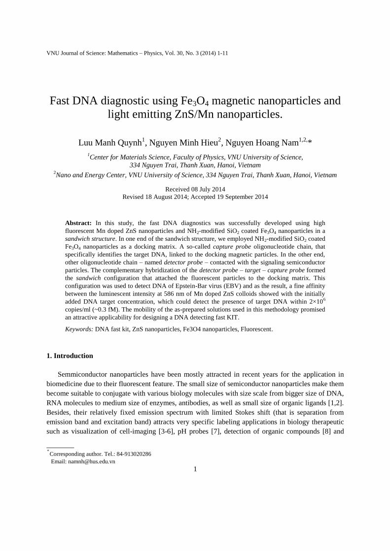
Figure 3.1. Typical XRD (A) and TEM image (B) of ZnS/Mn-NH2 particles prepared by co-precipitation, 4-
ATP surface-modification.
L.M. Quynh et al. / VNU Journal of Science: Mathematics – Physics, Vol. 30, No. 3 (2014) 1-11
6
Typical X-ray diffraction (XRD) of ZnS/Mn-NH2 particles is shown in Figure 3.1. The diffraction
peaks corresponding to (111), (220), (311) planes are separated explicitly at 29.12˚, 47.56˚and 54.42˚,
which mostly agree with standard close-packed face central cubic (FCC) structure of sphalerite
mineral (JCPDS number 05-0566). The data was fitted to the three peaks. We used Debye-Scherer
formula applied for these peaks to calculate the average size of crystalline of the particles, which is
shown in table 3.1. From the table, we assumed that the particles have spherical shape and average
size ~ 3nm, which agrees with the transferred electron microscopic (TEM) result (Panel B, Figure 3.1).
Table 3.1. Gaussian multi-peak fitting results of ZnS/Mn-NH2 particles’ XRD spectrum.
Miller index FHMW θ Calculated average size (nm)
(111) 2.805 14.280˚ 2.92±0.02
(220) 3.274 23.783˚ 2.66±0.04
(311) 3.114 28.214˚ 2.87±0.07
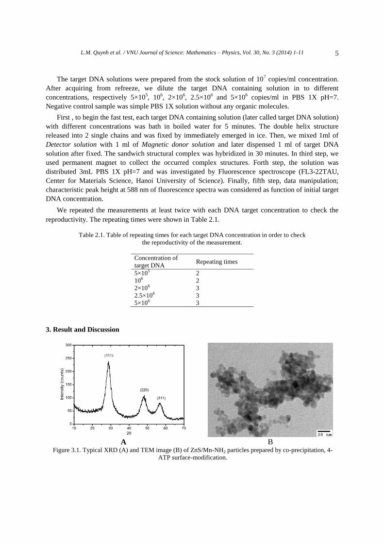
We did the same measurements (XRD and TEM investigations) with the Fe3O4/SiO2-NH2
nanoparticles. The results are shown in Figure 3.2. In panel A, there is a broadened peak occurred at
about 24o, which responses to the amorphous phase of SiO2 coat. The peaks obtained at 2θ of 30.07
o,
35.55o, 43.05
o, 53.59
o, 57.20
o and respectively at 62.68
o agree with the standard close-packed FCC
thiospinel structure of magnetite chalcogenide (JCPDS No. 79-0417).
A B
Figure 3.2. XRD (A) and TEM image (B) of Fe3O4/SiO2-NH2 particles.
Magnetic properties of the nanoparticles
At small size of 15-20 nm (Figure 3.2B) the ferrite oxide particles showed typical
superparamagnetic property with comparatively high saturated magnetization Ms of about 61.2 emu/g,
while the semiconductor particles illustrated very low saturated magnetization (~0.5 emu/g).
High external permanent magnet treatment (not shown) demonstrates that the Fe3O4/SiO2-NH2
particles could be totally collected under 15 min, while it was not experienced in the case of ZnS/Mn-
NH2 particles. Figure 3.3 shows the magnetization of Fe3O4/SiO2-NH2 and ZnS/Mn-NH2 particles

L.M. Quynh et al. / VNU Journal of Science: Mathematics – Physics, Vol. 30, No. 3 (2014) 1-11
7
depending on the applied field. The saturate-magnetization of magnetic donor colloids reaches
relatively high quantity of 61 emu/g at 15 kOe applied field, while that quantity of Detector particles
obtained at 0.5 emu/g. The reversibility of both lines demonstrates the superparamagnetic and
paramagnetic property of the Magnetic donor particles and the Detectors, respectively.
Figure 3.3. Magnetization curves of Fe3O4/SiO2-NH2 and ZnS/Mn-NH2 nanoparticles.
Photo luminescent property
The emission intensity of Mn2+
from ZnS:Mn clusters was obtained at 588 nm that agrees with
early published results [13-15]. The observed intensity decreases depending on decreasing of the
solution concentration (Figure 3.4). As visualized in g and h line (A panel), the observed signals could
not be distinguished when the solution was diluted to 5×10³ times and 10
4 times. In log scale, the
photoluminescence intensity was proportion with the solution concentration when the dilute rate was
smaller than 103 times, corresponding to a, b, c, d and e points in Panel B of the figure.
A

L.M. Quynh et al. / VNU Journal of Science: Mathematics – Physics, Vol. 30, No. 3 (2014) 1-11
8
B
Figure 3.4. Photo-Luminescence property of ZnS/Mn-NH2 Detector particles with different dilution time. A
panel: a. 2 times, b. 5 times, c. 10 times, d. 100 times, e. 1000 times, f. 10000 times, g. 50000 times and h.
100000 time dilution. B panel: illustration of the peak intensity of the ZnS/Mn-NH2 Detector solution at 588
nm depending on the dilution times in log-scale.
Diagnostic measurement
Figure 3.4 demonstrates the schematic progress of the DNA test experiment. The A, B, C, D panel
correspond to first 4 steps of respectively sandwich hybridization, magnetic collection, washing and
redistribution of the collected sandwich complex to PBS 1X pH=7.
In the first step, the sandwich formula of magnetic donor – DNA – Detector accumulates in
solution. During this step, the complementary hybridization was macroscopic stochastic progress. The
concentrations of accumulated sandwich structures are normally proposal with the initial target DNA
concentrations (Panel A). After applying outside permanent magnet, all magnetic colloids orientate to
the magnetic side. The remained uncoupled Detector particles distribute in solution (panel B). By
washing, the uncouple Detector particles sweeps out (panel C) and the coupled Detector particles (in
the complex) proportional with the number of the target DNA are distributed in the ending solution
(Panel D). The photo-luminescence (PL) intensity of the ending solution is the PL intensity of
Detector particles formulated in the complexes.
The observed photoluminescence spectra of the sandwich complex (data not showed) indicated
that when the concentration of initially added concentration increased the characteristic peak of Mn2+
at 588 nm increased. The detection limit was about 2×10⁶ copies/ml.
In Figure 3.5, the emitting intensity at 588 nm gave better concentration distinguishing when the
initial DNA concentration higher than 2×10⁶ copies/ml, and would obtain misinterpretation with
typical error at lower concentrations. Under same measuring condition with the upward measurements
of Detector solution, the obtained limited distinguishable intensity was nearly 106 counts at 588 nm.
Below this magnification, the equipment could not detect accurately the presence of the Detector
particles (here the negative control shown relatively low intensity at 588 nm – data not shown). Above

L.M. Quynh et al. / VNU Journal of Science: Mathematics – Physics, Vol. 30, No. 3 (2014) 1-11
9
the intensity of 106 counts, the equipment can detect the presence of the Detector particles quite well
(see Figure 3.4B) and is able to recognize the present of the DNA in the solution, which corresponds
to the 2×10⁶ copies/ml initial concentration of target DNA. We can assume that this diagnostic method
can help detect the presence of the EBV DNA higher than 2×10⁶ copies/ml (it is about 0.3 fM).
Figure 3.4. Schematic progress of the diagnostic experiment.
Figure 3.5. Dependence of obtained PL intensity at 588 nm on initial target DNA.
L.M. Quynh et al. / VNU Journal of Science: Mathematics – Physics, Vol. 30, No. 3 (2014) 1-11
10
4. Conclusion
The fast KIT of DNA detection was successfully developed using multifunctional nanoparticles in
a sandwich structure. The measurement, however, need to be developed to have the suitable form for
scene detection of present DNA. The solutions – Detector solution, Magnetic donor solution – are
comfortably mobile, but we still have to use the laboratorial equipment, that is the PL3-22TAU, which
has relatively big size. The light source, as inventing, could be replaced by 335 nm emitting Deep UV
LED and the emitted light could be collected by visible light detector, which are commercially
purchasable. Hence, the method promises a fine applicability for fast viral DNA detection, early
disease warning and scene-criminal detection.
Acknowledgements
The authors would thank Vietnam National University, Hanoi for financial supporting (Project no.
QG.12.03). We sincerely thank to Dr. Nguyen Thi Van Anh’s Group of Key Laboratory of Protein and
Enzyme, Hanoi University of Science for supporting the biological molecules and fruitful advices.
References
[1] Marcel Bruchez Jr., Mario Moronne, Peter Gin, Shimon Weiss, A. Paul Alivisatos, Semiconductor nanocrystals
as fluorescent biological labels, Sience 281(1998), 2013-2016.
[2] Lijia Shao, Yanfang Gao and Feng Yan, Semiconductor quantum dots for biomedical applications, Sensor
11(2011), 11736-11751.
[3] Xiao Gao, Lily Yang, John A Petros, Fray F Mashall, Jonathan W Simons and Shuming Nie, In vivo molecular
and cellular imaging with quantum dots, Current Opinion in Biotechnology 16 (2005), 63-72.
[4] Andrew M. Smith, Hongwei Duan, Aaron M. Mohs and Shuming Nie, Bioconjugated quantum dots for in vivo
molecular and cellular imaging, Adv. Drug. Deliv Rev. 60(11) (2008), 1226-1240.
[5] Brad A. Kairdolf, Andrew M. Smith, Todd H. Stokes, May D. Wang, Andrew N. Young and Shuming Nie,
Semiconductor quantum dots for bioimaging and biodiagnostic applications, Annu Rev Anal Chem. 6(1) (2013),
143-162.
[6] Igor L. Medintz, H. Tetsuo Uyeda, Ellen R. Goldman and Hedi Mattoussi, Quantum dot bioconjugates for
imaging, labeling and sensing, Nature Materials 4(2005), 435-446.
[7] Takashi Jin, Akira Sasaki, Masataka Kinjo and Jun Myazaki, Quantum dot-based ratiometric pH sensor,
Chemical Communications 46 (2010), 2408-2410.
[8] Manuela F. Frasco and Nikos Chaniotakis, Semiconductor quantum dots in chemical sensors and biosensors,
Sensors 9(2009), 7266-7286.
[9] Wei Lian, Sally A. Litherland, Hassan Badrane, Weihong Tan, Donghai Wu, Henry V. Baker, Paul A. Gulig,
Daniel V. Lim, Shouguang Jin, Ultrasensitive detection of biomolecules with fluorescent dye-doped
nanoparticles, Analitical biochemistry 334 (2004),135-144.
[10] Jiang Zhengran, Li Ruiyun, Todd Nevins W., Stass Sanford A., Jiang Feng, Detecting genomic aberrations by
fluorescence in situ hybridization with quantum dots-labeled probes, Journal of Nanoscience and Nanotechnology
12 (2007), 4254-4259.
[11] Cheng Chi, Fernando Vargas-Lara, Alexei V. Tkachenko, Francis W. Starr and Oleg Gang, Internal structure of
nanoparticle dimers linked by DNA, American Chemical Society 6(8) (2012), 6793-6802.
L.M. Quynh et al. / VNU Journal of Science: Mathematics – Physics, Vol. 30, No. 3 (2014) 1-11
11
[12] Vaishali Bagalkot, Liangfang Zhang, Etgar Levy-Nissenbaum, Sangyong Jon, Philip W. Kantoff, Robert Langer
and Omid C. Farokhzad, Quantum dot-aptamer conjugates for synchronous cancer imaging, therapy, and sensing
of drug delivery based on bi-fluorescence resonance energy transfer, Nano Letter 7(10) (2007), 3065-3070.
[13] Ram Kripal, Atul K. Gupta, Sheo K. Mishra, Rajneesh K. Srivastava, Avinash C. Pandey, S.G. Prakash,
Photoluminescence and photoconductivity of ZnS:Mn2+ nanopaticles synthesized via co-precipitation method,
Spectrochimica Acta Part A 76 (2010), 523-530.
[14] H.C. Warad. S.C. Ghosh, B. Hemtanon, C. Thananchayanont, J. Dutta, Luminescent nanopaticles of Mn doped
ZnS passivated with soldium hexametaphosphate, Science and Technology of Advanced Materials 6(2005), 296-
301.
[15] Tran Thi Quynh Hoa, Ngo Duc The, Stephen McVitie, Nguyen Hoang Nam, Le Van Vu, Ta Dinh Canh and
Nguyen Ngoc Long, Optical properties of Mn-doped ZnS semiconductor nanoclusters synthesized by a
hydrothermal process. Optical Materials 33(3) (2011), 308-314.
[16] Dongyeon Son, Dae-Ryong Jung, Jongmin Kim, Taeho Moon, Chunjoong Kim and Byungwoo Park, Synthesis
and photoluminescence of Mn-doped zinc sulfide nanoparticles, Applied Physics Letter 9 (2007) 0, 101910.
[17] Yu Z, Ma X, Yu B, Pan Y, Liu Z, Synthesis and characterization of ZnS:Mn/ZnS core/shell nanoparticles for
tumor targeting and imaging in vivo, J. Biomater Appl. 28(2) (2013), 232-240.
[18] Tran Thi Quynh Hoa, Le Thi Thanh Binh, Le Van Vu, Nguyen Ngoc Long, Vu Thi Hong Hanh, Vu Duc Chinh,
Pham Thu Nga, Luminescent ZnS:Mn/thioglycerol and ZnS:Mn/ZnS core/shell nanocrystals: synthesis and
characterization, Optical Materials 35(2) (2012), 136-140.
Dmitry Zimnisky, Chaoyang Jiang, Jun Xu, Zhiqun Lin anf Vladimir V. Tsukruk, Substrate and time-dependent
photoluminescence of quantum dots inside the ultrathin polymer LbL film, Langmuir 23 (2007), 4509-4515.
[19] Tian Long Zhang, yun Sheng Xia, Xue Lian Diao, Chang Quing Zhu, Preparation and formation mechanism of
strong violet luminescent CdS quantum dots by using a ligand exchange strategy, Journal of Nanoparticles
Research 10(1) (2008), 59-67.
[20] Goutam Palui, Tommaso Avellini, Naiquain Zhan, Feng Pan, David Gray, Igor Alabugin and Hedi Matoussi,
Photoinduced phase transfer of luminescent quantum dots for polar and aqueous media, J.A.C.S. 134(39) (2012),
16370-16378.
[21] Dao Van Quy, Nguyen Minh Hieu, Pham Thi Tra, Nguyen Hoang Nam, Nguyen Hoang Hai, Nguyen Thai Son,
Phan Tuan Nghia, Nguyen Thi Van Anh, Tran Thi Hong and Nguyen Hoang Luong, Synthesis of silica-coated
magnetic nanoparticles and application in the detection of pathogenic viruses, Journal of Nanomaterials 2013 ID.
063940.
[22] Jerina Majeed, Lina Pradhan, Raghumani Singh Ningthoujam, Rajesh Kumar Vatsa, Dhirendra Bahadur, Avesh
Kumar Tyagi, Enhanced specific absorption rate in silanol functioncalized Fe3O4 core-shell nanoparticles: study
of Fe leaching in Fe3O4 and hyperthermia in L929 and HeLa cells, Colloids and Surface B 122 (2014), 396-403.
[23] Nathalie Zammatteo, Laurent Jeanmart, Sandrine Hamels, Stephane Courtois, Pierre Louette, Laszlo Hevesi and
Jose Remacle, Comparison between different strategies of covalent attachment of DNA to glass surfaces to build
DNA microarray, Analitical Biochemistry 28 (2000), 143-150.
[24] Nguyen Hoang Hai, Nguyen Chau, Nguyen Hoang Luong, Nguyen Thi Van Anh, Phan Tuan Nghia, Applications
of magnetic nanoparticles for water treatment and for DNA and cell separation. Journal of the Korean Physical
Society 53(2008), 1601-1606.
[25] Nguyen Thuy Trang, Luu Manh Quynh, Tran Van Nam and Hoang Nam Nhat, Charge transfer at organic–
inorganic interface of surface-activated PbS by DFT method, Surface Science 608(2013), 67-73.
Copyright © 2022 FDOKUMEN


![Fluorescent cellulose aerogels containing covalently immobilized (ZnS) ₓ (CuInS ₂) ₁₋ ₓ/ZnS (core/shell) quantum dots [2013]](https://static.fdokumen.com/doc/165x107/63372dc94554fe9f0c05b209/fluorescent-cellulose-aerogels-containing-covalently-immobilized-zns-cuins.jpg)