Modular Broad-Host-Range Expression Vectors for Single-Protein and Protein Complex Purification
10
APPLIED AND ENVIRONMENTAL MICROBIOLOGY, Feb. 2004, p. 712–721 Vol. 70, No. 2 0099-2240/04/$08.000 DOI: 10.1128/AEM.70.2.712–721.2004 Copyright © 2004, American Society for Microbiology. All Rights Reserved. Modular Broad-Host-Range Expression Vectors for Single-Protein and Protein Complex Purification Barna D. Fodor, 1 A ´ kos T. Kova ´cs, 1,2 Ro ´bert Csa ´ki, 2 E ´ va Hunyadi-Gulya ´s, 3 E ´ va Klement, 3 Gergely Maro ´ti, 1,2 Lı ´via S. Me ´sza ´ros, 1,2 Katalin F. Medzihradszky, 3 Ga ´bor Ra ´khely, 1,2 and Korne ´l L. Kova ´cs 1,2 * Institute of Biophysics 1 and Mass Spectrometry Facility, 3 Biological Research Center, Hungarian Academy of Sciences, and Department of Biotechnology, 2 University of Szeged, H-6726 Szeged, Hungary Received 7 July 2003/Accepted 4 November 2003 A set of modular broad-host-range expression vectors with various affinity tags (six-His-tag, FLAG-tag, Strep-tag II, T7-tag) was created. The complete nucleotide sequences of the vectors are known, and these small vectors can be mobilized by conjugation. They are useful in the purification of proteins and protein complexes from gram-negative bacterial species. The plasmids were easily customized for Thiocapsa roseopersicina, Rhodobacter capsulatus, and Methylococcus capsulatus by inserting an appropriate promoter. These examples demonstrate the versatility and flexibility of the vectors. The constructs harbor the T7 promoter for easy overproduction of the desired protein in an appropriate Escherichia coli host. The vectors were useful in purifying different proteins from T. roseopersicina. The FLAG-tag–Strep-tag II combination was utilized for isolation of the HynL-HypC 2 protein complex involved in hydrogenase maturation. These tools should be useful for protein purification and for studying protein-protein interactions in a range of bacterial species. Purification of a gene product for characterization or anti- body production is greatly simplified by cloning and expressing the gene in question, usually fused to an affinity tag, in Esch- erichia coli. However, the heterologous expression approach does not always allow formation of multiprotein complexes. In these cases, the complexes should be assembled in and isolated from the original host. A generalized method (tandem affinity purification) for protein complex purification from yeast has been described (2, 32). In this method, two tags are fused to the target protein of interest, and proteins interacting with the target are isolated by using two successive affinity purification steps. The components of protein complexes are later sepa- rated in and isolated from sodium dodecyl sulfate (SDS)-poly- acrylamide gels for mass spectrometric (MS) identification. Tools that can facilitate a similar approach in a wide range of bacteria have not been developed yet. Protein overproduction in E. coli sometimes has other lim- itations, especially when a foreign gene is expressed. No ex- pression or a low efficiency of expression, degradation, toxicity, and protein insolubility are the most common problems. Pro- viding other subunits and factors needed for posttranslational modification, such as processing of signal sequences, protein cleavage, folding, and incorporation of prosthetic groups, is also problematic, and the absence of these subunits and factors results in an inactive protein (27). Some of these problems can be solved if the protein is expressed and purified from the original bacterial host by employing specific expression vectors or one of the broad-host-range expression vectors available (4, 5, 13, 15). Usually, these are not available commercially, and it is hard to find one that fulfills all the requirements needed for a particular study or organism. Existing vectors are compli- cated to redesign; moreover, it is laborious and time-consum- ing to change or add required properties because of the lack of sequence data, the large size, and often the need for several cloning steps. Our modular concept was to combine a broad-host-range vector backbone, containing all the necessary properties gen- erally needed for protein expression and purification, with the possibility of easy insertion of desired promoters or replace- ment of various features. The resulting vectors are small and mobilizable, and their sequences are known. Different fusion tags are available to help protein purification, or they can be omitted if desired. The tandem FLAG-tag (17)–Strep-tag II (35) combination was designed to allow purification and study of protein complexes. Promoter regions from Thiocapsa roseo- persicina, Rhodobacter capsulatus, and Methylococcus capsula- tus, inserted upstream from the expression cassettes, were uti- lized to express proteins in these hosts at different levels depending on the inserted promoter’s activity. In addition, it was demonstrated that the same construct was able to over- produce the protein in the appropriate E. coli host. The tandem FLAG-tag–Strep-tag II combination was uti- lized in a study of hydrogenase maturation in T. roseopersicina. Assembly of the active site, located in the large subunit of hydrogenases (containing Ni, Fe, CO, and CN ), is a complex process assisted by several proteins (8, 11). Two of the hy- drogenase maturation-assisting proteins of T. roseopersicina (HypC 2 and HupK) (28) were used in coaffinity purification experiments to test the utility of the tandem FLAG-tag–Strep- tag II combination for detecting protein-protein interactions and its usefulness for studying hydrogenase maturation. MATERIALS AND METHODS Bacterial strains, plasmids, growth conditions, and conjugation. The strains and plasmids used are listed in Tables 1 and 2. E. coli strains were maintained on * Corresponding author. Mailing address: Department of Biotech- nology, University of Szeged, H-6726 Szeged, Temesva ´ri krt. 62, Hun- gary. Phone: 36 62 544 351. Fax: 36 62 544 352. E-mail: kornel @nucleus.szbk.u-szeged.hu. 712
-
Upload
independent -
Category
Documents
-
view
2 -
download
0
Transcript of Modular Broad-Host-Range Expression Vectors for Single-Protein and Protein Complex Purification

APPLIED AND ENVIRONMENTAL MICROBIOLOGY, Feb. 2004, p. 712–721 Vol. 70, No. 20099-2240/04/$08.00�0 DOI: 10.1128/AEM.70.2.712–721.2004Copyright © 2004, American Society for Microbiology. All Rights Reserved.
Modular Broad-Host-Range Expression Vectors for Single-Proteinand Protein Complex Purification
Barna D. Fodor,1 Akos T. Kovacs,1,2 Robert Csaki,2 Eva Hunyadi-Gulyas,3 Eva Klement,3Gergely Maroti,1,2 Lıvia S. Meszaros,1,2 Katalin F. Medzihradszky,3
Gabor Rakhely,1,2 and Kornel L. Kovacs1,2*Institute of Biophysics1 and Mass Spectrometry Facility,3 Biological Research Center, Hungarian Academy of Sciences,
and Department of Biotechnology,2 University of Szeged, H-6726 Szeged, Hungary
Received 7 July 2003/Accepted 4 November 2003
A set of modular broad-host-range expression vectors with various affinity tags (six-His-tag, FLAG-tag,Strep-tag II, T7-tag) was created. The complete nucleotide sequences of the vectors are known, and these smallvectors can be mobilized by conjugation. They are useful in the purification of proteins and protein complexesfrom gram-negative bacterial species. The plasmids were easily customized for Thiocapsa roseopersicina,Rhodobacter capsulatus, and Methylococcus capsulatus by inserting an appropriate promoter. These examplesdemonstrate the versatility and flexibility of the vectors. The constructs harbor the T7 promoter for easyoverproduction of the desired protein in an appropriate Escherichia coli host. The vectors were useful inpurifying different proteins from T. roseopersicina. The FLAG-tag–Strep-tag II combination was utilized forisolation of the HynL-HypC2 protein complex involved in hydrogenase maturation. These tools should be usefulfor protein purification and for studying protein-protein interactions in a range of bacterial species.
Purification of a gene product for characterization or anti-body production is greatly simplified by cloning and expressingthe gene in question, usually fused to an affinity tag, in Esch-erichia coli. However, the heterologous expression approachdoes not always allow formation of multiprotein complexes. Inthese cases, the complexes should be assembled in and isolatedfrom the original host. A generalized method (tandem affinitypurification) for protein complex purification from yeast hasbeen described (2, 32). In this method, two tags are fused tothe target protein of interest, and proteins interacting with thetarget are isolated by using two successive affinity purificationsteps. The components of protein complexes are later sepa-rated in and isolated from sodium dodecyl sulfate (SDS)-poly-acrylamide gels for mass spectrometric (MS) identification.Tools that can facilitate a similar approach in a wide range ofbacteria have not been developed yet.
Protein overproduction in E. coli sometimes has other lim-itations, especially when a foreign gene is expressed. No ex-pression or a low efficiency of expression, degradation, toxicity,and protein insolubility are the most common problems. Pro-viding other subunits and factors needed for posttranslationalmodification, such as processing of signal sequences, proteincleavage, folding, and incorporation of prosthetic groups, isalso problematic, and the absence of these subunits and factorsresults in an inactive protein (27). Some of these problems canbe solved if the protein is expressed and purified from theoriginal bacterial host by employing specific expression vectorsor one of the broad-host-range expression vectors available (4,5, 13, 15). Usually, these are not available commercially, and itis hard to find one that fulfills all the requirements needed for
a particular study or organism. Existing vectors are compli-cated to redesign; moreover, it is laborious and time-consum-ing to change or add required properties because of the lack ofsequence data, the large size, and often the need for severalcloning steps.
Our modular concept was to combine a broad-host-rangevector backbone, containing all the necessary properties gen-erally needed for protein expression and purification, with thepossibility of easy insertion of desired promoters or replace-ment of various features. The resulting vectors are small andmobilizable, and their sequences are known. Different fusiontags are available to help protein purification, or they can beomitted if desired. The tandem FLAG-tag (17)–Strep-tag II(35) combination was designed to allow purification and studyof protein complexes. Promoter regions from Thiocapsa roseo-persicina, Rhodobacter capsulatus, and Methylococcus capsula-tus, inserted upstream from the expression cassettes, were uti-lized to express proteins in these hosts at different levelsdepending on the inserted promoter’s activity. In addition, itwas demonstrated that the same construct was able to over-produce the protein in the appropriate E. coli host.
The tandem FLAG-tag–Strep-tag II combination was uti-lized in a study of hydrogenase maturation in T. roseopersicina.Assembly of the active site, located in the large subunit ofhydrogenases (containing Ni, Fe, CO, and CN�), is a complexprocess assisted by several proteins (8, 11). Two of the hy-drogenase maturation-assisting proteins of T. roseopersicina(HypC2 and HupK) (28) were used in coaffinity purificationexperiments to test the utility of the tandem FLAG-tag–Strep-tag II combination for detecting protein-protein interactionsand its usefulness for studying hydrogenase maturation.
MATERIALS AND METHODS
Bacterial strains, plasmids, growth conditions, and conjugation. The strainsand plasmids used are listed in Tables 1 and 2. E. coli strains were maintained on
* Corresponding author. Mailing address: Department of Biotech-nology, University of Szeged, H-6726 Szeged, Temesvari krt. 62, Hun-gary. Phone: 36 62 544 351. Fax: 36 62 544 352. E-mail: [email protected].
712
Luria-Bertani agar plates. For protein overexpression, 2YT medium was used(1). Genetic manipulations were performed in strain XL1-Blue MRF� or DH5�.Strain BL21(DE3) was used as a host for overexpression of �-glucuronidasefused to six histidine residues at its N terminus (6His-UidA). T. roseopersicinastrains were grown photosynthetically in Pfennig’s mineral medium as describedpreviously (20, 30). To obtain a higher yield of biomass for protein purification,2 g of sodium acetate per liter was added to the basic medium. R. capsulatus was
maintained on YPS plates (containing [per 1,000 ml] 3 g of yeast extract, 3 g ofpeptone, 2 ml of 1 M CaCl2, and 2 ml of 1 M MgCl2), and liquid cultures werecultivated in mineral RCV medium (38). M. capsulatus was grown in NMSmedium (39) containing 5.0 �M CuSO4. Low-copper medium was preparedwithout CuSO4. Antibiotics were used at the following concentrations: 100 �g ofampicillin per ml, 25 �g of kanamycin per ml, 25 �g of streptomycin per ml, and15 �g of tetracycline per ml for E. coli; 10 �g of kanamycin per ml and 5 �g of
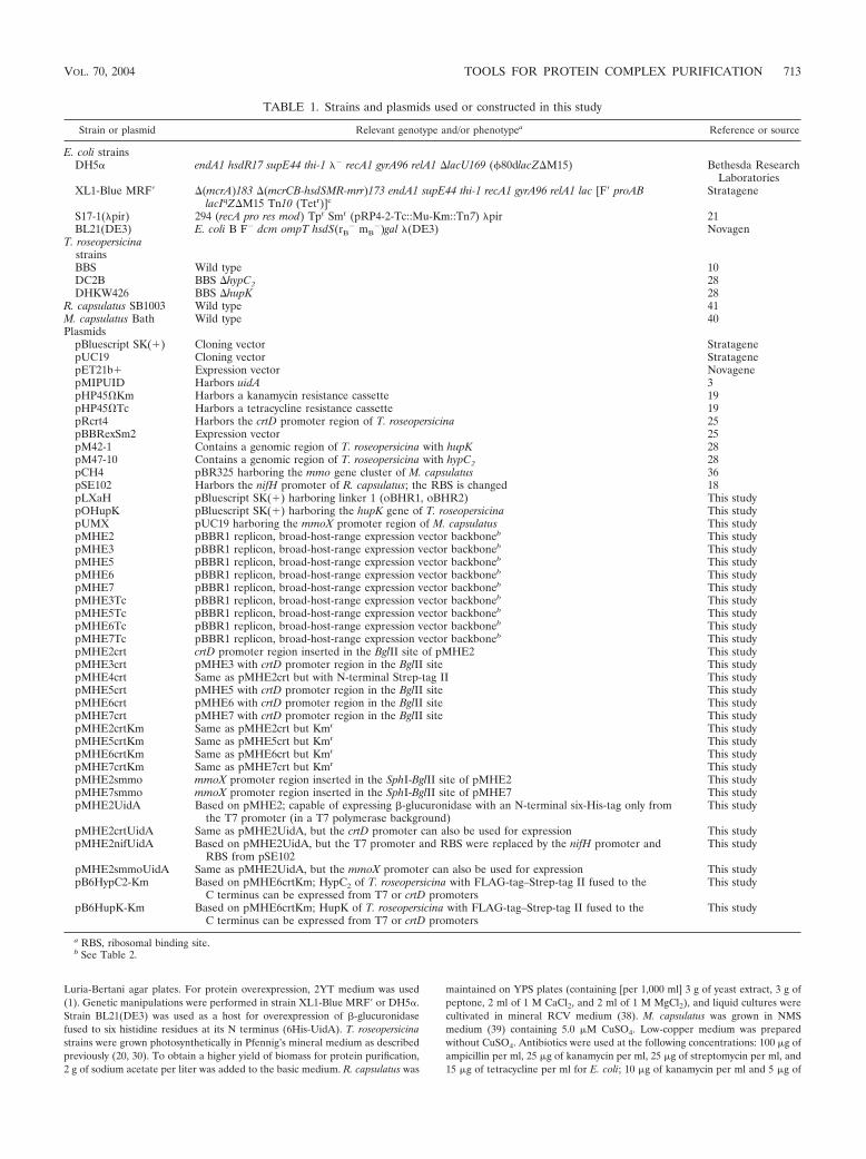
TABLE 1. Strains and plasmids used or constructed in this study
Strain or plasmid Relevant genotype and/or phenotypea Reference or source
E. coli strainsDH5� endA1 hsdR17 supE44 thi-1 �� recA1 gyrA96 relA1 �lacU169 (80dlacZ�M15) Bethesda Research
LaboratoriesXL1-Blue MRF� �(mcrA)183 �(mcrCB-hsdSMR-mrr)173 endA1 supE44 thi-1 recA1 gyrA96 relA1 lac [F� proAB
lacIqZ�M15 Tn10 (Tetr)]cStratagene
S17-1(�pir) 294 (recA pro res mod) Tpr Smr (pRP4-2-Tc::Mu-Km::Tn7) �pir 21BL21(DE3) E. coli B F� dcm ompT hsdS(rB
� mB�)gal �(DE3) Novagen
T. roseopersicinastrainsBBS Wild type 10DC2B BBS �hypC2 28DHKW426 BBS �hupK 28
R. capsulatus SB1003 Wild type 41M. capsulatus Bath Wild type 40Plasmids
pBluescript SK(�) Cloning vector StratagenepUC19 Cloning vector StratagenepET21b� Expression vector NovagenepMIPUID Harbors uidA 3pHP45Km Harbors a kanamycin resistance cassette 19pHP45Tc Harbors a tetracycline resistance cassette 19pRcrt4 Harbors the crtD promoter region of T. roseopersicina 25pBBRexSm2 Expression vector 25pM42-1 Contains a genomic region of T. roseopersicina with hupK 28pM47-10 Contains a genomic region of T. roseopersicina with hypC2 28pCH4 pBR325 harboring the mmo gene cluster of M. capsulatus 36pSE102 Harbors the nifH promoter of R. capsulatus; the RBS is changed 18pLXaH pBluescript SK(�) harboring linker 1 (oBHR1, oBHR2) This studypOHupK pBluescript SK(�) harboring the hupK gene of T. roseopersicina This studypUMX pUC19 harboring the mmoX promoter region of M. capsulatus This studypMHE2 pBBR1 replicon, broad-host-range expression vector backboneb This studypMHE3 pBBR1 replicon, broad-host-range expression vector backboneb This studypMHE5 pBBR1 replicon, broad-host-range expression vector backboneb This studypMHE6 pBBR1 replicon, broad-host-range expression vector backboneb This studypMHE7 pBBR1 replicon, broad-host-range expression vector backboneb This studypMHE3Tc pBBR1 replicon, broad-host-range expression vector backboneb This studypMHE5Tc pBBR1 replicon, broad-host-range expression vector backboneb This studypMHE6Tc pBBR1 replicon, broad-host-range expression vector backboneb This studypMHE7Tc pBBR1 replicon, broad-host-range expression vector backboneb This studypMHE2crt crtD promoter region inserted in the BglII site of pMHE2 This studypMHE3crt pMHE3 with crtD promoter region in the BglII site This studypMHE4crt Same as pMHE2crt but with N-terminal Strep-tag II This studypMHE5crt pMHE5 with crtD promoter region in the BglII site This studypMHE6crt pMHE6 with crtD promoter region in the BglII site This studypMHE7crt pMHE7 with crtD promoter region in the BglII site This studypMHE2crtKm Same as pMHE2crt but Kmr This studypMHE5crtKm Same as pMHE5crt but Kmr This studypMHE6crtKm Same as pMHE6crt but Kmr This studypMHE7crtKm Same as pMHE7crt but Kmr This studypMHE2smmo mmoX promoter region inserted in the SphI-BglII site of pMHE2 This studypMHE7smmo mmoX promoter region inserted in the SphI-BglII site of pMHE7 This studypMHE2UidA Based on pMHE2; capable of expressing �-glucuronidase with an N-terminal six-His-tag only from
the T7 promoter (in a T7 polymerase background)This study
pMHE2crtUidA Same as pMHE2UidA, but the crtD promoter can also be used for expression This studypMHE2nifUidA Based on pMHE2UidA, but the T7 promoter and RBS were replaced by the nifH promoter and
RBS from pSE102This study
pMHE2smmoUidA Same as pMHE2UidA, but the mmoX promoter can also be used for expression This studypB6HypC2-Km Based on pMHE6crtKm; HypC2 of T. roseopersicina with FLAG-tag–Strep-tag II fused to the
C terminus can be expressed from T7 or crtD promotersThis study
pB6HupK-Km Based on pMHE6crtKm; HupK of T. roseopersicina with FLAG-tag–Strep-tag II fused to theC terminus can be expressed from T7 or crtD promoters
This study
a RBS, ribosomal binding site.b See Table 2.
VOL. 70, 2004 TOOLS FOR PROTEIN COMPLEX PURIFICATION 713

streptomycin per ml for T. roseopersicina; 10 �g of streptomycin per ml for R.capsulatus; and 15 �g of streptomycin per ml for M. capsulatus. Conjugation wasperformed as previously described for T. roseopersicina (20), M. capsulatus (14),and R. capsulatus (12).
DNA manipulation and PCR, sequencing. Preparation of plasmid DNA, DNAmanipulation, cloning, and PCR were carried out by using the general proce-dures described previously (1) or the manufacturers’ instructions. Sequencingwas done with an Applied Biosystems 373 Stretch DNA sequencer.
Construction of plasmids. Relevant steps for construction of plasmids areoutlined in Fig. 1.
(i) Vectors used as a starting point. For construction of pLXaH, the oBHR1(5�CCATGGGGCATCATCATCATCATCATATCGAGGGAAGGCCTG3�)and oBHR2 (5�TCGACAGGCCTTCCCTCGATATGATGATGATGATGATGCCCCATGG3�) oligonucleotides were annealed to produce linker 1 with ablunt end and a SalI end. This linker was ligated to the KpnI (blunted)- andSalI-digested pBluescript SK(�) vector and sequenced. For construction ofpMHE2, PCR was performed with the pLXaH template by using the reverse andM13(�20) primers. The PCR product was cut with NcoI and BamHI, and the86-bp fragment was cloned into the NcoI-BamHI site of pBBRexSm2 and se-quenced. For construction of pMHE3, the 729-bp XbaI-SspI fragment ofpET21b� was ligated to the 5,565-bp XbaI-SspI fragment of pBBRexSm2. Forconstruction of pMHE3Tc, the 2,036-bp DraI fragment from pHP45Tc wasligated to the 4,348-bp DraI fragment of pMHE3. The orientation of the tetra-cycline resistance gene was opposite that of the T7 promoter.
(ii) Vectors with the crtD promoter region of T. roseopersicina. For constructionof pMHE2crt and pMHE3crt, the 124-bp BamHI-HindIII fragment of pRcrt4was treated with T4 polymerase and ligated into the blunted BglII sites ofpMHE2 and pMHE3, yielding pMHE2crt and pMHE3crt, respectively. Forconstruction of pMHE5crt, linker 2 was created by annealing and filling (with Pfupolymerase) oligonucleotides oflag1 (5�GTACTGCAGCTCGAGGGATCCGACTACAAGGACGACGACGACAAGAACTGGAGCCAT3�) and ostrepII3 (5�GATAGATCTTCACTTCTCGAACTGCGGATGGCTCCAGTTCTTGT3�).This linker was cut with PstI-BglII and was ligated into the BamHI-PstI site ofpMHE2crt. For construction of pMHE7crt, linker 3 was created by annealingand filling (with Pfu polymerase) oligonucleotides oflag2 (5�AGTACCATGGACGACTACAAGGACGACGACGACAAGCTCGAGGGCAACTGGAGCCATCCG3�) and ostII2 (5�TCGACAGGCCTTCCCTCGATCTTCTCGAACTGCGGATGGCTCCAGTTGCC3�). This linker was cut with NcoI and StuI andwas ligated into the same restriction sites of pMHE2crt. For construction ofpMHE4crt, linker 4 was created by mixing oligonucleotides ostII1 (5�CATGGGCAACTGGAGCCATCCGCAGTTCGAGAAGATCGAGGGAAGGCCTG3�) and ostII2 (see above) and was ligated into the NcoI-SalI site of pMHE2crt.In all cases, inserted linkers and joints were verified by sequencing. For con-struction of pMHE6crt, the 1,490-bp MscI-HindIII fragment of pMHE5crt wasligated to the 4,583-bp MscI-HindIII fragment of pMHE3crt. For construction ofpMHE*crt plasmids with kanamycin resistance, to create pMHE2crtKm, thestreptomycin cassette of pMHE2crt was removed with DraI and was replaced bythe 1,729-bp SmaI-DraI kanamycin cassette from pHP45Km. The 2,795-bpXbaI-NotI fragment of pMHE2crtKm harboring the kanamycin resistance cas-sette was used to replace the XbaI-NotI fragment (harboring the streptomycinresistance cassette) of pMHE7crt, pMHE6crt, and pMHE5crt to createpMHE7crtKm, pMHE6crtKm, and pMHE5crtKm, respectively.
(iii) Broad-host-range vector backbones with tandem FLAG-tag–Strep-tag II.For construction of pMHE5Tc, pMHE6Tc, and pMHE7Tc, the 2,974-bp XbaI-NotI fragment of pMHE3Tc (harboring the tetracycline resistance gene) wasligated to the expression cassette harboring fragments of pMHE5crtKm (3,058bp), pMHE6crtKm (3,065 bp), and pMHE7crtKm (3,040 bp), respectively. Forconstruction of pMHE5, pMHE6, and pMHE7, the 2,880 bp XbaI-NotI fragmentof pMHE3 (carrying the streptomycin resistance marker) was used to replace the
kanamycin resistance gene and the crtD promoter region of pMHE5crtKm,pMHE6crtKm, and pMHE7crtKm, respectively.
(iv) Vectors with the mmoX promoter region. For construction of pMHE2smmoand pMHE7smmo, a 507-bp fragment was amplified from pCH4 with primersoMXf (5�GTCTGCAGGAGGATCGAACAGGATTA3�) and oMXr (5� CAGGATCCATGATGAATGCCCGATGA 3�). The PCR product was digested withPstI and BamHI and then cloned into pUC19 and digested with the same en-zymes, yielding pUMX. After sequencing, the 508-bp SphI-BamHI fragment ofpUMX with the mmoX promoter region was cloned into the SphI-BglII restric-tion sites of pMHE2 and pMHE7, respectively.
(v) Vectors capable of expressing various tagged proteins. For construction ofpMHE2UidA and pMHE2crtUidA, pMIPUID was cut with NdeI and SmaI. Thefragment carrying the uidA gene was treated with T4 polymerase and cloned intothe polished SalI sites of pMHE2 and pMHE2crt, respectively. The clones wereverified by sequencing. For construction of pMHE2nifUidA, pMHE2UidA cutwith BglII (blunted) and NcoI was combined with pSE102 cut with HindIII(blunted) and NcoI (328 bp). For construction of pMHE2smmoUidA, the 1,863-bp Eco147I-EcoRI fragment from pMHE2UidA was cloned into the same re-striction sites of pMHE2smmo. For construction of pB6HypC2-Km, PCR wasperformed with pM47-10 by using primers otrc2N (5�TGTGTCTCGGTATCCCGATG3�) and otrc2H (5�CAACCTCGAGCCGTCCGCCG3�). The amplifiedfragment was cut with XhoI and cloned into NdeI (polished)- and XhoI-cutpMHE6crtKm. For construction of pB6HupK-Km, PCR was carried out withpM42-1 by using primers oNHupKndei (5�CATATGTCCGATCCGGGTGGAAG3�) and oCHupKxhoi (5�GATCTCGAGTGTGGCGCTTTTACAGGTGA3�). The product was cut with XhoI and cloned into the SmaI-XhoI site ofpBluescript SK(�). The resulting construct (pOHupK) was checked by sequenc-
FIG. 1. Outline of the cloning steps used to create the pMHE*vectors. See Materials and Methods for details. L1, L2, L3, and L4,linkers 1, 2, 3, and 4, respectively; F1, F2, and F3, XbaI-NotI fragmentsharboring the antibiotic resistance genes from pMHE2crtKm, pMHE3,and pMHE3Tc, respectively.
TABLE 2. Antibiotic resistance markers, fusion tags, and protease cleavage sites in the basic pMHE* plasmidsa
N terminus C terminus Streptomycinresistance
TetracyclineresistanceTag Protease cleavage site Tag Protease cleavage site
Six-His X-factor pMHE2T7 Six-His pMHE3 pMHE3TcSix-His X-factor FLAG-tag–Strep-tag II Enterokinase pMHE5 pMHE5TcT7 FLAG-tag–Strep-tag II Enterokinase pMHE6 pMHE6TcFLAG-tag–Strep-tag II Enterokinase X-factor pMHE7 pMHE7Tc
a The T7-lac operator sequence is present in all of the vectors. A second promoter can be cloned in a BglII site upstream of the expression cassette.
714 FODOR ET AL. APPL. ENVIRON. MICROBIOL.
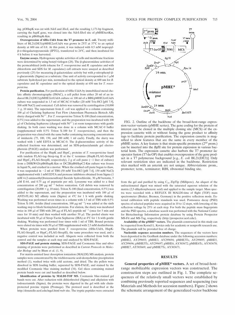
ing. pOHupK was cut with NdeI and XhoI, and the resulting 1,171-bp fragment,carrying the hupK gene, was cloned into the NdeI-XhoI site of pMHE6crtKm,resulting in pB6HupK-Km.
Overexpression of 6His-UidA from the T7 promoter in E. coli. Twenty milli-liters of BL21(DE3)/pMHE2crtUidA was grown in 2YT at 37°C to an opticaldensity at 600 nm of 0.8. At this point, it was induced with 0.5 mM isopropyl-�-D-thiogalactopyranoside (IPTG), transferred to 24°C, and then incubated for4 h before it was harvested.
Enzyme assays. Hydrogenase uptake activities of cells and membrane fractionswere determined by using benzyl viologen (28). The �-glucuronidase activities ofthe permeabilized (with toluene for T. roseopersicina and R. capsulatus and withchloroform and SDS for M. capsulatus) cell extracts were assayed as describedpreviously (25) for measuring �-galactosidase activity but with p-nitrophenyl-�-D-glucuronide (Sigma) as a substrate. One unit of activity corresponded to 1 �Msubstrate hydrolyzed per min, normalized to the optical density at 600 nm for R.capsulatus and M. capsulatus and to the optical density at 650 nm for T. roseo-persicina.
Protein purification. For purification of 6His-UidA by immobilized metal che-late affinity chromatography (IMAC), a cell pellet from either 20 ml of an in-duced BL21(DE3)/pMHE2crtUidA culture or 100 ml of a BBS/pMHE2crtUidAculture was suspended in 1.5 ml of MCAC-0 buffer (20 mM Tris-HCl [pH 7.9],500 mM NaCl) and sonicated. Cell debris was removed by centrifugation (10,000� g, 10 min). The supernatant from E. coli was applied to a column containing100 �l of Chelating Sepharose Fast Flow (Amersham Pharmacia Biotech AB)slurry charged with Ni2�. For T. roseopersicina Triton X-100 (final concentration,0.5%) was added to the supernatant, and the preparation was incubated with 100�l of Chelating Sepharose (charged with Ni2�) at room temperature with gentleshaking. In both cases washing was done in a column with MCAC-0 buffer(supplemented with 0.5% Triton X-100 for T. roseopersicina), and then thepreparation was eluted with the same buffer containing increasing concentrationsof imidazole (75, 100, 150, and 200 mM; 1 ml each). Finally, the slurry waswashed with 1 mM EDTA in MCAC-0. The �-glucuronidase activity of thecollected fractions was determined, and an SDS-polyacrylamide gel electro-phoresis (PAGE) analysis was performed.
For purification of the HupK and HypC2 proteins of T. roseopersicina fusedwith tandem FLAG-tag–Strep-tag II at the C terminus (HupK-FLAG-StrepIIand HypC2-FLAG-StrepII, respectively), 2 g of cell paste (�1 liter of culture)from a DHKW426/pB6HupK-Km or DC2B/pB6HypC2-Km culture was frozenin liquid N2 and crushed in a mortar. When the crushed cell paste began to thaw,it was suspended in �2 ml of TBS (50 mM Tris-HCl [pH 7.4], 150 mM NaCl)supplemented with 1 mM EDTA and protease inhibitors obtained from Sigma (1mM 4-(2-aminoethyl)benzenesulfonyl fluoride hydrochloride, 30 �M bestatin, 5�M E-64, and 0.75 �g of pepstatin per ml). Lysozyme was added to a finalconcentration of 200 �g ml�1 before sonication. Cell debris was removed bycentrifugation (20,000 � g, 10 min). Triton X-100 (final concentration, 0.5%) wasadded to the supernatant, and the preparation was incubated with 100 �l ofANTI-FLAG M2 affinity resin (Sigma) at 4°C for 1 h with gentle shaking.Washing was performed seven times in a column with 1.5 ml of TBS with 0.5%Triton X-100. Avidin (final concentration, 100 �g ml�1) was added at the sixthwashing step to block biotinylated proteins. For elution, the slurry was incubatedtwice in 100 �l of TBS with 200 �g of FLAG peptide ml�1 (once for 5 min andonce for 10 min) and then washed with another 50 �l. The pooled eluate wasincubated with 50 �l of Strep-Tactin Sepharose (IBA) at 4°C for 1 h with gentleshaking. Washing was performed four times with 1 ml of TBS. Bound proteinswere eluted six times with 50 �l of TBS supplemented with 2.5 mM desthiobiotin.
When proteins were purified from T. roseopersicina (6His-UidA, HupK-FLAG-StrepII, or HypC2-FLAG-StrepII), the same procedure was used, and anegative control was included as well. Aliquots were collected from both thecontrol and the samples at each step and analyzed by SDS-PAGE.
SDS-PAGE and protein staining. SDS-PAGE and Coomassie blue and silverstaining of proteins were performed as described in Current Protocols in Molec-ular Biology and by Blum et al. (1, 9).
For matrix-assisted laser desorption ionization (MALDI) MS analysis, proteinsamples were concentrated by the trichloroacetic acid-deoxycholate precipitationmethod (1), washed twice with cold acetone, and dried. The dry pellets weredissolved in SDS loading buffer, separated by SDS-PAGE, and stained by themodified Coomassie blue staining method (34). Gel slices containing stainedprotein bands were cut and handled as described below.
Identification of proteins by MALDI-TOF MS. Coomassie blue-stained gelbands were cut. After reduction with dithiothreitol (Sigma) and alkylation withiodoacetamide (Sigma), the proteins were digested in the gel with side chain-protected porcine trypsin (Promega). The protocol used is described at thehttp://donatello.ucsf.edu/ingel.html web site. The tryptic peptides were extracted
from the gel and purified by using C18 ZipTip (Millipore). An aliquot of theunfractionated digest was mixed with the saturated aqueous solution of thematrix (2,5-dihydroxybenzoic acid) and applied to the sample target. Mass spec-tra were recorded with a REFLEX III MALDI-time of flight (TOF) massspectrometer (Bruker, Bremen, Germany) in the positive reflectron mode. Ex-ternal calibration with peptide standards was used. Postsource decay (PSD)spectra of selected peptides were acquired in 10 to 12 steps, with lowering of thereflectron voltage by 25% at each step. For both the peptide mass fingerprintsand the PSD spectra, a database search was performed with the National Centerfor Biotechnology Information protein database by using Protein ProspectorMS-Fit and MS-Tag, respectively (http://prospector.ucsf.edu/).
Availability of the pMHE* vectors. The plasmids constructed in this study canbe requested from Kornel L. Kovacs only for academic or nonprofit research use.The plasmids will be provided free of charge.
Nucleotide sequence accession numbers. The sequences of the vectors havebeen deposited in the GenBank database under the following accession numbers:pMHE2, AY299693; pMHE3, AY299694; pMHE3Tc, AY299695; pMHE5,AY299696; pMHE5Tc, AY299697; pMHE6, AY303672; pMHE6Tc, AY303670;pMHE7, AY303669; and pMHE7Tc, AY303671.
RESULTS
General properties of pMHE* vectors. A set of broad-host-range mobilizable expression vectors was constructed. Theconstruction steps are outlined in Fig. 1. The complete se-quences of the relatively small vectors were established bycombining previously reported sequences and sequencing (seeMaterials and Methods for accession numbers). Figure 2 showsthe schematic arrangement of the generalized vector backbone
FIG. 2. Outline of the backbone of the broad-host-range expres-sion vector variants (pMHE series). The gene coding for the protein ofinterest can be cloned in the multiple cloning site (MCS) of the ex-pression cassette with or without fusing the gene product to affinitytags to facilitate protein purification. The expression cassette is mag-nified to show features that are the same in every member of thepMHE series. A key feature is that strain-specific promoters (2nd prom.)can be inserted into the BglII site for protein expression in various bac-terial hosts. The expression cassette also harbors the T7 promoter lacoperator fusion (T7-lacOP) that enables overexpression of the gene prod-uct in a T7 polymerase background [e.g., E. coli BL21(DE3)]. Onlyrelevant restriction sites are indicated in the backbone. Restrictionsites marked with an asterisk are not unique. Abbreviations: prom.,promoter; term., terminator; RBS, ribosomal binding site.
VOL. 70, 2004 TOOLS FOR PROTEIN COMPLEX PURIFICATION 715

and the expression cassette present in all vectors. The uniqueproperties of each vector variant are summarized in Table 2and Fig. 3. The combination of streptomycin and tetracyclineresistance genes with the broad-host-range pBBR1 repliconenables all of the vectors to be maintained in a wide range ofgram-negative bacteria (24). The vectors are also mobilizableand can be introduced by conjugation into the target strain iftransformation or electroporation protocols are not available.Rho-independent T4 transcription termination signals andtranslation stop codons flank the antibiotic resistance genes,and a T7 transcriptional terminator is located after the expres-sion cassettes. Several unique restriction sites present in theexpression cassettes’ polylinker enable cloning of the gene tobe (over)expressed. The T7 promoter and T7 terminator prim-er (Novagen) binding sites are convenient for sequencing theinsert. Various N- and C-terminal tags can be fused to an(over)expressed protein in various combinations (Table 2).These can facilitate purification and detection of the fusionprotein or isolation of protein complexes in which the taggedprotein is present. The N-terminal tags (except the T7 tag) andthe C-terminal Strep-tag II can be removed by sequence-spe-cific proteases if necessary. If fusion of tags to a protein isundesirable, NcoI or NdeI restriction sites that overlap thestart codon downstream of a Shine-Dalgarno sequence can beused for cloning. The expression cassette harbors the T7 pro-moter lac operator fusion (T7-lacOP) that enables overexpres-sion of the gene product in a T7 polymerase background [e.g.,E. coli BL21(DE3)]. The BglII site (BglII is compatible withseveral restriction enzymes) provides a simple way of cloning asecond promoter upstream of the T7 promoter, which widensboth the range of bacteria in which protein expression can takeplace and the mode of regulation and level of expression,creating a dual expression system. The same construct can beused in various hosts and in E. coli to produce proteins fromthe T7 promoter and/or the second promoter. Other regionsof the vectors were also designed in a modular fashion; if nec-essary, the ribosomal binding site can be changed by using theXbaI-NcoI/NdeI sites, oriV can be replaced by using the Bpu10I-MscI combination, the mob region can be removed with SacII-Bst1107I, and the resistance markers of the pMHE* and theinterposon carrying vectors created by Fellay et al. (19) areinterchangeable with DraI and SanDI restriction enzymes.
Expression in different gram-negative bacteria from a sec-ond upstream promoter. A promoterless reporter gene codingfor �-glucuronidase from E. coli (uidA) was introduced intothe versions of pMHE2 containing promoters from differentbacteria. Extra amino acids, including six histidine residues,were added to the N terminus of the UidA enzyme. SinceUidA is frequently used as a reporter enzyme fused to the Cterminus of other proteins, this tag was not expected to affectits activity (3). The negative control contained the reportergene alone (pMHE2UidA). pMHE2crtUidA carried the crtDpromoter region of T. roseopersicina that is active under pho-tosynthetic growth conditions (25), pMHE2smmoUidA carriedthe Cu2�-regulated mmoX promoter from M. capsulatus (14),and pMHE2nifUidA carried the NH4
�-regulated nifH promot-er of R. capsulatus (29). The levels of �-glucuronidase expres-sion were measured in the different host bacteria under variousgrowth conditions by using the appropriate vectors (Table 3).The data clearly demonstrated that the presence of the homol-
ogous promoters significantly increased the level of expressioncompared to that of the negative control. The mmoX promoterin M. capsulatus and the nifH promoter in R. capsulatus exhib-ited regulated expression by Cu2� and NH4
�, respectively. How-ever, expression from the mmoX promoter was not completelyrepressed by Cu2�. We also found that the crtD promoterregion of T. roseopersicina worked in R. capsulatus, althoughthe level of �-glucuronidase activity detected was significantlylower than that in the original host. The same thing was ob-served for the nifH promoter of R. capsulatus in T. roseopersi-cina, but the NH4
�-regulated phenotype was retained.Overexpression and protein purification from T7 polymer-
ase-expressing E. coli. To demonstrate that overproductionfrom the T7 promoter is possible in the presence of a secondupstream inserted promoter, pMHE2crtUidA was introducedinto a T7 polymerase-expressing E. coli strain, BL21(DE3).6His-UidA was expressed and purified as described in Mate-rials and Methods (Fig. 4). Approximately 70% of the enzymeproduced was found in inclusion bodies (data not shown), aphenomenon often encountered in overexpressing systems(27). Expression and purification steps were followed by SDS-PAGE and measurement of �-glucuronidase activity. 6His-UidA was overproduced and could be purified, as expected.
Protein purification from T. roseopersicina by IMAC.pMHE2crtUidA was used to demonstrate that protein purifi-cation from a different host is possible by employing the secondpromoter of the same construct used for overproduction inE. coli. The amount of 6His-UidA produced in T. roseopersi-cina BBS/pMHE2crtUidA (by using the crtD promoter region)was not large enough to be visualized as an extra bandcompared to the negative control (BBS/pMHE2crt) whentotal protein or crude extracts were separated by SDS-PAGE(data not shown). Each purification step was performed thesame way with both strains, and parallel samples were appliedto an SDS—8% polyacrylamide gel (Fig. 5). An extra �70-kDaband was found in the fractions that eluted with 100 to 200 mMimidazole from the strain harboring pMHE2crtUidA, whichcorresponded to the expected molecular mass of 6His-UidA
TABLE 3. Utilization of the pMHE vector derivatives for proteinexpression in three bacterial speciesa
Host Promoter Induction UidA activity
T. roseopersicina 7crtD 74nifH �NH4
� 0.1nifH �NH4
� 5R. capsulatus 0.01
crtD 0.8nifH �NH4
� 0.1nifH �NH4
� 15M. capsulatus �Cu2� 0.2
�Cu2� 0.2mmoX �Cu2� 1.5mmoX �Cu2� 8.7
a �-Glucuronidase (UidA) was used as a reporter enzyme to monitor ex-pression from various promoter regions. The hosts were T. roseopersicina,R. capsulatus, and M. capsulatus. The plasmids (promoter regions) used werepMHE2crtUidA (crtD), pMHE2nifUidA (nifH), and pMHE2smmoUidA(mmoX). The nifH and mmoX promoters were induced in the absence of NH4
�
and Cu2�, respectively. pMHE2UidA was the negative control without a pro-moter. The experimental error was within 10%.
716 FODOR ET AL. APPL. ENVIRON. MICROBIOL.

FIG
.3.
Expression
cassettesofthe
basicpM
HE
*vectors.R
estrictionsites
marked
with
anasterisk
arenotunique
inthe
vectorsw
itha
tetracyclineresistance
gene.T
heregions
codingfor
theaffinity
tagsand
proteaserecognition
sitesare
alsoindicated.A
secondprom
otercan
beinserted
intothe
BglII
siteto
facilitateprotein
expressionin
variousbacteria.R
BS,ribosom
albindingsite.
VOL. 70, 2004 TOOLS FOR PROTEIN COMPLEX PURIFICATION 717

and the results of the previous experiments with E. coli. TheUidA activity of the fractions (data not shown) correlated withthe results of the SDS-PAGE (Fig. 5). Several contaminatingprotein bands which were retained nonspecifically by the col-umn were detected. This demonstrated the drawback of IMACwhen the ratio of tagged protein to total protein was low, incontrast to the E. coli overexpression experiment. However,
significant purification could be achieved with this one stepalone, and in many cases the quality might be satisfactory forfurther applications.
Purification of HupK-FLAG-StrepII. For certain uses, a ho-mogeneous protein preparation is needed. Therefore, a tan-dem combination of more specific affinity tags (namely, theFLAG-tag and Strep-tag II) was used to reduce backgroundcontamination under relatively low-expression conditions. Thesestudies were done with the HupK protein of T. roseopersicinafused with FLAG-tag–Strep-tag II at the C terminus. The con-struct expressing HupK-FLAG-StrepII was introduced into a�hupK mutant of T. roseopersicina (DHKW426). Hydrogenaseuptake measurements demonstrated that the tagged proteincomplemented the �hupK mutation (Table 4). After ANTI-FLAG M2 agarose affinity purification, only minor contamina-tion was present in the eluted protein fraction. A second pu-rification with Strep-Tactin Sepharose removed practically alldetectable contamination (Fig. 6). The two remaining proteinbands (�42 and �62 kDa) were cut from the gel, digested withtrypsin, and analyzed by MALDI-TOF MS. As expected fromits calculated molecular mass, the �42-kDa band was identi-fied as HupK-FLAG-StrepII (18 of 23 peptides [78%] detectedmatched this protein, providing 47% sequence coverage). The
FIG. 4. Induced expression and affinity purification of �-glucuron-idase fused to six histidine residues at its N terminus (6His-UidA) fromE. coli BL21(DE3). Cells were induced with IPTG at the late logarith-mic phase to express the modified uidA from the T7 promoter. 6His-UidA was purified from the extracts of the centrifuged cells by metalchelate affinity chromatography. Fractions were collected during puri-fication and then were electrophoresed on an SDS—8% PAGE geland stained with Coomassie brilliant blue. Lanes 1 and 2, total proteinextract from induced cells carrying only the vector (pMHE2crt) andthe vector with the cloned uidA gene (pMHE2crtUidA), respectively;lanes 3 to 6, fractions from purification of the 6His-UidA from BL21(DE3)/pMHE2crtUidA (lane 3, supernatant; lanes 4 and 5, elutionwith 0 to 75 mM and 100 to 200 mM imidazole, respectively; lane 6,elution with EDTA.).
FIG. 5. Affinity purification of �-glucuronidase fused to six histi-dine residues at its N terminus (6His-UidA) from T. roseopersicina.Wild-type cells carrying only the vector (BBS/pMHE2crt) (lanes �) orthe vector with the cloned uidA gene (BBS/pMHE2crtUidA) (lanes �)were grown photosynthetically. 6His-UidA was purified from the ex-tracts of the collected cells by metal chelate affinity chromatography,and the negative control was treated in the same way. Proteins of thefractions eluted by imidazole were electrophoresed on an SDS—8%PAGE gel and silver stained. An extra �70-kDa band (correspondingto 6His-UidA) appeared in the proper lanes (corresponding to thepurified fractions from BBS/pMHE2crtUidA) (lanes �).�-Glucuroni-dase activity was also detected in the same fractions.
FIG. 6. Expression and purification of HupK hydrogenase acces-sory protein fused to two affinity purification tags (FLAG-tag andStrep-tag II) at its C terminus from T. roseopersicina. Photosyntheti-cally grown cells carrying only the vector (BBS/pMHE6crt) (lanes �)or the vector with the cloned hupK gene (DHKW426/pB6HupK-Km)(lanes �) were collected, and the extracts were used in two succes-sive affinity purification steps. Proteins from the fractions of the puri-fication procedure were separated by SDS–PAGE with a 20 to 8%polyacrylamide gradient and silver stained. (A) Fractions after ANTI-FLAG M2 agarose affinity purification. (B) Fractions after ANTI-FLAG M2 agarose affinity purification and Strep-Tactin Sepharoseaffinity purification.
TABLE 4. Ability of the tagged hydrogenase maturation proteins(HupK and HypC2) to complement the corresponding mutations inT. roseopersicina from the appropriate plasmids (pB6HupK-Km forHupK-FLAG-StrepII, pB6HypC2-Km for HypC2-FLAG-StrepII)a
StrainHydrogenase uptake activity (%)
Whole cells Membrane fractions
BBS 100 100DHKW426 (�hupK) 6DHKW426/pB6HupK-Km 62DC2B (�hypC2) 13DC2B/pB6HypC2-Km 100
a Hydrogenase uptake activity values for wild-type, mutant, and comple-mented strains are shown. The results are expressed as percentages of the activityof the wild-type strain (100%). The experimental error was within 10%.
718 FODOR ET AL. APPL. ENVIRON. MICROBIOL.

�62-kDa band most likely contained a putative 60-kDa GroELchaperonin, which apparently coeluted with HupK-FLAG-StrepII, because the corresponding band did not appear in thenegative control (Fig. 6). The sequence of GroEL from T.roseopersicina is not known, but a database search of the PSDspectrum at m/z 1181.6 identified an ELLPVLEAVAK se-quence (PSD cannot differentiate isomeric Ile and Leu resi-dues) that is identical to EI/LLPVLEAVAK, which is found inGroEL proteins of several bacterial species (e.g., Azotobactervinelandii, Actinobacillus ureae, etc.). The sequences of GroELproteins from these species are highly conserved. Six to eightadditional peaks from the spectrum of the �62-kDa proteincould be explained by using the MS fingerprint data of theseGroEL proteins created in silico.
Detection of an expected protein-protein interaction: puri-fication of the HypC2-HynL complex from T. roseopersicina.We investigated whether the tandem FLAG-tag–Strep-tag IIcombination could be used in coaffinity purification experi-ments to purify interacting proteins. The HypC2 protein ofT. roseopersicina was used for these experiments. pB6HypC2-Km (expressing HypC2-FLAG-StrepII) was conjugated intothe T. roseopersicina DC2B strain (�hypC2). Hydrogenase ac-tivity measurements clearly demonstrated that the taggedHypC2 protein was able to replace the wild-type protein (Table4). A negative control that did not express any tagged proteinswas treated in the same way. According to the MALDI-TOFanalysis, the 62-kDa band corresponded to the large subunit ofone of the T. roseopersicina hydrogenases (HynL), the 36- and34-kDa bands corresponded to the N terminus of the sameprotein, and the 25-kDa band represented the C-terminal frag-ment of HynL (Fig. 7). Peptide mass fingerprint-based data-base search results were confirmed by PSD analysis of selectedcomponents. The results of the MS analyses are summarized in
Table 5. The 36-, 34-, and 25-kDa bands might be degradationproducts of the intact 62-kDa HynL large subunit.
DISCUSSION
The majority of the currently available protein expressionand affinity tag-based purification systems are limited to anarrow range of hosts (predominantly E. coli) (37). The fewsimilar broad-host-range systems with affinity tags can be usedfor only a relatively narrow section of the gram-negative bac-terial species (4, 13). This is the result of the limited number ofantibiotic resistance genes or promoters employed or the lackof conjugal gene transfer ability. Thus, the use of affinity tag-based methods to study protein-protein interactions, multisub-unit proteins, and posttranslational modifications in the origi-nal host is also limited. An ideal broad-host-range expressionsystem should be functional in every bacterium. It is virtuallyimpossible to harmonize the needs of every experimenter andto create a consensus expression vector for all bacterial species.The set of broad-host-range expression vectors described hereoffers the possibility of expressing and purifying proteins (withor without tags) from different bacteria (Table 2). This wasachieved by utilizing a modular approach rather than a univer-sal or specialized approach (4, 5, 13, 15). This means that thekey elements of the vectors can be replaced separately andeasily. For proper control of expression, strain-specific promot-ers must be used (Fig. 2 and 3). This is advantageous becausepractically any promoter can be chosen, in addition to thepromoters that are routinely employed in broad-host-rangevectors. The outstanding flexibility of the modular vector de-sign gives an experimenter much greater freedom and widensthe range of possible hosts. In the tandem promoter arrange-ment, the expression vector backbone that already harbors aT7 promoter is customized for various gram-negative bacteriaby inserting a second promoter that works in the target host.For example, the promoter regions of T. roseopersicina crtD,R. capsulatus nifH, and M. capsulatus mmoX were built intoone of the vectors and successfully utilized in the original hosts(Table 3). With the tandem promoter system it is possible toexpress cloned genes in both E. coli and the target host withoutthe need to design and construct two separate plasmids, as wedemonstrated for 6His-UidA (Fig. 4 and 5). Depending on thesecond promoter, other hosts can be tested in parallel. Severalfactors (e.g., solubility problems or lack of cofactor insertion)can make non-E. coli expression necessary (15, 27). If the bio-
FIG. 7. Purification of the HypC2-HynL complex from T. roseoper-sicina. Photosynthetically grown cells carrying only the vector (BBS/pMHE6crt) (lanes �) or the vector with the cloned hypC2 gene fusedto the sequence coding for the FLAG-tag and Strep-tag II (DC2B/pB6HypC2-Km) (lanes �) were collected, and the extracts were usedin two successive affinity purification steps. Proteins from fractionsobtained during the purification procedure were separated by SDS–PAGE with a 20 to 8% polyacrylamide gradient and silver stained.(A) Fractions after ANTI-FLAG M2 agarose affinity purification.(B) Fractions after ANTI-FLAG M2 agarose affinity purification andStrep-Tactin Sepharose affinity purification. The HynL hydrogenaselarge subunit copurified with HypC2-FLAG-StrepII. The 62-kDa intactHynL protein and 36-kDa, 34-kDa (N-terminal), and 25-kDa (C-ter-minal) degradation products were detected.
TABLE 5. Identification of proteins that copurified withHypC2-FLAG-StrepII as determined by
MALDI-TOF MSa
Gel band(kDa)
Match
Coverage(%)
PSD sequence(position)%
No. ofmatchingpeptides
25 78 18/23 27 (C terminus) QPIEILR (541–547)34 50 11/22 20 (N terminus) DAWAFAQR (52–59)36 61 11/18 20 (N terminus) IELPPNAQLIR (82–92)62 84 26/31 44 (Whole protein) GALGHWIVIK (488–497)
a The intact HynL protein (62 kDa) and its degradation derivatives weredetected.
VOL. 70, 2004 TOOLS FOR PROTEIN COMPLEX PURIFICATION 719
logical activity of the target protein is not known or if there isno assay for it but a mutant with a known phenotype is avail-able, it is possible to test the functionality of the tagged proteinby complementation, as demonstrated for HupK and HypC2
(Table 4). If the tag has no negative effect, large quantities ofthe protein can be purified from E. coli for further study, or ifthe protein is inactive, it still can be used for raising antibodies.The effects of the affinity tags on protein expression, folding,and stability have to be monitored in each individual case (26).Other regions of the vectors are also easily replaced to cus-tomize them for different bacteria, although it is probablynecessary to change these segments less frequently.
The utility of different affinity tags for protein purificationwas tested in T. roseopersicina when the amount of the ex-pressed protein was moderate. The FLAG-tag–Strep-tag IIcombination turned out to be more efficient than the six-Histag alone under these circumstances (Fig. 5 to 7). An interac-tion between HupK and a putative GroEL was detected (Fig.6). Most probably the putative GroEL protein is involved inthe folding of HupK and has no specific function in hydroge-nase maturation. It is also conceivable that HupK was pro-duced at a higher level from the crtD promoter region thanfrom its own promoter in the wild-type T. roseopersicina andthat this triggered the interaction with the putative GroELprotein. GroEL copurification was also reported for theFLAG-tag-based expression-purification system constructedfor Pseudomonas (13). However, the possibility that a GroELhomolog protein plays an important and specific role in hydro-genase metallocenter assembly cannot be excluded in T. roseo-persicina. For example, previously it was demonstrated thatnickel incorporation into the E. coli HycGE large subunit isGroEL dependent (33). Furthermore, the final insertion of theiron-molybdenum cofactor into the molybdenum-iron proteinof nitrogenase in A. vinelandii requires GroEL (31). As an-other example, the role of hsc70-type Hsc66/Hsc20 chaperoneswas demonstrated in the assembly of iron-sulfur clusters. Inthis case Hsc66/Hsc20 directly and specifically interacts withthe scaffold protein IscU (22). It is worth mentioning that ascaffold function has been suggested for HupK in hydrogenasematuration as well (23). A general role of chaperones andchaperonins in metal center assembly was previously suggestedby Ribbe and Burgess (31). This is very reasonable, since sev-eral conformational changes take place during these processes,and a number of steps may be assisted by chaperones andchaperonins. The significance of the HupK-GroEL interactionin hydrogenase maturation must be studied further.
Remarkably, the FLAG tag–Strep-tag II combination couldbe utilized in the purification of protein complexes. This wasalso demonstrated by the isolation of an intermediate proteincomplex formed during maturation of the HynL hydrogenaselarge subunit (HynL-HypC2) (Fig. 7). The chaperone-likeHypC2 protein was tagged at the C terminus (6, 7, 16, 28). Theexpression of HynL was not modified, and as a consequence,the level of HynL in the cell was presumably close to the wild-type level. Moreover, the HynL found in the purified complexmay represent only a portion of the total HynL pool, becauseactive HynSL was also detected (Table 4). Interestingly, mostof the isolated HynL subunits were cleaved to form an N-terminal �36-kDa fragment and a C-terminal �25-kDa frag-ment. The degradation was likely due to proteolysis before or
during purification, in spite of the protease inhibitors used. Asthe results show, other proteins purified from T. roseopersicina(UidA, HupK, and the copurifying putative GroEL) were notsignificantly degraded by the same system. Nevertheless, themild purification conditions did not interfere with the interac-tion among the three polypeptides (HypC2-FLAG-StrepII andHynL N and C termini). In future studies, the copurificationmethod will be used in assigning the two HypC proteins to thematuration of the three hydrogenase large subunits present inT. roseopersicina (28).
In conclusion, the expression vectors described here havebroad application potential for studying proteins and protein-protein interactions, and results obtained with derivatives ofthese vectors adapted to other bacteria hopefully will confirmtheir practical value.
ACKNOWLEDGMENTS
This work was supported by EU 5th Framework Programme proj-ects QLK5-1999-01267, QLK3-2000-01528, QLK3-2001-01676, andICA1-CT-2000-70026, by OTKA grant T037916 to K.F.M., and by OMKFHAT grants OMFB-00525/02, OMFB-00242/02, OMFB-00768/03,and NKFP OM-00072/01 to K.L.K. B.D.F. was supported in part by theDr. Rollin D. Hotchkiss Foundation.
We thank Annette Colbeau and Sylvie Elsen (DBMS, CEA-CENG,Grenoble, France) for the R. capsulatus SB1003 strain and pSE102vector and Albrecht Klein (Philipps-Universitat Marburg, Marburg,Germany) for pMIPUID. We gratefully acknowledge Istvanne Ver-ebely for excellent technical assistance and Jennifer Tusz for assistingwith the English corrections.
REFERENCES
1. Ausubel, F. M., R. Brent, R. E. Kingston, D. D. Moore, J. G. Seidman, andJ. A. Smith (ed.). 1996. Current protocols in molecular biology. Wiley, NewYork, N.Y.
2. Bauer, A., and B. Kuster. 2003. Affinity purification-mass spectrometry.Powerful tools for the characterization of protein complexes. Eur. J. Bio-chem. 270:570–578.
3. Beneke, S., H. Bestgen, and A. Klein. 1995. Use of the Escherichia coli uidAgene as a reporter in Methanococcus voltae for the analysis of the regulatoryfunction of the intergenic region between the operons encoding selenium-free hydrogenases. Mol. Gen. Genet. 248:225–228.
4. Bertani, I., G. Devescovi, and V. Venturi. 1999. Controlled specific expres-sion and purification of 6 � His-tagged proteins in Pseudomonas. FEMSMicrobiol. Lett. 179:101–106.
5. Blatny, J. M., T. Brautaset, H. C. Winther-Larsen, K. Haugan, and S. Valla.1997. Construction and use of a versatile set of broad-host-range cloning andexpression vectors based on the RK2 replicon. Appl. Environ. Microbiol.63:370–379.
6. Blokesch, M., and A. Bock. 2002. Maturation of [NiFe]-hydrogenases inEscherichia coli: the HypC cycle. J. Mol. Biol. 324:287–296.
7. Blokesch, M., A. Magalon, and A. Bock. 2001. Interplay between the specificchaperone-like proteins HybG and HypC in maturation of hydrogenases 1, 2,and 3 from Escherichia coli. J. Bacteriol. 183:2817–2822.
8. Blokesch, M., A. Paschos, E. Theodoratou, A. Bauer, M. Hube, S. Huth, andA. Bock. 2002. Metal insertion into NiFe-hydrogenases. Biochem. Soc. Trans.30:674–680.
9. Blum, H., H. Beier, and H. S. Gross. 1987. Improved silver staining of plantproteins, RNA and DNA in polyacrylamide gels. Electrophoresis 8:93–99.
10. Bogorov, L. V. 1974. The properties of Thiocapsa roseopersicina, strain BBS,isolated from an estuary of the White Sea. Mikrobiologija (Belgrade) 43:326–332.
11. Casalot, L., and M. Rousset. 2001. Maturation of the [NiFe] hydrogenases.Trends Microbiol. 9:228–237.
12. Colbeau, A., A. Godfroy, and P. M. Vignais. 1986. Cloning of DNA fragmentscarrying hydrogenase genes of Rhodopseudomonas capsulata. Biochimie 68:147–155.
13. Couch, R., H. Seidle, and R. Parry. 2002. Construction of expression vectorsto produce affinity-tagged proteins in Pseudomonas. BioTechniques 32:1230,1232, 1234, 1236.
14. Csaki, R., L. Bodrossy, J. Klem, J. C. Murrell, and K. L. Kovacs. 2003.Genes involved in the copper-dependent regulation of soluble methanemonooxygenase of Methylococcus capsulatus (Bath): cloning, sequencing andmutational analysis. Microbiology 149:1785–1795.
720 FODOR ET AL. APPL. ENVIRON. MICROBIOL.

15. De Smet, L., V. Kostanjevecki, Y. Guisez, and J. Van Beeumen. 2001. A novelsystem for heterologous expression of flavocytochrome c in phototrophicbacteria using the Allochromatium vinosum rbcA promoter. Arch. Microbiol.176:19–28.
16. Drapal, N., and A. Bock. 1998. Interaction of the hydrogenase accessoryprotein HypC with HycE, the large subunit of Escherichia coli hydrogenase3 during enzyme maturation. Biochemistry 37:2941–2948.
17. Einhauer, A., and A. Jungbauer. 2001. The FLAG peptide, a versatile fusiontag for the purification of recombinant proteins. J. Biochem. Biophys. Meth-ods 49:455–465.
18. Elsen, S., O. Duche, and A. Colbeau. 2003. Interaction between the H2sensor HupUV and the histidine kinase HupT controls HupSL hydrogenasesynthesis in Rhodobacter capsulatus. J. Bacteriol.185:7111–7119.
19. Fellay, R., J. Frey, and H. Krisch. 1987. Interposon mutagenesis of soil andwater bacteria: a family of DNA fragments designed for in vitro insertionalmutagenesis of gram-negative bacteria. Gene 52:147–154.
20. Fodor, B., G. Rakhely, A. T. Kovacs, and K. L. Kovacs. 2001. Transposonmutagenesis in purple sulfur photosynthetic bacteria: identification of hypF,encoding a protein capable of processing [NiFe] hydrogenases in alpha, beta,and gamma subdivisions of the proteobacteria. Appl. Environ. Microbiol.67:2476–2483.
21. Herrero, M., V. Lorenzo, and K. N. Timmis. 1990. Transposon vectorscontaining non-antibiotic resistance selection markers for cloning and stablechromosomal insertion of foreign genes in gram-negative bacteria. J. Bacte-riol. 172:6557–6567.
22. Hoff, K. G., J. J. Silberg, and L. E. Vickery. 2000. Interaction of the iron-sulfur cluster assembly protein IscU with the Hsc66/Hsc20 molecular chap-erone system of Escherichia coli. Proc. Natl. Acad. Sci. 97:7790–7795.
23. Imperial, J., L. Rey, J. M. Palacios, and T. Ruiz-Argueso. 1993. HupK, ahydrogenase-ancillary protein from Rhizobium leguminosarum, shares struc-tural motifs with the large subunit of NiFe hydrogenases and could be ascaffolding protein for hydrogenase metal cofactor assembly. Mol. Microbiol.9:1305–1306.
24. Kovach, M. E., P. H. Elzer, D. S. Hill, G. T. Robertson, M. A. Farris, R. M.Roop, and K. M. Peterson. 1995. Four new derivatives of the broad-host-range cloning vector pBBR1MCS, carrying different antibiotic-resistancecassettes. Gene 166:175–176.
25. Kovacs, A. T., G. Rakhely, and K. L. Kovacs. 2003. Genes involved in thebiosynthesis of photosynthetic pigments in the purple sulfur photosyntheticbacterium Thiocapsa roseopersicina. Appl. Environ. Microbiol. 69:3093–3102.
26. Lee, S. H., and G. A. Altenberg. 2003. Expression of functional multidrug-resistance protein 1 in Saccharomyces cerevisiae: effects of N- and C-terminalaffinity tags. Biochem. Biophys. Res. Commun. 306:644–649.
27. Makrides, S. C. 1996. Strategies for achieving high-level expression of genesin Escherichia coli. Microbiol. Rev. 60:512–538.
28. Maroti, G., B. D. Fodor, G. Rakhely, A. T. Kovacs, S. Arvani, and K. L.Kovacs. 2003. Accessory proteins functioning selectively and pleiotropicallyin the biosynthesis of [NiFe] hydrogenases in Thiocapsa roseopersicina. Eur.J. Biochem. 270:2218–2227.
29. Masepohl, B., T. Drepper, A. Paschen, S. Gross, A. Pawlowski, K. Raabe,K. U. Riedel, and W. Klipp. 2002. Regulation of nitrogen fixation in thephototrophic purple bacterium Rhodobacter capsulatus. J. Mol. Microbiol.Biotechnol. 4:243–248.
30. Pfennig, N., and H. G. Truper. 1991. The family Chromatiaceae, p. 3200–3221. In A. Balows, H. G. Truper, M. Dworkin, W. Harder, and K. H.Schleifer (ed.), The prokaryotes. Springer-Verlag, Berlin, Germany.
31. Ribbe, M. W., and B. K. Burgess. 2001. The chaperone GroEL is requiredfor the final assembly of the molybdenum-iron protein of nitrogenase. Proc.Natl. Acad. Sci. 98:5521–5525.
32. Rigaut, G., A. Shevchenko, B. Rutz, M. Wilm, M. Mann, and B. Seraphin.1999. A generic protein purification method for protein complex character-ization and proteome exploration. Nat. Biotechnol. 17:1030–1032.
33. Rodrigue, A., N. Batia, M. Muller, O. Fayet, R. Bohm, M. A. Mandrand-Berthelot, and L. F. Wu. 1996. Involvement of the GroE chaperonins in thenickel-dependent anaerobic biosynthesis of NiFe-hydrogenases of Esche-richia coli. J. Bacteriol. 178:4453–4460.
34. Rosenfeld, J., J. Capdevielle, J. C. Guillemot, and P. Ferrara. 1992. In-geldigestion of proteins for internal sequence analysis after one- or two-dimen-sional gel electrophoresis. Anal. Biochem. 203:173–179.
35. Skerra, A., and T. G. Schmidt. 2000. Use of the Strep-Tag and streptavidinfor detection and purification of recombinant proteins. Methods Enzymol.326:271–304.
36. Stainthorpe, A. C., J. C. Murrell, G. P. Salmond, H. Dalton, and V. Lees.1989. Molecular analysis of methane monooxygenase from Methylococcuscapsulatus (Bath). Arch. Microbiol. 152:154–159.
37. Terpe, K. 2003. Overview of tag protein fusions: from molecular and bio-chemical fundamentals to commercial systems. Appl. Microbiol. Biotechnol.60:523–533.
38. Weaver, P. F., J. D. Wall, and H. Gest. 1975. Characterization of Rhodo-pseudomonas capsulata. Arch. Microbiol. 105:207–216.
39. Whittenbury, R., and H. Dalton. 1981. The methylotrophic bacteria, p. 894–902. In M. P. Starr, H. Stolp, H. G. Truper, A. Balows, and H. G. Schlegel(ed.), The prokaryotes. Springer-Verlag KG, Berlin, Germany.
40. Whittenbury, R., K. C. Phillips, and J. F. Wilkinson. 1970. Enrichment,isolation and some properties of methane-utilizing bacteria. J. Gen. Micro-biol. 61:205–218.
41. Yen, H. C., and B. Marrs. 1976. Map of genes for carotenoid and bacterio-chlorophyll biosynthesis in Rhodopseudomonas capsulata. J. Bacteriol. 126:619–629.
VOL. 70, 2004 TOOLS FOR PROTEIN COMPLEX PURIFICATION 721