Modifications in Bone Matrix of Estrogen-Deficient Rats Treated with Intermittent PTH
12
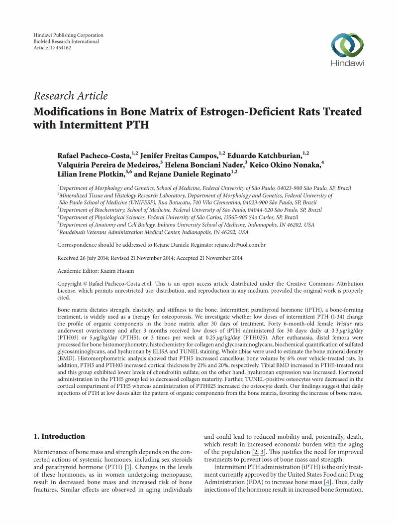
Research Article Modifications in Bone Matrix of Estrogen-Deficient Rats Treated with Intermittent PTH Rafael Pacheco-Costa, 1,2 Jenifer Freitas Campos, 1,2 Eduardo Katchburian, 1,2 Valquíria Pereira de Medeiros, 3 Helena Bonciani Nader, 3 Keico Okino Nonaka, 4 Lilian Irene Plotkin, 5,6 and Rejane Daniele Reginato 1,2 1 Department of Morphology and Genetics, School of Medicine, Federal University of S˜ ao Paulo, 04023-900 S˜ ao Paulo, SP, Brazil 2 Mineralized Tissue and Histology Research Laboratory, Department of Morphology and Genetics, Federal University of S˜ ao Paulo School of Medicine (UNIFESP), Rua Botucatu, 740 Vila Clementino, 04023-900 S˜ ao Paulo, SP, Brazil 3 Department of Biochemistry, School of Medicine, Federal University of S˜ ao Paulo, 04044-020 S˜ ao Paulo, SP, Brazil 4 Department of Physiological Sciences, Federal University of S˜ ao Carlos, 13565-905 S˜ ao Carlos, SP, Brazil 5 Department of Anatomy and Cell Biology, Indiana University School of Medicine, Indianapolis, IN 46202, USA 6 Roudebush Veterans Administration Medical Center, Indianapolis, IN 46202, USA Correspondence should be addressed to Rejane Daniele Reginato; [email protected] Received 26 July 2014; Revised 21 November 2014; Accepted 21 November 2014 Academic Editor: Kazim Husain Copyright © Rafael Pacheco-Costa et al. is is an open access article distributed under the Creative Commons Attribution License, which permits unrestricted use, distribution, and reproduction in any medium, provided the original work is properly cited. Bone matrix dictates strength, elasticity, and stiffness to the bone. Intermittent parathyroid hormone (iPTH), a bone-forming treatment, is widely used as a therapy for osteoporosis. We investigate whether low doses of intermittent PTH (1-34) change the profile of organic components in the bone matrix aſter 30 days of treatment. Forty 6-month-old female Wistar rats underwent ovariectomy and aſter 3 months received low doses of iPTH administered for 30 days: daily at 0.3 g/kg/day (PTH03) or 5 g/kg/day (PTH5); or 3 times per week at 0.25 g/kg/day (PTH025). Aſter euthanasia, distal femora were processed for bone histomorphometry, histochemistry for collagen and glycosaminoglycans, biochemical quantification of sulfated glycosaminoglycans, and hyaluronan by ELISA and TUNEL staining. Whole tibiae were used to estimate the bone mineral density (BMD). Histomorphometric analysis showed that PTH5 increased cancellous bone volume by 6% over vehicle-treated rats. In addition, PTH5 and PTH03 increased cortical thickness by 21% and 20%, respectively. Tibial BMD increased in PTH5-treated rats and this group exhibited lower levels of chondroitin sulfate; on the other hand, hyaluronan expression was increased. Hormonal administration in the PTH5 group led to decreased collagen maturity. Further, TUNEL-positive osteocytes were decreased in the cortical compartment of PTH5 whereas administration of PTH025 increased the osteocyte death. Our findings suggest that daily injections of PTH at low doses alter the pattern of organic components from the bone matrix, favoring the increase of bone mass. 1. Introduction Maintenance of bone mass and strength depends on the con- certed actions of systemic hormones, including sex steroids and parathyroid hormone (PTH) [1]. Changes in the levels of these hormones, as in women undergoing menopause, result in decreased bone mass and increased risk of bone fractures. Similar effects are observed in aging individuals and could lead to reduced mobility and, potentially, death, which result in increased economic burden with the aging of the population [2, 3]. is justifies the need for improved treatments to prevent loss of bone mass and strength. Intermittent PTH administration (iPTH) is the only treat- ment currently approved by the United States Food and Drug Administration (FDA) to increase bone mass [4]. us, daily injections of the hormone result in increased bone formation. Hindawi Publishing Corporation BioMed Research International Article ID 454162
-
Upload
independent -
Category
Documents
-
view
1 -
download
0
Transcript of Modifications in Bone Matrix of Estrogen-Deficient Rats Treated with Intermittent PTH
Research ArticleModifications in Bone Matrix of Estrogen-Deficient Rats Treatedwith Intermittent PTH
Rafael Pacheco-Costa12 Jenifer Freitas Campos12 Eduardo Katchburian12
Valquiacuteria Pereira de Medeiros3 Helena Bonciani Nader3 Keico Okino Nonaka4
Lilian Irene Plotkin56 and Rejane Daniele Reginato12
1Department of Morphology and Genetics School of Medicine Federal University of Sao Paulo 04023-900 Sao Paulo SP Brazil2Mineralized Tissue and Histology Research Laboratory Department of Morphology and Genetics Federal University ofSao Paulo School of Medicine (UNIFESP) Rua Botucatu 740 Vila Clementino 04023-900 Sao Paulo SP Brazil3Department of Biochemistry School of Medicine Federal University of Sao Paulo 04044-020 Sao Paulo SP Brazil4Department of Physiological Sciences Federal University of Sao Carlos 13565-905 Sao Carlos SP Brazil5Department of Anatomy and Cell Biology Indiana University School of Medicine Indianapolis IN 46202 USA6Roudebush Veterans Administration Medical Center Indianapolis IN 46202 USA
Correspondence should be addressed to Rejane Daniele Reginato rejanedruolcombr
Received 26 July 2014 Revised 21 November 2014 Accepted 21 November 2014
Academic Editor Kazim Husain
Copyright copy Rafael Pacheco-Costa et al This is an open access article distributed under the Creative Commons AttributionLicense which permits unrestricted use distribution and reproduction in any medium provided the original work is properlycited
Bone matrix dictates strength elasticity and stiffness to the bone Intermittent parathyroid hormone (iPTH) a bone-formingtreatment is widely used as a therapy for osteoporosis We investigate whether low doses of intermittent PTH (1-34) changethe profile of organic components in the bone matrix after 30 days of treatment Forty 6-month-old female Wistar ratsunderwent ovariectomy and after 3 months received low doses of iPTH administered for 30 days daily at 03120583gkgday(PTH03) or 5 120583gkgday (PTH5) or 3 times per week at 025 120583gkgday (PTH025) After euthanasia distal femora wereprocessed for bone histomorphometry histochemistry for collagen and glycosaminoglycans biochemical quantification of sulfatedglycosaminoglycans and hyaluronan by ELISA and TUNEL staining Whole tibiae were used to estimate the bone mineral density(BMD) Histomorphometric analysis showed that PTH5 increased cancellous bone volume by 6 over vehicle-treated rats Inaddition PTH5 and PTH03 increased cortical thickness by 21 and 20 respectively Tibial BMD increased in PTH5-treated ratsand this group exhibited lower levels of chondroitin sulfate on the other hand hyaluronan expression was increased Hormonaladministration in the PTH5 group led to decreased collagen maturity Further TUNEL-positive osteocytes were decreased in thecortical compartment of PTH5 whereas administration of PTH025 increased the osteocyte death Our findings suggest that dailyinjections of PTH at low doses alter the pattern of organic components from the bone matrix favoring the increase of bone mass
1 Introduction
Maintenance of bone mass and strength depends on the con-certed actions of systemic hormones including sex steroidsand parathyroid hormone (PTH) [1] Changes in the levelsof these hormones as in women undergoing menopauseresult in decreased bone mass and increased risk of bonefractures Similar effects are observed in aging individuals
and could lead to reduced mobility and potentially deathwhich result in increased economic burden with the agingof the population [2 3] This justifies the need for improvedtreatments to prevent loss of bone mass and strength
Intermittent PTHadministration (iPTH) is the only treat-ment currently approved by the United States Food and DrugAdministration (FDA) to increase bone mass [4] Thus dailyinjections of the hormone result in increased bone formation
Hindawi Publishing CorporationBioMed Research InternationalArticle ID 454162
2 BioMed Research International
Part of the anabolic effect of iPTH has been ascribed toits ability to prolong osteoblast lifespan resulting in theaccumulation of bone-forming cells with consequent increaseof bone mass and mechanical resistance [5ndash7] The actions ofiPTHare not only restricted to formation ofmineralized bonemediated by osteoblasts Indeed iPTH administration altersthe pattern of polysaccharides present in the bonematrix Forexample expression of the hyaluronan glycosaminoglycan isincreased in periosteal osteoblasts treated with iPTH [8] Inaddition altered synthesis of other glycosaminoglycans aswell as of proteoglycans was reported in a model of micelacking parathyroid-hormone related protein a moleculewith similar effect to PTH but with localized action [9]Changes in the bone matrix environment can affect thefate of osteoblasts and osteoclasts [10ndash15] and thereforereduction or overexpression of bone matrix molecules couldalter the result of the iPTH therapy Indeed the balance ofglycosaminoglycans and proteoglycans is crucial for mainte-nance of bone and mice with targeted-deletion of biglycanproteoglycan exhibit low bone mass similar to osteoporosis[16]
Based on these premises studies showing whether inter-mittent PTH administration alters the pattern of organiccomponents with focus on glycosaminoglycans and collagenare needed Although a recent study investigated the pro-teoglycans in human bone tissue after iPTH administrationthat study focused on the general proteoglycan content [17]In addition it is known that there is a relationship betweencollagen cross-linking and mineralization of bone matrix inmonkeys and humans treated with low doses of iPTH at longterm indicating that iPTH influences thematurity of collagenfibers [18 19]
Thus we aimed to investigate the effects of low dosesof iPTH at short term on the main organic bone matrixconstituents in rats in which osteopenia was induced bysex steroid removal through ovariectomy We found outthat intermittent administration of PTH alters the patternof organic components from the bone matrix potentiallyfavoring bone formation
2 Material and Methods
21 Animals and Treatment Forty 6-month-old femaleWistar rats (260 to 270 g) underwent bilateral ovariec-tomy (OVX) under intraperitoneal anesthesia with ketamine(40mgkg) and xylazine (20mgkg) to induce osteopeniawithin 3 months after surgery [20ndash22] Rats were randomlyassigned to four groups (119899 = 10group)The animals receivedsaline injections subcutaneously (abbreviated as OVX) orPTH at 03 120583gkgday (abbreviated as PTH03) or 5120583gkgday(abbreviated as PTH5) 7 times a week for 30 consecutivedays totalizing 30 injections An additional group of animalsreceived 025 120583gkg3 times a week (abbreviated as PTH025)only on Mondays Wednesdays and Fridays for 30 daystotalizing 12 injections Human PTH (1-34) (CalbiochemDarmstadt Germany) was dissolved in saline before admin-istration Rats were euthanized 24 hours after last injection byanesthesia overdose and then distal femora and whole tibiae
were collected All protocols involving rats were approved bythe Institutional Animal Care and Use Committee of FederalUniversity of Sao Paulo (UNIFESP process number 064308)
22 Vaginal Smears Collection Vaginal smears were collectedfrom all rats for 4 consecutive days in order to confirmthe success of the ovariectomy procedure by analyzing theperiodicity for the estrous cycle 21 days after removal of theovaries For this a cotton swabwas dampenedwith saline andthen carefully introduced into a vaginal cavity and slightlyrotated The secretion containing cells was placed on glassslides and fixed in ether and 95 ethanol (1 1) for 20minand evidenced by Shorr staining [23] As inclusion criteriaonly rats that were at diestrus (or anestrous) for at least threeconsecutive days were used in this study In the current studyall 40 rats ovariectomized were arrested at diestrus phase(Figure 1)
23 Histological Preparations Distal femora were fixed in4 formaldehyde (freshly derived from paraformaldehyde)buffered at pH 72 with 01M sodium phosphate at roomtemperature for 4 days Bones were subsequently decalcifiedin 25 formic acid for 30 days replacing once a weekthe decalcification solution Samples were then dehydratedin graded concentrations of ethanol embedded in paraffinand consecutive 5 120583m thick sections were processed forhistomorphometry histochemistry and TUNEL staining asdescribed below
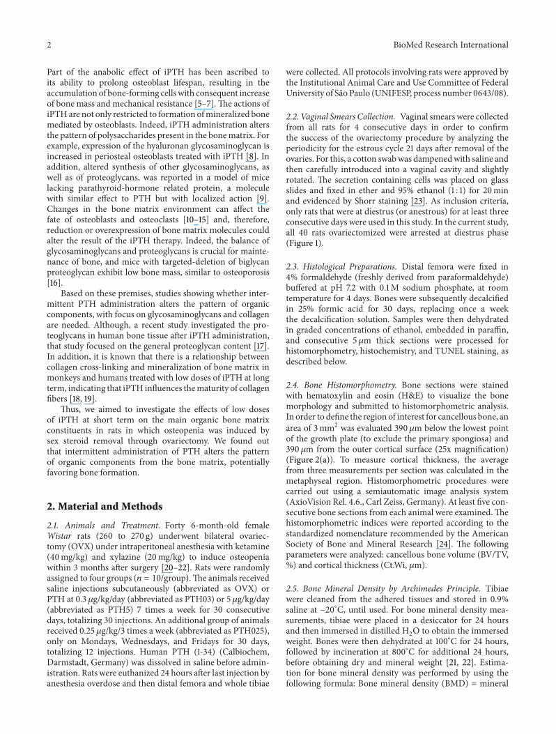
24 Bone Histomorphometry Bone sections were stainedwith hematoxylin and eosin (HampE) to visualize the bonemorphology and submitted to histomorphometric analysisIn order to define the region of interest for cancellous bone anarea of 3mm2 was evaluated 390120583m below the lowest pointof the growth plate (to exclude the primary spongiosa) and390 120583m from the outer cortical surface (25x magnification)(Figure 2(a)) To measure cortical thickness the averagefrom three measurements per section was calculated in themetaphyseal region Histomorphometric procedures werecarried out using a semiautomatic image analysis system(AxioVision Rel 46 Carl Zeiss Germany) At least five con-secutive bone sections from each animal were examinedThehistomorphometric indices were reported according to thestandardized nomenclature recommended by the AmericanSociety of Bone and Mineral Research [24] The followingparameters were analyzed cancellous bone volume (BVTV) and cortical thickness (CtWi 120583m)
25 Bone Mineral Density by Archimedes Principle Tibiaewere cleaned from the adhered tissues and stored in 09saline at minus20∘C until used For bone mineral density mea-surements tibiae were placed in a desiccator for 24 hoursand then immersed in distilled H
2O to obtain the immersed
weight Bones were then dehydrated at 100∘C for 24 hoursfollowed by incineration at 800∘C for additional 24 hoursbefore obtaining dry and mineral weight [21 22] Estima-tion for bone mineral density was performed by using thefollowing formula Bone mineral density (BMD) = mineral
BioMed Research International 3
Proestrous
(a)
Estrous
(b)
Metaestrous
(c)
Diestrous
(d)
Figure 1 Characterization of estrous cycle in ovariectomized mice in relation to proportion of cells (a) proestrous showing a predominanceof polynucleated and rounded cells (b) estrous showing decreased amount of epithelial cells and predominance of anucleated cornified cells(c) metaestrous containing the same proportion of leukocytes and nucleated epithelial cells and (d) diestrous (in this case anestrous withoutpermanent hormonal stimulus) containing a predominance of leukocytes in the smear The scale bar represents 10 120583m
ROI
CtMa
Gp
Tb
390120583m
390120583m
3000000 120583m2
(a)
lowast
lowast lowast
lowast
0
10
20
30
40BVTV
0
200
400
600CtWi
OVX PTH5PTH03 PTH025
OVX PTH5PTH03 PTH025
()
(120583m)
(b)
lowast
lowastlowast
Tibial BMD
04
06
08
10
OVX PTH5PTH03 PTH025
(c
mg
3)
(c)
Figure 2 Increased bone mass in rats treated with low doses of intermittent PTH (a) Photo from whole femur The black box indicatesthe region of interest (ROI) in the distal femora that histomorphometry histochemistry TUNEL assay and GAGs quantification wereperformed Observe a light micrograph from bone section stained with hematoxylin and eosin (HampE) showing the region of interest forhistomorphometric analysis (black square) An area of 3mm2 was evaluated at 390 120583mbelow the lowest point of the growth plate and 390 120583mfrom the outer cortical surface To measure cortical thickness the average from three cortical thicknesses per section was calculated in themetaphyseal region using the mean of three measurements from the same box used to measure the cancellous area Ct cortical Ma bonemarrow Tb trabecula Gp growth plate Scale bar represents 200 120583m (b) Cancellous bone volume (BVTV) and cortical thickness (CtWi)were measured by histomorphometry in the ROI of estrogen-deficient rats treated with vehicle or at indicated doses of iPTH (c) Tibial BMDwas measured in whole tibia by Archimedes principle Data are expressed as mean plusmn SD119873 = 9ndash11 lowast119875 lt 005 by one-way ANOVA
4 BioMed Research International
weightbone volume Bone volume = weight after 24 h insidea desiccator minus immersed weightwater density
26 Histochemistry for Glycosaminoglycans Bone sectionswere stained by the alcian blue method in order to identifyglycosaminoglycans in the distal femur from 390120583m belowfrom the lowest point of the growth plate in the same areaused to histomorphometry analyses Sulfated GAGs weredetected at pH 05 and all glycosaminoglycans includinghyaluronan at pH 25 [21 22] Briefly sections were deparaf-finized rehydrated and incubated in either 05M HCl or3 acetic acid for 2min before staining with alcian blueat pH 05 or pH 25 respectively for 40 minutes Sectionswere dehydrated cleaned and mounted in entellan [2122] Glycosaminoglycans were identified and quantified bymeasuring the distribution and intensity of the bluish staining(ImageLab 2000 system Diracom Brazil)
27 Histochemistry for Collagen Fibers andHyaluronan Bonesections were stained using a modified picrosirius red tech-nique previously described and the birefringence of collagenfibers was evaluated in the distal femur from 390 120583m belowthe lowest point of the growth plate [21 22 25] Sections weredeparaffinized rehydrated and incubated with 02 aqueousphosphomolybdic acid for 10 minutes to render the cyto-plasm colorless and then stained with 01 solution of siriusred diluted in saturated aqueous picric acid for 90 minutesNext sections were rinsed with 001M HCl for 2 minutesdehydrated cleaned and mounted One section from eachanimal was captured with a microscope (Axioskop 40 CarlZeiss Germany) equipped with polarizating filters using adigital camera (AxioCamMRc 5 Carl Zeiss Germany)
To detect the hyaluronan expression distal femur bonesections were deparaffinized rehydrated and incubated with3 hydrogen peroxide to block endogenous peroxidaseactivity followed by incubation with 1 BSA for 40min toblock nonspecific binding sites Next sectionswere incubatedovernightwith biotinylated hyaluronan-binding protein pro-duced byNader et al [26 27] After rinsingwith PBS sectionswere incubated with streptavidin-peroxidase solution (SantaCruz Biotechnology Santa Cruz CA) for 1 hour at roomtemperature in a dark chamber Samples were developedusing DAB substrate-chromogen system (DakoNorth Amer-ica Inc Carpinteria CA) for up to 5min Sections werethen rinsed and counterstained with Carrazirsquos hematoxylinThe final product of the reaction presented a brownish colorcorresponding to the bindings of endogenous hyaluronan tothe probe [26 28]
28 TUNEL Assay for Cell Death For detection of DNAbreaks the Apop-Tag Peroxidase In Situ Apoptosis Detec-tion Kit (Chemicon Internacional Temecula CA) was usedas previously described [29] TUNEL-positive hypertrophicchondrocytes were observed at the growth plate serving as aninternal positive control for each section Negative controlswere prepared by replacing TdT enzyme by distilled waterThree animalsgroup and two sectionsrat were scored fordetermination of the number of TUNEL-positive osteocytes
29 Quantification of Sulfated Glycosaminoglycans (GAGs)and Hyaluronan In a preliminary study we compared 10EDTA and 25 formic acid as decalcification solutions inorder to evaluate which one showed better efficiency forreleasing smaller amount of glycosaminoglycans at differenttime points This standardization was crucial to not overesti-mate or subestimate the concentration of glycosaminoglycanexpressed on bone tissue indicating that 25 acid formic wasmore efficient in 3 days allowing the maceration and releas-ing a low amount of glycosaminoglycans After removing thecartilage from the articular cartilage up to growth plate fromthe distal femora the samples were incubated in phosphatebuffered solution containing protease inhibitors for 3 hoursfollowed by decalcification in 25 formic acid for 3 days Fivesamples per group were then macerated with liquid nitrogendried and incubated for 18 h at 60∘C in 01M phosphatebuffer-cysteine pH 65 containing 2mgmL papain and002Methylenediaminetetraacetic acid (EDTA) to release theGAGs (Calbiochem Darmstadt Germany) Ninety percenttrichloroacetic acid was then added to a final concentrationof 10 to precipitate the proteins and nucleic acids at 4∘Cfollowed by two volumes of cold methanol with shakingto precipitate the GAGs and incubated at minus20∘C for 24 hThe supernatant was then discarded and the material wasdried at room temperature The pellet dissolved in water wasapplied to a 055 agarose gel in 005M 13-diaminopropaneacetate buffer (pH 90) [30] The gel was fixed with 01N-cetyl-NNN-trimethylammonium bromide solution for 2hours dried and stained with 01 toluidine blue in aceticacidethanol The intensity of the bands was measured usinga QuickScan 2000 densitometer (Helena Laboratories CorpBeaumont TX) [21 28] Hyaluronan concentration wasdetermined using an ELISA-like assay [27] Briefly standardconcentrations (0ndash500120583gL) of hyaluronan from umbilicalcord (Sigma Aldrich) and aliquots from bones obtainedafter proteolysis were diluted blocking buffer and 100120583Lfrom each sample was added in triplicate to 96-well platescoated with hyaluronan-binding protein After incubationfor 18 hours at 4∘C the plates were washed 3 times withwashing buffer containing 005M Tris-HCl 015M NaCl005 Tween 20 002mM EDTA III 77mM sodium azideand pH 775 and followed by addition of 100120583L of biotinylatedhyaluronan-binding protein (1mgmL) to each well Plateswere maintained shaking for 2 hours at room temperatureand then washed 6 times with washing buffer before adding100 120583L of europium-labeled streptavidin After 30 minutes ofincubation at room temperature the plates were washed 6times After that 200120583L of enhancement solution (Perkin-Elmer Life Sciences) was added and then shaken for 10minutes to release the europium-bound streptavidin Thereleased europium was read at 340 and 630 nm (excitationand emission filters resp) [26 31]
210 Statistical Analysis The data were analyzed by usingGraphPad Prism (GraphPad Software Inc La Jolla CA)The groups were compared using one-way ANOVA followedby Tukeyrsquos multiple comparison test to evaluate differencesamong groups Significance was established at 119875 lt 005 All
BioMed Research International 5
Table 1 Histochemical and biochemical quantification of glycosaminoglycans
Histochemical analysis Biochemical analysisGeneral GAGs pH 25 () Sulfated GAGs pH 05 () Chondroitin sulfate (120583gmg tissue) Hyaluronan (120583gg tissue)
OVX 368 plusmn 221 298 plusmn 154 090 plusmn 008 2918 plusmn 1308PTH03 302 plusmn 197 181 plusmn 099 067 plusmn 011 3324 plusmn 1216PTH5 140 plusmn 083lowast 124 plusmn 155lowast 053 plusmn 008lowast 5871 plusmn 1348lowast
PTH025 172 plusmn 126lowast 138 plusmn 128lowast 057 plusmn 011 3366 plusmn 1316Data are expressed as mean plusmn SD 119875 lt 005 lowastversus OVX
Table 2 Quantification of TUNELmdashpositive osteocytes in thecortical compartment
TUNEL positive osteocytes (total numbersection)OVX 27 plusmn 16PTH03 18 plusmn 1PTH5 5 plusmn 5PTH025 56 plusmn 11lowast
Data are expressed as mean plusmn SD 119875 lt 005 lowastversus OVX
numerical values were reported asmeanplusmn standard deviation(SD)
3 Results
31 Low Dose of Intermittent PTH Increases Bone Massin Cancellous and Cortical Compartment Analysis in thefemoral bone microarchitecture from rats treated with lowdoses of intermittent PTH (1-34) for one month showed anincrease of 6 in the cancellous bone volume (BVTV) ofPTH5 compared to the OVX group and of 8 compared tothe PTH03 group (Figure 2(b)) On the other hand therewere no statistical differences in BVTV between PTH03 andPTH025 compared to the OVX group In addition there isalso no difference in PTH025 when compared to PTH5 andPTH03 group Cortical thickness (CtWi) increased by 21 inPTH03 and 20 in PTH5 groups compared to OVX whilethe effect of PTH025 did not reach statistical significanceFurthermore tibial BMD increased only in PTH5 groupand was significantly higher compared to all other groups(Figure 2(c))
32 Glycosaminoglycans Profile Is Altered after Treatment withiPTH We found a decrease in sulfated and carboxylatedGAGs at pH 25 and also in sulfated GAGs only at pH05 of PTH5 and PTH025 groups when compared to theOVX group (Table 1) In addition bone sections from theOVX group evidenced the highest predominance of GAG atpH 05 (Figure 3(a) blue staining) compared to the lowestpredominance in PTH5 rats (Figure 3(b))
A similar electrophoretic mobility of chondroitin sulfatewas found in the distal femur lysates whereas dermatansulfate and heparan sulfate were not detected (not shown)Quantification of chondroitin sulfate showed a decreased inall iPTH-injected groups compared to control but reachedsignificance only in PTH5 group with an approximately 41reduction Moreover hyaluronan quantification by ELISA
showed an increase only in the PTH5 group compared tocontrol (Table 1) Consistent with that the expression ofhyaluronan in femoral bone sections was slightly stronger inthe PTH5 group (Figure 3(d)) compared to the OVX group(Figure 3(c))
33 Behavior of Collagen Birefringence Changed in iPTH-Treated Rats In order to examine the effect of iPTH onthe pattern of collagen in femoral bone sections we usedthe picrosirius-polarized light method Bones from the OVXgroup exhibited a predominance of reddish birefringence inthe cortical and cancellous compartment (Figures 4(a) and4(b)) in comparison with reduced reddish birefringence andincreased greenish birefringence in all iPTH-treated groups(Figures 4(c)ndash4(h)) in particular in the bone sections fromthe PTH5 group (Figures 4(e) and 4(f)) suggesting an alteredpattern of collagen induced by iPTH administration anindicative of collagen immaturity
34 Decreased Osteocytes Death in the PTH-Injected GroupsTo investigate whether inhibition of cell death induced byiPTH is associated with bone matrix modifications wequantified the prevalence of osteocytes death in corticalbone stained using the TUNEL assay In comparison tothe OVX group (Figure 3(e)) we detected a decrease inTUNEL-positive osteocytes in the cortical compartmentof PTH03 and PTH5 group however these changes didnot reach the significance but PTH5 exhibited a tendencytoward increase (Table 2 and Figure 3(f)) On the other handPTH025 increased significantly the number of TUNEL-positive osteocytes by 50 when compared to OVX group(Table 2)
4 Discussion
We show herein that low doses of intermittent PTH are ableto increase bone mass in estrogen-deficient rats and thatthis effect is accompanied by modifications in the profile ofsecreted components in the bone matrix Daily treatmentwith 5 120583gkgday of iPTH for one month increased bothcancellous and cortical bone mass However administrationof 03 120583gkgday every day only increased cortical bone thick-ness These results suggest that cancellous and cortical bonedisplay a different sensitivity to low doses of iPTH IndeedIida-Klein et al [32] showed that long bones responded toiPTH already between 1-2 weeks whereas the increase invertebral bone was only detected after 7 weeks inmice treated
6 BioMed Research International
TbTb
Ma
OVX
(a)
Tb
Ma
Tb
PTH5
(b)
Ma
Tb
Ct
GpPS
(c)
Ma
TbCt
GpPS
(d)
CtMa
Ps
(e)
Ps
Ct
(f)
Figure 3 (a and b) Modifications in the glycosaminoglycans after iPTH administration Representative images of 10 sectionsgroup underbright field micrographs Sections of distal femora were stained by alcian blue specific for sulfated GAGs (at pH 05) (a) OVX rats exhibitingmore expression of sulfated GAGs compared with (b) PTH5 group Scale bars represent 10 120583m (c and d) Representatives light micrographsof femoral sections stained for hyaluronan Bone sections from (c) OVX rats exhibiting slightly less expression of hyaluronan expression(brownish color) compared with (d) PTH5-treated rats Scale bars represent 200120583m Ps periosteum Ct cortical Ma bone marrow Tbtrabecula Gp growth plate (e and f) Representatives light micrographs of distal femora from rats stained by TUNEL for the detection ofDNA breaks (e) Femoral bone sections from OVX rats exhibit more TUNEL-positive when compared with (f) bone sections from PTH5group Arrowhead points at TUNEL-negative osteocytes and the arrow points at TUNEL-positive osteocytes Inset shows TUNEL-positivehypertrophic chondrocytes (arrows) at the growth plate serving as an internal positive control for each section119873 = 3 sectionsgroup Scalebars represent 10120583mThe number of TUNEL-positive osteocytes is expressed as mean plusmn SD Ct cortical Ma bone marrow Tb trabecula
with a dose of 40120583gkgday Other studies also reportedthat the hormone has skeletal site-specific action promotingthe cortical increase through a mechanism that involvesincreased in both bone formation and resorption in thecortical bone [33ndash36]
Consistent with the anabolic action of iPTH tibial BMDincreased in rats receiving a dose of 5 120583gkgday Severalauthors have reported that doses of iPTH from 20 up to100 120583gkg also have anabolic effects increasing bone massand reducing risk for fractures [5 7 32 37] We now report
BioMed Research International 7
OV
X
Cortical
(a)
Tb
Ma
Cancellous
(b)
PTH
03
(c)
Tb
Ma
(d)
PTH
5
(e)
Ma
Tb
(f)
PTH
025
(g)
Ma
Tb
(h)
Figure 4 Altered collagen pattern in iPTH-treated rats Representatives light micrographs from 10 sections of distal femurgroup evidencedby picrosirius under polarized light Cortical and cancellous compartments bone of the groups (a b) OVX (c d) PTH03 (e f) PTH5 and(g h) PTH025 Scale bars represent 200120583m Ma bone marrow Tb trabecula
the effectiveness of 03120583gkg and 5 120583gkg doses adminis-trated 7 days a week by 30 days in ovariectomy-inducedbone loss an experimental model of low bone density inrats [20ndash22 38] thus suggesting that daily injections of PTHincrease cancellous and cortical bone in very low doses
Instead administration of 025120583gkg only 3 times a weekhad little if any effect on bone mass showing that shorttime applied in this study or the low cumulative dose wasnot enough to increase cancellous andor cortical bone massSimilarly a previous study by Turner et al [39] showed that a
8 BioMed Research International
dose of 1 120583gkgday PTH prevented the bone loss induced byunloading but did not increase BMD after 2 weeks of therapy
Although other studies also have reported the effec-tiveness of low doses of iPTH the attention was limitedto the effect of the hormone on BMD In our study wefocused particularly on modifications in the pattern of bonematrix organic components which might drive cell fate andconsequently act in favor of formation andor resorptiondepending on molecules induced by iPTH administrationAdministration of low doses of iPTH might favor thedetection of changes in the bone matrix at the end of 30-day treatment since higher doses could accelerate the bonematrix modifications missing the opportunity to detect thechanges in the components of the bone matrix
The current clinical dose of iPTH approved to severeosteoporotic patients is 20120583g independently of weighthowever considering that patients weigh between 60 and70 kg the direct final dose injected is sim03 120583gkg (PTH03)[40]Thus we created the first iPTH-treated group (PTH03)receiving this doseThe other iPTH-treated group (PTH025)was created in an attempt to detect possible changes in bonecomponents the same changes expect to be found in PTH03but in rats receiving PTHonly 3 times a weekWe also createda group receiving 5 120583gkgday approximately 17 times morePTH than PTH03 but still considered a low dose in order toenhance the changes in bone tissue constituents
Collagen is the most abundant component of organicbonematrix [41] and altered collagen is associated with bonefragility in animals and humans [42ndash46] In the current studywe evaluated the collagen birefringence behavior by picrosir-ius red-polarization a well-known method to assess collagenmaturation [25 47] We observed a dramatic increase ingreenish birefringence in all iPTH-injected groups especiallyin that group receiving the highest dose (PTH5) Immatureand mature collagen fibrils of bone are differentiated bytheir colors under polarized light [48] Against a blackbackground thick fibers are mainly type I mature collagenconsequently present intense birefringence of yellow to redcolor while thin fibrils formed mainly by type I immaturecollagen (including procollagen intermediaries and evenaltered collagen) and display a weak birefringence of greenishcolor [48ndash50] Since the PTH5 group exhibited the highestpredominance of thin fibers we propose that in this groupthe hormone stimulates new bone tissue deposition initiallyreflecting on immature type I collagen content In additionwe cannot rule out the possibility that rats treated with PTH5are facing a higher rate of collagen fibers remodeling at theend of 30-day treatment when compared to the OVX groupCollagen birefringence changes also can be attributed to ori-entation of the fibers [51] In addition greenish birefringenceis also associated with accumulation of type III collagen[52] We propose that in our current study this birefringenceis associated with changes in morphology rather than withincreased synthesis of type III collagen Consistent with thisnotion the expression of the gene encoding the type IIIcollagen in rats is not altered when PTH is administered inthe intermittent fashion [53]
Studies have associated changes in collagen morphologyand thickness with the presence of glycosaminoglycans in
particular those containing sulfated residues [21 22 54 55]Moreover sulfated GAGs have important roles in the initialprocess of mineralization [12 55] Based on this idea weinvestigated the effect of the treatments on the levels ofchondroitin sulfate themost abundant sulfated GAG in bonetissue [56] Expression of sulfated GAG is reduced in thebone sections from PTH5 and PTH025 group assessed byhistochemistry but the results obtained with the biochemicalquantification revealed that only rats receiving PTH5 exhib-ited a reduction in chondroitin sulfate The difference on thefindings between histochemistry for GAGs and biochemicalanalysis in the PTH025 group might result from the factthat while the histochemistry method detects other sulfatedGAGs on the bone sections like dermatan and heparanbiochemistry analysis detects exclusively chondroitin Inaddition differences in the sensitivity of the methods couldalso explain this discrepancy Nevertheless it has been shownthat fusion and thickening of collagen fibrils are triggeredby removal of sulfated GAGs [54 55 57] Supporting thisidea in a deletion study using biglycan- and decorin-deficientmice chondroitin sulfate-rich proteoglycans showed alteredmorphology such as decreased diameter and size of collagenfibrils [54] Thus the removal of chondroitin sulfate mighthave led to the changes observed in the birefringence of colla-gen fibers The changes in collagen birefringence observed inour study could be at least partially explained by a reductionin sulfated GAGs
We found that hyaluronan expression is increased in bonelysates of rats treated with iPTH mainly with the highestdose It has been reported the accumulation of hyaluronanin periosteal area of long bones and increased expression incells of the osteoblastic lineage after treatment with iPTH[8 58] However since it was not our intention to detecthyaluronan expression in the periosteum we scrapped offthis layer from the bone when preparing it Neverthelesswe detected small areas of periosteum exhibiting increasedexpression for hyaluronan similar to what was reported byMidura and coworkers [8] Further studies are needed todetermine the role of the increase in hyaluronan induced byiPTH on the periosteal surface
It has been proposed that osteoporosis and other age-related diseases are caused in part by apoptosis of bone-forming cells [59ndash61] iPTH is thought to improve osteoporo-sis by stimulating bonematrix production and by suppressingosteoblast and osteocyte apoptosis [59 62]Herewe show thatthe hormone also reduced osteocyte death in cortical bonesections from the PTH03 and PTH5 groups However wefound a dramatic increase in osteocyte apoptosis in the ratstreated with PTH025 Although the reason for this increaseis not known we can speculate that is due to the fact thatthe hormone is not administered daily and the cell death isan initial event during iPTH administration that is delayedin this group compared to other iPTH-treated groups andcan be detected at the end of 30-day treatment Consistentwith this possibility a study showed that daily injectionswith a higher dose of PTH (80120583gkgday) resulted in anincrease in apoptotic osteocytes in cancellous bone after 7days of treatment in rats and that the percentage of apoptoticcells return to basal levels 28 days after initiating hormone
BioMed Research International 9
administration [63] Taking into account that the iPTHdose used in the above study was much higher than thoseadministrated by us it is reasonable to propose that in thegroup injected with the very low concentration of hormoneand only 3 times a week (PTH025) it still is not returned tothe basal levels (as observed after 28 days by Stanislaus etal [63]) at the end of the treatment period (30 days) Thesepieces of evidence suggest that the low iPTH doses at shorttime could induce transient increase in osteocyte death thatwill be followed by a reversion of the effect
Terminal deoxynucleotidyl transferase (TdT) mediateddUTP nick-end-labeling (TUNEL) is widely considered as aldquogold standardrdquo assay for assessment of apoptosis Howeverthere are exceptions for TUNEL specificity [6 60] sincethis assay can identify nucleotide excision repair (NER) [6465] NER involves the creation of DNA breaks through anendonuclease driven excision of damage nucleotides creating31015840OH groups [65 66] TdT the enzyme used to label nickedDNA in the TUNEL reaction recognizes 31015840OH groups atnicked ends in DNA identifying DNA breaks in fixed cells [665] In our study some TUNEL-positives osteocytes could beundergoing DNA repair Schnoke et al [6] hypothesized thatPTHmight suppress osteoblast apoptosis by enhancing DNArepair and they showed that the attachment of nucleotides tothe 31015840OHends of fixedDNAvia the TUNEL reaction in PTH-treated nuclei was more likely generated by NER-mediatednicking and not apoptotic cleavage Whether the TUNEL-positive cells are indeed apoptotic or whether DNA repair istaking place will be the subject of future studies
The signals that trigger bone resorption are not com-pletely understood One important event in the regulation ofremodeling seems to be apoptosis of osteocytes The deathosteocytes release chemotactic signals that attract osteoclaststriggering localized bone resorption [67] During the boneremodeling process the matrix is resorbed before the newtissue is produced If as proposed an early effect of PTHadministration is to induce osteocyte death this will befollowed by localized bone resorption and the consequentremodeling of the matrix Our results suggest that in thePTH5 group at the dose of 5 120583gkgday the osteocyte deathphase hasmost likely already occurred and the stage detectedhere is associated with the strong remodeling of the matrixdemonstrated by collagen fiber turnover as indicated by thegreenish birefringence low concentration of sulfated GAGsand high expression of hyaluronan
One limitation of our study is that we did not use agroup of rats without undergoing ovariectomy (sham group)However the initial propose of this study was to comparewhether the low doses of iPTH changes the profile of organicbone matrix components after 30 days of treatment in ratswith osteopenia Although we cannot evaluate if bone matrixcomponents are altered compared to sham controls wecan analyze the changes in bone matrix induced by iPTHadministration and compare it with the OVX model
In summary we show herein that low dose of daily iPTHadministration results in changes in organic component ofbone matrix such as in glycosaminoglycans and collagenThese events together with increased bone mass and reduc-tion in osteocyte cell death could contribute to increased bone
strength in individuals receiving intermittent administrationof the drug
Conflict of Interests
The authors declare that there is no conflict of interestsregarding the publication of this paper
Acknowledgments
This work was supported by Coordination of Improvementof Higher Level Personnel (CAPES) andNational Council forScientific and Technological Development (CNPq) Brazil
References
[1] A Teti ldquoBone development overview of bone cells and signal-ingrdquo Current Osteoporosis Reports vol 9 no 4 pp 264ndash2732011
[2] DW Dempster and R Lindsay ldquoPathogenesis of osteoporosisrdquoThe Lancet vol 341 no 8848 pp 797ndash801 1993
[3] S Khosla M J Oursler and D G Monroe ldquoEstrogen and theskeletonrdquo Trends in Endocrinology and Metabolism vol 23 no11 pp 576ndash581 2012
[4] J A Kanis E V McCloskey H Johansson C Cooper R Riz-zoli and J Y Reginster ldquoEuropean guidance for the diagnosisand management of osteoporosis in postmenopausal womenrdquoOsteoporosis International vol 24 no 1 pp 23ndash57 2013
[5] T Bellido A A Ali L I Plotkin et al ldquoProteasomal degra-dation of Runx2 shortens parathyroid hormone-induced anti-apoptotic signaling in osteoblasts a putative explanation forwhy intermittent administration is needed for bone anabolismrdquoThe Journal of Biological Chemistry vol 278 no 50 pp 50259ndash50272 2003
[6] M Schnoke S B Midura and R J Midura ldquoParathyroidhormone suppresses osteoblast apoptosis by augmenting DNArepairrdquo Bone vol 45 no 3 pp 590ndash602 2009
[7] R L Jilka R SWeinstein T Bellido P Roberson A M Parfittand S C Manolagas ldquoIncreased bone formation bypreventionof osteoblast apoptosis with parathyroid hormonerdquoThe Journalof Clinical Investigation vol 104 no 4 pp 439ndash446 1999
[8] R J Midura X Su J A Morcuende M Tammi and R TammildquoParathyroid hormone rapidly stimulates hyaluronan synthesisby periosteal osteoblasts in the tibial diaphysis of the growingratrdquo Journal of Biological Chemistry vol 278 no 51 pp 51462ndash51468 2003
[9] Y Ogihara N Suda V E Hammond P V Senior F Beckand M Yanagishita ldquoBiosynthesis of proteoglycan in boneand cartilage of parathyroid hormone-related protein knockoutmicerdquo Journal of Bone andMineral Metabolism vol 19 no 1 pp4ndash12 2001
[10] A D Berendsen E L Pinnow A Maeda et al ldquoBiglycanmodulates angiogenesis and bone formation during fracturehealingrdquoMatrix Biology vol 35 pp 223ndash231 2014
[11] C W Prince ldquoRoles of hyaluronan in bone resorptionrdquo BMCMusculoskeletal Disorders vol 5 article 12 2004
[12] CW Prince F Rahemtulla andWT Butler ldquoMetabolismof ratbone proteoglycans in vivordquo Biochemical Journal vol 216 no 3pp 589ndash596 1983
10 BioMed Research International
[13] P Bianco L W Fisher M F Young J D Termine andP G Robey ldquoExpression and localization of the two smallproteoglycans biglycan and decorin in developing humanskeletal and non-skeletal tissuesrdquo Journal of Histochemistry andCytochemistry vol 38 no 11 pp 1549ndash1563 1990
[14] N Rucci A RufoMAlamanou et al ldquoThe glycosaminoglycan-binding domain of PRELP acts as a cell type-specific NF-120581B inhibitor that impairs osteoclastogenesisrdquo Journal of CellBiology vol 187 no 5 pp 669ndash683 2009
[15] S R Amend O UluckanMHurchla et al ldquoThrombospondin-1 regulates bone homeostasis through effects on bone matrixintegrity and nitric oxide signaling in osteoclastsrdquo Journal ofBone and Mineral Research 2014
[16] T Xu P Bianco L W Fisher et al ldquoTargeted disruption of thebiglycan gene leads to an osteoporosis-like phenotype in micerdquoNature Genetics vol 20 no 1 pp 78ndash82 1998
[17] B Hofstetter S Gamsjaeger F Varga et al ldquoBone quality of thenewest bone formed after two years of teriparatide therapy inpatients who were previously treatment-naıve or on long-termalendronate therapyrdquo Osteoporosis International vol 25 no 12pp 2709ndash2719 2014
[18] E P Paschalis EVGlassDWDonley andE F Eriksen ldquoBonemineral and collagen quality in iliac crest biopsies of patientsgiven teriparatide new results from the fracture preventiontrialrdquo Journal of Clinical Endocrinology andMetabolism vol 90no 8 pp 4644ndash4649 2005
[19] E P Paschalis D B Burr R Mendelsohn J M Hock and AL Boskey ldquoBone mineral and collagen quality in humeri ofovariectomized cynomolgus monkeys given rhPTH(1ndash34) for18 monthsrdquo Journal of Bone and Mineral Research vol 18 no4 pp 769ndash775 2003
[20] D N Kalu ldquoThe ovariectomized rat model of postmenopausalbone lossrdquo Bone and Mineral vol 15 no 3 pp 175ndash191 1991
[21] R Florencio-SilvaMA Santos V PDeMedeiros et al ldquoEffectsof soy isoflavones and mechanical vibration on rat bone tissuerdquoClimacteric vol 16 no 6 pp 709ndash717 2013
[22] M A Santos R Florencio-Silva V P Medeiros et al ldquoEffectsof different doses of soy isoflavones on bone tissue of ovariec-tomized ratsrdquo Climacteric vol 17 pp 393ndash401 2014
[23] E Shorr ldquoA new technic for staining vaginal smears III a singledifferential stainrdquo Science vol 94 no 2449 pp 545ndash546 1941
[24] D W Dempster J E Compston M K Drezner et al ldquoStan-dardized nomenclature symbols and units for bone histo-morphometry a 2012 update of the report of the ASBMRHistomorphometryNomenclature Committeerdquo Journal of Boneand Mineral Research vol 28 no 1 pp 2ndash17 2013
[25] L C U Junqueira G Bignolas and R R Brentani ldquoPicrosiriusstaining plus polarization microscopy a specific method forcollagen detection in tissue sectionsrdquoTheHistochemical Journalvol 11 no 4 pp 447ndash455 1979
[26] R C Teixeira Gomes C Verna H B Nader et al ldquoConcen-tration and distribution of hyaluronic acid in mouse uterusthroughout the estrous cyclerdquo Fertility and Sterility vol 92 no2 pp 785ndash792 2009
[27] J RMMartins C C Passerotti RM BMaciel L O SampaioC P Dietrich and H B Nader ldquoPractical determinationof hyaluronan by a new noncompetitive fluorescence-basedassay on serum of normal and cirrhotic patientsrdquo AnalyticalBiochemistry vol 319 no 1 pp 65ndash72 2003
[28] C E Franciozi V A F Tarini R D Reginato et al ldquoGradualstrenuous running regimen predisposes to osteoarthritis due to
cartilage cell death and altered levels of glycosaminoglycansrdquoOsteoarthritis and Cartilage vol 21 no 7 pp 965ndash972 2013
[29] A P S Faloni E Sasso-Cerri E Katchburian and P SCerri ldquoDecrease in the number and apoptosis of alveolarbone osteoclasts in estrogen-treated ratsrdquo Journal of PeriodontalResearch vol 42 no 3 pp 193ndash201 2007
[30] C P Dietrich and S M C Dietrich ldquoElectrophoretic behaviourof acidic mucopolysaccharides in diamine buffersrdquo AnalyticalBiochemistry vol 70 no 2 pp 645ndash647 1976
[31] L A de Souza Merli V P de Medeiros L Toma et al ldquoThe lowlevel laser therapy effect on the remodeling of bone extracellularmatrixrdquo Photochemistry and Photobiology vol 88 no 5 pp1293ndash1301 2012
[32] A Iida-Klein H Zhou S S Lu et al ldquoAnabolic action ofparathyroid hormone is skeletal site specific at the tissue andcellular levels in micerdquo Journal of Bone and Mineral Researchvol 17 no 5 pp 808ndash816 2002
[33] S Arita S Ikeda A Sakai et al ldquoHuman parathyroid hormone(1ndash34) increases mass and structure of the cortical shell withresultant increase in lumbar bone strength in ovariectomizedratsrdquo Journal of Bone andMineral Metabolism vol 22 no 6 pp530ndash540 2004
[34] Y Rhee M R Allen K Condon et al ldquoPTH receptor signalingin osteocytes governs periosteal bone formation and intracorti-cal remodelingrdquo Journal of Bone and Mineral Research vol 26no 5 pp 1035ndash1046 2011
[35] J C Gallagher C J Rosen P Chen D A Misurski and RMarcus ldquoResponse rate of bone mineral density to teriparatidein postmenopausal womenwith osteoporosisrdquo Bone vol 39 no6 pp 1268ndash1275 2006
[36] M Montero D Serfati S Luna et al ldquoThe effectiveness ofintermittent rat parathyroid hormone (1ndash34) treatment on lowbone mass due to oestrogen or androgen depletion in skeletallymature ratsrdquo Aging Male vol 13 no 1 pp 59ndash73 2010
[37] A Del Fattore A Cappariello M Capulli et al ldquoAn experimen-tal therapy to improve skeletal growth and prevent bone loss inamousemodel overexpressing IL-6rdquoOsteoporosis Internationalvol 25 no 2 pp 681ndash692 2014
[38] U Comelekoglu S Bagis S Yalin et al ldquoBiomechanical eval-uation in osteoporosis ovariectomized rat modelrdquo ClinicalRheumatology vol 26 no 3 pp 380ndash384 2007
[39] R T Turner G L Evans S Lotinun P D Lapke U T Iwaniecand E Morey-Holton ldquoDose-response effects of intermittentPTH on cancellous bone in hindlimb unloaded ratsrdquo Journal ofBone and Mineral Research vol 22 no 1 pp 64ndash71 2007
[40] R M Neer C D Arnaud J R Zanchetta et al ldquoEffect ofparathyroid hormone (1ndash34) on fractures and bone mineraldensity in postmenopausal women with osteoporosisrdquoTheNewEngland Journal of Medicine vol 344 no 19 pp 1434ndash14412001
[41] B Clarke ldquoNormal bone anatomy and physiologyrdquo ClinicalJournal of theAmerican Society ofNephrology vol 3 supplement3 pp S131ndashS139 2008
[42] M-G Ascenzi V P Liao B M Lee et al ldquoParathyroidhormone treatment improves the cortical bone microstructureby improving the distribution of type I collagen in post-menopausal women with osteoporosisrdquo Journal of Bone andMineral Research vol 27 no 3 pp 702ndash712 2012
[43] H Oxlund M Barckman G Ortoft and T T AndreassenldquoReduced concentrations of collagen cross-links are associatedwith reduced strength of bonerdquo Bone vol 17 no 4 pp 365Sndash371S 1995
BioMed Research International 11
[44] A J Bailey T J Sims E N Ebbesen J PMansell J SThomsenand L Mosekilde ldquoAge-related changes in the biochemicalproperties of human cancellous bone collagen relationship tobone strengthrdquo Calcified Tissue International vol 65 no 3 pp203ndash210 1999
[45] A J Bailey S F Wotton T J Sims and P W ThompsonldquoBiochemical changes in the collagen of human osteoporoticbone matrixrdquo Connective Tissue Research vol 29 no 2 pp 119ndash132 1993
[46] A J Bailey S F Wotton T J Sims and P WThompson ldquoPost-translational modifications in the collagen of human osteo-porotic femoral headrdquo Biochemical and Biophysical ResearchCommunications vol 185 no 3 pp 801ndash805 1992
[47] L C U Junqueira G S Montes and E M Sanchez ldquoTheinfluence of tissue section thickness on the study of collagenby the Picrosirius-polarizationmethodrdquoHistochemistry vol 74no 1 pp 153ndash156 1982
[48] A Hirshberg M Lib A Kozlovsky and I Kaplan ldquoTheinfluence of inflammation on the polarization colors of collagenfibers in the wall of odontogenic keratocystrdquoOral Oncology vol43 no 3 pp 278ndash282 2007
[49] M Szendroi G Vajta and L Kovacs ldquoPolarization colours ofcollagen fibres a sign of collagen production activity in fibroticprocessesrdquo Acta Morphologica Hungarica vol 32 no 1 pp 47ndash55 1984
[50] L B Retamoso TDMA daCunha L AH Knop R L Shint-covsk and O M Tanaka ldquoOrganization and quantification ofthe collagen fibers in bone formation during orthodontic toothmovementrdquoMicron vol 40 no 8 pp 827ndash830 2009
[51] A S Acerbo A T Kwaczala L Yang S Judex and L MMiller ldquoAlterations in collagen and mineral nanostructureobserved in osteoporosis and pharmaceutical treatments usingsimultaneous small- and wide-angle X-ray scatteringrdquo CalcifiedTissue International vol 95 no 5 pp 446ndash456 2014
[52] G SMontes ldquoStructural biology of the fibres of the collagenousand elastic systemsrdquo Cell Biology International vol 20 no 1 pp15ndash27 1996
[53] J E Onyia L M Helvering L Gelbert et al ldquoMolecular profileof catabolic versus anabolic treatment regimens of parathyroidhormone (PTH) in rat bone an analysis by DNA microarrayrdquoJournal of Cellular Biochemistry vol 95 no 2 pp 403ndash418 2005
[54] A Corsi T Xu X-D Chen et al ldquoPhenotypic effects ofbiglycan deficiency are linked to collagen fibril abnormalitiesare synergized by decorin deficiency andmimic Ehlers-Danlos-like changes in bone and other connective tissuesrdquo Journal ofBone and Mineral Research vol 17 no 7 pp 1180ndash1189 2002
[55] K Hoshi S Kemmotsu Y Takeuchi N Amizuka and HOzawa ldquoThe primary calcification in bones follows removalof decorin and fusion of collagen fibrilsrdquo Journal of Bone andMineral Research vol 14 no 2 pp 273ndash280 1999
[56] N S Gandhi and R LMancera ldquoThe structure of glycosamino-glycans and their interactions with proteinsrdquo Chemical Biologyand Drug Design vol 72 no 6 pp 455ndash482 2008
[57] T Yamamoto N Nagaoka A Hirata et al ldquoUltrastructural andimmunohistochemical studies of medullary bone calcificationwith special reference to sulphated glycosaminoglycansrdquo Jour-nal of Electron Microscopy vol 54 no 1 pp 29ndash34 2005
[58] R J Midura S P Evanko and V C Hascall ldquoParathyroidhormone stimulates hyaluronan synthesis in an osteoblast-likecell linerdquo Journal of Biological Chemistry vol 269 no 18 pp13200ndash13206 1994
[59] R L Jilka ldquoMolecular and cellular mechanisms of the anaboliceffect of intermittent PTHrdquo Bone vol 40 no 6 pp 1434ndash14462007
[60] R L Jilka R S Weinstein A M Parfitt and S C ManolagasldquoQuantifying osteoblast and osteocyte apoptosis challenges andrewardsrdquo Journal of Bone and Mineral Research vol 22 no 10pp 1492ndash1501 2007
[61] J Sastre C Borras D Garcıa-Sala A Lloret F V Pallardo andJ Vina ldquoMitochondrial damage in aging and apoptosisrdquoAnnalsof the NewYork Academy of Sciences vol 959 pp 448ndash451 2002
[62] S Lotinun J D Sibonga and R T Turner ldquoDifferential effectsof intermittent and continuous administration of parathyroidhormone on bone histomorphometry and gene expressionrdquoEndocrine vol 17 no 1 pp 29ndash36 2002
[63] D Stanislaus X Yang J D Liang et al ldquoIn vivo regulationof apoptosis in metaphyseal trabecular bone of young rats bysynthetic human parathyroid hormone (1ndash34) fragmentrdquo Bonevol 27 no 2 pp 209ndash218 2000
[64] F Rohwer and F Azam ldquoDetection of DNA damage in prokary-otes by terminal deoxyribonucleotide transferase-mediateddUTP nick end labelingrdquo Applied and Environmental Microbi-ology vol 66 no 3 pp 1001ndash1006 2000
[65] M Kanoh G Takemura J Misao et al ldquoSignificance ofmyocytes with positiveDNA in situ nick end-labeling (TUNEL)in hearts with dilated cardiomyopathy not apoptosis but DNArepairrdquo Circulation vol 99 no 21 pp 2757ndash2764 1999
[66] J E Cleaver ldquoXeroderma pigmentosum a human disease inwhich an initial stage of DNA repair is defectiverdquo Proceedings ofthe National Academy of Sciences of the United States of Americavol 63 no 2 pp 428ndash435 1969
[67] L I Plotkin ldquoApoptotic osteocytes and the control of targetedbone resorptionrdquo Current Osteoporosis Reports vol 12 pp 121ndash126 2014
Submit your manuscripts athttpwwwhindawicom
Stem CellsInternational
Hindawi Publishing Corporationhttpwwwhindawicom Volume 2014
Hindawi Publishing Corporationhttpwwwhindawicom Volume 2014
MEDIATORSINFLAMMATION
of
Hindawi Publishing Corporationhttpwwwhindawicom Volume 2014
Behavioural Neurology
EndocrinologyInternational Journal of
Hindawi Publishing Corporationhttpwwwhindawicom Volume 2014
Hindawi Publishing Corporationhttpwwwhindawicom Volume 2014
Disease Markers
Hindawi Publishing Corporationhttpwwwhindawicom Volume 2014
BioMed Research International
OncologyJournal of
Hindawi Publishing Corporationhttpwwwhindawicom Volume 2014
Hindawi Publishing Corporationhttpwwwhindawicom Volume 2014
Oxidative Medicine and Cellular Longevity
Hindawi Publishing Corporationhttpwwwhindawicom Volume 2014
PPAR Research
The Scientific World JournalHindawi Publishing Corporation httpwwwhindawicom Volume 2014
Immunology ResearchHindawi Publishing Corporationhttpwwwhindawicom Volume 2014
Journal of
ObesityJournal of
Hindawi Publishing Corporationhttpwwwhindawicom Volume 2014
Hindawi Publishing Corporationhttpwwwhindawicom Volume 2014
Computational and Mathematical Methods in Medicine
OphthalmologyJournal of
Hindawi Publishing Corporationhttpwwwhindawicom Volume 2014
Diabetes ResearchJournal of
Hindawi Publishing Corporationhttpwwwhindawicom Volume 2014
Hindawi Publishing Corporationhttpwwwhindawicom Volume 2014
Research and TreatmentAIDS
Hindawi Publishing Corporationhttpwwwhindawicom Volume 2014
Gastroenterology Research and Practice
Hindawi Publishing Corporationhttpwwwhindawicom Volume 2014
Parkinsonrsquos Disease
Evidence-Based Complementary and Alternative Medicine
Volume 2014Hindawi Publishing Corporationhttpwwwhindawicom
2 BioMed Research International
Part of the anabolic effect of iPTH has been ascribed toits ability to prolong osteoblast lifespan resulting in theaccumulation of bone-forming cells with consequent increaseof bone mass and mechanical resistance [5ndash7] The actions ofiPTHare not only restricted to formation ofmineralized bonemediated by osteoblasts Indeed iPTH administration altersthe pattern of polysaccharides present in the bonematrix Forexample expression of the hyaluronan glycosaminoglycan isincreased in periosteal osteoblasts treated with iPTH [8] Inaddition altered synthesis of other glycosaminoglycans aswell as of proteoglycans was reported in a model of micelacking parathyroid-hormone related protein a moleculewith similar effect to PTH but with localized action [9]Changes in the bone matrix environment can affect thefate of osteoblasts and osteoclasts [10ndash15] and thereforereduction or overexpression of bone matrix molecules couldalter the result of the iPTH therapy Indeed the balance ofglycosaminoglycans and proteoglycans is crucial for mainte-nance of bone and mice with targeted-deletion of biglycanproteoglycan exhibit low bone mass similar to osteoporosis[16]
Based on these premises studies showing whether inter-mittent PTH administration alters the pattern of organiccomponents with focus on glycosaminoglycans and collagenare needed Although a recent study investigated the pro-teoglycans in human bone tissue after iPTH administrationthat study focused on the general proteoglycan content [17]In addition it is known that there is a relationship betweencollagen cross-linking and mineralization of bone matrix inmonkeys and humans treated with low doses of iPTH at longterm indicating that iPTH influences thematurity of collagenfibers [18 19]
Thus we aimed to investigate the effects of low dosesof iPTH at short term on the main organic bone matrixconstituents in rats in which osteopenia was induced bysex steroid removal through ovariectomy We found outthat intermittent administration of PTH alters the patternof organic components from the bone matrix potentiallyfavoring bone formation
2 Material and Methods
21 Animals and Treatment Forty 6-month-old femaleWistar rats (260 to 270 g) underwent bilateral ovariec-tomy (OVX) under intraperitoneal anesthesia with ketamine(40mgkg) and xylazine (20mgkg) to induce osteopeniawithin 3 months after surgery [20ndash22] Rats were randomlyassigned to four groups (119899 = 10group)The animals receivedsaline injections subcutaneously (abbreviated as OVX) orPTH at 03 120583gkgday (abbreviated as PTH03) or 5120583gkgday(abbreviated as PTH5) 7 times a week for 30 consecutivedays totalizing 30 injections An additional group of animalsreceived 025 120583gkg3 times a week (abbreviated as PTH025)only on Mondays Wednesdays and Fridays for 30 daystotalizing 12 injections Human PTH (1-34) (CalbiochemDarmstadt Germany) was dissolved in saline before admin-istration Rats were euthanized 24 hours after last injection byanesthesia overdose and then distal femora and whole tibiae
were collected All protocols involving rats were approved bythe Institutional Animal Care and Use Committee of FederalUniversity of Sao Paulo (UNIFESP process number 064308)
22 Vaginal Smears Collection Vaginal smears were collectedfrom all rats for 4 consecutive days in order to confirmthe success of the ovariectomy procedure by analyzing theperiodicity for the estrous cycle 21 days after removal of theovaries For this a cotton swabwas dampenedwith saline andthen carefully introduced into a vaginal cavity and slightlyrotated The secretion containing cells was placed on glassslides and fixed in ether and 95 ethanol (1 1) for 20minand evidenced by Shorr staining [23] As inclusion criteriaonly rats that were at diestrus (or anestrous) for at least threeconsecutive days were used in this study In the current studyall 40 rats ovariectomized were arrested at diestrus phase(Figure 1)
23 Histological Preparations Distal femora were fixed in4 formaldehyde (freshly derived from paraformaldehyde)buffered at pH 72 with 01M sodium phosphate at roomtemperature for 4 days Bones were subsequently decalcifiedin 25 formic acid for 30 days replacing once a weekthe decalcification solution Samples were then dehydratedin graded concentrations of ethanol embedded in paraffinand consecutive 5 120583m thick sections were processed forhistomorphometry histochemistry and TUNEL staining asdescribed below
24 Bone Histomorphometry Bone sections were stainedwith hematoxylin and eosin (HampE) to visualize the bonemorphology and submitted to histomorphometric analysisIn order to define the region of interest for cancellous bone anarea of 3mm2 was evaluated 390120583m below the lowest pointof the growth plate (to exclude the primary spongiosa) and390 120583m from the outer cortical surface (25x magnification)(Figure 2(a)) To measure cortical thickness the averagefrom three measurements per section was calculated in themetaphyseal region Histomorphometric procedures werecarried out using a semiautomatic image analysis system(AxioVision Rel 46 Carl Zeiss Germany) At least five con-secutive bone sections from each animal were examinedThehistomorphometric indices were reported according to thestandardized nomenclature recommended by the AmericanSociety of Bone and Mineral Research [24] The followingparameters were analyzed cancellous bone volume (BVTV) and cortical thickness (CtWi 120583m)
25 Bone Mineral Density by Archimedes Principle Tibiaewere cleaned from the adhered tissues and stored in 09saline at minus20∘C until used For bone mineral density mea-surements tibiae were placed in a desiccator for 24 hoursand then immersed in distilled H
2O to obtain the immersed
weight Bones were then dehydrated at 100∘C for 24 hoursfollowed by incineration at 800∘C for additional 24 hoursbefore obtaining dry and mineral weight [21 22] Estima-tion for bone mineral density was performed by using thefollowing formula Bone mineral density (BMD) = mineral
BioMed Research International 3
Proestrous
(a)
Estrous
(b)
Metaestrous
(c)
Diestrous
(d)
Figure 1 Characterization of estrous cycle in ovariectomized mice in relation to proportion of cells (a) proestrous showing a predominanceof polynucleated and rounded cells (b) estrous showing decreased amount of epithelial cells and predominance of anucleated cornified cells(c) metaestrous containing the same proportion of leukocytes and nucleated epithelial cells and (d) diestrous (in this case anestrous withoutpermanent hormonal stimulus) containing a predominance of leukocytes in the smear The scale bar represents 10 120583m
ROI
CtMa
Gp
Tb
390120583m
390120583m
3000000 120583m2
(a)
lowast
lowast lowast
lowast
0
10
20
30
40BVTV
0
200
400
600CtWi
OVX PTH5PTH03 PTH025
OVX PTH5PTH03 PTH025
()
(120583m)
(b)
lowast
lowastlowast
Tibial BMD
04
06
08
10
OVX PTH5PTH03 PTH025
(c
mg
3)
(c)
Figure 2 Increased bone mass in rats treated with low doses of intermittent PTH (a) Photo from whole femur The black box indicatesthe region of interest (ROI) in the distal femora that histomorphometry histochemistry TUNEL assay and GAGs quantification wereperformed Observe a light micrograph from bone section stained with hematoxylin and eosin (HampE) showing the region of interest forhistomorphometric analysis (black square) An area of 3mm2 was evaluated at 390 120583mbelow the lowest point of the growth plate and 390 120583mfrom the outer cortical surface To measure cortical thickness the average from three cortical thicknesses per section was calculated in themetaphyseal region using the mean of three measurements from the same box used to measure the cancellous area Ct cortical Ma bonemarrow Tb trabecula Gp growth plate Scale bar represents 200 120583m (b) Cancellous bone volume (BVTV) and cortical thickness (CtWi)were measured by histomorphometry in the ROI of estrogen-deficient rats treated with vehicle or at indicated doses of iPTH (c) Tibial BMDwas measured in whole tibia by Archimedes principle Data are expressed as mean plusmn SD119873 = 9ndash11 lowast119875 lt 005 by one-way ANOVA
4 BioMed Research International
weightbone volume Bone volume = weight after 24 h insidea desiccator minus immersed weightwater density
26 Histochemistry for Glycosaminoglycans Bone sectionswere stained by the alcian blue method in order to identifyglycosaminoglycans in the distal femur from 390120583m belowfrom the lowest point of the growth plate in the same areaused to histomorphometry analyses Sulfated GAGs weredetected at pH 05 and all glycosaminoglycans includinghyaluronan at pH 25 [21 22] Briefly sections were deparaf-finized rehydrated and incubated in either 05M HCl or3 acetic acid for 2min before staining with alcian blueat pH 05 or pH 25 respectively for 40 minutes Sectionswere dehydrated cleaned and mounted in entellan [2122] Glycosaminoglycans were identified and quantified bymeasuring the distribution and intensity of the bluish staining(ImageLab 2000 system Diracom Brazil)
27 Histochemistry for Collagen Fibers andHyaluronan Bonesections were stained using a modified picrosirius red tech-nique previously described and the birefringence of collagenfibers was evaluated in the distal femur from 390 120583m belowthe lowest point of the growth plate [21 22 25] Sections weredeparaffinized rehydrated and incubated with 02 aqueousphosphomolybdic acid for 10 minutes to render the cyto-plasm colorless and then stained with 01 solution of siriusred diluted in saturated aqueous picric acid for 90 minutesNext sections were rinsed with 001M HCl for 2 minutesdehydrated cleaned and mounted One section from eachanimal was captured with a microscope (Axioskop 40 CarlZeiss Germany) equipped with polarizating filters using adigital camera (AxioCamMRc 5 Carl Zeiss Germany)
To detect the hyaluronan expression distal femur bonesections were deparaffinized rehydrated and incubated with3 hydrogen peroxide to block endogenous peroxidaseactivity followed by incubation with 1 BSA for 40min toblock nonspecific binding sites Next sectionswere incubatedovernightwith biotinylated hyaluronan-binding protein pro-duced byNader et al [26 27] After rinsingwith PBS sectionswere incubated with streptavidin-peroxidase solution (SantaCruz Biotechnology Santa Cruz CA) for 1 hour at roomtemperature in a dark chamber Samples were developedusing DAB substrate-chromogen system (DakoNorth Amer-ica Inc Carpinteria CA) for up to 5min Sections werethen rinsed and counterstained with Carrazirsquos hematoxylinThe final product of the reaction presented a brownish colorcorresponding to the bindings of endogenous hyaluronan tothe probe [26 28]
28 TUNEL Assay for Cell Death For detection of DNAbreaks the Apop-Tag Peroxidase In Situ Apoptosis Detec-tion Kit (Chemicon Internacional Temecula CA) was usedas previously described [29] TUNEL-positive hypertrophicchondrocytes were observed at the growth plate serving as aninternal positive control for each section Negative controlswere prepared by replacing TdT enzyme by distilled waterThree animalsgroup and two sectionsrat were scored fordetermination of the number of TUNEL-positive osteocytes
29 Quantification of Sulfated Glycosaminoglycans (GAGs)and Hyaluronan In a preliminary study we compared 10EDTA and 25 formic acid as decalcification solutions inorder to evaluate which one showed better efficiency forreleasing smaller amount of glycosaminoglycans at differenttime points This standardization was crucial to not overesti-mate or subestimate the concentration of glycosaminoglycanexpressed on bone tissue indicating that 25 acid formic wasmore efficient in 3 days allowing the maceration and releas-ing a low amount of glycosaminoglycans After removing thecartilage from the articular cartilage up to growth plate fromthe distal femora the samples were incubated in phosphatebuffered solution containing protease inhibitors for 3 hoursfollowed by decalcification in 25 formic acid for 3 days Fivesamples per group were then macerated with liquid nitrogendried and incubated for 18 h at 60∘C in 01M phosphatebuffer-cysteine pH 65 containing 2mgmL papain and002Methylenediaminetetraacetic acid (EDTA) to release theGAGs (Calbiochem Darmstadt Germany) Ninety percenttrichloroacetic acid was then added to a final concentrationof 10 to precipitate the proteins and nucleic acids at 4∘Cfollowed by two volumes of cold methanol with shakingto precipitate the GAGs and incubated at minus20∘C for 24 hThe supernatant was then discarded and the material wasdried at room temperature The pellet dissolved in water wasapplied to a 055 agarose gel in 005M 13-diaminopropaneacetate buffer (pH 90) [30] The gel was fixed with 01N-cetyl-NNN-trimethylammonium bromide solution for 2hours dried and stained with 01 toluidine blue in aceticacidethanol The intensity of the bands was measured usinga QuickScan 2000 densitometer (Helena Laboratories CorpBeaumont TX) [21 28] Hyaluronan concentration wasdetermined using an ELISA-like assay [27] Briefly standardconcentrations (0ndash500120583gL) of hyaluronan from umbilicalcord (Sigma Aldrich) and aliquots from bones obtainedafter proteolysis were diluted blocking buffer and 100120583Lfrom each sample was added in triplicate to 96-well platescoated with hyaluronan-binding protein After incubationfor 18 hours at 4∘C the plates were washed 3 times withwashing buffer containing 005M Tris-HCl 015M NaCl005 Tween 20 002mM EDTA III 77mM sodium azideand pH 775 and followed by addition of 100120583L of biotinylatedhyaluronan-binding protein (1mgmL) to each well Plateswere maintained shaking for 2 hours at room temperatureand then washed 6 times with washing buffer before adding100 120583L of europium-labeled streptavidin After 30 minutes ofincubation at room temperature the plates were washed 6times After that 200120583L of enhancement solution (Perkin-Elmer Life Sciences) was added and then shaken for 10minutes to release the europium-bound streptavidin Thereleased europium was read at 340 and 630 nm (excitationand emission filters resp) [26 31]
210 Statistical Analysis The data were analyzed by usingGraphPad Prism (GraphPad Software Inc La Jolla CA)The groups were compared using one-way ANOVA followedby Tukeyrsquos multiple comparison test to evaluate differencesamong groups Significance was established at 119875 lt 005 All
BioMed Research International 5
Table 1 Histochemical and biochemical quantification of glycosaminoglycans
Histochemical analysis Biochemical analysisGeneral GAGs pH 25 () Sulfated GAGs pH 05 () Chondroitin sulfate (120583gmg tissue) Hyaluronan (120583gg tissue)
OVX 368 plusmn 221 298 plusmn 154 090 plusmn 008 2918 plusmn 1308PTH03 302 plusmn 197 181 plusmn 099 067 plusmn 011 3324 plusmn 1216PTH5 140 plusmn 083lowast 124 plusmn 155lowast 053 plusmn 008lowast 5871 plusmn 1348lowast
PTH025 172 plusmn 126lowast 138 plusmn 128lowast 057 plusmn 011 3366 plusmn 1316Data are expressed as mean plusmn SD 119875 lt 005 lowastversus OVX
Table 2 Quantification of TUNELmdashpositive osteocytes in thecortical compartment
TUNEL positive osteocytes (total numbersection)OVX 27 plusmn 16PTH03 18 plusmn 1PTH5 5 plusmn 5PTH025 56 plusmn 11lowast
Data are expressed as mean plusmn SD 119875 lt 005 lowastversus OVX
numerical values were reported asmeanplusmn standard deviation(SD)
3 Results
31 Low Dose of Intermittent PTH Increases Bone Massin Cancellous and Cortical Compartment Analysis in thefemoral bone microarchitecture from rats treated with lowdoses of intermittent PTH (1-34) for one month showed anincrease of 6 in the cancellous bone volume (BVTV) ofPTH5 compared to the OVX group and of 8 compared tothe PTH03 group (Figure 2(b)) On the other hand therewere no statistical differences in BVTV between PTH03 andPTH025 compared to the OVX group In addition there isalso no difference in PTH025 when compared to PTH5 andPTH03 group Cortical thickness (CtWi) increased by 21 inPTH03 and 20 in PTH5 groups compared to OVX whilethe effect of PTH025 did not reach statistical significanceFurthermore tibial BMD increased only in PTH5 groupand was significantly higher compared to all other groups(Figure 2(c))
32 Glycosaminoglycans Profile Is Altered after Treatment withiPTH We found a decrease in sulfated and carboxylatedGAGs at pH 25 and also in sulfated GAGs only at pH05 of PTH5 and PTH025 groups when compared to theOVX group (Table 1) In addition bone sections from theOVX group evidenced the highest predominance of GAG atpH 05 (Figure 3(a) blue staining) compared to the lowestpredominance in PTH5 rats (Figure 3(b))
A similar electrophoretic mobility of chondroitin sulfatewas found in the distal femur lysates whereas dermatansulfate and heparan sulfate were not detected (not shown)Quantification of chondroitin sulfate showed a decreased inall iPTH-injected groups compared to control but reachedsignificance only in PTH5 group with an approximately 41reduction Moreover hyaluronan quantification by ELISA
showed an increase only in the PTH5 group compared tocontrol (Table 1) Consistent with that the expression ofhyaluronan in femoral bone sections was slightly stronger inthe PTH5 group (Figure 3(d)) compared to the OVX group(Figure 3(c))
33 Behavior of Collagen Birefringence Changed in iPTH-Treated Rats In order to examine the effect of iPTH onthe pattern of collagen in femoral bone sections we usedthe picrosirius-polarized light method Bones from the OVXgroup exhibited a predominance of reddish birefringence inthe cortical and cancellous compartment (Figures 4(a) and4(b)) in comparison with reduced reddish birefringence andincreased greenish birefringence in all iPTH-treated groups(Figures 4(c)ndash4(h)) in particular in the bone sections fromthe PTH5 group (Figures 4(e) and 4(f)) suggesting an alteredpattern of collagen induced by iPTH administration anindicative of collagen immaturity
34 Decreased Osteocytes Death in the PTH-Injected GroupsTo investigate whether inhibition of cell death induced byiPTH is associated with bone matrix modifications wequantified the prevalence of osteocytes death in corticalbone stained using the TUNEL assay In comparison tothe OVX group (Figure 3(e)) we detected a decrease inTUNEL-positive osteocytes in the cortical compartmentof PTH03 and PTH5 group however these changes didnot reach the significance but PTH5 exhibited a tendencytoward increase (Table 2 and Figure 3(f)) On the other handPTH025 increased significantly the number of TUNEL-positive osteocytes by 50 when compared to OVX group(Table 2)
4 Discussion
We show herein that low doses of intermittent PTH are ableto increase bone mass in estrogen-deficient rats and thatthis effect is accompanied by modifications in the profile ofsecreted components in the bone matrix Daily treatmentwith 5 120583gkgday of iPTH for one month increased bothcancellous and cortical bone mass However administrationof 03 120583gkgday every day only increased cortical bone thick-ness These results suggest that cancellous and cortical bonedisplay a different sensitivity to low doses of iPTH IndeedIida-Klein et al [32] showed that long bones responded toiPTH already between 1-2 weeks whereas the increase invertebral bone was only detected after 7 weeks inmice treated
6 BioMed Research International
TbTb
Ma
OVX
(a)
Tb
Ma
Tb
PTH5
(b)
Ma
Tb
Ct
GpPS
(c)
Ma
TbCt
GpPS
(d)
CtMa
Ps
(e)
Ps
Ct
(f)
Figure 3 (a and b) Modifications in the glycosaminoglycans after iPTH administration Representative images of 10 sectionsgroup underbright field micrographs Sections of distal femora were stained by alcian blue specific for sulfated GAGs (at pH 05) (a) OVX rats exhibitingmore expression of sulfated GAGs compared with (b) PTH5 group Scale bars represent 10 120583m (c and d) Representatives light micrographsof femoral sections stained for hyaluronan Bone sections from (c) OVX rats exhibiting slightly less expression of hyaluronan expression(brownish color) compared with (d) PTH5-treated rats Scale bars represent 200120583m Ps periosteum Ct cortical Ma bone marrow Tbtrabecula Gp growth plate (e and f) Representatives light micrographs of distal femora from rats stained by TUNEL for the detection ofDNA breaks (e) Femoral bone sections from OVX rats exhibit more TUNEL-positive when compared with (f) bone sections from PTH5group Arrowhead points at TUNEL-negative osteocytes and the arrow points at TUNEL-positive osteocytes Inset shows TUNEL-positivehypertrophic chondrocytes (arrows) at the growth plate serving as an internal positive control for each section119873 = 3 sectionsgroup Scalebars represent 10120583mThe number of TUNEL-positive osteocytes is expressed as mean plusmn SD Ct cortical Ma bone marrow Tb trabecula
with a dose of 40120583gkgday Other studies also reportedthat the hormone has skeletal site-specific action promotingthe cortical increase through a mechanism that involvesincreased in both bone formation and resorption in thecortical bone [33ndash36]
Consistent with the anabolic action of iPTH tibial BMDincreased in rats receiving a dose of 5 120583gkgday Severalauthors have reported that doses of iPTH from 20 up to100 120583gkg also have anabolic effects increasing bone massand reducing risk for fractures [5 7 32 37] We now report
BioMed Research International 7
OV
X
Cortical
(a)
Tb
Ma
Cancellous
(b)
PTH
03
(c)
Tb
Ma
(d)
PTH
5
(e)
Ma
Tb
(f)
PTH
025
(g)
Ma
Tb
(h)
Figure 4 Altered collagen pattern in iPTH-treated rats Representatives light micrographs from 10 sections of distal femurgroup evidencedby picrosirius under polarized light Cortical and cancellous compartments bone of the groups (a b) OVX (c d) PTH03 (e f) PTH5 and(g h) PTH025 Scale bars represent 200120583m Ma bone marrow Tb trabecula
the effectiveness of 03120583gkg and 5 120583gkg doses adminis-trated 7 days a week by 30 days in ovariectomy-inducedbone loss an experimental model of low bone density inrats [20ndash22 38] thus suggesting that daily injections of PTHincrease cancellous and cortical bone in very low doses
Instead administration of 025120583gkg only 3 times a weekhad little if any effect on bone mass showing that shorttime applied in this study or the low cumulative dose wasnot enough to increase cancellous andor cortical bone massSimilarly a previous study by Turner et al [39] showed that a
8 BioMed Research International
dose of 1 120583gkgday PTH prevented the bone loss induced byunloading but did not increase BMD after 2 weeks of therapy
Although other studies also have reported the effec-tiveness of low doses of iPTH the attention was limitedto the effect of the hormone on BMD In our study wefocused particularly on modifications in the pattern of bonematrix organic components which might drive cell fate andconsequently act in favor of formation andor resorptiondepending on molecules induced by iPTH administrationAdministration of low doses of iPTH might favor thedetection of changes in the bone matrix at the end of 30-day treatment since higher doses could accelerate the bonematrix modifications missing the opportunity to detect thechanges in the components of the bone matrix
The current clinical dose of iPTH approved to severeosteoporotic patients is 20120583g independently of weighthowever considering that patients weigh between 60 and70 kg the direct final dose injected is sim03 120583gkg (PTH03)[40]Thus we created the first iPTH-treated group (PTH03)receiving this doseThe other iPTH-treated group (PTH025)was created in an attempt to detect possible changes in bonecomponents the same changes expect to be found in PTH03but in rats receiving PTHonly 3 times a weekWe also createda group receiving 5 120583gkgday approximately 17 times morePTH than PTH03 but still considered a low dose in order toenhance the changes in bone tissue constituents
Collagen is the most abundant component of organicbonematrix [41] and altered collagen is associated with bonefragility in animals and humans [42ndash46] In the current studywe evaluated the collagen birefringence behavior by picrosir-ius red-polarization a well-known method to assess collagenmaturation [25 47] We observed a dramatic increase ingreenish birefringence in all iPTH-injected groups especiallyin that group receiving the highest dose (PTH5) Immatureand mature collagen fibrils of bone are differentiated bytheir colors under polarized light [48] Against a blackbackground thick fibers are mainly type I mature collagenconsequently present intense birefringence of yellow to redcolor while thin fibrils formed mainly by type I immaturecollagen (including procollagen intermediaries and evenaltered collagen) and display a weak birefringence of greenishcolor [48ndash50] Since the PTH5 group exhibited the highestpredominance of thin fibers we propose that in this groupthe hormone stimulates new bone tissue deposition initiallyreflecting on immature type I collagen content In additionwe cannot rule out the possibility that rats treated with PTH5are facing a higher rate of collagen fibers remodeling at theend of 30-day treatment when compared to the OVX groupCollagen birefringence changes also can be attributed to ori-entation of the fibers [51] In addition greenish birefringenceis also associated with accumulation of type III collagen[52] We propose that in our current study this birefringenceis associated with changes in morphology rather than withincreased synthesis of type III collagen Consistent with thisnotion the expression of the gene encoding the type IIIcollagen in rats is not altered when PTH is administered inthe intermittent fashion [53]
Studies have associated changes in collagen morphologyand thickness with the presence of glycosaminoglycans in
particular those containing sulfated residues [21 22 54 55]Moreover sulfated GAGs have important roles in the initialprocess of mineralization [12 55] Based on this idea weinvestigated the effect of the treatments on the levels ofchondroitin sulfate themost abundant sulfated GAG in bonetissue [56] Expression of sulfated GAG is reduced in thebone sections from PTH5 and PTH025 group assessed byhistochemistry but the results obtained with the biochemicalquantification revealed that only rats receiving PTH5 exhib-ited a reduction in chondroitin sulfate The difference on thefindings between histochemistry for GAGs and biochemicalanalysis in the PTH025 group might result from the factthat while the histochemistry method detects other sulfatedGAGs on the bone sections like dermatan and heparanbiochemistry analysis detects exclusively chondroitin Inaddition differences in the sensitivity of the methods couldalso explain this discrepancy Nevertheless it has been shownthat fusion and thickening of collagen fibrils are triggeredby removal of sulfated GAGs [54 55 57] Supporting thisidea in a deletion study using biglycan- and decorin-deficientmice chondroitin sulfate-rich proteoglycans showed alteredmorphology such as decreased diameter and size of collagenfibrils [54] Thus the removal of chondroitin sulfate mighthave led to the changes observed in the birefringence of colla-gen fibers The changes in collagen birefringence observed inour study could be at least partially explained by a reductionin sulfated GAGs
We found that hyaluronan expression is increased in bonelysates of rats treated with iPTH mainly with the highestdose It has been reported the accumulation of hyaluronanin periosteal area of long bones and increased expression incells of the osteoblastic lineage after treatment with iPTH[8 58] However since it was not our intention to detecthyaluronan expression in the periosteum we scrapped offthis layer from the bone when preparing it Neverthelesswe detected small areas of periosteum exhibiting increasedexpression for hyaluronan similar to what was reported byMidura and coworkers [8] Further studies are needed todetermine the role of the increase in hyaluronan induced byiPTH on the periosteal surface
It has been proposed that osteoporosis and other age-related diseases are caused in part by apoptosis of bone-forming cells [59ndash61] iPTH is thought to improve osteoporo-sis by stimulating bonematrix production and by suppressingosteoblast and osteocyte apoptosis [59 62]Herewe show thatthe hormone also reduced osteocyte death in cortical bonesections from the PTH03 and PTH5 groups However wefound a dramatic increase in osteocyte apoptosis in the ratstreated with PTH025 Although the reason for this increaseis not known we can speculate that is due to the fact thatthe hormone is not administered daily and the cell death isan initial event during iPTH administration that is delayedin this group compared to other iPTH-treated groups andcan be detected at the end of 30-day treatment Consistentwith this possibility a study showed that daily injectionswith a higher dose of PTH (80120583gkgday) resulted in anincrease in apoptotic osteocytes in cancellous bone after 7days of treatment in rats and that the percentage of apoptoticcells return to basal levels 28 days after initiating hormone
BioMed Research International 9
administration [63] Taking into account that the iPTHdose used in the above study was much higher than thoseadministrated by us it is reasonable to propose that in thegroup injected with the very low concentration of hormoneand only 3 times a week (PTH025) it still is not returned tothe basal levels (as observed after 28 days by Stanislaus etal [63]) at the end of the treatment period (30 days) Thesepieces of evidence suggest that the low iPTH doses at shorttime could induce transient increase in osteocyte death thatwill be followed by a reversion of the effect
Terminal deoxynucleotidyl transferase (TdT) mediateddUTP nick-end-labeling (TUNEL) is widely considered as aldquogold standardrdquo assay for assessment of apoptosis Howeverthere are exceptions for TUNEL specificity [6 60] sincethis assay can identify nucleotide excision repair (NER) [6465] NER involves the creation of DNA breaks through anendonuclease driven excision of damage nucleotides creating31015840OH groups [65 66] TdT the enzyme used to label nickedDNA in the TUNEL reaction recognizes 31015840OH groups atnicked ends in DNA identifying DNA breaks in fixed cells [665] In our study some TUNEL-positives osteocytes could beundergoing DNA repair Schnoke et al [6] hypothesized thatPTHmight suppress osteoblast apoptosis by enhancing DNArepair and they showed that the attachment of nucleotides tothe 31015840OHends of fixedDNAvia the TUNEL reaction in PTH-treated nuclei was more likely generated by NER-mediatednicking and not apoptotic cleavage Whether the TUNEL-positive cells are indeed apoptotic or whether DNA repair istaking place will be the subject of future studies
The signals that trigger bone resorption are not com-pletely understood One important event in the regulation ofremodeling seems to be apoptosis of osteocytes The deathosteocytes release chemotactic signals that attract osteoclaststriggering localized bone resorption [67] During the boneremodeling process the matrix is resorbed before the newtissue is produced If as proposed an early effect of PTHadministration is to induce osteocyte death this will befollowed by localized bone resorption and the consequentremodeling of the matrix Our results suggest that in thePTH5 group at the dose of 5 120583gkgday the osteocyte deathphase hasmost likely already occurred and the stage detectedhere is associated with the strong remodeling of the matrixdemonstrated by collagen fiber turnover as indicated by thegreenish birefringence low concentration of sulfated GAGsand high expression of hyaluronan
One limitation of our study is that we did not use agroup of rats without undergoing ovariectomy (sham group)However the initial propose of this study was to comparewhether the low doses of iPTH changes the profile of organicbone matrix components after 30 days of treatment in ratswith osteopenia Although we cannot evaluate if bone matrixcomponents are altered compared to sham controls wecan analyze the changes in bone matrix induced by iPTHadministration and compare it with the OVX model
In summary we show herein that low dose of daily iPTHadministration results in changes in organic component ofbone matrix such as in glycosaminoglycans and collagenThese events together with increased bone mass and reduc-tion in osteocyte cell death could contribute to increased bone
strength in individuals receiving intermittent administrationof the drug
Conflict of Interests
The authors declare that there is no conflict of interestsregarding the publication of this paper
Acknowledgments
This work was supported by Coordination of Improvementof Higher Level Personnel (CAPES) andNational Council forScientific and Technological Development (CNPq) Brazil
References
[1] A Teti ldquoBone development overview of bone cells and signal-ingrdquo Current Osteoporosis Reports vol 9 no 4 pp 264ndash2732011
[2] DW Dempster and R Lindsay ldquoPathogenesis of osteoporosisrdquoThe Lancet vol 341 no 8848 pp 797ndash801 1993
[3] S Khosla M J Oursler and D G Monroe ldquoEstrogen and theskeletonrdquo Trends in Endocrinology and Metabolism vol 23 no11 pp 576ndash581 2012
[4] J A Kanis E V McCloskey H Johansson C Cooper R Riz-zoli and J Y Reginster ldquoEuropean guidance for the diagnosisand management of osteoporosis in postmenopausal womenrdquoOsteoporosis International vol 24 no 1 pp 23ndash57 2013
[5] T Bellido A A Ali L I Plotkin et al ldquoProteasomal degra-dation of Runx2 shortens parathyroid hormone-induced anti-apoptotic signaling in osteoblasts a putative explanation forwhy intermittent administration is needed for bone anabolismrdquoThe Journal of Biological Chemistry vol 278 no 50 pp 50259ndash50272 2003
[6] M Schnoke S B Midura and R J Midura ldquoParathyroidhormone suppresses osteoblast apoptosis by augmenting DNArepairrdquo Bone vol 45 no 3 pp 590ndash602 2009
[7] R L Jilka R SWeinstein T Bellido P Roberson A M Parfittand S C Manolagas ldquoIncreased bone formation bypreventionof osteoblast apoptosis with parathyroid hormonerdquoThe Journalof Clinical Investigation vol 104 no 4 pp 439ndash446 1999
[8] R J Midura X Su J A Morcuende M Tammi and R TammildquoParathyroid hormone rapidly stimulates hyaluronan synthesisby periosteal osteoblasts in the tibial diaphysis of the growingratrdquo Journal of Biological Chemistry vol 278 no 51 pp 51462ndash51468 2003
[9] Y Ogihara N Suda V E Hammond P V Senior F Beckand M Yanagishita ldquoBiosynthesis of proteoglycan in boneand cartilage of parathyroid hormone-related protein knockoutmicerdquo Journal of Bone andMineral Metabolism vol 19 no 1 pp4ndash12 2001
[10] A D Berendsen E L Pinnow A Maeda et al ldquoBiglycanmodulates angiogenesis and bone formation during fracturehealingrdquoMatrix Biology vol 35 pp 223ndash231 2014
[11] C W Prince ldquoRoles of hyaluronan in bone resorptionrdquo BMCMusculoskeletal Disorders vol 5 article 12 2004
[12] CW Prince F Rahemtulla andWT Butler ldquoMetabolismof ratbone proteoglycans in vivordquo Biochemical Journal vol 216 no 3pp 589ndash596 1983
10 BioMed Research International
[13] P Bianco L W Fisher M F Young J D Termine andP G Robey ldquoExpression and localization of the two smallproteoglycans biglycan and decorin in developing humanskeletal and non-skeletal tissuesrdquo Journal of Histochemistry andCytochemistry vol 38 no 11 pp 1549ndash1563 1990
[14] N Rucci A RufoMAlamanou et al ldquoThe glycosaminoglycan-binding domain of PRELP acts as a cell type-specific NF-120581B inhibitor that impairs osteoclastogenesisrdquo Journal of CellBiology vol 187 no 5 pp 669ndash683 2009
[15] S R Amend O UluckanMHurchla et al ldquoThrombospondin-1 regulates bone homeostasis through effects on bone matrixintegrity and nitric oxide signaling in osteoclastsrdquo Journal ofBone and Mineral Research 2014
[16] T Xu P Bianco L W Fisher et al ldquoTargeted disruption of thebiglycan gene leads to an osteoporosis-like phenotype in micerdquoNature Genetics vol 20 no 1 pp 78ndash82 1998
[17] B Hofstetter S Gamsjaeger F Varga et al ldquoBone quality of thenewest bone formed after two years of teriparatide therapy inpatients who were previously treatment-naıve or on long-termalendronate therapyrdquo Osteoporosis International vol 25 no 12pp 2709ndash2719 2014
[18] E P Paschalis EVGlassDWDonley andE F Eriksen ldquoBonemineral and collagen quality in iliac crest biopsies of patientsgiven teriparatide new results from the fracture preventiontrialrdquo Journal of Clinical Endocrinology andMetabolism vol 90no 8 pp 4644ndash4649 2005
[19] E P Paschalis D B Burr R Mendelsohn J M Hock and AL Boskey ldquoBone mineral and collagen quality in humeri ofovariectomized cynomolgus monkeys given rhPTH(1ndash34) for18 monthsrdquo Journal of Bone and Mineral Research vol 18 no4 pp 769ndash775 2003
[20] D N Kalu ldquoThe ovariectomized rat model of postmenopausalbone lossrdquo Bone and Mineral vol 15 no 3 pp 175ndash191 1991
[21] R Florencio-SilvaMA Santos V PDeMedeiros et al ldquoEffectsof soy isoflavones and mechanical vibration on rat bone tissuerdquoClimacteric vol 16 no 6 pp 709ndash717 2013
[22] M A Santos R Florencio-Silva V P Medeiros et al ldquoEffectsof different doses of soy isoflavones on bone tissue of ovariec-tomized ratsrdquo Climacteric vol 17 pp 393ndash401 2014
[23] E Shorr ldquoA new technic for staining vaginal smears III a singledifferential stainrdquo Science vol 94 no 2449 pp 545ndash546 1941
[24] D W Dempster J E Compston M K Drezner et al ldquoStan-dardized nomenclature symbols and units for bone histo-morphometry a 2012 update of the report of the ASBMRHistomorphometryNomenclature Committeerdquo Journal of Boneand Mineral Research vol 28 no 1 pp 2ndash17 2013
[25] L C U Junqueira G Bignolas and R R Brentani ldquoPicrosiriusstaining plus polarization microscopy a specific method forcollagen detection in tissue sectionsrdquoTheHistochemical Journalvol 11 no 4 pp 447ndash455 1979
[26] R C Teixeira Gomes C Verna H B Nader et al ldquoConcen-tration and distribution of hyaluronic acid in mouse uterusthroughout the estrous cyclerdquo Fertility and Sterility vol 92 no2 pp 785ndash792 2009
[27] J RMMartins C C Passerotti RM BMaciel L O SampaioC P Dietrich and H B Nader ldquoPractical determinationof hyaluronan by a new noncompetitive fluorescence-basedassay on serum of normal and cirrhotic patientsrdquo AnalyticalBiochemistry vol 319 no 1 pp 65ndash72 2003
[28] C E Franciozi V A F Tarini R D Reginato et al ldquoGradualstrenuous running regimen predisposes to osteoarthritis due to
cartilage cell death and altered levels of glycosaminoglycansrdquoOsteoarthritis and Cartilage vol 21 no 7 pp 965ndash972 2013
[29] A P S Faloni E Sasso-Cerri E Katchburian and P SCerri ldquoDecrease in the number and apoptosis of alveolarbone osteoclasts in estrogen-treated ratsrdquo Journal of PeriodontalResearch vol 42 no 3 pp 193ndash201 2007
[30] C P Dietrich and S M C Dietrich ldquoElectrophoretic behaviourof acidic mucopolysaccharides in diamine buffersrdquo AnalyticalBiochemistry vol 70 no 2 pp 645ndash647 1976
[31] L A de Souza Merli V P de Medeiros L Toma et al ldquoThe lowlevel laser therapy effect on the remodeling of bone extracellularmatrixrdquo Photochemistry and Photobiology vol 88 no 5 pp1293ndash1301 2012
[32] A Iida-Klein H Zhou S S Lu et al ldquoAnabolic action ofparathyroid hormone is skeletal site specific at the tissue andcellular levels in micerdquo Journal of Bone and Mineral Researchvol 17 no 5 pp 808ndash816 2002
[33] S Arita S Ikeda A Sakai et al ldquoHuman parathyroid hormone(1ndash34) increases mass and structure of the cortical shell withresultant increase in lumbar bone strength in ovariectomizedratsrdquo Journal of Bone andMineral Metabolism vol 22 no 6 pp530ndash540 2004
[34] Y Rhee M R Allen K Condon et al ldquoPTH receptor signalingin osteocytes governs periosteal bone formation and intracorti-cal remodelingrdquo Journal of Bone and Mineral Research vol 26no 5 pp 1035ndash1046 2011
[35] J C Gallagher C J Rosen P Chen D A Misurski and RMarcus ldquoResponse rate of bone mineral density to teriparatidein postmenopausal womenwith osteoporosisrdquo Bone vol 39 no6 pp 1268ndash1275 2006
[36] M Montero D Serfati S Luna et al ldquoThe effectiveness ofintermittent rat parathyroid hormone (1ndash34) treatment on lowbone mass due to oestrogen or androgen depletion in skeletallymature ratsrdquo Aging Male vol 13 no 1 pp 59ndash73 2010
[37] A Del Fattore A Cappariello M Capulli et al ldquoAn experimen-tal therapy to improve skeletal growth and prevent bone loss inamousemodel overexpressing IL-6rdquoOsteoporosis Internationalvol 25 no 2 pp 681ndash692 2014
[38] U Comelekoglu S Bagis S Yalin et al ldquoBiomechanical eval-uation in osteoporosis ovariectomized rat modelrdquo ClinicalRheumatology vol 26 no 3 pp 380ndash384 2007
[39] R T Turner G L Evans S Lotinun P D Lapke U T Iwaniecand E Morey-Holton ldquoDose-response effects of intermittentPTH on cancellous bone in hindlimb unloaded ratsrdquo Journal ofBone and Mineral Research vol 22 no 1 pp 64ndash71 2007
[40] R M Neer C D Arnaud J R Zanchetta et al ldquoEffect ofparathyroid hormone (1ndash34) on fractures and bone mineraldensity in postmenopausal women with osteoporosisrdquoTheNewEngland Journal of Medicine vol 344 no 19 pp 1434ndash14412001
[41] B Clarke ldquoNormal bone anatomy and physiologyrdquo ClinicalJournal of theAmerican Society ofNephrology vol 3 supplement3 pp S131ndashS139 2008
[42] M-G Ascenzi V P Liao B M Lee et al ldquoParathyroidhormone treatment improves the cortical bone microstructureby improving the distribution of type I collagen in post-menopausal women with osteoporosisrdquo Journal of Bone andMineral Research vol 27 no 3 pp 702ndash712 2012
[43] H Oxlund M Barckman G Ortoft and T T AndreassenldquoReduced concentrations of collagen cross-links are associatedwith reduced strength of bonerdquo Bone vol 17 no 4 pp 365Sndash371S 1995
BioMed Research International 11
[44] A J Bailey T J Sims E N Ebbesen J PMansell J SThomsenand L Mosekilde ldquoAge-related changes in the biochemicalproperties of human cancellous bone collagen relationship tobone strengthrdquo Calcified Tissue International vol 65 no 3 pp203ndash210 1999
[45] A J Bailey S F Wotton T J Sims and P W ThompsonldquoBiochemical changes in the collagen of human osteoporoticbone matrixrdquo Connective Tissue Research vol 29 no 2 pp 119ndash132 1993
[46] A J Bailey S F Wotton T J Sims and P WThompson ldquoPost-translational modifications in the collagen of human osteo-porotic femoral headrdquo Biochemical and Biophysical ResearchCommunications vol 185 no 3 pp 801ndash805 1992
[47] L C U Junqueira G S Montes and E M Sanchez ldquoTheinfluence of tissue section thickness on the study of collagenby the Picrosirius-polarizationmethodrdquoHistochemistry vol 74no 1 pp 153ndash156 1982
[48] A Hirshberg M Lib A Kozlovsky and I Kaplan ldquoTheinfluence of inflammation on the polarization colors of collagenfibers in the wall of odontogenic keratocystrdquoOral Oncology vol43 no 3 pp 278ndash282 2007
[49] M Szendroi G Vajta and L Kovacs ldquoPolarization colours ofcollagen fibres a sign of collagen production activity in fibroticprocessesrdquo Acta Morphologica Hungarica vol 32 no 1 pp 47ndash55 1984
[50] L B Retamoso TDMA daCunha L AH Knop R L Shint-covsk and O M Tanaka ldquoOrganization and quantification ofthe collagen fibers in bone formation during orthodontic toothmovementrdquoMicron vol 40 no 8 pp 827ndash830 2009
[51] A S Acerbo A T Kwaczala L Yang S Judex and L MMiller ldquoAlterations in collagen and mineral nanostructureobserved in osteoporosis and pharmaceutical treatments usingsimultaneous small- and wide-angle X-ray scatteringrdquo CalcifiedTissue International vol 95 no 5 pp 446ndash456 2014
[52] G SMontes ldquoStructural biology of the fibres of the collagenousand elastic systemsrdquo Cell Biology International vol 20 no 1 pp15ndash27 1996
[53] J E Onyia L M Helvering L Gelbert et al ldquoMolecular profileof catabolic versus anabolic treatment regimens of parathyroidhormone (PTH) in rat bone an analysis by DNA microarrayrdquoJournal of Cellular Biochemistry vol 95 no 2 pp 403ndash418 2005
[54] A Corsi T Xu X-D Chen et al ldquoPhenotypic effects ofbiglycan deficiency are linked to collagen fibril abnormalitiesare synergized by decorin deficiency andmimic Ehlers-Danlos-like changes in bone and other connective tissuesrdquo Journal ofBone and Mineral Research vol 17 no 7 pp 1180ndash1189 2002
[55] K Hoshi S Kemmotsu Y Takeuchi N Amizuka and HOzawa ldquoThe primary calcification in bones follows removalof decorin and fusion of collagen fibrilsrdquo Journal of Bone andMineral Research vol 14 no 2 pp 273ndash280 1999
[56] N S Gandhi and R LMancera ldquoThe structure of glycosamino-glycans and their interactions with proteinsrdquo Chemical Biologyand Drug Design vol 72 no 6 pp 455ndash482 2008
[57] T Yamamoto N Nagaoka A Hirata et al ldquoUltrastructural andimmunohistochemical studies of medullary bone calcificationwith special reference to sulphated glycosaminoglycansrdquo Jour-nal of Electron Microscopy vol 54 no 1 pp 29ndash34 2005
[58] R J Midura S P Evanko and V C Hascall ldquoParathyroidhormone stimulates hyaluronan synthesis in an osteoblast-likecell linerdquo Journal of Biological Chemistry vol 269 no 18 pp13200ndash13206 1994
[59] R L Jilka ldquoMolecular and cellular mechanisms of the anaboliceffect of intermittent PTHrdquo Bone vol 40 no 6 pp 1434ndash14462007
[60] R L Jilka R S Weinstein A M Parfitt and S C ManolagasldquoQuantifying osteoblast and osteocyte apoptosis challenges andrewardsrdquo Journal of Bone and Mineral Research vol 22 no 10pp 1492ndash1501 2007
[61] J Sastre C Borras D Garcıa-Sala A Lloret F V Pallardo andJ Vina ldquoMitochondrial damage in aging and apoptosisrdquoAnnalsof the NewYork Academy of Sciences vol 959 pp 448ndash451 2002
[62] S Lotinun J D Sibonga and R T Turner ldquoDifferential effectsof intermittent and continuous administration of parathyroidhormone on bone histomorphometry and gene expressionrdquoEndocrine vol 17 no 1 pp 29ndash36 2002
[63] D Stanislaus X Yang J D Liang et al ldquoIn vivo regulationof apoptosis in metaphyseal trabecular bone of young rats bysynthetic human parathyroid hormone (1ndash34) fragmentrdquo Bonevol 27 no 2 pp 209ndash218 2000
[64] F Rohwer and F Azam ldquoDetection of DNA damage in prokary-otes by terminal deoxyribonucleotide transferase-mediateddUTP nick end labelingrdquo Applied and Environmental Microbi-ology vol 66 no 3 pp 1001ndash1006 2000
[65] M Kanoh G Takemura J Misao et al ldquoSignificance ofmyocytes with positiveDNA in situ nick end-labeling (TUNEL)in hearts with dilated cardiomyopathy not apoptosis but DNArepairrdquo Circulation vol 99 no 21 pp 2757ndash2764 1999
[66] J E Cleaver ldquoXeroderma pigmentosum a human disease inwhich an initial stage of DNA repair is defectiverdquo Proceedings ofthe National Academy of Sciences of the United States of Americavol 63 no 2 pp 428ndash435 1969
[67] L I Plotkin ldquoApoptotic osteocytes and the control of targetedbone resorptionrdquo Current Osteoporosis Reports vol 12 pp 121ndash126 2014
Submit your manuscripts athttpwwwhindawicom
Stem CellsInternational
Hindawi Publishing Corporationhttpwwwhindawicom Volume 2014
Hindawi Publishing Corporationhttpwwwhindawicom Volume 2014
MEDIATORSINFLAMMATION
of
Hindawi Publishing Corporationhttpwwwhindawicom Volume 2014
Behavioural Neurology
EndocrinologyInternational Journal of
Hindawi Publishing Corporationhttpwwwhindawicom Volume 2014
Hindawi Publishing Corporationhttpwwwhindawicom Volume 2014
Disease Markers
Hindawi Publishing Corporationhttpwwwhindawicom Volume 2014
BioMed Research International
OncologyJournal of
Hindawi Publishing Corporationhttpwwwhindawicom Volume 2014
Hindawi Publishing Corporationhttpwwwhindawicom Volume 2014
Oxidative Medicine and Cellular Longevity
Hindawi Publishing Corporationhttpwwwhindawicom Volume 2014
PPAR Research
The Scientific World JournalHindawi Publishing Corporation httpwwwhindawicom Volume 2014
Immunology ResearchHindawi Publishing Corporationhttpwwwhindawicom Volume 2014
Journal of
ObesityJournal of
Hindawi Publishing Corporationhttpwwwhindawicom Volume 2014
Hindawi Publishing Corporationhttpwwwhindawicom Volume 2014
Computational and Mathematical Methods in Medicine
OphthalmologyJournal of
Hindawi Publishing Corporationhttpwwwhindawicom Volume 2014
Diabetes ResearchJournal of
Hindawi Publishing Corporationhttpwwwhindawicom Volume 2014
Hindawi Publishing Corporationhttpwwwhindawicom Volume 2014
Research and TreatmentAIDS
Hindawi Publishing Corporationhttpwwwhindawicom Volume 2014
Gastroenterology Research and Practice
Hindawi Publishing Corporationhttpwwwhindawicom Volume 2014
Parkinsonrsquos Disease
Evidence-Based Complementary and Alternative Medicine
Volume 2014Hindawi Publishing Corporationhttpwwwhindawicom
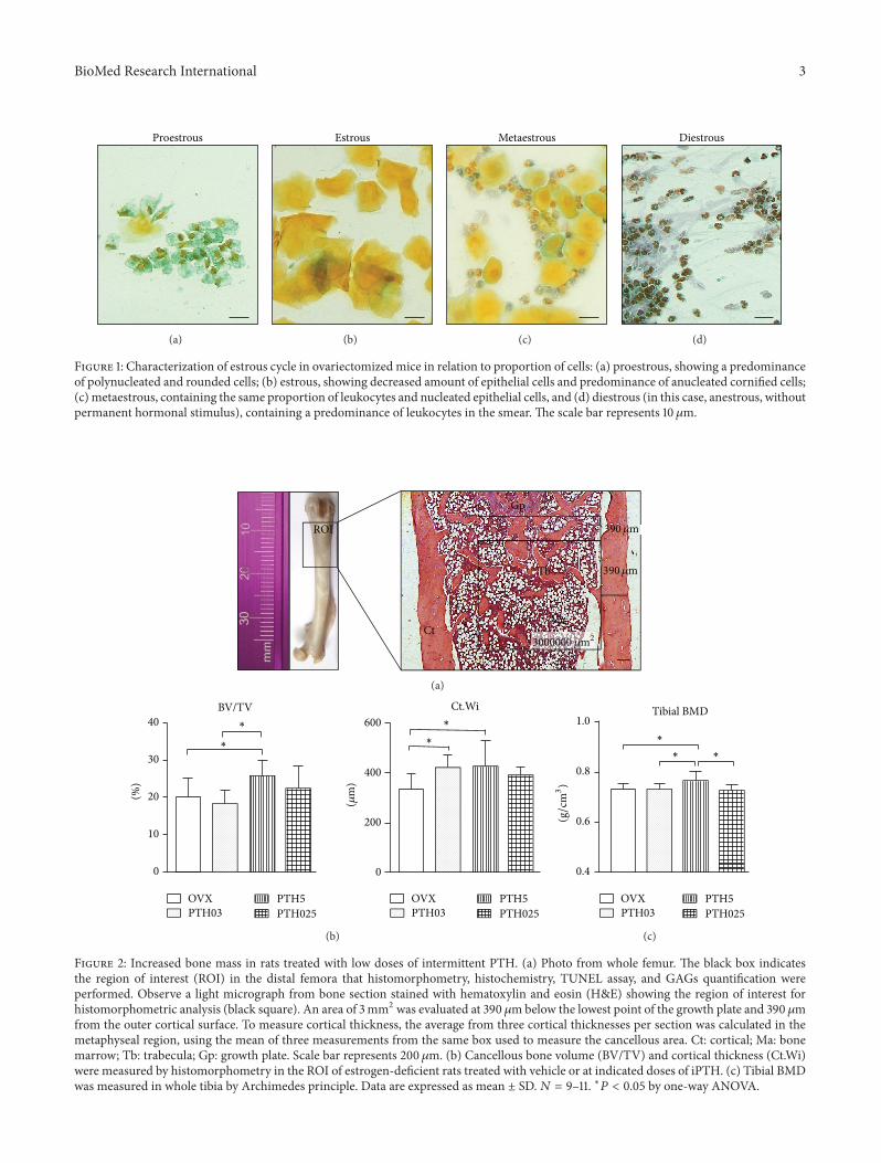
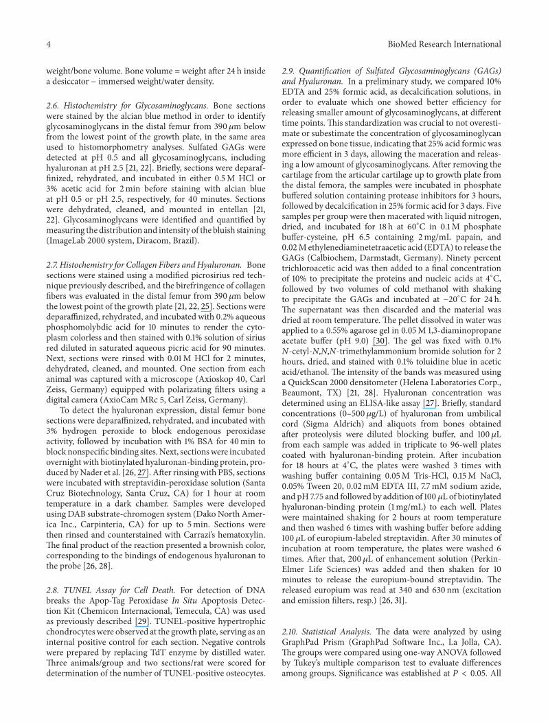
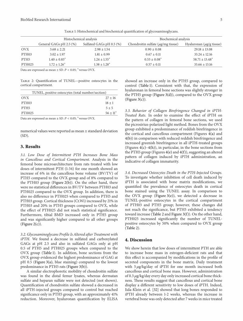
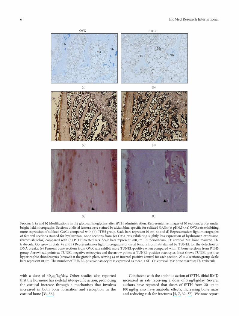
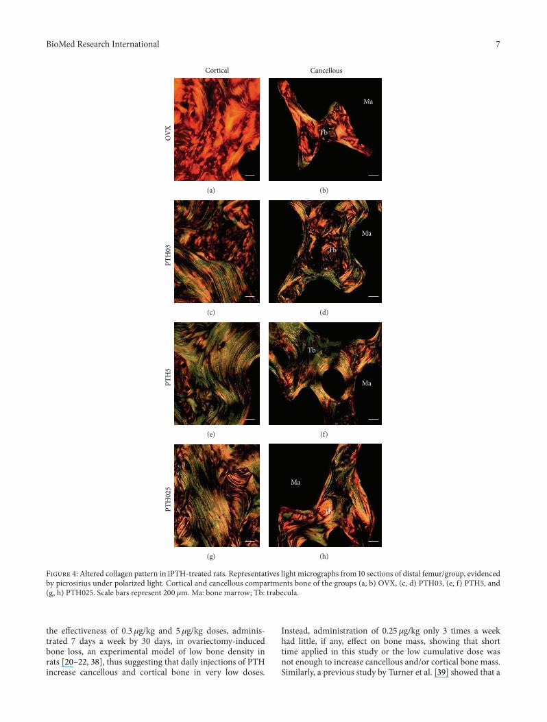