Microstructure, nanostructure and composition of the shell of Concholepas concholepas (Gastropoda,...
9
Microstructure, nanostructure and composition of the shell of Concholepas concholepas (Gastropoda, Muricidae) Yannicke Dauphin a, *, Nury Guzman b , Alain Denis a , Jean-Pierre Cuif a , Luc Ortlieb b a Département des sciences de la terre, bât. 504, Université Paris XI-Orsay, 91405 Orsay cedex, France b UR055 paléotropique, IRD, 32 av. H. Varagnat, 93143 Bondy cedex, France Received 29 November 2002; accepted 23 January 2003 Abstract Among the gastropods, some muricid shells are composed of an inner aragonitic crossed lamellar layer and a calcitic prismatic outer layer. The analysis of the structure and composition of the two layers of Concholepas shows that the crossed lamellar layer is similar to those of other Mollusc taxa. Sr, Mg and S contents are low in both layers. According to infrared spectrometry, the organic content of the outer calcitic layer is higher than that of the aragonitic crossed lamellar layer. The study of the nanostructure allows for the proposal of a new 3D interpretation of the fine structure of the crossed lamellar layer. The calcitic prismatic layer is compared with the outer calcitic prismatic layer of an archaeogastropod genus: Haliotis. © 2003 Éditions scientifiques et médicales Elsevier SAS and Ifremer/IRD/Inra/Cemagref. All rights reserved. Résumé Microstructure, nanostructure et composition de la coquille de Concholepas concholepas (Gastéropode, Muricidés). Parmi les Gastéropodes, quelques Muricidés ont une coquille formée par une couche interne aragonitique à structure lamellaire croisée, et une couche externe calcitique prismatique. L’analyse de la structure et de la composition des deux couches de la coquille de Concholepas montre que la couche lamellaire croisée est similaire à celle des autres Mollusques. Les teneurs en Sr, Mg et S sont faibles dans les deux couches. Selon les spectres infrarouges, le contenu en composants organiques est plus élevé dans la couche externe calcitique que dans la couche interne aragonitique. L’étude des nanostructures de la couche aragonitique a permis de proposer un nouveau modèle de la structure lamellaire croisée. La couche prismatique externe est comparée à celle d’un archéogastéropode : Haliotis. © 2003 Éditions scientifiques et médicales Elsevier SAS and Ifremer/IRD/Inra/Cemagref. Tous droits réservés. Keywords: Microstructure; Composition; Shell; Muricidae; Concholepas 1. Introduction Concholepas concholepas, also called south pacific aba- lone, is a carnivorous muricid gastropod, typical from the waters of Peru and Chile. There has long been a small local market for Loco (the Spanish name for Concholepas), and intertidal harvesting is still commonplace. The commercial export market began in 1976. Concholepas, the single most economically important shellfish in Chile, has been overex- ploited. Demand was so high that the government closed the fishery from 1989 to 1992. Another interest was due to the haemocyanin (an immunogenic protein), isolated and puri- fied from the lymph of Concholepas. Several projects includ- ing culture, population studies in protected zones and com- parison of larval behaviours in natural and artificial settlements have shown the role of human predation, climate and environmental factors on the abundance of Concholepas (Rodriguez et al., 1995; Moreno et al., 1998; Manriquez and Castilla, 2001; Poulin et al., 2002). It is well known that mollusk shells have a high environ- mental significance and provide information on physiologi- cal events (duration of spawning period, growth, etc.); more- over, they are clearly related with taxonomy and phylogeny. However, few data are available on the structure and compo- sition of the shell of Concholepas. * Corresponding author. E-mail address: [email protected] (Y. Dauphin). Aquatic Living Resources 16 (2003) 95–103 www.elsevier.com/locate/aquliv © 2003 Éditions scientifiques et médicales Elsevier SAS Ifremer/IRD/Inra/Cemagref. All rights reserved. DOI: 10.1016/S0990-7440(03)00022-6
Transcript of Microstructure, nanostructure and composition of the shell of Concholepas concholepas (Gastropoda,...

Microstructure, nanostructure and composition of the shellof Concholepas concholepas (Gastropoda, Muricidae)
Yannicke Dauphin a,*, Nury Guzman b, Alain Denis a, Jean-Pierre Cuif a, Luc Ortlieb b
a Département des sciences de la terre, bât. 504, Université Paris XI-Orsay, 91405 Orsay cedex, Franceb UR055 paléotropique, IRD, 32 av. H. Varagnat, 93143 Bondy cedex, France
Received 29 November 2002; accepted 23 January 2003
Abstract
Among the gastropods, some muricid shells are composed of an inner aragonitic crossed lamellar layer and a calcitic prismatic outer layer.The analysis of the structure and composition of the two layers of Concholepas shows that the crossed lamellar layer is similar to those of otherMollusc taxa. Sr, Mg and S contents are low in both layers. According to infrared spectrometry, the organic content of the outer calcitic layeris higher than that of the aragonitic crossed lamellar layer. The study of the nanostructure allows for the proposal of a new 3D interpretation ofthe fine structure of the crossed lamellar layer. The calcitic prismatic layer is compared with the outer calcitic prismatic layer of anarchaeogastropod genus: Haliotis.
© 2003 Éditions scientifiques et médicales Elsevier SAS and Ifremer/IRD/Inra/Cemagref. All rights reserved.
Résumé
Microstructure, nanostructure et composition de la coquille de Concholepas concholepas (Gastéropode, Muricidés). Parmi lesGastéropodes, quelques Muricidés ont une coquille formée par une couche interne aragonitique à structure lamellaire croisée, et une coucheexterne calcitique prismatique. L’analyse de la structure et de la composition des deux couches de la coquille de Concholepas montre que lacouche lamellaire croisée est similaire à celle des autres Mollusques. Les teneurs en Sr, Mg et S sont faibles dans les deux couches. Selon lesspectres infrarouges, le contenu en composants organiques est plus élevé dans la couche externe calcitique que dans la couche internearagonitique. L’étude des nanostructures de la couche aragonitique a permis de proposer un nouveau modèle de la structure lamellaire croisée.La couche prismatique externe est comparée à celle d’un archéogastéropode : Haliotis.
© 2003 Éditions scientifiques et médicales Elsevier SAS and Ifremer/IRD/Inra/Cemagref. Tous droits réservés.
Keywords: Microstructure; Composition; Shell; Muricidae; Concholepas
1. Introduction
Concholepas concholepas, also called south pacific aba-lone, is a carnivorous muricid gastropod, typical from thewaters of Peru and Chile. There has long been a small localmarket for Loco (the Spanish name for Concholepas), andintertidal harvesting is still commonplace. The commercialexport market began in 1976. Concholepas, the single mosteconomically important shellfish in Chile, has been overex-ploited. Demand was so high that the government closed thefishery from 1989 to 1992. Another interest was due to the
haemocyanin (an immunogenic protein), isolated and puri-fied from the lymph of Concholepas. Several projects includ-ing culture, population studies in protected zones and com-parison of larval behaviours in natural and artificialsettlements have shown the role of human predation, climateand environmental factors on the abundance of Concholepas(Rodriguez et al., 1995; Moreno et al., 1998; Manriquez andCastilla, 2001; Poulin et al., 2002).
It is well known that mollusk shells have a high environ-mental significance and provide information on physiologi-cal events (duration of spawning period, growth, etc.); more-over, they are clearly related with taxonomy and phylogeny.However, few data are available on the structure and compo-sition of the shell of Concholepas.
* Corresponding author.E-mail address: [email protected] (Y. Dauphin).
Aquatic Living Resources 16 (2003) 95–103
www.elsevier.com/locate/aquliv
© 2003 Éditions scientifiques et médicales Elsevier SAS Ifremer/IRD/Inra/Cemagref. All rights reserved.DOI: 10.1016/S0990-7440(03)00022-6
In the first extensive study of molluscan shell structures,(Boggild 1930) described and classified the main categoriesfrom mineralogical, crystallographic and microstructuralcharacters. The most widespread structure was the aragoniticcrossed lamellar layer. However, distinctive microstructuraland mineralogical features were noticed in different families.Some Muricidae showed an unusual association of severalaragonitic crossed lamellar sublayers and an outer thin layerof “longitudinal lamellae” (1930: p. 314). Despite this prom-ising beginning, studies were mainly directed towards bi-valve shells in subsequent decades (Taylor et al., 1969, 1973;Kobayashi, 1969, 1980; Kobayashi and Akai, 1994).(Petitjean, 1965) worked on the structural, mineralogical,and chemical characteristics of the Muricids, and was inter-ested in finding characters that could be useful in systemat-ics. His work included observations of ultra thin shell sec-tions, but no electron microscopy. The author recognized acalcitic “cortex” in various species, and an aragonitic “os-tracum”; he used the presence or absence of a calcitic cortexas a subfamilial taxonomic characteristic. For example, theRapaninae have a cortex, while the Muricinae do not. InC. concholepas, the calcitic cortex was always found, andthis species was thus assigned to the subfamily Rapaninae.
The aim of this paper is to study the micro- and nanostruc-tures, as well as the chemical composition of the shell ofgastropod C. concholepas by using microscopic, energydispersive spectroscopy (EDS) and infrared spectroscopy(FTIR) analyses.
2. Material and methods
2.1. Material
C. concholepas (Bruguière, 1789): class Gastropoda, Or-thogastropoda. Caenogastropoda, Sorbeoconcha, Hypsogas-tropoda, Neogastropoda.
Muricoidea Rafinesque, 1815; Muricidae Rafinesque,1815; Rapaninae Gray, 1853.
Samples of adult C. concholepas were collected by localfishermen in the Antofagasta area (Santa Maria Island, Chile)at a maximal depth of 5 m. The soft parts of the animal wereremoved and shells were rinsed with seawater, and then withtap water.
2.2. Methods
2.2.1. Light microscopyFirst insights were obtained by observing thin (~40 µm)
and ultra-thin (~5 µm) sections of longitudinal and transver-sal plans of shell ridges, with both normal and crossed nicolslight microscopy.
2.2.2. Scanning electron microscopyFractures and polished sections were prepared. Polished
sections and some fractures were etched with various acidsolutions and enzymes to reveal microstructural features.
Fixative and etching solutions such as a mixture of aceticacid 1% + glutaraldehyde 12% were used to preserve theorganic matrices. These matrices were partially destroyed byenzymes at 38 °C under a gentle rocking (Alcalase—Merck,1 mg/ml Protease—Sigma in HEPES pH 7.8, 1 mg/mlTrypsine—Sigma in HEPES pH 7.8) to reveal the shape ofmicrocrystals. Basic pH was used to minimize the dissolu-tion of the carbonate of the shell. Scanning electron micros-copy (SEM) observations were conducted with Philips SEM505 and XL30 microscopes housed at the Laboratoire degéologie, Université d’Orsay— Paris XI.
2.2.3. Atomic force microscopeScanning probe microscopy encompasses a family of
techniques that measures surface topography and propertiesat the atomic scale. The atomic force microscope (AFM)maps the topography of surface. In phase imaging, a variantof tapping mode, the phase lag of the cantilever oscillationrelative to the signal sent to the cantilver’s piezo driver isused as a basis for image generation. Phase images can begenerated as a consequence of variations in material proper-ties such as friction, viscoelasticity, and adhesion. AFMobservations were conducted with Digital Instruments(Veeco) Nanoscope III Dimension 3100 (Universitéd’Orsay), at room temperature and air. The probe consistedof a cantilever with integrated Si3N4 tips (Digital Instru-ments). Micron scale images were acquired by using thetapping mode. The procedures of the sample preparations aregiven in the figure legends.
2.2.4. Energy dispersive spectroscopyChemical analyses were done on an electron microprobe
by EDS. Energy dispersive X-ray microanalysis was under-taken with a Cambridge Stereoscan 200 SEM, equipped witha solid-state X-ray detector. Quantitative analysis was doneusing the Link AN10000 analysis system by a ZAF/PB pro-gram that estimates peak-to-background ratios (IRD,Bondy). Samples were embedded in resin and polished withdiamond pastes. The surface was then lightly etched in 5%formic acid for 15 s to reveal anystructural details within thesamples (e.g. different layers), so that the analysis positionscould be related to any observed structural features. Mea-surements were made using a lifetime of 100 s. Operatingconditions were an accelerating voltage of 15 kV. A cobaltstandard provided the instrument calibration.
Elements analysed were Ca, Na, Mg, Sr, Al, S, P, K, Fe,and Mn. Minimum of 10 analyses were made at variouslocations on each layer of each shell (5). These punctualanalyses have been averaged to obtain an individual mean.
2.2.5. Infrared spectrometryFirst, the epibionts found in the outer surface of the shells
were mechanically removed. Then the two layers of thecleaned C. concholepas shells were separated. The smallpieces obtained were immersed in 3% NaClO to removeorganic contaminants, rinsed with Milli-Q water, dried, and
96 Y. Dauphin et al. / Aquatic Living Resources 16 (2003) 95–103
ground into powder. The powdered samples were dried in anoven at 38 °C for one night. All spectra were recorded at4 cm–1 resolution with 64 scans using a strong Norton-Beerapodization on a Perkin Elmer model 1 600 Fourier trans-form infrared spectrometer (FTIR) from 4000 to 450 cm–1.The spectrometer was equipped with a diffuse reflectanceaccessory, which permits DRIFT measurements with highsensitivity on powders. All spectra were corrected by usingthe Kubelka-Munk function.
3. Observations
C. concholepas has a thick, round and white-gray shell(Fig. 1) (Guzman et al., 1998, 2001). The outer surface of theshell, often encrusted with epibionts such as small barnacles,shows strong lamellose ribs (Fig. 1A). The smooth andglossy inner surface of the shell is white (Fig. 1A).
3.1. Micro- and nanostructures
Thin sections show that the shell is composed of two mainlayers (Fig. 2a–d). The thickness of the shell near the apex isabout 1620 µm (calcite = 480 µm, aragonite = 1140 µm) and2250 µm near the pallial margin (calcite = 2130 µm, arago-nite = 120 µm). Depending on the presence/absence of theouter ridges, the thickness is variable. The calcitic outer layeris thin near the apex (Fig. 2a) but becomes thicker towardsthe pallial margin (Fig. 2c–d). The aragonitic inner layer issubdivided into several sublayers (Fig. 2a,e), and becomesvery thin near the growing edge of the shell (Fig. 2c–d).
Thin sections of the calcitic outer layer show numerousgrowth lines in large crystals (Fig. 2a, C–D), with longitudi-nal axes oblique or perpendicular to the shell surface(Fig. 2b). This structure has been called prismatic (Boggild,1930). These “prisms” do not show regular polygonal sec-tions, and they do not have regular diameter. In most of theshell, the size of the prisms increases from the outer surfaceto the crossed lamellar layer (Fig. 2a–d). The thin sectionsalso show that the extinctions of adjacent prisms are notsimultaneous under crossed nicols (Fig. 2a,b,d). The SEM
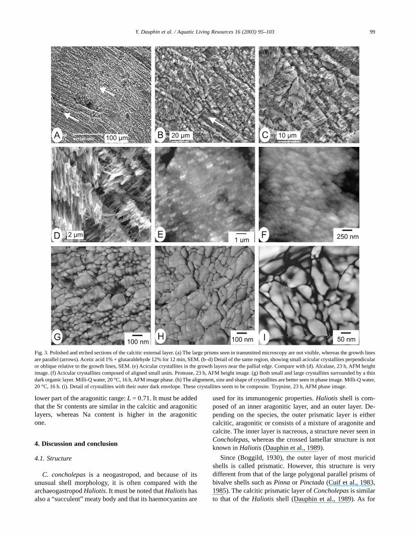
micrographs confirm that the growth lines are well developed(Fig. 3a,b), and the prismatic units are irregular (Fig. 3b,c).Prisms are composed of small acicular crystallites (lengthabout 2 µm) more or less perpendicular to the growth lines(Fig. 3c,d). AFM micrographs also show the irregularity ofthe components. Several etchings were used, but the acicularcrystallites are not regularly visible in height (=topographic)images (Fig. 3f–g). Phase images, more sensitive to thecomposition of the sample, are more explicit. Acicular crys-tallites are composed of smaller units, aligned in small rows(Fig. 3g,i). Each unit is surrounded by a thin dark layer inimage phase (Fig. 3i), whereas no layer is seen in a heightimage. It is suggested that the thin layer is composed oforganic matrix. The size of the smaller units varies from 15 to140 nm (Fig. 3g,i). Three-dimensional images of the calciticlayer show more or less regular alignment of the crystallites(Fig. 4a–b). According to the orientation of the crystallitesrelative to the sections, the rows are more or less visible. Theparallel rows in Fig. 3g are more visible in a 3D image(Fig. 4b).
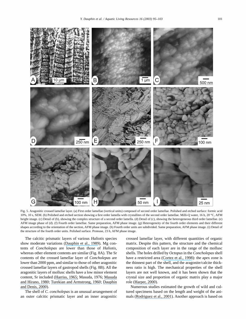
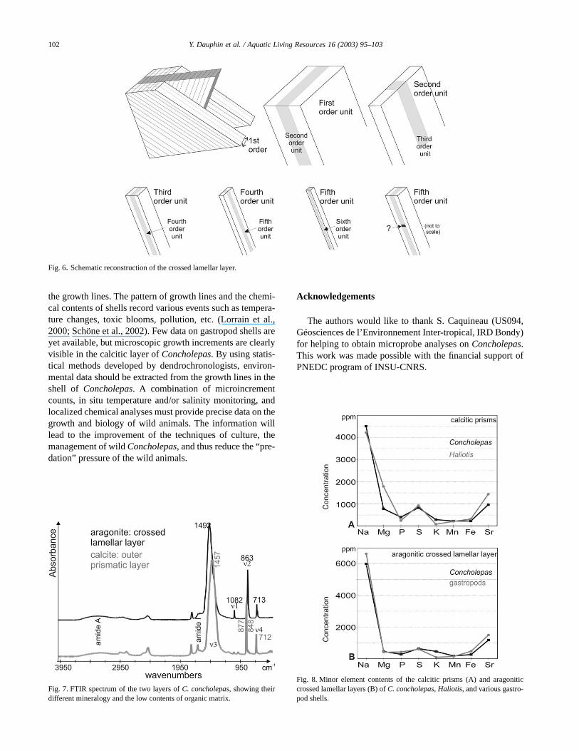
The aragonitic inner layer is composed of crossed lamellarsublayers (Fig. 2a,e). The basic structures of each sublayerare similar; only the orientation of the units changes, acommon feature of the crossed lamellar shells. The width ofthe first order lamellae is about 10 µm or more (Fig. 5a). Eachlamella is composed of stacked second order lamellae(Fig. 5a), which size is approximately 1.2 µm. The complexstructure of the second order lamella is visible in the SEM(Fig. 5a) and AFM (Fig. 5b,c) micrographs. The third orderunits have different orientations in two contiguous secondorder lamellae. Then third order lamellae are composed ofsmaller or fourth order elongated units: width 105 nm, length630 nm (Fig. 5d,e). Phase image shows the change of orien-tation of the fourth units in contiguous third order lamellae(Fig. 5e,g). 3D images of similar zones show the fourthlamellae as elongated crystallites (Fig. 4c–d). This change inorientation in contiguous units is a constant characteristic ofthe crossed lamellar structure (Fig. 6). Thus, according to thefracture plane, lamellae seem to be elongated or roundedunits (Fig. 5f,g). The fourth order lamella is also a composite
Fig. 1. Outer (A) and inner (B) views of the shell of C. concholepas.
97Y. Dauphin et al. / Aquatic Living Resources 16 (2003) 95–103

structure, as shown by the phase images (Fig. 5h–i). The darklayer surrounding the units may be indicative of an organiccomposition. The width of a granule is approximately 42 nm.
3.2. Bulk composition
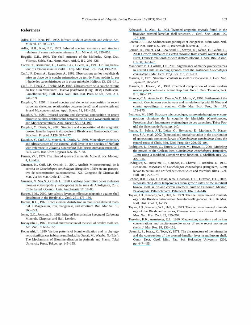
The infrared spectra of the calcite and aragonite groupsare characterized by three major bands attributed to thecarbonate radical CO3
2–: m3 at 1429 cm–1, the m2 doublet877–848 cm–1, and m4 at 713 cm–1 for the calcite group; m3 at1 471 cm–1 and two doublets: m2 at 877–848 cm–1 and m4 at713–700 cm–1 for the aragonite group (Adler and Kerr, 1962,1963; Farmer, 1974; Jones and Jackson, 1993). A compari-son between chemical compositions and FTIR data showsthat there is a linear correlation between the Mg and Srcontents and the wavelength of some FTIR bands. In arago-nite, there is a negative correlation between the m2 wave-lengths and the Sr contents, and a negative correlation be-tween m2 wavelengths and the Mg contents (Dauphin, 1997):a part of this doublet varies from 863.7 to 858.8 cm–1. Theother part (844 cm–1) of this doublet is not altered. In bio-genic calcite, there is a positive linear correlation between them4 wavelength and Mg concentrations (Dauphin, 1999).
The spectra of the powdered outer layer confirm the cal-citic mineralogy (Fig. 7): the m2 doublet 877–848 cm–1, m1 at1013 cm–1 and m4 at 713 cm–1. However, the m3 band issubdivided: 1457, 1445 and 1436 cm–1. Other bands usuallypresent in calcite are also visible: 1176 and 1084 cm–1. The
m2 wavelength is indicative of low Mg and Sr contents of thelayer (<1000 ppm). Several other bands may be attributed tothe organic matrix: 3274 cm–1 band may be assigned toamide A, 1685–1653–1636 cm–1 bands to the amide I.
The thin inner layer is aragonitic (Fig. 7). However, the m3band is also subdivided (1492 and 1458 cm–1), and only oneband of the m2 is seen at 863 cm–1 and m1 is seen at 1083cm–1. This wavelength is indicative of low Sr (<2500 ppm)and Mg (<800 ppm) contents. The amide A band is at 3310cm–1, and the amide I region is composed of two bands (1654and 1636 cm–1).
To estimate the organic/mineral ratio from FTIR spectra,two ratios are used: amide I/860, and amide A/860 for arago-nitic layers (Dauphin and Denis, 2000). The first ratio isequal to 0.14 and the second one to 0.08 in the crossedlamellar layer. In calcitic layers, the 878 cm–1 band was usedand the ratios are 0.23 and 0.07, respectively. According tothese data, the organic content of the outer calcitic layer ishigher than that of the aragonitic crossed lamellar layer.
3.3. Chemical composition (EDS data)
The outer prismatic calcitic layer shows a low minorelement content, including Mg (Fig. 8). In fact, Mg content islower than Sr. K and Mn contents, usually correlated withcalcite, are also low. Aragonitic crossed lamellar layers areknown to have low minor element contents, and Concholepasis not different (Fig. 8). The Loreau ratio (1982) is in the
Fig. 2. (a–f) Thin sections of C. concholepas. (a) External calcitic layer (P) and crossed lamellar layer (CL) near the apex. Growth lines are present in theprismatic layer (dotted arrows). Crossed nicols. (b) External calcitic layer (P) and crossed lamellar layer (CL) in the middle region of the shell. The prisms arelarge in the inner part of the layer and small in the outer part. Crossed nicols. (c) External calcitic layer (P) and crossed lamellar layer (CL) near the pallial edge.(d) Idem, crossed nicols. (e) Detail of the sublayers of the crossed lamellar layer. Crossed nicols. (f) Haliotis: outer calcitic prismatic layer (P) and aragoniticnacreous layer (N). Crossed nicols.
98 Y. Dauphin et al. / Aquatic Living Resources 16 (2003) 95–103
lower part of the aragonitic range: L = 0.71. It must be addedthat the Sr contents are similar in the calcitic and aragoniticlayers, whereas Na content is higher in the aragoniticone.
4. Discussion and conclusion
4.1. Structure
C. concholepas is a neogastropod, and because of itsunusual shell morphology, it is often compared with thearchaeogastropod Haliotis. It must be noted that Haliotis hasalso a “succulent” meaty body and that its haemocyanins are
used for its immunogenic properties. Haliotis shell is com-posed of an inner aragonitic layer, and an outer layer. De-pending on the species, the outer prismatic layer is eithercalcitic, aragonitic or consists of a mixture of aragonite andcalcite. The inner layer is nacreous, a structure never seen inConcholepas, whereas the crossed lamellar structure is notknown in Haliotis (Dauphin et al., 1989).
Since (Boggild, 1930), the outer layer of most muricidshells is called prismatic. However, this structure is verydifferent from that of the large polygonal parallel prisms ofbivalve shells such as Pinna or Pinctada (Cuif et al., 1983,1985). The calcitic prismatic layer of Concholepas is similarto that of the Haliotis shell (Dauphin et al., 1989). As for
Fig. 3. Polished and etched sections of the calcitic external layer. (a) The large prisms seen in transmitted microscopy are not visible, whereas the growth linesare parallel (arrows). Acetic acid 1% + glutaraldehyde 12% for 12 min, SEM. (b–d) Detail of the same region, showing small acicular crystallites perpendicularor oblique relative to the growth lines, SEM. (e) Acicular crystallites in the growth layers near the pallial edge. Compare with (d). Alcalase, 23 h, AFM heightimage. (f) Acicular crystallites composed of aligned small units. Protease, 23 h, AFM height image. (g) Both small and large crystallites surrounded by a thindark organic layer. Milli-Q water, 20 °C, 16 h, AFM image phase. (h) The alignment, size and shape of crystallites are better seen in phase image. Milli-Q water,20 °C, 16 h. (i). Detail of crystallites with their outer dark envelope. These crystallites seem to be composite. Trypsine, 23 h, AFM phase image.
99Y. Dauphin et al. / Aquatic Living Resources 16 (2003) 95–103

Concholepas, the size of the prisms decreases from the innerpart to the outer part of the layer (Fig. 2f).
The structure of the crossed lamellar layer is similar inbivalve and gastropod shells, and several 3D graphs havebeen proposed since Boggild (1930). The width of the firstorder lamellae varies from 10 to 30 µm. The minimal lengthof third order units is about 5 µm, and their width is between0.3 and 2 µm (Uozumi et al., 1971). The various units of theConcholepas layer show similar sizes. Uozumi et al. (1971:p. 452) noticed that the third order lamella are subdivided insmaller units “parallel to the longitudinal axes of the crys-tals”. They also described oblique regular dark stripes, sup-posed “to reflect the lattice structures of aragonite crystals”.However, these minute structures are not illustrated. Accord-ing to the SEM observations, it has been supposed that a thirdorder lamella was divided in small fourth order cubes (Cuif etal., 1985; Dauphin and Denis, 2000). However, our AFMobservations have shown that the third order lamellae arelongitudinally divided, as are the fourth and fifth order units
(Fig. 6). Each subdivision is perpendicular to the previousone as seen in a transverse plane, but parallel to the longitu-dinal axis of the unit. The question of a transverse striation allalong the length of the fifth or sixth order units is not yetresolved.
4.2. Composition
FTIR analyses on mollusc shells are scarce, and no dataare available on calcitic prismatic structures similar to theouter layer of Concholepas. FTIR spectra of the crossedlamellar layers of the gastropods Cypraea, Phalium andStrombus are similar and are also indicative of low Sr and Mgcontents. However, these genera differ in their organic bands:the ratios calculated from FTIR spectra to estimate the or-ganic contents vary from 0.20 to 0.33 (amide I/860), andfrom 0.07 to 0.19 (amide A/860) (Dauphin and Denis, 2000).Thus, the organic/mineral ratio of the crossed lamellar layerof Concholepas seems low, despite the paucity of data.
Fig. 4. (a–b) Polished and etched section showing the size and arrangement of the small acicular calcitic crystallites. Milli-Q water, 20 °C, 16 h. (c–d) Samesample, showing the various shapes of the third order units of the aragonitic crossed lamellar layer according to the sections, and their complex internal structure.AFM image phase.
100 Y. Dauphin et al. / Aquatic Living Resources 16 (2003) 95–103
The calcitic prismatic layers of various Haliotis speciesshow moderate variations (Dauphin et al., 1989). Mg con-tents of Concholepas are lower than those of Haliotis,whereas other element contents are similar (Fig. 8A). The Srcontents of the crossed lamellar layer of Concholepas arelower than 2000 ppm, and similar to those of other aragoniticcrossed lamellar layers of gastropod shells (Fig. 8B). All thearagonitic layers of mollusc shells have a low minor elementcontent, Sr included (Harriss, 1965; Masuda, 1976; Masudaand Hirano, 1980; Turekian and Armstrong, 1960; Dauphinand Denis, 2000).
The shell of C. concholepas is an unusual arrangement ofan outer calcitic prismatic layer and an inner aragonitic
crossed lamellar layer, with different quantities of organicmatrix. Despite this pattern, the structure and the chemicalcomposition of each layer are in the range of the molluscshells. The holes drilled by Octopus in the Concholepas shellhave a restricted area (Cortez et al., 1998): the apex zone isthe thinnest part of the shell, and the aragonite/calcite thick-ness ratio is high. The mechanical properties of the shelllayers are not well known, and it has been shown that thecrystal size and proportion of organic matrix play a majorrole (Harper, 2000).
Numerous studies estimated the growth of wild and cul-tured specimens based on the length and weight of the ani-mals (Rodriguez et al., 2001). Another approach is based on
Fig. 5. Aragonitic crossed lamellar layer. (a) First order lamellae (vertical units) composed of second order lamellae. Polished and etched surface: formic acid10%, 10 s, SEM. (b) Polished and etched section showing a first order lamella with crystallites of the second order lamellae. Milli-Q water, 16 h, 20 °C, AFMheight image. (c) Detail of (b), showing the complex structure of a second order lamella. (d) Detail of (c), showing the heterogeneous third order lamellae. (e)AFM image phase of (d). (f) Fourth order lamellae. Same preparation, AFM phase image. (g) Heterogeneity of the fourth order elements and their differentshapes according to the orientation of the section, AFM phase image. (h) Fourth order units are subdivided. Same preparation, AFM phase image. (i) Detail ofthe structure of the fourth order units. Polished surface. Protease, 23 h, AFM phase image.
101Y. Dauphin et al. / Aquatic Living Resources 16 (2003) 95–103
the growth lines. The pattern of growth lines and the chemi-cal contents of shells record various events such as tempera-ture changes, toxic blooms, pollution, etc. (Lorrain et al.,2000; Schöne et al., 2002). Few data on gastropod shells areyet available, but microscopic growth increments are clearlyvisible in the calcitic layer of Concholepas. By using statis-tical methods developed by dendrochronologists, environ-mental data should be extracted from the growth lines in theshell of Concholepas. A combination of microincrementcounts, in situ temperature and/or salinity monitoring, andlocalized chemical analyses must provide precise data on thegrowth and biology of wild animals. The information willlead to the improvement of the techniques of culture, themanagement of wild Concholepas, and thus reduce the “pre-dation” pressure of the wild animals.
Acknowledgements
The authors would like to thank S. Caquineau (US094,Géosciences de l’Environnement Inter-tropical, IRD Bondy)for helping to obtain microprobe analyses on Concholepas.This work was made possible with the financial support ofPNEDC program of INSU-CNRS.
Fig. 6. Schematic reconstruction of the crossed lamellar layer.
Fig. 7. FTIR spectrum of the two layers of C. concholepas, showing theirdifferent mineralogy and the low contents of organic matrix.
Fig. 8. Minor element contents of the calcitic prisms (A) and aragoniticcrossed lamellar layers (B) of C. concholepas, Haliotis, and various gastro-pod shells.
102 Y. Dauphin et al. / Aquatic Living Resources 16 (2003) 95–103
References
Adler, H.H., Kerr, P.F., 1962. Infrared study of aragonite and calcite. Am.Mineral. 47, 700–717.
Adler, H.H., Kerr, P.F., 1963. Infrared spectra, symmetry and structurerelations of some carbonate minerals. Am. Mineral. 48, 839–853.
Boggild, O.B., 1930. The shell structure of the Mollusks. Kong. Dsk.Vidensk. Selsk. Skr., Natur. Math. Afd. 9, II 2, 230–326.
Cortez, T., Bernardino, G., Castro, B.G., Guerra, A., 1998. Drilling behav-iour of Octopus mimus Gould. J. Exp. Mar. Biol. Ecol. 224, 199–203.
Cuif, J.P., Denis, A., Raguideau, A., 1983. Observations sur les modalités demise en place de la couche prismatique du test de Pinna nobilis L. parl’étude des caractéristiques de la phase minérale. Haliotis 13, 131–141.
Cuif, J.P., Denis, A., Triclot, M.P., 1985. Ultrastructure de la couche externedu test d’un Veneracea: Dosinia ponderosa (Gray, 1838) (Mollusque,Lamellibranche). Bull. Mus. Natl. Hist. Nat. Paris 4è sér., Sect. A 4,741–759.
Dauphin, Y., 1997. Infrared spectra and elemental composition in recentcarbonate skeletons: relationships between the n2 band wavelength andSr and Mg concentrations. Appl. Spectr. 51, 141–152.
Dauphin, Y., 1999. Infrared spectra and elemental composition in recentbiogenic calcites: relationships between the n4 band wavelength and Srand Mg concentrations. Appl. Spectr. 53, 184–190.
Dauphin, Y., Denis, A., 2000. Structure and composition of the aragoniticcrossed lamellar layers in six species of Bivalvia and Gastropoda. Comp.Biochem. Physiol. A126, 367–377.
Dauphin, Y., Cuif, J.P., Mutvei, H., Denis, A., 1989. Mineralogy, chemistryand ultrastructure of the external shell-layer in ten species of Haliotiswith reference to Haliotis tuberculata (Mollusca: Archaeogastropoda).Bull. Geol. Inst. Univ. Uppsala N.S. 15, 7–38.
Farmer, V.C., 1974. The infrared spectra of minerals. Mineral. Soc. Monogr.4, London.
Guzman, N., Cuif, J.P., Ortlieb, L., 2001. Analisis Microestructural de laconcha de Concholepas concholepas (Bruguière 1789) en una perspec-tiva de reconstitucion paleoambiental. XXI Congreso de Ciencias delMar, Via del Mar. Chile 47, 1789.
Guzman, N., Saa, S., Ortlieb, L., 1998. Catalogo descriptivo de los moluscoslitorales (Gastropoda y Pelecypoda) de la zona de Antofagasta, 23 S,Chile. Estud. Oceanol. Univ. Antofagasta 17, 17–86.
Harper, E.M., 2000. Are calcitic layers an effective adaptation against shelldissolution in the Bivalvia? J. Zool. 251, 179–186.
Harriss, R.C., 1965. Trace element distribution in molluscan skeletal mate-rial. I. Magnesium, iron, manganese, and strontium. Bull. Mar. Sci. 15,265–273.
Jones, G.C., Jackson, B., 1993. Infrared Transmission Spectra of CarbonateMinerals. Chapman and Hall, London.
Kobayashi, I., 1969. Internal microstructure of the shell of bivalve molluscs.Am. Zool. 9, 663–672.
Kobayashi, I., 1980. Various patterns of biomineralization and its phyloge-netic significances in bivalve mollusks. In: Omori, M., Watabe, N. (Eds.),The Mechanisms of Biomineralization in Animals and Plants. TokaiUniversity Press, Tokyo, pp. 145–155.
Kobayashi, I., Akai, J., 1994. Twinned aragonite crystals found in thebivalvian crossed lamellar shell structure. J. Geol. Soc. Japan 100,177–180.
Loreau, J.P., 1982. Sédiments aragonitiques et leur genèse. Mém. Mus. Natl.Hist. Nat. Paris N.S., sér. C, sciences de la terre 47, 1–312.
Lorrain, A., Paulet, Y.M., Chauvaud, L., Savoye, N., Nézan, E., Guérin, L.,2000. Growth anomalies in Pecten maximus from coastal waters (Bay ofBrest, France): relationships with diatoms blooms. J. Mar. Biol. Assoc.UK 80, 667–673.
Manriquez, P.H., Castilla, J.C., 2001. Significance of marine protected areasin central Chile as seeding grounds from the gastropod Concholepasconcholepas. Mar. Ecol. Prog. Ser. 215, 201–211.
Masuda, F., 1976. Strontium contents in shell of Glycymeris. J. Geol. Soc.Japan 82, 565–572.
Masuda, F., Hirano, M., 1980. Chemical composition of some modernmarine pelecypod shells. Scient. Rep. Inst. Geosc. Univ. Tsukuba, Sect.B 1, 163–177.
Moreno, C.A., Asencio, G., Duarte, W.E., Marin, V., 1998. Settlement of themuricid Concholepas concholepas and its relationship with El Nino andcoastal upwellings in southern Chile. Mar. Ecol. Prog. Ser. 167,171–175.
Petitjean, M., 1965. Structure microscopique, nature minéralogique et com-position chimique de la coquille de Muricidés (Gastéropodes,Prosobranches). Importance systématique de ces caractères. Thèse Fac-ulté des Sciences de Paris. pp. 140.
Poulin, E., Palma, A.T., Leiva, G., Hernadez, E., Martinez, P., Navar-rete, S.A., et al., 2002. Temporal and spatial variation in the distributionof epineustonic competent larvae of Concholepas concholepas along thecentral coast of Chile. Mar. Ecol. Prog. Ser. 229, 95–104.
Rodriguez, L., Daneri, G., Torres, C., Leon, M., Bravo, L., 2001. Modelingthe growth of the Chilean loco, Concholepas concholepas (Bruguière,1789) using a modified Gompertz-type function. J. Shellfish Res. 20,309–315.
Rodriguez, S., Riquelme, C., Campos, E., Chavez, P., Brandan, E., 1995.Behavioral responses of Concholepas concholepas (Bruguière, 1789)larvae to natural and artifical settlement cues and microbial films. Biol.Bull. 189, 272–279.
Schöne, B.R., Lega, J., Flessa, K.W., Goodwin, D.H., Dettman, D.L., 2002.Reconstructing daily temperatures from growth rates of the intertidalbivalve mollusk Chione cortezi (northern Gulf of California. Mexico.Palaeogeogr. Palaeoclimatol. Palaeoecol. 184, 131–146.
Taylor, J.D., Kennedy, W.J., Hall, A., 1969. The shell structure and mineral-ogy of the Bivalvia. Introduction. Nuculacae–Trigonacae. Bull. Br. Mus.Natl. Hist. Zool. 3, 1–125.
Taylor, J.D., Kennedy, W.J., Hall, A., 1973. The shell structure and mineral-ogy of the Bivalvia–Lucinacea, Clavagellacea, conclusions. Bull. Br.Mus. Natl. Hist. Zool. 22, 255–294.
Turekian, K.K., Armstrong, R.L., 1960. Magnesium, strontium and bariumconcentrations and calcite-aragonite ratios of some recent molluscanshells. J. Mar. Res. 18, 133–151.
Uozumi,, S., Iwata,, K., Togo, Y., 1971. The ultrastructure of the mineral inand the construction of the crossed-lamellar layer in molluscan shell.Contr. Dept. Geol. Min., Fac. Sci. Hokkaido University 1236,pp. 447–455.
103Y. Dauphin et al. / Aquatic Living Resources 16 (2003) 95–103


![Палеогеновые архитектонициды (Gastropoda) юга СССР / Paleogene architectonicids (Gastropoda) of southern USSR [In Russian]](https://static.fdokumen.com/doc/165x107/631f3e30198185cde201015e/paleogenovie-arkhitektonitsidi-gastropoda-yuga-sssr.jpg)