Federico Bellomi e l'incisione - Federico Bellomi and etching
Upload
khangminh22Category
view
7download
0
Etching to Reveal Microstructure
George F. Vander Voort
Buehler Ltd
Lake Bluff, Illinois USA

Guidelines
• Low magnification evaluation; deeper etch• Time is less reliable than appearance• Best results occur when etching right after
polishing, time can create a passive surface• High magnification evaluation; shallow etch • If you are not satisfied, an etch-repolish-etch
sequence may improve the results

Etching
• Techniques– Swab– Immersion– Electrolytic
• To Reveal– Grain boundaries– Constituents– Banding, segregation– Deformation
100 µm

Etching Reveals
• Grain boundaries• Phases• Constituents• Homogeneity• Coatings and Platings• Interfaces• Heat affected zones• Reaction zones
• Dendritic patterns• Segregation• Deformation

Techniques
• Swab
• Immersion
• Electrolytic
Attack controlled by chemical selection, time, current, voltage

Etchant Selection
Low-carbon sheet steel
2% NitalReveals ferrite grain boundaries and cementite
4% PicralReveals cementite
Beraha’s reagentColored grains based on crystallographic orientation

4% Picral 2% Nital
Low-carbon sheet steel (Fe – 0.05% C – 0.08% Mn – 0.20% Si) etched with picral and nital. Picral reveals the fine cementite particles and patches of pearlite. Nital reveals the
ferrite grain boundaries and the pearlite patches, but the fine cementite particles are hard to see, especially if the are at grain boundaries. Originals at 500X.

4% Picral 2% Nital
Low-carbon sheet steel etched with picral and nital. Picral reveals the fine cementite particles and patches of pearlite. Nital reveals the ferrit grain boundaries and the pearlite patches, but the fine cementite particles are hard to see, especially if the are at grain boundaries. Originals at 500X.

4% Picral 2% Nital
Microstructure of hot-rolled Fe – 0.11% C – 0.85% Mn – 0.21% Si etched with picral and nital. Picral reveals the cementite and pearlite, but not the ferrite grain boundaries. Nital reveals the
ferrite grain boundaries and the pearlite, but the fine cementite particles are difficult to see. Originals at 500X.

4% Picral 2% Nital
Microstructure of hot-rolled Fe – 0.12% C – 0.23% Mn – 0.17% Si – 0.74% Mo revealing a ferrite-pearlite microstructure with small patches of martensite (arrows). The many small black particles
are small patches of pearlite, not inclusions. Originals at 1000X.

4% Picral 2% Nital
Microstructure of hot-rolled Fe – 0.16% C – 1.26% Mn – 0.22% Si – 0.15% Cr etched with picral and nital. Picral reveals the cementite and pearlite, but not the ferrite grain boundaries. Nital
reveals the ferrite grain boundaries and the pearlite, but the fine cementite particles are difficult to see. Originals at 500X.

2% NitalFerritePearlite
Aqueous 10% Sodium Metabisulfite
Microstructure of an as-rolled, continuously cast HSLA steel (Fe – 0.19% C – 1.24% Mn – 0.37% Si –0.08% V) containing segregation (and some cracks – yellow arrows at left) etched with nital (left in
bright field) and aqueous 10% Na2S2O5 (right in polarized light plus sensitive tint). The normal structure is ferrite and pearlite but bainite (yellow-green arrows, both images) is observed in the
segregated regions (greater hardenability). Average hardness values were 180, 260 and 325 HV for the ferrite, pearlite and bainite streaks, respectively. The magnification bars are 50 µm.

Aqueous 10% Na2S2O3 + 3% K2S2O5 Klemm’s I
Microstructure of an as-rolled, continuously-cast HSLA steel (Fe – 0.19% C – 1.24% Mn – 0.37% Si– 0.08% V) containing segregation etched with Beraha’s “10/3” etch (left) and with Klemm’s I
(right), both images in polarized light plus sensitive tint. The normal structure is ferrite and pearlite but bainite (yellow-green arrows, both images) is observed in the segregated regions. The segregated
regions are hard to detect using Klemm’s I as it darkens the ferrite in the bainite as heavily as the matrix ferrite. The magnification bars are 50 µm long.

Aqueous 3% K2S2O5 + 1% H2NSO3H
Microstructure of an as-rolled, continuously-cast HSLA steel (Fe – 0.19% C – 1.24% Mn – 0.37% Si –0.08% V) containing segregation etched with Beraha’s sulfamic acid etch (No. 1) and viewed with
polarized light plus sensitive tint. The normal structure is ferrite and pearlite but bainite (yellow-green arrows) is observed in the segregated regions. The segregated regions are easier to detect with this etch
than using Klemm’s I (previous slide), but 10% sodium metabisulfite and Beraha’s “10/3” reagents were better. The magnification bar is 50 µm long.

Weld HAZ Base Metal
Montage showing the structure of a large weld in a carbon steel as revealed using 2% nital. The heat-affected zone is larger than usual. Note that the grain size and shape change dramatically from the fusion line (arrows)
to the base metal. The originals are at 50X magnification in bright field illumination.

Weld HAZ Base
Montage showing the structure of a large weld in a carbon steel as revealed by Klemm’s reagent. The heat-affected zone is larger than usual. Note that the grain size and shape change dramatically from the fusion
line (arrows) to the base metal. The originals are at 50X magnification in polarized light plus sensitive tint. The magnification bar is 200 µm long.

4% Picral 2% Nital
Microstructure of hot-rolled Fe – 0.23% C – 0.85% Mn – 0.22% Si revealing a moderately banded structure of ferrite and pearlite. Originals at 500X. The hot working axis is horizontal. The arrow (right picture) points to a large MnS inclusion. Note that it is quite malleable and has been highly
elongated by hot rolling.

4% Picral 2% Nital
Microstructure of hot-rolled Fe – 0.31% C – 0.84% Mn – 0.29% Si steel revealing a banded ferrite-pearlite structure (hot rolling direction is horizontal). The arrows point to some of the highly
elongated MnS inclusions. Originals at 500X.

4% Picral 2% Nital
Microstructure of hot-rolled Fe – 0.29% C – 0.40% Mn – 0.16% Si – 2.88% Ni revealing a slightly banded ferrite-pearlite microstructure. The arrows point to highly elongated manganese
sulfide, MnS, inclusions. Originals at 500X.

4% Picral 2% Nital
Microstructure of hot-rolled Fe – 0.30% C – 0.73% Mn – 0.23% Si – 0.97% Cr alloy steel revealing a banded ferrite-pearlite structure. Originals at 500X.

Vilella’s Reagent 2% Nital
Microstructure of quenched and tempered (treatment not known) Fe – 0.30% C – 0.53% Mn –0.17% Si – 0.79% Cr – 3.34% Ni – 0.50% Mo alloy steel with a tempered lath martensite matrix.
Originals at 1000X.

4% Picral 2% Nital
Microstructure of quenched and tempered (treatment unknown) Fe – 0.32% C – 0.31% Mn –0.16% Si – 1.37% Cr – 4.12% Ni alloy steel revealing a tempered lath martensite matrix.
Originals at 1000X.

2% Nital4% Picral
Microstructure of annealed 1040 carbon steel (0.40% C – 0.68% Mn – 0.12% Si) revealing ferrite and pearlite. Originals are at 1000X.

4% Picral 2% Nital
Microstructure of annealed (871 °C, 1600 °F – 1 hour, slow cool) SAE 4140 alloy steel (Fe – 0.4% C – 0.85% Mn – 0.95% Cr – 0.2% Mo) revealing proeutectoid ferrite and coarse lamellar pearlite.
Note that nital, because it is sensitive to crystal orientation, does not reveal the cementite lamellae well in all packets (arrows point to examples). Originals at 1000X.

2% Nital 4% Picral
Upper bainite (dark or outlined) and as-quenched martensite (gray or white) in 5160 alloy steel (Fe – 0.6% C - 0.85% Mn – 0.25% Si – 0.8% Cr) that was austenitized at 830 °C (1525 °F) for 30 min., isothermally held at 538 °C (1000 F°) for 30 sec to partially transform the austenite,
and then water quenched (untransformed austenite forms martensite).

2% Nital 4% Picral
Microstructure of 4140 alloy steel (Fe – 0.4% C – 0.9% Mn – 0.2% Si – 1% Cr – 0.2% Mo) austenitized at 843 °C (1550 °F), isothermally transformed at 566 °C (1050 °F) for 15 min., and water quenched, to partially transform austenite to upper bainite (arrow in segregation streak
shows the initiation of bainitic ferrite surrounded by martensite; the untransformed austenite was converted to martensite). 4% picral etch does not reveal the bainitic ferrite surrounded by
martensite as well as nital.

2% Nital 4% Picral
Lower bainite (dark) and as-quenched martensite (white/gray) in 5160 alloy steel (Fe – 0.6% C -0.85% Mn – 0.25% Si – 0.8% Cr) that was austenitized at 830 °C (1525 °F) for 30 min.,
isothermally held at 343 °C (650 F°) for 20 minutes to partially transform the austenite, and then water quenched (untransformed austenite forms martensite).

2% Nital 4% Picral
4140 alloy steel (Fe – 0.4% C – 0.9% Mn – 0.2% Si – 1% Cr – 0.2% Mo) austenitized at 843 °C (1550 °F), isothermally transformed at 413 °C (775 °F) for 30 sec., and water quenched, to partially transform austenite to lower bainite (surrounded by martensite; the untransformed austenite was
converted to martensite).

4% Picral 2% Nital
Microstructure of hot-rolled Fe – 0.68% C – 0.84% Mn – 0.33% Si revealing a nearly fully pearlitic structure. Originals at 1000X.

2% Nital4% Picral
Microstructure of hot-rolled Fe – 0.94% C – 0.51% Mn – 0.32% Si – 1.34% Cr alloy steel revealing a fully pearlitic matrix. Picral revealed a network of cementite in the prior-austenite
grain boundaries (arrows). This is not visible using nital. Originals at 1000X.

2% Nital4% Picral
Microstructure of as-rolled Fe – 1.31% C – 0.35% Mn – 0.25% Si high-carbon water hardenable tool steel. Note the Widmanstätten intragranular cementite that precipitated as pro-
eutectoid cementite before the eutectoid reaction. Originals at 1000X.

Microstructure of the as-rolled Fe – 1.31% C – 0.35% Mn – 0.25% Si specimen with the intergranular carbide network clearly visible after etching with alkaline sodium picrate, 90 °C –60 s. Original at 500X magnification. Note also some intragranular Widmanstätten cementite.

Beraha’s Sulfamic Acid Reagent Klemm’s I Reagent
Color etching of the as-rolled hypereutectoid Fe-1.31% C – 0.35% Mn – 0.25% Si specimen clearly revealed the intergranular cementite films. Beraha’s sulfamic acid etch (100 mL
water, 3 g K2S2O5 and 2 g NH2SO3H) (left) and Klemm’s I reagent (right) were used. Original magnifications were 500X. Taken with polarized light and sensitive tint.

Carburized 8620 – Lower Bainite Case/Lath Martensite Core
Polarized Light + Sensitive Tint
Bainitic Case
Bright Field
Microstructure of the case of carburized (0.95% C potential) 8620 alloy steel. It was carburized at 1750 °F, then quenched into a 50/50 mix of sodium nitrite and potassium
nitrate at 480 °F and held 120 minutes. It was air cooled and then tempered at 480 °F for 240 minutes to an aim case hardness of 52-60 HRC (etched with 10% sodium metabisulfite).
The lower bainite case performs better under low-cycle fatigue conditions.

Carburized 8620 – Lower Bainite Case/Lath Martensite Core
Lath Martensite Core
Bright Field Polarized Light + Sensitive Tint
Lath martensitic core structure, with some ferritic areas, revealed using 10% sodium metabisulfite.

Carburized 8620 – Lower Bainite Case/Lath Martensite Core
Case – Lower Bainite Core – Lath Martensite
Case and core structure revealed using 2% nital.

Vilella’s Reagent 2% Nital
Microstructure of spheroidize annealed T1 high speed steel (0.72% C – 0.25% Mn – 0.2% Si –4% Cr – 1.15% V – 18.25% W) revealing spheroids of alloy carbides in a ferritic matrix. Etching with nital, which is sensitive to crystal orientation, did not reveal the spheroids well in all of the
ferrite grains (arrows indicated examples). Originals at 1000X.

Aqueous 10% Na2S2O5 Vilella’s Reagent
C
Microstructure of Ni-Hard cast iron (Fe – 3.3% C – 0.9% Mn – 0.9% Si – 1.8% Cr – 4.4% Ni – 0.4% Mo) revealing massive cementite (C) and patches of plate martensite and retained austenite. Originals
at 1000X. Some graphite (arrows) is also present.

Vilella’s Reagent Alkaline Sodium Picrate
Proeutectic cementite in heat treated NiHard cast iron (Fe – 3.4% C – 0.9% Mn – 0.9% Si - 1.7% Cr –4.5% Ni – 0.4% Mo). Etching with Vilella’s reagent (left) reveals the cementite (white, unetched) and
tempered martensite. Graphite is present but hard to see. Etching with boiling alkaline sodium picrate (right) colors the cementite and the gray graphite is easily seen against the unetched
martensite. Magnification bar is 50 µm in length.

Delta ferrite colored by Murakami’s reagent (at 100 °C) in a solution annealed and aged 1038 °C – air cool, 482 °C – 3 h, air cool) specimen of 17-4PH martensitic precipitation
hardened stainless steel (original at 200X).

7 Mo duplex stainless steel (UNS S32900) annealed and aged at 816 °C for 48 h converting some of the ferrite (tan) to sigma (orange) and new austenite (arrows). The specimen was
electrolytically etched with aqueous 20% NaOH at 3 V dc for 10s. Austenite is not colored. The magnification bar is 10 µm long.

312 stainless steel weld metal (used to weld 316 stainless) after aging at 816 °C for 160 h which converted the delta ferrite to sigma phase. Sigma was colored orange and green. The specimen was
etched with Murakami’s reagent at 80 °C for 60 seconds. The magnification bar is 20 µm long.

20% NaOH 1.5 V dc LB 1 Reagent
As-cast ASTM A890, Grade 5A (UNS J93404) (Fe – 0.03% C – 1.5% Mn – 25% Cr – 7% Ni –4.5% Mo – 0.2% N) duplex stainless steel after etching with aqueous 20% NaOH (3 V dc, 10 s) to color the ferrite (left) or with 100 mL water – 20 g NH4FHF – 0.5 g K2S2O5 to color the austenite
(right) . Originals at 100X magnification. The composition is adjusted so that ferrite and austenite are stable at room temperature.

Murakami’s Reagent
Microstructure of as-cast ASTM A 890-5A duplex stainless steel (Fe – <0.03% C - <1.5% Mn - <1% Si– 25% Cr – 7% Ni – 4.5% Mo – 0.2% N) in the solution annealed condition. Etched with Murakami’s
reagent (80 °C). Original at 100X. Ferrite is colored and austenite is unaffected.

Microstructure of 7-Mo PLUS duplex stainless steel (Fe - <0.03C - <2% Mn – 27.5% Cr –4.85% Ni – 1.75% Mo – 0.25% N) etched electrolytically with 10% CrO3. This outlined the
minor phase (austenite). The magnification bar is 25 µm long.

Microstructure of 7-Mo PLUS duplex stainless steel (Fe - <0.03C - <2% Mn – 27.5% Cr –4.85% Ni – 1.75% Mo – 0.25% N) etched with 15% HCl in methanol. This outlined the minor
phase (austenite). The magnification bar is 50 µm long.

Microstructure of 7-Mo PLUS duplex stainless steel (Fe - <0.03C - <2% Mn – 27.5% Cr –4.85% Ni – 1.75% Mo – 0.25% N) etched with Beraha’s reagent (15 mL HCl – 85 mL water – 1
g K2S2O5). Original at 200X. Ferrite is colored and austenite is unaffected.

Microstructure of CDA 101 (>99.99% Cu) etched with 75 mL water – 25 mL HCl – 5 g Fe(NO3)3 after polishing (left) and after re-polishing (right) showing a transition from a grain
contrast etch to a “flat” etch (right). The magnification bars are both 50 µm long.

Wrought cartridge brass, Cu – 30% Zn, cold reduced 50%, and annealed at 704 °C (1300 °F) –30 minutes producing a fully recrystallized, coarse grained, equiaxed FCC grain structure with
annealing twins. The specimen was etched with equal parts of ammonium hydroxide and hydrogen peroxide (3% conc.). The magnification bar is 200 µm long.

Klemm’s I
Wrought cartridge brass, Cu – 30% Zn, cold reduced 50%, and annealed at 704 °C (1300 °F) – 30 minutes produced a fully recrystallized, coarse grained, equiaxed FCC grain structure with annealing twins. The specimen was tint etched with Klemm’s I reagent for 3 minutes producing a lightly colored
image in bright field. The structure was imaged with polarized light and sensitive tint (which dramatically improved the color contrast). The magnification bar is 200 µm long.

Klemm’s II
Wrought cartridge brass, Cu – 30% Zn, cold reduced 50%, and annealed at 704 °C (1300 °F) – 30 minutes produced a fully recrystallized, coarse grained, equiaxed FCC grain structure with
annealing twins. The specimen was tint etched with Klemm’s II reagent for 2 minutes. This version line etches many of the alpha grains. Imaged with polarized light plus sensitive tint. The
magnification bar is 200 µm long.

Klemm’s III
Wrought cartridge brass, Cu – 30% Zn, cold reduced 50%, and annealed at 704 °C (1300 °F) – 30 min produced a fully recrystallized, coarse grained, equiaxed FCC grain structure with annealing twins. The specimen was tint etched with Klemm’s III reagent for 3 minutes. It was imaged with
polarized light and sensitive tint. The magnification bar is 200 µm long.

Beraha’s PbS
Wrought cartridge brass, Cu – 30% Zn, cold reduced 50%, and annealed at 704 °C (1300 °F) – 30 minutes to produce a fully recrystallized, coarse grained, equiaxed FCC grain structure with
annealing twins. Tint etched with Beraha’s PbS. Viewed with polarized light and sensitive tint. The magnification bar is 200 µm long.

Wrought, annealed and cold drawn alpha/beta brass, Cu – 40% Zn (longitudinal axis is vertical). Note the elongation in the grains. It was tint etched with Klemm’s I which colors the beta phase. There is a light coloring of the alpha grains and the annealing twins and
slip lines are visible. Knoop testing revealed 178 HK in the alpha and 185 HK in the beta phase. Original at 500X.
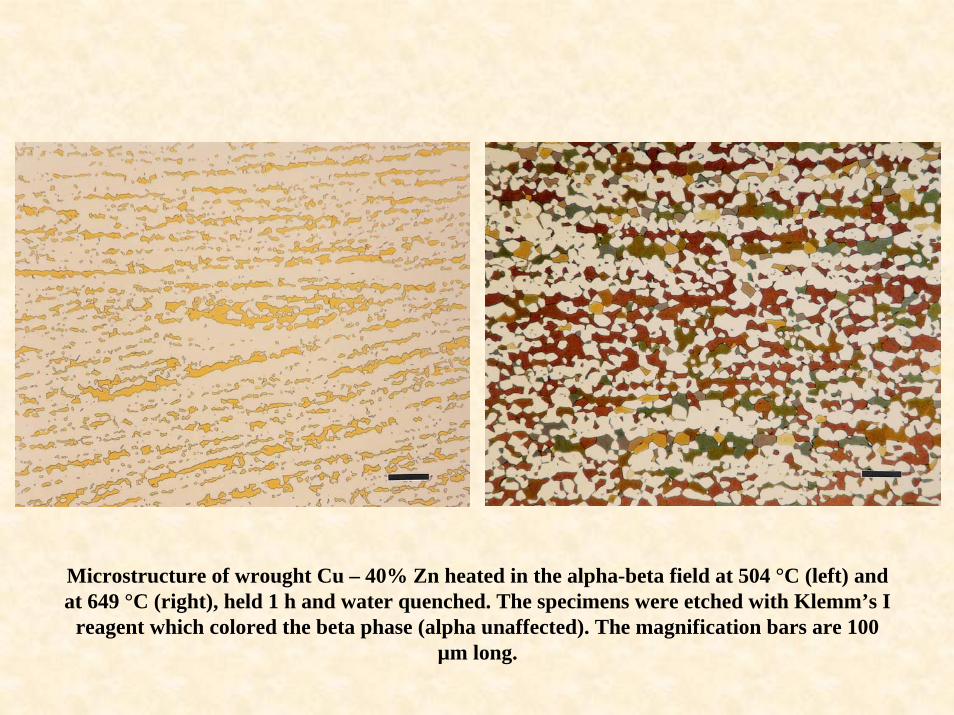
Microstructure of wrought Cu – 40% Zn heated in the alpha-beta field at 504 °C (left) and at 649 °C (right), held 1 h and water quenched. The specimens were etched with Klemm’s I
reagent which colored the beta phase (alpha unaffected). The magnification bars are 100 µm long.

Microstructure of wrought Cu – 40% Zn heated in the alpha-beta field at 716 °C, held 1 h and water quenched. The specimen was etched with Klemm’s I reagent which colored the beta phase
(alpha unaffected). The specimen on the left was viewed with bright field while the one on the right was viewed with Nomarski DIC revealing the annealing twins in the austenite. The
magnification bars are 100 µm long.

Microstructure of wrought Cu – 40% Zn heated in the beta field at 843 °C, held 1 h and water quenched. The specimen was etched with Klemm’s I reagent which colored the beta phase (alpha unaffected). We see three prior-beta grains with ordered beta (colored) and alpha (white) within
each grain. The magnification bar is 100 µm long.


Ammonium Persulfate and NH4OH in H2O NH4OH and H2O2 (3%)
Annealed and cold drawn Naval Brass (Cu – 39.7% Zn – 0.8% Sn) viewed on a transverse plane after etching (left) with 100 mL water, 3 g ammonium persulfate and 1 mL ammonium
hydroxide, which darkens the beta phase (alpha not affected); and, after etching (right) with equal parts ammonium hydroxide and hydrogen peroxide (3% conc.), which darkens the alpha
phase (note the annealing twins in the alpha). Some evidence of slip lines can be seen in some beta grains. Beta-beta boundaries are visible. Originals at 500X.

Klemm’s I Reagent Beraha’s Selenic Acid Reagent
Annealed and cold drawn Naval Brass (Cu – 39.7% Zn – 0.8% Sn) viewed on a transverse plane after etching (left) with Klemm’s I reagent, which colors the beta phase (some detail of twins in the alpha grains can be seen faintly); and, after etching with Beraha’s selenic acid reagent and
viewing with polarized light and sensitive tint, which colors the alpha phase (note annealing twins) and the beta phase (color is non-uniform). Originals at 200X.

Partially transformed alpha/beta brass, Cu – 42% Zn. Heat treatment: heated to 800 °C (1472 °F) into the beta field, held 1 hour, furnace cooled to 600 °C (1112 °F) and water quenched. Etched with 3g ammonium persulfate – 1 mL ammonium hydroxide – 100
mL water which colored the beta phase; alpha is white. Original at 50X.

Microstructure of hypoeutectic Cu – 11.2% Al (annealed at 982 °C, air cool) revealed using Beraha’s PbS reagent that colored the alpha phase but not the γ2 phase. The magnification bar
is 20 µm long.

Aluminum bronze (Cu – 10.5% Al – 3.8% Fe – 0.3% Ni) aged to precipitate Al4Cu9 (γ2). The structure was revealed using Beraha’s PbS reagent which colored the alpha matrix. The
small gray rosettes are insoluble, elemental Fe particles. The magnification bar is 20 µm long.

The eutectic in Cu – 8.4% P (sand cast) revealed using equal parts of ammonium hydroxide and hydrogen peroxide (3% conc.). The magnification bar is 20 µm long.

Potassium dichromate did a better job of revealing the Cu – 8.4% P eutectic structure as it colored the Cu-rich phase orange while leaving the copper phosphide (Cu3P) unaffected. The
magnification bar is 20 µm long.

Microstructure of sand cast, hypereutectic Cu – 10.5% P revealed using Klemm’s II reagent which colored the Cu-rich phase but not the Cu3P phase. The magnification bar is 20 µm long.

Microstructure of sand cast, hypereutectic Cu – 10.5% P revealed using Beraha’s PbSreagent which colored the Cu-rich phase and revealed mechanical twins in the copper
phosphide. Original magnification 500X.

Bright Field
Polarized Light
100 µm 100 µm
PL + Sensitive Tint
Grain structure of wrought 1100 grade foil after electrolytic polishing and anodizing with Barker’s reagent (20 V dc, 2 min), and viewed with bright field (top), polarized light (left) and with polarized light plus a sensitive tint filter (right). Note that color is not observed in
bright field because an interference film is not formed using Barker’s reagent.

Equiaxed alpha grains in the interior of a super-pure aluminum specimen anodized with Barker’s reagent, 30 V dc, 2 min. Viewed with crossed polars + sensitive tint. Original at 50X. The dark spots are
intermetallic phases.

Grain boundary precipitates in super-pure aluminum specimen (shown previously) revealed by aqueous 0.5% HF etch. Magnification bar is 20 µm long.

2011-0Keller’s NaF-NaOH
B&W –Bars are
20-µm long500X
Color is 200XGraff-Sargent Weck’s

2011-T3Keller’s NaF-NaOH
B&W –Bars are
20-µm long500X
Color is 500XGraff-Sargent Weck’s

3003Keller’s NaF-NaOH
B&W –Bars are
20-µm long500X
Color is 500XGraff-Sargent Weck’s

Differential Interference ContrastBright Field
As-cast 3004 aluminum (Al – 1.25% Mn – 1.05% Mg) etched with Keller’s reagent. Magnification bars are both 20 µm long and the same field is shown, in bright field (left) and in Nomarski DIC (right). The dendrites are visible using DIC but not in bright field. The intermetallic particles between the dendrites are easier to see
in bright field.

4032-T6Keller’s NaF-NaOH
B&W –Bars are
20-µm long500X
Color is 500XGraff-Sargent Weck’s

4147Keller’s NaF-NaOH
B&W –Bars are
20-µm long500X
Color is 500XGraff-Sargent Weck’s

5083-H321Keller’s NaF-NaOH
B&W –Bars are
20-µm long500X
Color is 500X Weck’sGraff-Sargent

6013-T8Keller’s (longitudinal) NaF-NaOH (transverse)
B&W –Bars are
20-µm long500X
Color is 500X Weck’sGraff-Sargent

6061-T6511Keller’s NaF-NaOH
B&W –Bars are
20-µm long500X
Color is 200XGraff-Sargent Weck’s

6262-T9Keller’s NaF-NaOH
B&W –Bars are
20-µm long500X
Color is 200XGraff-Sargent Weck’s

7075-T651Keller’s NaF-NaOH
B&W –Bars are
20-µm long500X
Color is 200XGraff-Sargent Weck’s

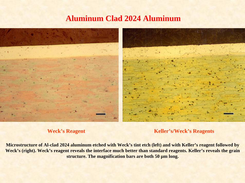
Aluminum Clad 2024 Aluminum
Bright Field Nomarski DIC
Microstructure of Al-clad 2024 aluminum etched with Keller’s reagent. The specimen was mounted in Epo-Thin epoxy resin. The magnification bars are both 50 µm long. The interface is not distinct in bright
field but is more clearly seen in DIC.

Aluminum Clad 2024 Aluminum
Weck’s Reagent Keller’s/Weck’s Reagents
Microstructure of Al-clad 2024 aluminum etched with Weck’s tint etch (left) and with Keller’s reagent followed by Weck’s (right). Weck’s reagent reveals the interface much better than standard reagents. Keller’s reveals the grain
structure. The magnification bars are both 20 µm long.
Aluminum Clad 2024 Aluminum
Weck’s Reagent Keller’s/Weck’s Reagents
Microstructure of Al-clad 2024 aluminum etched with Weck’s tint etch (left) and with Keller’s reagent followed by Weck’s (right). Weck’s reagent reveals the interface much better than standard reagents. Keller’s reveals the grain
structure. The magnification bars are both 50 µm long.

AM60 AZ91D
Microstructures of AM60 and AZ91D pressure die cast specimens after etching with the aqueous 10% HF reagent (magnification bars are 10 µm long). Note that
this etchant darkened the Mg17Al12 intermetallic constituent.

Alpha Alloys
Microstructure of CP Ti, ASTM F 67, Grade 4 (UNS R50700) (longitudinal plane, specimen was annealed at 704 °C) prepared using the three-step method and etched with Kroll’s reagent to reveal the
grain structure. Original image at 200X. Magnification bar is 50 µm long.

Alpha Alloys
Microstructure of CP Ti, ASTM F 67, Grade 2 (UNS R50400; specimen was in the as-rolled condition) prepared using the three-step method and viewed with polarized light to reveal the grain structure. Original
image at 100X. Magnification bar is 100 µm long. Note the mechanical twins in the grains (arrows).

Alpha Alloys
Microstructure of CP Ti, ASTM F 67, Grade 2 (UNS R50400; specimen in the as-rolled condition) prepared using the three-step method, followed by a vibratory polish, and viewed with crossed polarized light to reveal the grain structure. Original image at 100X. Magnification bar is 100 µm long. Note the
deformation twins (arrows).

Alpha Alloys
Microstructure of CP Ti, ASTM F 67, Grade 4 (transverse plane, specimen was annealed) prepared using the three-step method, heat tinted on a laboratory hot plate, and viewed with polarized light plus
sensitive tint to reveal the grain structure.

Microstructure of the silver-copper eutectic, pure phases of Ag and Cu (colored) after etching with Klemm’s I reagent. The magnification bar is 20 µm long.

Microstructure of a W – 27% Cu powder metallurgy composite material after hot isostaticpressing. The specimen was etched with Klemm’s III for 20 seconds and imaged with
bright field. The magnification bar is 20 µm long.

Metal-Matrix Composites
Microstructure of iron containing 10% TiC. The magnification bars are 100 and 50 µm long, respectively (left and right).
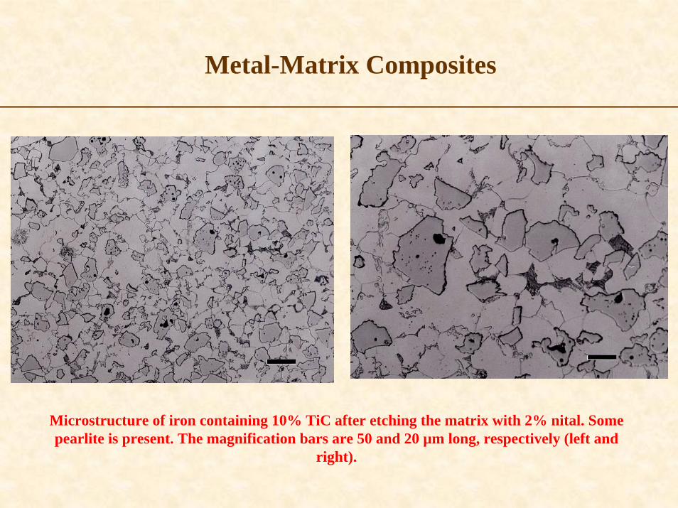
Metal-Matrix Composites
Microstructure of iron containing 10% TiC after etching the matrix with 2% nital. Some pearlite is present. The magnification bars are 50 and 20 µm long, respectively (left and
right).

Metal-Matrix Composites
Microstructure of iron containing 10% TiC after etching the matrix with 2% nital and darkening the TiC with Murakami’s reagent. The magnification bars are 50 and 20 µm long, respectively
(left and right).


Copyright © 2022 FDOKUMEN