importance of microstructure, chemical composition and aging
183
THE MECHANICAL BEHAVIOR OF DENTIN: IMPORTANCE OF MICROSTRUCTURE, CHEMICAL COMPOSITION AND AGING Carolina Montoya Mesa Universidad Eafit A dissertation submitted to the Universidad Eafit for the Degree of Doctor of Philosophy Universidad Eafit, Engineering Department, February 2017.
-
Upload
khangminh22 -
Category
Documents
-
view
1 -
download
0
Transcript of importance of microstructure, chemical composition and aging

THE MECHANICAL BEHAVIOR OF DENTIN:
IMPORTANCE OF MICROSTRUCTURE, CHEMICAL
COMPOSITION AND AGING
Carolina Montoya Mesa
Universidad Eafit
A dissertation submitted to the Universidad Eafit for the Degree of Doctor of
Philosophy
Universidad Eafit, Engineering Department, February 2017.

2
Preface and Declaration
The work described in this dissertation was carried out at Universidad Eafit between
January 2003 and February 2017.
I would like to thank my supervisor Dr Alex Ossa. for his guidance and invaluable
discussions. Special thanks to Dr. Dwayne. Arola from the Materials Science and Engineering
department at the University of Washington for the continuous support during this
investigation. Also to Prof. Santiago Arango and Dr. Alejandro Peláez from the Dental Clinic
of Universidad Cooperativa de Colombia for providing teeth for this study.
Financial support for this project was given to me by the Departamento Administrativo
de Ciencia, Tecnología e Innovación, Colciencias. This dissertation is the result of my own
work, except where specific reference has been made to the work of others. No part of the
work has been, or is currently being, submitted for any degree, diploma or other qualification.

3
THE MECHANICAL BEHAVIOR OF DENTIN: IMPORTANCE
OF MICROSTRUCTURE, CHEMICAL COMPOSITION AND AGING
Carolina Montoya Mesa
Summary
Dental fracture is one of the three most common forms of failure of restored teeth and
the most common cause of tooth loss or extraction in elderly patients. Previous investigations
conducted on aging of hard tissues have identified that there is a considerable reduction in the
mechanical properties (i.e. fracture toughness, fatigue and flexural resistance) of dentin with
aging and that may predispose tooth fracture. These declines in properties have been attributed
to microstructural and chemical composition changes over time. However, these aging
processes have not been really quantified and related with the changes in mechanical
properties. Accordingly, the aim of this work is to evaluate the aging process of coronal dentin
in terms of the evolution of microstructure, changes in chemical composition and mechanical
properties from selected age groups (young and old donors). The changes in these properties
were evaluated in three different regions (outer, middle and inner) in order to identify spatial
variations within the crown.
A brief description of the main literature on composition, microstructure and
mechanical behavior of dentin is presented in chapter 2.

4
An extensive experimental study was carried out in chapter 3 to identify the changes in
microstructure of dentin with aging by means of optical and electron microscopy; while
changes in chemical composition were analyzed using Raman Spectroscopy to calculate the
mineral-to-collagen ratio. Changes in mechanical properties were measured using Vickers
micro-hardness.
Chapter 4 describes the importance of tubule density to the fracture toughness of dentin
for young and old donor’s groups. An approach previously proposed to study the mechanical
behavior of porous materials was used to model the fracture toughness of coronal dentin in
terms of the tubule characteristics. Results were then compared with published results from
previous studies.
The time-dependent deformation response of dentin was analyzed via spherical
indentation experiments at different indentation loads in Chapter 5. From the experimental
observations was proposed a simple model to describe the time dependent deformation
behavior of dentin. This model was based on previously proposed theories for indentation of
time dependent materials, showing that the effective strain rate of dentin depends on its
chemical composition (i.e. mineral-to-collagen ratio) and microstructure (i.e. lumen area
fraction). The descriptions of the model were compared with the experimental results showing
good agreement. The same model was validated with experimental results of aged dentin,
finding a low change in the deformation response of dentin with aging, as presented in
chapter 6.
Finally, preliminary results made on the mechanical properties of dentin have shown
that the microstructure of aged human dentin can vary depending on the ethnic background of
the donor and that this quality is critically important to the mechanical properties of the tissue.
In chapter 7 preliminary results on the comparison between the microstructure, chemical

5
composition and mechanical properties of Colombian, Chinese and American donors is
presented. Finally, conclusions for the study are presented in chapter 8.

6
Products
As a result of this doctoral research the following products have been obtained:
Peer reviewed publications
Montoya, C., Arola, D., Ossa, E. A. (2017). Time Dependent Deformation Behavior of
Dentin. Archives of Oral Biology, 76, 20-29.
Montoya, C., Arola, D., Ossa, E. A. (2016). Importance of tubule density to the fracture
toughness of dentin. Archives of Oral Biology, 67, 9-14.
Montoya, C., Arango-Santander, S., Peláez-Vargas, A., Arola, D., Ossa, E. A. (2015).
Effect of aging on the microstructure, hardness and chemical composition of dentin. Archives
of oral biology, 60 (12), 1811-1820.
Oral conference presentations
Montoya, C., Arola, D., Ossa, E. A. Prediction of fracture toughness of human dentin.
6th International Conference on Mechanics of Biomaterials and Tissues, 2015, Hawaii, USA.
Montoya, C., Arola, D., Ossa, E. A. Time dependent behavior of human dentin. TMS
2015, Orlando, USA.
Montoya, C. Propiedades mecánicas de la dentina. XX Jornada de Investigación-
Facultad de Odontología UCC, 2014, Medellín, Colombia.
Montoya, C., Arola, D., Ossa, E. A. Effect of aging on the microstructure, hardness and
chemical composition of human Dentin. TMS 2014, 2014, San Diego, USA

7
Montoya, C., Ossa, E. A. Composición química y microestructura de la dentina de
pacientes colombianos. VII Congreso Internacional de Materiales, 2013, Medellín, Colombia.
Conference proceedings publications
Montoya, C., Ossa, E. A. Composición química y microestructura de la dentina de
pacientes colombianos. Revista colombiana de Materiales, 5, 73-78.

8
Contents
Preface and Declaration ................................................................................................... 2
Summary .......................................................................................................................... 3
Products ............................................................................................................................ 6
Peer reviewed publications....................................................................................... 6
Oral conference presentations .................................................................................. 6
Conference proceedings publications....................................................................... 7
Contents ............................................................................................................................ 8
List of Tables .................................................................................................................. 14
List of Figures ................................................................................................................ 15
Chapter 1 ........................................................................................................................ 20
Introduction .................................................................................................................... 20
Chapter 2 ........................................................................................................................ 23
Review of previous research on the mechanical behavior and aging of human dentin . 23
2. 1 Introduction ...................................................................................................... 23
2. 2 Tissues of the human body .............................................................................. 23
2. 3 Microstructure and chemical composition of dentin ....................................... 24
2. 4 Mechanical Properties of dentin ...................................................................... 27

9
2.3. 1 Hardness and Elastic Modulus .................................................................. 27
2.3. 2 Flexure Strength ........................................................................................ 29
2.3. 3 Fracture Toughness ................................................................................... 30
2.3. 4 Compressive behavior ............................................................................... 31
2.3. 5 Fatigue strength ......................................................................................... 32
2.3. 6 Viscoelastic Properties .............................................................................. 32
2. 5 Aging process of dentin ................................................................................... 35
2. 6 Conclusions ...................................................................................................... 38
2. 7 Tables ............................................................................................................... 40
2. 8 Figures ............................................................................................................. 44
Chapter 3 ........................................................................................................................ 45
Effect of aging on hardness, microstructure and chemical composition of dentin ........ 45
3. 1 Introduction ...................................................................................................... 45
3. 2 Experimental investigation .............................................................................. 46
3. 3 Experimental Results ....................................................................................... 49
Microstructure ........................................................................................................ 49
Hardness ................................................................................................................. 51
Chemical composition ............................................................................................ 52
3. 4 Discussion ........................................................................................................ 53
Microstructure ........................................................................................................ 53

10
Hardness ................................................................................................................. 55
Chemical composition ............................................................................................ 57
3. 5 Conclusions ...................................................................................................... 59
3. 6 Tables ............................................................................................................... 61
3. 7 Figures ............................................................................................................. 62
Chapter 4 ........................................................................................................................ 73
Importance of Tubule Density to the Fracture Toughness of Dentin ............................. 73
4. 1 Introduction ...................................................................................................... 73
4. 2 Experimental investigation .............................................................................. 74
4. 3 Experimental results ........................................................................................ 75
4. 4 Discussion ........................................................................................................ 76
4. 5 Conclusions ...................................................................................................... 81
4. 6 Figures ............................................................................................................. 82
Chapter 5 ........................................................................................................................ 86
Time dependent deformation behavior of human dentin ............................................... 86
5. 1 Introduction ...................................................................................................... 86
5. 2 Background ...................................................................................................... 87
5. 3 Experimental investigation .............................................................................. 89
Spherical indentation tests ...................................................................................... 90
Chemical Composition Analysis ............................................................................ 90
11
Microstructural Analysis ........................................................................................ 91
5. 4 Results .............................................................................................................. 91
Spherical indentation tests ...................................................................................... 91
Chemical Composition Analysis ............................................................................ 93
Microstructural Analysis ........................................................................................ 94
5. 5 Time dependent deformation model for dentin ............................................... 94
5. 6 Approximate calibration of the model ............................................................. 96
5. 7 Discussion ........................................................................................................ 98
5. 8 Conclusion ..................................................................................................... 101
5. 9 Tables ............................................................................................................. 103
5. 10 Figures ........................................................................................................ 106
Chapter 6 ...................................................................................................................... 117
Contributions of aging to the time dependent deformation of dentin .......................... 117
6. 1 Introduction .................................................................................................... 117
6. 2 Experimental investigation ............................................................................ 118
Sample collection and preparation ....................................................................... 118
Spherical Indentation Tests .................................................................................. 118
Chemical composition analysis ............................................................................ 118
Microstructural Analysis ...................................................................................... 119
Statistical Analysis ............................................................................................... 120

12
6. 3 Experimental results ...................................................................................... 120
Spherical Indentation............................................................................................ 120
Chemical composition analysis ............................................................................ 121
Microstructure ...................................................................................................... 123
6. 4 Discussion ...................................................................................................... 124
6. 5 Conclusions .................................................................................................... 129
6. 6 Tables ............................................................................................................. 130
6. 7 Figures ........................................................................................................... 131
Chapter 7 ...................................................................................................................... 138
Ethnic Background influence on the aging process of dentin: Preliminary Results .... 138
7. 1 Introduction .................................................................................................... 138
7. 2 Experimental investigation ............................................................................ 140
7. 3 Experimental results ...................................................................................... 141
Microstructure ...................................................................................................... 141
Spherical Indentation Tests .................................................................................. 142
Chemical Composition ......................................................................................... 143
7. 4 Discussion ...................................................................................................... 143
7. 5 Conclusions .................................................................................................... 149
7. 6 Tables ............................................................................................................. 151
7. 7 Figures ........................................................................................................... 152
13
Chapter 8 ...................................................................................................................... 158
Conclusions .................................................................................................................. 158
Chapter 2: Review of previous research on the mechanical behavior and aging of
human dentin ................................................................................................................... 158
Chapter 3: Effect of aging on hardness, microstructure and chemical composition
of dentin ........................................................................................................................... 159
Chapter 4: Importance of Tubule Density to the Fracture Toughness of Dentin . 160
Chapter 5: Time dependent deformation behavior of human dentin ................... 160
Chapter 6: Contributions of aging on the time dependent deformation of dentin 161
Chapter 7: Ethnic Background influence on the aging process of dentin ............ 161
Bibliography ................................................................................................................. 163

14
List of Tables
Table 2. 1. Results reported in the literature for the hardness of dentin. ....................... 40
Table 2. 2. Results reported in the literature for the Young Modulus of dentin. ........... 41
Table 2. 3. Results reported in the literature for the flexural strength of dentin. ........... 42
Table 2. 4. Results reported in the literature for the fracture toughness of dentin. ........ 43
Table 3. 1. Results from the ANOVA (p-values) in comparing the microstructure of
dentin from young and old donor teeth. Note the statistically significant differences for the
occlusion ratio for the middle and outer dentin. ........................................................................ 61
Table 5. 1. Indentation model parameters and c as a function of the power-law
exponent n (reproduced from Bower et al. (1993)). ................................................................ 103
Table 5. 2. Dentin parameters obtained from the proposed model. ............................. 104
Table 5. 3. Parameters describing the basic power-law creep behavior for different hard
tissues. ..................................................................................................................................... 105
Table 6. 1. Dentin parameters obtained to describe the time dependent behavior of
aged dentin. .............................................................................................................................. 130
Table 7. 1. Power-law parameter obtained for each group of dentin analyzed. ........... 151

15
List of Figures
Figure 2. 1. Scheme samples used by Iwamonto and Ruse (2003) to measure the
fracture toughness of dentin. Image taken from Iwamonto and Ruse (2003). .......................... 44
Figure 3. 1. Schematic diagram of a sectioned molar after (a) longitudinal (A-A), and
(b) transverse (A’-A’) cutting. The specimen is then embedded in cold-cure epoxy resin with
the sectioned surface facing outwards. ...................................................................................... 62
Figure 3. 2. Micrographs of the dentin microstructure. a) single tubule; b-c) Outer
dentin; d-e) Middle dentin; f-g) Inner dentin. Note the obliterated dentinal tubules for the old
donor teeth in the middle and outer regions (evident in (e) and (g)). ........................................ 63
Figure 3. 3. Micrographs of dentinal tubules from outer dentin. a) young donor; b) old
donor. ......................................................................................................................................... 64
Figure 3. 4. Comparison of microstructure as a function of depth in the coronal dentin.
a) tubule density; b) tubule diameter; c) peritubular dentin diameter. ...................................... 65
Figure 3. 5. Effect of indentation load on the Vickers hardness of dentin from a young
donor tooth. a) change in hardness with indentation load. (b) indentation at 0.23 N; c)
indentation at 9.80 N. ................................................................................................................ 66
Figure 3. 6. Vickers hardness obtained for dentin from young and old donor teeth
according to depth. The direction of applied load is parallel to the dentinal tubules. ............... 67
Figure 3. 7. Indentation of dentin for determination of Vickers hardness. The
indentation locations are within the outer dentin of a young (a) and old (b) donor tooth. ........ 68
1 6
Fi g ur e 3. 8. Distri b uti o n of mi n er al -t o-c oll a g e n r ati o of d e nti n fr o m y o u n g a n d ol d
d o n or t e et h a c c or di n g t o d e pt h. ................................................................................................. 6 9
Fi g ur e 3. 9. C o m p aris o n of t h e h ar d n ess a n d c h e mi c al c o m p ositi o n distri b uti o ns i n a
t o ot h as e vi d e nt fr o m l o n git u di n al s e cti o ni n g. a) h ar d n ess a n d b) c h e mi c al c o m p ositi o n
( mi n er al t o c oll a g e n r ati o) fr o m t h e t o ot h of a 1 8 y e ar ol d d o n or, c) h ar d n ess a n d d) c h e mi c al
c o m p ositi o n fr o m t h e t o ot h of a 6 5 y e ar ol d d o n or. .................................................................. 7 0
Fi g ur e 3. 1 0. O c cl usi o n r ati o f or t h e t hr e e diff er e nt r e gi o ns of c or o n al d e nti n a n d a
c o m p aris o n of r es ult s f or t h e y o u n g a n d ol d d o n or t e et h. ......................................................... 7 1
Fi g ur e 3. 1 1. Vi c k ers h ar d n ess o bt ai n e d f or d e nti n of y o u n g a n d ol d d o n or t e et h
a c c or di n g t o d e pt h. T h e a p pli e d l o a d i s p ar all el (//) a n d p er p e n di c ul ar ( ) t o t h e d e nti n al
t u b ul es. C ol u m ns wit h si g nifi c a nt diff er e n c es ( p ≤ 0. 0 5) ar e gr o u p e d i n a li n e a n d m ar k e d wit h
a cr oss ( +) a n d a ast eris k ( *). ..................................................................................................... 7 2
Fi g ur e 4. 1. Mi cr o gr a p hs of t h e mi cr ostr u ct ur e f or t h e y o u n g a n d ol d d e nti n as a
f u n cti o n of l o c ati o n. a-b) O ut er d e nti n; c -d) Mi d dl e d e nti n; e -f) I n n er d e nti n. N ot e t h e
o blit er at e d d e nti n al t u b ul es i n mi cr o gr a p hs f or t h e mi d dl e a n d o u t er d e nti n of t h e ol d d o n or
t e et h. .......................................................................................................................................... 8 2
xFi g ur e 4. 2. L u m e n ar e a fr a cti o n ( ) f or t h e t hr e e diff er e nt r e gi o ns of c or o n al d e nti n
a n d a c o m p aris o n of r es ult s f or t h e y o u n g a n d ol d d o n or t e et h. ................................................ 8 3
Fi g ur e 4. 3. C o m p aris o n of e x p eri m e nt al a n d pr e di ct e d fr a ct ur e t o u g h n ess of d e nti n as
a f u n cti o n of t h e l u m e n ar e a fr a cti o n x( ). T h e e x p eri m e nt al d at a c orr es p o n ds t o I v a n ci k a n d
Ar ol a ( 2 0 1 3). ............................................................................................................................. 8 4

1 7
Fi g ur e 4. 4. Esti m at e d fr a ct ur e t o u g h n ess f or diff er e nt r e gi o ns of c or o n al d e nti n f or t h e
y o u n g a n d ol d d o n or t e et h. T h es e esti m at es ar e o bt ai n e d fr o m t h e l u m e n ar e a fr a cti o n x( )
m e as ur e m e nts a n d t h e us e of t h e B als hi n e q u ati o n ( B als hi n, 1 9 4 9 ). ........................................ 8 5
Fi g ur e 5. 1. S c h e m ati c di a gr a m of a h alf -s pa c e u n d er i n d e nt ati o n b y a ri gi d s p h er e.
T h e v ari a bl es F, R, h a n d a r e pr es e nt t h e i n d e nt ati o n f or c e, i n d e nt or r a di us of c ur v at ur e, d e pt h
of i n d e nt ati o n a n d t h e r a di us of p er m a n e nt i n d e nt ati o n, r es p e cti v el y. .................................... 1 0 6
Fi g ur e 5. 2. S c h e m ati c di a gr a m of a s e cti o n e d m ol ar wit h t h e e x p os e d d e nti n e m b e d d e d
i n c ol d c ur e d e p o x y r e a d y f or t h e i n d e nt ati o n t e st. T h e l ett ers D a n d E r ef er t o d e nti n a n d
e n a m el, r es p e cti v el y. ............................................................................................................... 1 0 7
Fi g ur e 5. 3. S el e ct e d i n d e nt ati o n d e pt h v ers us ti m e r es ult s f or i n n er d e nti n at a p pli e d
l o a ds of 1, 1 0 a n d 5 0 N e wt o ns. ............................................................................................... 1 0 8
Fi g ur e 5. 4. St e a d y st at e i n d e nt ati on r at e ( ) v ers u s i n d e nt ati o n l o a d r es p o ns e f or t h e
t hr e e r e gi o ns of d e nti n e v al u at e d. ............................................................................................ 1 0 9
Fi g ur e 5. 5. C o m p aris o n of t h e e x p eri m e nt al st e a d y -st at e eff e cti v e str ess a n d ef f e cti v e
str ai n r at e of c or o n al d e nti n ( m ar k ers) wit h pr e di ct e d r es p o ns es (li n es). ................................ 1 1 0
Fi g ur e 5. 6. D e p e n d e n c e of t h e r ef er e n c e eff e cti v e str ai n r at e ( ) o n t h e mi n er al-t o
c oll a g e n r ati o ( ) wit hi n t h e t hr e e diff er e nt r e gi o n s of c or o n al d e nti n. .................................. 1 1 1
Fi g ur e 5. 7. Eff e ct of i n d e nt ati o n l o a d o n t h e eff e cti v e str ai n r at e f or diff er e nt r e gi o ns
of c or o n al d e nti n. C ol u m ns wit h o ut si g nifi c a nt diff er e n c es ( p > 0. 0 5) ar e gr o u p e d wit h a li n e.
................................................................................................................................................. 1 1 2
Fi g ur e 5. 8. Distri b uti o n of t h e mi n er al -t o-c oll a g e n r ati o of d e nti n a c c or di n g t o t h e
n or m ali z e d dist a n c e fr o m t h e p ul p. ......................................................................................... 1 1 3

18
Figure 5. 9. Schematic diagram showing how changes in the degree of mineralization
affect the response of the reference strain rate . .................................................................. 114
Figure 5. 10. Experimental results reported for the steady state creep rate for bone
(Rimnac et al. 1993) and radicular dentin (Jantarat et al. 2002) and comparison with results of
the current study. ..................................................................................................................... 115
Figure 5. 11. Schematic representation of how the dentinal tubules are distributed in
dentin. a) and b) top views of an indentation test on the regions indicated............................. 116
Figure 6. 1. Selected indentation depth versus time results for different regions of
dentin at a constant applied load of 60N. The results correspond from an incisor of a 70-year-
old donor. ................................................................................................................................. 131
Figure 6. 2. Comparison of the experimental steady-state effective stress and effective
strain rate of coronal aged dentin (markers) with predicted responses (lines). ....................... 132
Figure 6. 3. Effect of indentation load on the effective strain rate of different regions of
coronal dentin. Columns without significant differences (p ≤ 0.05) are grouped with a line. 133
Figure 6. 4. Comparison of some dentin characteristics obtained using Raman
Spectroscopy as a function of depth in the coronal dentin. a) Full width at half maximum
(FWHM) of the 1 peak; b) Carbonate-to-phosphate ratio; c) Collagen cross-linking; d)
Mineral-to-collagen ratio. Columns without significant differences are grouped with a line. 134
Figure 6. 5. Micrographs of the dentin microstructure. a) Obliterated dentinal tubules;
b) Outer dentin; c) Middle dentin; Inner dentin. Note the obliterated dentinal tubules for the
middle and outer regions. The percentages correspond to the fraction lumen area obtained for
each region of dentin. .............................................................................................................. 135

19
Figure 6. 6. Comparison of the effective stress and effective strain rate of young (lines)
and old (markers) dentin. ......................................................................................................... 136
Figure 6. 7. Comparison of fraction lumen area () of old incisors and canines and
molars. ..................................................................................................................................... 137
Figure 7. 1. Micrographs of the outer dentin microstructure. a, b, c) China donors; d, e,
f) Colombia donors; g, h, i) United States donors. .................................................................. 152
Figure 7. 2. A comparison of the microstructure as a function of age in the outer dentin
from donor teeth of China, Colombian and US. a) lumen density; b) lumen diameter. .......... 153
Figure 7. 3. Selected indentation depth versus time results for outer china dentin at
applied loads of 5, 10 and 50 Newtons. The results correspond to a Chinese donor of 56 years
of age. ...................................................................................................................................... 154
Figure 7. 4. Comparison of the experimental steady-state effective stress and effective
strain for the outer dentin of the three groups analyzed (markers) with the power-law fit
(lines). ...................................................................................................................................... 155
Figure 7. 5. Mineral-to-collagen ratio of outer dentin from young and old donor teeth
according to the donor’s origin region. ................................................................................... 156
Figure 7. 6. Change in the proportion of obliterated dentinal tubules with aging. ..... 157

20
Chapter 1
Introduction
The effect of aging on the microstructure and mechanical properties of bone has been
studied extensively due to its importance to the elderly and their quality of life (e.g. Currey et
al., 1996; Zioupos et al., 1998; Zioupos et al., 1999; Wang et al., 2002; Wang et al., 2004;
Nalla et al., 2004a; Ural and Vashishth 2006; Ural and Vashishth, 2007). However, the effect
of aging on dental hard tissues (including dentin and enamel) has received rather limited
attention. That is surprising when one considers the importance of human teeth to mastication
and dietary intake.
Previous studies conducted on aging of dentin have identified that there is a
considerable reduction in fatigue tolerance, fracture toughness and flexure resistance over time
(Arola et al., 2010; Nazari et al., 2009). These changes generated by aging predispose dental
fractures, which along with the presence of caries and gum deterioration are the three most
common causes of dental repair and failure; about a third of the teeth repaired daily
correspond to any of these causes (White et al., 1996). Further, some authors have linked tooth
fracture with the completion of restoration processes (root canal treatments and tooth filling
procedures), since cracks or stress concentrators may be generated, promoting tooth fracture
(Lawrence Livermore National Laboratory, 2008). This behavior can be even seen in both
young and old patients.

21
Microstructural changes occurring in dentin with aging have been associated with
obliteration of dentinal tubules and variations on chemical composition (Kinney et al., 2005;
Rosen et al., 1999). However, the aging process of human dentin have not been really
quantified and related with the changes in mechanical properties. Therefore, the aim of this
doctoral work is to quantitatively evaluate the aging process of coronal dentin in terms of the
evolution of microstructure, changes in chemical composition and mechanical properties from
selected age groups (young and old donors). The changes in these properties were evaluated in
three different regions (outer, middle and inner dentin) in order to identify spatial variations
within the crown.
The overall hypothesis of this project is that aging of dentin and the decrease in
mechanical properties is directly correlated with the change in the microstructure and chemical
composition. To achieve this objective, the following aims were developed:
Specific Aim 1: Test the hypothesis that human dentin undergoes a significant change
in the microstructure and obliteration of dentinal tubules with increasing patient age.
Specific Aim 2: Test the hypothesis that human dentin undergoes significant changes in
its chemical composition with increasing patient age.
Specific Aim 3: Test the hypothesis that human dentin undergoes a significant change
in hardness and in its distribution within the crown with increasing patient age.
Specific Aim 4: Test the hypothesis that the viscoelastic or time dependent deformation
response of dentin change along the tooth and undergo significant changes with increasing
patient age.
With the results obtained in this investigation, it is expected to understand how the
decrease in mechanical properties of aged dentin are related with the changes in
microstructure and chemical composition, and therefore in the long-term development of

22
dental restorative materials with differential properties as patient age, achieving thus a
reduction in the recurrence of fractures and tooth extractions.
23
Chapter 2
Review of previous research on the mechanical behavior and aging of
human dentin
2. 1 Introduction
A brief description of the main literature on microstructure, chemical composition and
mechanical behavior of dentin and its changes with aging is presented in this chapter. Detailed
reviews can be found elsewhere (e. g. Pashley et al., 1989; Kinney et al., 2003; Zhang et al.,
2014). Additionally, throughout the entire document additional information on previous
research can be found where necessary to complement the discussion.
2. 2 Tissues of the human body
A tissue is a collection of cells having similar structure and function, usually having a
common embryonic origin and working together to develop specialized activities (Gomez de
Ferraris and Campos Munoz, 2009). According to the embryonic origin the tissues in the
human body can be classified as (Henrikson and Kaye, 1986):
Epithelium: Responsible of cover and protect organs, constituting the inner lining of
the cavities, hollow organs and ducts of the body as well as form mucous and glands.
Connective tissue: Protect, support and bind together different types of tissues and
organs in the body.
24
Muscular tissue: Produces movement and according to the location and function can be
skeletal or striated muscle, smooth or non-striated muscle, and cardiac muscle.
Nervous tissue: Initiates and transmits potentials that help coordinate activities.
These tissues, depending on their level of mineralization and therefore their hardness,
may be classified as soft or hard tissues.
Soft tissues are responsible of connect, support, or surround other structures and organs
of the body (i.e. ligaments, tendons and skin) (Derby and Akhta, 2015). While hard tissues are
those that have been mineralized, and therefore serve as protective shield or structural support
(Grumezescu, 2016). The hard tissues of humans are bone, tooth enamel, dentin, and
cementum. Detailed reviews about each of these tissues and their chemical composition,
formation and chemical properties can be found in: Kambic (1994), Rho et al. (1998), Meyes
et al. (2008) and Lloyd et al. (2015).
2. 3 Microstructure and chemical composition of dentin
Dentin is a hard tissue that occupies the majority of the human tooth. By volume it
consists of approximately 45% mineral material, 33% organic material (collagen type I) and
22% water (Nanci, 2012). Although this chemical composition is assumed to dentin, it has
been widely reported in literature that these percentages change within the tooth from the pulp
to the Dentin Enamel Junction (DEJ) and along radicular dentin (Ryou et al., 2011; Xu et al.,
2009; Tesch et al., 2001).
The thickness of dentin (i.e. from the pulp to the DEJ) is largely dependent on tooth
type, but generally ranges from roughly 2 mm for mandibular incisors up to 3 mm in canines
and molars. Furthermore, the thickness of dentin tends to increase with aging as a result of

25
appositional growth and deposition of secondary dentin (Gomez de Ferraris and Campos
Munoz, 2009).
The microstructure of dentin is largely dominated by its tubules, which are responsible
for housing the odontoblastic processes and maintain dentin vitality. The tubules extend from
the pulp to the DEJ with a double “S” shape in coronal dentin and only one curvature in the
dentin root (radicular dentin) (Nanci, 2012). These are called primary curvatures and are
formed as the progressive stacking of dentinal tubules during the formation of dentin. As a
result of this process there are a higher number of dentinal tubules near the pulp (40.000
tubules/mm2) and fewer near the DEJ (17.000 tubules/mm2) (Fehrenbach and Popowics,
2015).
Dentinal tubule diameters also vary along dentin, diameters of dentinal tubules range
from approximately 1 to 3 μm, depending on patient age and its location, having larger
diameters near the pulp (2.00m) and smaller near the DEJ (1.20m) (Ide Ingle et al.,
2008). Additionally, dentinal tubules have collateral ramifications that contribute to dentinal
permeability and sensitivity; this branching have been reported as more evident in root dentin
than in the crown. This condition promotes bacterial penetration and therefore periodontal
disease in elderly patients (Pashley and Pashley, 1991).
A highly-mineralized cuff of peritubular dentin (PTD) containing mainly apatite
crystals and a small proportion of organic proteins, surrounds the lumens of each tubule. The
tissue located between the tubules is called intertubular dentin (ITD) and contains a matrix of
collagen fibers reinforced by apatite (Marshall et al., 1997). Peritubular dentin is characterized
by the absence of collagen type I in its chemical composition and its high mineral content (Xu
and Wang, 2012). Formation of PTD takes place after mineralization of intertubular dentin is

26
complete and is slowly deposited centripetally after the development of the tooth (Kinney et
al., 1996). This process of formation makes that PTD show three clearly distinguishable areas:
a hypo-calcified region near the outer edge, a highly-mineralized zone that is in continuous
formation and a hypo-mineralized area, which is continuously forming and mineralizing
throughout life (Gomez de Ferraris and Campos Munoz, 2009).
The process of formation and mineralization of dentin, known as dentinogenesis takes
place during the stage of apposition of the tooth; where enamel, cementum and dentin are
secreted in layers by ameloblasts, cementoblasts, and odontoblasts respectively (Goldberg et
al., 2011). During dentinogenesis, odontoblasts release a non-mineralized matrix of collagen
fibers that are later mineralized in the maturation stage by the formation of hydroxyapatite
crystals (Fehrenbach and Popowics, 2015). This process takes place as long as the tooth is
alive and that is why dentin is considered a live tissue (Nanci, 2012).
Since dentin is not a uniform tissue due to its variation in chemical composition,
dentinal tubule density and diameter of dentinal tubules, various types of dentin have been
identified (Nanci, 2012; Gomez de Ferraris and Campos Munoz, 2009; Spangberg, 1989):
Primary dentin: This dentin forms the external shape of the tooth. Depending on its
location can be named as mantle dentin (first formed dentin) and circumpulpar dentin
(forms the remainder tissue). This tissue is formed during odontogenesis and until the
tooth enters occlusion.
Secondary dentin: Since dentin continues its formation through life, dentin formed
since the tooth enters in occlusion is called secondary dentin. The formation pattern of
this dentin is the responsible of forming the “S” shape of dentinal tubules, obliteration
of dentinal tubules and decrease of the pulp chamber size with aging.

27
Tertiary dentin: Also named reactionary or reparative dentin. This dentin is formed as
a result of external stimuli (i.e. caries or trauma) in order to protect the sensitive pulp.
This dentin formation occurs irregularly depending on the aggressiveness of the
stimulus.
Based on its differences in chemical composition and microstructure, dentin is
considered a hierarchical biological composite (Ziskind et al., 2011) and its mechanical
properties change along different regions of dentin and with patient age.
2. 4 Mechanical Properties of dentin
The mechanical properties of dentin have been studied using techniques commonly
used for the characterization of ceramic materials. Some of the properties that have been
determined are hardness, Young’s modulus, flexural strength, fracture toughness, fatigue
strength, compressive strength and some viscoelastic properties. Some of the results obtained
are described below:
2.3. 1 Hardness and Elastic Modulus
Hardness and elastic modulus of dentin have been determinated in different types and
regions of dentin. Some of the techniques used include nanoindentation, Atomic Force
Microscopy (AFM) and microindentation.
By means of Atomic Force Microscopy (AFM) have been found values of hardness
between 2.23 GPa and 2.54 GPa for peritubular dentin and between 0.49 and 0.52 GPa for
intertubular dentin. From these results was found that the values for peritubular dentin are
independent of the location on the tooth, while hardness of intertubular dentine was found to
be dependent on the position, being larger near the DEJ and lower near the pulp (Kinney et al.,
1996). The Young’s modulus for peritubular dentin showed to have values of 25 GPa and

28
between 18 GPa and 22 GPa for intertubular dentin. The higher value obtained for peritubular
dentin was attributed to a higher mineralization degree of this tissue when comparing with
intertubular dentin (Kinney et al., 1996). By means of nanoindentation Ryou et al. (2012)
found hardness values of 2.38 GPa and 1.31 GPa for peritubular and intertubular dentin,
respectively; while the Young´s modulus had values of 29.8 GPa for peritubular dentin and
19.4 GPa for intertubular dentin. From the results, the authors found that intertubular dentin is
almost isotropic and the anisotropic behavior of dentin is determined by the direction of
dentinal tubules and the direction in which testing is performed.
An average hardness of 0.5 GPa has been reported using a Vickers microindentation
techniques, with no significant dependence on indentation load or indentation time
(Chuenarrom et al., 2009). Additionally, Gutiérrez-Salazar and Reyes-Gasga (2003) used
Vickers hardness to determinate how the tooth hardness change from outer enamel surface to
inner dentin layer finding that enamel’s hardness ranges from 2.65 GPa to 3.53 GPa and from
0.49 GPa to 0.58 GPa for dentin.
These properties have been measured not only in permanent teeth but also for
deciduous teeth. For example, Angke et al. (2003) measured the hardness and elastic modulus
of a deciduous molars using a Berkovich indenter. Measurements were made in coronal dentin
from the cusps to the pulp. For both hardness and elastic modulus, an increase in the
penetration depth was found closer to the pulp and a decrease near the DEJ. An average
hardness value of 0.52 GPa was found for zones near de pulp and 0.91 GPa for the region near
the DEJ. The elastic modulus near the pulp was 11.59 GPa while a value of 16.91 GPa was
found near the DEJ. The statistical analysis showed that the values obtained for middle and
near DEJ were not statistically significant. A summary of the reported results for the hardness
and Young modulus of dentin are shown in the tables 2.1. and 2.2 respectively.
29
Dentin hardness has been widely studied and some of the differences obtained between
authors can be attributed to the specimen preparation procedure, indentation load or type of
tooth analyzed. Nonetheless, how dentin hardness changes spatially within the tooth and how
this differences relates with the chemical composition and tubular characteristics (i.e. tubule
density and diameter of dentinal tubules) have not been completely understood.
2.3. 2 Flexure Strength
Different types of bending tests have been performed to dentin in order to compare its
properties with those obtained for restorative materials or adhesives used in restoration
procedures. For example, Ryou et al. (2011) used specimens of two regions of coronal dentin
to perform flexural test - the regions analyzed were named as “top” (region near DEJ) and
“inner” dentin (region near pulp) -. All the samples showed a linear elastic behavior with a
small zone of plastic deformation before failure. Further, it was found that the “top” dentin
had the higher elastic modulus and strength when comparing with the inner regions. The
flexure strength was found ranging from roughly 130 MPa to 180 MPa and the strain ranging
from 0.008 mm/mm to 0.011 mm/mm. The fracture of the specimens was found to begin in
the area subjected to tension stresses. Similar tests were performed by Plotino et al. (2007) on
root dentin, in this particular case in order to evaluate the strength of different synthetic root
materials like carbon and glass fiber composites, zirconia, gold, stainless steel and titanium.
The flexure modulus for root dentin showed values of 17.5 3.8 GPa and flexure strength of
212.9 41.9 MPa. A comparison of results for natural and synthetic materials showed similar
elastic moduli for the fiber-reinforced composite posts while other synthetic materials showed
higher values. Finally, Staninec et al. (2008) also conducted tests on coronal dentin reporting

30
an average flexure strength of 164.4 ± 9.1 MPa. A summary of the flexure strength of dentin
results is shown in the table 2.3.
The changes in flexure strength for the different regions of dentin have been attributed
to the chemical composition and microstructure of each region, where the higher values of
flexural strength were found for outer dentin and radicular dentin where a lower number of
dentinal tubules were found.
2.3. 3 Fracture Toughness
Various approaches have been used to measure the fracture toughness of dentin,
including notched and compact tension specimens with different orientations of dentinal
tubules with respect to the direction of crack propagation. Knowledge of the fracture
toughness of dentin is important to establish the effects of a restoration procedures in the
advent and propagation of cracks in dentin.
Triangular samples were used by Iwamoto et al. (2003) to study the fracture toughness
of dentin. Samples were classified in three groups; i) those with load applied perpendicular to
the plane of dentinal tubules, ii) parallel and aligned to the plane of dentinal tubules; and iii)
the plane of crack propagation parallel and transverse to the plane of dentinal tubules. A
scheme of the samples used is shown in Figure 2.1. From the results, they argued that the
orientation of dentinal tubules with respect to the direction of the load had a statistically
significant effect on fracture toughness of the tissue. Values of 1.13 MPa*m0.5 were found for
samples with the load applied perpendicular to the plane of dentinal tubules and an average of
2.00 MPa*m0.5 was found when the load was applied parallel to the dentinal tubules with the
crack propagating across the boundary between inter- and peritubular dentin.

31
Another type of test that has been used to study the fracture toughness of dentin
involves notched specimens in three-point bending and two directions of crack propagation, in
plane and anti-plane, with respect to dentinal tubules. The results obtained were used to
calculate the stress intensity factor, showing that this factor is higher when the load is applied
anti-plane due to a higher proportion of peritubular dentin with a higher mineralization degree
(Yan et al., 2009).
Compact tension specimens were used by Ivancik and Arola (2013) to examine how
fracture toughness changes in outer, middle, and inner coronal dentin. They found that outer
dentin required 50% greater stress to propagate the crack as compared with inner dentin. This
behavior was attributed to differences in tubule density and diameter of dentinal tubules along
the tooth.
Additional test performed to measure the fracture toughness of dentin were reported by
Imbeni et al. (2003), Nalla et al. (2003a) and Wang (2005); who found similar results to those
mentioned above. A summary of the reported results are shown in table 2.4
2.3. 4 Compressive behavior
Human teeth are subjected to compressive loads and friction during mastication of
food, whereas dentin works under compression stresses only. This makes that the
understanding of the compressive behavior of dentin become very important for this tissue.
Several studies have found compressive strength values that range between 275 MPa and 300
MPa (Craig and Peyton, 1958) and elastic modulus 14 GPa (Kinney et al., 2003). The elastic
modulus obtained under compression loads is similar to the values obtained using indentation
techniques like AFM and nanoindentation, as mentioned in the previous section (section
2.3.1).
32
2.3. 5 Fatigue strength
The fatigue strength of dentin is of great importance as teeth are continuously subjected
to cyclic stresses during mastication. Likewise, after a restoration process is common to find
cracks that can propagate even with low stresses but applied continuously. A detailed review
on the fatigue behavior of dentin can be found in Kruzic and Ritchie (2008).
Studies on the fatigue behavior of human dentin have distinguished that dentin exhibits
the traditional S–N response (Arola et al., 2010; Arola and Reprogel, 2005; Nalla et al.,
2003b). From these results, has been established that dentin exhibits an apparent endurance
limit that ranges from approximately 20–50 MPa, and that it is dependent on the frequency of
loading (Nalla et al., 2003b, Kruzic et al., 2003), the stress applied (Nalla et al., 2004b), and
tubule orientation with respect to the loading direction (Arola and Reprogel, 2006).
The dependence of these factors can be explained by the variations in the
microstructure of dentin (i.e. Tubule density and diameter) and changes in chemical
composition (i.e. collagen content) along the tooth.
2.3. 6 Viscoelastic Properties
Although the viscoelasticity of dentin is assumed due to its collagen content, little is
known about its time-dependent behavior and its relation to the microstructure and mineral
content. The mechanical properties of dentin involving stress-strain relations due to tension,
bending and shear have been investigated under quasi-static conditions (see for instance
sections 2.3.1 through 2.3.5). However, just a few studies have analyzed viscoelasticity and
stress relaxation using various techniques and in different types and regions of dentin. One of
the first tests conducted to determine the viscoelastic properties of dentin was performed by
Duncanson and Korostoff (1975). They used radicular dentin cylinders under compressive

33
stresses to analyze the relaxation behavior of the tissue, finding that dentin showed a linear
dependence on the logarithm of time. They found that the relaxation modulus of dentin
showed a linear dependence on the logarithm of time. The mathematical model of
viscoelasticity previously presented by Alfrey and Doty (1945) was further compared with
their experimental results finding that the stress relaxation response of dentin followed a linear
viscoelastic behavior. Despite of these results, it is not clear if the same behavior can be
extrapolated to coronal dentin, which supports different loads and shows distinct
microstructure. On the other hand, Jantarat et al. (2002) also used radicular dentin cylinders
manufactured from incisors and canines. Compressive loads of 100 N, 300 N, 500 N and 700
N were applied and held for time periods of 90 min. Strain recovery was measured for 60 min
after removing the load. A typical viscoelastic behavior was found for the creep measurements
with a strain increase over time while the stress was kept constant. Statistical differences were
found between stress and creep rate when comparing for the different loads applied. Similar
test made on radicular dentin have been performed by Trengrove et al. (1995) and Jafarzadeh
et al. (2004) finding some viscoelastic behavior for the tissue when a load in constant for a
period of time.
Viscoelastic test on coronal dentin performed by Pashley et al. (2003) in order to study
the stress relaxation of demineralized dentin under tension showed that the dentin matrix
exhibits both stress–relaxation and creep behavior. However, the stress–relaxation and tensile
creep were independent of the initial strain applied.
Other techniques reported in the literature for measuring the viscoelasticity of dentin
involved Atomic Force Microscopy (AFM) and nano-Dynamic Mechanical Analysis (DMA).
For instance, Balooch et al. (1998) measured the viscoelastic properties of fully hydrated
demineralized dentin using an AFM-based indentation method finding that the behavior of the

34
demineralized material is nearly elastic, and that hardness and elastic modulus do not change
with different maximum loads. According to these results, it was established that collagen in
dentin does not contribute to the elastic modulus but it does to dentin strength and toughness.
On the other hand, Ryou et al. (2012) studied the viscoelastic behavior of peritubular and
intertubular dentin. Indentations were made using loads of 400, 700 and 1000 μN with a
corresponding dynamic load of 20 μN and frequencies varying from 2 to 100 Hz. Complex
modulus, loss moduli, storage moduli and tan showed an increase in magnitude with loading
frequency. The complex and storage moduli of peritubular dentin were significantly larger
than for intertubular dentin. No significant differences in the loss modulus and tan between
intertubular and peritubular dentin were found, implying similar viscoelastic behavior in the
two types of dentin. Recently, Chuang et al. (2015) performed nanoindentation creep tests to
study the viscoelastic properties of dentin after de- and re-mineralization processes finding
that the demineralization process increase the primary and secondary creep regimes, while the
remineralization reduces the primary creep of dentin without increasing viscoelasticity.
Indentation techniques have been widely used to analyze the viscoelastic properties of
tissues and biological materials (i.e. bone and teeth) due to its ability to obtain reliable results
without damaging the samples when the loads used are low (Cheneler et al., 2013; Staines et
al., 1981; Ahearne et al., 2007). Various types of indenters have been used for these tests such
as spherical, conical and pyramidal. Sharp indenters introduce a discontinuity at the tip
followed by immediate inelasticity, while spherical indenters produce a uniform and
axisymmetric distribution of stresses, allowing a soft transition between elastic and
viscoelastic regimes during the indenter penetration, simplifying the viscoelastic behavior
analysis of the material (Bower et al., 1993). For instance, da Silva et al. (2008) used a

35
1.5 mm diameter tungsten-carbide ball to analyze the Hertzian response (i.e. contact modulus)
of dentin with different loading rates and indentation directions as a function of dentinal
tubules orientation (i.e. parallel and perpendicular), finding significant differences in the
response of dentin with respect to loading rate but with no differences with tubules orientation.
2. 5 Aging process of dentin
Geriatric dentistry has attracted attention in recent years given the need to preserve and
improve the quality of life of elder people. Additionally, maintaining the physiological
function of the different organs in the aging population could help to reduce the burden on the
existing medical systems as older individuals consume medical services (Sieck, 2003). The
aging process begins since the day of birth and continues throughout life; however, the effects
of the aging process are more evident in the third decade of life and are increasingly obvious
after that (Vaughn, 2011).
The aging process of teeth includes wear of enamel, tooth loss due to alveolar
resorption and periodontal disease and color change (darkening) (Nanci, 2012). On the other
hand, in the case of dentin it has been found that the aging process can generate the following
changes (Murray et al., 2002):
Increase in dentin thickness and decrease of the pulp chamber due to the deposition of
secondary dentin throughout life: An approximate rate of secondary deposition of
43 µm per year, or 0.119 μm per day have been estimated (Solheim et al., 1992).
Reduction in the number of odontoblasts in the pulp chamber: Quantitative studies
have reported a reduction in total pulp cell numbers by 50% between the ages of 20
and 70 years (Frolich et al., 1970).

36
Obliteration of dentinal tubules: The most obvious change in dentin with aging is the
microstructural change due to the obliteration of dentinal tubules with sclerotic or
transparent dentin. This process changes the chemical composition of dentin
increasing its mineral content (Nanci, 2012).
As can be seen, all these changes are related and have an effect not only on the
chemical composition as mentioned earlier, but on the mechanical behavior, permeability and
sensitivity of the tissue (Arola and Reprogel, 2005). A few studies have been published
aiming at identify the changes in mechanical properties of dentin with aging. An extensive
review of the aged dentin properties was published by Arola et al. (2009). Some of the
changes found for the mechanical properties of dentin include:
A decrease in dentin tubule diameter due to deposition of secondary coronal dentin
(Nanci, 2012). Nonetheless, these changes have not been quantified. This process of
obliteration is known to begin at the root and move towards the crown of the tooth
(Vasiliadis et al., 1983).
An increase in the mineral content of dentin has been found in aged dentin (Koester et
al., 2008a). Likewise, changes in crystallinity of the mineral material have been
reported; intertubular dentin mineral crystallites are 7–19% smaller in aged than in
young dentin, while the dentin mineral crystals deposited - obliterating - in dentinal
tubules are chemically similar to the intertubular mineral (Porter et al., 2005).
Increases in elastic modulus and hardness have been also found for radicular dentin,
being more significant in the cervical portion of the root (Xu et al., 2014).
Increases in the elastic modulus of 5% and 9% in the hardness of the outer layer of
dentin have been found by Senawongse et al. (2006) when comparing young and old

37
dentin. On the other hand, Zheng et al. (2005) analyzed the changes in hardness and
Young’s modulus of dentin with aging and reported that dentin does not undergo a
significant change in hardness or Young’s modulus with age in the middle and inner
dentin. However, they found an increase of 16% in hardness and around 5% in
Young’s modulus within outer dentin.
There is a decrease in flexural strength of dentin of almost 20 MPa per decade of life
that begins shortly after reaching adulthood (30 years) (Arola et al., 2009).
A reduction in fatigue strength of dentin was found in aged dentin at higher levels of
stress. However, at low stress levels transparent dentin appears to have the same
behavior than young dentin (Kinney et al., 2005).
There is reduction in the fatigue crack growth resistance of dentin with increasing age.
The average rate of fatigue crack growth in old dentin is greater by a factor of 102 in
comparison to young dentin (Arola et al., 1999).
A reduction of 75% in the energy required to fracture dentin between young (age≤30)
and old patients (age>55) was also reported (Arola et al., 2009).
Ryou et al. (2015) found a difference in the damping behavior of dentin with aging and
therefore in its viscoelastic properties using nanoscopic dynamic mechanical analysis
(nanoDMA). The results showed that either complex or storage modulus depend on
age for peritubular and intertubular dentin while the loss modulus and tan are lower
than the values obtained for young dentin.

38
2. 6 Conclusions
After reviewing the studies available on the mechanical properties of dentin and its
aging process, the follow conclusions can be drawn to support the development of this
doctoral project:
1. It has been widely reported in literature that aging causes a change in dentin
microstructure, product of the obliteration of dentinal tubules. Most of these studies
have been performed on root dentin, where aging begins, and the results obtained
have been analyzed qualitatively using microscopy technics. However, these
changes have not been quantified and there is no information available on how the
obliteration of dentinal tubules changes spatially in coronal dentin. Obtaining this
information for coronal dentin is important as the crown is responsible to support
most of the stresses during mastication. Identifying these quantitative changes is
essential to find relations between the decrease in mechanical properties due to the
aging process.
2. It has been reported in literature that aging causes a change in the chemical
composition of dentin. These changes have been related with an increase in mineral
content due to the obliteration of dentinal tubules. However, the literature review
shows no studies aimed at determine and quantify the changes in chemical
composition of aged coronal dentin (spatial variations). These changes might be
related with an increase in mineralization, crosslinking of collagen or changes in
crystallinity.
3. Dentin hardness has been shown to have considerable local variations for young
patients. For aged dentin, although it has been reported that hardness increases with
aging, the changes obtained have been related to the amount of “free spaces” (i.e.

39
dentinal tubules density), but not with chemical composition changes that occur in
the tooth. For this correlation, old dentin can include other additional changes that
occur with aging such as obliteration of dentinal tubules. Due to differences in
microstructural features of dentin it is expected to find a correlation between
hardness and age.
4. The viscoelastic properties of coronal dentin have been measured in the bulk
dentin. However, the fact that dentin is an anisotropic material, and that mechanical
properties change spatially within the tooth suggests that the viscoelastic properties
of dentin will too. Also, the existing literature shows only one study regarding the
viscoelastic properties of aged dentin, considering intertubular and peritubular
dentin, ignoring how these properties could change along different regions of
dentin. The results obtained about the changes in viscoelastic properties of dentin
may be related with the chemical composition and microstructure. The relative
decrease of viscoelastic properties of the tissue can be assessed as a function of age.

40
2. 7 Tables
Table 2. 1. Results reported in the literature for the hardness of dentin.
Author Technique Region /Type of
dentin
Hardness
GPa)
Kinney et al. (1996) AFM PTD1 2.40
ITD2 0.51
Ryou et al. (2012) Nanoindentation PTD 2.38
ITD 1.31
Chuenarrom et al. (2009) Microindentation/
Vickers Bulk dentin 0.50
Gutiérrez-Salazar and
Reyes-Gasga (2003)
Microindenttion/
Vickers
Outer 0.49
Inner 0.58
Angke et al. (2003) Berkovich indenter.
Primary dentin/
Outer 0.91
Primary dentin/
Inner 0.52
1 PTD: Peritubular dentin
2 ITD: Intertubular dentin

41
Table 2. 2. Results reported in the literature for the Young Modulus of dentin.
Author Technique Region /Type of
dentin
Young
modulus
(GPa)
Kinney et al. (1996) AFM PTD 25.0
ITD 20.0
Ryou et al. (2012) Nanoindentation PTD 29.8
ITD 19.4
Angke et al. (2003) Berkovich indenter.
Primary dentin/
Outer 16.9
Primary dentin/
Inner 11.6
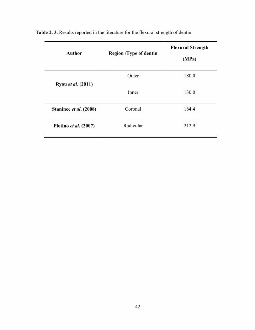
42
Table 2. 3. Results reported in the literature for the flexural strength of dentin.
Author Region /Type of dentin Flexural Strength
(MPa)
Ryou et al. (2011)
Outer 180.0
Inner 130.0
Staninec et al. (2008) Coronal 164.4
Plotino et al. (2007) Radicular 212.9

43
Table 2. 4. Results reported in the literature for the fracture toughness of dentin.
Autor Sample Type /Region
of dentin
Crack propagation
direction
Fracture
Toughness)
(MPa*m0.5)
Iwamoto et al.
(2003)
Triangular
sample Coronal
⊥ 1.13
/ / 1.97
Ivancik and
Arola (2013) Compact tension
Outer
⊥
3.4
Middle 2.7
Inner 2.2
Yan et al. (2009) Beam Coronal / / 2.3
Imbeni et al
(2003)
Notched Beam
(pre-crack)
Coronal ⊥
1.79
Notched Beam
(without pre-
crack)
2.72

44
2. 8 Figures
Figure 2. 1. Scheme samples used by Iwamonto and Ruse (2003) to measure the fracture
toughness of dentin. Image taken from Iwamonto and Ruse (2003).

45
Chapter 3
Effect of aging on hardness, microstructure and chemical composition of
dentin
3. 1 Introduction
Within the field of dentistry, the importance of aging has become of greater interest in
recent years due to its impact on the practice of restorative dentistry. Indeed, the tooth
undergoes certain changes with age, including wear of enamel, the formation of transparent
dentin, a decrease in the number of odontoblasts and an increase in dentin thickness as well as
a production of reactionary dentin (Nanci, 2012). The changes in dentin microstructure
produce variations in its mechanical properties, which are important for the introduction of
restorative treatments and the greater potential for tooth fractures.
Studies have shown that after the third decade of life there is a transition in the
microstructure of dentin, in which the tubules become gradually filled with inorganic material
(Kinney et al., 2005). After a significant number of tubules have been filled, the tissue appears
transparent, and is generally considered as "sclerotic". This process results in an increase in
the mineral content of dentin, opposed to what occurs in bone where there is largely a decrease
in mineral content with aging (Rosen et al., 1999). Furthermore, this increase in mineral
content has been usually associated with increasing dentin fragility and, therefore, causes a
variation in its mechanical properties (Koester et al., 2008b; Nazari et al., 2009).

46
Changes in mechanical properties of dentin with aging have largely been attributed to
the increase in mineralization due to filling of the dentinal tubules. However, it remains
unclear whether these changes can be attributed to the mineral occupying the dentinal tubules,
a complimentary change of the mineral of the intertubular dentin, or crosslinking of collagen
by non-enzymatic processes (Miura et al., 2014). In fact, little information is available on the
relationship between the changes in microstructure of dentin with age and spatial variations in
chemical composition. Thus, the aim of this chapter is to identify the changes in
microstructure, chemical composition and hardness of dentin with aging from selected age
groups of Colombian patients.
3. 2 Experimental investigation
Human third molars were obtained from selected patients after written consent and
following all the protocols required by the Dental Clinic at Universidad Cooperativa de
Colombia (UCC). Exclusion criteria included presence of caries and previous restorations. The
teeth were obtained from donors residing in Medellín, Colombia, and were divided into two
age groups, namely a “young” group with donors between 18 and 25 years of age (N=12), and
an “old” group with donors between 47 and 65 years of age (N=8). There were an equal
number of male and female samples in both groups. Immediately after extraction, all the
specimens were kept in Hank’s Balanced Salt Solution (HBSS) at 2°C to avoid dehydration
and loss of mineral (Habelitz et al., 2001). In addition, the specimens were tested within two
weeks of extraction to limit the loss of mineral and organic materials.
Each molar was sectioned along its longitudinal axis (section A-A in Fig. 3.1a) using
diamond abrasive slicing equipment with continuous water coolant. Secondary sections were
cut transversely (Section A’-A’) in order to expose the dentin as shown in Figure 3.1. For

47
indentation analysis and microscopic evaluations, the specimens were embedded in cold-cured
epoxy resin, following similar procedures used by other researchers (Park et al., 2008; Rivera
et al, 2013; Brauer et al., 2011). The exposed dentin in the resin mount was polished using
silicon carbide abrasive paper with successive smaller particle sizes until reaching 1200 grit.
Further polishing by means of standard red felt polishing cloth wheels was then performed
using diamond particle suspensions of 3 m in size. After polishing, all samples were
ultrasonically cleaned in an HBSS bath for 30 min before microscopic observation in order to
eliminate particles of the diamond particle suspension or tissue resulting from the polishing
process. The polished specimens were then kept in a HBSS bath solution prior to testing.
Vickers testing was used to study the variation in hardness as a function of dentin
depth. Microindentation was performed using a micro-hardness tester (Wilson Instruments,
Model 402 MVD, Norward, MA, USA) with a Vickers diamond indenter. Ten indentations
were made on each surface, starting at the DEJ. Grinding and polishing was then performed to
remove approximately 500 m of material, after which another 10 indentations were
performed. This procedure was repeated until the pulpal surface was reached. Indentations
were made using an indentation load of 1.96 N and dwell times of 10 sec. These testing
conditions generate an indentation large enough so that the hardness corresponds to the overall
dentin hardness, which includes hardness of intertubular and peritubular dentin. Indentations
were carefully made with a distance of at least 10 diagonals in length from each other in order
to avoid any deformation from neighboring indentations.
The Vickers hardness number (HV) was estimated following the ASTM C1327 (2008)
standard according to:

48
where F is the indentation load and d is the indentation diagonal.
The same specimens were used to evaluate the dentin microstructure using an optical
microscope (Axiovert 40 MAT, Carl Zeiss Microscopy, NY). Tubule density, diameter of the
tubule lumens and diameter of the peritubular dentin were measured and calculated at each
depth. A determination of such parameters was carried across the sample surface. Seven
images, each with constant area, were randomly selected from every polished surface. In each
image, the amount of tubules was calculated and expressed as tubules/mm2. The tubule
diameter and peritubular dentin diameter were also obtained. Values from the seven images
were averaged to obtain information from each depth.
The results obtained for hardness and microstructure at each depth were normalized to
the dentin thickness and then classified as outer (normalized depth between 0.76 to 1.00),
middle (between 0.36 to 0.75) or inner (depth between 0.00 to 0.35) dentin. Differences in
hardness between the outer, middle and inner dentin, as well as between young and old
patients were evaluated using a two-way analysis of variance (ANOVA), defining significance
of results by p-value ≤ 0.05, a Tukey post-hoc analysis was then performed.
For the analysis of chemical composition, selected molars from each of the two age
groups were sectioned along the longitudinal axis (section A-A in Fig. 3.1a) and embedded in
cold-cure epoxy resin with the sectioned surface facing outwards. The section surfaces were
polished following the process previously explained. Chemical composition analysis of outer,
middle and inner dentin was performed using Raman spectroscopy and the results were used
to determine the mineral-to-collagen ratio for the three regions (i.e. inner, middle and outer). A
confocal Raman spectrometer (Horiba Jobin Yvon LabRAM HR) was used and the spectrums

49
were obtained over the spectral region of 400 to 1100 cm-1. The Raman spectrometer had a
laser diode with a wavelength of 785 nm and a spot diameter of approximately 1.1 µm. The
mineral-to-collagen ratio was calculated from the ratio of area under the 4PO4 peak at
589 cm-1, which is associated with the phosphate bending of hydroxyapatite, and the area
under the amide III peak at 1254 cm-1, which is associated with movements of the peptide
bond present in collagen (Goodyear et al., 2009; Kazanci et al., 2006). These bands were
selected for analysis as they are reportedly less susceptible to orientation effects and the
polarization direction of the incident light (Kazanci et al., 2006).
Differences in the mineral-to-collagen ratio between the outer, middle and inner dentin,
as well as between young and old patients were evaluated using a two-way analysis of
variance (ANOVA), defining significance of results by p-value ≤ 0.05; a Tukey post-hoc
analysis was then performed. Representative maps of hardness and mineral-to-collagen ratio
were also obtained from selected teeth to convey the spatial variations present.
3. 3 Experimental Results
Microstructure
The microstructure of dentin from selected young and old donor teeth is shown in
Figure 3.2. The microstructures shown for each group correspond to the three regions of
coronal dentin evaluated, including the outer (Figs. 3.2b and 3.2c), middle (Figs. 3.2d and
3.2e) and inner (Figs. 3.2f and 3.2g) regions. As evident from the figures, there was an
increase in tubule density and diameter of the tubules as the distance from the DEJ increased.
Peritubular dentin can be seen surrounding each dentinal tubule (e.g. Fig. 3.2a). There was no
differences in appearance between the peritubular dentin from young and old donor teeth. In
comparing the two age groups, some of the tubules in the old donor group appeared to be
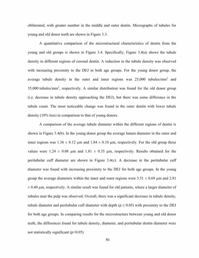
50
obliterated, with greater number in the middle and outer dentin. Micrographs of tubules for
young and old donor teeth are shown in Figure 3.3.
A quantitative comparison of the microstructural characteristics of dentin from the
young and old groups is shown in Figure 3.4. Specifically, Figure 3.4(a) shows the tubule
density in different regions of coronal dentin. A reduction in the tubule density was observed
with increasing proximity to the DEJ in both age groups. For the young donor group, the
average tubule density in the outer and inner regions was 25,000 tubules/mm2 and
35,000 tubules/mm2, respectively. A similar distribution was found for the old donor group
(i.e. decrease in tubule density approaching the DEJ), but there was some difference in the
tubule count. The most noticeable change was found in the outer dentin with lower tubule
density (10% less) in comparison to that of young donors.
A comparison of the average tubule diameter within the different regions of dentin is
shown in Figure 3.4(b). In the young donor group the average lumen diameter in the outer and
inner regions was 1.36 ± 0.12 µm and 1.84 ± 0.10 µm, respectively. For the old group these
values were 1.24 ± 0.08 µm and 1.81 ± 0.35 µm, respectively. Results obtained for the
peritubular cuff diameter are shown in Figure 3.4(c). A decrease in the peritubular cuff
diameter was found with increasing proximity to the DEJ for both age groups. In the young
group the average diameters within the inner and outer regions were 3.51 ± 0.69 µm and 2.81
± 0.40 µm, respectively. A similar result was found for old patients, where a larger diameter of
tubules near the pulp was observed. Overall, there was a significant decrease in tubule density,
tubule diameter and peritubular cuff diameter with depth (p ≤ 0.05) with proximity to the DEJ
for both age groups. In comparing results for the microstructure between young and old donor
teeth, the differences found for tubule density, diameter, and peritubular dentin diameter were
not statistically significant (p>0.05).

51
Hardness
The influence of indentation load on hardness was studied in order to identify the
proper load for measuring the hardness of dentin. Results of that evaluation are shown in
Figure 3.5. The measured hardness values decrease from approximately 1.5 GPa at 0.23 N
indentation load to 0.7 GPa at 1.96 N load. A plateau in hardness is observed for loads of
1.96 N and greater. Consequently, all further hardness measures were conducted with a load of
1.96 N. The average hardness for the three regions of dentin are shown in Figure 3.6 for both
the young and old donor teeth. These values correspond to hardness measured with load
applied parallel to the dentinal tubules, as shown in Figs 3.5b and 3.5c.
Representative indentations made within the outer dentin of young and old donor teeth
are shown in Fig. 3.7a and 3.7b, respectively. Indentations within the outer dentin covered an
average of 36 tubules within the body of the indentation; for the inner dentin that number was
170 tubules. For the old donor teeth, an average of 27 and 156 tubules were included in the
outer and inner dentin, respectively. It is worth noting that no cracks were observed emanating
from the indentation corners for either group, even at the highest indentation load (9.8N).
According to the distribution in measurements, an increase in hardness was found with
proximity to the DEJ for both age groups. Average hardness values of 0.65 ± 0.03 GPa and
0.68 ± 0.01 GPa were found for the young and old donor groups, respectively. When
comparing hardness results with respect to the specific regions of dentin between young and
old donors, significant differences were found for the outer and inner dentin between young
and old dentin (p-value ≤0.05).

52
Chemical composition
The distribution of mineral-to-collagen ratio as a function of distance across the
coronal dentin for young and old donor teeth is shown in Figure 3.8. The dentin in both age
groups showed a similar behavior, with increasing mineral-to-collagen ratio approaching the
DEJ. A higher ratio indicates a lower proportion of organic material. The differences found
among the areas evaluated in the young donors group were not statistically significant
(p>0.05), as opposed to the old donors group where they were statistically significant
(p≤0.05).
When comparing the mineral-to-collagen ratios between the young and old patients,
nearly a 4% difference was found nearest the pulp within the inner dentin. However, the
difference in mineral-to-collagen ratio was approximately 40% in the middle dentin and 70%
in the outer dentin. In comparing results for the mineral-to-collagen ratio between young and
old donor teeth, the differences were statistically significant (p≤0.05).
Comparisons of hardness and mineral-to-collagen ratio distributions in representative
teeth from each age group after longitudinal sectioning can be seen in Figure 3.9. The results
correspond to a young donor (18 years of age) and an old donor (65 years of age). For the
young donor tooth there was an increase in hardness from the pulp up to the DEJ (Figs. 3.9a).
As for the chemical composition of the young dentin (Fig. 3.9b), there was an increase in the
mineral-to-collagen ratio with increasing proximity to the DEJ. In comparing Figures 3.9a and
3.9b it is seen that lower hardness values were obtained in regions of lower mineral-to-
collagen ratio (inner dentin). Conversely, where a higher proportion of mineral was present,
the hardness was greater.
Results of the hardness and mineral-to-collagen ratio measurements for an old donor
tooth are shown in Figures 3.9(c) and 3.9(d). The chemical composition map for the old donor

53
tooth shows a distorted pulp chamber due to deposition of secondary dentin within. This
process can lead to complete pulp obliteration over sufficient time (Tronstad, 2003).
3. 4 Discussion
Microstructure
Consistent with the findings of earlier studies, there were differences in tubule density
and dentinal tubule diameter as conveyed from the measures within the three regions of dentin
evaluated (Fig. 3.2). The results obtained for tubule density in the young patients group are
consistent with those of Marshall et al. (1997), who reported values of 20,000 tubules/mm2
(outer) to 43,000 tubules/mm2 (inner).
For the dentin of old donor teeth only full or partially opened tubules were considered
in the tubule density measurements. Obliterated tubules were excluded from the measurements
to assess how dentinal tubule density changes with aging as caused by the filling process.
Clearly a reduction in the number of open tubules contributes to changes in dentin
permeability and potentially other aspects of its physical behavior. For example, the changes
with aging could be important to dentinal sensitivity and the resistance to tooth fracture.
There was a decrease in tubule diameter from the pulp to the DEJ for both age groups
evaluated. The measures of tubule diameters are consistent with those reported by Ivancik et
al. (2014) for the dentin from young U.S. patients. Interestingly, larger tubule diameters have
been reported for the dentin of Brazilian donor teeth (diameters of 2.99 m and 2.42 m for
inner and outer dentin, respectively) and no significant differences with depth (Lopes et al.,
2009). When comparing the measured values from young and old donor teeth of the present
study, similar results were obtained for the two groups in the inner and middle regions. There

54
was some difference in the measures for diameter in the outer dentin, with 9% lower values in
the old dentin. Undoubtedly this difference is attributed to the onset of sclerosis.
In general, the measures of peritubular cuff diameter were approximately twice the
diameter of the tubule lumens. The formation of peritubular dentin occurs after the
mineralization of intertubular dentin has completed (Gomez de Ferraris and Campos Munoz,
2009). Deposition of dentin is continued by odontoblasts following a circular pattern
surrounding the dentinal tubules. During this process, and as the odontoblasts produce several
layers of dentin, the pulpal chamber reduces its size, odontoblasts move to the interior of the
pulp, and odontoblastic processes remain inside the dentinal tubules (Gomez de Ferraris and
Campos Munoz, 2009).
To identify the change in number of obliterated dentinal tubules relative to the total
number of open tubules for each region of dentin, the occlusion ratio was calculated according
to:
A higher occlusion ratio indicates a higher fraction of obliterated dentinal tubules.
Measured estimates for the occlusion ratio in the three regions of dentin evaluated are shown
in Figure 3.10. As expected, the occlusion ratio in young donor teeth is approximately zero in
all three regions. Indeed, the visible changes in microstructure of dentin with aging begin in
the third decade of life (Gomez de Ferraris and Campos Munoz, 2009; Nazari et al., 2009;
Curtis and Watson, 2014). On the other hand, an increase in occlusion ratio was noted as one
approaches the DEJ. Hence, the highest proportion of obliterated tubules in the tooth crown is
located in the outer dentin and decreases with depth. The occlusion ratio measurements

55
correspond to an average of 1170 obliterated tubules/mm2 in outer dentin and 120 obliterated
tubules/mm2 in inner dentin. Thus, in the tooth crown filling of the lumens begins at the ends
of the tubules.
In comparing the results obtained for microstructure between young and old patients
(e.g. tubule density, diameter, and peritubular dentin diameter) there was no significant
difference (p > 0.05). However, there was a significant difference in the occlusion ratio
between the young and old groups, and between the outer and inner dentin (p ≤ 0.05) as shown
in Table 3.1.
Hardness
There was load-dependency in the measured hardness of dentin as shown in Figure
3.5a. This behavior is at least partly explained by the number of tubules involved within the
indentation area. The difference in number of tubules involved in the indentation response is
evident from a comparison of Figures 3.5b and 3.5c. Load-dependent behavior has been
previously reported in the indentation response of dental enamel (Rivera et al., 2013; Park et
al., 2008). In enamel, which is absent of tubules, the load dependence was described in terms
of the change in mechanisms of deformation with increasing load. That could also play a role
in the response of dentin through the relative viscous behavior and degree of water movement.
Results of the hardness measurements show that there are limited differences in the
hardness between the three regions of dentin evaluated. An overall average hardness of 0.65±
0.03 GPa was obtained for the young dentin, which is in agreement with results from previous
studies (Fuentes et al., 2003). Fuentes et al. (2003) found that the hardness of dentin ranged
between 0.60 GPa and 0.61 GPa, with only little variation along the tooth. Nevertheless, the
spatial variations in hardness identified in the present study are not as significant as those

56
found by other authors, where values between 0.25 GPa to 0.8 GPa have been reported
(Pashley et al., 1985). It is important to note that previous studies have generally involved
data derived from the teeth of patients living in North America. Differences in the
microstructure and mechanical behavior of dentin have been noted in a previous comparison
of donor groups from North and South America (Ivancik et al., 2014). That may be relevant
here.
An increase in hardness occurred with increasing proximity of the DEJ for both age
groups. The lower hardness of inner dentin is at least partly associated with the higher number
(and area) of dentinal tubules (Pashley et al., 1985). The area occupied by dentinal tubules in
the inner dentin is approximately 20% higher than the outer dentin (Pashley et al., 1985;
Ivancik and Arola, 2013). This behavior might be attributed to differences in chemical
composition and variations between the amount of organic and inorganic material within
dentin. However, according to the results obtained for the mineral-to-collagen ratio, these
differences were not statistically significant in the young patients group.
An analysis of the hardness results for the young and old donor groups showed that
there were significant differences for outer and inner dentin between young and old groups
(p≤0.05). These results are in agreement with those found for the occlusion ratio and
obliteration of dentinal tubules. Nevertheless, the results are not consistent with some previous
results concerning the hardness of dentin and age. For instance, Senawongse et al. (2006) used
nano-indentation techniques to determine changes in hardness of different types of dentin with
age and did identify significant differences. However, the secondary and mantle dentin did
show an increase in hardness. Zheng et al. (2005), used nano-indentation to investigate the
changes in hardness of dentin with age. In that investigation there were no significant
differences in dentin hardness between age and location. However, the transparent regions of

57
dentin from old donor teeth did exhibit significantly greater hardness. The discrepancy
between the earlier studies and present results may be explained by the use of nano-
indentation techniques, and differences in the representation of peritubular or intertubular
dentin with single indents. A load-dependency may also contribute to unique responses for the
intertubular and peritubular components. More work is needed in this area.
Chemical composition
An increase in mineral-to-collagen ratio was found with increasing proximity to the
DEJ for both young and old donors, but no significant differences were found along the tooth.
These results contradict the findings by Ryou et al (2011), who found a reduction in the
mineral-to-collagen ratio from the pulp to the DEJ in an evaluation of coronal dentin from US
donors using FTIR. Ryou et al (2011) used the bands associated with the ʋ3PO4 peak and the
Amide I peak to find the mineral-to-collagen ratio of dentin, contrary to the bands used in this
study. Authors like Kazanci et al. (2006) have argued that it is important to consider the effect
of orientation and polarization of different bands for Raman Spectroscopy analysis. They have
also found that Amide III band (used in this study) is less susceptible to polarization effects
than other amide bands. However, Märten et al (2010) measured the volume fraction of
mineral within the coronal dentin using Small-angle X-ray scattering (SAXS) and found that
the mineral volume fraction in dentin is uniform and only found statistically significant
differences near the DEJ.
A lower mineral content within the inner dentin could be attributed to differences in the
mineralization of the intertubular dentin at different regions. For example, Kinney et al. (1996)
measured the hardness of intertubular and peritubular dentin using AFM; the results obtained
established a lower hardness in intertubular dentin near the pulp and an increase towards the

58
DEJ. A correlation made by Pashley et al. (1985) between the change in hardness and tubule
density demonstrated that the decrease in hardness of dentin towards the pulp can be
accounted for by the decreased hardness of the intertubular dentin and that correlation with
tubule density may be coincidental.
It is important to note that most of the results found in the literature were obtained from
studies using teeth of mostly anglo-saxons living in the USA. Therefore, the differences in
dentin features from Colombian donors (i.e. the variation in the mineral-to-collagen ratio
along the tooth) might be attributed to individual characteristics, such as oral health and
nutritional status, among others. In addition, several authors have suggested that a relation
between ethnicity and some dental features, such as tooth size (Merz et al., 1991; Bishara et
al., 1989), enamel thickness (Hall et al., 2007) and tooth formation rate (Olze et al., 2007),
might exist. Merz et al. (1991) found that dental arches in black patients are significantly
wider and deeper than the ones in white patients; while Hall et al. (2007) found thicker enamel
on the distal aspect of the black donor tooth. Differences between tooth dimensions of
permanent teeth in three populations: Egyptians, Mexicans, and the North Americans were
found by Bishara et al. (1989). However, only a few studies on dentin characteristics and how
they might be determined by ethnicity have been carried out (Bajaj et al., 2008).
The results obtained for the mineral-to-collagen ratio are in agreement with those
found for hardness distribution, where higher hardness values were associated with areas
where there was a higher amount of mineral.
The observed distribution in hardness and chemical composition of the old donors
group corresponds to the progression of aging. It has been well established that aging of dentin
starts from root dentin and continues in the coronal direction Drusini et al. (1991). It has also

59
been reported that the total obliteration of tubules for coronal dentin might occur near the age
of 70 (Tronstad, 2003).
It is important to note that the results shown in the hardness maps (Figs 3.9a and 3.9c)
were obtained after longitudinal sectioning of the tooth (section A-A in Fig. 3.1a).
Consequently, the indentation load was applied perpendicular to the dentinal tubules. A
comparison of hardness measurements from the parallel and perpendicular loading orientation
is shown in Figure 3.11. In comparing the results for these two directions, significant
differences in hardness were found between the middle and inner dentin (p ≤0.05) of young
donor teeth. For the old dentin there were no significant differences (p > 0.05). The unique
behavior of the inner and middle dentin may be attributed to a greater number of tubules in
these regions, which yields a higher proportion of voids and lower hardness. For old dentin
there is a consistent increase in hardness approaching the DEJ with both orientations of
indentation. In this case, the increase is more significant than the one discussed earlier (load
applied parallel to dentinal tubules) due to the presence of large areas of peritubular dentin
within the indentation as a result of obliteration of the dentinal tubules.
3. 5 Conclusions
According to the results obtained, the following conclusions were drawn:
1. The tubule density in the dentin of young and old donor teeth ranged from
approximately 22,000 to 35,000 tubules/mm2. The tubule diameter ranged from
approximately 1.2 m to 1.8 m. There was a significant decrease in tubule density
from the pulp to the DEJ in both age groups studied (p ≤ 0.05).
2. There was a significant difference (p ≤ 0.05) in the measures of occlusion ratio
between the outer (4.71%) and middle dentin (2.50%) of the old donor teeth. The

60
largest proportion of obliterated dentinal tubules was near the DEJ and decreased
towards the pulp.
3. A significant decrease in hardness was found with increasing distance from the DEJ
for both young and old donor teeth (p ≤ 0.05). A larger hardness was found in the
old dentin when compared to young dentin. There were significant differences (p ≤
0.05) in the hardness in the outer and middle dentin between the young and old
donor teeth.
4. The mineral-to-collagen ratio was found to increase with proximity to the DEJ for
both young and old patients. This behavior is opposed to previous results reported
in the literature for dentin from US donors, which in turn raises questions whether
the chemical composition of dentin and its percentages of organic material and
collagen might be affected by ethnicity.
5. The outer dentin exhibited the highest mineral-to-collagen ratio, while lower values
were found in the areas near the pulp. A greater mineral-to-collagen ratio was found
for the old dentin, with the highest values found in the outer dentin.
6. A comparison of the hardness and mineral-to-collagen ratio distributions showed
that the regions with the highest mineral-to-collagen ratio also exhibited the highest
hardness.

61
3. 6 Tables
Table 3. 1. Results from the ANOVA (p-values) in comparing the microstructure of dentin
from young and old donor teeth. Note the statistically significant differences for the occlusion
ratio for the middle and outer dentin.
Young vs. Old
Region Tubule
Density
Occlusion
Ratio
Tubule
Diameter
Peritubular
Diameter
Outer 0.1425 0.0002 0.2123 0.6466
Middle 0.4333 0.0001 0.7358 0.4379
Inner 0.9703 0.3739 0.8497 0.6694

62
3. 7 Figures
Figure 3. 1. Schematic diagram of a sectioned molar after (a) longitudinal (A-A), and (b)
transverse (A’-A’) cutting. The specimen is then embedded in cold-cure epoxy resin with the
sectioned surface facing outwards.
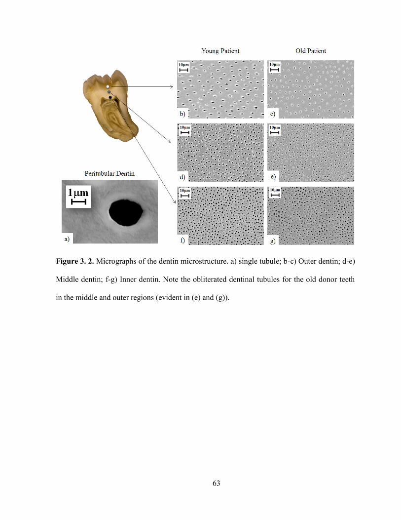
63
Figure 3. 2. Micrographs of the dentin microstructure. a) single tubule; b-c) Outer dentin; d-e)
Middle dentin; f-g) Inner dentin. Note the obliterated dentinal tubules for the old donor teeth
in the middle and outer regions (evident in (e) and (g)).

64
Figure 3. 3. Micrographs of dentinal tubules from outer dentin. a) young donor; b) old donor.

65
Figure 3. 4. Comparison of microstructure as a function of depth in the coronal dentin.
a) tubule density; b) tubule diameter; c) peritubular dentin diameter.

66
Figure 3. 5. Effect of indentation load on the Vickers hardness of dentin from a young donor
tooth. a) change in hardness with indentation load. (b) indentation at 0.23 N; c) indentation at
9.80 N.

67
Figure 3. 6. Vickers hardness obtained for dentin from young and old donor teeth according to
depth. The direction of applied load is parallel to the dentinal tubules.

68
Figure 3. 7. Indentation of dentin for determination of Vickers hardness. The indentation
locations are within the outer dentin of a young (a) and old (b) donor tooth.

69
Figure 3. 8. Distribution of mineral-to-collagen ratio of dentin from young and old donor teeth
according to depth.

70
Figure 3. 9. Comparison of the hardness and chemical composition distributions in a tooth as
evident from longitudinal sectioning. a) hardness and b) chemical composition (mineral to
collagen ratio) from the tooth of a 18 year old donor, c) hardness and d) chemical composition
from the tooth of a 65 year old donor.

71
Figure 3. 10. Occlusion ratio for the three different regions of coronal dentin and a
comparison of results for the young and old donor teeth.

72
Figure 3. 11. Vickers hardness obtained for dentin of young and old donor teeth according to
depth. The applied load is parallel (//) and perpendicular () to the dentinal tubules. Columns
with significant differences (p ≤ 0.05) are grouped in a line and marked with a cross (+) and a
asterisk (*).

73
Chapter 4
Importance of Tubule Density to the Fracture Toughness of Dentin
4. 1 Introduction
The “cracked tooth syndrome” was coined by Cameron (1964) over a half-century ago
to describe the prominence of cusp fractures observed in restored teeth. Fracture is no less
important to the field of restorative dentistry today (e.g. Opdam et al., 2007; Barreto et al.,
2015).
The human dentition is subjected to a variety of cyclic stresses, including those of
mastication and other para-functional activities. Acute stresses can promote the formation of
micro cracks in the tooth (Homewood, 1998), in addition to other events including trauma and
restorative procedures (Bastone et al., 2000; Majd et al., 2012; Lee et al., 2014). Damage and
microcracks in dentin may not cause fracture, but can facilitate the failure of restored teeth
through fatigue crack growth (Arola et al., 1999; Nalla et al., 2003b; Bajaj et al., 2008).
Spatial variations in the resistance to fracture of dentin can contribute to this process and is an
important factor to consider.
Due to its importance, the fracture toughness of coronal dentin has been evaluated, with
primary emphasis on the importance of tubule orientation (see section 2.3.3). With the
increase in senior dentate, aging has become of greater importance to the field of restorative
dentistry ((McNally et al., 2014; Yellowitz and Schneiderman, 2014). Human teeth undergo

74
changes with increasing age, including a decrease in the number of odontoblasts, an increase
in dentin thickness and the formation of transparent dentin (Toto et al., 1971; Bernick and
Nedelman, 1975; Murray et al., 2002; Nanci, 2012; Timiras, 2007). In addition, there are
changes in mechanical properties, such as an increase in elastic modulus and hardness, a
decrease in strength and decrease in fatigue crack growth resistance (see section 2.5). There is
also a reduction in fracture toughness with increasing age (Kinney et al., 2005; Koester et al.,
2008a; Nazari et al., 2009). Changes in the fracture toughness with aging may be related to
dehydration (Toto et al., 1971), increases in the degree of mineralization (Porter et al., 2005),
or an increase in the degree of crosslinking of the collagen fibrils (Miura et al., 2014).
However, as mentioned earlier the most visible change is the decrease in diameter and density
of the tubules, which results from an increase in mineral deposited within the tubules.
To the authors’ knowledge, no investigation has examined the fracture toughness of
coronal dentin in terms of tubule characteristics and number of obliterated tubules that occur
with increasing age. Hence, the objective of this investigation was to examine the effect of
tubule density on the fracture toughness of dentin from young and old donors and utilize an
approach previously proposed for porous materials to quantify the spatial variations in fracture
resistance.
4. 2 Experimental investigation
Human third molars were obtained from selected patients after written consent and
following all the protocols required by the Dental Clinic at Universidad Cooperativa de
Colombia (UCC). Exclusion criteria included presence of caries and previous restorations. The
teeth were obtained from donors residing in Medellín, Colombia, and were divided into two
age groups, namely a “young” group with donors between 18 and 25 years of age (N=12), and

75
an “old” group with donors between 47 and 65 years of age (N=8). There were an equal
number of male and female samples in both groups. Immediately after extraction, all the
specimens were kept in Hank’s Balanced Salt Solution (HBSS) at 2°C to prevent dehydration
(Habelitz et al., 2001). In addition, the specimens were tested within two weeks of extraction
to limit the loss of mineral and degradation of organic materials. All species were prepared
according to the procedure explained in section 3.2.
Measurement of the tubule density (t) and tubule lumen diameter (l) was performed
using commercial image analysis software (AxioVision LE). Seven randomly selected images
with a constant area (approximate size of each image 80 µm x 100 µm) were obtained over the
polished surface. The mean tubule diameter and number of tubules were calculated for each
image. Values from the seven images were averaged and used to estimate the lumen area
fraction () within the three regions of evaluation as:
where Al is the area (mm2) occupied by lumens and AT is the total area of dentin measured
(mm2) in each image. The average lumen area was calculated using the measures of tubule
diameter and density.
4. 3 Experimental results
The microstructure of dentin from selected young and old donor teeth is shown in
Figure 4.1. Representative images are presented from the three regions of evaluation including
the outer (Figs. 4.1a and 4.1b), middle (Figs. 4.1c and 4.1d) and inner (Figs. 4.1e and 4.1f)
dentin. For both age groups the peritubular dentin can be seen surrounding each dentinal
tubule. Several obliterated tubules are evident in the images for the old donor group, with

76
greater number of filled tubules in the middle and outer regions. Micrographs of tubules
within the outer dentin of young and old donor teeth obtained at higher magnification are
shown in Figures 3.3(a) and 3.3(b), respectively. An example of a tubule that has become
filled with mineral is shown in Figure 3.3(b). No obliterated tubules were evident in the dentin
of the young donor group regardless of the region of evaluation.
Figure 4.2 shows the estimates for the lumen area fractions () in the three regions of
dentin evaluated. Overall, there was a significant decrease in with proximity to the DEJ for
both age groups (p ≤ 0.05). In the young donor group the average in the outer and inner
regions was 3.7±0.6% and 9.3±1.0%, respectively. For the old group these values were
2.9±0.4% and 9.6±1.1%, respectively. The primary difference between the two age groups
was the lower value of (37% less) in the outer region of dentin for the old group. This
difference is attributed to the reduction in diameter of the dentin tubules due to deposition of
mineral within the lumens as a result of sclerosis. When comparing the results for in each
region between the young and old donors, significant differences were found only for outer
dentin (p ≤0.05).
4. 4 Discussion
While the dentin tubules play an important role in tooth sensitivity and pain stimuli
(Magloire et al., 2010), they also serve as a form of porosity that contributes to the mechanical
behavior of dentin. Several models have been proposed to describe the mechanical behavior
of porous materials (Ryshkewitch, 1953; Schiller, 1985; Hasselman, 1963; Ji et al., 2006).
According to these models, it is possible to define relationships between porosity and physical
properties such as Young’s modulus, hardness, and strength (Ji et al., 2006; Salomon and
Kosmač, 2013; Luo and Stevens, 1999; Kováčik, 1999). However, the lack of uniformity in

77
the distribution and shape of pores has been considered a limitation of these approaches in
their general application to materials.
Balshin (1949) developed an empirical model for the prediction of mechanical
properties for materials with microstructures similar to dentin. According to this approach, the
variation in fracture toughness of dentin can be described as a function of the porosity or the
lumen area fraction () by
where Kic and Kico are the Mode I fracture toughness of dentin with specific porosity
and of solid dentin (i.e. with no lumens), respectively, and m is a constant that depends on the
degree of stress concentration developed around the lumens (i.e. lumen geometry). According
to Bocaccini et al. (1996) the value of m varies from 1, for long cylindrical pores orientated
parallel to the stress direction (z/x ~∞), up to about 7 for oblate spheroids (axial ratio z/x < 1).
The axial ratio z/x is related to the shape of the pores, where z and x are associated with the
length and width of the pore, respectively.
The reported range in fracture toughness for human dentin is roughly 1.1 MPa·m0.5 ≤
Kic ≤ 3.5 MPa·m0.5. The lowest values have been obtained for cracks oriented perpendicular to
the tubules (Iwamoto and Ruse, 2003), whereas higher values are reported for cracks
extending in-plane with the tubules (Yan et al., 2009) and for outer dentin, which exhibits a
low tubule density (Pashley et al., 1985). Nonetheless, estimates for the fracture toughness of
dentin without the presence of dentinal tubules (solid dentin) have not been reported.
Recently, Ivancik and Arola (2013) evaluated the fracture behavior of coronal dentin
obtained from the teeth of young donors residing in the US. They obtained estimates of Kic for
the outer, middle, and inner dentin. The average fracture toughness for the outer coronal

78
dentin (with lowest density of tubules) was 3.40 MPa·m0.5. Their results are shown in Figure
4.3 along with a prediction from the Balshin model (Eq. 4.2) using Kico = 3.40 MPa·m0.5
(outer dentin) and m=3, which is appropriate for long cylindrical pores (e.g. dentinal tubules)
oriented perpendicular to the direction of stress (Bocaccini et al., 1996). As evident in Figure
4.3, the model predicts a decrease in fracture toughness of dentin with increasing tubule area
fraction (porosity). That distribution correctly captures the decrease in fracture toughness
approaching the pulp due to a higher proportion of dentin tubules and larger average diameter.
A reasonable correlation was found between the model and experimental results (R2= 0.89).
An alternate approach was also used to obtain the parameters of Eq. (4.2) by
performing a best-fit to the experimental results (Fig. 4.3), which resulted in Kico = 3.76
MPa·m0.5 and m=4.5. A comparison of the model to experimental data with these “best-fit”
parameters results in R2=0.998, which indicates very good agreement. The value obtained for
Kico is similar to the maximum value obtained by Ivancik and Arola (2013) of 3.74 MPa·m0.5,
which was obtained for the outer dentin with a lumen area of just under 2%. Therefore, the
value of Kico obtained in this study is effectively related to dentin without tubules. According
to Boccaccini et al. (1996), a value of m=4.5 corresponds to a porous microstructure
composed of oblate spheroids with z/x = 0.3 and an orientation of = 30o, which is a
reasonable approximation to the mean shape and orientation of the S-shape lumens in dentin.
Therefore, the model provides a reasonable description for the effect of on the fracture
toughness of coronal dentin.
Estimates for the fracture toughness of dentin from the teeth of young and old
Colombian donors were obtained using Eqn. (4.2) and are shown in Figure 4.4. These
predictions were obtained using the best-fit estimates for Kico and m, along with the lumen

79
area fraction measurements () from the Colombian donor teeth in Figure 4.2. As evident in
this figure, there is a significant increase (p ≤0.05) in fracture toughness with increasing
proximity to the DEJ for the middle and outer regions of young dentin, which agrees with the
reported results (Ivancik and Arola, 2013). Furthermore, in comparing the estimated Kic
between the young and old dentin of the three regions there is a significant difference (p
≤0.05) in values for the outer dentin only; the Kic for the old dentin is 4.0% greater. These
results contrast those previously reported for the fracture toughness of dentin from senior
patients (Kinney et al., 2005; Nazari et al., 2009) where a substantial reduction (20%) in
fracture toughness was noted with aging, regardless of the direction of crack growth (Koester
et al., 2008a; Ivancik and Arola, 2013).
According to the adopted model (Eq. 4.2), the microstructural changes in dentin that
occur with aging would cause an increase in fracture toughness. This behavior is related to the
reduction in number and diameter of stress concentrators in the material (as represented by
lower ). But if the tubules are obliterated by deposited mineral with low fracture toughness,
the overall toughness should be higher than that of a porous material (with empty tubules) if
the mechanisms of fracture do not change with aging. An obvious limitation of the proposed
model is that only geometrical features are considered (i.e: lumen area). The question that
arises is “If the dentin of old patients undergoes a decrease in the number of stress
concentrators, why is there a reduction in the fracture toughness with aging?”.
The primary mechanisms of crack growth toughening in dentin involve crack
deflection, uncracked-ligament bridging, crack branching and collagen fibril bridging (Nalla et
al., 2003; Nalla et al., 2006). In situ observations of crack growth in dentin have shown that
the obliteration of tubules in old dentin causes changes to the path of crack growth (Koester et

80
al., 2008a) and the mechanisms of toughening. Specifically, tubules that are filled with
mineral resist penetration of the crack. Consequently, crack extension occurs about the
interface of the peritubular cuff and intertubular dentin. In young dentin, the peritubular cuffs
located within the K-dominant region undergo fracture. As a consequence, crack growth
occurs by the “linking” of these spurious microcracked tubules or so-called daughter cracks
(Nalla et al., 2006; Ivancik et al., 2012). Due to the suppression of peritubular cuff fracture in
old dentin, crack deflection and uncracked-ligament bridging (two of the dominant
mechanisms of toughening) are essentially inactivated. Koester et al. (2008a) commented that
the most commonly observed change in fracture characteristics with aging was a decrease in
crack branching. That process occurs by crack extension from tubule to tubule and the
propensity for branching is determined by the number of unfilled tubules. Therefore, aging
causes a decrease in fracture toughness of dentin due to a reduction in the number of
contributing toughening mechanisms as well as a potential reduction in the potency of
remaining mechanisms. A definitive description for the changes in toughening mechanisms
that occurs in dentin with aging has not been presented. A quantitative description for the
reduction in toughness related to the individual mechanisms, including the increase in
mineralization of intertubular dentin, cross linking or degradation of the collagen matrix,
would be valuable.
The changes in toughening mechanisms with aging necessitates that new values of Kico
and m are obtained for the model (Eq. 4.2) when applied to old dentin. Some modifications in
the Balshin model parameters may also be required due to differences in the fracture resistance
of dentin between the teeth of US and Colombian donors. Ivancik et al., (2014) compared the
microstructure and fracture resistance of dentin from US and Colombian donor teeth. Results
showed that there were significant differences in the tubule lumen diameters between the two
81
groups in the inner and outer dentin. Furthermore, it was found that the fatigue crack growth
resistance for the dentin of teeth from the Colombian donors was independent of location,
which contradicts observed trends in the dentin of US donors. Thus, the value of Kico should
be tuned to the degree of mineralization that is related to age, and the other unique physical
characteristics of the tissue.
4. 5 Conclusions
According to the results obtained, the following conclusions were drawn:
1. A quantitative analysis of the changes in tubules occurring in coronal dentin with aging
showed that the primary difference between young and old dentin was a reduction in
the lumen area fraction. In the outer dentin there was nearly a 40% reduction in the
lumen area with aging. This difference is attributed to obliteration of dentinal tubules
with formation of sclerotic dentin.
2. An approach previously proposed to study the mechanical behavior of porous materials
was employed to model the fracture toughness of dentin and the influence of spatial
variations in dentinal tubules. The model showed a strong correlation to experimental
results previously reported in the literature for the dentin of young donor teeth.
However, results for the old dentin were not in agreement due to differences in the
mechanisms of fracture.

82
4. 6 Figures
Figure 4. 1. Micrographs of the microstructure for the young and old dentin as a function of
location. a-b) Outer dentin; c-d) Middle dentin; e-f) Inner dentin. Note the obliterated dentinal
tubules in micrographs for the middle and outer dentin of the old donor teeth.

8 3
Fi g u r e 4 . 2 . xL u m e n ar e a fr a cti o n ( ) f or t h e t hr e e diff er e nt r e gio ns of c or o n al d e nti n a n d a
c o m p aris o n of r es ult s f or t h e y o u n g a n d ol d d o n or t e et h.

8 4
Fi g u r e 4 . 3 . C o m p aris o n of e x p eri m e nt al a n d pr e di ct e d fr a ct ur e t o u g h n ess of d e nti n as a
f u n cti o n of t h e l u m e n ar e a fr a cti o n x( ). T h e e x p eri m e nt al d at a c orr es p o n ds t o I v a n ci k a n d
Ar ol a ( 2 0 1 3).

8 5
Fi g u r e 4 . 4 . Esti m at e d fr a ct ur e t o u g h n ess f or diff er e nt r e gi o ns of c or o n al d e nti n f or t h e y o u n g
a n d ol d d o n or t e et h. T h es e esti m at es ar e o bt ai n e d fr o m t h e l u m e n ar e a fr a cti o n x( )
m e as ur e m e nts a n d t h e us e of t h e B als hi n e q u ati o n ( B als hi n, 1 9 4 9 ).

86
Chapter 5
Time dependent deformation behavior of human dentin
5. 1 Introduction
Most biological tissues exhibit viscoelastic behavior, which is considered to be
important to their function. For instance, the time dependent deformation behavior of dentin
potentially contributes to the distribution of loads within the tooth and the resistance to
fracture (Kinney et al., 2003; Trengrove et al., 1995; Duncanson and Korostoff, 1975).
The viscoelastic behavior of dentin and its ability to undergo time dependent
deformation are considered to be important to oral functions and its capacity to resist fracture.
There are spatial variations in the microstructure of dentin within the crown, which could be
important to the viscous behavior. Despite previous studies for the viscoelastic properties of
dentin, little is known about its time-dependent deformation and the effect of spatial variations
within the crown. Understanding the time-dependent loading deformation behavior of dentin is
important to comprehend the structural behavior of teeth and in the development of new dental
materials with mechanical properties consistent with those of the hard tissue. Therefore, the
aim of this work was to develop a simple model for the time dependent loading deformation
behavior of coronal dentin, considering the spatial variations in microstructure and
composition within dentin. First, an experimental study of the spherical indentation behavior
of coronal dentin is presented. Based on the experimental results, a model based on previously
proposed theories for indentation of time dependent materials is proposed and validated.
87
5. 2 Background
Several models have been proposed to determine the displacement and stress fields
produced in a time dependent material under a rigid indenter (e.g. Bower et al., 1993; Graham,
1965; Lee and Radok, 1960; Hunter, 1960). Bower et al. (1993) solved the problem of
axisymmetric indentation of a half-space comprised of power-law creep of the form:
, (5.1)
using the similarity transformations suggested by Hill et al. (1989), where o and are the
reference stress and strain rates, and n the power law creep exponent of the material. These
transformations are based on the observation that at any given instant, the velocity, strain rate
and stress fields in the half-space only depend on the size of the contact a and the indentation
rate , and are independent of the loading history (see Fig. 5.1). Thus, the general indentation
problem is reduced to calculating stresses and displacements in a non-linear elastic solid,
indented to a unit depth by a rigid flat punch of unit radius (in the axisymmetric problem). For
indentation by a frictionless spherical indenter, the similarity solutions dictate that the contact
radius a is related to the indentation depth h by:
Rhca 2 , (5.2)
where a is the contact radius and R is the radius of the indenter. The constant c is only a
function of the material constant n and may be thought of as the ratio of the true to nominal
contact radius, where the nominal contact radius is . Similarly, the applied load F is
related to the indentation rate via:
, (5.3)

8 8
w h er e t h e c o nst a nt is a g ai n o nl y a f u n cti o n of t h e p o w er-l a w e x p o n e nt n . V al u es of c a n d
f or s el e ct e d v al u es of n w er e d e d u c e d b y B o w er et al . ( 1 9 9 3) fr o m a s eri es of fi nit e el e m e nt
c al c ul ati o ns a n d ar e li st e d i n T a bl e 5. 1. E qs. ( 5. 2 ) a n d (5. 3 ) c a n b e writt e n i n t erms of a n
eff e cti v e str ess a n d eff e cti v e str ai n u n d er t h e i n d e nt er. T h e eff e cti v e str ess s eff u n d er t h e
i n d e nt er is d efi n e d as
2a
Feff
, ( 5. 4)
w hil e t h e eff e cti v e str ai n a n d str ai n r at e u n d er t h e i n d e nt er ar e s p e cifi e d as
R
hceff
2
, ( 5. 5)
a n d
, ( 5. 6)
r es p e cti v el y. S u bstit uti n g t h es e d efi niti o ns i n (5. 2) a n d ( 5. 3 ) gi v es t h e e m piri c al r es ult s of
M ul h e ar n a n d T a b or ( 1 9 6 0):
, ( 5. 7)
f or t h e str ai n r at e u n der a n i n d e nt er i n a s oli d t h at u n d er g o es p o w er -l a w cr e e p.
D e nti n is a p or o us s oli d. S e v er al m o d els h a v e b e e n pr o p os e d i n t h e lit er at ur e t o
d es cri b e t h e m e c h a ni c al b e h a vi or of p or o us a n d c o m p osit e m at eri als (s e e f or i nst a n c e Ji et al .,
2 0 0 6 ; J o n es, 1 9 7 5 ). P er h a ps t h e m ost c o n v e ni e nt a p pr o a c h utili z es t h e r ul e of mi xt ur es, w h er e
t h e pr o p ert y of i nt er est is esti m at e d a c c or di n g t o t h e s u m of c o ntri b uti o ns fr o m e a c h i n di vi d u al
c o m p o n e nt of t h e mi xt ur e. F oll o wi n g t hi s a p pr o a c h, t h e eff e cti v e str ess s u p p ort e d b y d e nti n
as a f u n cti o n of t h e l u m e n ar e a fr a cti o n ( ) (i. e. p or osit y) c a n b e s p e cifi e d as:

8 9
s eff = s 1 - x( )
, ( 5. 8)
effw h er e is t h e eff e cti v e str ess s u p p ort e d b y d e nti n f or a gi v e n l u m e n ar e a fr a cti o n ( )
a n d is t h e a p pli e d str ess. Wit h t h e us e of t hi s e x pr essi o n it is ass u m e d t h at t h e str ess a p pli e d
i n d e nti n i s s u p p ort e d o nl y b y d e nti n a n d t h e l u m e ns ar e c o nsi d er e d t o h a v e n ull str e n gt h.
5. 3 E x p e ri m e nt al i n v esti g ati o n
H u m a n t hir d m ol ars w er e o bt ai n e d fr o m s el e ct e d p ati e nts wit h si g n e d writt e n c o ns e nt
a n d f oll o wi n g all t h e pr ot o c ols r e q u ir e d b y t h e D e nti str y Cli ni c at U ni v ersi d a d C o o p er ati v a d e
C ol o m bi a ( U C C). T h e t e et h w er e o bt ai n e d fr o m d o n ors li vi n g wit hi n M e d ellí n, C ol o m bi a,
b et w e e n 1 8 a n d 2 5 y e ars of a g e, a n d wit h n e arl y e q u al n u m b er of m al es a n d f e m al es.
E x cl usi o n crit eri a i n cl u d e d pr es e n c e of c ari es a n d pr e vi o us r est or ati o ns. I m m e di at el y aft er
e xtr a cti o n, all t h e s p e ci m e ns w er e k e pt i n H a n k’s B al a n c e d S alt S ol uti o n ( H B S S) at 2 ° C t o
a v oi d d e h y dr ati o n a n d l oss of mi n er al ( H a b elit z et al., 2 0 0 2 ). F urt h er m or e, t h e s p e ci m e ns
w er e t est e d wit hi n t w o w e e ks of e xtr a cti o n t o li mit t h e l oss of mi n er al a n d d e gr a d ati o n of t h e
or g a ni c m at eri als.
T h e t e et h w er e s e cti o n e d usi n g di a m o n d a br asi v e sli ci n g e q ui p m e nt wit h c o nti n u o us
w at er c o ol a nt t o o bt ai n s p e ci m e ns fr o m o ut er ( N = 3 0), mi d dl e ( N = 3 5) a n d i n n er ( N = 2 7)
r e gi o ns of d e nti n, w hi c h w er e l o c at e d a p pr o xi m at el y at 0. 5 m m, 2. 0 m m a n d 3. 5 m m a w a y
fr o m t h e D EJ, r es p e cti v el y. T h e s p e ci m e ns w er e e m b e d d e d i n c ol d -c ur e d e p o x y r esi n w it h t h e
s e cti o n e d s urf a c e f a ci n g o ut w ar ds as s h o w n i n Fi g ur e 5. 2. T h e e x p os e d d e nti n w as t h e n
p oli s h e d usi n g sili c o n c ar bi d e a br asi v e p a p er wit h s u c c essi v e s m all er p arti cl e si z es fr o m # 6 0 0
t o # 1 2 0 0 grit. F urt h er p oli s hi n g w as t h e n p erf or m e d b y m e a ns of st an d ar d r e d f elt p oli s hi n g
cl ot h w h e el a n d di a m o n d p arti cl e s us p e nsi o ns u ntil r e a c hi n g 3 m p arti cl e si z e.
90
Spherical indentation tests
Indentation tests were performed using a dedicated fixture with tungsten carbide sphere
of 12.67 mm of diameter, allowing low depth of penetration at the low loads used. This
indentation diameter allows low depth of penetration at low loads, but generates a contact area
large enough so that the response of the test corresponds to the overall dentin and not only
regions of peritubular or intertubular dentin, and as such the variability in microstructure with
depth is limited. Also, the use of spherical indenter generates a more homogeneous stress
distribution.
Loading was performed using a universal testing machine (Instron Model 3366)
equipped with a 500 N load cell and accuracy of 0.1% of full scale. Indentations were made
using five different loads (e.g. 1, 5, 10, 30 and 50 N) applied instantaneously (1N/s) and then
held constant until reaching steady state deformation behavior (corresponding to
approximately 12 h, 2 h, 1 h, 30 min and 15 min, respectively, for each load). Displacement
versus time curves were recorded for the duration of the tests. Between 5 and 8 tests were
performed for each load and region. The specimens were kept in the HBSS solution during
testing to avoid dehydration and minimize friction between the indenter and dentin. A
schematic diagram of the test is shown in Figure 5.1. The results were evaluated using a one-
way ANOVA with a Tukey post-hoc analysis. Significance was defined by p ≤ 0.05 to
establish differences between the groups.
Chemical Composition Analysis
A chemical composition analysis of dentin was performed by means of Raman
spectroscopy. The measurements were performed at the same locations of the indentation tests
in order to find the mineral-to-collagen ratio of dentin at these specific locations. A confocal

91
Raman spectrometer (Horiba Jobin Yvon LabRAM HR) was used and the spectra were
obtained over the spectral region of 400 to 1400 cm-1. The Raman spectrometer had a laser
diode with a wavelength of 785 nm, with a spot diameter of approximately 1.1 µm. The
mineral-to-collagen ratio () was calculated from the ratio of area under the 4PO4 peak at a
wavelength of 589 cm-1, which is associated with the phosphate bending of hydroxyapatite,
and the area under the amide III peak at 1254 cm-1, which is associated with the collagen
amide III band (Kazanci et al., 2006; Goodyear et al., 2009).
Microstructural Analysis
Previous to each indentation test the specimens were used to evaluate the dentin
microstructure using optical microscopy (Axiovert 40 MAT, Carl Zeiss Microscopy, NY). The
tubule density and diameter were determined from measures obtained at seven unique
locations with an approximate window size of 80 µm x 100 µm. In each image, the number of
tubules was calculated and expressed as tubules/mm2. The values from the seven images were
averaged and used to determinate the tubule density for each dentin region.
5. 4 Results
Spherical indentation tests
Figure 5. 3 shows the evolution of indentation depth (h) within inner dentin as a
function of time for selected applied loads. The curves displayed an initial “instantaneous”
elastic def
ormation followed by a decreasing indentation rate (primary or transient creep regime).
Thereafter, the response exhibited an approximately constant indentation rate corresponding to
the steady-state creep regime. A difference in the elastic deformation was obtained for all

92
indentation loads and in different regions of dentin. The higher values of elastic deformation
were found at inner dentin with a decrease as approaching the DEJ. Approximate values of 25
µm were found for middle dentin using an indentation load of 1 N and 125 µm using an
indentation load of 50 N. It is worth mentioning that the maximum depth reached from the
tests was of around 500m, allowing to consider all the tests as being performed on a “half-
space”, without any substrate effect underneath the indentation.
The indentation rate ( h ) was extracted from the steady-state region of each test
performed. The distribution in the indentation rate as a function of the load is shown in Figure
5.4. There was a significant increase in the indentation rate with load for the three different
regions evaluated (p ≤0.05). Furthermore, the indentation rates were higher for inner dentin in
comparison to results for the middle and outer dentin.
At each of the three depths evaluated the results for indentation creep rate exhibited a
power law response that could be described in the form:
on
oo F
Fhh
, (5.9)
where oh is a reference indentation rate, Fo is a reference load, F is the applied
indentation load and no is the power law exponent for dentin. The power law exponent was
determined to be constant (no=1.44) for all three depths evaluated as estimated through least-
squares error evaluation. This response shows that dentin exhibit a creep behavior in which
deformation occurs at constant loads for long periods of time; therefore, eqs. 5.2 to 5.6 and the
model proposed by Bower et al. (1993) can be used to describe the mechanical behavior of
dentin.

93
Results for the effective stress versus effective strain rate calculated from the
indentation test results and obtained using equations (5.4) and (5.6) are presented in Figure
5.5. These results follow a power law behavior with a creep rate exponent of n=3.38. The
individual values of the parameters found using equation (5.7) are listed in Table 5.2. A strong
correlation was found between effective stress and effective strain rate for all regions of dentin
(r2=0.90 for inner, r2=0.94 for middle and r2=0.89 for outer dentin). The values of o where
found to be constant (o = 0.5 MPa) and not dependent on the region of dentin analyzed. On
the other hand, the reference strain rate was found to exhibit a strong exponential
dependence on the mineral to collagen ratio (), and an increase with proximity to the pulp
(Figure 5.6), which corresponds to decreasing mineral content.
A comparison of the values for effective strain rate obtained over the range of applied
indentation loads and for the different regions of dentin analyzed is shown in Figure 5.7. An
increase in effective strain rate occurred with increase in indentation load within each of the
three regions. Furthermore, there was a decrease in effective strain rate from inner to outer
dentin regions, regardless of the indentation load. Significant differences (p ≤0.05) were
observed with increasing distance for most indentation loads as shown in Figure 5.7.
Chemical Composition Analysis
The mineral-to-collagen ratio () of dentin is shown as a function of the normalized
distance from the pulp to the DEJ (ranging from 0 to 1) in Figure 5.8. There is an approximate
linear increase in mineral-to-collagen ratio approaching the DEJ. A higher mineral to collagen
ratio indicates a higher proportion of inorganic material.

94
Microstructural Analysis
Micrographs detailing the microstructure of outer (Fig. 4.1a), middle (Fig. 4.1c), and
inner (Fig. 4.1e) dentin are shown for a representative donor tooth in Figure 4.1. A highly
mineralized peritubular cuff is evident surrounding each dentinal tubule, which is most evident
in Figures 4.1(a) and 4.1(c). As clear from comparison of the micrographs, there is an
increase in tubule density and diameter of dentinal tubules as the distance from the DEJ
increases. The average density ranged from ~25,000 tubules/mm2 within the outer dentin to
~35,000 tubules/mm2 within the inner dentin. Similarly, the average diameter of the tubules
ranged from ~1.36 µm at the outer dentin to ~1.81 µm at the inner dentin. Based on these
measurements there was a significant increase in tubule density and tubule diameter with
increasing distance from the DEJ (p ≤ 0.05).
The results obtained for tubule density and diameter in the three regions of evaluation
were used to calculate the lumen area fraction (), which is defined by the ratio between the
area occupied by lumens and the total area of dentin measured. The average lumen area
fraction corresponding to the outer and inner regions was 3.68 ± 0.63% and 9.28 ± 1.04%,
respectively. Based on the statistical analysis, there was significant increase (p ≤ 0.05) in the
lumen area fraction as the distance from the DEJ increased.
5. 5 Time dependent deformation model for dentin
A time dependent model is proposed here to capture the steady-state deformation
behavior of dentin in response to monotonic loads. The model is motivated by the following
experimental observations:
The spherical indentation response of dentin was adequately described by a power law
model, following the formulation of Bower et al. (1993).

95
The deformation response within dentin was found to be a function of the distance
from the pulp to DEJ. Furthermore, this response was found to be affected by spatial
variations in the chemical composition (i.e. mineral to collagen ratio) and geometrical
factors (i.e. lumen area fraction).
The total strain rate of dentin is written as the sum of the elastic e and time
dependent (viscous) strain rates v . Thus, for an arbitrary loading history, the total strain rate
is described by:
, (5.10)
The elastic response of dentin to an applied load is described by
Ee
, (5.11)
where E is the Young’s modulus of dentin. The time dependent response can be described
using an extension of equation (5.7) as:
2
1 2coi
n
o
ieffv
, (5.12)
where is the effective strain rate at the normalized position i, is the stress resulting
from applied load, i is the lumen area fraction of dentin at position i, n is the creep stress
exponent of dentin, c and are parameters of dentin that depend uniquely on n, is a
reference effective strain rate at position i and o is the reference stress for dentin.
The effective strain rate oi at position i is dependent on the chemical composition
and can be calculated according to:
iMooi e
, (5.13)
96
where o is the reference effective strain rate of a “reference” dentin with the lowest degree of
mineral content (e.g. with no mineral), is the spatial trend in chemical composition of
dentin, which is described by the slope of the change of mineral-to-collagen ratio () with the
normalized distance from pulp to DEJ (), as shown in Figure 5.8; is the normalized position
and M is the mineralization rate decay of dentin. This parameter describes the spatial
variations in mineralization of dentin. The importance of M to the decay in reference strain
rate is shown schematically in Figure 5.9. Note that a large value of M corresponds to a dentin
with larger gradient in from the pulp to the DEJ, and vice versa.
To obtain the strain resulting from an applied stress history it is then necessary to
integrate Eqs. (5.10) to (5.12) with respect to time. The model was then used to describe the
time dependent behavior of dentin using the parameters listed in Table 5.2 in the three
normalized positions of =0.25 (for inner dentin), =0.55 (for middle dentin) and =0.85 (for
outer dentin). These results for the model are added in Figure 5.5. As evident from the
comparison of model and experiment, there is good agreement for all three regions evaluated.
5. 6 Approximate calibration of the model
Six parameters - , n, oi , o, and M - are required in the proposed model to fully
describe the effective strain rate of coronal dentin at a given spatial location. A four-step
approximate calibration procedure requiring a minimum of four spherical indentations and two
dentin depths is described below.
Step 1: The first step is to characterize the steady-state spherical indentation behavior
of dentin at a given depth 1. Experience indicates that it is adequate to perform two spherical
indentation tests at two given constant loads (e.g.: F1=10 N, F2 = 40N). From those tests

9 7
m e as ur e t h e st e a d y -st at e i n d e nt ati o n r at es a n d , r es p e cti v el y. E q u ati o n ( 5. 9) c a n b e us e d
t o o bt ai n:
21
21
/l o g
/l o g
FF
hhn o
, ( 5. 1 4)
a n d c c a n b e f o u n d fr o m T a bl e 5. 1. N o w us e e q u ati o ns ( 5. 4) a n d ( 5. 6) t o c al c ul at e
s ieffa n d f or e a c h of t h e t ests p erf or m e d a n d t h e n fi n d n , usi n g:
, ( 5. 1 5)
a n d c al c ul at e t h e n e w v al u es of a n d c . T h e r ef er e n c e str ess o c a n b e ass u m e d
c o nst a nt wit h a v al u e of 0. 5 M P a. Wit h t his, c a n b e f o u n d f or d e pt h 1 as:
, ( 5. 1 6)
St e p 2: R e p e at st e p 1 f or a diff er e nt d e pt h t o o bt ai n .
St e p 3: O n e a c h of t h e d e pt hs t est e d m e as ur e t h e l u m e n ar e a fr a cti o ns ( 1 a n d 2 ) a n d
mi n er al -c oll a g e n r ati os ( 1 a n d 2 ). T h e s p ati al tr e n d i n c h e mi c al c o m p ositi o n c a n b e f o u n d
as:
v =c 2 - c 1
d 2 - d 1
, ( 5. 1 7)
St e p 4: T h e mi n er al r a t e d e c a y M c a n b e f o u n d as:
1
2
12
l n1
o
oM
, ( 5. 1 8)
98
Thus, a minimum of two depths with four spherical indentation tests are necessary to
calibrate the model proposed. More tests and depths can be conducted to improve the
statistical accuracy of the fitted parameters.
5. 7 Discussion
The creep behavior of coronal dentin was studied using spherical indentation, and the
results supported the development of a model describing the time dependent response as a
function of spatial variations of the dentinal tubules density (lumen area fraction) and
chemical composition (mineral-to-collagen ratio). The relationship between effective stress
and effective strain rate showed that dentin exhibits a non-linear creep behavior with a stress
exponent of n=3.38, which is indicative of a non-linear viscous solid.
The power-law behavior of hard tissues has been previously reported in the literature.
Figure 5.10 shows a comparison of the results obtained for the steady state creep rate for
coronal dentin and published data for compact bovine bone (in uniaxial tension, Rimnac et al.,
1993) and radicular dentin (in uniaxial compression Jantarat et al., 2002). The results reported
by these authors were used to calculate the parameters of the power law creep model (Eq. 5.1)
shown in Table 5.3.
The creep stress exponents reported for compact bovine bone (n=5.2) and radicular
dentin (n=1.5) are in general agreement with that estimated here for coronal dentin. Based on
the creep exponents, coronal dentin (e.g. this study) and compact bovine bone exhibit more
extensive non-linear behavior than radicular dentin. This behavior could be attributed to the
creep characteristics of the type I collagen present in both materials. While type I collagen is
the primary organic component of both tissues, the hydroxylysine content is higher in dentin
than in bone (Kumar, 2010). Hydroxylysine content has been connected with covalent cross-

99
linking of the collagen chains and manifests as an increase in the stiffness and strength of
collagen (Cardinale, 2011). This difference in collagen conformation could explain, in part,
why compact bone exhibits higher non-linear behavior than dentin. Conversely, radicular
dentin displays a value of stress exponent closer to true linear viscoelastic behavior (n=1). The
difference in viscous response between coronal and radicular dentin could be associated with
differences in the dentinal tubule orientation relative to the applied stress. In compression of
radicular dentin the tubules are oriented perpendicular to the stress direction. In addition,
radicular dentin has higher tubule density and smaller lumens in comparison to those qualities
of the crown (Arola et al., 2009), which could contribute to larger volume fraction of
peritubular dentin without collagen fibrils (Xu and Wang, 2012). These qualities could be
responsible for the lower degree of time dependent deformation in radicular dentin.
From the results obtained from both the model and experimental data, the inner dentin
exhibited the highest effective strain rate ( eff ), and there was a decrease approaching the
DEJ. This behavior is related to the composition and mineralization pattern during
dentinogenesis. In the course of odontogenesis, odontoblasts produce primary dentin until
eruption of the tooth (Schour, 1948). As soon as the tooth erupts, the formation of secondary
dentin begins and continues throughout life (Goldberg et al., 2011). Prior to mineralization of
dentin, an extracellular organic matrix is produced (Almushayt et al., 2006), making the dentin
located nearest the pulp (newly produced by odontoblasts) to display the lowest degree of
mineralization, leading to higher strain rates than for the subsequent dentin layers.
Furthermore, the larger tubule diameters of inner dentin provide a larger volume of “free
spaces” occupied by dentinal fluid during normal function of the tooth. This fluid has been
reported to have a similar composition to plasma (e. g.: 90-92% water and the remaining

100
corresponding to solids) (Driessens and Verbeeck, 1990; Chatterjea and Shinde, 2011). The
water has been suggested to have a plasticizing effect in dentin, participating in its viscoelastic
behavior (Jameson et al., 1993; Kishen and Asundi, 2005).
Although results from the current study provide further understanding of the time
dependent deformation behavior of human dentin, there are recognized limitations that should
be considered. There has been a good deal of discrepancy concerning the distribution in
mineral to collagen ratio of coronal dentin. Some researchers have found that the amount of
mineral content increases from the pulp to the DEJ (Hayakawa et al., 2000; Angker et al.,
2004; Clementino-Luedemann and Kunzelmann, 2006), while others have reported the
opposite trend, namely a reduction in this direction (e.g. Xu et al., 2009; Ryou et al., 2011).
This contradiction would not require changes to the proposed model since a higher mineral-to-
collagen ratio (higher mineral content) in any position of dentin will be accounted for directly
by the term i in eq. (5.13). However, if the model is applied to the dentin of seniors, then
the model may need adjustment to account for the changes in microstructure, as well as for the
potential effects of collagen cross-linking (Ivancik et al., 2012, Miura et al., 2014).
It is worth noting here that the model in its present form can be applied only to coronal
dentin and wherein the indentation load is applied parallel to the dentinal tubules. If the
direction of the indentation load is changed, a correction factor must be considered,
corresponding to the stress concentration posed by the dentinal tubules. These factors are
commonly used in the study of mechanical properties of porous materials (Balshin, 1949;
Boccaccini et al., 1996) and even to predict the fracture toughness of dentin (Chapter 4). A
correction of the stress concentration factor is expected to be important if grinding or
clenching problems are investigated, where shear forces are deemed to be critical.

101
Also of concern, it is often reported that dentinal tubules possess sigmoid (“S”) shape
distribution in coronal dentin, as shown schematically in Figure 5.11a (Nanci, 2008; Gómez de
Ferraris and Campos Munoz, 2009). This sigmoid shape it is well known have a significant
effect on the mechanical response of dentin, when a load is applied, dentinal tubules have
different orientations (i.e. angles) relative to the direction of the load; making the tissue
behavior even more anisotropic; this behavior was analyzed by Han et al. (2012) who
developed a model to evaluate the mechanical and material properties of dentin tubules with
various orientation angles.
However, the time dependent model proposed assumes that dentinal tubules extend
straight from the pulp up to the DEJ and that the three regions of dentin analyzed (outer,
middle, and inner) are homogenous and isotropic. It is then expected that the sigmoid shape
might have an effect on the model results depending on the specific orientation of the tubules.
For instance, if the dentinal area described by the model corresponds to straight areas of the
tubules (Fig. 5.11b), the model results will be unaffected. On the other hand, if the description
corresponds to wavy regions of the lumens (Fig. 5.11c), some discrepancy between model and
reality could be expected.
An analysis of the error level of the model due to these geometrical factors an others
well-known characteristics of dentin like the branching of dentinal tubules, change of the
diameter of peritubular dentin with depth are beyond the scope of this study and is a topic for
future study.
5. 8 Conclusion
The time dependent deformation behavior of coronal dentin was studied using
instrumented spherical indentation. The experimental results showed that the deformation

102
behavior followed a power law behavior, which was appropriately captured by an extension to
previously proposed models for the time dependent deformation behavior of structural
materials. The model accounts for the spatial variations of chemical composition (e.g. mineral
to collagen ratio) and microstructure (e.g. tubule density) of coronal dentin. Regions with
higher mineral to collagen ratio exhibited a stiffening effect, with reduced strain rate for
indentation loads. Similarly, a reduction in tubule density also caused a decrease in the
deformation behavior. The power law creep exponent for dentin was found to have a value of
n=3.38 which was independent of the spatial variations in composition and microstructure.
Furthermore, this parameter was found to be dependent on the specific composition of the
collagen present in the tissue and not on its amount.

103
5. 9 Tables
Table 5. 1. Indentation model parameters and c as a function of the power-law exponent n
(reproduced from Bower et al. (1993)).
n c
1.00 0.849 0.707
1.11 1.085 0.747
1.25 1.332 0.788
1.43 1.602 0.831
1.66 1.886 0.875
2.00 2.176 0.920
2.50 2.465 0.966
3.33 2.734 1.013
5.00 2.973 1.065
10.00 3.110 1.128
100.00 3.051 1.201

1 0 4
T a bl e 5. 2 . D e nti n p ar a m et ers o bt ai n e d fr o m t h e pr o p os e d m o d el.
P a r a m et e r V al u e
3. 3 8
2. 7 5
1. 0 1
s o
( M P a) 0. 5 0
(s-1 ) 0. 0 1 3
1 7. 0 2
0. 4 8
o ut er ( %) 3. 6 8
mi d dl e ( %) 6. 0 0
mi d dl e ( %) 9. 2 8

1 0 5
T a bl e 5. 3 . P ar a m et ers d es cri bi n g t h e b asi c p o w er-l a w cr e e p b e h a vi or f or diff er e nt h ar d
ti ss u es.
P ar a m et er
C o m p a ct b o vi n e
b o n e
( Ri m n a c et al.
1 9 9 3)
R a di c ul ar d e nti n
(J a nt ar at et al.
2 0 0 2)
C or o n al D e nti n
5. 2 0 1. 4 7 3. 3 8
s o ( M P a) 0. 5 0 0. 5 0 0. 5 0
(s-1 ) 4. 2 1 x 1 0 -1 5 7. 0 0 x 1 0 -9
O ut er: 7. 0 7 x 1 0 -9
Mi d dl e: 9. 6 3 x 1 0 -8
I n n er: 7. 3 1 x 1 0-7
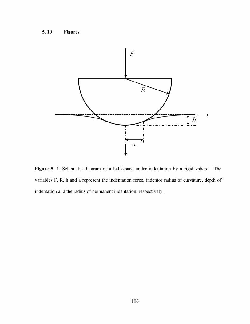
106
5. 10 Figures
Figure 5. 1. Schematic diagram of a half-space under indentation by a rigid sphere. The
variables F, R, h and a represent the indentation force, indentor radius of curvature, depth of
indentation and the radius of permanent indentation, respectively.

107
Figure 5. 2. Schematic diagram of a sectioned molar with the exposed dentin embedded in
cold cured epoxy ready for the indentation test. The letters D and E refer to dentin and enamel,
respectively.
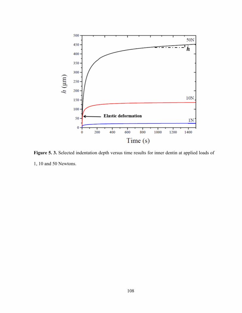
108
Figure 5. 3. Selected indentation depth versus time results for inner dentin at applied loads of
1, 10 and 50 Newtons.
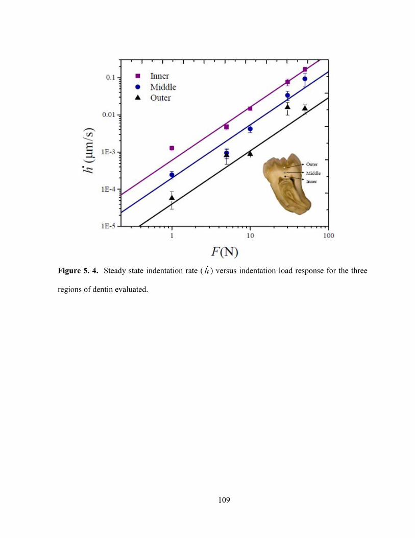
109
Figure 5. 4. Steady state indentation rate ( ) versus indentation load response for the three
regions of dentin evaluated.

110
Figure 5. 5. Comparison of the experimental steady-state effective stress and effective strain
rate of coronal dentin (markers) with predicted responses (lines).
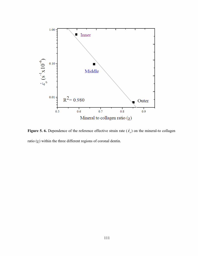
111
Figure 5. 6. Dependence of the reference effective strain rate ( ) on the mineral-to collagen
ratio () within the three different regions of coronal dentin.
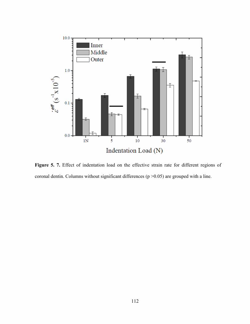
112
Figure 5. 7. Effect of indentation load on the effective strain rate for different regions of
coronal dentin. Columns without significant differences (p >0.05) are grouped with a line.

113
Figure 5. 8. Distribution of the mineral-to-collagen ratio of dentin according to the normalized
distance from the pulp.
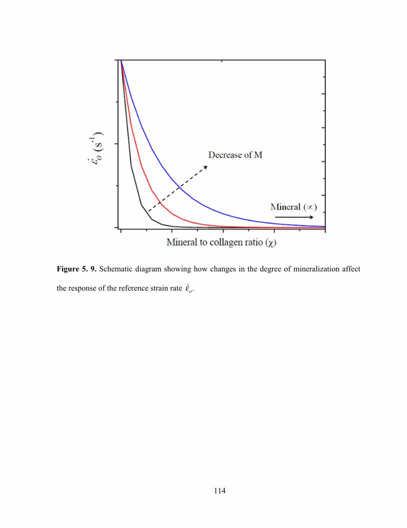
114
Figure 5. 9. Schematic diagram showing how changes in the degree of mineralization affect
the response of the reference strain rate .

115
Figure 5. 10. Experimental results reported for the steady state creep rate for bone (Rimnac et
al. 1993) and radicular dentin (Jantarat et al. 2002) and comparison with results of the current
study.

116
Figure 5. 11. Schematic representation of how the dentinal tubules are distributed in dentin. a)
and b) top views of an indentation test on the regions indicated.
117
Chapter 6
Contributions of aging to the time dependent deformation of dentin
6. 1 Introduction
Previous chapters have described the mechanical response of dentin and its dependence
on changes in the density and diameter of its dentinal tubules (i.e porosity), and changes in
chemical composition along the tooth. Several studies have previously reported the
viscoelastic properties of coronal, intertubular and peritubular dentin. However, little is known
about the time-dependent behavior of aged dentin and the importance of its spatial structure
and composition variations.
The time dependent deformation behavior of young coronal dentin and its spatial
variations along the tooth have been analyzed in chapter 5 using instrumented spherical
indentation. The experimental results indicated that dentin follows a power law deformation
behavior and that the strain rate of dentin changes along its thickness depending on the
chemical composition (e.g. mineral to collagen ratio) and microstructure (e.g. tubule density).
From these results a model to predict the time dependent deformation of dentin was proposed
and validated. Based on this model, in this chapter the spherical indentation response of aged
coronal dentin was analyzed for outer, middle and inner dentin, and its time dependent
deformation response was modeled in terms of its microstructure (i.e. tubules) and chemical
composition. Likewise, the changes that suffer the mineral and organic materials with aging
were analyzed using Raman spectroscopy.

118
6. 2 Experimental investigation
Sample collection and preparation
Human incisors and canines (N=40) and third molars (N= 10) were obtained from
selected patients after consent and following all the protocols required by the Dental Clinic at
Universidad Cooperativa de Colombia (UCC). Exclusion criteria included presence of caries
and previous restorations. The teeth were obtained from local patients (Medellín, Colombia),
and were classified in two age groups named “young” and “old” with donors between 18 and
25 years of age (molars) and 54 and 85 years of age (incisors and canines), respectively.
Before two weeks after extraction, teeth were sectioned and polished using the same procedure
explained in section 5.3.
Spherical Indentation Tests
Indentation tests of the “old” samples group were performed using the same fixture
reported in section 5.3. Indentations were made using six different loads (e.g. 1, 5, 10, 30, 50
and 60 N) applied instantaneously and then held constant until reaching a steady state
deformation regime. Between 5 and 9 tests were performed for each load and dentin depth. A
schematic of the test is shown in figure 5.1.
Chemical composition analysis
Sectioned and polished samples of outer, middle and inner regions of dentin from both
age groups were analyzed using Raman spectroscopy in order to determinate changes in the
mineral or collagen matrix with aging. Ten measurements covering the entire sample surface
were made using the same equipment previously reported (Section 5.3).

119
The spectrums were normalized and the baseline was corrected using data processing
software. Spectral decompositions and curve fitting were used in the amide I peak present in
collagen (between 1590 and 1720 cm-1) (Lefèvre et al., 2007) and in the 13PO4 bands
present in hydroxyapatite (between 990 and 1100 cm-1) (Farlay et al., 2010). The spectrums
were decomposed with mixtures of Lorentzian and Gaussian functions and the following
parameters were calculated:
Crystallinity of hydroxyapatite: Calculated as the full width at half maximum (FWHM)
of 1 peak at 960 cm-1 (Xu et al., 2012), associated to the symmetric stretches of the
phosphate group (Wopenka et al., 2008).
Carbonate-to-phosphate ratio: Calculated as the ratio of the area under the 1070 cm-1
peak associated to the symmetric stretching of the CO3 groups (B-type carbonate
apatite) (de Castro et al., 2014) and under the 960 cm-1 peak.
Collagen cross-linking: Found as the area under the 1664 cm-1 peak, corresponding to
the stretching vibration of the C=O bond of the amide I band of collagen
(Rabotyagova, 2008) and under the 1684 cm-1 peak associated to type I Beta () turns
in collagen (Gąsior-Głogowska, et al., 2013).
Mineral to collagen ratio: Calculated as the ratio of area under the 4PO4 peak at a
wavelength of 589 cm-1, which is associated with the phosphate bending of
hydroxyapatite, and the area under the amide III peak at 1254 cm-1, which is associated
with the collagen amide III band (Goodyear, et al., 2009).
Microstructural Analysis
Samples of the “old” group were used to evaluate the dentin microstructure using
optical microscopy (Axiovert 40 MAT, Carl Zeiss Microscopy, NY). Tubule density and

1 2 0
di a m et er of t h e t u b ul es w er e m e as ur e d f or e a c h r e gi o n of d e nti n, a n d t h e r es ult s w er e us e d t o
c al c ul at e t h e l u m e n ar e a fr a cti o n ( ) as t h e r ati o b et w e e n t h e ar e a o c c u pi e d b y l u m e ns a n d t h e
t ot al ar e a of d e nti n m e as ur e d .
St atisti c al A n al ysis
Diff er e n c es i n t h e r es ult s o bt ai n e d f or t h e i n di vi d u al t ests w er e e v al u at e d usi n g a o n e -
w a y A N O V A wit h a si g nifi c a n c e of 0. 0 5, a n d a T u k e y p ost -h o c a n al ysis w er e us e d t o l o c at e
diff er e n c es b et w e e n t h e gr o u ps ( y o u n g vs. ol d d o n ors) .
6. 3 E x p e ri m e nt al r es ults
S p h eri c al I n d e nt ati o n
Fi g ur e 6. 1 s h o ws t h e e v ol uti o n of i n d e nt ati o n d e pt h (h ) as a f u n cti o n of ti m e f or
s el e ct e d s a m pl es of t h e ol d d o n ors gr o u p at diff er e nt r e gi o ns of d e nti n a n d at a c o nst a nt l o a d
of 6 0 N f or a p eri o d of 9 0 0 s. All t h e s a m pl e s s h o w e d a n i niti al “i nst a nt a n e o us ” el asti c
d ef or m ati o n f oll o w e d b y a d e cr e asi n g i n d e nt ati o n r at e ( pri m ar y or tr a nsi e nt r e gi m e).
T h er e aft er t h e r es p o ns e e x hi bit e d a n a p pr o xi m at el y c o nst a nt i n d e nt ati o n r at e c orr es p o n di n g t o
t h e st e a d y-st at e r e gi m e of d ef or m ati o n .
Fr o m e a c h of t h e t ests p erf or m e d , t h e i n d e nt ati o n r at e ( ) w as e xtr a ct e d as t h e sl o p e i n
t h e st e a d y-st at e r e gi o n. Fr o m t h e r es ult s, a si g nifi c a nt di ff er e n c e i n t h e i nd e nt ati o n r at e w as
o bt ai n e d o nl y f or s o m e r e gi o ns a n d s o m e i n d e nt ati o n l o a ds , i m pl yi n g t h at t h e i n d e nt ati o n
d e pt h d o es n ot c h a n g e s p ati all y si g nifi c a ntl y wit h i n d e nt ati o n l o a d.
Usi n g t h e d at a o bt ai n e d fr o m t h e s p h eri c al i n d e nt ati o n t ests p erf or m e d o n a g e d d e nti n,
s efft h e eff e cti v e str ess ( ) a n d t h e eff e cti v e str ai n r at e ( ) w er e c al c ul at e d usi n g E qs. ( 5 .4 )
a n d ( 5 .6 ), r es p e cti v el y t o g et t h e r es ult s s h o w n i n fi g ur e 6 . 2. T h e r es ult s s h o w t h at t h e r el ati o n
1 2 1
s effb et w e e n a n d of a g e d d e nti n f oll o w a n a p pr o xi m at e li n e ar b e h a vi or i n t h e l o g -l o g pl ot
t h at c a n b e fitt e d t o a p o w er l a w r es p o ns e wit h a cr e e p e x p o n e nt n = 2. 1 .
Fi g ur e 6. 3 s h o ws a c o m p aris o n b et w e e n t h e v al u es of str ai n r at e o bt ai n e d f or t h e
diff er e nt r e gi o ns of d e nti n wit h t h e a p pli e d i n d e n t ati o n l o a ds. A n i n cr e a s e i n str ai n r at e w as
f o u n d as t h e i n d e nt ati o n l o a d i n cr e as es f or t h e s a m e r e gi o n of d e nti n. F urt h er, t h e diff er e nt
r e gi o ns of d e nti n s h o w a d e cr e as e i n t h e str ai n r at e fr o m i n n er t o o ut er d e nti n usi n g t h e s a m e
i n d e nt ati o n l o a d. T h es e res ult s s h o w si g nifi c a nt diff er e n c es i n s o m e c as es ( p -v al u e ≤ 0. 0 5).
C ol u m ns wit h o ut si g nifi c a nt diff er e n c es ( p ≤ 0. 0 5) ar e gr o u p e d wit h a li n e . T h e l o w st atisti c al
si g nifi c a n c e of t h e d at a s h o ws t h at t h e ol d d e nti n d o es n ot h a v e si g nifi c a nt c h a n g es al o n g
di ff er e nt r e gi o ns of d e nti n o n it s ti m e d e p e n d e nt b e h a vi or. T h es e littl e diff er e n c es b et w e e n t h e
t hr e e r e gi o ns of d e nti n m a y als o b e s e e n i n fi g ur e 6. 2 w er e t h e d at a of t h e t hr e e diff er e nt
r e gi o ns ar e cl os e t o e a c h ot h er.
C h e mi c al c o m p ositi o n a n al ysis
T h e r e s ult s o bt ai n e d f or s o m e of t h e d e nti n c h ar a ct eristi cs usi n g R a m a n S p e ctr os c o p y
ar e s h o w e d i n fi g ur e 6. 4. T h e r es ult s o bt ai n e d f or t h e cr yst alli nit y of h y dr o x y a p atit e ( F W H M
of t h e 1 p e a k) as a f u n cti o n of dist a n c e a cr oss t h e c or o n al d e nti n f or y o u n g a n d ol d d o n or
t e et h ar e s h o w n i n Fi g ur e 6. 4 a . D e nti n i n t h e y o u n g d o n or gr o u p s h o w e d a si mil ar v al u e f or
t h e t hr e e r e gi o ns of st u d y; m e a n w hil e , f or t h e ol d d o n or gr o u p w as f o u n d a n i n cr e as e i n t h e
F W H M as a p pr o a c hi n g t h e D EJ. A hi g h er F W H M ( wi d er p e a k ) i n di c at es l o w er cr yst alli nit y.
A d e cr e as e i n cr yst alli nit y d e n ot es t h at a gr e at er pr o p orti o n of cr yst al s h a v e l ess p erf e ct
l atti c es, wit h hi g h er s u bstit uti o ns ( F arl a y et al. , 2 0 1 0). T h e diff er e n c es f o u n d a m o n g d e nti n
r e gi o ns i n t h e y o u n g d o n ors gr o u p w er e n ot st atisti c all y si g nifi c a nt ( p > 0. 0 5), as o p p os e d t o t h e
ol d d o n ors gr o u p w h er e t h er e w as a st atisti c all y si g nifi c a nt i n cr e as e i n t h e F W H M f or t h e

122
outer dentin when comparing with the middle and inner regions of the aged tissue. On the
other hand, when analyzing the results with respect to the specific regions of dentin between
young and old donors, significant differences were only found for the outer dentin between
young and old donors.
Carbonate-to-phosphate ratio results for the three different regions of coronal dentin are
presented in Figure 6.4b. The carbonate-to-phosphate ratio is related with the substitution of
the PO43- group by CO3
2- (B-type carbonate apatite) groups (Penel et al., 1998). An increase in
this ratio (an increase in the substitution) is normally associated with a mineral material with
increased amorphous regions (Sahar, 2009). The results obtained for the young donor group
showed a similar value for the three regions of dentin with no statistically significant
differences (p>0.05). On the other hand, for the old donor group, an increase in the carbonate-
to-phosphate ratio was found from inner to outer dentin with statistically significant
differences between the three different regions (p≤0.05). When comparing the results between
young and old donors (for specific regions), significant differences were found for the young
and old outer dentin.
Figure 6.4c shows the results obtained for collagen cross-linking. An increase in
collagen cross-linking has shown to be proportional to the relative amounts of the trivalent
cross-links in the collagen matrix (Mandair and Morris, 2015). The fibril structure of collagen
tends to form intermolecular and interfibrillar cross-links, in this process the divalent cross-
links present in the structure react with another telopeptide aldehyde group to form a trivalent
mature bond linking three tropocollagen molecules (Eyre and Wu, 2005; Depalle et al., 2015).
The results for the young donors group are not statistically significant (p>0.05) finding the
same values for the three regions of dentin; but when comparing the collagen cross-linking
between young and old patients, nearly a 29% difference was found near the pulp at inner

123
dentin. However, the difference in the collagen cross-linking was approximately 4% at middle
and 30% at outer dentin.
Figure 6.4d shows the distribution of mineral-to-collagen ratio () of dentin as a
function of the normalized distance from the pulp to DEJ - measured in the direction shown
in fig. 5.2 -. For both age groups an increase in mineral-to-collagen ratio approaching the DEJ
was found. A higher mineral-to-collagen ratio indicates a higher proportion of inorganic
material. On the young donor group the differences found were not statistically significant
along the crown. While for the old donor group, an increase in the mineral to collagen ratio is
observed when compared to young dentin. This increase is more evident as approaching the
DEJ (outer dentin). An increase of 15% was found for inner dentin while for the middle and
inner regions an increase of ~62% was found.
When comparing the results between the young and old donor groups, significant
differences were found for the outer and middle dentin when comparing specific regions.
Microstructure
The microstructure of dentin from a selected old donor incisor evaluated is shown in
Figure 6.5. Representative micrographs correspond to the three regions of evaluation; outer
(Fig. 6.5b), middle (Fig. 6.5c) and inner (Fig. 6.5d) dentin. For each region, a peritubular
dentin cuff can be seen surrounding each dentinal tubule, being more evident this cuff in the
middle and outer regions. Several obliterated tubules are evident in the images corresponding
to outer and middle dentin, an example of some obliterated dentinal tubules found in outer
dentin is also shown in fig. 6.5a.
From the microstructural analysis was found a reduction in the tubule density with
increasing proximity to the DEJ. The average tubule density in the outer and inner regions was
124
of 14,000 tubules/mm2 and 29,800 tubules/mm2, respectively. As for the diameter of dentinal
tubules, an average of 1.84 ± 0.18 µm was found for inner dentin and 1.16 ± 0.17 µm for outer
dentin. For both characteristics, the differences obtained along different regions of dentin were
statistically significant (p ≤ 0.05). From these results the lumen area fraction () at each depth
was calculated. The average for outer and inner regions being of 1.15±0.30% and
7.82±1.00%, respectively (p ≤ 0.05).
6. 4 Discussion
The time dependent deformation behavior of aged coronal dentin was studied using
spherical indentation. The experimental results obtained for the indentation depth versus time
were used to calculate the steady-state strain rate of dentin in three different regions - inner,
middle and outer dentin-. The relationship between effective stress and effective strain rate
showed that old dentin exhibits a non-linear behavior with a creep rate exponent of n=2.1 and
was found to be independent of the region of dentin analyzed, finding similar results for inner,
middle and outer regions. The creep exponent n governs the non-linear behavior of a material
with a creeping solid behavior and can range from n =1, associated with a linear viscous solid,
to n= representing a perfectly viscous solid.
According to the model proposed in chapter 5 (section 5.5), the time dependent loading
behavior of dentin can be described using different tissue parameters depending not only on
the creep exponent of the tissue, but on its chemical composition and microstructural features.
With the results obtained for the microstructure and chemical composition of aged dentin, the
model parameters (Eqs. 5.12 and 5.13) were calculated and the results are listed in table 6.1.
These parameters were then used to describe the time dependent behavior of aged dentin in the
normalized positions =0.25 (for inner dentin), =0.55 (for middle dentin) and =0.85 (for
1 2 5
o ut er d e nti n). T h e r es ult s ar e als o s h o w n i n fi g ur e 6. 2 (s oli d li n es). Fr o m t h e r es ult s, t h er e c a n
b e s e e n a g o o d a gr e e m e nt b et w e e n e x p eri m e nt al r es ult s a n d m o d el pr e di cti o ns f or t h e i nn er
a n d o ut er d e nti n , fi n di n g r 2 = 0. 9 6 7 f or i n n er a n d 0. 9 5 0 f or o ut er d e nti n, w hil e f or mi d dl e
d e nti n t h e r 2 w as 0. 7 6 1 .
I n or d er t o d et er mi n at e h o w t h e a gi n g pr o c ess aff e cts t h e ti m e d e p e n d e nt b e h a vi or of
d e nti n, th e r es ult s o b t ai n e d f or t h e ol d d e nti n d o n ors w er e c o m p ar e d wit h t h os e o bt ai n e d
pr e vi o usl y f or y o u n g d e nti n, as s h o w n i n fi g ur e 6. 6. Fr o m t h es e r es ult s, a d e cr e as e i n - f or
s efft h e s a m e - c a n b e s e e n f or t h e i n n er a n d mi d dl e d e nti n. W hil e f or o ut er d e nti n a n i n cr e as e
i n w as o bs er v e d f or t h e ol d d o n ors gr o u p. A d e cr e as e o n is r el at e d wit h a l o w er
a bilit y t o u n d er g o ti m e -d e p e n d e nt d ef or m ati o n of t h e ti ss u e. T his b e h a vi or f or t h e i n n er a n d
mi d dl e d e nti n c a n b e attri b ut e d n ot o nl y t o t h e c h a n g e i n t u b ul e d e nsit y d u e t o o blit er ati o n of
d e nti n al t u b ul es, b ut t o t h e c h a n g es i n c h e mi c al c o m p ositi o n - i n cr e as e i n mi n er al c o nt e nt as
s h o w n i n fi g. 6. 4d - a n d or g a ni z ati o n a n d i nt er a cti o n b et w e e n t h e mi n er al a n d or g a ni c p h as e s.
W hil e t h e r es ult s o bt ai n e d f or o ut er d e nti n ar e c o ntr ar y t o w h at w o ul d b e e x p e c t e d f or t hi s
r e gi o n, w h er e it is w ell k n o w n t h at t h e a gi n g pr o c ess of c or o n al d e nti n b e gi ns ( o blit er ati o n of
d e nti n al t u b ul es b e gi ns n e ar t h e D EJ a n d pr o gr ess t o w ar ds t h e p ul p) ( Dr usi ni et al. , 1 9 9 1 ); t hi s
p att er n of a gi n g will i m pl y a hi g h er c h a n g e i n t h e vis c o el asti c pr o p erti es of o ut er d e nti n wi t h
a gi n g, w hi c h i n t his c as e w as n ot f o u n d.
T h e m ost o b vi o us c h a n g e o bs er v e d w h e n c o m p ari n g t h e ti m e d e p e n d e nt d ef or m ati o n of
y o u n g a n d ol d d o n or s’ d e nti n is t h e c h a n g e i n t h e cr e e p r at e e x p o n e nt ( n ). T h e cr e e p r a t e
e x p o n e nt f or y o u n g d e nti n h as b e e n pr e vi o usl y r e p ort e d t o h a v e a v al u e of 3. 3 8 , w hil e t he
v al u e o bt ai n e d f or t h e ol d d o n or gr o u p w as f o u n d t o b e of 2. 1 . Pr e vi o us st u di es o n t h e ti m e
d e p e n d e nt d ef or m ati o n of y o u n g d e nti n ar g u e d t h at t h e vis c o el asti c b e h a vi or of d e nti n is
126
mainly attributed to the creep characteristics of the collagen type I present in the tissue
(Kinney et al., 2003; Chuang et al., 2015; Bertassoni et al., 2015). Therefore, this decrease in
the creep exponent with aging implies that the collagen of the tissue is suffering significant
changes in its deformation capacity over time. The change in collagen characteristics can be
seen in Figure 6.4c, where the collagen crosslinking for different regions of dentin are shown.
Although no statistically significant differences were found between different regions of
dentin, an increase in crosslinking is observed when comparing young and old dentin groups.
Moreover, an increase in the amount of mineral material (increase in mineral-to-collagen ratio
showed in figure 6.4d) may also have an effect on the deformation capability of collagen when
subjected to stresses. Since the collagen present in intertubular dentin form a three-
dimensional network reinforced by apatite crystals (Bertassoni et al., 2012), an increase in the
mineral material or more imperfect crystals (as showed in fig. 6.4a and 6.4b with a decrease in
crystallinity and an increase in the carbonate-to-phosphate ratio respectively) could have an
effect on the interaction between the two phases and a change in the mobility of the softer
organic phase.
From the results obtained in the chemical composition analysis was found an increase
with aging in the Carbonate-to-phosphate ratio, product of the filling of dentinal tubules with
carbonated apatite (fig. 6.4b), being more significant at outer dentin where the obliteration was
higher. This change has a direct effect on the crystallinity and perfection of the HA lattice
which is in agreement with the results reported in fig. 6.4a. A decrease in crystallinity and an
increase in the Carbonate-to-phosphate ratio have been linked to a detriment in mechanical
properties such as hardness and elastic modulus in other hard tissues (i.e. enamel) (Xu et al.,
2012); which could explain in part the low change in the time dependent behavior of outer
dentin. However, the results obtained for the decrease in crystallinity of dentin by Raman

127
spectroscopy could be the result of two phenomena reported in literature that are contrary to
each other. For intertubular dentin a decrease in the crystal width of the hydroxyapatite has
been reported (which increases crystallinity), while on the other hand the dentinal tubules are
occluded by larger crystals when comparing to normal dentin (decreases crystallinity); this
phenomenon was reported by Porter et al. (2005) who argued that smaller crystals on
intertubular dentin could be the result of a dissolution process of the mineral from intertubular
dentin, which is then deposited into the lumens resulting in the obliteration process. This
dissolution and re-precipitation of minerals in the peritubular cuff can reduce the poroelastic
processes related to water movement in dentin, as the precipitated particles can fill the
transverse secondary tubules and restrict water and dentinal fluid movement, which can have
an impact on the deformation capability of the tissue.
In this study the differences found in the time dependent behavior between young and
old dentin specimens is not limited to age, some differences can be attributed to the intrinsic
differences between a molar tooth (young samples) and incisors and canines (old samples) and
the use to which the samples have been subjected. In the case of third molars (young donors
group) these samples can be regarded as "new" teeth that have not been subjected to
mastication stresses. On the other hand, the incisors and canines used for testing the elder
donor’s groups are samples that can be considered as "used”, with a possible cumulative
damage generated not only by mastication but by brushing (Addy, 2005) that can produce
enamel wear (Liu et al., 2014). These changes can be denominated “dental erosion” which is a
non-carious deterioration process that can include: abrasion, demastication, attrition,
abfraction, resorption and erosion (Imfeld, 1996). Dental erosion does not only affect enamel,
but can also affect dentin, causing hypersensitivity, or in extreme cases, pulp exposure and
even tooth fracture (Addy et al., 1987). The surface of erosive lesions is hypo-mineralized and

128
its incidence can increase by consuming soft drinks, sugar and bleaching procedures; and
although it can be found at any age, in the elderly is a normal feature. Some previous studies
have reported that aging teeth tend to develop more dental erosion on its occlusal and incisal
surfaces. However, these studies also suggest that more evidence is required to ensure that age
is a determining factor in dental erosion (Bartlett and Dugmore, 2008). Since the indentation
tests performed in this study were superficial and the indentation depth was only of a few
microns, if the tested tissue has a significant dentinal erosion which penetrates dentin, it seems
normal to think that this dentin will display higher indentation depths and strain rates.
Likewise, differences would also be expected between the two groups of samples
(incisors/canines and molars) in terms of the orientation of the incremental lines -that show the
deposition pattern of secondary dentin-, collagen fibril (Arola et al., 2009), and between
tubule density, diameter of dentinal tubules and chemical composition. The differences
between old molars, incisors and canines in terms of microstructural characteristics are
presented in figure 6.7. The results correspond to the distribution of their lumen area fraction
() across the three regions of aged coronal dentin. Overall, there was a significant decrease in
with proximity to the DEJ for both groups (p ≤ 0.05). In the molar group the average is
higher when comparing with the incisors and canines group. Values of for outer dentin of
2.86% and 1.15% were found for molars and for the canines and incisors group, respectively.
The mechanisms contributing to the changes in the time dependent deformation of
dentin and its spatial changes with aging are not clearly understood and there is no consensus
on how each mechanism affect the mechanical behavior of the tissue, which one is the primary
mechanism or if there is a balance between them. Decrease in crystallinity and increase in the
carbonate-to-phosphate ratio could promote deformation of dentin. While increase in mineral

129
content, obliteration of dentinal tubules with sclerotic dentin with lower crystal size and
collagen cross-linking have the opposite effect. Further work is necessary to isolate the
mechanisms responsible for the changes on the mechanical behavior of dentin with aging and
in order to understand what happen with these properties between the ages of 30 and 50 years,
where there is little available information.
6. 5 Conclusions
According to the results obtained, the following conclusions can be drawn:
1. The time dependent deformation and chemical composition of aged dentin was
evaluated in outer, middle and inner coronal dentin. The experimental results
showed to follow a power law deformation behavior with a decrease in the stress
exponent when comparing with young dentin.
2. Significant changes in the mineral-to-collagen ratio (increase), crystallinity
(decrease) and carbonate to phosphate ratio (increase) were found with increasing
donor age. Most of these changes were found to be more significant in the middle
and outer dentin.
3. A model previously proposed to predict the time dependent deformation response
of dentin was employed to model the creep behavior of aged dentin. The model
showed a strong correlation between the experimental and the predicted results. A
decrease in the time-dependent behavior of middle and inner aged dentin was found
when compared with young dentin.

1 3 0
6. 6 T a bl es
T a bl e 6. 1 . D e nti n p ar a m et ers o bt ai n e d t o d es cri b e t h e ti m e d e p e n d e nt b e h a vi or of a g e d
d e nti n.
P a r a m et e r V al u e
2. 1 0
2. 2 2
0. 9 3
s o
( M P a) 0. 5 0
(s-1 ) 8. 0 x 1 0 -7
1. 1
0. 9 9

131
6. 7 Figures
Figure 6. 1. Selected indentation depth versus time results for different regions of dentin at a
constant applied load of 60N. The results correspond from an incisor of a 70-year-old donor.

132
Figure 6. 2. Comparison of the experimental steady-state effective stress and effective strain
rate of coronal aged dentin (markers) with predicted responses (lines).

133
Figure 6. 3. Effect of indentation load on the effective strain rate of different regions of
coronal dentin. Columns without significant differences (p ≤ 0.05) are grouped with a line.

134
Figure 6. 4. Comparison of some dentin characteristics obtained using Raman Spectroscopy
as a function of depth in the coronal dentin. a) Full width at half maximum (FWHM) of the 1
peak; b) Carbonate-to-phosphate ratio; c) Collagen cross-linking; d) Mineral-to-collagen ratio.
Columns without significant differences are grouped with a line.

135
Figure 6. 5. Micrographs of the dentin microstructure. a) Obliterated dentinal tubules; b)
Outer dentin; c) Middle dentin; Inner dentin. Note the obliterated dentinal tubules for the
middle and outer regions. The percentages correspond to the fraction lumen area obtained for
each region of dentin.

136
Figure 6. 6. Comparison of the effective stress and effective strain rate of young (lines) and
old (markers) dentin.

137
Figure 6. 7. Comparison of fraction lumen area () of old incisors and canines and molars.
138
Chapter 7
Ethnic Background influence on the aging process of dentin: Preliminary
Results
7. 1 Introduction
Although the spatial variations in microstructure of dentin were well-recognized and
extensively studied in chapter 3 for dentin from Colombian donors. Some authors have found
significant differences between ages and sexes, when comparing results obtained for some
dental features (Kumagai et al., 2012). For example, some authors have suggested that a
relation between ethnicity and race and some dental characteristics, such as tooth size (Merz et
al., 1991; Altherr et al., 2007; Bishara et al., 1989), enamel thickness (Hall et al., 2007) and
tooth formation rate (Olze et al., 2007) might exist. For instance, Merz et al. (1991) found that
dental arches in black patients are significantly wider and deeper than those from white
patients; similar results were found by Altherr et al. (2007), who reported larger teeth for
black male patients in comparison with white donor samples. On the other hand, possible
differences between tooth dimensions of permanent teeth in three ethnic groups: Egyptians,
Mexicans, and the North Americans were analyzed by Bishara et al. (1989), finding not only
significant differences between the three populations in tooth dimensions but between sexes,
with males having larger canines and first molars than women. Regarding the dimensions of
dental tissues; Hall et al. (2007) found thicker enamel on the distal aspect of a black donor

139
tooth when comparing with white donors, this thicker enamel explained in some cases wider
teeth, but not in all cases.
Most previous research on dentin has been made mostly from American donors.
However, previous results reported on the chemical composition and microstructure of
Colombian dentin has shown different results not only for the chemical composition but for
some microstructural characteristics. For example, the results showed in Chapter 3 for the
mineral-to-collagen ratio of Colombian dentin showed an increase in the mineral material as
one approaches the DEJ, however the differences reported in this study along the crown were
not statistically significant. Nevertheless, Ryou et al. (2011) found for samples of American
(US) donors a lower amount of mineral material near DEJ when comparing with the regions
near the pulp, and its differences were statically significant. Also, some microstructural
differences have been found regarding tubule density. For instance, Ivancik et al. (2014) found
significant differences in the tubule lumen diameters between the dentin of two groups from
US and Colombia; while the US samples showed a significant change in the diameter of the
lumens from the pulp to the DEJ, the average lumen diameter of dentin from the Colombian
donors did not change significantly.
Despite of this, only a few studies on dentin mechanical behavior and how it might be
affected by ethnicity have been carried out. For instance, Ivancik et al. (2014) found that the
fatigue resistance properties of dentin from American donors are dependent on the location
(varies from pulp to the DEJ), while Colombian dentin showed a homogeneous behavior
without significant spatial variations. On the other hand, a comparison of the rate of crack
growth between American and Chinese donors found that the coronal dentin of molars
obtained from Chinese donors was larger than that of US patients. However, the
microstructural analyses showed no significant differences in the average lumen diameter for

140
the two groups (Bajaj et al., 2008). These differences have been explained not only by the
ethnic backgrounds of the patient but by individual aspects as diet, water quality (fluoride
content) and hygiene habits (Nikiforuk, 1970; Robinson et al., 2004; Vieira et al., 2005).
It is well known that the aging process of coronal dentin begins in the outer regions of
the crown - near the DEJ - and its microstructural changes lead to a reduction in the fatigue
strength and fracture toughness of dentin. Further, as the information comparing both
microstructure and mechanical properties of dentin donors with different ethnic backgrounds
is scarce, this chapter aims to compare the microstructure, chemical composition and
mechanical properties (i.e. time dependent behavior) of outer dentin from residents of South
America (Colombia), North America (USA) and Asia (China) in order to explore the potential
importance of ethnic background and their dental hard tissues specific characteristics.
7. 2 Experimental investigation
Human dental pieces were obtained from selected patients after written consent and
following all the protocols required and approved by the IRB of the Dentistry Clinic
at Universidad Cooperativa de Colombia (UCC) located in Medellin (Colombia), University
of Washington School of Dentistry located in Seattle (United States) and in the Tianjin
Stomatology Hospital of Nankai University located in Tianjin, (China). Exclusion criteria
included presence of caries and previous restorations.
The teeth were divided into three groups, named “Colombia” (N=20), “US” (N=21)
and “China” (N=19) depending on the place of origin of the donor. Samples were in an age
range between 18 and 85 years and there were almost an equal number of male and female
samples in each group. The samples were also classified according to their age and named as:
young (<30 years), middle age (30- 50 years) and old samples (> 50 years).

141
Immediately after extraction, all the specimens were prepared and tested according to
the procedure explained in section 5.3 for indentation tests, microstructural and chemical
composition analysis.
7. 3 Experimental results
Microstructure
The microstructure of dentin from selected samples of each age group is shown in
Figure 7.1. The microstructures shown for each group correspond to outer dentin from China
(Fig 7.1a, 7.1b and 7.1c), Colombia (Fig 7.1d, 7.1e, and 7.1f) and United States (US) (Fig
7.1g, 7.1h and 7.1i). As evident from the figures, a peritubular dentin ring can be seen
surrounding each dentinal tubule regardless of donor age. When comparing the samples
according to age, dentinal tubules in the young samples seem to be open, while as for the old
samples a considerable number of obliterated dentinal tubules can be seen.
A quantitative comparison of the microstructural characteristics of dentin and how it
changes according to age is shown in Figures 7.2. The tubule density in the three origin groups
and ages can be seen in Figure 7.2a, where a reduction in tubule density can be observed with
increasing donor age in all three origin regions. For the young donor’s groups, the average
tubule density was 22,000 tubules/mm2 without significant differences between the three
origins analyzed (p>0.05). While for the old group, an average of 14,000 tubules/mm2 was
found, with a significant difference in the US group; finding an average of 9,500 tubules/mm2.
A comparison of the average tubule diameter for the three age groups is shown in
Figure 7.2b. In the young donor group, the average lumen diameter was 1.34 ± 0.02 µm, with
very few differences between the three groups of origin. While for the old group an average of
1.23 ± 0.02 µm was found. For the middle-age group an average diameter of 1.22 ± 0.09 µm

1 4 2
w as f o u n d b ut wit h a hi g h er v ari ati o n , w hi c h s h o ws t h at d uri n g t hi s a g e t h er e is a tr a nsiti o n i n
t h e ti ss u e wit h s o m e p arti all y o p e ne d a n d cl os e d t u b ul es. O v er all, t h er e is a r e d u cti o n i n t u b ul e
d e nsit y a n d di a m et er of d e nti n al t u b ul es f or t h e t hr e e r e gi o ns of ori gi n a n al y z e d wit h
i n cr e asi n g d o n or a g e (p ≤ 0. 0 5) . A c o m p aris o n of t h e r es ult s a c c or di n g t o t h eir or i gi n s h o ws
t h at t h e diff er e n c es f or t u b ul e d e nsit y a n d di a m et er of d e nti n al t u b ul es f or t h e y o u n g s a m pl es
w er e n ot st atisti c all y si g nifi c a nt ( p > 0. 0 5), w hil e f or t h e ol d s a m pl es a l o w er di a m et er of
d e nti n al t u b ul es w as f o u n d f or t h e U S gr o u p (p ≤ 0. 0 5 ).
S p h eri c al I n d e nt ati o n T ests
Fi g u r e 7. 3 s h o ws t h e e v ol uti o n of i n d e nt ati o n d e pt h ( h ) i n d e nti n as a f u n cti o n of ti m e
f or s el e ct e d a p pli e d l o a ds of a n ol d s a m pl e fr o m C hi n a. T h e r es ult s of all t h e gr o u ps a n al y z e d
s h o w e d a n i niti al “i nst a nt a n e o us ” el asti c d ef or m ati o n f oll o w e d b y a d e cr e asi n g i n d e nt ati o n
r at e (tr a nsi e nt cr e e p r e gi m e). T h er e aft er, t h e r es p o ns e e x hi bit e d a n a p pr o xi m at el y c o nst a nt
i n d e nt ati o n r at e c orr es p o n di n g t o t h e st e a d y-st at e cr e e p r e g i m e. T h e i n d e nt ati o n r at e ( ) w as
e xtr a ct e d fr o m t h e st e a d y -st at e r e gi o n of e a c h t e st p erf or m e d. Fr o m t h e e x p eri m e nts w er e
o bt ai n e d s efft h e eff e cti v e str ess ( ) a n d t h e eff e cti v e str ai n r at e ( ) b y usi n g E q s. ( 5. 4 ) a n d
( 5. 6), r es p e cti v el y t o g et t h e r es ult s s h o w n i n fi g ur e 7. 4 . T h e r es ult s o bt ai n e d f or all t h e gr o u ps
f oll o we d a p o w er l a w b e h a vi or. T h e i n di vi d u al p o w er -l a w e x p o n e nts o bt ai n e d f or e a c h gr o u p
ar e li st e d i n T a bl e 7. 1. T h e r es ult s c orr es p o n d o nl y t o t h e ol d a n d y o u n g s a m pl es gi v e n t h e
diffi c ult y t o fi n d e n o u g h s a m pl es t o b uil d t h e mi d dl e-a g e gr o u p r es p o ns e.
Fr o m t h e r es ult s c a n b e s e e n t h at t h e y o u n g d e nti n s h o w e d a si mil ar p o w er -l a w
e x p o n e nt r e gar dl ess of t h e d o n or ori gi n, fi n di n g a n a v er a g e v al u e of 3. 1 1 . O n t h e ot h er h a n d, a
r e d u cti o n i n th e p o w er -l a w e x p o n e nt w as f o u n d f or t h e ol d gr o u p w h e n c o m p ari n g wit h t h e
y o u n g d o n ors f or all ori gi ns a n al y z e d. W h e n c o m p ari n g t h e r e d u cti o n b et w e e n ol d a n d y o u n g

143
donors according to their origin, a reduction of almost of 50% was found for the China and US
group, while for the Colombia group a reduction of only 27% was found.
Chemical Composition
Figure 7.5 shows the results obtained for the mineral-to-collagen ratio from young and
old donor teeth for the three origin regions analyzed. An increase in the mineral-to-collagen
ratio was found when comparing the young and old donors’ groups and independent of the
region of origin. A higher mineral-to-collagen ratio is related with a more mineralized tissue
(i.e. higher mineral content). This increase in the mineral content with aging is related with the
obliteration of dentinal tubules due to the deposition of secondary dentin. An increase of
around 55% was found for the Colombia and China groups in comparison with young tissues,
while for the US group the mineral-to-collagen ratio is twice higher in the old donors than in
the young samples.
When comparing the results for young samples according to their region of origin, the
US and Colombia samples showed to be statistically equal (p>0.05), while the China samples
have a statistically higher mineral-to-collagen ratio when comparing with the other two
regions (~40% higher mineral-to- Collagen ratio). For the old donor’s samples, the China and
US samples are statistically equal (p>0.05); meanwhile the Colombian samples are statistically
lower.
7. 4 Discussion
Only a few studies have been developed aiming at understand the effect of ethnic
differences on the microstructure and mechanical properties of dentin. In this study, the
microstructure, chemical composition and viscoelastic properties of outer dentin of three
different regions (China, Colombia and United States) were studied.

144
The microstructure for the young samples (<30 years) were within the ranges
previously reported for tubule density and diameter of dentinal tubules regardless of the origin
of the donor (Pashley, 1989, 2015; Ivancik et al., 2014). Average values of 22,000
tubules/mm2 and 1.33 µm were found for the tubule density and diameter of dentinal tubules,
respectively. Surprisingly, although the microstructure characteristics were consistent between
the three origin groups in the young samples, for the old samples (>50 years) a decrease in
tubule density was found, but this decrease is more evident for the US group. For the
Colombian and Chinese groups a decrease of approximately 30% in tubule density compared
to the group of young donors was found, while for the US donors a decrease of almost 50%
was found. In the case of the diameter of dentinal tubules, a similar decrease (~7.0%) was
found for the three origin regions when comparing with the young samples.
A decrease in the microstructural parameters was also found for the middle-age group.
However, as can be seen in Figure 7.2b, the data corresponding to the diameters of the
dentinal tubules show a high standard deviation. This variation may be related to the fact that a
transition occurs in the microstructure in that age range (30-50 years) in which there is a
variety of partially open tubules and with different diameters. This decrease in tubule density
and in the diameter of dentinal tubules for the old and middle-age groups is related with the
progressive deposition of secondary dentin since the tooth enters into occlusion and continues
throughout life; this dentin is deposited in dentinal tubules decreasing its diameter
progressively and in the long term obliterating it completely (Gómez de Ferraris and Campos
Munoz, 2009).
In order to identify how significant is the change in tubule density taking place with
aging, with respect to the amount of initial dentinal tubules (young donors), the proportion of
obliterated dentinal tubules was calculated for each sample according to:

1 4 5
M e as ur e d esti m at es f or t h e pr o p orti o n of o blit er at e d d e nti n al t u b ul es f or e a c h s a m pl e
w er e e v al u at e d a n d s h o w n i n Fi g ur e 7. 6 . As e x p e ct e d, t h e pr o p orti o n of o blit er at e d d e nti n al
t u b ul es i n t h e y o u n g d o n or’s gr o u ps is a p pr o xi m at el y z er o i n all t hr e e ori gi n r e gi o ns u ntil
a b o ut 3 0 y e ars of a g e , w h er e a n i n cr e as e i n t h e n u m b er of o blit er at e d d e nti n al t u b ul es ca n b e
s e e n. I n t h e C hi n es e gr o u p, a m a xi m u m pr o p orti o n of o blit er at e d d e nti n al t u b ul es of 4 7 % w as
f o u n d i n a 5 3 -y e ar -ol d d o n or . Whil e f or t h e U S , t h e m a xi m u m v al u e o bt ai n e d w as 4 1 % i n a
4 7 -y e ar -ol d ( alt h o u g h t h e ol d est s a m pl e is of 8 3 y e ars), a n d i n t h e Col o m bi a n s a m pl es t h e
m a xi m u m o blit er ati o n f o u n d w as 9 % i n a 4 7 -y e ar -ol d s a m pl e . T h e a n al ysi s of t hi s r es ult s a n d
h o w f ast o blit er ati o n of d e nti n al t u b ul es o c c urs, dr a w att e nti o n t o t w o as p e cts: Th e i n cr e as e o n
t h e pr o p orti o n of o blit er at e d d e nti n al t u b ul es wit h a g e is hi g h er i n t h e C hi n a a n d U S gr o u p s
w h e n c o m p ari n g w it h t h e C ol o m bi a n s a m pl es. O n t h e ot h er h a n d, t h e o blit er ati o n of d e nti n al
t u b ul es d o es n ot s e e m t o i n cr e as e pr o p orti o n at el y wit h a g e, as s o m e s a m pl es of t h e mi d dl e -
a g e gr o u p h a v e hi g h er o blit er ati o n t h a n s a m pl es of al m ost 9 0 y e ars; t hi s r es ult m a y i m pl y t h at
t h e o blit er ati o n of d e nti n al t u b ul es ( a gi n g) c o ul d n ot o nl y d e p e n d o n t h e d o n or a g e b ut o n
s o m e c h ar a ct eristi cs a n d c ust o m s of t h e d o n or.
T h e vis c o el asti c b e h a vi or of o ut er -y o u n g a n d ol d - d e nti n w as st u di e d usi n g s p h eri c al
i n d e nt ati o n, t h e r es ult s o bt ai n e d i n t er ms of i n d e nt ati o n d e pt h a n d ti m e w er e us e d t o c al c ul at e
t h e eff e cti v e str esses s eff( ) ( E q. 5. 4) a n d str ai n r at e s ( ) ( E q. 5. 6 ); usi n g t h e s ol uti o n
pr e vi o usl y pr o p os e d f or t h e s p h eri c a l i n d e nt ati o n of a cr e e pi n g s oli d m at eri al (M ul h e ar n a n d
T a b or, 1 9 6 0) T h e r el ati o ns hi p b et w e e n eff e cti v e str ess a n d eff e cti v e str ai n r at e pr es e nt e d i n
fi g ur e 7. 4 s h o w e d t h at all t h e gr o u ps of d e nti n a n al y z e d e x hi bit e d a n o n -li n e ar cr e e p b e h a vi or,

146
which is indicative of a non-linear viscous solid. The results obtained in table 7.1 showed that
the young samples for all the origins analyzed have a similar stress exponent, with an average
value of 3.11. This stress exponent is similar to the stress exponents reported on earlier studies
on other hard tissues like bone (Rimnac et al., 1993), where a stress exponent of 5.2 was found
for compact bovine bone using uniaxial tensile test. However, Bowman et al. (1994) analyzed
the creep behavior of cortical bone using compressive tests, from the results the creep curve
seems to be independent of the apparent density of the tissue and the steady-state creep rate vs.
applied normalized stress were fit to a power-law with an exponent of 17.65, an exponent
considerably higher than the one found for the dentin in this study. These differences in the
stress exponent between studies could be a result of the magnitude of the loads used during the
tests. Fondrk et al. (1988) analyzed the creep behavior of cortical bone using tensile test; from
his results two different regimes of creep behavior were found, a regime associated with lower
stresses and a regime associated with higher stresses, where higher stress exponents were
found. The limit between these two regimes was found to be near 73 MPa for human bone
and 117 MPa for bovine bone, which correspond to 360 and 580 N, for their test
configurations respectively (Fondrk et al., 1988). During mastication, the forces to which the
tooth is subjected can vary depending on the type of tooth and food, but can be near 100N for
molars, 40 for premolars and 50 N for incisors (Bates et al., 1976); enamel absorbs most of
these loads due to its higher stiffness and only a low proportion of these reach dentin; whereby
the dentin during normal function never will be above the higher stress creep regime.
However, the studies reported on the creep behavior of hard tissues seem to agree that organic
content (i.e. collagen) and its characteristics are responsible for the time-dependent
deformation behavior of the tissue (Bowman et al., 1999; Deymier-Black et al., 2012; Shen et
al., 2011).

147
A comparison of the stress exponent obtained for old donors shows a decrease for all
the regions of origin. Since the creep behavior of dentin depends on the collagen
characteristics, this decrease with aging could be attributed to a lower formability of collagen
due possibly to an increase in collagen crosslinking (Bentley, 1979; Walters and Eyre, 1983),
and dehydration (Toto et al., 1971; Wang et al., 2012). Additionally, the obliteration of
dentinal tubules found in the microstructural analysis may have an effect on the mobility of
dentinal fluid within the dentin and therefore limiting the movement of the organic phase.
The reduction in the power- law exponent with aging was found to be different in each
origin group. The most significant decreases were in the Chinese and US groups, where the
stress exponent of the old groups correspond to almost half the value found for young samples.
These results are in agreement with those found for the microstructural analysis and the
proportion of obliterated dentinal tubules, where the samples of these two regions (China and
US) have the highest number of obliterated tubules. As mentioned earlier, a lower proportion
of open tubules can limit the mobility of dentinal fluid and therefore its ability to plasticize the
collagen (Nalla et al., 2005). The results obtained for the mineral-to-collagen ratio seem to
support the decrease in the stress exponent in the US old samples, since the mineral-to-
collagen ratio found in the old samples group doubles the value of the young donors samples.
For the Chinese samples, although the increase in the mineral-to-collagen ratio is less
significant (55%), the young samples of this group were found to be more mineralized in
comparison with samples of the other two regions. Furthermore, a reduction of only 27% was
found in the stress exponent of the Colombian samples with aging, which is reflected in the
low proportion of obliterated dentinal tubules found in the old samples.

148
After the analysis of these results a question remain: ¿Does the origin of the donor (or
location where aging occurs) have an effect on the mineralization degree during the
development of dentin or its aging process?
From the results obtained in the chemical composition analysis of the China young
samples, a significant higher mineral-to-collagen ratio was found when comparing with the
other two regions. These results agree with those reported by Bajaj et al. (2008) regarding the
fatigue crack growth response of US and China dentin. In this study the rate of crack growth
of the China samples were 100 times greater than that in the dentin of US donors. Likewise,
the apparent stress intensity threshold of the China samples was lower than the US.
The results showed that some of the characteristics of dentin and how the aging process
take place (and its effect on the mechanical properties) could be related to the ethnic
characteristics of the donor. The ethnic characteristics of a person, should not to be confused
with its race. Ethnicity involves ways of being, norms, manners, and rituals; while the race is a
biological feature that typically is described in relation to physical descriptions such as skin
color or facial features (Williams and Wilson, 2001). The effect of ethnicity on the chemical
composition, mechanical properties and aging of hard tissues has been previously reported.
The most studied has been the bone tissue, where differences in bone mineral density
according not only to age but ethnicity have been reported. For example, Looker et al. (1998)
reported that Black-Americans had a higher bone mineral density in the femoral neck and hip
compared to White-American men; while Marshall et al. (2008) analyzed osteoporotic
fractures of elder donors, finding that Black-American and East Asian-American men reported
higher bone strength compared to White-American men due to greater volumetric bone
mineral density. Other results regarding the possible effect of ethnicity on the properties and
aging of bone can be found in the literature (Nelson et al., 2011; Travison et al., 2008).

149
According to these results, it is possible to think that as dentin and bone are two hard tissues
with similar characteristics, ethnicity will also be an important factor in determining some of
the characteristics of the dental tissue. Although it is well known that the aging process of
dentin (increase in mineral content) occurs differently to bone (decrease in mineral content), it
has been reported that the aging process depends largely on individual characteristics
determinated by gender, genetic, medication status, dietary intakes, oral health and water
quality (Rosen et al. 2009).
The identification of differences in the microstructure of the aged dentin (i.e.
proportion of obliterated dentinal tubules) between the three regions analyzed raises questions
pertaining to the principal cause that could be related with differences in the water quality
between the three regions, nutritional status of the donor and oral cleaning habits. However,
one of the main limitations of this study is that in the samples used were not taken into account
individual characteristics of the donor besides age and gender.
7. 5 Conclusions
According to the results obtained, the following conclusions were drawn:
1. A reduction in the tubule density was observed with increasing donor age for the
three regions of origin analyzed: China, Colombia and United States. For the young
samples the average tubule density was 22,000 tubules/mm2 (p>0.05). While for the
old group an average of 14,000 tubules/mm2 was found, with significant difference
in the US group (9,500 tubules/mm2).
2. The average lumen diameter of the young sample was 1.34±0.02 µm, with very few
differences between the three groups of origin (p> 0.05). While for the old group an

150
average of 1.23±0.02 µm was found. The lower diameter of dentinal tubules was
found for the US group (p ≤ 0.05).
3. The time dependent deformation behavior of dentin was studied using instrumented
spherical indentation. The experimental results showed that the deformation
behavior for both young and old samples followed a power law behavior. From the
results can be seen that the young dentin showed a power-law exponent of 3.11
regardless of their origin. A reduction in the power- law exponent was found for the
old group. A reduction of almost of 50% was found for the China and US group,
while for the Colombian group a reduction of only 27% was found.
4. The largest proportion of obliterated dentinal tubules found was of 47% in the
China group in a 53-year-old sample. While for the US and Colombian samples,
the maximum values obtained were 41% and 9%, respectively, in both cases in a
sample of 47-year-old sample.
5. The mineral-to-collagen ratio of the young US and Colombia samples showed to be
statistically equal (p>0.05), while the China samples have a statistically higher
mineral-to-collagen ratio (~40% higher). An increase in the mineral-to-collagen
ratio was found in the old samples for all the regions; the results obtained showed
that the old China and US samples are statistically equal (p>0.05); meanwhile the
Colombia samples are statistically lower.
6. The differences found on the obliteration of dentinal tubules, chemical composition
and mechanical properties between the regions analyzed showed that the aging
process of dentin could be related not only with ethnicity but also to individual
characteristic of the donor.

151
7. 6 Tables
Table 7. 1. Power-law parameter obtained for each group of dentin analyzed.
Origin Age Group n
China Young 3.23
Old 1.63
Colombia Young 3.00
Old 2.18
US Young 3.10
Old 1.67
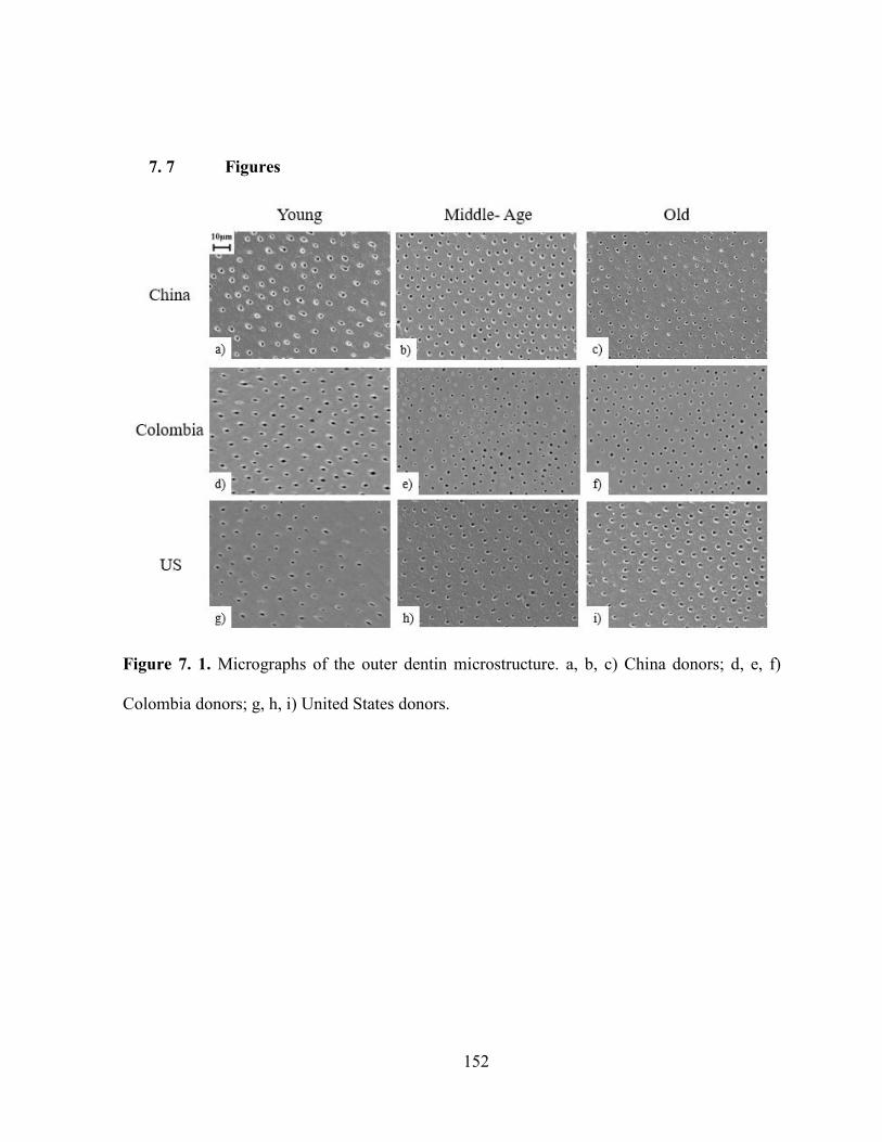
152
7. 7 Figures
Figure 7. 1. Micrographs of the outer dentin microstructure. a, b, c) China donors; d, e, f)
Colombia donors; g, h, i) United States donors.

153
Figure 7. 2. A comparison of the microstructure as a function of age in the outer dentin from
donor teeth of China, Colombian and US. a) lumen density; b) lumen diameter.

154
Figure 7. 3. Selected indentation depth versus time results for outer china dentin at applied
loads of 5, 10 and 50 Newtons. The results correspond to a Chinese donor of 56 years of age.

155
Figure 7. 4. Comparison of the experimental steady-state effective stress and effective strain
for the outer dentin of the three groups analyzed (markers) with the power-law fit (lines).

156
Figure 7. 5. Mineral-to-collagen ratio of outer dentin from young and old donor teeth
according to the donor’s origin region.

157
Figure 7. 6. Change in the proportion of obliterated dentinal tubules with aging.

158
Chapter 8
Conclusions
This chapter summarizes the main conclusions of this dissertation:
Chapter 2: Review of previous research on the mechanical behavior and aging of
human dentin
The aging process of dentin aging causes a change in dentin microstructure, product of
the obliteration of dentinal tubules; these changes have been related with an increase in
mineral content. However, the literature review shows no studies aimed at determine and
quantify the changes in chemical composition of aged coronal dentin (spatial variations).
These changes might be related with an increase in mineralization, crosslinking of collagen or
changes in crystallinity.
Dentin hardness has been shown to have considerable local variations for young
patients. For aged dentin, although it has been reported that hardness increases with aging, the
changes obtained have been related to the amount of “free spaces” (i.e. dentinal tubules
density), but not with chemical composition changes that occur in the tooth. For this
correlation old dentin can include other additional changes that occur with aging such besides
obliteration of dentinal tubules.
The viscoelastic properties of coronal dentin have been measured in the bulk dentin.
However, the fact that dentin is an anisotropic material, and that mechanical properties change
spatially within the tooth suggests that the viscoelastic properties of dentin will too. Also, only
159
one study regarding the viscoelastic properties of aged dentin is available, considering
intertubular and peritubular dentin, ignoring how these properties could change along different
regions of dentin.
Chapter 3: Effect of aging on hardness, microstructure and chemical composition of
dentin
The tubule density in the dentin of young and old donor teeth ranged from
approximately 22,000 to 35,000 tubules/mm2. The tubule diameter ranged from approximately
1.2 m to 1.8 m. There was a significant decrease in tubule density from the pulp to the DEJ
in both young and old dentin (p ≤ 0.05).
There was a significant difference (p ≤ 0.05) in the measures of occlusion ratio between
the outer (4.71%) and middle dentin (2.50%) of the old donor teeth. The largest proportion of
obliterated dentinal tubules being near the DEJ and decrease towards the pulp.
A significant decrease in hardness was found with increasing distance from the DEJ for
both young and old donor teeth (p ≤ 0.05). A larger hardness was found in the old dentin when
compared to young dentin. There were significant differences (p ≤ 0.05) in the hardness in the
outer and middle dentin between young and old patients.
The mineral-to-collagen ratio was found to increase with proximity to the DEJ for both
young and old patients. This behavior is opposed to previous results reported in the literature
for dentin from US donors, which in turn raises questions whether the chemical composition
of dentin and its percentages of organic material and collagen might be affected by ethnicity.
The outer dentin exhibited the highest mineral-to-collagen ratio, while lower values
were found in the areas near the pulp. A greater mineral-to-collagen ratio was found for the
old dentin, with the highest values found in the outer dentin.

160
A comparison of the hardness and mineral-to-collagen ratio distributions showed that
the regions with the highest mineral-to-collagen ratio exhibited the highest hardness values.
Chapter 4: Importance of Tubule Density to the Fracture Toughness of Dentin
A quantitative analysis of the changes in tubules occurring in coronal dentin with aging
showed that the primary difference between young and old dentin was a reduction in the
lumen area fraction. In the outer dentin there was nearly a 40% reduction in the lumen area
with aging. This difference is attributed to obliteration of dentinal tubules with formation of
sclerotic dentin.
An approach previously proposed to study the mechanical behavior of porous materials
was employed to model the fracture toughness of dentin and the influence of spatial variations
in dentinal tubules. The model showed a strong correlation to experimental results previously
reported in the literature for the dentin of young donor teeth. However, results for the old
dentin were not in agreement due to differences in the mechanisms of fracture.
Chapter 5: Time dependent deformation behavior of human dentin
The time dependent deformation behavior of coronal dentin was studied using
instrumented spherical indentation. The experimental results showed that the deformation
behavior followed a power law behavior, which was appropriately captured by an extension to
previously proposed models for the time dependent deformation behavior of structural
materials. The model accounts for the spatial variations of chemical composition (e.g. mineral
to collagen ratio) and microstructure (e.g. tubule density) of coronal dentin. Regions with
higher mineral to collagen ratio exhibited a stiffening effect, with reduced strain rate for
indentation loads. Similarly, a reduction in tubule density also caused a decrease in the
deformation behavior. The power law creep exponent for dentin was found to have a value of

161
n=3.38 which was independent of the spatial variations in composition and microstructure.
Furthermore, this parameter was found to be dependent on the specific composition of the
collagen present in the tissue and not on the amount.
Chapter 6: Contributions of aging on the time dependent deformation of dentin
The time dependent deformation and chemical composition of aged dentin was
evaluated in outer, middle and inner coronal dentin. The experimental results showed to
follow a power law deformation behavior with a decrease in the stress exponent when
comparing with young dentin.
Significant changes in the mineral to collagen ratio, crystallinity and carbonate to
phosphate ratio were found with increasing donor age. Most of these changes were found to be
more significant in the middle and outer dentin.
A model previously proposed to predict the time dependent deformation response of
dentin was employed to model the creep behavior of aged dentin. The model showed a strong
correlation between the experimental and the predicted results. A decrease in the time-
dependent behavior of middle and inner aged dentin was found when compared with young
dentin.
Chapter 7: Ethnic Background influence on the aging process of dentin
A reduction in the tubule density was observed with increasing donor age for the three
regions of origin analyzed: China, Colombian and United States. For the young samples the
average tubule density was 22,000 tubules/mm2 (p>0.05). While for the old group an average
of 14,000 tubules/mm2 was found, with a significant difference in the US group (9,500
tubules/mm2).

162
The average lumen diameter of the young sample was 1.34 ± 0.02 µm, with very few
differences between the three groups of origin (p> 0.05). While for the old group an average of
1.23 ± 0.02 µm was found. The lower diameter of dentinal tubules was found for the US group
(p ≤ 0.05).
The time dependent deformation behavior of dentin was studied using instrumented
spherical indentation. The experimental results showed that the deformation behavior for both
young and old samples followed a power law behavior; from the results, can be seen that the
young dentin showed a power-law exponent of 3.1 regardless of their area of origin. A
reduction in the power- law exponent was found for the old group, a reduction of almost of
50% was found for the China and US group, while for the Colombia group a reduction of only
27% was found.
The largest proportion of obliterated dentinal tubules found was 47% in the China
group in a 53-year-old sample. While for the US and Colombian samples, the maximum
values obtained were 41% and 9%, respectively, in both cases in a sample of 47-year-old
sample.
The mineral-to-collagen ratio of the young US and Colombia samples showed to be
statistically equal (p>0.05), while the China samples have a statistically higher mineral-to-
collagen ratio (~40% higher). An increase in the mineral-to-collagen ratio was found in the old
samples for all the regions; the results obtained showed that the old China and US samples are
statistically equal (p>0.05); meanwhile the Colombia samples are statistically lower.
The differences found on the obliteration of dentinal tubules, chemical composition and
mechanical properties between the regions analyzed, showed that the aging process of dentin
could be related not only with ethnicity but individual characteristic of the donor.

163
Bibliography
Addy, M. (2005). Tooth brushing, tooth wear and dentine hypersensitivity—are they
associated?. International dental journal, 55(S4), 261-267.
Addy, M., Absi, E. G., & Adams, D. (1987). Dentine hypersensitivity. The effects in
vitro of acids and dietary substances on root-planed and burred dentine. Journal of clinical
periodontology, 14(5), 274-279.
Ahearne, M., Yang, Y., Then, K. Y., & Liu, K. K. (2007). An indentation technique to
characterize the mechanical and viscoelastic properties of human and porcine corneas. Annals
of biomedical engineering, 35(9), 1608-1616.
Alfrey, T., & Doty, P. (1945). The methods of specifying the properties of viscoelastic
materials. Journal of applied physics, 16(11), 700-713.
Almushayt, A., Narayanan, K., Zaki, A. E., & George, A. (2006). Dentin matrix protein
1 induces cytodifferentiation of dental pulp stem cells into odontoblasts. Gene therapy, 13(7),
611-620.
Altherr, E. R., Koroluk, L. D., & Phillips, C. (2007). Influence of sex and ethnic tooth-
size differences on mixed-dentition space analysis. American journal of orthodontics and
dentofacial orthopedics, 132(3), 332-339.
Angker, L., Nockolds, C., Swain, M. V., & Kilpatrick, N. (2004). Quantitative analysis
of the mineral content of sound and carious primary dentine using BSE imaging. Archives of
oral biology, 49(2), 99-107.

164
Angker, L., Swain, M. V., & Kilpatrick, N. (2003). Micro-mechanical characterisation
of the properties of primary tooth dentine. Journal of dentistry,31(4), 261-267.
Arola, D. D., & Reprogel, R. K. (2006). Tubule orientation and the fatigue strength of
human dentin. Biomaterials, 27(9), 2131-2140.
Arola, D., & Reprogel, R. K. (2005). Effects of aging on the mechanical behavior of
human dentin. Biomaterials, 26(18), 4051-4061.
Arola, D., Bajaj, D., Ivancik, J., Majd, H., & Zhang, D. (2010). Fatigue of biomaterials:
hard tissues. International journal of fatigue, 32(9), 1400-1412.
Arola, D., Huang, M. P., & Sultan, M. B. (1999). The failure of amalgam dental
restorations due to cyclic fatigue crack growth. Journal of materials science: Materials in
medicine, 10(6), 319-327.
Arola, D., Ivancik, J., Majd, H., Fouad, A., Bajaj, D., Zhang, X. Y., & Eidelman, N.
(2009). Microstructure and mechanical behavior of radicular and coronal dentin. Endodontic
Topics, 20(1), 30-51.
Bajaj, D., Ivancik, J., & Arola, D. (2008). Ethnic background influences the crack
growth resistance of dentin. J Dent Res, 85(Spec Issue), 0438.
Bajaj, D., Sundaram, N., & Arola, D. (2008). An examination of fatigue striations in
human dentin: in vitro and in vivo. Journal of Biomedical Materials Research Part B: Applied
Biomaterials, 85(1), 149-159.
Balooch, M., Wu-Magidi, I. C., Balazs, A., Lundkvist, A. S., Marshall, S. J., Marshall,
G. W., Siekhaus, W.J., & Kinney, J. H. (1998). Viscoelastic properties of demineralized
human dentin measured in water with atomic force microscope (AFM)-based
indentation. Journal of biomedical materials research, 40(4), 539-544.
165
Balshin, M., (1949). Relation of mechanical properties of powder metals and their
porosity and the ultimate properties of porous-metal ceramic materials. Dokl Askd SSSR, 67,
831-834.
Bartlett, D., & Dugmore, C. (2008). Pathological or physiological erosion—is there a
relationship to age?. Clinical oral investigations, 12(1), 27-31.
Barreto, B.C., Van Meerbeek, B., Van Ende, A., Junior Boaventura de Sousa, S., da
Silva, G. R., Soares, P. V., & Soares, C. J. (2015). Biomechanical Behavior of Extensively
Restored Premolars: Cusp Deformation, Marginal Integrity, and Fracture Resistance. Journal
of Adhesive Dentistry, 17(3).
Bastone, E. B., Freer, T. J., & McNamara, J. R. (2000). Epidemiology of dental trauma:
a review of the literature. Australian dental journal, 45(1), 2-9.
Bates, J. F., Stafford, G. D., & Harrison, A. (1976). Masticatory function—a review of
the literature. Journal of Oral Rehabilitation, 3(1), 57-67.
Bentley, J. P. (1979). Aging of collagen. Journal of Investigative Dermatology,73(1),
80-83.
Bernick, S., & Nedelman, C. (1975). Effect of aging on the human pulp. Journal of
Endodontics, 1(3), 88-94.
Bertassoni, L. E., Orgel, J. P., Antipova, O., & Swain, M. V. (2012). The dentin
organic matrix–limitations of restorative dentistry hidden on the nanometer scale. Acta
biomaterialia, 8(7), 2419-2433.
Bertassoni, L. E., Kury, M., Rathsam, C., Little, C. B., & Swain, M. V. (2015). The
role of proteoglycans in the nanoindentation creep behavior of human dentin. Journal of the
mechanical behavior of biomedical materials, 55, 264-270.

166
Bishara, S. E., Jakobsen, J. R., Abdallah, E. M., & Garcia, A. F. (1989). Comparisons
of mesiodistal and bnccolingnal crown dimensions of the permanent teeth in three populations
from Egypt, Mexico, and the United States. American Journal of Orthodontics and
Dentofacial Orthopedics, 96(5), 416-422.
Boccaccini, A. R., Ondracek, G., & Mombello, E. (1996). Determination of stress
concentration factors in porous materials. Journal of materials science letters, 15(6), 534-536.
Bower, A. F., Fleck, N. A., Needleman, A., & Ogbonna, N. (1993). Indentation of a
power law creeping solid. In Proceedings of the Royal Society of London A: Mathematical,
Physical and Engineering Sciences, 441(1911), 97-124.
Bowman, S. M., Keaveny, T. M., Gibson, L. J., Hayes, W. C., & McMahon, T. A.
(1994). Compressive creep behavior of bovine trabecular bone. Journal of biomechanics,
27(3), 301-310.
Bowman, S. M., Gibson, L. J., Hayes, W. C., & McMahon, T. A. (1999). Results from
demineralized bone creep tests suggest that collagen is responsible for the creep behavior of
bone. Journal of biomechanical engineering, 121(2), 253-258.
Brauer, D. S., Hilton, J. F., Marshall, G. W., & Marshall, S. J. (2011). Nano-and
micromechanical properties of dentine: investigation of differences with tooth side. Journal of
biomechanics, 44(8), 1626-1629.
Cameron, C. E. (1964). Cracked-tooth syndrome. The Journal of the American Dental
Association, 68(3), 405-411.
Cardinale, M., Newton, R., & Nosaka, K. (Eds.). (2011). Strength and conditioning:
biological principles and practical applications. John Wiley & Sons.
Chatterjea, M. N., & Shinde, R. (2011). Textbook of medical biochemistry. Wife Goes
On.

167
Cheneler, D., Mehrban, N., & Bowen, J. (2013). Spherical indentation analysis of stress
relaxation for thin film viscoelastic materials. Rheologica Acta, 52(7), 695-706.
Chuang, S. F., Lin, S. Y., Wei, P. J., Han, C. F., Lin, J. F., & Chang, H. C. (2015).
Characterization of the elastic and viscoelastic properties of dentin by a nanoindentation creep
test. Journal of biomechanics, 48(10), 2155-2161.
Chuenarrom, C., Benjakul, P., & Daosodsai, P. (2009). Effect of indentation load and
time on knoop and vickers microhardness tests for enamel and dentin. Materials Research,
12(4), 473-476.
Clementino-Luedemann, T. N. R., & Kunzelmann, K. H. (2006). Mineral concentration
of natural human teeth by a commercial micro-CT. Dental materials journal, 25(1), 113-119.
Craig, R. G., & Peyton, F. A. (1958). Elastic and mechanical properties of human
dentin. Journal of Dental Research, 37(4), 710-718.
Currey, J. D., Brear, K., & Zioupos, P. (1996). The effects of ageing and changes in
mineral content in degrading the toughness of human femora. Journal of biomechanics, 29(2),
257-260.
Curtis, R. V., & Watson, T. F. (Eds.). (2014). Dental biomaterials: imaging, testing and
modelling. Elsevier.
da Silva, N. R. F. A., Lalani, F., Coelho, P. G., Clark, E. A., de Oliveira Fernandes, C.
A., & Thompson, V. P. (2008). Hertzian contact response of dentin with loading rate and
orientation. archives of oral biology, 53(8), 729-735.
de Castro, I. C. V., Rosa, C. B., dos Reis Júnior, J. A., Moreira, L. G. P., Aragão, J. S.,
dos Santos Barbosa, A. F., Silveira, L. Jr. & Pinheiro, A. L. (2014). Assessment of the use of
LED phototherapy on bone defects grafted with hydroxyapatite on rats with iron-deficiency

168
anemia and nonanemic: a Raman spectroscopy analysis. Lasers in medical science, 29(5),
1607-1615.
Depalle, B., Qin, Z., Shefelbine, S. J., & Buehler, M. J. (2015). Influence of cross-link
structure, density and mechanical properties in the mesoscale deformation mechanisms of
collagen fibrils. Journal of the mechanical behavior of biomedical materials, 52, 1-13.
Derby, B., & Akhtar, R. (Eds.). (2015). Mechanical Properties of Aging Soft Tissues.
Springer.
Deymier-Black, A. C., Yuan, F., Singhal, A., Almer, J. D., Brinson, L. C., & Dunand,
D. C. (2012). Evolution of load transfer between hydroxyapatite and collagen during creep
deformation of bone. Acta biomaterialia, 8(1), 253-261.
Driessens, F. C., & Verbeeck, R. K. (1990). Biominerals. CRC Press.
Drusini, A., Calliari, I., & Volpe, A. (1991). Root dentine transparency: age
determination of human teeth using computerized densitometric analysis.American journal of
physical anthropology, 85(1), 25-30.
Duncanson, M. G., & Korostoff, E. (1975). Compressive viscoelastic properties of
human dentin: I. Stress-relaxation behavior. Journal of dental research, 54(6), 1207-1212.
Eyre, D. R., & Wu, J. J. (2005). Collagen cross-links. In Collagen (207-229). Springer
Berlin Heidelberg.
Farlay, D., Panczer, G., Rey, C., Delmas, P. D., & Boivin, G. (2010). Mineral maturity
and crystallinity index are distinct characteristics of bone mineral. Journal of bone and
mineral metabolism, 28(4), 433-445.
Fehrenbach, M. J., & Popowics, T. (2015). Illustrated dental embryology, histology,
and anatomy. Elsevier Health Sciences.

169
Fondrk, M., Bahniuk, E., Davy, D. T., & Michaels, C. (1988). Some viscoplastic
characteristics of bovine and human cortical bone. Journal of biomechanics, 21(8), 623-630.
Frolich, E. Altersverunderungen de pulp und des paradontium. Dt. Zahnurtl
Z. 1970(25), 175–183 (in German).
Fuentes, V., Toledano, M., Osorio, R., & Carvalho, R. M. (2003). Microhardness of
superficial and deep sound human dentin. Journal of Biomedical Materials Research Part
A, 66(4), 850-853.
Gąsior-Głogowska, M., Komorowska, M., Hanuza, J., Mączka, M., Zając, A., Ptak, M.,
Będziński, R., Kobielarz, M., Maksymowicz, K., Kuropka, P. & Szotek, S. (2013). FT-Raman
spectroscopic study of human skin subjected to uniaxial stress. Journal of the mechanical
behavior of biomedical materials, 18, 240-252.
Goodyear, S. R., Gibson, I. R., Skakle, J. M., Wells, R. P., & Aspden, R. M. (2009). A
comparison of cortical and trabecular bone from C57 Black 6 mice using Raman
spectroscopy. Bone, 44(5), 899-907.
Goldberg, M., Kulkarni, A. B., Young, M., & Boskey, A. (2011). Dentin: Structure,
Composition and Mineralization: The role of dentin ECM in dentin formation and
mineralization. Frontiers in bioscience (Elite edition), 3, 711.
Gomez de Ferraris, M. E. G., & Muñoz, A. C. (2009). Histologa, embriologa e
ingeniería tisular bucodental/ Histology, embryology and oral tissue engineering. Ed. Médica
Panamericana
Graham, G. A. (1965). The contact problem in the linear theory of
viscoelasticity. International Journal of Engineering Science, 3(1), 27-46.
Grumezescu, A. (Ed.). (2016). Nanobiomaterials in Hard Tissue Engineering:
Applications of Nanobiomaterials. William Andrew.

170
Gutiérrez-Salazar, M. D. P., & Reyes-Gasga, J. (2003). Microhardness and chemical
composition of human tooth. Materials Research, 6(3), 367-373.
Habelitz, S., Marshall, S. J., Marshall, G. W., & Balooch, M. (2001). Mechanical
properties of human dental enamel on the nanometre scale. Archives of Oral Biology, 46(2),
173-183.
Habelitz, S., Marshall, G. W., Balooch, M., & Marshall, S. J. (2002). Nanoindentation
and storage of teeth. Journal of biomechanics, 35(7), 995-998.
Hall, N. E., Lindauer, S. J., Tüfekçi, E., & Shroff, B. (2007). Predictors of variation in
mandibular incisor enamel thickness. The Journal of the American Dental Association, 138(6),
809-815.
Hasselman, D. P. H. (1963). Relation between effects of porosity on strength and on
Young's modulus of elasticity of polycrystalline materials. Journal of the American Ceramic
Society, 46(11), 564-565.
Hayakawa, T., Mishima, H., Yokota, I., SakaE, T., Kozawa, Y., & Nemoto, K. (2000).
Application of high resolution microfocus X-ray CT for the observation of human
tooth. Dental materials journal, 19(1), 87-95.
Henrikson, R. C., & Kaye, G. I. (1986). Key Facts in Histology. Churchill Livingstone.
Hill, R. F. R. S., Storakers, B., & Zdunek, A. B. (1989). A theoretical study of the
Brinell hardness test. In Proceedings of the Royal Society of London A: Mathematical,
Physical and Engineering Sciences, 423(1865), 301-330.
Homewood, C. I. (1998). Cracked tooth syndrome-incidence, clinical findings and
treatment. Australian dental journal, 43(4), 217-222.
Hunter, S. C. (1960). The Hertz problem for a rigid spherical indenter and a
viscoelastic half-space. Journal of the Mechanics and Physics of Solids, 8(4), 219-234.

171
Imbeni, V., Nalla, R. K., Bosi, C., Kinney, J. H., & Ritchie, R. O. (2003). In vitro
fracture toughness of human dentin. Journal of Biomedical Materials Research Part A, 66(1),
1-9.
Imfeld, T. (1996). Dental erosion. Definition, classification and links. European journal
of oral sciences, 104(2), 151-155.
Ingle, J. I. (2008). Ingle's endodontics 6. PMPH-USA.
Ivancik, J., Majd, H., Bajaj, D., Romberg, E., & Arola, D. (2012). Contributions of
aging to the fatigue crack growth resistance of human dentin. Acta biomaterialia, 8(7), 2737-
2746.
Ivancik, J., & Arola, D. D. (2013). The importance of microstructural variations on the
fracture toughness of human dentin. Biomaterials, 34(4), 864-874.
Ivancik, J., Naranjo, M., Correa, S., Ossa, A., Tay, F. R., Pashley, D. H., & Arola, D.
(2014). Differences in the microstructure and fatigue properties of dentine between residents
of North and South America. Archives of oral biology, 59(10), 1001-1012.
Iwamoto, N., & Ruse, N. D. (2003). Fracture toughness of human dentin. Journal of
Biomedical Materials Research Part A, 66(3), 507-512.
Jafarzadeh, T., Erfan, M., & Watts, D. C. (2004). Creep and Viscoelastic Behaviour of
Human Dentin. Journal of Dentistry of Tehran University of Medical Sciences, 1(1), 5-14.
Jameson, M. W., Hood, J. A. A., & Tidmarsh, B. G. (1993). The effects of dehydration
and rehydration on some mechanical properties of human dentine. Journal of
biomechanics, 26(9), 1055-1065.
Jantarat, J., Palamara, J. E., Lindner, C., & Messer, H. H. (2002). Time-dependent
properties of human root dentin. Dental materials, 18(6), 486-493.

172
Ji, S., Gu, Q., & Xia, B. (2006). Porosity dependence of mechanical properties of solid
materials. Journal of Materials Science, 41(6), 1757-1768.
Jones, R. M. (1975). Mechanics of composite materials (Vol. 193). Washington, DC:
Scripta Book Company.
Kambic, H. E., & Yokobori, A. T. (1994). Biomaterials' mechanical properties. ASTM.
Kazanci, M., Roschger, P., Paschalis, E. P., Klaushofer, K., & Fratzl, P. (2006). Bone
osteonal tissues by Raman spectral mapping: orientation–composition. Journal of structural
biology, 156(3), 489-496.
Kinney, J. H., Balooch, M., Marshall, S. J., Marshall, G. W., & Weihs, T. P. (1996).
Hardness and Young's modulus of human peritubular and intertubular dentine. Archives of
Oral Biology, 41(1), 9-13.
Kinney, J. H., Balooch, M., Marshall, S. J., Marshall, G. W., & Weihs, T. P. (1996).
Atomic force microscope measurements of the hardness and elasticity of peritubular and
intertubular human dentin. Journal of biomechanical engineering, 118(1), 133-135.
Kinney, J. H., Marshall, S. J., & Marshall, G. W. (2003). The mechanical properties of
human dentin: a critical review and re-evaluation of the dental literature. Critical Reviews in
Oral Biology & Medicine, 14(1), 13-29.
Kinney, J. H., Nalla, R. K., Pople, J. A., Breunig, T. M., & Ritchie, R. O. (2005). Age-
related transparent root dentin: mineral concentration, crystallite size, and mechanical
properties. Biomaterials, 26(16), 3363-3376.
Kishen, A., & Asundi, A. (2005). Experimental investigation on the role of water in the
mechanical behavior of structural dentine. Journal of Biomedical Materials Research Part
A, 73(2), 192-200.

173
Koester, K. J., Ager III, J. W., & Ritchie, R. O. (2008a). Aging and fracture of human
cortical bone and tooth dentin. Jom, 60(6), 33-38.
Koester, K. J., Ager, J. W., & Ritchie, R. O. (2008b). The effect of aging on crack-
growth resistance and toughening mechanisms in human dentin. Biomaterials, 29(10), 1318-
1328.
Kováčik, J. (1999). Correlation between Young's modulus and porosity in porous
materials. Journal of materials science letters, 18(13), 1007-1010.
Kruzic, J. J., Nalla, R. K., Kinney, J. H., & Ritchie, R. O. (2003). Crack blunting, crack
bridging and resistance-curve fracture mechanics in dentin: effect of
hydration. Biomaterials, 24(28), 5209-5221.
Kruzic, J. J., & Ritchie, R. O. (2008). Fatigue of mineralized tissues: cortical bone and
dentin. Journal of the mechanical behavior of biomedical materials,1(1), 3-17.
Kumagai, A., Fujita, Y., Endo, S., & Itai, K. (2012). Concentrations of trace element in
human dentin by sex and age. Forensic science international, 219(1), 29-32.
Kumar, C. S. (Ed.). (2010). Biomimetic and bioinspired nanomaterials (Vol. 3). John
Wiley & Sons.
Lawrence Livermore National Laboratory (2008). The Role of Dentin in Tooth
Fracture. Science & Technology Review. 17-19.
Lee, E. H., & Radok, J. R. M. (1960). The contact problem for viscoelastic
bodies. Journal of Applied Mechanics, 27(3), 438-444.
Lee, H. H., Majd, H., Orrego, S., Majd, B., Romberg, E., Mutluay, M. M., & Arola, D.
(2014). Degradation in the fatigue strength of dentin by cutting, etching and adhesive
bonding. Dental Materials, 30(9), 1061-1072.

174
Lefèvre, T., Rousseau, M. E., & Pézolet, M. (2007). Protein secondary structure and
orientation in silk as revealed by Raman spectromicroscopy. Biophysical journal, 92(8), 2885-
2895.
Liu, B., Zhang, M., Chen, Y., & Yao, Y. (2014). Tooth wear in aging people: an
investigation of the prevalence and the influential factors of incisal/occlusal tooth wear in
northwest China. BMC oral health, 14(1), 1.
Lloyd, A. A., Wang, Z. X., & Donnelly, E. (2015). Multiscale contribution of bone
tissue material property heterogeneity to trabecular bone mechanical behavior. Journal of
biomechanical engineering, 137(1). 010801.
Looker, A. C., Wahner, H. W., Dunn, W. L., Calvo, M. S., Harris, T. B., Heyse, S. P.,
Johnston, C.C. Jr. & Lindsay, R. (1998). Updated data on proximal femur bone mineral levels
of US adults. Osteoporosis International, 8(5), 468-490.
Lopes, M. B., Sinhoreti, M. A., Gonini Júnior, A., Consani, S., & Mccabe, J. F. (2009).
Comparative study of tubular diameter and quantity for human and bovine dentin at different
depths. Brazilian dental journal, 20(4), 279-283.
Luo, J., & Stevens, R. (1999). Porosity-dependence of elastic moduli and hardness of
3Y-TZP ceramics. Ceramics International, 25(3), 281-286.
Magloire, H., Christophe Maurin, J., Couble, M. L., Shibukawa, Y., Tsumura, M., &
Bleicher, F. (2010). Topical review. Dental pain and odontoblasts: facts and
hypotheses. Journal of orofacial pain, 24(4).
McNally, M. E., Matthews, D. C., Clovis, J. B., Brillant, M., & Filiaggi, M. J. (2014).
The oral health of ageing baby boomers: a comparison of adults aged 45–64 and those 65
years and older. Gerodontology, 31(2), 123-135.

175
Majd, H., Viray, J., Porter, J. A., Romberg, E., & Arola, D. (2012). Degradation in the
fatigue resistance of dentin by bur and abrasive air-jet preparations. Journal of dental
research, 91(9), 894-899.
Mandair, G. S., & Morris, M. D. (2015). Contributions of Raman spectroscopy to the
understanding of bone strength. BoneKEy reports, 4.
Marshall, G. W., Marshall, S. J., Kinney, J. H., & Balooch, M. (1997). The dentin
substrate: structure and properties related to bonding. Journal of dentistry, 25(6), 441-458.
Marshall, L. M., Zmuda, J. M., Chan, B. K., Barrett‐Connor, E., Cauley, J. A., Ensrud,
K. E., Lang, T.F. & Orwoll, E. S. (2008). Race and ethnic variation in proximal femur
structure and BMD among older men. Journal of Bone and Mineral Research, 23(1), 121-130.
Märten, A., Fratzl, P., Paris, O., & Zaslansky, P. (2010). On the mineral in collagen of
human crown dentine. Biomaterials, 31(20), 5479-5490.
Merz, M. L., Isaacson, R. J., Germane, N., & Rubenstein, L. K. (1991). Tooth
diameters and arch perimeters in a black and a white population. American Journal of
Orthodontics and Dentofacial Orthopedics, 100(1), 53-58.
Meyers, M. A., Chen, P. Y., Lin, A. Y. M., & Seki, Y. (2008). Biological materials:
structure and mechanical properties. Progress in Materials Science,53(1), 1-206.
Miura, J., Nishikawa, K., Kubo, M., Fukushima, S., Hashimoto, M., Takeshige, F., &
Araki, T. (2014). Accumulation of advanced glycation end-products in human
dentine. Archives of oral biology, 59(2), 119-124.
Mulhearn, T.O., Tabor, D. (1960). Creep and Hardness of metals: A physical study. J.
Inst. Met.89, 7-12.

176
Murray, P. E., Stanley, H. R., Matthews, J. B., Sloan, A. J., & Smith, A. J. (2002). Age-
related odontometric changes of human teeth. Oral Surgery, Oral Medicine, Oral Pathology,
Oral Radiology, and Endodontology, 93(4), 474-482.
Nalla, R. K., Kinney, J. H., & Ritchie, R. O. (2003a). Effect of orientation on the in
vitro fracture toughness of dentin: the role of toughening mechanisms. Biomaterials, 24(22),
3955-3968.
Nalla, R. K., Imbeni, V., Kinney, J. H., Staninec, M., Marshall, S. J., & Ritchie, R. O.
(2003b). In vitro fatigue behavior of human dentin with implications for life prediction.
Journal of Biomedical Materials Research Part A, 66(1), 10-20.
Nalla, R. K., Kruzic, J. J., Kinney, J. H., & Ritchie, R. O. (2004a). Effect of aging on
the toughness of human cortical bone: evaluation by R-curves. Bone,35(6), 1240-1246.
Nalla, R. K., Kinney, J. H., Marshall, S. J., & Ritchie, R. O. (2004b). On the in vitro
fatigue behavior of human dentin: effect of mean stress. Journal of dental research, 83(3),
211-215.
Nalla, R. K., Balooch, M., Ager, J. W., Kruzic, J. J., Kinney, J. H., & Ritchie, R. O.
(2005). Effects of polar solvents on the fracture resistance of dentin: role of water
hydration. Acta Biomaterialia, 1(1), 31-43.
Nalla, R. K., Kinney, J. H., Tomsia, A. P., & Ritchie, R. O. (2006). Role of alcohol in
the fracture resistance of teeth. Journal of dental research, 85(11), 1022-1026.
Nanci, A. (2012). Ten Cate's Oral Histology Development, Structure, and Function,
8/e. Elsevier India.
Nazari, A., Bajaj, D., Zhang, D., Romberg, E., & Arola, D. (2009). Aging and the
reduction in fracture toughness of human dentin. Journal of the mechanical behavior of
biomedical materials, 2(5), 550-559.

177
Nelson, D. A., Beck, T. J., Wu, G., Lewis, C. E., Bassford, T., Cauley, J. A., LeBoff,
M.S., Going, S.B. & Chen, Z. (2011). Ethnic differences in femur geometry in the women's
health initiative observational study. Osteoporosis international, 22(5), 1377-1388.
Nikiforuk, G. (1970). Posteruptive effects of nutrition on teeth. Journal of dental
research, 49(6), 1252-1261.
Olze, A., Van Niekerk, P., Ishikawa, T., Zhu, B. L., Schulz, R., Maeda, H., &
Schmeling, A. (2007). Comparative study on the effect of ethnicity on wisdom tooth
eruption. International journal of legal medicine, 121(6), 445-448.
Opdam, N. J., Bronkhorst, E. M., Roeters, J. M., & Loomans, B. A. (2007). Longevity
and reasons for failure of sandwich and total-etch posterior composite resin
restorations. Journal of Adhesive Dentistry, 9(5).
Park, S., Wang, D. H., Zhang, D., Romberg, E., & Arola, D. (2008). Mechanical
properties of human enamel as a function of age and location in the tooth. Journal of
Materials Science: Materials in Medicine, 19(6), 2317-2324.
Park, S., Quinn, J. B., Romberg, E., & Arola, D. (2008). On the brittleness of enamel
and selected dental materials. dental materials, 24(11), 1477-1485.
Pashley, D., Okabe, A., & Parham, P. (1985). The relationship between dentin
microhardness and tubule density. Dental Traumatology, 1(5), 176-179.
Pashley, D. H. (1989). Dentin: a dynamic substrate--a review. Scanning
microscopy, 3(1), 161-74.
Pashley, D. H., & Pashley, E. L. (1991). Dentin permeability and restorative dentistry:
a status report for the American Journal of Dentistry. American journal of dentistry, 4(1), 5-9.

178
Pashley, D. H., Agee, K. A., Wataha, J. C., Rueggeberg, F., Ceballos, L., Itou, K.,
Yoshiyama, M., Carvalho, R.M., & Tay, F. R. (2003). Viscoelastic properties of
demineralized dentin matrix. Dental materials, 19(8), 700-706.
Penel, G., Leroy, G., Rey, C., & Bres, E. (1998). MicroRaman spectral study of the
PO4 and CO3 vibrational modes in synthetic and biological apatites. Calcified Tissue
International, 63(6), 475-481.
Plotino, G., Grande, N. M., Bedini, R., Pameijer, C. H., & Somma, F. (2007). Flexural
properties of endodontic posts and human root dentin. Dental materials, 23(9), 1129-1135.
Porter, A. E., Nalla, R. K., Minor, A., Jinschek, J. R., Kisielowski, C., Radmilovic, V.,
Kinney, J.H., Tomsia, A.P., & Ritchie, R. O. (2005). A transmission electron microscopy
study of mineralization in age-induced transparent dentin. Biomaterials, 26(36), 7650-7660.
Rabotyagova, O. S. (2008). Structure to function: Spider silk and human collagen.
ProQuest.
Rimnac, C. M., Petko, A. A., Santner, T. J., & Wright, T. M. (1993). The effect of
temperature, stress and microstructure on the creep of compact bovine bone. Journal of
biomechanics, 26(3), 219-228.
Rivera, C., Arola, D., & Ossa, A. (2013). Indentation damage and crack repair in
human enamel. Journal of the mechanical behavior of biomedical materials,21, 178-184.
Rho, J. Y., Kuhn-Spearing, L., & Zioupos, P. (1998). Mechanical properties and the
hierarchical structure of bone. Medical engineering & physics, 20(2), 92-102.
Robinson, C., Connell, S., Kirkham, J., Brookes, S. J., Shore, R. C., & Smith, A. M.
(2004). The effect of fluoride on the developing tooth. Caries research,38(3), 268-276.
Rosen, C. J., Glowacki, J., & Bilezikian, J. P. (Eds.). (1999). The aging skeleton.
Academic Press.

179
Ryou, H., Amin, N., Ross, A., Eidelman, N., Wang, D. H., Romberg, E., & Arola, D.
(2011). Contributions of microstructure and chemical composition to the mechanical
properties of dentin. Journal of Materials Science: Materials in Medicine, 22(5), 1127-1135.
Ryou, H., Romberg, E., Pashley, D. H., Tay, F. R., & Arola, D. (2012). Nanoscopic
dynamic mechanical properties of intertubular and peritubular dentin. Journal of the
mechanical behavior of biomedical materials, 7, 3-16.
Ryou, H., Romberg, E., Pashley, D. H., Tay, F. R., & Arola, D. (2015). Importance of
age on the dynamic mechanical behavior of intertubular and peritubular dentin. Journal of the
mechanical behavior of biomedical materials,42, 229-242.
Ryshkewitch, E. (1953). Compression strength of porous sintered alumina and
zirconia. Journal of the American Ceramic Society, 36(2), 65-68.
Sahar, N. D. (2009). Investigating the Effects of Age and Exercise on Bone
Composition and the Impact of Composition on Mechanical Integrity. ProQuest.
Salomon, D., Kosmač, T., 2013. Advanced Ceramics, in: Shen, J., Kosmač, T. (Eds.),
Advanced Ceramics for Dentistry. Butterworth-Heinemann, Waltham. M.A., pp.116.
Schiller, K., 1985. Porosity and strength of brittle solids, in: Walton, W.H. (Ed),
Mechanical Properties of Non-Metallic Brittle Materials. Butterworth, London, pp. 35.
Schour, I. (1948). Development and growth of teeth. Oral Surgery, Oral Medicine,
Oral Pathology, 1(4), 346-354.
Senawongse, P., Otsuki, M., Tagami, J., & Mjör, I. (2006). Age-related changes in
hardness and modulus of elasticity of dentine. Archives of Oral Biology,51(6), 457-463.
Shen, Z. L., Kahn, H., Ballarini, R., & Eppell, S. J. (2011). Viscoelastic properties of
isolated collagen fibrils. Biophysical journal, 100(12), 3008-3015.

180
Sieck, G. C. (2003). Physiology of aging. Journal of Applied Physiology, 95(4), 1333-
1334.
Solheim, T. (1992). Amount of secondary dentin as an indicator of age. European
Journal of Oral Sciences, 100(4), 193-199.
Spangberg, L. S. (1989). Experimental endodontics. CRC Press.
Staines, M., Robinson, W. H., & Hood, J. A. A. (1981). Spherical indentation of tooth
enamel. Journal of materials science, 16(9), 2551-2556.
Staninec, M., Nguyen, H., Kim, P., Marshall, G. W., Ritchie, R. O., & Marshall, S. J.
(2008). Four-point bending evaluation of dentin-composite interfaces with various stresses.
Medicina Oral Patologia Oral y Cirugia Bucal, 13(1), 81.
Tesch, W., Eidelman, N., Roschger, P., Goldenberg, F., Klaushofer, K., & Fratzl, P.
(2001). Graded microstructure and mechanical properties of human crown dentin. Calcified
Tissue International, 69(3), 147-157.
Timiras, P. S. (Ed.). (2007). Physiological basis of aging and geriatrics. CRC Press.
Toto, P. D., Kastelic, E. F., Duyvejonck, K. J., & Rapp, G. W. (1971). Effect of age on
water content in human teeth. Journal of dental research, 50(5).
Travison, T. G., Beck, T. J., Esche, G. R., Araujo, A. B., & McKinlay, J. B. (2008).
Age trends in proximal femur geometry in men: variation by race and ethnicity. Osteoporosis
International, 19(3), 277-287.
Trengrove, H. G., Carter, G. M., & Hood, J. A. (1995). Stress relaxation properties of
human dentin. Dental Materials, 11(5), 305-310.
Tronstad, L. (2003). Clinical endodontics: a textbook. Thieme.
Ural, A., & Vashishth, D. (2006). Cohesive finite element modeling of age-related
toughness loss in human cortical bone. Journal of biomechanics,39(16), 2974-2982.

181
Ural, A., & Vashishth, D. (2007). Anisotropy of age-related toughness loss in human
cortical bone: a finite element study. Journal of biomechanics, 40(7), 1606-1614.
Vasiliadis, L., Darling, A. I., & Levers, B. G. H. (1983). The histology of sclerotic
human root dentine. Archives of Oral Biology, 28(8), 693-700.
Vaughn, D. (2011). Your Guide to Health: Anti-Aging: "Secrets to Help You Slow
Down the Aging Process. Adams Media
Vieira, A. P. G. F., Hancock, R., Dumitriu, M., Schwartz, M., Limeback, H., &
Grynpas, M. (2005). How does fluoride affect dentin microhardness and mineralization?.
Journal of dental research, 84(10), 951-957.
Walters, C., & Eyre, D. R. (1983). Collagen crosslinks in human dentin: increasing
content of hydroxypyridinium residues with age. Calcified tissue international, 35(1), 401-
405.
Wang, X., Shen, X., Li, X., & Agrawal, C. M. (2002). Age-related changes in the
collagen network and toughness of bone. Bone, 31(1), 1-7.
Wang, X., & Puram, S. (2004). The toughness of cortical bone and its relationship with
age. Annals of biomedical engineering, 32(1), 123-135.
Wang, R. (2005). Anisotropic fracture in bovine root and coronal dentin. Dental
Materials, 21(5), 429-436.
Wang R, Mao S, Romberg E, Arola D, Zhang D. (2003). Importance of aging to
dehydration shrinkage of human dentin. Applied Mathematics and Mechanics. 33(3), 333-344.
White, B. A., Albertini, T. F., Brown, L. J., Larach-Robinson, D., Redford, M., &
Selwitz, R. H. (1996). Selected restoration and tooth conditions: United States, 1988-1991.
Journal of dental research, 75, 661-671.

182
Williams, D. R., & Wilson, C. M. (2001). Race, ethnicity, and aging. Handbook of
aging and the social sciences, 5, 160-178.
Wopenka, B., Kent, A., Pasteris, J. D., Yoon, Y., & Thomopoulos, S. (2008). The
tendon-to-bone transition of the rotator cuff: a preliminary Raman spectroscopic study
documenting the gradual mineralization across the insertion in rat tissue samples. Applied
spectroscopy, 62(12), 1285-1294.
Xu, C., Yao, X., Walker, M. P., & Wang, Y. (2009). Chemical/molecular structure of
the dentin–enamel junction is dependent on the intratooth location. Calcified tissue
international, 84(3), 221-228.
Xu, C., & Wang, Y. (2012). Chemical composition and structure of peritubular and
intertubular human dentine revisited. Archives of oral biology, 57(4), 383-391.
Xu, C., Reed, R., Gorski, J. P., Wang, Y., & Walker, M. P. (2012). The distribution of
carbonate in enamel and its correlation with structure and mechanical properties. Journal of
materials science, 47(23), 8035-8043.
Xu, H., Zheng, Q., Shao, Y., Song, F., Zhang, L., Wang, Q., & Huang, D. (2014). The
effects of ageing on the biomechanical properties of root dentine and fracture. Journal of
dentistry, 42(3), 305-311.
Yan, J., Taskonak, B., & Mecholsky, J. J. (2009). Fractography and fracture toughness
of human dentin. Journal of the mechanical behavior of biomedical materials, 2(5), 478-484.
Yellowitz, J. A., & Schneiderman, M. T. (2014). Elder's oral health crisis. Journal of
Evidence Based Dental Practice, 14, 191-200.
Zhang, Y. R., Du, W., Zhou, X. D., & Yu, H. Y. (2014). Review of research on the
mechanical properties of the human tooth. International journal of oral science, 6(2), 61-69.

183
Zheng, L., Nakajima, M., Higashi, T., Foxton, R. M., & Tagami, J. (2005). Hardness
and Young's modulus of transparent dentin associated with aging and carious disease. Dental
materials journal, 24(4), 648-653.
Zioupos, P., & Currey, J. D. (1998). Changes in the stiffness, strength, and toughness
of human cortical bone with age. Bone, 22(1), 57-66.
Zioupos, P., Currey, J. D., & Hamer, A. J. (1999). The role of collagen in the declining
mechanical properties of aging human cortical bone. Journal of biomedical materials
research, 45(2), 108-116.
Ziskind, D., Hasday, M., Cohen, S. R., & Wagner, H. D. (2011). Young’s modulus of
peritubular and intertubular human dentin by nano-indentation tests. Journal of structural
biology, 174(1), 23-30.