![Aqua(4,4'-bipyridine-[kappa]N)bis(1,4-dioxo-1 ... - ScienceOpen](https://static.fdokumen.com/doc/165x107/63262349e491bcb36c0aa51f/aqua44-bipyridine-kappanbis14-dioxo-1-scienceopen.jpg)
Aqua(4,4'-bipyridine-[kappa]N)bis(1,4-dioxo-1 ... - ScienceOpen
* Correspondence: [email protected]
RESEARCH Open Access
MicroRNA profiling reveals age-dependentdifferential expression of nuclear factor �B andmitogen-activated protein kinase in adipose andbone marrow-derived human mesenchymal stemcellsAmitabh C Pandey1, Julie A Semon1, Deepak Kaushal2, Regina P O’Sullivan3, Julie Glowacki3, Jeffery M Gimble4
and Bruce A Bunnell1,5,6*
Abstract
Introduction: Mesenchymal stem cells (MSCs) play a central role in mediating endogenous repair of cell and tissuedamage. Biologic aging is a universal process that results in changes at the cellular and molecular levels. In thepresent study, the role of microRNA (miRNA) in age-induced molecular changes in MSCs derived from adiposetissue (ASCs) and bone marrow (BMSCs) from young and old human donors were investigated by using anunbiased genome-wide approach.
Methods: Human ASCs and BMSCs from young and old donors were cultured, and total RNA was isolated. ThemiRNA fraction was enriched and used to determine the expression profile of miRNA in young and old donorMSCs. Based on miRNA expression, differences in donor MSCs were further investigated by using differentiationassays, Western blot, immunocytochemistry, and bioinformatics.
Results: Biologic aging demonstrated reduced osteogenic and adipogenic potential in ASCs isolated from olderdonors, whereas cell size, complexity, and cell-surface markers remained intact with aging. Analysis of miRNAprofiles revealed that small subsets of active miRNAs changed secondary to aging. Evaluation of miRNA showedsignificantly decreased levels of gene expression of inhibitory kappa B kinase (I�B), interleukin-1a, inducible nitricoxide synthase (iNOS), mitogen-activated protein kinase/p38, ERK1/2, c-fos, and c-jun in MSCs from older donors byboth bioinformatics and Western blot analysis. Nuclear factor kappa B (NF-�B), myc, and interleukin-4 receptormRNA levels were significantly elevated in aged cells from both the adipose and bone marrow depots.Immunocytochemistry showed nuclear localization in young donors, but a cytosolic predominance ofphosphorylated NF-�B in ASCs from older donors. Western blot demonstrated significantly elevated levels of NF-�Bsubunits, p65 and p50, and AKT.
Conclusions: These findings suggest that differential expression of miRNA is an integral component of biologicaging in MSCs.
1Center for Stem Cell Research and Regenerative Medicine, School ofMedicine, Tulane University, New Orleans, LA, USAFull list of author information is available at the end of the article
Pandey et al. Stem Cell Research & Therapy 2011, 2:49http://stemcellres.com/content/2/6/49
© 2011 Pandey et al.; licensee BioMed Central Ltd. This is an open access article distributed under the terms of the Creative CommonsAttribution License (http://creativecommons.org/licenses/by/2.0), which permits unrestricted use, distribution, and reproduction inany medium, provided the original work is properly cited.
IntroductionAge-related changes occur in all biologic systems, fromthe phenotypic to the molecular level, leading to activa-tion and deactivation of cellular pathways. Recent stu-dies suggest that mesenchymal stem cells (MSCs) aresubject to changes that accompany biologic aging [1-3].MSCs, also known as mesenchymal stromal cells, are amultipotent, heterogeneous population of cells that pos-sess the ability to differentiate along a variety of celllineages. MSCs have been isolated from numerous tissuesources, including the bone marrow (BMSCs) and adi-pose tissue (ASCs), and have been shown to retain theability to differentiate into numerous terminally differen-tiated cell types, including bone, cartilage, fat, muscle,and skin [4-6]. Studies also have investigated the role ofMSCs as therapeutic agents in many disease states [4,7].It has been suggested that populations of MSCs aredepleted with age and that reduction in MSC pools con-tributes to human aging and the onset of age-relateddisease processes [8,9].Biologic aging can affect not only the absolute num-
bers of MSCs, but also the expression profile of thesecells [9-11]. Indeed, MSCs appear to be as susceptible asother cells to molecular alterations that result from invivo biologic aging [2,3,12]. It has been suggested thatMSCs isolated from older donors have an overall declinein differentiation potential or may show a greater pro-pensity toward adipogenesis than toward other cell fates;however, most of these studies focused solely on BMSCs[1,2,13]. Other reports allude to a more complex patternof events, especially with regard to the adipogenicpotential of MSCs and aging [14]. However, the changesexhibited by MSCs due to aging have not been fullydelineated. Moreover, the effect of aging on the thera-peutic potential of MSCs for regenerative medicineremains to be fully elucidated. It has been suggestedthat microRNAs (miRNAs) play an integral role in theregulation of aging and subsequent changes associatedwith the aging process [15-18]. Specifically, miRNAs,which are small 19- to 27-nucleotide (nt) RNA frag-ments, function in the translational regulation of geneexpression. They are members of a large class of smallnoncoding RNAs. Degradation and repression of targetmRNA transcripts are the primary mechanisms wherebymiRNAs regulate gene expression and influence cellularprocesses and signaling mechanisms [19,20]. It has beenestimated that approximately two thirds of the wholemammalian genome may be influenced by translationalregulation of gene expression by miRNA activity [21].Indeed, miRNAs appear to be integral regulators of geneexpression, influencing processes that include aging,apoptosis, cancer, and inflammation [15,22,23].Recent studies have investigated the role of miRNAs
in MSCs as they progress from undifferentiated states to
differentiated end-cell fates, in a variety of species[24,25]. No investigations to date, however, have delvedinto the effects of biologic age-induced miRNA changeson MSCs. Given the potential of MSCs as cellular thera-peutic agents, it is imperative to gain a full understand-ing of the effects of biologic aging on MSC properties,as well as the potential benefits and risks of using olderor younger donor MSCs as treatment modalities.In the current study, the alterations in the miRNA
profiles of MSCs isolated from old and young donorswere investigated, and significant downstream altera-tions that may be manifested secondary to biologicaging have been identified.
Materials and methodsMaterialsCell-culture materials, including Hank’s buffered salinesolution (HBSS), a-modified Eagle’s medium (a-MEM),L-glutamine, penicillin/streptomycin, phosphate-bufferedsaline (PBS), and trypsin/EDTA were obtained fromInvitrogen (Carlsbad, CA). Fetal bovine serum (FBS) waspurchased from Atlanta Biological (Atlanta, GA). Allreagents for miRNA and mRNA arrays were obtainedfrom SABiosciences (Frederick, MD), including wholehuman genome miRNA array, signal-transduction path-way finder arrays, and RT2 First Strand Kits. Proteaseand phosphatase inhibitor cocktails, RIPA buffer, andBCA protein quantification kits were ordered fromThermo Scientific (Rockford, IL). Western blot reagentswere obtained from Invitrogen; these included loadingbuffer, reducing agent, 4% to 12% Bis-Tris SDS-PAGEgels, iblot nitrocellulose membrane and blotting compo-nents, and chemiluminescence HRP developer kits. Thechemiluminescence blocker was acquired from Millipore(Billerica, MA). All antibodies, both primary and sec-ondary, were obtained from Santa Cruz Biotechnology(Santa Cruz, CA). All other chemicals used were mole-cular biology reagent grade.
Mesenchymal stem cell isolation and expansionASCs and BMSCs were isolated and expanded as pre-viously reported [26]. In brief, ASCs were isolated fromsubcutaneous white adipose tissue. BMSCs wereobtained from the iliac crest or marrow discarded dur-ing orthopedic procedures [2,26]. In brief, BMSCs wereseparated over a Ficoll gradient by centrifugation for 30minutes at 1,800 g. Similarly, ASCs were isolated fromsubcutaneous white adipose tissue, after which the adi-pose tissue was digested with 0.075% collagenase for 30minutes at 37°C to isolate ASCs. Subsequently, all cellswere washed in HBSS or PBS, and then centrifuged at300 g (ASC) or 1,000 g (BMSC) for 10 minutes. The pel-leted nucleated cells were cultured overnight in a humi-dified atmosphere at 37°C with 5% CO2 in complete
Pandey et al. Stem Cell Research & Therapy 2011, 2:49http://stemcellres.com/content/2/6/49
Page 2 of 18
culture medium (CCM) for BMSCs, which consisted ofa-MEM, 20% FBS, 1% L-glutamine, and 1% penicillin/streptomycin or stromal medium (SM) for ASCs(DMEM/F12, 10% FBS, 1% penicillin/streptomycin/amphotericin). The CCM was replaced every third dayuntil the cells reached 70% to 80% confluence. All cellsused were between passages 3 and 5 and identified asyoung donors (younger than 50 years) or old donors(older than 50 years). Young donors of ASCs were anaverage age of 31.5 ± 10.4 years, whereas BMSCs were31.5 ± 8.7 years; old donors of ASCs were an averageage of 63 ± 6.0 years, and old donors of BMSCs were anaverage age of 56.3 ± 5.0 years. Investigators wereblinded to donor information about the ASCs andBMSCs; however, donor age, race, and other selecteddemographics were obtained. All donor groups had sig-nificant differences in age, but no other significantdemographic differences, including BMI. All cells wereisolated after review and approval by the institutionalreview board (IRB) of Tulane University School of Med-icine, Pennington Biomedical Research Center, or Brig-ham and Women’s Hospital, with informed patientconsent.
Flow cytometryFlow cytometry was performed as previously described[27]. After MSCs were 70% confluent, CCM was aspi-rated, and cells were washed twice with PBS. Cells wereharvested with trypsin, and resuspended in PBS for ana-lysis. Cells were assessed for size by using forward andside light-scatter measurements. In addition, all cellswere characterized by examination for cell-surface mar-kers by using an extensive panel of antibodies developedby our Center over the past several-years period [28].Both BMSCs and ASCs expressed known MSC markersincluding CD29, CD44, CD90, CD105, CD166, and HLAclass I. Both cell types were negative for lymphohemato-poietic lineage markers, such as CD3, CD34, CD45,CD11b, and CD19.
Differentiation assayThe ability of MSCs to differentiate along osteogenicand adipogenic lineages was adapted from our pre-viously described methods [29,30]. In brief, cells wereplated in 24-well plates and cultured in CCM until theyattained 70% confluence. Culture medium was thenaspirated and replaced with differentiation-specific med-ium. Osteogenic differentiation was assessed by stainingfor bone mineralization with Alizarin Red. Assessmentof lipid inclusions, which indicate differentiation to adi-pocytes, was done by staining with Oil Red O solution,followed by microscopic examination on a Nikon EclipseTE200 microscope (Nikon Instruments, Melville, NY).Differentiation was quantified as previously described
[31]. In brief, after cells were stained, they weredestained by using 10% cetylpyridinium chloride forAlzarian Red and isopropyl alcohol for Oil Red O. Col-lected samples were then analyzed by using a microplatereader at 580 nm to assess the optical density (OD) ofthe collected samples. Sample OD values were then nor-malized to protein concentrations of the differentiatedcells.
miRNA profiling arrays of MSCsThe microRNA profiling was performed by using theSABiosciences quantitative polymerase chain reaction(qPCR) array platform according to the protocols of theSABiosciences service core. In total, 16 donors, compris-ing four young and four old donors of ASCs andBMSCs, respectively, were analyzed with the wholehuman genome miRNA qPCR array. Samples of ASCsand BMSCs were pelleted and quantified to containapproximately 1 × 10⁷ cells per donor. Cell pellets wereflash frozen with liquid nitrogen and sent to the SABios-ciences service core (Frederick, MD) for total RNA iso-lation, miRNA enrichment, qPCR-based array, and datareport. The miRNA profile was analyzed for hierarchicclustering of miRNA by using Genesis to generate heat-maps (Genesis, Austria) [32]. Genesis was also used tocluster donor profiles to identify individual donorvariability.
Target generation from miRNA dataIn addition to targets for miRNA action that have beenvalidated in reported studies, potential targets formiRNA action were predicted by using putative targetsgenerated from TargetScan Human V5.1. TargetScanHuman predicts putative targets based on factors thatinclude sequence homology, predicted biologic function,and verified targets. The predicted targets were rankedin order of conserved sequences and the prospect ofregulation of gene expression via miRNA activity byusing a context score, and previous studies have shownthat context scores less than -0.3 are commonly consid-ered biologically relevant [33]. The predicted gene tar-gets were analyzed with Ingenuity Pathways Assessment(IPA) and the Ingenuity Knowledgebase (Version 8.7,Ingenuity Systems, Redwood City, CA).
Bioinformatics analysis of generated miRNA targetsTargets generated by TargetScan were entered into IPAto delineate further the interactions and functions ofmiRNA on mRNA levels and the protein expression.IPA also generated lists of focus molecules, which weredefined by direct and indirect molecules. Direct mole-cules were those input directly into IPA, such as the tar-gets from TargetScan. Indirect molecules were thosethat have been described in previous studies, are
Pandey et al. Stem Cell Research & Therapy 2011, 2:49http://stemcellres.com/content/2/6/49
Page 3 of 18
commonly associated with the direct molecules, and arepart of the Ingenuity Knowledgebase. IPA was used togain insight into the interacting networks of the focusmolecules, canonic pathway involvement, biologic func-tions, and cellular processes influenced by miRNAactions. Canonic pathways are those pathways in whichmiRNA may play integral roles in regulation. IPA analy-sis generates these canonic pathways based on the inte-gration of inputted mRNA targets that were generatedfrom significantly changed miRNA. Biologic functionsare generated by IPA analysis investigating which phy-siological and pathological functions may be influencedby the inputted targets. Both the canonic pathways andbiologic functions are then evaluated for statisticalsignificance.
mRNA arrays of MSCsSignal-transduction qPCR-based array was obtainedfrom SABiosciences and was performed according tothe manufacturer’s instructions. In brief, total RNA iso-lated for the miRNA arrays was obtained from two ASCsamples based on clustering of the miRNA profile ofdonors. Representative samples from young and olddonors were selected for analysis. Total RNA samples(500 ng) were processed for cDNA synthesis by using aRT2 First Strand Kit according to the manufacturer’sprotocol. The Signal Transduction Pathway Finder arrayfrom SABiosciences was performed on a Bio-Rad CFX96real-time PCR machine (Bio-Rad, Hercules, CA), accord-ing to the manufacturer’s instructions. Data from thearray were analyzed with the SABiosciences software byobserved Ct values and determining the relative foldchange as compared with housekeeping genes. House-keeping genes used as internal controls included b2-microglobulin, hypoxanthine phosphoribosyltransferase1, ribosomal protein L13a, glyceraldehyde-3-phosphatedehydrogenase, and b-actin.
Subcellular localization of NF-�B subunits byimmunocytochemistryRepresentative ASCs from old and young donors wereselected based on hierarchic clustering of donors frommiRNA analysis. Cells were plated at a density of 1 ×10⁴ cells per 0.4 cm2. The next day, cells were fixed for15 minutes at room temperature, washed, permeabilizedfor 5 minutes at room temperature, and washed again.Cells were incubated overnight at 4°C with a 1:200 dilu-tion of p50 and p65 subunits of NF-�B, followed by 1hour at room temperature with a 1:1,000 dilution of agoat anti-rabbit FITC secondary antibody. Slides werecounterstained with DAPI, photographed with a LeicaDMRXA2 microscope, and rendered with Slidebooksoftware (Denver, CO).
Cell lysate and protein isolationASCs were grown in 15-cm2 dishes to 70% confluenceat 37°C, as described earlier. Cells were washed twicewith PBS and harvested with trypsin/EDTA. Cells werecentrifuged, collected, flash frozen in liquid nitrogen,and stored at -80°C. Cell pellets were lysed in RIPA buf-fer (reference for RIPA buffer) containing protease andphosphatase inhibitor cocktails. Cell lysate was incu-bated on ice for 15 minutes with intermittent agitation,and clarified by centrifuging at 14,000 g for 15 minutesat 4°C. The supernatant was collected, and the proteinconcentration of samples was assessed by using a BCAkit and microplate reader (Optica). Protein sampleswere stored at -80°C until assayed.
Western blot analysisAliquots of 10 μg of total cellular protein from ASCsfrom old and young donor sample buffered with redu-cing agent were reduced, boiled, and placed on ice. Bis-Tris 4% to 12% SDS-PAGE gels were used to separateproteins of interest, which were transferred to a nitro-cellulose membrane. The membranes were developedwith chemiluminescence HRP developer according tothe manufacturer’s directions, and then immediatelyimaged by using the Image Quant LAS 4000 imager (GEHealthcare Life Sciences, Piscataway, NJ). Analysis anddensitometry were performed with the Image Quantsoftware and Image Quant imager.
Statistical analysisThe miRNA data were analyzed for relative fold changesfrom Ct values by using the 2-ΔΔCt method [34]. Statisti-cal significance was determined by comparisons of rela-tive fold regulation of older versus younger donors, witha P value < 0.05 indicating significance. Data for miRNAwere normalized to the average of four small RNA mole-cules, SNORD48, SNORD47, SNORD44, and RNU6. Foranalysis of mRNA data, relative fold changes were deter-mined from Ct values by using the 2-ΔΔCt method. Statis-tical significance was determined by comparison of foldregulation of older with younger donors, with fold regula-tion more than equal or less than equal to ± 2, indicatingsignificance. Additionally, P values were calculated forthe miRNA expression profiles, and a P < 0.05 was usedto identify those miRNAs whose fold changes were sig-nificant. IPA analysis used the right-tailed Fisher Exacttest to calculate P values, with a P < 0.05 indicating sig-nificance of association to predicted targets and potentialinvolvement in canonic pathways, biologic function, andnetworks assessment. All other data were analyzed withSigma Plot by using a Student test, with P < 0.05 indicat-ing statistical significance. All data are presented as mean± standard error of the mean (SEM).
Pandey et al. Stem Cell Research & Therapy 2011, 2:49http://stemcellres.com/content/2/6/49
Page 4 of 18
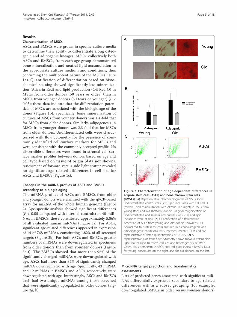
ResultsCharacterization of MSCsASCs and BMSCs were grown in specific culture mediato determine their ability to differentiate along osteo-genic and adipogenic lineages. MSCs, collectively bothASCs and BMSCs, from each age group demonstratedbone mineralization and neutral lipid accumulation inthe appropriate culture medium and conditions, thusconfirming the multipotent nature of the MSCs (Figure1a). Quantification of differentiation based on histo-chemical staining showed significantly less mineraliza-tion (Alizarin Red) and lipid production (Oil Red O) inMSCs from older donors (50 years or older) than inMSCs from younger donors (50 tears or younger) (P <0.05); these data indicate that the differentiation poten-tials of MSCs are associated with the biologic age of thedonor (Figure 1b). Specifically, bone mineralization ofcultures of MSCs from younger donors was 1.4-fold thatfor MSCs from older donors. Similarly, adipogenesis inMSCs from younger donors was 2.3-fold that for MSCsfrom older donors. Undifferentiated cells were charac-terized with flow cytometry for the presence of com-monly identified cell-surface markers for MSCs andwere consistent with the commonly accepted profile. Nodiscernible differences were found in stromal cell-sur-face marker profiles between donors based on age andcell type based on tissue of origin (data not shown).Assessment of forward versus side light scatter revealedno significant age-related differences in cell size forASCs and BMSCs (Figure 1c).
Changes in the miRNA profiles of ASCs and BMSCssecondary to biologic agingThe miRNA profiles of ASCs and BMSCs from olderand younger donors were analyzed with the qPCR-basedarray for miRNA of the whole human genome (Figure2). Age-specific analysis showed significant differences(P < 0.05 compared with internal controls) in 45 miR-NAs in BMSCs; these constituted approximately 5.86%of all evaluated human miRNAs (Figure 3a). For ASCs,significant age-related differences appeared in expressionof 14 of 768 miRNAs, constituting 1.82% of all screenedtargets (Figure 3b). For both ASCs and BMSCs, greaternumbers of miRNAs were downregulated in specimensfrom older donors than from younger donors (Figure3c-f). The BMSCs showed that more than 95% of thesignificantly changed miRNAs were downregulated withage. ASCs had more than 85% of significantly changedmiRNA downregulated with age. Specifically, 43 miRNAand 12 miRNAs in BMSCs and ASCs, respectively, weredownregulated with age. Interestingly, ASCs and BMSCseach had two unique miRNAs among those screenedthat were significantly upregulated in older donors (Fig-ure 3g, h).
MicroRNA target prediction and bioinformaticsassessmentsLists of predicted genes associated with significant miR-NAs differentially expressed secondary to age-relateddifferences within a subset grouping (for example,downregulated BMSCs in older versus younger donors)
Figure 1 Characterization of age-dependent differences inadipose stem cells (ASCs) and bone marrow stem cells(BMSCs). (a) Representative photomicrographs of MSCs showundifferentiated control cells (left), lipid inclusions with Oil Red O(middle), and mineralization with Alizarin Red (right) in ASCs fromyoung (top) and old (bottom) donors. Original magnification ofundifferentiated and mineralized cultures was ×10, and lipidinclusions were at ×40. (b) Quantification of differentiationpotentials of ASCs from young and old donors shown as ODnormalized to protein for cells cultured in osteoblastogenic andadipocytogenic conditions. Bars represent mean ± SEM and arerepresentative of three quantifications. *P < 0.05. (c) Arepresentative plot from flow cytometry shows forward versus sidelight scatter used to assess cell size and heterogeneity of MSCs.Green plots demonstrate ASCs, and red plots indicate BMSCs. Datafor young donors are on the right, and for old donors, on the left.
Pandey et al. Stem Cell Research & Therapy 2011, 2:49http://stemcellres.com/content/2/6/49
Page 5 of 18
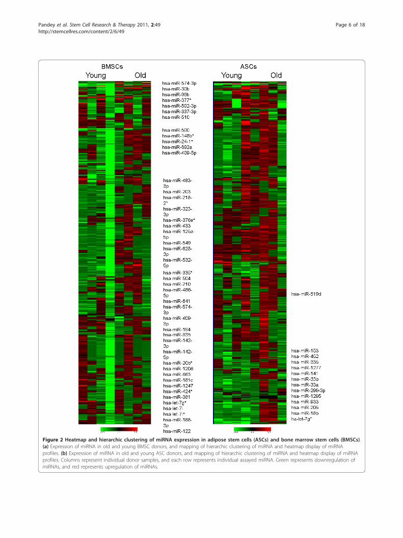
Figure 2 Heatmap and hierarchic clustering of miRNA expression in adipose stem cells (ASCs) and bone marrow stem cells (BMSCs).(a) Expression of miRNA in old and young BMSC donors, and mapping of hierarchic clustering of miRNA and heatmap display of miRNAprofiles. (b) Expression of miRNA in old and young ASC donors, and mapping of hierarchic clustering of miRNA and heatmap display of miRNAprofiles. Columns represent individual donor samples, and each row represents individual assayed miRNA. Green represents downregulation ofmiRNAs, and red represents upregulation of miRNAs.
Pandey et al. Stem Cell Research & Therapy 2011, 2:49http://stemcellres.com/content/2/6/49
Page 6 of 18
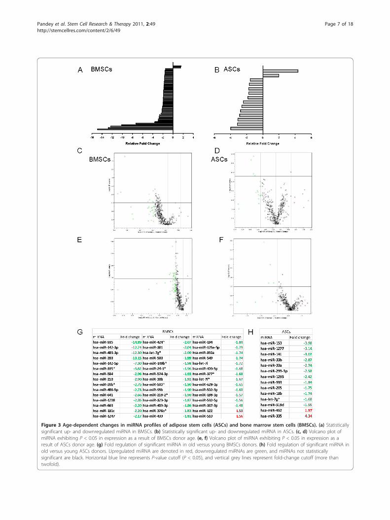
Figure 3 Age-dependent changes in miRNA profiles of adipose stem cells (ASCs) and bone marrow stem cells (BMSCs). (a) Statisticallysignificant up- and downregulated miRNA in BMSCs. (b) Statistically significant up- and downregulated miRNA in ASCs. (c, d) Volcano plot ofmiRNA exhibiting P < 0.05 in expression as a result of BMSCs donor age. (e, f) Volcano plot of miRNA exhibiting P < 0.05 in expression as aresult of ASCs donor age. (g) Fold regulation of significant miRNA in old versus young BMSCs donors. (h) Fold regulation of significant miRNA inold versus young ASCs donors. Upregulated miRNA are denoted in red, downregulated miRNAs are green, and miRNAs not statisticallysignificant are black. Horizontal blue line represents P-value cutoff (P < 0.05), and vertical grey lines represent fold-change cutoff (more thantwofold).
Pandey et al. Stem Cell Research & Therapy 2011, 2:49http://stemcellres.com/content/2/6/49
Page 7 of 18
were generated from TargetScan (data not shown). IPAwas then used to generate canonic pathway involvement,biologic functions, and network analysis for the pre-dicted targets. Canonic pathways generated for up- anddownregulated miRNAs in BMSCs from older as com-pared with younger donors demonstrated the involve-ment of many pathways (Figure 4a, b). The pathwaysassociated with the largest age-related decreases inmiRNA levels of BMSCs (P ≤ 0.05) included thoseinvolved in molecular mechanisms of cancer, PTEN sig-naling, mTOR signaling, and RAN signaling. It would bepredicted that those pathways would have increasedactivity because the inhibitory miRNA levels were down-regulated. Among the canonic pathways associated withthe largest age-related increases in miRNA of olderdonor BMSCs were Wnt/b-catenin signaling, tight-junc-tion signaling, cleavage and polyadenylation of pre-mRNA, and SAPK/JNK signaling; these observationssuggest decreased activity of those component moleculesin the older BMSCs. Analysis of downregulated miRNAsin ASCs revealed significant involvement of the canonicpathways, including molecular mechanisms of cancer,axonal guidance signaling, ephrin receptor signaling, andPPARa/RXRa activation (Figure 4c, d). The canonicpathways associated with the largest age-relatedincreases in miRNA levels of older donor ASCs includedRAN signaling, AMPK signaling, and cell-cycle regula-tion; however, none achieved the P-value threshold.Subsequently, the gene lists of putative targets were ana-
lyzed with IPA software for biologic functions manifestedsecondary to age-dependent miRNA expression in MSCs.For age-related decreased miRNA targets of BMSCs, thetop functions mapped included increased gene expression,organismal development, and cardiovascular disease (Fig-ure 5a). For age-related increased miRNA targets inBMSCs, those with inhibition of biologic functionsincluded cell morphology, cancer, and diseases of thereproductive system (Figure 5b). For ASCs, age-relateddecreased miRNA targets predict greater involvement ofgastrointestinal disease, geriatric disorders, and inflamma-tory disease states (Figure 5c). For age-related increasedmiRNA targets in ASCs, the biologic functions that wouldbe inhibited included cellular development, embryonicdevelopment, and gene expression (Figure 5d).From the data for canonic pathways and biologic func-
tions, IPA assessed networks of focus molecules, whichwere then grouped for the generation of networks ofdirectly and indirectly involved molecules (data notshown). For age-related decreased miRNA in BMSCs,the major networks were related to cellular movement,cell signaling, cell death, and inflammatory diseases (Fig-ure 6a, b). For age-related increased miRNA in BMSCs,major network functions involved cellular compromise,
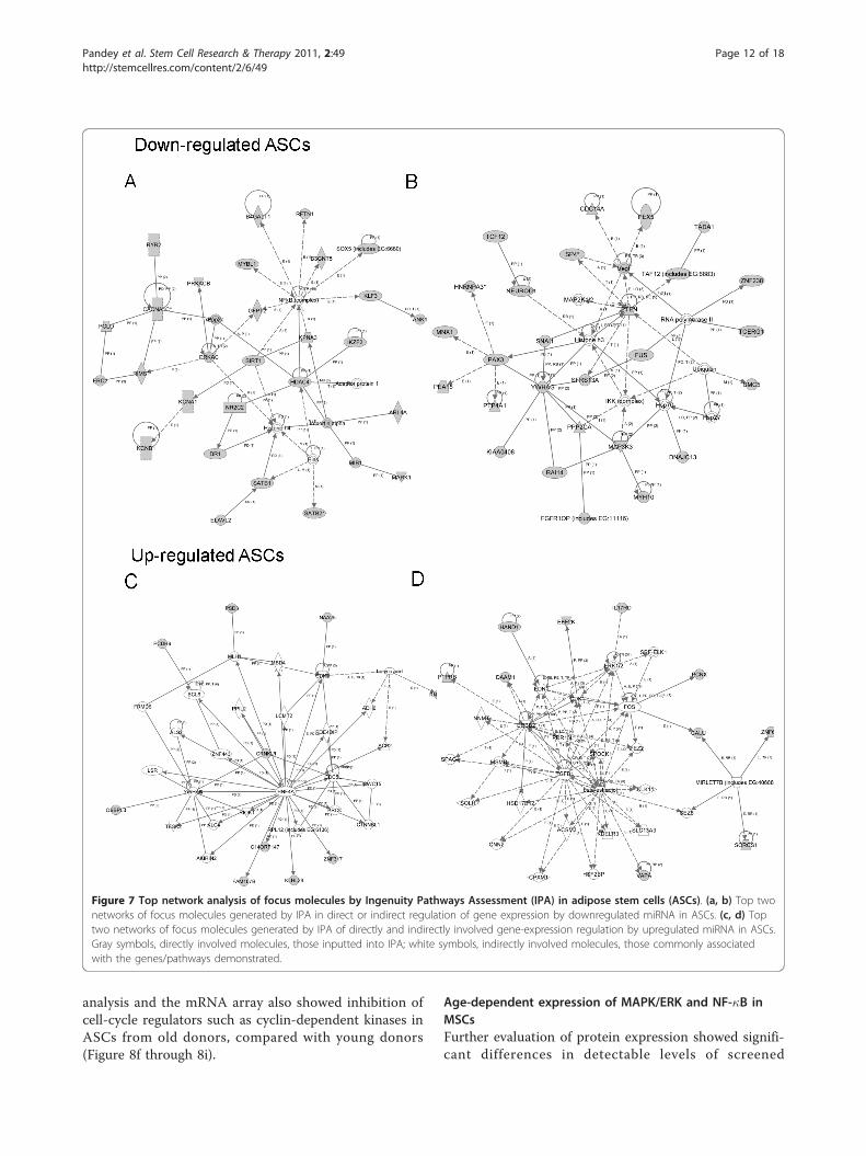
antigen presentation, cellular growth and proliferation,cell death, and cancer (Figure 6a, b). For age-relateddecreased miRNA in ASCs, the major significant net-works were related to cell signaling, molecular transport,cell cycle, and gene expression (Figure 7a, b). For age-related increased miRNA in ASCs, major network func-tions were related to the cell cycle, cell-to-cell signalingand interaction, and cellular development (Figure 7a, b).The focus molecules were mapped by IPA, which gener-ated networks of interaction between focus moleculesand predicted or commonly associated target molecules.Major network pathways were created for up- anddownregulated ASCs and BMSCs. Collectively, canonicpathways generated by IPA and analysis of the focusmolecules networks reveal potential targets that miRNAdifferentially expressed in MSCs aging. Comprehensiveevaluation of these major focus molecules from IPArevealed that miRNAs influencing NF-�B, ERK1/2, andI�B were directly involved.
Age-related differences in constitutive mRNA expressionin MSCs secondary to miRNA regulationRepresentative ASCs from both age groups wereselected based on microarray data clustering for furtherevaluation of the predicted effects of miRNA on consti-tutive mRNA expression. These representative ASCswere analyzed by qPCR-based signal-transduction array.This analysis confirmed that mRNA levels of the MAPKelements, iNOS, VCAM1, and IKK, as well as other NF-�B-pathway-related molecules were downregulated inASCs from older donors compared with those fromyounger donors (Figure 8a through 8c). In contrast,mRNA for NF-�B and non-classically activated NF-�Btargets, such as IL-4 receptor and myc, were significantlyelevated. Other significantly upregulated mRNA in ASCsfrom older donors included WNT/b-catenin pathwayconstituents. Analysis of mRNA levels by using IPAshowed similar canonic pathways and biologic functionsas predicted from the miRNA data (Figure 7d through7e). Analysis of focus molecules showed that majorcomponents of the MAPK/ERK pathway were downre-gulated in older donors and predicted depressed expres-sion of related molecules. Furthermore, although thearray showed elevated levels of NF-�B mRNA that IPApredicted based on the array data, other traditionallyassociated components of the inflammatory cascadewere downregulated (Figure 8f through 8i). The excep-tion to this was tumor necrosis factor-a (TNF-a), whichIPA predicted would be increased due to a downregula-tion of miRNAs that act on it. Evaluation of mRNAlevels of TNF-a showed increased levels; the fold differ-ence compared with younger donors, however, was notsignificant. IPA and the Ingenuity Knowledgebase
Pandey et al. Stem Cell Research & Therapy 2011, 2:49http://stemcellres.com/content/2/6/49
Page 8 of 18
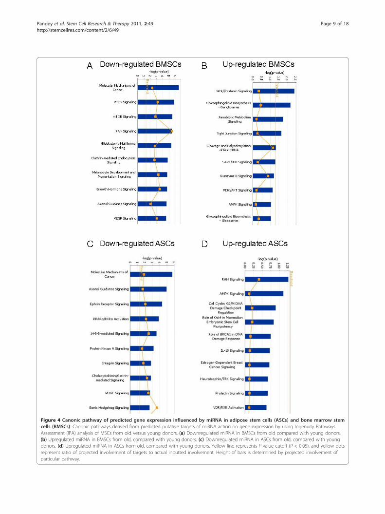
Figure 4 Canonic pathway of predicted gene expression influenced by miRNA in adipose stem cells (ASCs) and bone marrow stemcells (BMSCs). Canonic pathways derived from predicted putative targets of miRNA action on gene expression by using Ingenuity PathwaysAssessment (IPA) analysis of MSCs from old versus young donors. (a) Downregulated miRNA in BMSCs from old compared with young donors.(b) Upregulated miRNA in BMSCs from old, compared with young donors. (c) Downregulated miRNA in ASCs from old, compared with youngdonors. (d) Upregulated miRNA in ASCs from old, compared with young donors. Yellow line represents P-value cutoff (P < 0.05), and yellow dotsrepresent ratio of projected involvement of targets to actual inputted involvement. Height of bars is determined by projected involvement ofparticular pathway.
Pandey et al. Stem Cell Research & Therapy 2011, 2:49http://stemcellres.com/content/2/6/49
Page 9 of 18
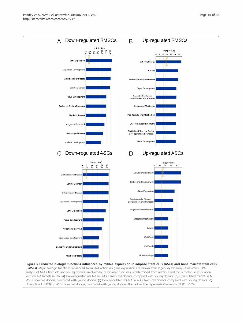
Figure 5 Predicted biologic functions influenced by miRNA expression in adipose stem cells (ASCs) and bone marrow stem cells(BMSCs). Major biologic functions influenced by miRNA action on gene expression are shown from Ingenuity Pathways Assessment (IPA)analysis of MSCs from old and young donors. Involvement of biologic functions is determined from network and focus molecule associationwith miRNA targets in IPA. (a) Downregulated miRNA in BMSCs from old donors, compared with young donors. (b) Upregulated miRNA in M-MSCs from old donors, compared with young donors. (c) Downregulated miRNA in ASCs from old donors, compared with young donors. (d)Upregulated miRNA in ASCs from old donors, compared with young donors. The yellow line represents P-value cutoff (P < 0.05).
Pandey et al. Stem Cell Research & Therapy 2011, 2:49http://stemcellres.com/content/2/6/49
Page 10 of 18
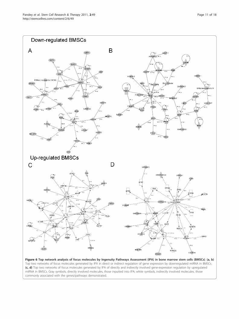
Figure 6 Top network analysis of focus molecules by Ingenuity Pathways Assessment (IPA) in bone marrow stem cells (BMSCs). (a, b)Top two networks of focus molecules generated by IPA in direct or indirect regulation of gene expression by downregulated miRNA in BMSCs.(c, d) Top two networks of focus molecules generated by IPA of directly and indirectly involved gene-expression regulation by upregulatedmiRNA in BMSCs. Gray symbols, directly involved molecules, those inputted into IPA; white symbols, indirectly involved molecules, thosecommonly associated with the genes/pathways demonstrated.
Pandey et al. Stem Cell Research & Therapy 2011, 2:49http://stemcellres.com/content/2/6/49
Page 11 of 18
analysis and the mRNA array also showed inhibition ofcell-cycle regulators such as cyclin-dependent kinases inASCs from old donors, compared with young donors(Figure 8f through 8i).
Age-dependent expression of MAPK/ERK and NF-�B inMSCsFurther evaluation of protein expression showed signifi-cant differences in detectable levels of screened
Figure 7 Top network analysis of focus molecules by Ingenuity Pathways Assessment (IPA) in adipose stem cells (ASCs). (a, b) Top twonetworks of focus molecules generated by IPA in direct or indirect regulation of gene expression by downregulated miRNA in ASCs. (c, d) Toptwo networks of focus molecules generated by IPA of directly and indirectly involved gene-expression regulation by upregulated miRNA in ASCs.Gray symbols, directly involved molecules, those inputted into IPA; white symbols, indirectly involved molecules, those commonly associatedwith the genes/pathways demonstrated.
Pandey et al. Stem Cell Research & Therapy 2011, 2:49http://stemcellres.com/content/2/6/49
Page 12 of 18
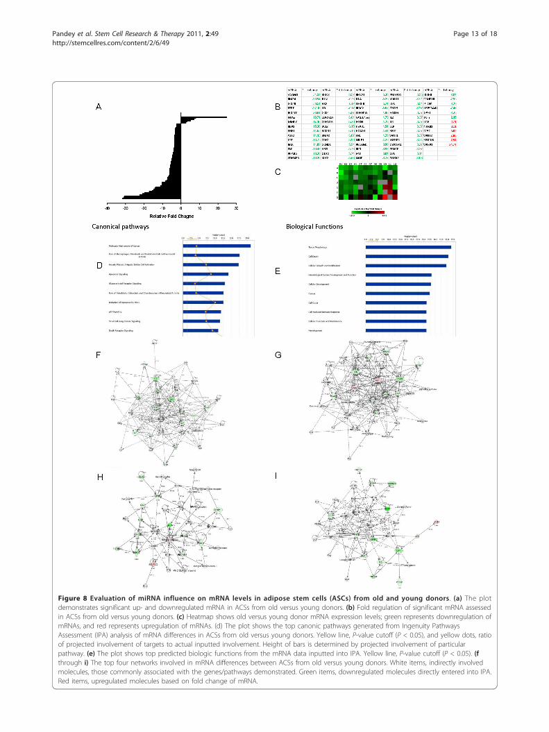
Figure 8 Evaluation of miRNA influence on mRNA levels in adipose stem cells (ASCs) from old and young donors. (a) The plotdemonstrates significant up- and downregulated mRNA in ACSs from old versus young donors. (b) Fold regulation of significant mRNA assessedin ACSs from old versus young donors. (c) Heatmap shows old versus young donor mRNA expression levels; green represents downregulation ofmRNAs, and red represents upregulation of mRNAs. (d) The plot shows the top canonic pathways generated from Ingenuity PathwaysAssessment (IPA) analysis of mRNA differences in ACSs from old versus young donors. Yellow line, P-value cutoff (P < 0.05), and yellow dots, ratioof projected involvement of targets to actual inputted involvement. Height of bars is determined by projected involvement of particularpathway. (e) The plot shows top predicted biologic functions from the mRNA data inputted into IPA. Yellow line, P-value cutoff (P < 0.05). (fthrough i) The top four networks involved in mRNA differences between ACSs from old versus young donors. White items, indirectly involvedmolecules, those commonly associated with the genes/pathways demonstrated. Green items, downregulated molecules directly entered into IPA.Red items, upregulated molecules based on fold change of mRNA.
Pandey et al. Stem Cell Research & Therapy 2011, 2:49http://stemcellres.com/content/2/6/49
Page 13 of 18
molecules, as predicted from the miRNA data. Immuno-cytochemical localization of p65 and p50 subunits ofNF-�B showed differential expression in cells from olderand younger donors (Figure 9a). In cells from youngerdonors, NF-�B appeared to localize predominantlywithin the nucleus. In cells from older donors, however,detectable NF-�B was present in the nucleus, but dra-matically elevated levels were observed in the cytoplasm.The pattern of distribution was similar for both p65 andp50 subunits of NF-�B, but was much more pronouncedfor p50. As assessed with Western blot, levels of phos-phorylated inhibitory kappa B (p-I�B) (P < 0.001), phos-phorylated inhibitory kappa B kinase (p-I�K) (P <0.001), and inducible nitric oxide (iNOS) (P < 0.001)were significantly decreased in ASCs from older donors,compared with those from younger donors (Figure 9b,d). Interestingly, this analysis showed that phosphory-lated NF-�B/p65 (p-NF-�B) levels were significantly ele-vated in ASCs from older donors as compared withyounger donors (P < 0.05). Furthermore, as predicted bymiRNA screening and mRNA data, Western blot analy-sis showed that levels of phosphorylated ERK1/2 (p-ERK1/2) (P < 0.01), phosphorylated c-fos (p-c-fos) (P <0. 01), phosphorylated c-jun (p-c-jun) (P < 0.001), andphosphorylated JNK (p-JNK) (P < 0.001) were all signifi-cantly decreased in ASC from older donors comparedwith younger donors (Figure 9c, e).
DiscussionIn the present study, the comprehensive miRNA profilesof undifferentiated ASCs and BMSCs were analyzed toidentify age-related differences in constitutive potentialbiologic functions at the mRNA and protein expressionlevels. The results showed that in MSCs isolated fromtwo distinct tissues/organs, small subsets of miRNAsdisplay age-related differences in the regulation of geneexpression involving specific cellular and molecularpathways of cell proliferation and inflammation, pre-sumably as regimented markers of aging. Specifically,significant age-related decreases were found in theMAPK/ERK pathway and NF-�B pathway expression.Originally, it was thought that the hallmark of MSCs
was their ability to differentiate into various end-celltypes for regenerative repair of damage due to injuryfrom disease processes [11,35,36]. Subsequently, manyreports suggested that the role of MSCs as treatmentmodalities may occur through the expression of cyto-kines and chemokines that promote angiogenesis orsquelch inflammatory responses, and adhesion mole-cules that would subsequently induce endogenous repairof injured cells and tissues [7]. In addition to thesemechanisms that MSCs use to ameliorate cellulardamage, their differentiation capabilities remain animportant defining characteristic. We confirmed that
ASCs from young donors, compared with those fromolder donors, had significantly elevated potential to dif-ferentiate toward osteogenic and adipogenic lineages.Other studies found similar changes while investigatingaging of MSCs from humans, mice, rhesus macaques,and cynomolgus monkeys [3,30,37,38]. Our flow-cyto-metry results failed to detect changes in cell-surfacemarkers secondary to the aging process. Thus, despiteother age-related changes in MSCs, the cells can beidentified by using cell-surface markers frequently usedto distinguish MSCs from other cell types, regardless ofthe age of the donor [27].Analysis revealed that individual targets selected from
the IPA analysis followed the predicted pattern ofexpression at the mRNA and protein levels. All evalu-ated upregulated miRNA targets showed decreasedlevels of both mRNA and functional protein, includingthose involved in the MAPK/ERK and NF-�B pathwaysand in cell-cycle control. This is in accordance withrecent results suggesting that although miRNAs pro-mote both mRNA degradation and posttranslationalrepression, they appear to act predominantly as regula-tors at the mRNA level [19]. Furthermore, the currentfindings suggest that the miRNA profile of MSCs can beused to identify potential signaling mechanisms involvedin MSCs functions, and perhaps even provide anotherway to classify MSCs function.Several recent reports have demonstrated the function
of the let-7 family of miRNA and its role in MSCs, espe-cially as markers and regulators of a differentiated state[39]. The let-7 family was shown to repress cellular repli-cation and inhibit self-renewal [40]. Our data showedthat let-7 family miRNA were significantly downregu-lated in both ASCs and BMSCs from older donors.Accordingly, it can be surmised that ASCs and BMSCs inolder donors may not be as efficient at self-renewal andwould have decreased overall proliferation, comparedwith younger donors. Other reports determined thathuman skeletal muscle cells from older donors demon-strate reduced expression of let-7 and decreased mRNAlevels of cell-cycle regulators such as CDK6 [25]. Ouranalysis of mRNA levels of cell-cycle regulatory mole-cules found that multiple cyclin-dependent kinases weredownregulated, contributing to a reduction in cell prolif-eration. The data also showed increased miRNA targetedtoward the Wnt/b-catenin-signaling pathway. Recent stu-dies have shown that prolonged activation of Wnt signal-ing promotes MSC proliferation and contributes to aging[41]. Thus, our results demonstrate that miRNA inhibitsWnt/b-catenin signaling to decrease cell proliferation inaged MSCs, and potentially plays a role in retarding theaging process in MSCs.The miRNAs directed toward the MAPK/ERK system
were expressed at higher levels in cells from older
Pandey et al. Stem Cell Research & Therapy 2011, 2:49http://stemcellres.com/content/2/6/49
Page 14 of 18
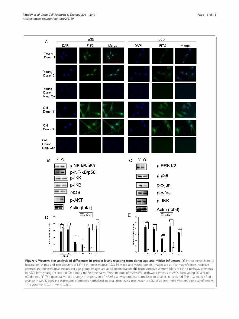
Figure 9 Western blot analysis of differences in protein levels resulting from donor age and miRNA influences. (a) Immunocytochemicallocalization of p65 and p50 subunits of NF-�B in representative ASCs from old and young donors. Images are at ×20 magnification. Negativecontrols are representative images per age group. Images are at ×5 magnification. (b) Representative Western blots of NF-�B pathway elementsin ASCs from young (Y) and old (O) donors. (c) Representative Western blots of MAPK/ERK pathway elements in ASCs from young (Y) and old(O) donors. (d) The quantitative fold change in expression of NF-�B pathway proteins normalized to total actin levels. (e) The quantitative foldchange in MAPK signaling expression of proteins normalized to total actin levels. Bars, mean ± SEM of at least three Western blot quantifications.*P < 0.05; **P < 0.01; ***P < 0.001).
Pandey et al. Stem Cell Research & Therapy 2011, 2:49http://stemcellres.com/content/2/6/49
Page 15 of 18
donors. Specifically, ERK1/2 and JNK gene expressionwere involved as putative targets for miRNA-mediatedgene-expression control. The downregulation of mRNAlevels for c-fos and c-jun were confirmed by using real-time PCR and, by Western blot, demonstrated decreasedprotein levels of the MAPK pathway. Collectively p38,p-ERK1/2, p-c-fos, p-c-jun, and p-JNK levels were allsignificantly reduced in the ASCs of older donors ascompared with those of younger donors, with the exten-sion to BMSCs due to a similar miRNA profile and IPAanalysis. Previous studies have indicated that BMSCsfrom older donors have decreased proliferation potential[2,42,43]. Additional reports have suggested that thedynamics of the aging process of MSCs is a determinantof cellular aging; however, the exact mechanism remainsunclear [10]. The identified differences in miRNA incells from older donors may represent the mechanismby which MSCs, through control over the MAPK/ERKsignaling cascade, decrease cellular proliferation rates,thereby contributing to reduced tissue renewal in aging.Although many of the fine details of aging in humans
are yet to be elucidated, the interplay of aging andinflammation has been intensively researched. Manyabnormalities in cellular processes have been found tooccur with aging, including the development of cancerand type II diabetes mellitus [17,44]. At the center ofthese disease processes lie the common denominators ofadvanced age and inflammation. Interestingly, elevatedlevels of activated NF-�B were observed in older-donorMSCs. Whereas regulatory and traditional componentsof the NF-�B pathway, including, among others, I�B,I�K, iNOS, and IL-1a, were downregulated, other non-traditionally associated molecules were upregulated,including IL-4 receptor and myc oncogene. Tradition-ally, protein factors responsive to NF-�B transcriptionalregulation would further amplify NF-�B expression,which was not observed in the current study [45]. Inaddition, the subcellular distribution of NF-�B observedin MSCs from older donors suggests the possibility ofan alternative role in MSCs aging. Aging and NF-�Bactivation may have more in common than was initiallypostulated. Previous reports indicated that NF-�B canfunction in resistance to apoptosis [46]. In addition, NF-�B was shown to repress apoptosis triggered by JNK[47]. These observations, combined with our data show-ing decreased levels of JNK, provide evidence that NF-�B may be central to a protective role in MSC aging.Indeed, our data demonstrate that elevated levels of NF-�B regulated by miRNA activity may play a central rolein the onset and progression of the aging process inMSCs.The results also suggest that a delicate balance is
maintained as a result of increased NF-�B expression inolder MSCs. Typically, elevated levels of NF-�B are
linked with pathologic processes; in old cells, however,elevated levels of NF-�B could prevent apoptosis [45].Prevention of apoptosis would be of substantial impor-tance to MSCs, especially given their function for endo-genous cellular repair. Most studies, however, havedemonstrated a potent anti-inflammatory role for MSCs[7]. The paradigm shift occurs with the notion of how apotently anti-inflammatory cell would maintain elevatedlevels of NF-�B. Our results demonstrated that althoughNF-�B levels are indeed elevated in older donors, pre-sumably to prevent activation of apoptotic pathways,other commonly associated molecules in the NF-�B andinflammatory cascade were downregulated. IPA analysis,mRNA expression, and protein levels demonstrated thatmolecules such as IL-1a, TNF-a, iNOS, and I�K weresignificantly downregulated in ASCs from older donors.In addition, JNK was also significantly downregulated inolder donors, giving support to the role of NF-�B as aninhibitor of apoptosis. That I�B levels decreased in theolder donor ASCs is indicative of prolonged NF-�Bactivity. Although the classic NF-�B activity simulta-neously causes de novo synthesis of I�B, it appears thatNF-�B levels that are elevated because of aging in MSCsdo not work through this mechanism. Intracellular loca-lization of NF-�B from the current study in cells fromyounger donors appeared more predominant in thenucleus, specifically in the nucleolus, whereas cells fromolder donors demonstrated accumulation of NF-�B inthe cytosol; these observations suggest that although itwas phosphorylated in both groups, NF-�B was func-tioning differently as a function of age. Alternatively,relocalization of NF-�B subunits to the cytosol mayindicate a lack of transcriptional activity, as further evi-denced by decreased pro-inflammatory cytokines andother molecules including IL-1a, IL-8, and iNOS mRNAlevels in cells from older donors compared with youngerdonors. Further investigation of the role of elevated NF-�B levels is required to gain a full understanding of themechanisms at work in the MSC aging process.
ConclusionsIn summary, miRNA profiling demonstrated that sub-sets of miRNAs are biologically active in human MSCs,with the profiles of miRNAs changing with aging inboth ASCs and BMSCs. Furthermore, miRNAs modu-late and regulate gene expression related to a variety offunctions, particularly cellular proliferation and inflam-mation, both of which play an integral role in the pro-cess of aging. ASCs from older donors also exhibitsignificantly elevated levels of NF-�B, perhaps inresponse to downregulation of the MAPK/ERK systemto prevent proliferation and inhibit apoptotic stimuli.Interestingly, other molecules commonly associated withNF-�B were downregulated, leading to a unique and
Pandey et al. Stem Cell Research & Therapy 2011, 2:49http://stemcellres.com/content/2/6/49
Page 16 of 18
novel constellation of expression of the core inflamma-tory molecules in a potently anti-inflammatory cell. Thecurrent study presents, to our knowledge, the firstreport that miRNAs have unique roles in the aging ofMSCs, maintaining their capacity to suppress inflamma-tion and promote endogenous cellular repair while, per-haps, allowing MSCs to escape the aging process.
AbbreviationsASCs: adipose-derived mesenchymal stem cells; BMSCs: bone marrow-derived mesenchymal stem cells; CCM: complete culture medium; IPA:Ingenuity Pathway Assessment; MAPK: mitogen activated protein kinase;mRNA: messenger ribonucleic acid; miRNA: micro ribonucleic acid; MSCs:mesenchymal stem cells; NF-κB: nuclear factor kappa B; PCR: polymerasechain reaction.
AcknowledgementsWe thank Alan Tucker for his excellent technical assistance in performingflow cytometry. We also thank Margie McCants and Christina McLennan fortheir technical assistance with cell culture and Dina Gaupp for hertremendous help with immunocytochemistry. We thank Drs. James Wade,Ann Reilley, and Michael Teague, together with their office staff andpatients, for donating adipose tissues to this project. JMG is the Founder ofLaCell, LLC, and serves as a consultant for Johnson & Johnson companiesATRM and Mentor. The research was supported by grant numbers RR00164from the National Center for Research Resources, R21-NS059665 NationalInstitutes of Neurological Disorders and Stroke, National Institutes of Health,Pennington Biomedical Research Center, and Tulane University.
Author details1Center for Stem Cell Research and Regenerative Medicine, School ofMedicine, Tulane University, New Orleans, LA, USA. 2Division of Bacteriologyand Parasitology, Tulane National Primate Research Center, Covington, LA,USA. 3Department of Orthopedic Surgery, Brigham and Women’s Hospital,Harvard Medical School, Boston, MA, USA. 4Stem Cell Laboratory, PenningtonBiomedical Research Center, Louisiana State University System, Baton Rouge,LA, USA. 5Department of Pharmacology, School of Medicine, TulaneUniversity, New Orleans, LA, USA. 6Division of Regenerative Medicine, TulaneNational Primate Research Center, Covington, LA, USA.
Authors’ contributionsACP and BAB conceived, designed, and executed the experiments. ACP, JS,and RPO characterized the cells. ACP, DK, and BAB preformed bioinformaticsand statistical analyses. ACP, BAB, and JMG analyzed the data. ACP and BABwrote the article. JMG, JS, and JG edited the article. All authors read andapproved the final manuscript.
Competing interestsJMG is the Founder of LaCell, LLC, and serves as a consultant for Johnson &Johnson companies ATRM and Mentor; he has also received fees fromToucan Capital and its subsidiary companies in the adipose stem cell area.The other authors declare that they have no competing interests.
Received: 28 June 2011 Revised: 3 November 2011Accepted: 14 November 2011 Published: 14 November 2011
References1. Tokalov SV, Gruner S, Schindler S, Wolf G, Baumann M, Abolmaali N: Age-
related changes in the frequency of mesenchymal stem cells in thebone marrow of rats. Stem Cells Dev 2007, 16:439-446.
2. Zhou S, Greenberger JS, Epperly MW, Goff JP, Adler C, Leboff MS,Glowacki J: Age-related intrinsic changes in human bone-marrow-derived mesenchymal stem cells and their differentiation to osteoblasts.Aging Cell 2008, 7:335-343.
3. Yu JM, Wu X, Gimble JM, Guan X, Freitas MA, Bunnell BA: Age-relatedchanges in mesenchymal stem cells derived from rhesus macaque bonemarrow. Aging Cell 2011, 10:66-79.
4. Gimble JM, Guilak F, Bunnell BA: Clinical and preclinical translation of cell-based therapies using adipose tissue-derived cells. Stem Cell Res Ther2010, 1:19.
5. Izadpanah R, Joswig T, Tsien F, Dufour J, Kirijan JC, Bunnell BA:Characterization of multipotent mesenchymal stem cells from the bonemarrow of rhesus macaques. Stem Cells Dev 2005, 14:440-451.
6. Prockop DJ: Repair of tissues by adult stem/progenitor cells (MSCs):controversies, myths, and changing paradigms. Mol Ther 2009,17:939-946.
7. Ortiz LA, Dutreil M, Fattman C, Pandey AC, Torres G, Go K, Phinney DG:Interleukin 1 receptor antagonist mediates the antiinflammatory andantifibrotic effect of mesenchymal stem cells during lung injury. ProcNatl Acad Sci USA 2007, 104:11002-11007.
8. Stolzing A, Jones E, McGonagle D, Scutt A: Age-related changes in humanbone marrow-derived mesenchymal stem cells: consequences for celltherapies. Mech Ageing Dev 2008, 129:163-173.
9. Van Zant G, Liang Y: The role of stem cells in aging. Exp Hematol 2003,31:659-672.
10. Carrington JL: Aging bone and cartilage: cross-cutting issues. BiochemBiophys Res Commun 2005, 328:700-708.
11. Rao MS, Mattson MP: Stem cells and aging: expanding the possibilities.Mech Ageing Dev 2001, 122:713-734.
12. Mareschi K, Ferrero I, Rustichelli D, Aschero S, Gammaitoni L, Aglietta M,Madon E, Fagioli F: Expansion of mesenchymal stem cells isolated frompediatric and adult donor bone marrow. J Cell Biochem 2006, 97:744-754.
13. Zheng H, Martin JA, Duwayri Y, Falcon G, Buckwalter JA: Impact of agingon rat bone marrow-derived stem cell chondrogenesis. J Gerontol A BiolSci Med Sci 2007, 62:136-148.
14. Karagiannides I, Tchkonia T, Dobson DE, Steppan CM, Cummins P, Chan G,Salvatori K, Hadzopoulou-Cladaras M, Kirkland JL: Altered expression of C/EBP family members results in decreased adipogenesis with aging. Am JPhysiol Regul Integr Comp Physiol 2001, 280:R1772-1780.
15. Hackl M, Brunner S, Fortschegger K, Schreiner C, Micutkova L, Muck C,Laschober GT, Lepperdinger G, Sampson N, Berger P, et al: miR-17, miR-19b, miR-20a, and miR-106a are down-regulated in human aging. AgingCell 2010, 9:291-296.
16. Liang R, Bates DJ, Wang E: Epigenetic control of microRNA expressionand aging. Curr Genomics 2009, 10:184-193.
17. Noren Hooten N, Abdelmohsen K, Gorospe M, Ejiogu N, Zonderman AB,Evans MK: microRNA expression patterns reveal differential expression oftarget genes with age. PLoS One 2010, 5:e10724.
18. Wang E: MicroRNA, the putative molecular control for mid-life decline.Ageing Res Rev 2007, 6:1-11.
19. Guo H, Ingolia NT, Weissman JS, Bartel DP: Mammalian microRNAspredominantly act to decrease target mRNA levels. Nature 2010,466:835-840.
20. Pillai RS, Bhattacharyya SN, Filipowicz W: Repression of protein synthesisby miRNAs: how many mechanisms? Trends Cell Biol 2007, 17:118-126.
21. Friedman RC, Farh KK, Burge CB, Bartel DP: Most mammalian mRNAs areconserved targets of microRNAs. Genome Res 2009, 19:92-105.
22. Subramanian S, Steer CJ: MicroRNAs as gatekeepers of apoptosis. J CellPhysiol 2010, 223:289-298.
23. Baek D, Villen J, Shin C, Camargo FD, Gygi SP, Bartel DP: The impact ofmicroRNAs on protein output. Nature 2008, 455:64-71.
24. Bae S, Ahn JH, Park CW, Son HK, Kim KS, Lim NK, Jeon CJ, Kim H: Gene andmicroRNA expression signatures of human mesenchymal stromal cells incomparison to fibroblasts. Cell Tissue Res 2009, 335:565-573.
25. Drummond MJ, McCarthy JJ, Sinha M, Spratt HM, Volpi E, Esser KA,Rasmussen BB: Aging and microRNA expression in human skeletalmuscle: a microarray and bioinformatics analysis. Physiol Genomics 2010,43:595-603.
26. Izadpanah R, Trygg C, Patel B, Kriedt C, Dufour J, Gimble JM, Bunnell BA:Biologic properties of mesenchymal stem cells derived from bonemarrow and adipose tissue. J Cell Biochem 2006, 99:1285-1297.
27. Tucker HA, Bunnell BA: Characterization of human adipose-derived stemcells using flow cytometry. Methods Mol Biol 2010, 702:121-131.
28. Pachon-Pena G, Yu G, Tucker A, Wu X, Vendrell J, Bunnell BA, Gimble JM:Stromal stem cells from adipose tissue and bone marrow of age-matched female donors display distinct immunophenotypic profiles.J Cell Physiol 2011, 226:843-851.
Pandey et al. Stem Cell Research & Therapy 2011, 2:49http://stemcellres.com/content/2/6/49
Page 17 of 18
29. Bunnell BA, Flaat M, Gagliardi C, Patel B, Ripoll C: Adipose-derived stemcells: isolation, expansion and differentiation. Methods 2008, 45:115-120.
30. Izadpanah R, Kaushal D, Kriedt C, Tsien F, Patel B, Dufour J, Bunnell BA:Long-term in vitro expansion alters the biology of adult mesenchymalstem cells. Cancer Res 2008, 68:4229-4238.
31. Ripoll CB, Bunnell BA: Comparative characterization of mesenchymalstem cells from eGFP transgenic and non-transgenic mice. BMC Cell Biol2009, 10:3.
32. Sturn A, Quackenbush J, Trajanoski Z: Genesis: cluster analysis ofmicroarray data. Bioinformatics 2002, 18:207-208.
33. Grimson A, Farh KK, Johnston WK, Garrett-Engele P, Lim LP, Bartel DP:MicroRNA targeting specificity in mammals: determinants beyond seedpairing. Mol Cell 2007, 27:91-105.
34. Livak KJ, Schmittgen TD: Analysis of relative gene expression data usingreal-time quantitative PCR and the 2(-Delta Delta C(T)) method. Methods2001, 25:402-408.
35. Krause DS, Theise ND, Collector MI, Henegariu O, Hwang S, Gardner R,Neutzel S, Sharkis SJ: Multi-organ, multi-lineage engraftment by a singlebone marrow-derived stem cell. Cell 2001, 105:369-377.
36. Pittenger MF, Mackay AM, Beck SC, Jaiswal RK, Douglas R, Mosca JD,Moorman MA, Simonetti DW, Craig S, Marshak DR: Multilineage potentialof adult human mesenchymal stem cells. Science 1999, 284:143-147.
37. Katsara O, Mahaira LG, Iliopoulou EG, Moustaki A, Antsaklis A, Loutradis D,Stefanidis K, Baxevanis CN, Papamichail M, Perez SA: Effects of donor age,gender and in vitro cellular aging on the phenotype, functional andmolecular characteristics of mouse bone marrow-derived mesenchymalstem cells. Stem Cells Dev 2011, 20:1549-1561.
38. Ren Z, Wang J, Zou C, Guan Y, Zhang YA: Comparative characterization ofmesenchymal stem cells from different age groups of cynomolgusmonkeys. Sci China Life Sci 2010, 53:563-572.
39. Hammond SM, Sharpless NE: HMGA2, microRNAs, and stem cell aging.Cell 2008, 135:1013-1016.
40. Johnson CD, Esquela-Kerscher A, Stefani G, Byrom M, Kelnar K,Ovcharenko D, Wilson M, Wang X, Shelton J, Shingara J, Chin L, Brown D,Slack FJ: The let-7 microRNA represses cell proliferation pathways inhuman cells. Cancer Res 2007, 67:7713-7722.
41. Castilho RM, Squarize CH, Chodosh LA, Williams BO, Gutkind JS: mTORmediates Wnt-induced epidermal stem cell exhaustion and aging. CellStem Cell 2009, 5:279-289.
42. Kassem M: Stem cells: potential therapy for age-related diseases. Ann N YAcad Sci 2006, 1067:436-442.
43. Stenderup K, Justesen J, Clausen C, Kassem M: Aging is associated withdecreased maximal life span and accelerated senescence of bonemarrow stromal cells. Bone 2003, 33:919-926.
44. Salminen A, Kaarniranta K: NF-kappaB signaling in the aging process.J Clin Immunol 2009, 29:397-405.
45. Salminen A, Ojala J, Kaarniranta K: Apoptosis and aging: increasedresistance to apoptosis enhances the aging process. Cell Mol Life Sci2010, 68:1021-1031.
46. Karin M: NF-kappaB as a critical link between inflammation and cancer.Cold Spring Harbor Perspect Biol 2009, 1:a000141.
47. Papa S, Bubici C, Zazzeroni F, Pham CG, Kuntzen C, Knabb JR, Dean K,Franzoso G: The NF-kappaB-mediated control of the JNK cascade in theantagonism of programmed cell death in health and disease. Cell DeathDiffer 2006, 13:712-729.
doi:10.1186/scrt90Cite this article as: Pandey et al.: MicroRNA profiling reveals age-dependent differential expression of nuclear factor �B and mitogen-activated protein kinase in adipose and bone marrow-derived humanmesenchymal stem cells. Stem Cell Research & Therapy 2011 2:49.
Submit your next manuscript to BioMed Centraland take full advantage of:
• Convenient online submission
• Thorough peer review
• No space constraints or color figure charges
• Immediate publication on acceptance
• Inclusion in PubMed, CAS, Scopus and Google Scholar
• Research which is freely available for redistribution
Submit your manuscript at www.biomedcentral.com/submit
Pandey et al. Stem Cell Research & Therapy 2011, 2:49http://stemcellres.com/content/2/6/49
Page 18 of 18
Copyright © 2022 FDOKUMEN