Cyclin D1 Is a Selective Modifier of Androgen-dependent Signaling and Androgen Receptor Function
Upload
independentCategory
view
3download
0
Biomarkers, Genomics, Proteomics, and Gene Regulation
NF-�B Regulates Androgen Receptor Expressionand Prostate Cancer Growth
Liying Zhang,* Saleh Altuwaijri,† Fangming Deng,‡
Lishi Chen,* Priti Lal,§ Umeshkumar K. Bhanot,*Ruslan Korets,¶ Sven Wenske,¶ Hans G. Lilja,¶
Chawnshang Chang,� Howard I. Scher,**and William L. Gerald*††
From the Departments of Pathology,* Urology and Clinical
Laboratory,¶ and Medicine,** and the Human Oncology and
Pathogenesis Program,†† Memorial Sloan-Kettering Cancer
Center, New York, New York; the Clinical Research Center,† Saad
Specialist Hospital, Al-Khobar, Saudi Arabia; the Department of
Pathology,‡ State University of New York, Upstate Medical
University, Syracuse, New York; the Department of Pathology and
Laboratory Medicine,§ University of Pennsylvania, Philadelphia,
Pennsylvania; and the George Whipple Laboratory for Cancer
Research,� the Department of Pathology, The Cancer Center,
University of Rochester, Rochester, New York
Prostate cancers that progress during androgen-dep-rivation therapy often overexpress the androgen re-ceptor (AR) and depend on AR signaling for growth.In most cases, increased AR expression occurs with-out gene amplification and may be due to alteredtranscriptional regulation. The transcription factornuclear factor (NF)-�B , which is implicated in tumor-igenesis, functions as an important downstream sub-strate of mitogen-activated protein kinase, phospha-tidylinositol 3-kinase, AKT, and protein kinase C andplays a role in other cancer-associated signaling path-ways. NF-�B is an important determinant of prostatecancer clinical biology, and therefore we investigatedits role in the regulation of AR expression. We foundthat NF-�B expression in prostate cancer cells signif-icantly increased AR mRNA and protein levels, ARtransactivation activity, serum prostate-specific anti-gen levels, and cell proliferation. NF-�B inhibitorsdecrease AR expression levels, prostate-specific anti-gen secretion, and proliferation of prostate cancercells in vitro. Furthermore, inhibitors of NF-�Bdemonstrated anti-tumor activity in androgen dep-rivation-resistant prostate cancer xenografts. In ad-dition, levels of both NF-�B and AR were stronglycorrelated in human prostate cancer. Our data sug-gest that NF-�B can regulate AR expression in pros-
tate cancer and that NF-�B inhibitors may havetherapeutic potential. (Am J Pathol 2009, 175:489–499;
DOI: 10.2353/ajpath.2009.080727)
Androgens play a critical role in normal prostate functionand in the development and progression of prostate can-cer.1–3 Prostate cancer cells are typically androgen de-pendent, and androgen deprivation is the standard sys-temic therapy for this disease. Virtually all prostatecancer patients treated with anti-androgens eventuallyprogress, an ominous clinical state for which no consis-tently effective therapy exists. The mechanisms involvedin the development of resistance are poorly understood.AR mutations, AR gene amplification, and activation ofthe AR through other signal transduction pathways havebeen implicated.2
The majority of tumors progressing during androgendeprivation therapy (referred to here as androgen depri-vation-resistant prostate cancer or ADRPC) expresshigher levels of AR transcript and protein suggesting thata marked increase in AR expression is a critical event intherapy resistance.4–7 Recent studies also demonstratethat increased AR expression is both necessary andsufficient to convert prostate cancer growth from a hor-mone therapy-sensitive to a resistant state in xenograftmodels.8 Since AR mRNA levels are often increased inADRPC without gene amplification,6,7 it is likely mediatedby transcription factors and transcription regulatingsignal transduction pathways that are altered duringprogression.
Nuclear Factor (NF)-�B is a family of transcription fac-tors composed of homo- and hetero-dimers initially iden-tified as an enhancer binding protein for the � immu-noglobulin light chain in B lymphocytes.9 Five knownmammalian subunits have been identified and include
Supported by National Institutes of Health/NCI (CA84999) to W.L.G. andDOD postdoctoral fellowship award (W81XWH-05-1-0107) to L.Z.
Accepted for publication May 14, 2009.
This paper is dedicated to the memory of Dr. William L. Gerald whoprovided strong leadership and support in this project.
Address reprint requests to Liying Zhang, M.D., Ph.D., Memorial Sloan-Kettering Cancer Center, 1275 York Avenue, Box 36, New York, NY10065. E-mail: [email protected].
The American Journal of Pathology, Vol. 175, No. 2, August 2009
Copyright © American Society for Investigative Pathology
DOI: 10.2353/ajpath.2009.080727
489
p65 (RelA), RelB, c-Rel, p50/p105, and p52/p100. NF-�Bdimers are retained in the cytoplasm complexed withinhibitory (I)�B proteins. Upon cellular stimulation, I�B isphosphorylated by I�B kinases and dissociates fromNF-�B subunits. As a consequence, NF-�B can translo-cate to the nucleus and regulate gene expression via in-teraction with �B enhancer elements.10
The NF-�B family of transcription factors are implicatedin oncogenesis by stimulating cell proliferation, inhibitingapoptosis, and promoting metastasis and angiogene-sis.11 There is growing evidence that NF-�B proteins playimportant roles in the development and progression of anumber of human malignancies.11–13 Constitutive NF-�Bactivity is common in solid tumors and has been demon-strated in cell lines of head and neck squamous cellcarcinoma,14 pancreatic adenocarcinoma,15 melano-ma,16 and breast cancer.17,18 Nuclear localization of p65,which is indicative of NF-�B activation, has also beenobserved in tumor samples from pancreatic adenocarci-noma,15 melanoma,19 hepatocellular carcinoma,20 thy-roid C-cell carcinoma,21 and breast cancer.18 Further-more constitutive activation of NF-�B has been proposedto be involved in the progression of breast cancers tohormone independent growth.17,18,22
Several studies have examined the expression ofNF-�B in human prostate cancer and its relationship toclinical features of the disease. NF-�B/p65 is overex-pressed in prostatic intraepithelial neoplasia and cancercompared with benign epithelium.23 Nuclear levels ofNF-�B/p65 correlate with NF-�B-dependent expressionof BclII, cyclin D1, matrix metalloproteinase-9, and vas-cular endothelial growth factor.24 Recent work indicatesthat NF-�B/p65 expression is predictive of biochemicalrecurrence in patients with positive surgical margins afterradical prostatectomy and nuclear localization of NF-�Bis increased in prostate cancer lymph node metastasis25
and can be used to predict patient outcome.26 Theseresults demonstrate that NF-�B/p65 is frequently acti-vated in human prostate adenocarcinoma and expres-sion may be related to progression.
NF-�B is constitutively activated in ADRPC xenograftswhere AR expression is also up-regulated.8,27 In addition,apigenin treatment, which inhibits the growth of andro-gen-responsive LNCaP human prostate carcinoma cells,results in a significant decrease in AR protein expressionand down-modulation of the constitutive expression ofNF-�B/p65.28 These studies suggest there is a positivecorrelation between the expression of NF-�B and AR inprostate cancer cell lines and xenograft models althoughthe underlying molecular relationship is not entirely clear.Furthermore, NF-�B has been shown to regulate tran-scription of the AR gene in rat Sertoli cells.29 No priorstudy has addressed the direct transcriptional regulatoryinteraction between NF-�B and AR in prostate cancer.We investigated the molecular mechanisms of regulationof AR by NF-�B using cellular and animal models anddemonstrated that NF-�B can directly regulate AR ex-pression and activity and proliferation of human prostatecancer cells. Our data suggest that inhibition of NF-�Bmight be a novel therapeutic strategy appropriate for
prostate cancers with activated NF-�B and AR signalingpathways.
Materials and Methods
Cell Culture
The human prostate cancer cell line LNCaP was obtainedfrom the American Type Culture Collection (Manassas,VA) and maintained in RPMI 1640 medium (InvitrogenCorporation, Carlsbad, CA) supplemented with 10% fetalbovine serum (FBS). The LNCaP/C5 and LNCaP/I�B�Mcell lines were kindly provided by Edward Gelmann(Georgetown University)30 and maintained in RPMI 1640medium supplemented with 10% FBS and 200 �g/ml ofG418. The LNCaP/V3 and LNCaP/p65 cell lines weredescribed previously31 and maintained in RPMI 1640medium supplemented with 10% FBS and 200 �g/ml ofG418.
Plasmids
The MMTV-Luc reporter and constructs encoding NF-�Bp50 and p65 subunits were kindly provided by TerryBrown (The Johns Hopkins University). The pNF�B-TA-Luc plasmid, as well as the plasmids encoding I�B� andI�B�M (it contains amino acid substitutions of serines 32and 36 to alanine) were purchased from BD Biosciences(Palo Alto, CA).
Transfection
LNCaP cells was plated at 200 � 103 cells/well in 12-wellplates and transfected 2 days later in accordance withthe manufacturer’s instructions using Lipofectamine Plusreagent (Invitrogen Life Technologies, Carlsbad, CA). Tominimize interference from androgen, transfected cellswere maintained in RPMI 1640 medium supplementedwith 10% charcoal-stripped serum. Two days after trans-fection, cells were harvested and the luciferase activity ofthe reporter constructs was measured with a dual lucif-erase assay kit (Promega, Madison, WI). Luciferase ac-tivity was normalized to Renilla luciferase from pRL-TKplasmid used as the normalization control.
Transfection with Small Interfering RNA
ON-TARGETplus SMARTpool p65 (RelA) and p50 (NF�B1)small interfering (si)RNAs each consists of four double-stranded siRNAs commercially designed and tested byDharmacon, Inc. (Dallas, TX). The pool of siRNAs containedthe p65-specific sequences 5�-GGAUUGAGGAGAAAC-GUAA-3�, 5�-CCCACGAGCUUGUAGGAAA-3�, 5�-GGCU-AUAACUCGCCUAGUG-3�, and 5�-CCACACAACUGAGC-CCAUG-3�. The pool of siRNAs contained the p50 specificsequences 5�-GGAGACAUCCUUCCGCAAA-3�, 5�-GAU-GGGAUCUGCACUGUAA-3�, 5�-GAAAUUAGGUCUGGG-GAUA-3�, and 5�-GCAGGAAGGACCUCUAGAA-3�. Con-trol (nonsilencing) siRNA (Qiagen, Germantown, MD) was
490 Zhang et alAJP August 2009, Vol. 175, No. 2
used as the negative control (Cat# 1027281). LNCaP cellsat 50% to 70% confluent were transfected with p65 (RelA)and p50 SMARTpool siRNAs (100 nmol/L each) or controlsiRNA (100 nmol/L) by LipofectAMINE 2000 (Invitrogen,Carlsbad, CA) for 4 to 6 hours at 37°C in six-well cultureplates in Opti-MEM (Invitrogen). Following transfection, cellswere grown in complete RPMI 1640 medium and collected48 hours later.
Immunohistochemistry
Multitissue blocks of formalin-fixed, paraffin-embeddedtissue from 52 cases of androgen-independent meta-static prostate cancer were prepared using a tissue ar-rayer (Beecher Instruments, Silver Spring, MD). Theblocks contained three representative 0.6-mm cores fromdiagnostic areas of each case. Immunohistochemical de-tection of p65 (SC-8008, Santa Cruz Biotechnology, Inc.,Santa Cruz, CA) and AR (Clone AR441, DAKO Corporation,Carpinteria, CA) was performed with standard streptavidin-biotin-peroxidase methodology as described.32 Immuno-histochemical studies were manually scored on a semi-quantitative scale: negative (0), weak, (1) moderate (2), andstrong (3).
Western Blot
Whole cell protein was extracted using the T-PER TissueProtein Extraction Reagent (Cat# 78510, Pierce Biotech-nology Inc., Rockford, IL). Protein concentration was de-termined (Bio-Rad, Hercules, CA) and Western blot anal-ysis was performed using standard procedures33 withantibodies against human AR (Clone AR441, DAKO Cor-poration, Carpinteria, CA), NF-�B p65 (SC-109, SantaCruz Biotechnology, Inc., Santa Cruz, CA), NF-�B p50(SC-1190, Santa Cruz Biotechnology, Inc., Santa Cruz,CA), and �-actin (C-15, Sigma, Saint Louis, MO).
Proliferation Assay
Cells were maintained in RPMI 1640 supplemented with10% FBS. Equal numbers of LNCaP cells and LNCaP/p65cells (100,000 cells/well) were seeded into 12-well tissueculture plates. Cells were stained by trypan blue and cellnumbers were determined by direct counting on hema-cytometers. For the treatment of Parthenolide, LNCaPcells (10,000 cells/well) were seeded in 96-well microtiterplates and maintained in RPMI supplemented with 10%FBS for 48 hours. Cells were treated with Parthenolide (0,2, 5, and 10 �g/ml) for 6 hours. Cell viability and prolif-eration were measured using the 3-(4,5 dimethylthiazol-2-yl)�2,5- diphenyl tetrazolium bromide colorimetric as-say (American Type Culture Collection, Manassas, VA)and quantified by measuring absorbance at 570 nm (Vic-tor V7 microplate reader, Perkin Elmer, Wellesley, MA).
Real-Time Reverse Transcription-PCR
Total RNA from cultured cells or mouse tissues was ex-tracted using RNeasy Mini Kit (Qiagen, Valencia, CA) and
0.25 �g of total RNA was reverse transcribed to cDNAusing the SuperScript III One-Step RT-PCR System withPlatium TaqDNA Polymerase (Invitrogen Life Technolo-gies, Carlsbad, CA). To construct the AR recombinantplasmid as standards, the primer/probe mix (AppliedBiosystems, Foster City, CA) was used for reverse tran-scription (RT)-PCR to amplify the target gene. The PCRfragment was gel purified and inserted into the pGEM-TEasy Vector according to manufacturer’s instructions(Promega Corporation, Madison, WI). To make TATA-binding protein (TBP) recombinant plasmid, similar strat-egy was used and the PCR product was cloned intoTOPO cloning vector pCR2.1 according to manufactur-er’s instructions (Invitrogen Inc., Carlsbad, CA). Plasmidconstructs were verified by DNA sequencing. Real-timequantitative RT-PCR was performed using iCycler (Bio-Rad Laboratories, Inc., Hercules, CA). Briefly, seriallydiluted plasmid DNA (10 � 106 copies) was used to gen-erate standard curves for absolute quantitation of thetarget genes, AR and TBP, in each sample. The AR PCRprimer/probe mix (20�) was purchased from ABI(Hs00171172_ml, Applied Biosystems, Foster City, CA).The sequence of primers for TBP is: (Forward) 5�-CAC-GAACCACGGCACTGATT-3�; (Reverse) 5�-TTTTCTT-GCTGCCAGTCTGGAC-3�. The sequence of the probefor TBP is 5�mGTGCACAGGAGCCAAGAGTGAAGA xp-3�(m: 6-FAM; xp: TAMRA) (Sigma, Saint Louis, MO). EachPCR reaction contained 25 �l TaqMan Universal PCRMaster Mix (Part number: 4304437, Applied Biosystems,Foster City, CA), 1� primer and probe mix for AR and 0.2�mol/L of each primer and probe for TBP, 10 �l cDNA(1:5 dilution) or 10 �l of serially diluted plasmid DNA asstandards, and water to make the final volume of 50 �l.Real time PCR reactions were performed in 96-well PCRplates (Bio-Rad Laboratories, Inc., Hercules, CA). Cy-cling conditions were 95°C for 8 minutes, 95°C for 30seconds (40 �), and 60°C for 1 minute (40�).
Measurements of PSA
To measure total PSA (tPSA), we used the commercial-ized version of a previously reported dual-label assay(DELFIA Prostatus PSA F/T Dual Assay, Perkin Elmer LifeSciences, Turku, Finland) that measures tPSA on anequimolar basis using H117/H50 monoclonal antibod-ies.34 Detection limit was 0.05 ng/ml.
Chromatin Immunoprecipitation
Chromatin Immunoprecipitation (ChIP) was performedusing the ChIP kit (Cat# 17-295, Upstate Biotechnology,Inc., Charlottesville, VA) according to manufacturer’s in-struction. 4 �g of rabbit polyclonal antibody specific top50 (Cat# ab7971-1, Abcam Inc., Cambridge, MA) or thesame amount of normal rabbit IgG (SC-2027, Santa CruzBiotechnology, Inc., Santa Cruz, CA) were used in eachreaction of ChIP. For PCR, 1 to 5 �l of DNA was used in25 to 30 cycles of amplification. The primer sequencesfor the putative NF-�B binding motifs were as follows: 1F:5�-ACAGTCTTCATCAAAGAAAG-3�; 1R: 5�-GGTATAC-
NK-�B Regulates AR and PC Growth 491AJP August 2009, Vol. 175, No. 2
ACCTACTTTTGTAAG-3�; 2F: 5�-CTACTTACATATGGTGA-GGTAT-3�; 2R: 5�-AAACCTAAACTAGTCTTCCA-3�; 34F:5�-GCACAAGATAAAGCTACAAC-3�; 34R: 5�-ATGGAAA-GAGAGAGAAAATG-3�; 56F: 5�-GGAGGCCCAGGGTCTC-TACTGACAT-3�; 56R: 5�-TGCAGCCGCTTGCTTTTCCA-3�;7F: 5�-CTTGGAAAAGCAAGCGGCTGCATA-3�; 7R: 5�-TA-GATCGGGCCTCGTGGCATTG-3�; 8F: 5�-ACAATGCCAC-GAGGCCCGAT-3�; 8R: 5�-AAACCAACAGGGTGGAGGC-GAG-3�. To demonstrate the binding specificity, two controlPCRs were performed using primers that bind to the regionlocated �5 kb region upstream of the AR transcription startsite. The primer sequences were: 1C F:5�-CAGCCAGGG-GAGAAG TTAG G-3�; 1C R: 5�-CCTCTGCTGATTTGTGGA-CATC-3�; 2C F: 5�-GATGAGAAAGTAGTCCATTATAAGC-3�; 2C R:5�-TGAACTGGATTGGAGATGAA-3�.
Tumor Animal Model Studies
Animal studies have been approved by the institutionalreview board of Memorial Sloan-Kettering Cancer Center.Athymic nude mice (NCI) aged 6 to 8 weeks old wereprocured and allowed to acclimatize for one week. Theandrogen independent prostate cancer line, CWR22Rv1(kindly provided by Dr. Yuhong Shi, Antitumor Assess-ment Core, Memorial Sloan-Kettering Cancer Center)was implanted into the subcutaneous tissue of the flank.Tumors were allowed to establish for 6 days before drugtreatment. The mice were treated with solvent control orparthenolide alone. Parthenolide was administered byoral gavage daily (QDx5/week) at 40 mg/kg daily. Themice were maintained in a pathogen-free environmentwith free access to food. Body weight and tumor volumewere measured twice weekly in three dimensions usingcalipers. At the end of experiment, mouse blood andtissues were collected for measurement of PSA and ARlevels.
Results
NF-�B Activates the AR Promoter in LNCaPCells
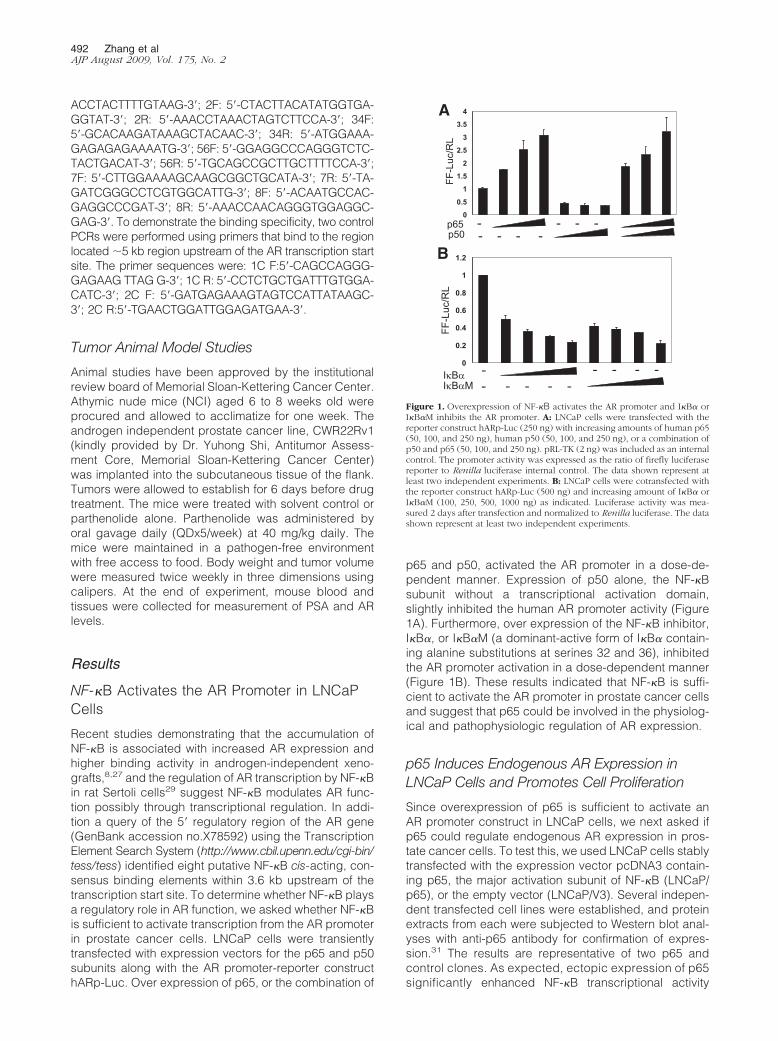
Recent studies demonstrating that the accumulation ofNF-�B is associated with increased AR expression andhigher binding activity in androgen-independent xeno-grafts,8,27 and the regulation of AR transcription by NF-�Bin rat Sertoli cells29 suggest NF-�B modulates AR func-tion possibly through transcriptional regulation. In addi-tion a query of the 5� regulatory region of the AR gene(GenBank accession no.X78592) using the TranscriptionElement Search System (http://www.cbil.upenn.edu/cgi-bin/tess/tess) identified eight putative NF-�B cis-acting, con-sensus binding elements within 3.6 kb upstream of thetranscription start site. To determine whether NF-�B playsa regulatory role in AR function, we asked whether NF-�Bis sufficient to activate transcription from the AR promoterin prostate cancer cells. LNCaP cells were transientlytransfected with expression vectors for the p65 and p50subunits along with the AR promoter-reporter constructhARp-Luc. Over expression of p65, or the combination of
p65 and p50, activated the AR promoter in a dose-de-pendent manner. Expression of p50 alone, the NF-�Bsubunit without a transcriptional activation domain,slightly inhibited the human AR promoter activity (Figure1A). Furthermore, over expression of the NF-�B inhibitor,I�B�, or I�B�M (a dominant-active form of I�B� contain-ing alanine substitutions at serines 32 and 36), inhibitedthe AR promoter activation in a dose-dependent manner(Figure 1B). These results indicated that NF-�B is suffi-cient to activate the AR promoter in prostate cancer cellsand suggest that p65 could be involved in the physiolog-ical and pathophysiologic regulation of AR expression.
p65 Induces Endogenous AR Expression inLNCaP Cells and Promotes Cell Proliferation
Since overexpression of p65 is sufficient to activate anAR promoter construct in LNCaP cells, we next asked ifp65 could regulate endogenous AR expression in pros-tate cancer cells. To test this, we used LNCaP cells stablytransfected with the expression vector pcDNA3 contain-ing p65, the major activation subunit of NF-�B (LNCaP/p65), or the empty vector (LNCaP/V3). Several indepen-dent transfected cell lines were established, and proteinextracts from each were subjected to Western blot anal-yses with anti-p65 antibody for confirmation of expres-sion.31 The results are representative of two p65 andcontrol clones. As expected, ectopic expression of p65significantly enhanced NF-�B transcriptional activity
A
IκBαIκBαM
FF-L
uc/R
L
p65p50
- - - -- - - -
FF-L
uc/R
L
B
- - - -- - - -
--
0
0.2
0.4
0.6
0.8
1
1.2
0
0.5
1
1.5
2
2.5
3
3.5
4
Figure 1. Overexpression of NF-�B activates the AR promoter and I�B� orI�B�M inhibits the AR promoter. A: LNCaP cells were transfected with thereporter construct hARp-Luc (250 ng) with increasing amounts of human p65(50, 100, and 250 ng), human p50 (50, 100, and 250 ng), or a combination ofp50 and p65 (50, 100, and 250 ng). pRL-TK (2 ng) was included as an internalcontrol. The promoter activity was expressed as the ratio of firefly luciferasereporter to Renilla luciferase internal control. The data shown represent atleast two independent experiments. B: LNCaP cells were cotransfected withthe reporter construct hARp-Luc (500 ng) and increasing amount of I�B� orI�B�M (100, 250, 500, 1000 ng) as indicated. Luciferase activity was mea-sured 2 days after transfection and normalized to Renilla luciferase. The datashown represent at least two independent experiments.
492 Zhang et alAJP August 2009, Vol. 175, No. 2

compared with control LNCaP/V3 cells when transientlytransfected with the pNF-�B-TA-Luc reporter (P � 0.05)(data not shown). To determine whether overexpressionof p65 had an effect on AR expression, we measured ARmRNA level using real-time RT-PCR. LNCaP/p65 linesexpressed a significantly higher level of AR (�2.5-fold),as compared with the control LNCaP/V3 (P � 0.05) (Fig-ure 2A). Consistent with the increased AR transcript level,the AR protein is also significantly increased in LNCaP/p65 cells (Figure 2B). The increased level of AR in cellsoverexpressing p65 was associated with enhancedtransactivation activity particularly in the presence of thenon-metabolizable AR ligand R1881 (Figure 2C). Impor-tantly, the increased sensitivity to low concentrations ofandrogen observed in LNCaP/p65 cells indicates thatactivation of p65 could contribute to enhanced growth inthe setting of low androgen levels during androgen-dep-rivation therapy. About 0.5 nmol/L of androgen is re-quired to give a half maximal response. To determinewhether the increased transactivation activity was phys-iologically relevant, we monitored the secretion of PSA, awell-known AR target gene. About fourfold more PSA wassecreted into the cell culture media from LNCaP/p65 cellsthan from LNCaP/V3 cells (P � 0.05) (Figure 2D).
The androgen response pathway is a critical growthregulatory mechanism in prostate cancer and knock-down of the androgen receptor attenuates ligand-inde-
pendent activation and delays tumor progression.35 Itfollows that regulation of AR activity by NF-�B may havea similar effect. To further explore the biological signifi-cance of p65 induced AR expression in prostate cancercells, we determined the effects on cell growth. Overex-pression of p65 resulted in an increased viable cell num-ber, detected as early as day 3 that reflected a neardoubling of growth (Figure 2E). Of interest, a similar, butmore modest, increase in cell growth was detected inandrogen-depleted media (data not shown). Cumula-tively, these data confirm that p65 can induce expressionof the endogenous AR in human prostate cancer cells,and that induction is associated with increased expres-sion of downstream AR targets and enhanced growthand/or survival of prostate cancer cells.
NF-�B Inhibition Decreases Endogenous ARExpression, AR Transactivation Activity, andNF-�B Binding in the AR Promoter in ProstateCancer Cells
NF-�B activity is specifically regulated by its inhibitorI�B�. To further validate the direct role of the NF-�Bsignaling pathway on AR expression in prostate cancer,we compared LNCaP cells stably transfected with an
BA3 5
C 200
LNCaP/V3
AR
AR
mR
NA
1.5
2.0
2.5
3.0
3.5
*
FF-L
uc/R
L
50
100
150LNCaP/V3
LNCaP/p65
LNCaP/V3 LNCaP/p65
A
0.0
0.5
1.0β-actin
R1881(nM) 0.1 0.2 0.5 1 10- + - + - + - + - +
0
50
D E 3300 *
PS
A(n
g/m
l)
x 10
6C
ells
/ml
1
1.5
2
2.5 LNCaP/V3LNCaP/p65
100
150
200
250
0.5
0
0.5
1
Day 0 Day 1 Day 3 Day 6 Day 9LNCaP/V3 LNCaP/p65
0
50
Figure 2. Overexpression of p65 (RelA) enhances AR expression in LNCaP cells and promotes cell proliferation. A: AR mRNA level is significantly higher inLNCaP/p65 compared with LNCaP/V3 cells. Real-time RT-PCR was performed to examine the AR mRNA level using iCycler. The AR mRNA level was expressedrelative to TBP internal housekeeping control. The level of AR mRNA in LNCaP/p65 cells was normalized to that in LNCaP/V3 cells. B: Western blot analysis forhuman AR performed on protein extracts derived from LNCaP/V3 and LNCap/p65. C: AR transcriptional activity. LNCaP/V3 and LNCap/p65 cells were transfectedwith MMTV-Luc and pRL-TK constructs and cultured in 10% charcoal-stripped FBS in the presence or absence of R1881 for 2 days. Data were normalized to Renillaluciferase. The data shown is representative of at least two independent experiments. D: Overexpression of p65 (RelA) induces endogenous PSA expression.LNCaP/V3 and LNCaP/p65 cell lines were cultured in RPMI1640 media supplemented with 10% FBS to about 70% to 80% confluence. The media were subjectedto enzyme-linked immunosorbent assay to determine PSA expression. E: p65 promotes prostate cancer cell proliferation. Equal numbers of LNCaP/V3 andLNCap/p65 cells (100,000 cells/well) were seeded into 12-well tissue culture plates. Cells were stained by trypan blue (1, 3, 6, and 9 days) and cell numbers weredetermined. The data represent means � SD from triplicate sets of three independent experiments.
NK-�B Regulates AR and PC Growth 493AJP August 2009, Vol. 175, No. 2

expression construct for I�B�M, a constitutively activemutant NF-�B inhibitor (LNCaP/I�B�M) and LNCaP/C5,which harbors the empty expression vector. The resultsare representative of two I�B� clones (LNCaP/I�B� andLNCaP/I�B�M) and two control clones. The results ofLNCaP/I�B� (data not shown) is very similar to thoseof LNCaP/I�B�M. The inhibitory effect of I�B�M on tran-scriptional activity of endogenous NF-�B was shown byreporter gene assay. As expected, ectopic expression ofI�B�M blocked NF-�B activity in LNCaP cells as shownby transient transfection with the NF-�B responsive re-porter construct, pNF-�BTA-Luc (Data not shown). Over-expression of I�B�M also significantly inhibited expres-sion of the endogenous AR gene at the mRNA level asdetermined by real-time RT-PCR (Figure 3A) and de-creased AR protein (Figure 3B). To examine the effects ofI�B�M on AR transactivation function, we transientlytransfected LNCaP/I�B�M and LNCaP/C5 cells with theAR dependent MMTV-Luc reporter construct. Consistentwith the reduced AR level in LNCaP/I�B�M, the transac-tivation activity of AR in these cells is significantly de-creased even in the presence of R1881 (Figure 3C).Reduced AR activity by inhibition of NF-�B is also dem-onstrated by decreased PSA secretion (Figure 3D), a wellvalidated endogenous transcriptional target of AR. Tofurther demonstrate the inhibition of AR expression byI�B�M at the transcriptional level, we performed ChIPassay and observed decreased binding of p50 (the DNAbinding subunit of NF-�B p65/p50 heterodimer) to one of
the NF-�B binding sites, site 5/6 (compare lane 5 v.s. lane2, Figure 3E). Based on the inhibitory effects of I�B�M onAR expression, we next examined if specific inhibition ofp65 and p50 expression could affect AR expression inLNCaP cells. Co-transfection of p65 and p50 siRNA (100nmol/L each) significantly inhibited AR mRNA (P � 0.05)and protein expression (Figure 3F and 3G). Decreasedexpression AR by specific inhibition of NF-�B (overex-pression of I�B�M or co-transfection of p65 and p50siRNAs) provided strong evidence that this transcriptionfactor plays an important regulatory role in this process.
NF-�B p65 Is Coordinately Expressed withthe AR in Human ADRPC
Recent studies indicate that NF-�B is overexpressed inprostate cancer and its expression correlates with dis-ease progression during androgen deprivation thera-py.23,24,36,37 Our experiments have further shown thatNF-�B can regulate expression of the AR in prostatecancer cell lines even in the setting of androgen deple-tion. To determine the relationship between NF-�B andAR expression in human prostate cancers progressingduring androgen deprivation therapy, we examined pro-tein expression of AR and p65 by immunohistochemistryin 52 metastatic ADRPC (Figure 4a-d). In 22 out of 52(42%) cases examined, p65 and AR were coordinatelyexpressed at moderate to high levels (spearman rho P �
Figure 3. Overexpression of I�B�M inhibits AR expression, AR transactivation activity, and NF-�B binding in the AR promoter in LNCaP cells. NF-�B inhibition by p65and p50 siRNAs also inhibits endogenous AR expression. AR mRNA (A), AR protein level (B), AR transcriptional activity (C), and PSA secretion (D) were determined inLNCaP/p65 (LNCaP cells stably transfected with p65) and LNCaP/V3 (which harbors empty expression vector) as outlined in Materials and Methods. The data representmeans � SD from triplicate sets of three independent experiments. *P � 0.05. E: Overexpression of I�B�M decreased the binding of p50 to the AR promoter region. M:1kb plus DNA marker; Lanes 1 and 4, input control; Lanes 2 and 5, immunoprecipitation with p50 specific antibody; Lanes 3 and 6, immunoprecipitation with controlantibody (normal rabbit IgG). F: Co-transfection of p65 and p50 siRNAs (100 nmol/L each) significantly inhibited AR mRNA expression in LNCaP cells. G: Co-transfectionof p65 and p50 siRNAs (100 nmol/L each) inhibited AR protein expression in LNCaP cells.
494 Zhang et alAJP August 2009, Vol. 175, No. 2

0.03). Twelve out of 51 (23%) are weakly stained ornegative for both p65 and AR, and in 18 cases (35%), theimmunostaining was moderate for one protein and weak/negative for the other protein (Table 1). To a certainextent, heterogeneity was observed for both AR and p65staining in human ADRPC. Besides the overall correlationbetween AR and p65 expression, prostate cancer cellswith moderate or strong staining of p65 also have corre-sponding moderate or strong nuclear AR staining (Figure4). In addition, when AR is strongly stained in the nucleus,we observed p65 nuclear staining (Figure 4, A and B).Although circumstantial, the common coincident expres-sion of p65 and AR in human ADRPC is consistent with acausal relationship as suggested by the in vitro experi-mental data presented here.
NF-�B Binds the 5� Regulatory Region of theAR Gene, which Can Be Inhibited byParthenolide
We identified eight putative NF-�B binding sites within the�3.6 kb 5� regulatory region of the AR gene (Figure 5A).To determine whether transcriptional regulation of AR byNF-�B is mediated by these binding motifs, we con-ducted ChIP experiments to define the occupancy ofNF-�B on the AR promoter. Soluble chromatin was pre-pared from LNCaP/p65 cells after formaldehyde treat-ment and specific antibody against p50 (the subunit thatcontains the DNA-binding domain) was used to immuno-precipitate p50-bound genomic DNA fragments. Four mi-
Figure 4. Correlation between AR and p65 immunostaining in human ADRPC. Moderate AR labeling (2�) is shown in (A), this corresponds to moderate p65labeling (B). Nuclear p65 is shown in the inset. Strong AR labeling (3�) in (C) corresponds to strong p65 immunostaining (D).
Table 1. Expression of NF-�B/p65 and Nuclear AR in Metastatic Prostate Cancer Specimens
NF-�B/p65 immunoreactivity
AR nuclear immunoreactivity Negative Weak Moderate Strong Total
Negative 6 2 1 0 9Weak 2 2 2 0 6Moderate 3 8 6 5 22Strong 1 3 6 5 15Total 12 15 15 10 52
The expression of NF-�B/p65 and nuclear AR was evaluated on staining intensity of the tissues as: negative (0), weak (1), moderate (2) and strong(3). In 22 out of 52 (42%) cases examined, p65 and AR were coordinately expressed at moderate to high levels (spearman rho P � 0.03). Twelve outof 51 (23%) are weakly stained or negative for both p65 and AR, and in 18 cases (35%), the immunostaining was moderate for one protein andweak/negative for the other protein.
NK-�B Regulates AR and PC Growth 495AJP August 2009, Vol. 175, No. 2

crograms of rabbit polyclonal antibody specific to p50 orthe same amount of normal rabbit IgG were used in eachreaction of ChIP. To demonstrate the binding specificity,two control PCRs were performed to amplify fragmentswithin the �5 kb region that do not contain a putative NF-�Bsites (Figure 5C). NF-�B recruitment to the AR promoter wasdetected in all of the putative NF-�B binding sites assayedby PCR (Figure 5B) and not detected in the negative con-trols, indicating that the binding of p50 to these sites wasspecific. As the average length of the genomic DNA frag-ments produced in these experiments is �1000 bp andsome NF-�B binding sites are very close to each other, wecannot distinguish with certainty whether some or all of theNF-�B binding motifs are bound. Direct binding of theNF-�B transcription factor to the AR promoter is consistentwith a role in transcriptional regulation of this gene.
Parthenolide, the active ingredient of the herbal rem-edy feverfew (Tanacetum parthenium), is able to inhibitNF-�B DNA binding and prostate cancer cell growth invitro.23 This effect is at least in part due to inhibition ofNF-�B signaling38,39 and we therefore expected that thisdrug is able to interfere with NF-�B transcriptional regu-lation of the AR gene. ChIP assays were performed aftertreatment with parthenolide and NF-�B occupancy of theNF-�B binding sites was markedly decreased (Figure5D). The inhibition of NF-�B occupancy correlated withthe inhibition of AR transcription by parthenolide, whichwas maximal at 6 hours (data not shown). These data addsupport to the hypothesis that NF-�B regulates AR. Fur-thermore, parthenolide could be used to test this hypoth-esis in vitro and in vivo.
Pathenolide, an NF-�B Inhibitor, DecreasesAR Expression, PSA Secretion and CellProliferation in Prostate Cancer Cells
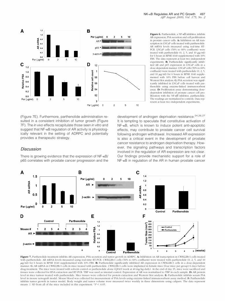
To test whether parthenolide is able to regulate AR ex-pression, we analyzed LNCaP cells treated with parthe-
nolide (Figure 6, A and B). There was a dose-dependentdecrease in AR mRNA and protein after 6 hours of par-thenolide treatment, a previously determined time of max-imal inhibition. Interestingly, parthenolide also decreasedlevels of p65 while having no observable effects on �-ac-tin (Figure 6B). The inhibition of AR expression correlatedwith reduction of PSA secretion (Figure 6C) and resultedin a dose-dependent decrease in viable prostate cancercells (Figure 6D). These experiments were also per-formed in LAPC4, a prostate cancer cell line with a wild-type AR, with similar results (data not shown).
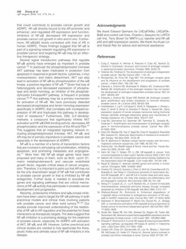
Parthenolide Treatment Inhibits AR Expression,PSA Secretion, and Tumor Growth in aXenograft Model of ADRPC
Our data have demonstrated that parthenolide inhibitsAR expression and prostate cancer cell proliferation invitro using the androgen-dependent LNCaP cell line (Fig-ure 6). To test whether parthenolide treatment is effectivein the setting of ADRPC, we used the CWR22Rv1 prostatecancer xenograft model.40–42 We first tested whethertreatment of parthenolide inhibited the AR expression invitro. There was a dose-dependent decrease in ARmRNA and protein after 6 hours of parthenolide treatmentin CWR22Rv1 cells (Figure 7A and 7B). We then ex-amined the effect of parthenolide treatment in vivo.CWR22Rv1 tumors were allowed to establish for 6 daysbefore drug treatment. The mice were treated with sol-vent control or parthenolide (QDx5/week at 40 mg/kgdaily). Body weight and tumor volume were measuredtwice weekly and mouse blood and tumor tissues werecollected for measurement of PSA and AR levels. Parthe-nolide inhibited AR expression significantly (both mRNAand protein) in most of the CWR22Rv1 tumors (Figure 7,C and D). In concordance with the inhibition in AR ex-pression, mice treated with parthenolide produced sig-nificantly lower levels of circulating PSA in the serum
Figure 5. NF-�B interacts with the 5� regulatoryregion of the AR gene, which can be inhibited byParthenolide treatment. A: Schematic diagram ofthe AR gene regulatory region. Solid boxes depictputative NF-�B binding sites, and 1F/1R, 2F/2R,34F/34R, 56F/56R, 7F/7R, 8F/8R, C1F/C1R, C2F/C2R are primer pairs used for amplifying corre-sponding DNA fragments. Numbers are the posi-tions upstream of the AR gene transcription startsite. B: ChIP assays of NF-�B occupancy on the ARgene regulatory region. LNCaP/p65 cells were cul-tured in RPMI 1640 medium supplemented with10% FBS. Soluble chromatin was prepared fromformaldehyde-cross-linked and sonicated cell cul-tures. Specific antibody against p50, as well ascontrol serum (normal rabbit IgG), was used toimmunoprecipitate protein-bound DNA frag-ments. These fragments were amplified by PCRusing primers described in (A). M, 1-kb plusmarker (for 1F/1R) or 100-bp markers (for theother sites); 1, input; 2, p50 IP; 3, rabbit serum IP.C: Two control PCRs were performed using prim-ers that bind to the region located �5 kb regionupstream of the AR transcription start site. M: 1kbplus marker; 1, input; 2, p50 IP. D: Inhibition of ARexpression by parthenolide is associated with al-terations in the recruitment of NF-�B to the 5�regulatory region of the AR gene.
496 Zhang et alAJP August 2009, Vol. 175, No. 2
(Figure 7E). Furthermore, parthenolide administration re-sulted in a consistent inhibition of tumor growth (Figure7F). The in vivo effects recapitulate those seen in vitro andsuggest that NF-�B regulation of AR activity is physiolog-ically relevant in the setting of ADRPC and potentiallyprovides a therapeutic strategy.
Discussion
There is growing evidence that the expression of NF-�B/p65 correlates with prostate cancer progression and the
development of androgen deprivation resistance.24,26,27
It is tempting to speculate that constitutive activation ofNF-�B, which is known to induce potent anti-apoptoticeffects, may contribute to prostate cancer cell survivalfollowing androgen withdrawal. Increased AR expressionis also a critical event in the development of prostatecancer resistance to androgen deprivation therapy. How-ever, the signaling pathways and transcription factorsinvolved in the regulation of AR expression are not clear.Our findings provide mechanistic support for a role ofNF-�B in regulation of the AR in human prostate cancer
Figure 6. Parthenolide, a NF-�B inhibitor, inhibitsAR expression, PSA secretion and cell proliferationin prostate cancer cells. A: Inhibition on AR tran-scription in LNCaP cells treated with parthenolide.AR mRNA levels measured using real-time RT-PCR. LNCaP cells (50% to 60% confluent) weretreated with parthenolide (0, 2, 5, and 10 �g/ml)for 6 hours in RPMI 1640 supplemented with 10%FBS. The data represent at least two independentexperiments. B: Parthenolide significantly inhib-ited AR and p65 expression in LNCaP cells in adose-dependent manner. LNCaP cells (50% to 60%confluent) were treated with parthenolide (0, 2, 5,and 10 �g/ml) for 6 hours in RPMI 1640 supple-mented with 10% FBS before cell harvest andWestern blot analysis. C: PSA secretion was signif-icantly inhibited in LNCaP cells treated with par-thenolide using enzyme-linked immunosorbentassay. D: Proliferation assay demonstrating dose-dependent inhibition of prostate cancer cell pro-liferation with the NF-�B inhibitor, parthenolide.The readings are normalized to controls. Data rep-resent at least two independent experiments.
Figure 7. Parthenolide treatment inhibits AR expression, PSA secretion and tumor growth in ADRPC. A: Inhibition on AR transcription in CWR22Rv1 cells treatedwith parthenolide. AR mRNA levels measured using real-time RT-PCR. CWR22Rv1 cells (50% to 60% confluent) were treated with parthenolide (0, 2, 5, and 10�g/ml) for 6 hours in RPMI 1640 supplemented with 10% FBS. B: Parthenolide significantly inhibited AR expression in CWR22Rv1 cells in a dose-dependentmanner. C: AR mRNA in CWR22Rv1 cells in mice treated with parthenolide. CWR22Rv1 cells were implanted in female mice (four mice per group) 6 days beforedrug treatment. The mice were treated with solvent control or parthenolide alone (QDx5/week at 40 mg/kg daily). At the end of day 35, mice were sacrificed andtissues were collected for RNA extraction and RT-PCR. TBP was used as internal control. Expression of AR was normalized to TBP in each sample. D: AR proteinlevel in mice tumors treated with parthenolide. Mice tissues were collected for protein extraction and Western blot analysis. E: Parthenolide inhibits serum PSAlevel in mouse xenograft model. Mouse blood was collected for measurement of PSA levels using enzyme-linked immunosorbent assay method. F: Parthenolideinhibits tumor growth in tumor model. Body weight and tumor volume were measured twice weekly in three dimensions using calipers. The data representmeans � SD from all of the mice included in this experiment. *P � 0.05.
NK-�B Regulates AR and PC Growth 497AJP August 2009, Vol. 175, No. 2
that could contribute to prostate cancer growth andADRPC. NF-�B directly bound to the AR promoter andenhancer, and regulated AR expression and function.Inhibitors of NF-�B decreased AR expression andprostate cancer cell growth in vitro and in vivo. Further-more, NF-�B/p65 levels correlated with that of AR inhuman ADRPC. These findings suggest that NF-�B ispart of a signaling network regulating AR expression inprostate cancer and targeting NF-�B may be an effec-tive therapeutic strategy.
Several signal transduction pathways that regulateNF-� activity have emerged as important in prostatecancer.43 In particular the phosphatidylinositol 3-kinase/AKT pathway has been implicated in protection fromapoptosis in response to growth factors, cytokines, c-mycoverexpression, and matrix detachment. AKT can alsolead to activation of NF-� by phosphorylation of the I�Bkinase, a positive regulator of NF-�B.44 Given that loss ofheterozygosity and decreased expression of phospha-tase and tensin homolog, an inhibitor of the phosphati-dylinositol 3-kinase/AKT pathway, is frequent in prostatecancer,45,46 this pathway may be a primary mechanismfor activation of NF-�B. We have previously detecteddecreased phosphatase and tensin homolog expressionspecifically in ADRPC, that correlates with increased ex-pression of AR and implicates this pathway in develop-ment of resistance.6 Furthermore, DIM, 3,3�-diindolyl-methane, a compound that significantly inhibits AKTactivation and NF-� DNA binding activity, decreases ARphosphorylation and the expression of the AR and PSA.47
This suggests that an integrated signaling network in-cluding phosphatidylinositol 3-kinase, AKT, NF-�, andAR may be of primary importance in prostate cancer andespecially in the development of ADRPC.
NF-�B is a member of a family of transcription factorsthat are involved in stimulating cell proliferation, inhibitingapoptosis, and promoting metastasis and angiogene-sis.11 More than 180 NF-�B target genes have beenproposed and many of them, such as BclII, cyclin D1,matrix metalloproteinase-9, and vascular endothelialgrowth factor, regulate critical steps of cancer progres-sion. Therefore, it is important to point out that AR may notbe the only downstream target of NF-� that contributesto prostate cancer growth or that is inhibited by NF-�antagonists. Further study is needed to elucidate thegenes and signaling pathways that are critical mecha-nisms of NF-� activity that participate in prostate cancerdevelopment and progression.
Recently, proteosome inhibitors and I�B� kinase inhib-itors have been used to target the NF-�B pathway in bothpreclinical models and clinical trials involving patientswith prostate cancer and other solid tumors.48–50 Ourstudies provide improved understanding of the relation-ship of NF-� to AR regulation and the potential for theseinteractions as therapeutic targets. The data suggest thatNF-�B inhibition is a promising strategy for the treatmentof prostate cancer, especially for ADRPC with activationof AKT, NF-�, and AR. However, further preclinical andclinical studies are needed to fully appreciate the thera-peutic index and ultimate value of NF-� inhibitors in thisdisease.
Acknowledgments
We thank Edward Gelmann for LNCaP/IkBa, LNCaP/Ik-BaM and control cell lines, Charles L.Sawyers for LAPC4cell line, Terry Brown for MMTV-Luc reporter and NF-�Bp50 and p65 expression vectors. We thank Hui-Kuan Linand Xiaodi Ren for advice and technical assistance.
References
1. Culig Z, Hobisch A, Hittmair A, Peterziel H, Cato AC, Bartsch G,Klocker H: Expression, structure, and function of androgen receptorin advanced prostatic carcinoma. Prostate 1998, 35:63–70
2. Feldman BJ, Feldman D: The development of androgen-independentprostate cancer. Nat Rev Cancer 2001, 1:34–45
3. Montgomery JS, Price DK, Figg WD: The androgen receptor geneand its influence on the development and progression of prostatecancer. J Pathol 2001, 195:138–146
4. Edwards J, Krishna NS, Mukherjee R, Watters AD, Underwood MA,Bartlett JM: Amplification of the androgen receptor may not explainthe development of androgen-independent prostate cancer. BJU Int2001, 88:633–637
5. Edwards J, Krishna NS, Grigor KM, Bartlett JM: Androgen receptorgene amplification and protein expression in hormone refractoryprostate cancer. Br J Cancer 2003, 89:552–556
6. Holzbeierlein J, Lal P, LaTulippe E, Smith A, Satagopan J, Zhang L,Ryan C, Smith S, Scher H, Scardino P, Reuter V, Gerald WL: Geneexpression analysis of human prostate carcinoma during hormonaltherapy identifies androgen-responsive genes and mechanisms oftherapy resistance. Am J Pathol 2004, 164:217–227
7. Linja MJ, Savinainen KJ, Saramaki OR, Tammela TL, Vessella RL,Visakorpi T: Amplification and overexpression of androgen receptorgene in hormone-refractory prostate cancer. Cancer Res 2001,61:3550–3555
8. Chen CD, Welsbie DS, Tran C, Baek SH, Chen R, Vessella R, RosenfeldMG, Sawyers CL: Molecular determinants of resistance to antiandro-gen therapy. Nat Med 2004, 10:33–39
9. Sen R, Baltimore D: Multiple nuclear factors interact with the immu-noglobulin enhancer sequences. Cell 1986, 46:705–716
10. Perkins ND: The Rel/NF-kappa B family: friend and foe. Trends Bio-chem Sci 2000, 25:434–440
11. Karin M, Cao Y, Greten FR, Li ZW: NF-kappaB in cancer: frominnocent bystander to major culprit. Nat Rev Cancer 2002, 2:301–310
12. Baldwin AS: Control of oncogenesis and cancer therapy resistance bythe transcription factor NF-kappaB. J Clin Invest 2001, 107:241–246
13. Rayet B, Gelinas C: Aberrant rel/nfkb genes and activity in humancancer. Oncogene 1999, 18:6938–6947
14. Ondrey FG, Dong G, Sunwoo J, Chen Z, Wolf JS, Crowl-Bancroft CV,Mukaida N, Van Waes C: Constitutive activation of transcription fac-tors NF-(kappa)B. AP-1, and NF-IL6 in human head and neck squa-mous cell carcinoma cell lines that express pro-inflammatory andpro-angiogenic cytokines. Mol Carcinog 1999, 26:119–129
15. Wang CY, Cusack JC, Jr., Liu R, Baldwin AS, Jr: Control of induciblechemoresistance: enhanced anti-tumor therapy through increasedapoptosis by inhibition of NF-kappaB. Nat Med 1999, 5:412–417
16. Shattuck-Brandt RL, Richmond A: Enhanced degradation ofI-kappaB alpha contributes to endogenous activation of NF-kap-paB in Hs294T melanoma cells. Cancer Res 1997, 57:3032–3039
17. Nakshatri H, Bhat-Nakshatri P, Martin DA, Goulet RJ, Jr., SledgeGW, Jr: Constitutive activation of NF-kappaB during progression ofbreast cancer to hormone-independent growth. Mol Cell Biol 1997,17:3629 –3639
18. Sovak MA, Bellas RE, Kim DW, Zanieski GJ, Rogers AE, Traish AM,Sonenshein GE: Aberrant nuclear factor-kappaB/Rel expression and thepathogenesis of breast cancer. J Clin Invest 1997, 100:2952–2960
19. Dhawan P, Richmond A: A novel NF-kappa B-inducing kinase-MAPKsignaling pathway up-regulates NF-kappa B activity in melanomacells. J Biol Chem 2002, 277:7920–7928
20. Cooper CR, Chay CH, Gendernalik JD, Lee HL, Bhatia J, TaichmanRS, McCauley LK, Keller ET, Pienta KJ: Stromal factors involved inprostate carcinoma metastasis to bone. Cancer 2003, 97:739–747
498 Zhang et alAJP August 2009, Vol. 175, No. 2

21. Ludwig L, Kessler H, Wagner M, Hoang-Vu C, Dralle H, Adler G,Bohm BO, Schmid RM: Nuclear factor-kappaB is constitutively activein C-cell carcinoma and required for RET-induced transformation.Cancer Res 2001, 61:4526–4535
22. Cogswell PC, Guttridge DC, Funkhouser WK, Baldwin AS, Jr: Selec-tive activation of NF-kappa B subunits in human breast cancer:potential roles for NF-kappa B2/p52 and for Bcl-3, Oncogene 2000,19:1123–1131
23. Sweeney C, Li L, Shanmugam R, Bhat-Nakshatri P, Jayaprakasan V,Baldridge LA, Gardner T, Smith M, Nakshatri H, Cheng L: Nuclearfactor-kappaB is constitutively activated in prostate cancer in vitroand is overexpressed in prostatic intraepithelial neoplasia and ade-nocarcinoma of the prostate. Clin Cancer Res 2004, 10:5501–5507
24. Shukla S, MacLennan GT, Fu P, Patel J, Marengo SR, Resnick MI,Gupta S: Nuclear factor-kappaB/p65 (Rel A) is constitutively acti-vated in human prostate adenocarcinoma and correlates with dis-ease progression. Neoplasia 2004, 6:390–400
25. Ismail HA, Lessard L, Mes-Masson AM, Saad F: Expression of NF-kappaB in prostate cancer lymph node metastases. Prostate 2004,58:308–313
26. Lessard L, Mes-Masson AM, Lamarre L, Wall L, Lattouf JB, Saad F:NF-kappa B nuclear localization and its prognostic significance inprostate cancer. BJU Int 2003, 91:417–420
27. Chen CD, Sawyers CL: NF-kappa B activates prostate-specific anti-gen expression and is upregulated in androgen-independent pros-tate cancer. Mol Cell Biol 2002, 22:2862–2870
28. Gupta S, Afaq F, Mukhtar H: Involvement of nuclear factor-kappa B. Baxand Bcl-2 in induction of cell cycle arrest and apoptosis by apigenin inhuman prostate carcinoma cells. Oncogene 2002, 21:3727–3738
29. Zhang L, Charron M, Wright WW, Chatterjee B, Song CS, Roy AK,Brown TR: Nuclear factor-kappaB activates transcription of the an-drogen receptor gene in Sertoli cells isolated from testes of adult rats.Endocrinology 2004, 145:781–789
30. Kimura K, Gelmann EP: Propapoptotic effects of NF-kappaB in LN-CaP prostate cancer cells lead to serine protease activation. CellDeath Differ 2002, 9:972–980
31. Altuwaijri S, Lin HK, Chuang KH, Lin WJ, Yeh S, Hanchett LA, RahmanMM, Kang HY, Tsai MY, Zhang Y, Yang L, Chang C: Interruption ofnuclear factor kappaB signaling by the androgen receptor facilitates12-O-tetradecanoylphorbolacetate-induced apoptosis in androgen-sen-sitive prostate cancer LNCaP cells. Cancer Res 2003, 63:7106–7112
32. Lee SB, Kolquist KA, Nichols K, Englert C, Maheswaran S, Ladanyi M,Gerald WL, Haber DA: The EWS-WT1 translocation product inducesPDGFA in desmoplastic small round-cell tumour. Nat Genet 1997,17:309–313
33. Sambrook JE, Russell DW: Molecular cloning: A laboratory manual,3rd. Cold Spring Harbor Laboratory Press, Cold Spring Harbor, N.Y.,2001 A9.28
34. Mitrunen K, Pettersson K, Piironen T, Bjork T, Lilja H, Lovgren T:Dual-label one-step immunoassay for simultaneous measurement offree and total prostate-specific antigen concentrations and ratios inserum. Clin Chem 1995, 41:1115–1120
35. Cheng H, Snoek R, Ghaidi F, Cox ME, Rennie PS: Short hairpinRNA knockdown of the androgen receptor attenuates ligand-inde-pendent activation and delays tumor progression. Cancer Res2006, 66:10613–10620
36. Fradet V, Lessard L, Begin LR, Karakiewicz P, Masson AM, Saad F:Nuclear factor-kappaB nuclear localization is predictive of biochem-ical recurrence in patients with positive margin prostate cancer. ClinCancer Res 2004, 10:8460–8464
37. Ross JS, Kallakury BV, Sheehan CE, Fisher HA, Kaufman RP, Jr.,Kaur P, Gray K, Stringer B: Expression of nuclear factor-kappa B andI kappa B alpha proteins in prostatic adenocarcinomas: correlation ofnuclear factor-kappa B immunoreactivity with disease recurrence.Clin Cancer Res 2004, 10:2466–2472
38. Bork PM, Schmitz ML, Kuhnt M, Escher C, Heinrich M: Sesquiterpenelactone containing Mexican Indian medicinal plants and pure ses-quiterpene lactones as potent inhibitors of transcription factor NF-kappaB. FEBS Lett 1997, 402:85–90
39. Hehner SP, Heinrich M, Bork PM, Vogt M, Ratter F, Lehmann V,Schulze-Osthoff K, Droge W, Schmitz ML: Sesquiterpene lactonesspecifically inhibit activation of NF-kappa B by preventing the deg-radation of I kappa B-alpha and I kappa B-beta. J Biol Chem 1998,273:1288–1297
40. Nagabhushan M, Miller CM, Pretlow TP, Giaconia JM, Edgehouse NL,Schwartz S, Kung HJ, de Vere White RW, Gumerlock PH, Resnick MI,Amini SB, Pretlow TG: CWR22: the first human prostate cancer xeno-graft with strongly androgen-dependent and relapsed strains both invivo and in soft agar. Cancer Res 1996, 56:3042–3046
41. Sirotnak FM, She Y, Lee F, Chen J, Scher HI: Studies with CWR22xenografts in nude mice suggest that ZD1839 may have a role in thetreatment of both androgen-dependent and androgen-independenthuman prostate cancer. Clin Cancer Res 2002, 8:3870–3876
42. Wainstein MA, He F, Robinson D, Kung HJ, Schwartz S, Giaconia JM,Edgehouse NL, Pretlow TP, Bodner DR, Kursh ED, Resnick MI, SeftelA, Pretlow TG: CWR22: androgen-dependent xenograft model de-rived from a primary human prostatic carcinoma. Cancer Res 1994,54:6049–6052
43. Graff JR: Emerging targets in the AKT pathway for treatment ofandrogen-independent prostatic adenocarcinoma. Expert Opin TherTargets 2002, 6:103–113
44. Gustin JA, Maehama T, Dixon JE, Donner DB: The PTEN tumorsuppressor protein inhibits tumor necrosis factor-induced nuclearfactor kappa B activity. J Biol Chem 2001, 276:27740–27744
45. Suzuki H, Freije D, Nusskern DR, Okami K, Cairns P, Sidransky D,Isaacs WB, Bova GS: Interfocal heterogeneity of PTEN/MMAC1 genealterations in multiple metastatic prostate cancer tissues. Cancer Res1998, 58:204–209
46. Wang SI, Parsons R, Ittmann M: Homozygous deletion of the PTENtumor suppressor gene in a subset of prostate adenocarcinomas.Clin Cancer Res 1998, 4:811–815
47. Bhuiyan MM, Li Y, Banerjee S, Ahmed F, Wang Z, Ali S, Sarkar FH:Down-regulation of androgen receptor by 3,3�-diindolylmethane con-tributes to inhibition of cell proliferation and induction of apoptosis inboth hormone-sensitive LNCaP and insensitive C4-2B prostate can-cer cells. Cancer Res 2006, 66:10064–10072
48. Adams J: Proteasome inhibition: a novel approach to cancer therapy.Trends Mol Med 2002, 8:S49–S54
49. Elliott PJ, Ross JS: The proteasome: a new target for novel drugtherapies. Am J Clin Pathol 2001, 116:637–646
50. Orlowski RZ, Baldwin AS, Jr: NF-kappaB as a therapeutic target incancer, Trends Mol Med 2002, 8:385–389
NK-�B Regulates AR and PC Growth 499AJP August 2009, Vol. 175, No. 2
Copyright © 2022 FDOKUMEN


![Aqua(4,4'-bipyridine-[kappa]N)bis(1,4-dioxo-1 ... - ScienceOpen](https://static.fdokumen.com/doc/165x107/63262349e491bcb36c0aa51f/aqua44-bipyridine-kappanbis14-dioxo-1-scienceopen.jpg)