Adipose-Derived Stem Cells from Type 2 Diabetic Rats ... - MDPI
13
Citation: Quaade, M.L.; Dhumale, P.; Steffensen, S.G.C.; Beck, H.C.; Harvald, E.B.; Jensen, C.H.; Lund, L.; Andersen, D.C.; Sheikh, S.P. Adipose-Derived Stem Cells from Type 2 Diabetic Rats Retain Positive Effects in a Rat Model of Erectile Dysfunction. Int. J. Mol. Sci. 2022, 23, 1692. https://doi.org/10.3390/ ijms23031692 Academic Editor: Kazunori Sango Received: 23 November 2021 Accepted: 28 January 2022 Published: 1 February 2022 Publisher’s Note: MDPI stays neutral with regard to jurisdictional claims in published maps and institutional affil- iations. Copyright: © 2022 by the authors. Licensee MDPI, Basel, Switzerland. This article is an open access article distributed under the terms and conditions of the Creative Commons Attribution (CC BY) license (https:// creativecommons.org/licenses/by/ 4.0/). International Journal of Molecular Sciences Article Adipose-Derived Stem Cells from Type 2 Diabetic Rats Retain Positive Effects in a Rat Model of Erectile Dysfunction Marlene Louise Quaade 1,2,† , Pratibha Dhumale 1,3,† , Simon Gabriel Comerma Steffensen 4,5 , Hans Christian Beck 3,6 , Eva Bang Harvald 1,3 , Charlotte Harken Jensen 1,3 , Lars Lund 2,3,7 , Ditte Caroline Andersen 1,2,3 and Søren Paludan Sheikh 1,2,3, * 1 Laboratory of Molecular and Cellular Cardiology, Department of Clinical Biochemistry and Pharmacology, Odense University Hospital, 5000 Odense, Denmark; [email protected] (M.L.Q.); [email protected] (P.D.); [email protected] (E.B.H.); [email protected] (C.H.J.); [email protected] (D.C.A.) 2 Department of Cardiovascular and Renal Research, Institute of Molecular Medicine, University of Southern Denmark, 5000 Odense, Denmark; [email protected] 3 Institute of Clinical Research, University of Southern Denmark, 5000 Odense, Denmark; [email protected] 4 Department of Biomedicine, Aarhus University, 8000 Aarhus, Denmark; [email protected] 5 Department of Biomedical Sciences/Animal Physiology, Faculty of Veterinary, Central University of Venezuela, Maracay, Aragua 0243, Venezuela 6 Centre for Clinical Proteomics, Department of Clinical Biochemistry and Pharmacology, Odense University Hospital, 5000 Odense, Denmark 7 Department of Urology, Odense University Hospital, 5000 Odense, Denmark * Correspondence: [email protected]; Tel.: +45-21380410 † These authors contributed equally to this work. Abstract: Erectile dysfunction is a common complication associated with type 2 diabetes mellitus (T2DM) and after prostatectomy in relation to cancer. The regenerative effect of cultured adipose- derived stem cells (ASCs) for ED therapy has been documented in multiple preclinical trials as well as in recent Pase 1 trials in humans. However, some studies indicate that diabetes negatively affects the mesenchymal stem cell pool, implying that ASCs from T2DM patients could have impaired regenerative capacity. Here, we directly compared ASCs from age-matched diabetic Goto–Kakizaki (ASC GK ) and non-diabetic wild type rats (ASC WT ) with regard to their phenotypes, proteomes and ability to rescue ED in normal rats. Despite ASC GK exhibiting a slightly lower proliferation rate, ASC GK and ASC WT proteomes were more or less identical, and after injections to corpus cavernosum they were equally efficient in restoring erectile function in a rat ED model entailing bilateral nerve crush injury. Moreover, molecular analysis of the corpus cavernosum tissue revealed that both ASC GK and ASC WT treated rats had increased induction of genes involved in recovering endothelial function. Thus, our finding argues that T2DM does not appear to be a limiting factor for autologous adipose stem cell therapy when correcting for ED. Keywords: erectile dysfunction; adipose derived stem/stromal cells; type 2 diabetes mellitus; bilateral nerve crush injury 1. Introduction The epidemic-like increasing prevalence of type 2 diabetes mellitus (T2DM) is associ- ated with numerous complications and reduced life quality for the patients, in addition to substantial societal economic costs related to treatment [1,2]. Diabetic men rank erectile dys- function (ED) as their third-worst complication [3] and approximately 75% of the patients suffer from ED [4,5]. In addition, diabetics experience ED earlier in life and in a more severe form than otherwise seen in the healthy male population [6]. It has profound negative Int. J. Mol. Sci. 2022, 23, 1692. https://doi.org/10.3390/ijms23031692 https://www.mdpi.com/journal/ijms
-
Upload
khangminh22 -
Category
Documents
-
view
0 -
download
0
Transcript of Adipose-Derived Stem Cells from Type 2 Diabetic Rats ... - MDPI

�����������������
Citation: Quaade, M.L.; Dhumale, P.;
Steffensen, S.G.C.; Beck, H.C.;
Harvald, E.B.; Jensen, C.H.; Lund, L.;
Andersen, D.C.; Sheikh, S.P.
Adipose-Derived Stem Cells from
Type 2 Diabetic Rats Retain Positive
Effects in a Rat Model of Erectile
Dysfunction. Int. J. Mol. Sci. 2022, 23,
1692. https://doi.org/10.3390/
ijms23031692
Academic Editor: Kazunori Sango
Received: 23 November 2021
Accepted: 28 January 2022
Published: 1 February 2022
Publisher’s Note: MDPI stays neutral
with regard to jurisdictional claims in
published maps and institutional affil-
iations.
Copyright: © 2022 by the authors.
Licensee MDPI, Basel, Switzerland.
This article is an open access article
distributed under the terms and
conditions of the Creative Commons
Attribution (CC BY) license (https://
creativecommons.org/licenses/by/
4.0/).
International Journal of
Molecular Sciences
Article
Adipose-Derived Stem Cells from Type 2 Diabetic Rats RetainPositive Effects in a Rat Model of Erectile DysfunctionMarlene Louise Quaade 1,2,†, Pratibha Dhumale 1,3,†, Simon Gabriel Comerma Steffensen 4,5 ,Hans Christian Beck 3,6 , Eva Bang Harvald 1,3, Charlotte Harken Jensen 1,3 , Lars Lund 2,3,7,Ditte Caroline Andersen 1,2,3 and Søren Paludan Sheikh 1,2,3,*
1 Laboratory of Molecular and Cellular Cardiology, Department of Clinical Biochemistry and Pharmacology,Odense University Hospital, 5000 Odense, Denmark; [email protected] (M.L.Q.);[email protected] (P.D.); [email protected] (E.B.H.); [email protected] (C.H.J.);[email protected] (D.C.A.)
2 Department of Cardiovascular and Renal Research, Institute of Molecular Medicine,University of Southern Denmark, 5000 Odense, Denmark; [email protected]
3 Institute of Clinical Research, University of Southern Denmark, 5000 Odense, Denmark;[email protected]
4 Department of Biomedicine, Aarhus University, 8000 Aarhus, Denmark; [email protected] Department of Biomedical Sciences/Animal Physiology, Faculty of Veterinary,
Central University of Venezuela, Maracay, Aragua 0243, Venezuela6 Centre for Clinical Proteomics, Department of Clinical Biochemistry and Pharmacology,
Odense University Hospital, 5000 Odense, Denmark7 Department of Urology, Odense University Hospital, 5000 Odense, Denmark* Correspondence: [email protected]; Tel.: +45-21380410† These authors contributed equally to this work.
Abstract: Erectile dysfunction is a common complication associated with type 2 diabetes mellitus(T2DM) and after prostatectomy in relation to cancer. The regenerative effect of cultured adipose-derived stem cells (ASCs) for ED therapy has been documented in multiple preclinical trials as wellas in recent Pase 1 trials in humans. However, some studies indicate that diabetes negatively affectsthe mesenchymal stem cell pool, implying that ASCs from T2DM patients could have impairedregenerative capacity. Here, we directly compared ASCs from age-matched diabetic Goto–Kakizaki(ASCGK) and non-diabetic wild type rats (ASCWT) with regard to their phenotypes, proteomes andability to rescue ED in normal rats. Despite ASCGK exhibiting a slightly lower proliferation rate,ASCGK and ASCWT proteomes were more or less identical, and after injections to corpus cavernosumthey were equally efficient in restoring erectile function in a rat ED model entailing bilateral nervecrush injury. Moreover, molecular analysis of the corpus cavernosum tissue revealed that both ASCGK
and ASCWT treated rats had increased induction of genes involved in recovering endothelial function.Thus, our finding argues that T2DM does not appear to be a limiting factor for autologous adiposestem cell therapy when correcting for ED.
Keywords: erectile dysfunction; adipose derived stem/stromal cells; type 2 diabetes mellitus; bilateralnerve crush injury
1. Introduction
The epidemic-like increasing prevalence of type 2 diabetes mellitus (T2DM) is associ-ated with numerous complications and reduced life quality for the patients, in addition tosubstantial societal economic costs related to treatment [1,2]. Diabetic men rank erectile dys-function (ED) as their third-worst complication [3] and approximately 75% of the patientssuffer from ED [4,5]. In addition, diabetics experience ED earlier in life and in a more severeform than otherwise seen in the healthy male population [6]. It has profound negative
Int. J. Mol. Sci. 2022, 23, 1692. https://doi.org/10.3390/ijms23031692 https://www.mdpi.com/journal/ijms

Int. J. Mol. Sci. 2022, 23, 1692 2 of 13
effects on the life quality of not only the patients but also their partners. Endothelial dys-function with reduced nitric oxide (NO) release and impaired neurotransmission has beenimplied as the underlying pathological mechanisms that cause ED in diabetic men [4,5].Likewise, and independent of diabetes, a significant number of men experience ED afterprostatectomy in relation to prostate cancer. As in ED in diabetic men, the underlying causerelates to the neural system, where the nerves are damaged during prostatectomy. Conven-tional treatment i.e., oral dosages of phosphodiesterase 5 inhibitors are often ineffective,because their therapeutic efficacy is based on endothelial cells and nitric oxide signalingpathways [7]. Thus, there is a high demand for new alternative therapies for patients withneurogenic ED.
Stem cell therapy has long been considered a promising approach for correcting EDand the efficacy has been tested in different animal models [8,9]. Kim et al. have reviewedthe different sources from which stem cells have been derived and used for treatment ofED, i.e., bone marrow, adipose tissue, umbilical cord blood, mononuclear blood cells andneural crest stem cells [10]. Compared to other autologous tissue sources including bonemarrow, adipose tissue has the advantage that it can be easily obtained with less discomfortfor the patient and most importantly, it contains a large number of progenitor cells [3,11,12].Upon enzymatic digestion of adipose tissue, the freshly isolated heterogeneous populationof cells obtained is referred to as the stromal vascular fraction (SVF). When culturingSVF in a defined media it forms a homogenous population of mesenchymal stem cellsdubbed adipose stem cells (ASCs). ASCs are multipotent progenitor cells that can self-renew, differentiate into other cell types [13,14], and secrete multiple growth factors andcytokines resulting in regenerative effects in various diseases including ED [15–18]. We havepreviously demonstrated safety and potential therapeutic efficacy of freshly isolated SVFin a Phase I clinical trial treating ED patients following radical prostatectomy [19]. Thisunderscores the potential benefit of ASCs for correcting ED.
However, previous studies have suggested that Diabetes Mellitus (DM) impairsthe function of the progenitor cells themselves, including ASCs, and they have beenreported to exhibit decreased proliferation rates, growth factor secretion and angiogenicpotential [20–24]. However, detailed in vitro and in vivo studies investigating the regener-ative potential of diabetic and non-diabetic ASCs in relation to correcting ED remains tobe determined.
We therefore set out to directly determine and compare the regenerative potential ofdiabetic and healthy ASCs in a rat model of neurogenic ED.
2. Results2.1. Diabetic and Non-Diabetic ASCs Are Similar during In Vitro Conditions
To determine whether the type 2 diabetic state has an effect on the regenerativepotential of ASCs, we used the non-obese type 2 diabetic (GK) rats along with normalage-matched WT rats for isolation of donor ASCs. Initially, we isolated SVFs from sub-cutaneous adipose tissue (SAT, inguinal fat pads) and cultured them to obtain ASCs.As expected, the ASC cultures were relatively more homogenous than their parent SVF andconsisted of CD45−/CD31−/CD34−/CD44+/CD90+ cells (Figure S1A) in agreement withothers [25,26]. Moreover, as expected ASCs exhibited an adipogenic-, chondrogenic- andosteogenic differentiation potential as well as the ability to form capillary-like structures,when stimulated (Figure S1B). The established approach was then used in a new seriesof experiments on SAT from WT and GK rats. Prior to fat harvest, blood glucose levelswere measured and found to be significantly elevated in GK rats as compared to WTrats (217.9 ± 41.8 and 92.3 ± 19.7 mg/dL, (n = 3), p = 0.0092) (Figure 1A) confirming thediabetic state of the GK rats and underscoring the study design. We then obtained ASCsfrom GK- (ASCGK) and WT- (ASCWT) rats by harvesting SAT followed by SVF isolation asestablished above. No significant difference was observed in the yield of nucleated primarycells (SATGK: 3344 ± 846 cells/mg tissue and SATWT: 4967 ± 1915 cells/mg tissue, t-test(n = 3), p = 0.4) between wildtype and diabetic rats (Figure 1B). Similarly, the frequency of

Int. J. Mol. Sci. 2022, 23, 1692 3 of 13
stromal progenitor cells in the SVF from WT and GK rats were comparable as determinedby CFU-F assays as in general (Figure 1C). Upon 12 days culture, both ASCGK and ASCWTshowed sigmoid-shaped proliferation curves as expected. However, ASCGK displayed aslightly less steep growth between day 4-8 of culture resulting in a final lower number ofASCsGK as compared to ASCsWT at day 10 (p = 0.03) and day 12 (p < 0.0001) (Figure 1D).Flow cytometric analysis of classic mesenchymal stem cell markers on ASCWT and ASCGKrevealed that CD90, CD44, and CD140a were expressed in all ASCs independent of diabeticstatus whereas CD31 and CD34 were absent in agreement with our initial setup (Figure 1D).We did observe Itgb1 (CD29) to be expressed in 47.0 ± 2.7% of ASCWT and 23.3 ± 1.6% ofASCGK (p < 0.0001) (Figure 1B) likely reflected heterogeneity for CD29 expression in ASCsin general. To enable a more detailed comparison of ASCGK and ASCWT, we exploitedquantitative mass spectrometry of the ASCs themselves (Figure 2A). We identified a totalof 2144 proteins across all samples, where 1601 of them were present in all six samples.Ingenuity analysis confirmed that identified proteins originated from all parts of the cell(Figure S2) validating the approach used. However, clustering analysis (Figure 2B) andprincipal component analysis PCA (Figure 2C) plotting of all 6 samples showed no associa-tion between samples and the diabetic state of origin. With a False Discovery Rate (FDR)of 5%, we did not detect significant differences in any of the identified proteins betweenASCWT and ASCGK (data not shown). This similarity was also reflected by the appearanceof heatmap scattering despite ordering of samples into WT and diabetic origins (Figure 2D).Together these data thus suggest that the diabetic state of the GK rats does not significantlyaffect the overall derived phenotype of ASCs. Since the proteome data includes several IDswith missing values (305 of 1906), we speculate if there would be any change between dia-betic and non-diabetic ASCs when including these IDs. To that end, estimated values werecalculated for all missing values using the R package missMDA. As for the analysis above,this did not result in any difference between diabetic and non-diabetic ASCs (Figure S4).
2.2. The Diabetic State Does Not Impair the Effect of ASCs on Erectile Recovery In Vivo
Although ASCs of healthy and diabetic donor origin appear similar in vitro, confir-mation of functional ED recovery is required to evaluate the therapeutic potential of ASCswith a diabetic origin. To simulate erectile dysfunction in rats, we performed bilateralcavernous nerve crush injury in WT rats (BNCI; Figure 3A). We then injected 106 ASCsWTor ASCsGK into the penis of nine rats in each group and quantified the erectile function atday 28 post injury (Figure 3A). To obtain a robust functional output, we performed a seriesof electro-stimulations (2, 4, 6, and 8V) to the cavernous nerve, where the mean arterialpressure (MAP) and simultaneous recording of the ICP in the corpus cavernosum of thepenis were obtained. As previously described [27] we determined peak ICP, maximumICP (MICP) and MICP/MAP ratio (Figure 3B) to more firmly determine erectile function.This model has been widely used to evaluate stem cell therapy for ED (see additionalreferences) [28–30]. At day 28, the erectile function was substantially reduced in the vehiclegroup as compared to the sham group (Figure 3C and Figure S3A,B) thus validating theBNCI model. However, injections of both ASCs WT and ASCs GK rescued erectile functionas compared to vehicle control 28 days after BNCI (Figure 3C and Figure S3A,B). Impor-tantly, no significant differences were observed between ASCs WT and ASCs GK in erectilefunctional recovery at any voltages (2 V, 4 V, 6 V: p > 0.999, 8 V: p = 0.823). These data thusunderscore that the presence of diabetes in the ASC donor individual does not impair thetherapeutic potential of ASCs for correcting erectile dysfunction.

Int. J. Mol. Sci. 2022, 23, 1692 4 of 13
Int. J. Mol. Sci. 2022, 23, x FOR PEER REVIEW 3 of 13
= 0.4) between wildtype and diabetic rats (Figure 1B). Similarly, the frequency of stromal
progenitor cells in the SVF from WT and GK rats were comparable as determined by CFU-
F assays as in general (Figure 1C). Upon 12 days culture, both ASCGK and ASCWT showed
sigmoid-shaped proliferation curves as expected. However, ASCGK displayed a slightly
less steep growth between day 4-8 of culture resulting in a final lower number of ASCsGK
as compared to ASCsWT at day 10 (p = 0.03) and day 12 (p < 0.0001) (Figure 1D). Flow cy-
tometric analysis of classic mesenchymal stem cell markers on ASCWT and ASCGK revealed
that CD90, CD44, and CD140a were expressed in all ASCs independent of diabetic status
whereas CD31 and CD34 were absent in agreement with our initial setup (Figure 1D). We
did observe Itgb1 (CD29) to be expressed in 47.0 ± 2.7% of ASCWT and 23.3 ± 1.6% of ASCGK
(p < 0.0001) (Figure 1B) likely reflected heterogeneity for CD29 expression in ASCs in gen-
eral. To enable a more detailed comparison of ASCGK and ASCWT, we exploited quantita-
tive mass spectrometry of the ASCs themselves (Figure 2A). We identified a total of 2144
proteins across all samples, where 1601 of them were present in all six samples. Ingenuity
analysis confirmed that identified proteins originated from all parts of the cell ( Figure S2)
validating the approach used. However, clustering analysis (Figure 2B) and principal
component analysis PCA (Figure 2C) plotting of all 6 samples showed no association be-
tween samples and the diabetic state of origin. With a False Discovery Rate (FDR) of 5%,
we did not detect significant differences in any of the identified proteins between ASCWT
and ASCGK (data not shown). This similarity was also reflected by the appearance of
heatmap scattering despite ordering of samples into WT and diabetic origins (Figure 2D).
Together these data thus suggest that the diabetic state of the GK rats does not signifi-
cantly affect the overall derived phenotype of ASCs. Since the proteome data includes
several IDs with missing values (305 of 1906), we speculate if there would be any change
between diabetic and non-diabetic ASCs when including these IDs. To that end, estimated
values were calculated for all missing values using the R package missMDA. As for the
analysis above, this did not result in any difference between diabetic and non-diabetic
ASCs (Figure S4).
Figure 1. Characterization of ASC from WT and GK rats. (A) Blood glucose levels were measured
in tail vein blood from 17-week-old wildtype (WT) and diabetic Goto–Kakizaki (GK) rats to validate
their non-diabetic/diabetic status, respectively. (B) The primary yield of nucleated cells in SVF from
SAT of WT and GK expressed per mg of adipose tissue (cells/mg of tissue). (C) The frequency of
stromal progenitors in the original SVF was determined by CFU-F assays of parent SVFWT and SVFGK
cells. (D) Proliferation of ASCWT and ASCGK were followed by cell number assessment after 2, 4, 6,
8, 10 and 12 days of culture. (E) The presence of well-known mesenchymal stem cell surface markers
of ASCWT and ASCGK cells were analyzed by flow cytometry. For each marker, the percentage of
Figure 1. Characterization of ASC from WT and GK rats. (A) Blood glucose levels were measured intail vein blood from 17-week-old wildtype (WT) and diabetic Goto–Kakizaki (GK) rats to validatetheir non-diabetic/diabetic status, respectively. (B) The primary yield of nucleated cells in SVF fromSAT of WT and GK expressed per mg of adipose tissue (cells/mg of tissue). (C) The frequencyof stromal progenitors in the original SVF was determined by CFU-F assays of parent SVFWT andSVFGK cells. (D) Proliferation of ASCWT and ASCGK were followed by cell number assessmentafter 2, 4, 6, 8, 10 and 12 days of culture. (E) The presence of well-known mesenchymal stem cellsurface markers of ASCWT and ASCGK cells were analyzed by flow cytometry. For each marker, thepercentage of positive cells was obtained by gating using the appropriate isotype control. CD31 andCD34 expression were not detected (ND). Not significant (ns). Data are shown as mean ± SD, n = 3and statistical significance was tested using a nonparametric Mann–Whitney test in (A–C) and bytwo-way ANOVA test with Sidak’s multiple comparison test in (D,E). * p < 0.001, ** p < 0.0001.
Int. J. Mol. Sci. 2022, 23, x FOR PEER REVIEW 4 of 13
positive cells was obtained by gating using the appropriate isotype control. CD31 and CD34 expres-
sion were not detected (ND). Not significant (ns) . Data are shown as mean ± SD, n = 3 and statistical
significance was tested using a nonparametric Mann–Whitney test in (A–C) and by two-way
ANOVA test with Sidak’s multiple comparison test in (D,E). * p < 0.001, ** p < 0.0001.
Figure 2. Quantitative proteomic analysis on ASCs from WT and GK rats. (A) Schematic represen-
tation of the TMT proteomic study. (B,C) Cluster analysis of proteins expressed in ASCWT (n = 3) and
ASCGK (n = 3) with heatmap analysis was performed in “R”. Heatmap (B) and PCA plot (C) showing
no association between samples. (D) Heatmap with no dendrogram clustering of samples. The leg-
end color bar in (B,D) indicates the relation between scaled ratios and colors.
2.2. The Diabetic State Does Not Impair the Effect of ASCs on Erectile Recovery In Vivo
Although ASCs of healthy and diabetic donor origin appear similar in vitro, confir-
mation of functional ED recovery is required to evaluate the therapeutic potential of ASCs
with a diabetic origin. To simulate erectile dysfunction in rats, we performed bilateral
cavernous nerve crush injury in WT rats (BNCI; Figure 3A). We then injected 106 ASCsWT
or ASCsGK into the penis of nine rats in each group and quantified the erectile function at
day 28 post injury (Figure 3A). To obtain a robust functional output, we performed a series
of electro-stimulations (2, 4, 6, and 8V) to the cavernous nerve, where the mean arterial
pressure (MAP) and simultaneous recording of the ICP in the corpus cavernosum of the
penis were obtained. As previously described [27] we determined peak ICP, maximum
ICP (MICP) and MICP/MAP ratio (Figure 3B) to more firmly determine erectile function.
This model has been widely used to evaluate stem cell therapy for ED (see additional
references) [28–30]. At day 28, the erectile function was substantially reduced in the vehi-
cle group as compared to the sham group (Figure 3C and Figure S3A,B) thus validating
the BNCI model. However, injections of both ASCs WT and ASCs GK rescued erectile func-
tion as compared to vehicle control 28 days after BNCI (Figure 3C and Figure S3A,B).
Importantly, no significant differences were observed between ASCs WT and ASCs GK in
erectile functional recovery at any voltages (2 V, 4 V, 6 V: p > 0.999, 8 V: p = 0.823). These
data thus underscore that the presence of diabetes in the ASC donor individual does not
impair the therapeutic potential of ASCs for correcting erectile dysfunction.
Figure 2. Quantitative proteomic analysis on ASCs from WT and GK rats. (A) Schematic represen-tation of the TMT proteomic study. (B,C) Cluster analysis of proteins expressed in ASCWT (n = 3)and ASCGK (n = 3) with heatmap analysis was performed in “R”. Heatmap (B) and PCA plot (C)showing no association between samples. (D) Heatmap with no dendrogram clustering of samples.The legend color bar in (B,D) indicates the relation between scaled ratios and colors.

Int. J. Mol. Sci. 2022, 23, 1692 5 of 13Int. J. Mol. Sci. 2022, 23, x FOR PEER REVIEW 5 of 13
Figure 3. Assessment of erectile function following ASCWT or ASCGK treatment. (A) Schematic rep-
resentation of the in vivo experiment. Rats were subjected to bilateral nerve crush injury (BNCI)
followed by penile injection of vehicle (PBS) or ASCWT or ASCGK and compared with sham controls
(n = 9 in each group). 28 days after surgery the erectile function was evaluated by intracavernosal
pressure (ICP) (mmHg) recordings during electro-stimulation of 2, 4, 6 or 8V. (B) Representative
ICP and mean arterial pressure (MAP, mmHg) traces in response to 8V electro-stimulation for 50
sec (marked by a line below the x-axis) in Sham, BNCI+vehicle, BNCI+ASCWT and BNCI+ASCGK
treated rats are shown. (C) The maximum ICP increase (MICP) was normalized to MAP by calcu-
lating the MICP/MAP ratio in order to compare erectile function between animals. Data are repre-
sented as mean ± SD, and statistical significance was tested by mixed-effect model (REML) with
Tukey’s multiple comparison test. * p < 0.05, ** p < 0.01.
2.3. Independent of Donor Diabetic State, ASC Treatment Induces Endothelial Repair
We finally assessed the underlying mechanism for ASC mediated repair in both
ASCs WT and ASCs GK treated rats by analyzing the corpus cavernosum after termination
of the study. Quantifying endothelial and fibrosis markers, we found that endothelial re-
pair was markedly enhanced in both ASCs WT and ASCs GK treated rats as compared to
sham and PBS injected rats (Figure 4). As such, expression of the endothelial marker Cd31
was significantly elevated after treatment with both ASCWT and ASCGK as compared to
vehicle control (30.4 ± 13.1 and 18.7 ± 3.4 versus 2.1 ± 0.6, **** p = 0.0004). Likewise, endo-
thelial nitric oxide synthase (eNOS or Nos3) expression was significantly increased in both
ASCs treated groups (ASCWT; 19.9 ± 5.7, **** p < 0.0001 and ASCGK; 19.7 ± 1.8, **** p < 0.0001)
compared to the vehicle control (2.3 ± 0.7). On the opposite, expression of neuronal nitric
oxide synthase (nNOS or Nos1) was significantly decreased in the ASC treated groups
(ASCWT; 2.6 ± 1.5, *** p = 0.001 and ASCGK; 2.2 ± 0.9, *** p < 0.0003) as compared to the
vehicle control (95.85 ± 53.3). No difference was observed between ASCWT and ASCGK in
the expression of either eNOS or nNOS, but the ASCWT population seemed slightly supe-
rior to ASCGK to induce Cd31 expression (** p = 0.0041) (Figure 4). No difference was ob-
served in the expression of the fibrotic marker Pro-collagen 1 (Col1A1) between treated rat
groups.
These results thus indicate that ASC treatment, at least partly, corrects the ED phe-
notype by restoring the endothelium and the ability to release NO in the corpus cavern-
ousum. Moreover, these data further support that the diabetic ASC donor origin does not
overall affect the regenerative ability of the ASCs.
Figure 3. Assessment of erectile function following ASCWT or ASCGK treatment. (A) Schematicrepresentation of the in vivo experiment. Rats were subjected to bilateral nerve crush injury (BNCI)followed by penile injection of vehicle (PBS) or ASCWT or ASCGK and compared with sham controls(n = 9 in each group). 28 days after surgery the erectile function was evaluated by intracavernosalpressure (ICP) (mmHg) recordings during electro-stimulation of 2, 4, 6 or 8V. (B) RepresentativeICP and mean arterial pressure (MAP, mmHg) traces in response to 8V electro-stimulation for 50 s(marked by a line below the x-axis) in Sham, BNCI+vehicle, BNCI+ASCWT and BNCI+ASCGK treatedrats are shown. (C) The maximum ICP increase (MICP) was normalized to MAP by calculatingthe MICP/MAP ratio in order to compare erectile function between animals. Data are representedas mean ± SD, and statistical significance was tested by mixed-effect model (REML) with Tukey’smultiple comparison test. * p < 0.05, ** p < 0.01.
2.3. Independent of Donor Diabetic State, ASC Treatment Induces Endothelial Repair
We finally assessed the underlying mechanism for ASC mediated repair in both ASCsWT and ASCs GK treated rats by analyzing the corpus cavernosum after termination ofthe study. Quantifying endothelial and fibrosis markers, we found that endothelial repairwas markedly enhanced in both ASCs WT and ASCs GK treated rats as compared to shamand PBS injected rats (Figure 4). As such, expression of the endothelial marker Cd31 wassignificantly elevated after treatment with both ASCWT and ASCGK as compared to vehiclecontrol (30.4 ± 13.1 and 18.7 ± 3.4 versus 2.1 ± 0.6, **** p = 0.0004). Likewise, endothelialnitric oxide synthase (eNOS or Nos3) expression was significantly increased in both ASCstreated groups (ASCWT; 19.9 ± 5.7, **** p < 0.0001 and ASCGK; 19.7 ± 1.8, **** p < 0.0001)compared to the vehicle control (2.3 ± 0.7). On the opposite, expression of neuronal nitricoxide synthase (nNOS or Nos1) was significantly decreased in the ASC treated groups(ASCWT; 2.6 ± 1.5, *** p = 0.001 and ASCGK; 2.2 ± 0.9, *** p < 0.0003) as compared to thevehicle control (95.85 ± 53.3). No difference was observed between ASCWT and ASCGK inthe expression of either eNOS or nNOS, but the ASCWT population seemed slightly superiorto ASCGK to induce Cd31 expression (** p = 0.0041) (Figure 4). No difference was observedin the expression of the fibrotic marker Pro-collagen 1 (Col1A1) between treated rat groups.
These results thus indicate that ASC treatment, at least partly, corrects the ED pheno-type by restoring the endothelium and the ability to release NO in the corpus cavernousum.Moreover, these data further support that the diabetic ASC donor origin does not overallaffect the regenerative ability of the ASCs.

Int. J. Mol. Sci. 2022, 23, 1692 6 of 13Int. J. Mol. Sci. 2022, 23, x FOR PEER REVIEW 6 of 13
Figure 4. ASCs derived from both WT and GK rats induce endothelial gene expression in vivo:
mRNA expression of endothelial markers Cd31 (Pecam1) and Nos3 (eNOS), neuronal marker Nos1
(nNOS) and fibrotic marker Col1a1 (Pro-collagen 1) were analyzed by qRT-PCR of corpus cavernous
tissue from rats as indicated by treatment group (n = 9 in each). All data were normalized to two
stably expressed reference genes, Gapdh and Actb (β-actin). Data is represented as mean ± SD, and
statistical significance was tested by ordinary one-way ANOVA with Tukey’s multiple comparison
test. ** p < 0.01, *** p < 0.001, **** p < 0.0001.
3. Discussion
The therapeutic effect of ASCs has been widely studied in regenerative medicine in
both animals and clinical trials [31]. However, a considerable proportion of patients that
could benefit from adipose stem cell therapy are diabetic which prompted us to investi-
gate whether autologous ASCs from diabetic individuals retain the same regenerative ca-
pacity for ED repair. Overall, we found no apparent difference between ASC of diabetic
and non-diabetic origin, and they were equally effective in ED recovery in vivo.
We chose to use GK rats, a non-obese and spontaneous (genetic) T2DM experimental
model, that exhibits defective pancreatic β cell mass and function, similar to human dia-
betic patients [32]. While we did not observe any difference in the yield of SVF cells and
numbers of CFUs between GK and WT rats, others have recently reported both parame-
ters to be reduced in diabetic patients [24]. We did though, recognize a slightly compro-
mised ASC proliferation rate but as other groups have reported results varying from no
difference to lower and higher proliferation potential of “diabetic” ASCs [23,24,33,34], this
suggests that study design and use of methods may likely influence the outcome. Our
study design isolating wild type and diabetic adipose-derived cells and testing their ef-
fects in wildtype rats have not previously been employed precluding direct comparisons
with the literature. However, our principal conclusion is strongly supported by work in
mice from Wang et al. showing that ‘diabetic’ stem cells were less proliferative but re-
tained biological activity albeit on diabetes, while the effect on ED was not investigated.
Thus, ASCs from high-fat diet and streptozotocin-induced type 2 diabetes had inferior
proliferative capacity compared to cells from healthy controls, improved insulin sensitiv-
ity and less β cell death was seen in T2D mice receiving mesenchymal stem cell therapy
[35].
Diabetes is a multifaceted disease and has been reported to alter the expression of
many genes and related proteins [36,37], but to our knowledge comparative proteome
analysis of ASCs remains to be performed. Herein, we used quantitative mass spectrom-
etry of ASCs, but did not observe any significant differences between the proteomes of
diabetic and non-diabetic ASCs. However, since the quantitative method of mass spec-
trometry used herein only identifies the most abundantly expressed proteins, we cannot
Figure 4. ASCs derived from both WT and GK rats induce endothelial gene expression in vivo:mRNA expression of endothelial markers Cd31 (Pecam1) and Nos3 (eNOS), neuronal marker Nos1(nNOS) and fibrotic marker Col1a1 (Pro-collagen 1) were analyzed by qRT-PCR of corpus cavernoustissue from rats as indicated by treatment group (n = 9 in each). All data were normalized to twostably expressed reference genes, Gapdh and Actb (β-actin). Data is represented as mean ± SD, andstatistical significance was tested by ordinary one-way ANOVA with Tukey’s multiple comparisontest. ** p < 0.01, *** p < 0.001, **** p < 0.0001.
3. Discussion
The therapeutic effect of ASCs has been widely studied in regenerative medicine inboth animals and clinical trials [31]. However, a considerable proportion of patients thatcould benefit from adipose stem cell therapy are diabetic which prompted us to investigatewhether autologous ASCs from diabetic individuals retain the same regenerative capacityfor ED repair. Overall, we found no apparent difference between ASC of diabetic andnon-diabetic origin, and they were equally effective in ED recovery in vivo.
We chose to use GK rats, a non-obese and spontaneous (genetic) T2DM experimentalmodel, that exhibits defective pancreatic β cell mass and function, similar to human diabeticpatients [32]. While we did not observe any difference in the yield of SVF cells and numbersof CFUs between GK and WT rats, others have recently reported both parameters to bereduced in diabetic patients [24]. We did though, recognize a slightly compromised ASCproliferation rate but as other groups have reported results varying from no difference tolower and higher proliferation potential of “diabetic” ASCs [23,24,33,34], this suggests thatstudy design and use of methods may likely influence the outcome. Our study designisolating wild type and diabetic adipose-derived cells and testing their effects in wildtyperats have not previously been employed precluding direct comparisons with the literature.However, our principal conclusion is strongly supported by work in mice from Wanget al. showing that ‘diabetic’ stem cells were less proliferative but retained biologicalactivity albeit on diabetes, while the effect on ED was not investigated. Thus, ASCs fromhigh-fat diet and streptozotocin-induced type 2 diabetes had inferior proliferative capacitycompared to cells from healthy controls, improved insulin sensitivity and less β cell deathwas seen in T2D mice receiving mesenchymal stem cell therapy [35].
Diabetes is a multifaceted disease and has been reported to alter the expression ofmany genes and related proteins [36,37], but to our knowledge comparative proteomeanalysis of ASCs remains to be performed. Herein, we used quantitative mass spectrometryof ASCs, but did not observe any significant differences between the proteomes of diabeticand non-diabetic ASCs. However, since the quantitative method of mass spectrometryused herein only identifies the most abundantly expressed proteins, we cannot exclude that
Int. J. Mol. Sci. 2022, 23, 1692 7 of 13
less abundant proteins such as transcription factors may differ between diabetic and non-diabetic ASCs. Other proteomic studies of whole epididymal VAT from diabetic or insulin-resistant animals and patients have indeed been demonstrated to exhibit altered abundancesin many proteins particularly those involved in lipid metabolism and inflammation [38,39].Whether such discrepancies are explained by the methods used or by proteomes of whole fatand the fat-derived ASCs being different, we can only speculate. Yet, by our approach, noapparent difference was observed between any proteins identified in the mass spectrometryof diabetic and non-diabetic ASCs.
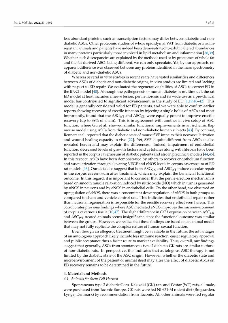
Whereas several in vitro studies in recent years have tested similarities and differencesbetween ASCs of diabetic and non-diabetic origins, in vivo studies are limited and lackingwith respect to ED repair. We evaluated the regenerative abilities of ASCs to correct ED inthe BNCI model [40]. Although the pathogenesis of human diabetes is multimodal, the ratED model at least includes a nerve lesion, penile fibrosis and its wide use as a pre-clinicalmodel has contributed to significant advancement in the study of ED [1,19,40–42]. Thismodel is generally considered valid for ED patients, and we were able to confirm earlierreports showing recovery of erectile function by injecting a single bolus of ASCs and mostimportantly, found that the ASCWT and ASCGK were equally potent to improve erectilerecovery (up to 89% of sham). This is in agreement with another in vivo setup of ASCfunction, where Gu et al. showed similar functional improvements in an ischemic flapmouse model using ASCs from diabetic and non-diabetic human subjects [43]. By contrast,Rennert et al. reported that the diabetic state of mouse SVF impairs their neovascularizationand wound healing capacity in vivo [22]. Yet, SVF is quite different from ASCs as alsorevealed herein and may explain the differences. Indeed, impairment of endothelialfunction, decreased levels of growth factors and cytokines along with fibrosis have beenreported in the corpus cavernosum of diabetic patients and also in preclinical models [44,45].In this respect, ASCs have been demonstrated by others to recover endothelium functionand vascularization through elevating VEGF and eNOS levels in corpus cavernosum of EDrat models [46]. Our data also suggest that both ASCGK and ASCWT induce vascular repairin the corpus cavernosum after treatment, which may explain the beneficial functionaloutcome. In this regard, it is important to consider that the penile erection mechanism isbased on smooth muscle relaxation induced by nitric oxide (NO) which in turn is generatedby nNOS in neurons and by eNOS in endothelial cells. On the other hand, we observed anupregulation of eNOS, there was a concomitant downregulation of nNOS in both groups ascompared to sham and vehicle control rats. This indicates that endothelial repair ratherthan neuronal regeneration is responsible for the erectile recovery effect seen herein. Thiscorroborates previous findings where ASC mediated eNOS improves the microenvironmentof corpus cavernous tissue [10,47]. The slight difference in Cd31 expression between ASCGKand ASCWT treated animals seems insignificant, since the functional outcome was similarbetween the groups. However, we realize that these findings are based on an animal modelthat may not fully replicate the complex nature of human sexual function.
Even though an allogenic treatment might be available in the future, the advantagesof an autologous approach likely include less immune reaction, easier regulatory approvaland public acceptance thus a faster route to market availability. Thus, overall, our findingssuggest that generally, ASCs from spontaneous type 2 diabetes GK rats are similar to thoseof non-diabetic rats. In perspective, this indicates that autologous ASC therapy is notlimited by the diabetic state of the ASC origin. However, whether the diabetic state andmicroenvironment of the patient or animal itself may alter the effect of diabetic ASCs onED recovery remains to be determined in the future.
4. Material and Methods4.1. Animals for Stem Cell Harvest
Spontaneous type 2 diabetic Goto–Kakizaki (GK) rats and Wistar (WT) rats, all male,were purchased from Taconic Europe. GK rats were fed NIH31-M rodent diet (Brogaarden,Lynge, Denmark) by recommendation from Taconic. All other animals were fed regular

Int. J. Mol. Sci. 2022, 23, 1692 8 of 13
altromin 1324 (Brogaarden). At 18 weeks of age, animals were sacrificed to harvest inguinaladipose tissue for SVF isolation and tissue culture. According to the manufacturer, theGK rats spontaneously develop type 2 diabetes at 14–16 weeks of age. After five hours offasting, blood glucose levels were evaluated in tail vein blood at 17 weeks of age by usingthe OneTouch, Ultra Easy instrument (Mediq, Brøndby, Denmark).
4.2. Cell Isolation and Culturing
For the generation of cultured ASCs for in vivo transplantation, GK rats and WTrats were euthanized using carbon dioxide, and the inguinal fat pads were collected.Adipose tissue was minced and enzymatically digested with collagenase (0.86 U/mLCollagenase NB 4 Standard Grade, Serva, Germany). Following red blood cell lysis andfiltration, the SVFs were isolated and counted using a NucleoCounter NC-200 instrument(ChemoMetec, Allerød, Denmark). SVF aliquots were either fixed immediately for flowcytometry, seeded for colony-forming-unit fibroblast CFU-F assay or cultured for in vitroexpansion to achieve ASCs.
For expanding cell number and increase homogeneity, 106 SVF were seeded in T75 cellculture flasks in growth medium (DMEM/1.0 g/L glucose/25 mM HEPES supplementedwith 4 mM Ultraglutamine/10% Fetal Bovine Serum (FBS) (all products from Lonza,UK)/1% PS) and cultured at 37 ◦C and 5% CO2. Non-adherent cells were removed after24 h while refreshing media. Hereafter, the growth medium was changed every third day,and the cells passaged until number 4 with approximately 80% of confluence.
4.3. Colony Forming Unit Assay
Freshly isolated SVF were seeded in triplicates in 6-well plates at a density of 100 cells/cm2
and cultured for twelve days. Following a PBS wash the cells were fixed in 4% neutralbuffered formalin (NBF) for 20 min, at room temperature RT, washed twice in PBS andstored at 4 ◦C until staining. For staining, the cells were rinsed in tap water and incu-bated 5 min, at RT with Mayer’s hematoxylin. Cells were washed and plates were leftinverted to dry overnight. The number of colonies (>30 cells) was determined using astereo microscope (Leica M80).
4.4. Flow Cytometry
Freshly isolated SVF or Cultured ASCs (passage 4) were washed in Hank’s balancedsalt solution (HBSS)/1% PS/5% FBS and fixed in HBSS/5% FBS/1% PS/1% NBF overnightat 4 ◦C. The cells were then washed twice and stored in HBSS/1%PS/5% FBS/0.05%sodium-azide at 4 ◦C until analysis. Fixed cells were washed in HBSS/1% PS/5% FBS andincubated 60 min with primary antibodies on ice while shaking. After, two washes the cellswere incubated 30 min with secondary antibodies on ice while shaking and finally washedtwice. Data acquisition and analysis were obtained using a FACSCalibur instrument(Becton Dickinson, 2150 Commerce Dr, San Jose, CA, USA) and FlowJo 10.0.6 software(Tree Star Inc., OR, USA), respectively. Primary antibodies were specific for rat CD45, CD90,CD44 (BD bioscience, 554,875 (1:100), 554,895 (1:50); and 554,869 (1:100), respectively), andfrom other companies CD29 (abcam, ab52971 (1:100)), CD34 (R&D Systems, MN, USA,AF6518 (1:72)), PDGFRα (cell signaling, 3164 (1:200) and CD31 (Santa Cruz, CA, USA,sc-1506 (1:100)). Isotypes included sheep IgG (R&D systems, 5-001-A), rabbit polyclonalIgG, rabbit monoclonal IgG (abcam, ab37415, ab125938), mouse IgG2a,k, and mouse IgG1,k(Sigma-Aldrich, Vandtårnsvej 62A, Søborg Denmark M 5409, M 5284). Alexa 488 or 647conjugated secondary donkey antibodies specific for rabbit IgG, mouse IgG, sheep IgG(all purchased at Invitrogen, 3 Fountain Drive Inchinnan Business Park, Paisley, UK, 1:200)were used for visualization.
4.5. Differentiation
StemPro Differentiation Kits (Gibco, Life Technologies, Scientific 151 Brook Drive,Milton Park Abingdon, UK) were used to verify the differentiation capacity of the cultured
Int. J. Mol. Sci. 2022, 23, 1692 9 of 13
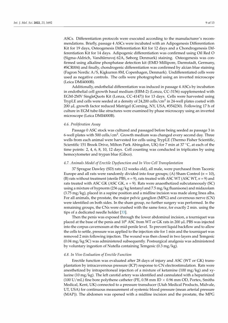
ASCs. Differentiation protocols were executed according to the manufacturer’s recom-mendations. Briefly, passage 4 ASCs were incubated with an Adipogenesis DifferentiationKit for 19 days, Osteogenesis Differentiation Kit for 12 days and a Chondrogenesis Dif-ferentiation Kit for 14 days. Adipogenic differentiation was confirmed using Oil Red O(Sigma-Aldrich, Vandtårnsvej 62A, Søborg Denmark) staining. Osteogenesis was con-firmed using alkaline phosphatase detection kit (EMD Millipore, Darmstadt, Germany,#SCR004) and finally, chondrogenic differentiation was confirmed by alcian blue staining(Fagron Nordic A/S, Kigkurren 8M, Copenhagen, Denmark). Undifferentiated cells wereused as negative controls. The cells were photographed using an inverted microscope(Leica DMI4000B).
Additionally, endothelial differentiation was induced in passage 4 ASCs by incubationin endothelial cell growth basal medium (EBM-2) (Lonza, CC-3156) supplemented withEGM-2MV SingleQuots Kit (Lonza, CC-4147)) for 13 days. Cells were harvested usingTrypLE and cells were seeded at a density of 24,200 cells/cm2 in 24-well plates coated with200 µL growth factor reduced Matrigel (Corning, NY, USA, #354230). Following 17 h ofculture in EGM tube-like structures were examined by phase microscopy using an invertedmicroscope (Leica DMI4000B).
4.6. Proliferation Assay
Passage 0 ASC stock was cultured and passaged before being seeded as passage 3 in6-well plates with 500 cells/cm2. Growth medium was changed every second day. Threewells from each animal were harvested for cells using TrypLE (Thermo Fisher Scientific,Scientific 151 Brook Drive, Milton Park Abingdon, UK) for 7 min at 37 ◦C, at each of thetime points: 2, 4, 6, 8, 10, 12 days. Cell counting was conducted in triplicates by usinghemocytometer and trypan blue (Gibco).
4.7. Animals Model of Erectile Dysfunction and In Vivo Cell Transplantation
37 Sprague Dawley (SD) rats (12 weeks old), all male, were purchased from TaconicEurope and all rats were randomly divided into four groups; (A) Sham Control (n = 10),(B) rats without treatment (sterile PBS, n = 9), rats treated with ASC WT (ASC WT, n = 9) andrats treated with ASC GK (ASC GK, n = 9). Rats were anaesthetized subcutaneously (SC)using a mixture of hypnorm (236 µg/kg fentanyl and 7.5 mg/kg fluanisone) and midazolam(3.75 mg/kg), placed in a supine position and a midline incision was made along linea alba.For all animals, the prostate, the major pelvic ganglion (MPG) and cavernous nerve (CN)were identified on both sides. In the sham group, no further surgery was performed. In theremaining groups, the CNs were crushed with the same force, for exactly 2 min. using thetips of a dedicated needle holder [33].
Then the penis was exposed through the lower abdominal incision, a tourniquet wasplaced at the base of the penis and 106 ASC from WT or GK rats in 200 µL PBS was injectedinto the corpus cavernosum at the mid-penile level. To prevent liquid backflow and to allowthe cells to settle, pressure was applied to the injection site for 1 min and the tourniquet wasremoved 2 min following injection. The wound was then closed in two layers and Temgesic(0.04 mg/kg SC) was administered subsequently. Postsurgical analgesia was administeredby voluntary ingestion of Nutella containing Temgesic (0.3 mg/kg).
4.8. In Vivo Evaluation of Erectile Function
Erectile function was evaluated after 28 days of injury and ASC (WT or GK) trans-plantation by intracavernous pressure (ICP) response to CN electrostimulation. Rats wereanesthetized by intraperitoneal injection of a mixture of ketamine (100 mg/kg) and xy-lazine (10 mg/kg). The left carotid artery was identified and cannulated with a heparinized(100 U/mL) fine bore polythene catheter (PE, 0.58 mm ID × 0.96 mm OD, Portex, SmithsMedical, Kent, UK) connected to a pressure transducer (Utah Medical Products, Midvale,UT, USA) for continuous measurement of systemic blood pressure (mean arterial pressure(MAP)). The abdomen was opened with a midline incision and the prostate, the MPG

Int. J. Mol. Sci. 2022, 23, 1692 10 of 13
and the CN were identified. The testicles were relocated to the upper abdomen to easethe access to the crus of the penis. The penis was exposed and dissection continued toexpose the crus under the ischiocavernous muscle, which was transected. A bipolar hookelectrode was placed on the CN distal to the crush injury. The crus of penis was thencannulated with a heparinized 25G needle with a PE-50 catheter (Portex, Smiths Medical)connected to a pressure transducer for measurement of ICP. The CN electrostimulationswere performed using a custom-made stimulator. In accordance with the majority of similarstudies, the stimulus parameters were 2, 4, 6 and 8V, 20 Hz, pulse width of 0.5 and durationof 50 s, with a minimum of 5 min rest interval between stimulations [41]. The CN andthe area were kept dry prior to stimulations. Following every stimulation series, the CNwas transected distal to the electrode and a stimulation of 8V was applied to check fornon-CN-mediated retrograde ICP response. In the case of such a response, the stimulationseries was rejected as a false positive and the contralateral CN was subjected to a new seriesof electro-stimulations. All animal experiments were approved by the Danish Council forSupervision with Experimental Animals (#2013-15-2934-00877). All data was collected byLabview (National Instruments, Austin, TX, USA) and analyzed using GraphPad Prism(9.0d Mac OS X, 2365 Northside Dr., San Diego, CA, USA).
4.9. RNA Isolation and qRT-PCR
For RNA extraction and subsequent qRT-PCR analysis, the corpora cavernosumwere dissected and rapidly frozen using dry ice. RNA isolation and relative quantitativereverse transcription-polymerase chain reaction (qRT-PCR) was used to examine relativegene expression in the corpus cavernosum. Accordingly, dissected corpus cavernosumsamples were homogenized and the total RNA was extracted using a TriReagent protocol(Thermo Fisher Scientific, Scientific 151 Brook Drive, Milton Park Abingdon, UK). RNApurity and quantity was assessed by nanodrop (Nanodrop® Technologies, Thermo FisherScientific, Scientific 151 Brook Drive, Milton Park Abingdon, UK) measurements. mRNAwas reverse transcribed to cDNA using a High Capacity cDNA kit (Applied Biosystems,Thermo Fisher Scientific, Scientific 151 Brook Drive, Milton Park Abingdon, UK) and theqRT-PCR reaction was carried out using Power SYBR® Green PCR kit (Applied Biosystems)and primers specific for Cd31, Procollagen1, αSMA, eNOS, nNOS (Primer sequences notshown). All qRT-PCR analyses were performed with the 7900HT Fast Real-Time PCRSystem (Applied Biosystems) instrument and data was normalized against stably expressedcontrol genes, GAPDH and β-actin (Mean M = 0,431 and Mean CV = 0.150) using theqBase+ software.
4.10. Proteome Analysis
ASCs (Passage 4) obtained from inguinal adipose tissue from GK or WT rats werecounted using the NucleoCounter instrument and 2 × 10⁶ ASCs were washed in PBS threetimes to avoid protein contamination from the FBS.
For protein isolation, cells were re-suspended in lysis buffer (10 mM EDTA, 300 mMNaCl, 0.2% Triton X100, 200 mM Tri-ethylammonium bicarbonate-TEAB, Complete miniprotease inhibitor) and disrupted by sonication. Cell debris was pelleted by centrifugation(20,000× g for 60 min at 4 ◦C) and proteins were isolated by transferring the supernatantto 5 equivalents ice-cold acetone. Proteins were then reduced in the presence of 5 mMdithiothreitol (DTT) and incubated for 30 min at 50 ◦C followed by blocking of the reducedsulfhydryl groups with 15 mM iodoacetamide for 30 min in darkness. Trypsin (Promega,Madison, WI, USA) was added at protein:trypsin ratio of 50:1 w/w, followed by overnightincubation at 37 ◦C.
4.11. Stable Isotope Labeling of Protein Samples with TMT-10 Plex
A 10-µg fraction of the tryptic digest was collected for labeling with 10-plex TMT-kit(Thermo Scientific). The content of each TMT reagent vial was re-suspended in anhydrousethanol, and a 40-µg sample was labeled. Labeled samples from each biological experiment

Int. J. Mol. Sci. 2022, 23, 1692 11 of 13
(n = 3) were pooled in equal ratios, dried in a vacuum centrifuge, re-dissolved in 50 µLtrifluoroacetic acid solution (0.1%), and purified loaded on a microcolumn packed withreversed phased material (equal w/w amounts of Poros R2 and Oligo R3 material) andfractionated into 7 fractions by high pH liquid chromatography virtually as previouslydescribed [48].
The Fractions were analyzed by RP-nanoLC-MS/MS analysis on an Orbitrap Eclipsemass spectrometer (Thermo Fisher Scientific) equipped with a nanoHPLC interface (DionexUltiMate 3000 nano HPLC). Briefly, samples were separated using linear 49 min gradients(fractions 1–3) and 77 min linear gradients (fractions 4–7) ranging from 93% solvent A(0.1% formic acid) to 34% solvent B (80% acetonitrile/0.1% formic acid). Mass spectra ofeluting peptides were acquired in positive ion mode applying automatic data-dependentswitch between an Orbitrap survey MS scan in the mass range of 400–1200 m/z followedby peptide fragmentation applying a normalized collisional energy of 40% in a 3-s dutycycle. The automatic gain control (AGC) target was set to “250%” at a resolution of 60,000 atm/z 200, and 200,000 ions at a resolution of 50,000 at m/z 200 for MS/MS scans. Ionselection threshold for MS/MS analysis was set to 50,000 counts. Selected sequenced ionswere dynamically excluded for 60 s. All raw data files were processed and quantifiedusing Proteome Discoverer version 2.4 (Thermo Scientific) also as previously described [49]with the exception that the database search was restricted to the rat database insteadof humans. For principal component analysis (PCA), all missing values were removed(a total of 1601 proteins were used) prior to reducing dimensions. ggplot2-based fviz_pca{factoextra} version 1.0.7 was used for graphical representation of individuals for the firsttwo components.
For analysis, hierarchical clustering and heatmap visualization heatmap3 {heatmap3}version 1.1.9 was used. All missing values were removed (a total of 1601 proteins wereused) and heatmaps were computed with and without reordering of columns. For imputingmissing values, the imputerPCA {missMDA} was used with ncp = 2.
4.12. Statistical Analysis
The data were analyzed using GraphPad Prism (9.0 Mac OS X, USA) software, andall data were expressed as mean ± standard deviation (SD) with a statistical significanceset at p < 0.05. For the proliferation assay and flow cytometry data, statistical significancewas tested using two-way ANOVA and Sidak’s multiple comparison test. Mixed-effectmodel analysis with Tukey’s multiple comparison test was used on analysis of ICP dataand qRT-PCR data. Finally, a Mann–Whitney t-test was used for comparison of fibroblastcolony formation unit (CFU-F) frequencies and cell yield. Statistics used for proteomic dataare outlined under mass spectrometry data analysis.
Supplementary Materials: The following are available online at https://www.mdpi.com/article/10.3390/ijms23031692/s1.
Author Contributions: M.L.Q., D.C.A. and S.P.S. conceived and designed the experiments; M.L.Q.,S.G.C.S. and H.C.B. performed the experiments; M.L.Q., P.D., C.H.J., H.C.B., S.G.C.S., L.L., E.B.H.,D.C.A. and S.P.S. analyzed and interpreted the data; M.L.Q., P.D., C.H.J. and D.C.A. wrote themanuscript and M.L.Q. and P.D. prepared the figures. L.L., D.C.A., C.H.J. and S.P.S. supervised theexperiments; D.C.A. and S.P.S. contributed to the funding. All authors have read and agreed to thepublished version of the manuscript.
Funding: Please add: This work was supported by the Faculty of Health Science, University ofSouthern Denmark. Odense Denmark, the Department of Clinical Biochemistry and Pharmacology,Odense University Hospital, Odense Denmark as well as the Innovation Foundation Denmark(#7051-00001A).
Institutional Review Board Statement: All animal experiments were approved by Danish councilfor supervision with experimental animals (#2013-15-2934-00877 and #2012-15-2934-00340).

Int. J. Mol. Sci. 2022, 23, 1692 12 of 13
Data Availability Statement: Data will be available from the corresponding author on reasonablerequest and with permission of Odense University Hospital Legal Department, Odense, Denmark.
Acknowledgments: We thank Ulf Simonsen (Aarhus University, Denmark) for introduction andtraining in the execution of the ED rat model, Trinity Bivalaqcua (John Hopkins Medical Institu-tion, Baltimore, MD, USA) for training in micro surgical techniques regarding the ED rat model,Jens Ole Pedersen (IMM/SDU/DK) for construction of electrical equipment and Tonja L. Jørgensen(LMCC/SDU/DK) for excellent technical assistance.
Conflicts of Interest: CHJ, DCA, and SPS have filed a patent (WO2019193052) indirectly related tothe study, and SPS is the CEO of Blue Cell Therapeutics.
References1. Albersen, M. Injections of adipose tissue-derived stem cells and stem cell lysate improve recovery of erectile function in a rat
model of cavernous nerve injury. J. Sex. Med. 2010, 7, 3331–3340. [CrossRef] [PubMed]2. Cannon, A. Burden of Illness in Type 2 Diabetes Mellitus. J. Manag. Care Spec. Pharm. 2018, 24, S5–S13. [CrossRef] [PubMed]3. Janssen, L.M.M. Burden of disease of type 2 diabetes mellitus: Cost of illness and quality of life estimated using the Maastricht
Study. Diabet. Med. 2020, 37, 1759–1765. [CrossRef] [PubMed]4. De Ugarte, D.A. Comparison of multi-lineage cells from human adipose tissue and bone marrow. Cells Tissues Organs 2003,
174, 101–109. [CrossRef]5. Vardi, Y. Microvascular Complications in Diabetic Erectile Dysfunction Do we need other alternatives? Diabetes Care 2009, 32,
S420–S422. [CrossRef]6. Chaiban, T.J.; Azar, S.T. Erectile dysfunction in diabetic patients. J. Med. Liban 2004, 52, 217–219.7. Daneshgari, F.; Moore, C. Diabetic uropathy. Semin. Nephrol. 2006, 26, 182–185. [CrossRef]8. Corona, G. Phosphodiesterase Type 5 (PDE5) Inhibitors in Erectile Dysfunction: The Proper Drug for the Proper Patient. J. Sex.
Med. 2011, 8, 3418–3432. [CrossRef]9. Qiu, X. Effects of intravenous injection of adipose-derived stem cells in a rat model of radiation therapy-induced erectile
dysfunction. J. Sex. Med. 2012, 9, 1834–1841. [CrossRef]10. Yang, J. Adipose-derived stem cells improve erectile function partially through the secretion of IGF-1, bFGF, and VEGF in aged
rats. Andrology 2018, 6, 498–509. [CrossRef]11. Kim, S.W.; Zhu, G.Q.; Bae, W.J. Mesenchymal Stem Cells Treatment for Erectile Dysfunction in Diabetic Rats. Sex. Med. Rev. 2020,
8, 114–121. [CrossRef] [PubMed]12. Mizuno, H.; Tobita, M.; Uysal, A.C. Concise Review: Adipose-Derived Stem Cells as a Novel Tool for Future Regenerative
Medicine. Stem. Cells 2012, 30, 804–810. [CrossRef] [PubMed]13. Li, X. Comprehensive characterization of four different populations of human mesenchymal stem cells as regards their immune
properties, proliferation and differentiation. Int. J. Mol. Med. 2014, 34, 695–704. [CrossRef] [PubMed]14. Tsuji, W.; Rubin, J.P.; Marra, K.G. Adipose-derived stem cells: Implications in tissue regeneration. World J. Stem. Cells 2014, 6,
312–321. [CrossRef]15. Zuk, P.A. Multilineage cells from human adipose tissue: Implications for cell-based therapies. Tissue Eng. 2001, 7, 211–228.
[CrossRef]16. Lombardi, F. Secretome of Adipose Tissue-Derived Stem Cells (ASCs) as a Novel Trend in Chronic Non-Healing Wounds: An.
Overview of Experimental In Vitro and In Vivo Studies and Methodological Variables. Int. J. Mol. Sci. 2019, 20, 3721. [CrossRef]17. Salgado, A.J. Adipose tissue derived stem cells secretome: Soluble factors and their roles in regenerative medicine. Curr. Stem.
Cell Res. Ther. 2010, 5, 103–110. [CrossRef]18. Damous, L.L. Cell-free therapy with the secretome of adipose tissue-derived stem cells in rats’ frozen-thawed ovarian grafts.
Stem. Cell Res. Ther. 2018, 9, 323. [CrossRef]19. Albersen, M.; Weyne, E.; Bivalacqua, T.J. Stem Cell Therapy for Erectile Dysfunction: Progress and Future Directions. Sex. Med.
Rev. 2013, 1, 50–64. [CrossRef]20. Haahr, M.K. Safety and Potential Effect of a Single Intracavernous Injection of Autologous Adipose-Derived Regenerative Cells
in Patients with Erectile Dysfunction Following Radical Prostatectomy: An. Open-Label Phase I Clinical Trial. EBioMedicine 2016,5, 204–210. [CrossRef]
21. El-Ftesi, S. Aging and diabetes impair the neovascular potential of adipose-derived stromal cells. Plast. Reconstr. Surg. 2009, 123,475–485. [CrossRef] [PubMed]
22. Rennert, R.C. Diabetes impairs the angiogenic potential of adipose-derived stem cells by selectively depleting cellular subpopula-tions. Stem. Cell Res. Ther. 2014, 5, 79. [CrossRef] [PubMed]
23. Dzhoyashvili, N.A. Disturbed angiogenic activity of adipose-derived stromal cells obtained from patients with coronary arterydisease and diabetes mellitus type 2. J. Transl. Med. 2014, 12, 337. [CrossRef] [PubMed]
24. Karina, K. Diabetes mellitus type 2 reduces the viability, proliferation, and angiogenic marker of adipose-derived stem cellscultured in low-glucose anti-oxidant-serum supplemented medium. Biomed. Res. Ther. 2019, 6, 3073–3082. [CrossRef]

Int. J. Mol. Sci. 2022, 23, 1692 13 of 13
25. Ramos, T.L. MSC surface markers (CD44, CD73, and CD90) can identify human MSC-derived extracellular vesicles by conven-tional flow cytometry. Cell Commun. Signal. 2016, 14, 1–14. [CrossRef]
26. Dominici, M. Minimal criteria for defining multipotent mesenchymal stromal cells. The International Society for Cellular Therapyposition statement. Cytotherapy 2006, 8, 315–317. [CrossRef] [PubMed]
27. Wu, H. Nanotechnology-assisted adipose-derived stem cell (ADSC) therapy for erectile dysfunction of cavernous nerve injury:In vivo cell tracking, optimized injection dosage, and functional evaluation. Asian J. Androl. 2018, 20, 442.
28. Chung, E.; de Young, L.; Brock, G.B. Investigative models in erectile dysfunction: A state-of-the-art review of current animalmodels. J. Sex. Med. 2011, 8, 3291–3305. [CrossRef]
29. Cellek, S. Common pitfalls in some of the experimental studies in erectile function and dysfunction: A consensus article. J. Sex.Med. 2012, 9, 2770–2784. [CrossRef]
30. Burnett, A.L. GGF2 is neuroprotective in a rat model of cavernous nerve injury-induced erectile dysfunction. J. Sex. Med. 2015, 12,897–905. [CrossRef]
31. Si, Z. Adipose-derived stem cells: Sources, potency, and implications for regenerative therapies. Biomed. Pharmacother. 2019,114, 108765. [CrossRef] [PubMed]
32. Akash, M.S.; Rehman, K.; Chen, S. Goto-Kakizaki rats: Its suitability as non-obese diabetic animal model for spontaneous type 2diabetes mellitus. Curr. Diabetes Rev. 2013, 9, 387–396. [CrossRef] [PubMed]
33. Yan, J.L. Type 2 Diabetes Restricts Multipotency of Mesenchymal Stem Cells and Impairs Their Capacity to Augment PostischemicNeovascularization in db/db Mice. J. Am. Heart Assoc. 2012, 1, e002238. [CrossRef]
34. Jin, P. Streptozotocin-Induced Diabetic Rat-Derived Bone Marrow Mesenchymal Stem Cells Have Impaired Abilities in Prolifera-tion, Paracrine, Antiapoptosis, and Myogenic Differentiation. Transplant. Proc. 2010, 42, 2745–2752. [CrossRef] [PubMed]
35. Wang, M. Therapeutic Effects of Adipose Stem Cells from Diabetic Mice for the Treatment of Type 2 Diabetes. Mol. Ther. 2018, 26,1921–1930. [CrossRef] [PubMed]
36. Thongboonkerd, V. Proteomics for diabetes research: An update and future perspectives. Expert Rev. Endocrinol. Metab. 2006, 1,507–515. [CrossRef]
37. Sundsten, T.; Ortsater, H. Proteomics in diabetes research. Mol. Cell Endocrinol. 2009, 297, 93–103. [CrossRef]38. Berti, D.A. Identification of intracellular peptides in rat adipose tissue: Insights into insulin resistance. Proteomics 2012, 12,
2668–2681. [CrossRef]39. Kim, S.J. A protein profile of visceral adipose tissues linked to early pathogenesis of type 2 diabetes mellitus. Mol. Cell Proteom.
2014, 13, 811–822. [CrossRef]40. Haney, N.M. Bilateral Cavernous Nerve Crush Injury in the Rat Model: A Comparative Review of Pharmacologic Interventions.
Sex. Med. Rev. 2018, 6, 234–241. [CrossRef]41. Hannan, J.L. Valproic Acid Prevents Penile Fibrosis and Erectile Dysfunction in Cavernous Nerve-Injured Rats. J. Sex. Med. 2014,
11, 1442–1451. [CrossRef] [PubMed]42. Gur, S. Review of erectile dysfunction in diabetic animal models. Curr. Diabetes Rev. 2014, 10, 61–73. [CrossRef] [PubMed]43. Gu, J.H. Neovascular potential of adipose-derived stromal cells (ASCs) from diabetic patients. Wound Repair Regen. 2012, 20,
243–252. [CrossRef]44. Dean, C.R.; Lue, T.F. Physiology of penile erection and pathophysiology of erectile dysfunction. Urol. Clin. North Am. 2005,
32, 379. [CrossRef] [PubMed]45. Gonzalez-Cadavid, N.F. Mechanisms of penile fibrosis. J. Sex. Med. 2009, 6, 353–362. [CrossRef] [PubMed]46. Zhou, F. Effects of adipose-derived stem cells plus insulin on erectile function in streptozotocin-induced diabetic rats. Int. Urol.
Nephrol. 2016, 48, 657–669. [CrossRef] [PubMed]47. Li, M. Stem Cell Therapy for Diabetic Erectile Dysfunction in Rats: A Meta-Analysis. PLoS ONE 2016, 11, e0154341. [CrossRef]48. Mulorz, J. Hyperlipidemia does not affect development of elastase-induced abdominal aortic aneurysm in mice. Atherosclerosis
2020, 311, 73–83. [CrossRef]49. Matchkov, V.V. A paradoxical increase of force development in saphenous and tail arteries from heterozygous ANO1 knockout
mice. Physiol. Rep. 2020, 8, e14645. [CrossRef]